Kyphosis is a condition that is most typically identified by a humped mid to upper back. It can be frustrating to find yourself diagnosed with kyphosis, both due to the unusual appearance it causes and the physical discomfort that can come with the condition. Fortunately, chiropractic offers a variety of treatment options for kyphosis that are non-invasive, drug-free and quite effective. With the right approach, it should be possible to get significant relief for your symptoms through chiropractic care.
Contents
Causes
Many times kyphosis is caused by poor posture. Years of hunching your back can cause the body to change the way the vertebrae sit atop one another�leading to stretching of soft tissues and a resting position that is hunched instead of upright.
Not all cases of kyphosis are the result of poor posture, however. There are some people who are born with kyphosis. There are also those that develop kyphosis following trauma or due to disease. One of the most common diseases that result in kyphosis is Scheuermann’s disease.
Chiropractic can often help resolve kyphosis that is caused by poor posture. If your kyphosis is due to trauma or other issues, chiropractic can still provide a much-needed relief from many of your symptoms�including pain and stiffness.
How Chiropractic Helps With Kyphosis
HEALTHCARE AND MEDICAL CONCEPT: KYPHOSIS
Diagnosing the Cause
Your first visit to the chiropractor will begin with a comprehensive examination to determine the cause of your condition. An accurate diagnosis is key to developing an effective treatment plan. The examination will most likely include imaging tests like an x-ray or MRI. It may also include a physical examination to figure out how your body currently moves, where you are tender and/or in pain, etc.
Treatment Plan
Your chiropractor will never move forward with treatment until he or she has explained your options and gotten your permission to begin. The treatment plan you are offered may include:
The joint in the human body, particularly the spinal joints, have an ideal alignment where everything fits together best. The more you can achieve and maintain this alignment, the less pain, stiffness and other disruptions you can expect to experience. Chiropractic adjustments aim to bring your body back into alignment.
Regular adjustments should help to improve your spinal alignment and straighten your back. In the case of poor posture, adjustments can aid you in learning how to improve your posture and keep those improvements over the long term.
Mobility Increased
The curving of the back typical of kyphosis can lead to a lot of stiffness in the spine and surrounding soft tissues. Lack of mobility makes everything you do more difficult, so it makes sense to try and increase your mobility when possible.
Chiropractic adjustments and complementary therapies are ideal for relaxing the soft tissues that may have become stiff as the kyphosis developed. One of the great things about improving the mobility of the spine is that it allows you to improve your posture. Over time, it should become easier to keep your back in a more healthy alignment.
Pain Lessened
Pain is common with kyphosis. The spine is not meant to hump as it does with kyphosis, which explains why pain is so typical with the condition. Chiropractic is very useful for decreasing pain related to spinal issues. Many patients get relief immediately following their first adjustment.
Schedule an Appointment With Us Today
If you are suffering from kyphosis, please contact us to schedule an appointment. Our chiropractic team is standing by to answer your questions and help you get treatment for your condition.
What is a Pronated Foot & What Can Custom Orthotics Do?
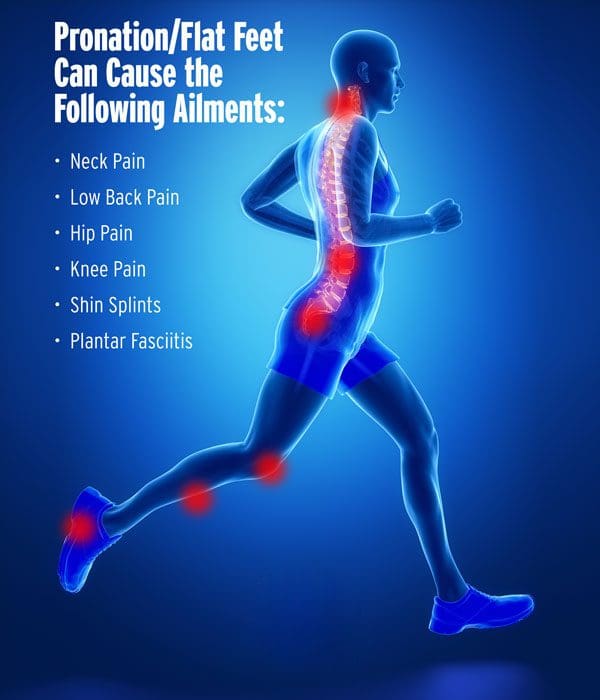
When a foot is pronated, it means its arches have fallen and the foot is flat. Flat feet do not absorb shock as well and make for a less stable base for�the bones, muscles, ligaments, and tendons. They can also:
Shift the body’s alignment out of order
Cause aches and pains in the feet, knees, hips, neck and/or spine
Lead to injury and problems like shin splints, Achilles tendinitis, and plantar fasciitis
Up to a third of people suffer from flat feet
Causes
The connective tissue, called the plantar fascia, on the underside of the foot is what maintains the foot�s healthy arch shape. Injury and health conditions can cause the fascia to stretch out and flatten. But so can everyday walking and standing. Once the fascia stretches out, it is unable to spring back. When the foot structure becomes flattened, the body�s foundation is prone to injury and health conditions.
Treatment
Chiropractic adjusting to ensure proper positioning of bones and joints
Custom orthotics in every pair of shoes for pronation control, support, and comfort
Get Rid of *Foot Pronation* with *FOOT ORTHOTICS* | El Paso, Tx (2019)
Pronation
Pronation describes the way that the foot rolls inward during its normal motion. The foot turns inward, flattening out, as the heel�s outer edge strikes the ground. For the foot to function correctly, there must be a significant degree of pronation. However, excessive pronation, or overpronation, can cause injury and damage to the foot and ankle. It creates the arch in the foot to flatten, and the ligaments, tendons, and muscles under the foot overstretch. For instance, overpronation of the foot causes a series of internal changes that extend up through the leg. The femur may rotate causing hip pain and inflammation of the sacroiliac joint which leads to back pain. Other misalignments in the body that are caused by foot problems can also lead to chronic lower back pain as well.
NCBI Resources
Sometimes there are abnormalities of the spine and it causes a misalignment of the natural curvatures or some curvatures may be exaggerated. These unnatural curvatures of the spine are characterized by three health conditions called�lordosis, kyphosis, and scoliosis. They gently curve, sloping slightly inward at the small of the back, and again slightly at the neck. The pull of gravity, combined with body movement, can put a great deal of stress on the spine and these slight curves help absorb some of the impact. Spinal manipulations for spinal curvature disorders�have been shown to be very effective. Chiropractic helps restore the spine�s natural balance even if the patient has one of these types of conditions.
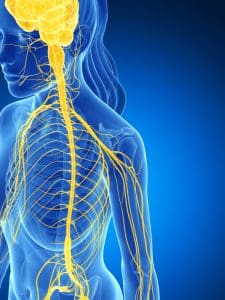
Foot drop is a health issue which identifies a weakening of the muscles which allow for flexing of the ankle and toes. This health issue causes the patient to drag the front part of the foot when walking. To compensate for this dragging, the patient will often flex the knee to raise the foot higher than in a standard stride, frequently referred to as high steppage gait. �
Foot drop generally affects the muscles in charge of moving the ankle and foot upward, especially the anterior tibialis, the extensor halluces longus, and the extensor digitorum longus. Although foot drop is a neuromuscular disorder which affects the muscles and the nerves, it’s not a disease. It is a symptom of an underlying health issue, perhaps a spinal health issue. �
Contents
What are the Symptoms of Foot Drop?
The most well-known symptom of foot drop, high steppage gait, is commonly characterized by raising the thigh up in an exaggerated manner while walking, similar to climbing the stairs. High steppage gait is associated with one of the following: �
Dragging of the foot and toes
Scraping of the toes throughout the floor
Uncontrolled slapping of the toes against the floor
The affected muscles are often utilized to maintain the foot off the ground during the swing-through phase of walking. If these are weak, they can’t maintain the foot up and the foot will scrape throughout the floor if the foot is not raised high. Aside from the common symptoms above, additional problems typically associated with foot drop can ultimately include: �
The inability to raise the foot at the ankle
The inability to point the toes upward in the human body, frequently referred to as dorsiflexion
Struggling to walk normally in a heel-to-toe manner
Other well-known symptoms also associated with foot drop may ultimately include one or a combination of the following: �
Excessive, swinging hip motion. With foot drop, the hip may swing out to prevent the toes from hitting the floor.
Limp foot. The affected foot may flop away from the individual.
Pain, tingling sensations, and numbness in the foot. The intensity of the symptoms can vary from person to person. Symptoms can make regular tasks, such as walking or driving, difficult. The foot pain may be associated with sciatica.
Difficulty engaging in exercise and/or physical activities requiring the utilization of the front of the foot. By way of instance, climbing stairs may become challenging for patients with foot drop.
Muscle atrophy in the lower extremities. Muscle atrophy refers to a muscle decreasing in mass and weakening. Because the anterior tibialis, the extensor halluces longus, and the extensor digitorum longus muscles are affected by foot drop, muscle atrophy may occur and make it more challenging to exert force in the lower extremities.
Foot drop may be experienced in one or both feet. Foot drop is generally experienced in a single foot.
What are the Causes of Foot Drop?
Foot drop is a symptom of an underlying health issue. Foot drop causes fall into one or a combination of three categories: �
Muscle damage or injury
Skeletal or anatomical abnormalities affecting the foot
Nerve damage
Specific conditions or diseases and other health issues which may cause foot drop may also include: �
A lumbar spine health issue
A stroke or tumor
Parkinson’s disease
Diabetes
Motor neuron disease
Multiple sclerosis
Adverse reactions to drugs and/or medications as well as alcohol
An injury to the leg or foot
There’s a variety of lumbar spine health issues which can ultimately affect the nerve roots, including the peroneal nerve and the sciatic nerve. Any compression or impingement of the nerve roots in the lower back may cause foot drop. Common lower back or lumbar spine health issues which can cause foot drop and a variety of other symptoms, such as sciatica, may include: �
Lumbar herniated disc
Lumbar spinal stenosis
Spondylolisthesis
Bone fractures or lacerations
It should be known that determining the underlying cause of foot drop is often necessary to be able to effectively treat it. �
How is Foot Drop Diagnosed?
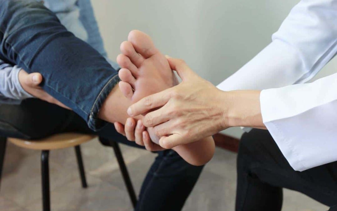
Foot drop is generally diagnosed through a physical evaluation. The healthcare professional will watch the patient walk and check their leg muscles for weakness. The doctor should also check for pain, tingling sensation, and numbness on the foot. �
Imaging Tests
Foot drop is sometimes caused by an overgrowth of bone in the spinal canal or by a tumor or cyst pressing on the nerve roots in the lumbar spine. Imaging tests will help pinpoint these kinds of health issues. These imaging tests can include: �
X-rays. Plain X-rays utilize a minimal amount of radiation to show a soft tissue mass or even a bone lesion.
Ultrasound. This technology, which utilizes sound waves to create images of internal structures, may check for tumors or cysts on the nerves or demonstrate swelling on the nerves due to compression or impingement.
CT scan. This combines X-ray images from distinct angles to form cross-sectional perspectives of structures.
Magnetic resonance imaging or MRI. This test utilizes radio waves and a strong magnetic field to produce detailed images. MRI is very useful in showing soft tissue lesions which may be irritating a nerve.
Nerve Tests
Electromyography, or EMG, and nerve conduction studies measure electrical activity in the muscles and nerves. These tests can be uncomfortable, but they’re helpful in determining the region of the damage or injury along the affected nerve. �
What is the Treatment for Foot Drop?
Treatment for foot drop depends on the cause of the health issue. If the cause is successfully treated, foot drop may improve or even disappear. If the cause can’t be treated, foot drop may be irreversible. Treatment for foot drop may include:
Braces or splints. A brace or splint on your ankle and foot which fits a shoe can maintain the foot in a normal position.
Chiropractic care or physical therapy. Alternative treatment options including stretches and exercises can help strengthen the lower extremities and their range of motion which may improve gait problems associated with foot drop. Stretching stretches and exercises are especially important to protect against stiffness in the heel. The healthcare professional may also provide custom foot orthotics to provide additional support, stability, and shock absorption.
Nerve stimulation. Occasionally stimulating the nerve which lifts the foot can help improve foot drop.
Surgery. Depending on the cause and if the patient’s foot drop is comparatively new, surgical interventions may be useful. If the patient’s foot drop is long-standing, the healthcare professional may suggest surgery which fuses the ankle and foot bones or a process which transfers an attached muscle and tendon to another region of the foot.
Foot drop is a health issue in which the raising of the front part of the foot is difficult. Foot drop is not a condition or disease but rather, a symptoms of an underlying health issue. Muscle and/or nerve damage caused by injury and/or an aggravated health issue, such as a lumbar herniated disc, spinal stenosis, spondylolisthesis, and/or fractures can ultimately cause foot drop. These health issues can also cause low back pain and sciatica as well as other painful symptoms.� – Dr. Alex Jimenez D.C., C.C.S.T. Insight
Low Back Pain
The purpose of the article is to understand how foot drop can be associated with sciatica and other symptoms. Sciatica is a collection of symptoms characterized by pain, tingling sensation, and numbness. The scope of our information is limited to chiropractic, musculoskeletal and nervous health issues as well as functional medicine articles, topics, and discussions. To further discuss the subject matter above, please feel free to ask Dr. Alex Jimenez or contact us at 915-850-0900 . �
Curated by Dr. Alex Jimenez �
Additional Topic Discussion: Foot Orthotics
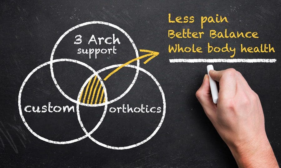
Low back pain and sciatica are common health issues which affect many individuals worldwide. However, did you know that chronic pain may be due to foot problems? Health issues originating in the foot may ultimately cause imbalances in the spine, such as poor posture, which can cause the well-known symptoms of low back pain and sciatica. Custom foot orthotics, individually designed with 3-arch support can help promote overall health and wellness by supporting and promoting good posture and correcting foot problems. Custom foot orthotics can ultimately help improve low back pain and sciatica. �
Formulas for Methylation Support
XYMOGEN�s Exclusive Professional Formulas are available through select licensed health care professionals. The internet sale and discounting of XYMOGEN formulas are strictly prohibited.
Proudly,�Dr. Alexander Jimenez makes XYMOGEN formulas available only to patients under our care.
Please call our office in order for us to assign a doctor consultation for immediate access.
If you are a patient of Injury Medical & Chiropractic�Clinic, you may inquire about XYMOGEN by calling 915-850-0900.
�
For your convenience and review of the XYMOGEN products please review the following link.*XYMOGEN-Catalog-Download �
* All of the above XYMOGEN policies remain strictly in force. �
Chiropractic can help with a wide variety of health conditions, particularly those involving the spine. Your chiropractor�s ability to adjust your spine makes it possible to correct many issues with the back and neck. Chiropractic also offers the advantage of being drug-free and non-invasive, so the side effects you need to worry about are minimal.
Contents
6 Spine Disorders that Chiropractic Helps
1. Degenerative Disc Disease
Most often, degenerative disc disease is a condition that strikes people as they get older. The discs that sit between your vertebrae are supposed to be relatively soft to provide cushion and flexibility to the spine. Unfortunately, years of use and various injuries can cause the discs to wear down. When they wear down it can lead to pressure on nerves and bone on bone contact.
2. Herniated Disc
Sometimes a disc can rupture, allowing the interior matter to protrude from the outer shell and press against nerves in the spine. The most common areas of the body to have a herniated disc are the lower back and the neck.
3. Sciatica
The sciatic nerve travels from the lower back and down the legs. The nerve can become irritated if there are issues with the lower back, such as degenerative disc disease, herniated disc or misalignment. Pressure on the nerve can cause symptoms like shooting pains, numbness, tingling, dull aching, and other discomforts.
4. Whiplash
Whiplash is most common in rear-end car accidents, where a car hits you from behind and causes your head to whip forward and backward. The force generated by the whipping motion can cause significant damage to your neck. Stiffness and pain are common symptoms. Whiplash should be treated as soon as possible following an accident.
5. Myofascial Pain
The fascia is the sheath-like material that surrounds your muscles. Adhesions can occur in the fascia due to overuse or injury. Sometimes the painful points can feel like hard knots under the skin. Myofascial pain often causes discomfort in other areas of the body that you would not assume are related to the problem area.
6. Back Sprains and Strains
There are many ways that the back can become sprained or strained. A sprain involves the ligaments that are attached to your bones, while a strain involves your muscles and/or tendons. Either condition can cause significant pain and a loss of mobility.
Chiropractic Can Help By
Your chiropractor will offer you different options for healing your spinal condition. These may include:
Adjustments
Many times the problems with your spine can be improved by realigning the vertebrae. Injuries and spinal conditions are often associated with misalignment�where the vertebrae are not sitting on top of one another as they should be. A chiropractic adjustment gently returns your vertebrae to alignment.
Massage Therapy
Massage is a great complementary therapy to chiropractic adjustments. It can help to relax muscles, eliminate adhesions and improve mobility. It can also help your spinal adjustments to stay in place as you go about your day to day life.
Ultrasound
Ultrasound treatments are an effective way to encourage your body�s own healing mechanisms without causing any unwanted side effects. The sound waves penetrate deep into the tissues to create heat energy, which encourages circulation and aids in healing.
Get Help with Your Spinal Disorder
If you are struggling with a spinal disorder like those mentioned above, we would love the opportunity to help. Our chiropractic team understands how to administer safe, effective treatments that are non-invasive and work together to improve healing outcomes.
Please contact us today to discuss your spinal condition and schedule an appointment.
Heel Spurs
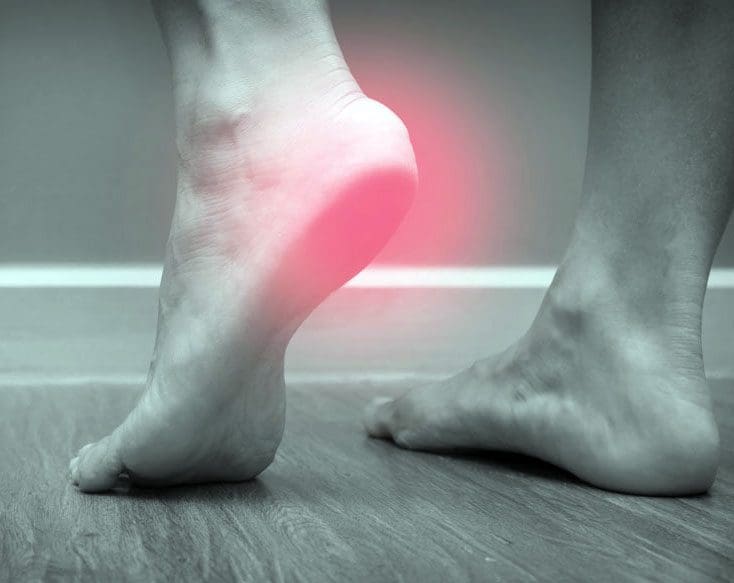
Do you have a sharp pain in your heel? When you take a step, does it feel like a knife is stabbing your heel? If the answer is yes, then you might have a heel spur.
A heel spur is a degenerative outgrowth of bone on the calcaneus. While the heel spur itself is sometimes painless, the condition is commonly associated with Plantar Fasciitis.
Though adjustments may help reduce symptoms, they do not restore plantar aponeurosis laxity or the resultant excessive pronation.
Custom orthotics support all 3 arches, which help control pronation and protect against heel shock.
Your healthcare provider and custom orthotic’s center can offer a special cut out�at the site of the insertion of the plantar fascia to reduce pressure.
Conclusion
Heel spur pain can be reduced by seeing your movement patterns of the foot and ankle. While there is no cure for heel spurs, chiropractic foot adjustments, orthotics and home stretching can help.
El Paso, TX Chiropractic Heel Spur Treatment
Blanca, born and raised in El Paso, TX, has been suffering from heel spurs for approximately two years. As a registered nurse, her symptoms appreciably affected her ability to work and her general quality of life. Determined to improve her health, Blanca believed care. Once she began treatment with Dr. Alex Jimenez, however, Blanca experienced enormous relief from her heel spurs, nearly immediately. Blanca highly recommends chiropractic care with Dr. Alex Jimenez as the nonsurgical selection for the treatment of heel spurs. A heel spur is a calcium residue resulting in a bony protrusion on the bottom of the heel bone.
Although heel spurs are often painless, they can lead to heel pain. They are often associated with plantar fasciitis, a painful inflammation of the fibrous band of connective tissue (plantar fascia) which runs across the bottom of the foot and connects the heel bone to the ball of the foot. Heel spurs are usually caused by strains on foot ligaments and tendons, stretching of the plantar fascia, and repeated tearing of the membrane that covers the heel bone. Heel spurs are especially common among athletes.
Orthotics
Foot Dysfunction can very easily cause a domino effect that extends all the way to the back. The feet are the foundation of the body and when there is a problem with the way they function it can cause the entire body to shift out of alignment. Overpronation and oversupination can cause a variety of injuries and conditions that affect not only the feet and ankles, but also the knees,�hips, and back as well.
Functional Hallux Limitus
Functional hallux limitus is unfortunately still considered to be a rare health issue which often goes unaddressed. Outcomes for many different foot disorders will be jeopardized if treatment doesn�t address functional hallux limitus. Foot and postural abnormalities have a basis in poor function of stabilization and the windlass mechanism of the foot structure through the plantar fascia.
NCBI Resources
Although many people try to treat their low back pain on their own first, one of the fastest and best ways to treat it is through custom orthotics, which actually optimize the performance of your entire body, not just your feet and lower back. For instance, overpronation of the foot causes a series of internal changes that extend up through the leg. The femur may rotate causing hip pain and inflammation of the sacroiliac joint which leads to back pain. Other misalignments in the body that are caused by foot problems can also lead to chronic lower back pain as well.
Heel spurs are a health issue which causes the development of a bony-like expansion, known as a calcium deposit, which develops between the heel bone and arch. Heel spurs generally begin in the front of the heel and may affect other regions of the foot. They’re generally about a quarter of an inch in length and they may not necessarily be visible to the naked eye. �
Diagnosing heel spurs can be challenging for healthcare professionals because these don’t necessarily trigger painful symptoms and not all heel pain is associated with heel spurs. The purpose of the following article is to discuss the symptoms, causes, treatment, and prevention of heel spurs as well as their association with radiating pain and sciatica symptoms. �
Contents
What are the Symptoms of Heel Spurs?
Common symptoms of heel spurs may include pain, discomfort, swelling, and inflammation in the front of the heel. Alongside the painful symptoms previously described, the affected region may also feel warm to the touch. The painful symptoms can also radiate or spread to the back of the foot. Over time, a small bony protrusion may become observable to the naked eye. �
Some heel spurs may also cause no painful symptoms. However, approximately 50 percent of people with heel spurs will experience heel pain.� Some heel spurs may also not result in any changes to the bones or soft tissues surrounding the heel. Moreover, the painful symptoms may affect an individual’s gait and posture, causing compensation which can ultimately result in a variety of other health issues. A spinal misalignment, or subluxation, may cause low back pain and sciatica. �
Heel spurs are frequently diagnosed utilizing X-rays and other clinical evaluations for foot health issues. Heel spurs are difficult to diagnose because the symptoms are similar to other types of heel pain. It’s fundamental to visit a healthcare professional to receive a proper diagnosis. The healthcare professional can then diagnose a heel spur utilizing X-rays. �
What are the Causes of Heel Spurs?
Heel spurs are caused by long-term muscle and ligament strain. The excess strain can affect the soft tissues of the heel and wear them out. Heel spurs generally develop over an extended period of time and often after the individual ignores early signs, such as heel pain.�Repetitive pressure from walking, running, or jumping on hard surfaces is a common cause of heel spurs. These may also develop from wearing shoes which don’t properly support the foot. Heel spurs may also be caused by: �
Arthritis
Bruising of the heel
Excess body weight
Poorly fitted shoes
Walking gait problems
Using flip-flops too often
Worn-out shoes
What is the Treatment for Heel Spurs?
Fortunately, there are a variety of treatment approaches for heel spurs. Treatment choices for heel spurs can include: �
Cold compresses. Applying ice packs after exercise and/or physical activity may be especially beneficial.
Anti-inflammatory injections. This helps alleviate pain and inflammation in the heel of the foot and arch.
Over-the-counter pain drugs and/or medications. These could include acetaminophen, aspirin, or ibuprofen.
Chiropractic care and physical therapy. These, alongside stretches and exercise, can help improve symptoms.
Rest. It is essential to rest the feet after standing or engaging in physical activities for an extended period of time.
Orthotic shoe inserts. These may help provide you arch support.
Healthcare professionals may recommend surgery as a last resort if other alternative treatment options don’t help improve heel spurs. This surgical intervention involves the removal of the heel spur. Sometimes it also involves releasing the plantar fascia muscle. Heel spur surgery can reduce painful symptoms and help boost mobility in the foot. Due to the safety and effectiveness of other alternative treatment options, surgery is generally not recommended for the treatment of heel spurs. �
Heel spurs are characterized as a degenerative outgrowth of bone on the calcaneus, or the heel bone. Although heel spurs may be commonly associated with heel pain and discomfort, not all cases of heel spurs cause painful symptoms. Long-term stress and/or pressure can cause heel spurs. Heel spurs can also cause low back pain and sciatica. Because of the altered gait during heel strike and foot-off due to the painful symptoms, heel spurs can cause hip imbalances and compensation health issues. Spinal misalingments, or subluxations, due to altered posture can ultimately cause low back pain and sciatica.� – Dr. Alex Jimenez D.C., C.C.S.T. Insight
Low Back Pain
The purpose of the article is to understand how heel spurs can be associated with sciatica and other symptoms. Sciatica is a collection of symptoms characterized by pain, tingling sensation, and numbness. The scope of our information is limited to chiropractic, musculoskeletal and nervous health issues as well as functional medicine articles, topics, and discussions. To further discuss the subject matter above, please feel free to ask Dr. Alex Jimenez or contact us at 915-850-0900 . �
Curated by Dr. Alex Jimenez �
Additional Topic Discussion: Foot Orthotics
Low back pain and sciatica are common health issues which affect many individuals worldwide. However, did you know that chronic pain may be due to foot problems? Health issues originating in the foot may ultimately cause imbalances in the spine, such as poor posture, which can cause the well-known symptoms of low back pain and sciatica. Custom foot orthotics, individually designed with 3-arch support can help promote overall health and wellness by supporting and promoting good posture and correcting foot problems. Custom foot orthotics can ultimately help improve low back pain and sciatica. �
Formulas for Methylation Support
XYMOGEN�s Exclusive Professional Formulas are available through select licensed health care professionals. The internet sale and discounting of XYMOGEN formulas are strictly prohibited.
Proudly,�Dr. Alexander Jimenez makes XYMOGEN formulas available only to patients under our care.
Please call our office in order for us to assign a doctor consultation for immediate access.
If you are a patient of Injury Medical & Chiropractic�Clinic, you may inquire about XYMOGEN by calling 915-850-0900.
�
For your convenience and review of the XYMOGEN products please review the following link.*XYMOGEN-Catalog-Download �
* All of the above XYMOGEN policies remain strictly in force. �
At least 80% of the U.S. population will experience back pain at some point in their life, which accounts for more than 3 million reported cases of lower back pain each year. Lower back pain is also one of the biggest reasons why people miss work.
There is a solution that doesn�t include trips to the doctor, insurance agency, or prescription medication.
Contents
Understand Low Back Pain
The symptoms of low back pain can be different for everyone, ranging from dull aches to sharp /stabbing pain.
Low back pain can inhibit our ability to stand up straight, move around, lift everyday items, and do our household chores e.g. laundry and garbage.
When the pain comes quick such as, after lifting something heavy or from an injury, it is considered acute.
If the pain lasts for more than three months, it is considered chronic.
Common Causes
Heavy lifting, exercise or sudden jerking movements and injury can cause acute pain.
Injury can also lead to more chronic issues like sciatica
Those of us with desk jobs are at risk for chronic low back pain.
When carrying a bag, be careful
Purses, backpacks, and briefcases carried over the shoulder put a greater risk of developing chronic back pain because it is the lower back that takes all the weight.
Instead, get a rolling bag/case or a backpack that distributes the weight evenly over the body, across both shoulders.
Other causes of lower back pain can be hormones, weight, lifestyle, and environmental factors.
The Solution to Low Back Pain
Many chiropractors encourage the use of custom orthotics to help treat a variety of problems, ranging from joint pain, foot discomfort, and posture issues.
Custom orthotics are insoles that are slipped into shoes and made especially for your body.
Wearing custom orthotics provides nearly immediate relief from lower back pain, and also helps prevent it from returning.
Custom orthotics are created by scanning and assessing your feet in less than five minutes.
By simply standing barefoot on a kiosk inside your chiropractor�s office, a one-of-a-kind solution is created.
No X-rays, MRI�s, or further testing is required.
The foot scan assesses the three arches in both of your feet, as well as how you distribute your weight on the foundation of your body.
Although many people try to treat their low back pain on their own first, one of the fastest and best ways to treat it is through custom orthotics, which actually optimize the performance of your entire body, not just your feet and lower back.
Over-the-counter orthotics do not provide proper support and can even cause more damage to the body.
Chronic lower back pain is not a normal thing that has to do with age or lifestyle.
If you are experiencing lower back pain, call a chiropractor and ask about how custom orthotics can help you.
Low Back Pain? Fix it with *FOOT ORTHOTICS* | El Paso, Tx
After a quick scan, your feet will be assessed and your custom orthotics will be designed and made especially for you.
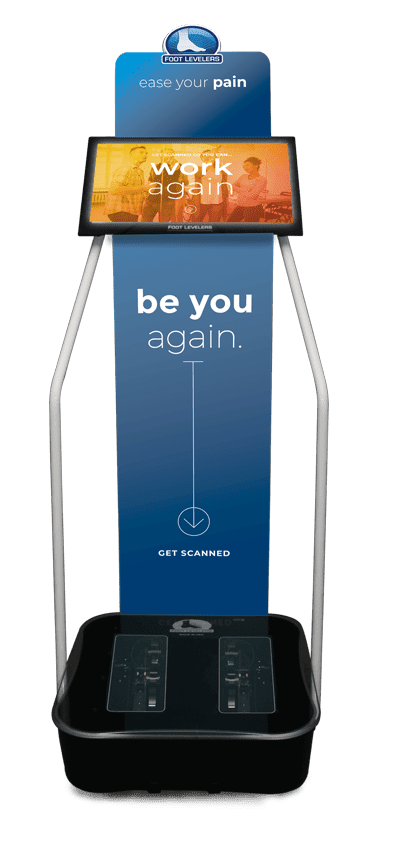
El Paso Back Clinic Foot Orthotic Kiosk
The Kiosk helps guide patients in selecting the best custom-made orthotics for their condition and lifestyle. It’s�
Fast:�Push the Start button and the scanner begins.
Easy to use:�User-friendly easy touch screen.
Engaging:�Videos explain the importance of healthy feet and the benefits of custom-made orthotics.
Cloud-based:�Results can be securely accessed from anywhere.
Comprehensive:�Easily retrieve previous scans to compare them to new scans and see the difference.
The Foot Levelers Kiosk saves time so you can spend more time living your life.
Orthotics
When there are problems with the feet, it can cause problems through the legs and all the way to the spine. This can cause the ankle to pronate, meaning it rolls inward. This alters the way the�bones of the foot line up which extends through the tibia, or shin bone. Ankle pain�and injury are not issues reserved solely for athletes. In the United States, more than 25,000 people deal with�ankle sprains�or pain every day. Studies show that somewhere around 40 percent of ankle sprains are not treated correctly or are misdiagnosed, which leads to disability or chronic ankle pain.
Ankle Manipulation
The typical treatment for pain, such as with a sprain, is R.I.C.E., which is rest, ice, compression and elevation. A somewhat newer treatment approach replaces the R with an M, meaning that instead of rest, movement is required instead. However, it is important that the movement is done safely and carefully. Certain types of�ankle injuries can be exacerbated by movement so it should be approached with care. Chiropractic can help reduce pain without prescription medication and the associated side effects. This alone is often a great draw for many patients. However, there are other benefits that chiropractic can provide for ankle pain.
31 million Americans experience low back pain at any given time. This condition affects many, but finding the exact cause can be a challenge. Chiropractors are spinal specialists that are trained to not only alleviate pain but also find and help correct the cause of the problem. A�chiropractor�is the ideal�medical professional to consult with for any unexplained pain in the musculoskeletal system. They are highly qualified professionals that their specialty is treating conditions like lower back pain and they are very affordable.
Achilles tendinitis is a common health issue which occurs when the large tendon that runs down the back of the lower leg becomes irritated and inflamed. The Achilles tendon is the largest tendon in the human body. It connects the calf muscles to the heel bone which allows you to walk, run, climb stairs, jump, and stand on your tiptoes. Although the Achilles tendon can withstand tremendous amounts of pressure, it can be prone to tendinitis, a state associated with overuse and degeneration. �
Achilles tendinitis is the inflammation of the Achilles tendon. Inflammation is the human body’s natural reaction to injury, infection, or disease and it generally causes pain, discomfort, irritation, swelling, and inflammation, among other symptoms. According to research studies, Achilles tendinitis can even cause low back pain and sciatica. The purpose of the article is to help understand Achilles tendinitis and how it can be associated with low back pain and sciatica, among other health issues. �
Contents
Achilles Tendinitis Causes
Achilles tendinitis is generally not associated with a specific injury and/or condition. The health issue commonly results from stress associated with overuse and degeneration. This frequently occurs when we push ourselves too much, too soon, however other factors may also ultimately increase the risk of developing Achilles tendinitis. These factors can include: �
Sudden increase in the intensity or amount of exercise� and physical activity
Having tight calf muscles and abruptly starting a fitness program can place additional stress on the Achilles tendon
Bone spurs or extra bone development where the Achilles tendon attaches to the heel bone
Achilles Tendinitis Symptoms
Achilles tendinitis can be characterized by a variety of common symptoms. These symptoms can include: �
Pain, discomfort, and stiffness along the Achilles tendon in the daytime
Painful symptoms throughout the tendon or back of the heel that worsens with activity
Severe pain and discomfort after exercise or physical activity
Thickening of the Achilles tendon
Bone spurs, also referred to as insertional tendinitis
Swelling and inflammation which worsens throughout the day with exercise or physical activity
Low back pain and sciatica due to compensation or altered gait and posture
If you experienced a sudden”pop” in the back of your calf or heel, you may have ruptured or torn your Achilles tendon. Make sure to seek immediate medical attention for a proper diagnosis if you feel that you may have damaged your Achilles tendon. �
The association between Achilles tendinitis, low back pain and sciatica have been investigated in a variety of research studies. According to one specific research study, in 138 patients who experienced Achilles tendinitis and in a group of individuals nominated by the patients, matched for age, sex, and occupation, low back pain and sciatica had been experienced by 63 of the patients and by 91 of the individuals in the control group. However, 35 of the patients had experienced sciatica before Achilles tendinitis. Researchers found a significant association between Achilles tendinitis, low back pain, and sciatica. This association may be due to impaired afferent signals from the lower extremities or to similar collagen or vascular anomalies of the intervertebral disc and the Achilles tendon, associated with compensation or altered gait and posture. �
Achilles Tendinitis Diagnosis
Once you seek immediate medical attention to receive a proper diagnosis for your Achilles tendinitis, the healthcare professional will examine your ankle and foot. Moreover, the healthcare professional will look for these symptoms: �
Swelling and inflammation along the Achilles tendon or at the back of the heel
Thickening or augmentation of the Achilles tendon
Bony spurs in the lower part of the tendon in the back of your heel
Points of maximum tenderness
Pain in the center of the tendon
Pain in the back of the heel at the lower portion of the tendon
Restricted range of motion in the ankle and foot, including a diminished ability to flex the foot
The healthcare professional may also order imaging tests, such as X-rays and magnetic resonance imaging, or MRI,� to make sure that symptoms are due to Achilles tendinitis. Differential diagnosis for Achilles tendinitis may cause similar symptoms. �
Achilles Tendinitis Treatment
In most instances, non-surgical treatment approaches may help provide Achilles tendinitis pain relief. Chiropractic care and physical therapy may also help decrease symptoms as well as increase strength and performance to promote faster recovery. The chiropractor or physical therapist may utilize a combination of treatment methods and techniques to improve overall health and wellness. Furthermore, because the bones and soft tissues of the ankle and foot are utilized throughout the various stages of walking and running, improper movement patterns of the ankle and foot can cause a variety of health issues, including Achilles tendonitis. Custom foot orthotics can provide support, stability, and shock absorption to prevent excess stress and pressure on the feet. For acute cases, your doctor may suggest that you consider surgery. �
Achilles tendinitis is commonly characterized as the swelling and/or inflammation of the Achilles tendon which runs from the calf muscles to the back of the heel bone. As previously mentioned above, Achilles tendonitis generally occurs due to overuse and degeneration. Achilles tendinitis is commonly reported among athletes, especially runners. Common symptoms associated with Achilles tendinitis includes pain and discomfort along the length of the Achilles tendon. – Dr. Alex Jimenez D.C., C.C.S.T. Insight
Low Back Pain
The purpose of the article is to understand Achilles tendinitis and its association with sciatica and other symptoms. Sciatica is a collection of symptoms characterized by pain, tingling sensation, and numbness. The scope of our information is limited to chiropractic, musculoskeletal and nervous health issues as well as functional medicine articles, topics, and discussions. To further discuss the subject matter above, please feel free to ask Dr. Alex Jimenez or contact us at 915-850-0900 . �
Curated by Dr. Alex Jimenez �
Additional Topic Discussion: Foot Orthotics
Low back pain and sciatica are common health issues which affect many individuals worldwide. However, did you know that chronic pain may be due to foot problems? Health issues originating in the foot may ultimately cause imbalances in the spine, such as poor posture, which can cause the well-known symptoms of low back pain and sciatica. Custom foot orthotics, individually designed with 3-arch support can help promote overall health and wellness by supporting and promoting good posture and correcting foot problems. Custom foot orthotics can ultimately help improve low back pain and sciatica. �
Formulas for Methylation Support
XYMOGEN�s Exclusive Professional Formulas are available through select licensed health care professionals. The internet sale and discounting of XYMOGEN formulas are strictly prohibited.
Proudly,�Dr. Alexander Jimenez makes XYMOGEN formulas available only to patients under our care.
Please call our office in order for us to assign a doctor consultation for immediate access.
If you are a patient of Injury Medical & Chiropractic�Clinic, you may inquire about XYMOGEN by calling 915-850-0900.
�
For your convenience and review of the XYMOGEN products please review the following link.*XYMOGEN-Catalog-Download �
* All of the above XYMOGEN policies remain strictly in force. �
When you struggle with anxiety, it can make living your day-to-day life more difficult. Anxiety comes in a variety of forms and varies by individual. The treatments that are most effective for one person may not be as effective for another, but there is some general guideline for dealing with anxiety that most sufferers can benefit from. Regular chiropractic care, including a focus on improving overall health, can serve as a foundation for navigating the difficulties of anxiety.
Contents
Ways Chiropractic Can Help
1. Keeping track of triggers.
Chiropractors are big fans of keeping diaries surrounding any type of health issue you are dealing with�and anxiety is no exception. Just like keeping a food diary can help you identify a food allergy, keeping an anxiety diary can help you see what things in your life are triggering your anxiety. Triggers for anxiety can include a wide range of things, not all of them related to human interactions. Some of these triggers can include:
Allergies to certain foods
Consumption of alcohol
Consumption of caffeine
Vitamin deficiencies, particularly B vitamins, calcium, and magnesium
Consumption of sugar
Your chiropractor can tell you how to keep a diary that will help you see what you do in your day-to-day life, and how those actions relate to your anxiety.
2. Keeping your body in Top Form.
The relationship between the body and the mind is still far from being fully understood. However, there is no denying the significant connection between our physical health and our mental health. When your body is healthier, your mood is more level and positive.
Chiropractic care is focused on treatments that improve your health. These can include chiropractic adjustments, massage therapy, spinal decompression, ultrasound and more. Which treatments are right for you will depend on the results of your physical examination with the chiropractor. What you can be sure of is that your chiropractor will do everything possible to ensure your body is healthy and functioning at an optimal level.
3. Maintaining Spinal Alignment.
There are many ways that the nervous system affects the body and the brain�and the full effects are not totally clear as of yet. What we do know is that many chiropractic patients report improvements in their health seemingly unrelated to the pain in their back or neck. A few adjustments into a treatment plan and patients discover that some other health issue gets resolved.
Adjustments are designed to put your vertebrae back into proper alignment. With proper alignment, your nervous system can function optimally. The results may or may not improve your anxiety directly, but they will make you feel better and keep your body running the way it was intended to.
4. Improving Your Diet.
What you eat plays a huge role in your overall health. Improving your diet could lead to an improvement in your anxiety symptoms, which is why your chiropractor will try to help you make healthy changes to what you eat. Chiropractors are trained in the latest research in nutrition and are well-versed in the benefits of various approaches to a healthy diet. You can work with your chiropractor to shift your diet from its current state to one that includes more healthy options.
Other Healthcare Providers & Your Chiropractor
If you are seeing a mental healthcare professional for your anxiety, your chiropractor will strive to work with your provider to ensure the best possible outcomes. Sometimes the best way to improve your health is to take a multi-pronged approach�which your chiropractor can help you with!
Please contact us today to schedule an appointment with our chiropractic team. We look forward to seeing you.
Depression & Chronic pain | El Paso, Tx
Chronic pain caused by accidents and/or aggravated conditions can often be one of the primary reasons for depression in patients. When painful symptoms induce patients to struggle with their everyday physical activities, their mental health can be tremendously influenced. Chiropractic care utilizes spinal adjustments and manual manipulations which could help restore the initial integrity of the backbone. Patients describe how chiropractic care has helped them recover their well-being and they highly recommend Dr. Alex Jimenez, doctor of chiropractic, as the non-surgical choice for chronic pain and depression, one of a variety of other common health issues.
Gait Related Low Back Pain
Currently, more than 2 million Americans are dependent on opioids. For many, their addiction comes from seeking pain management for an injury or chronic condition. Opioids are a convenient and fast remedy, but only provide pain relief and not actual treatment of the cause. Therefore, opioids should be a last resort when experiencing pain and discomfort. Starting at the foundation (feet) of the body with orthotics can help the rest of the body stay aligned.
NCBI Resources
If you are suffering from depression or anxiety, you may feel hopeless and helpless. You may be less apt to seek or follow treatment, believing there is nothing you can do to make it better. When you have a chronic medical condition, it doesn�t just impact your health. Often you can�t work or miss time at work, you may have financial problems. Relationships frequently suffer when one partner is sick. While these can be true for all chronic conditions, when you add in depression or anxiety, coping is even more difficult.
Sciatica can commonly occur due to the over-pronation of the foot, which causes flat-footedness as well as an abnormal gait and posture. This theory is widely accepted by podiatrists, physical therapists, chiropractors, and even sports medicine specialists, however, it is doubted by many healthcare professionals, especially in mild to moderate circumstances. �
Flat-footedness, also referred to as fallen arches, can be demonstrated unilaterally or bilaterally. This health issue can cause foot pain with a variety of other symptoms. However, many healthcare professionals still doubt that flat-footedness is considerable enough to cause sciatica, being how the human body can compensate for structural and functional irregularity. The purpose of the following article is to demonstrate how foot pronation can cause sciatica and other symptoms. �
Contents
What is Foot Pronation?
Foot pronation is characterized as a flattening of the frontal arch of the foot and inwards turning of the big toe. Pronated feet seem to roll inward at the ball of the foot, making the knife-edge of the foot seem to bend outwards at the ankle joint. Basically, the heel bone is further towards the outside of the human body in placement when compared to the ankle bone. This type of foot abnormality may be shown on one foot (unilateral expression) or in both feet (bilateral expression). �
Over-pronation can range considerably from mild to moderate and severe. It’s fundamental to understand that the action of pronation is a normal part of standing and walking. However, in the following article, we will discuss how over-pronation or flat-footedness, can cause a variety of health issues, including sciatica, a collection of painful signs and symptoms. �
Pronated Foot Mechanisms and Sciatica
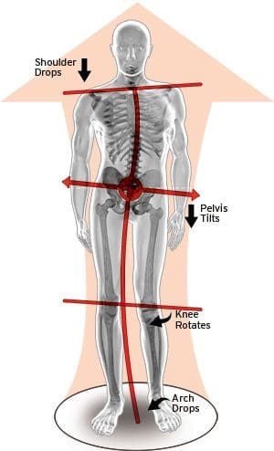
When the feet don’t come in contact with the floor, the human body will have to compensate from the feet all the way to the top of the head. Research studies have associated foot pronation with sciatica and many other debilitating signs and symptoms associated with this health issue. Moreover, over-pronation affects the angle in which the foot meets the ankle. �
In several patients, the joints of the spine, sacroiliac, hip, and knee, as well as the soft tissues which maintain and support these joints, will adjust to the over-pronation of the foot. This can develop a variety of signs and symptoms. For sciatica, several of the mechanisms make-up the several consequences of anatomical tilting and compensatory activities, including: �
Postural changes which can aggravate nerve root compression or impingement in the lumbar spine
Postural and gait changes which can affect the sacroiliac joint between the pelvic ilium and sacrum
Postural and functional changes which can affect the piriformis muscle and cause piriformis syndrome
The sciatic nerve may be affected by joint or soft tissue health issues. Compression or impingement is merely one of the mechanisms which might be created by muscular imbalances and joint misalignments as well as other prospective outcomes of over-pronation of the foot. Some people may also confuse other health issues for sciatica. These can also affect the hip, the knee, the ankle, and the foot, as a result of compensation of the human body in response to over-pronation of the foot. �
Foot Pronation Risks and Concerns
Many healthcare professionals believe that severe instances of foot pronation are the cause of foot pain, in addition to other well-known signs and symptoms. Nonetheless, healthcare professionals also discuss that mild to moderate cases of over-pronation, accounting for approximately 98 percent of diagnoses, are likely to be non-factors in sciatica signs and symptoms. �
Other healthcare professionals discuss that sciatica caused by foot pronation is plausible in severe over-pronation health issues, despite apparent postural and gait abnormalities. These healthcare professionals believe that sciatica caused by foot pronation, although not frequently well-known, is possible in several cases. This supports the findings of several research studies, where it is logical and plausible based on structural and functional evidence, in addition to absent in several cases where sciatica signs and symptoms would seem completely justifiable based on acute cases of flat-footedness. �
Foot pronation, or flat-footedness, can cause overall imbalances in the human body. Pronated feet or flat feet can ultimately cause pain and discomfort in the spine, hips, knees, and feet, as well as shift the entire human body out of alignment. Research studies have demonstrated that women with flat feet are approximately 50 percent more likely than those without to have low back pain and sciatica symptoms.� – Dr. Alex Jimenez D.C., C.C.S.T. Insight
Low Back Pain
The purpose of the article is to describe how foot pronation can be associated with sciatica and other health issues. Sciatica is a collection of symptoms characterized by pain, tingling sensation, and numbness. The scope of our information is limited to chiropractic, musculoskeletal and nervous health issues as well as functional medicine articles, topics, and discussions. To further discuss the subject matter above, please feel free to ask Dr. Alex Jimenez or contact us at 915-850-0900 . �
Curated by Dr. Alex Jimenez �
Additional Topic Discussion: Foot Orthotics
Low back pain and sciatica are common health issues which affect many individuals worldwide. However, did you know that chronic pain may be due to foot problems? Health issues originating in the foot may ultimately cause imbalances in the spine, such as poor posture, which can cause the well-known symptoms of low back pain and sciatica. Custom foot orthotics, individually designed with 3-arch support can help promote overall health and wellness by supporting and promoting good posture and correcting foot problems. Custom foot orthotics can ultimately help improve low back pain and sciatica. �
Formulas for Methylation Support
XYMOGEN�s Exclusive Professional Formulas are available through select licensed health care professionals. The internet sale and discounting of XYMOGEN formulas are strictly prohibited.
Proudly,�Dr. Alexander Jimenez makes XYMOGEN formulas available only to patients under our care.
Please call our office in order for us to assign a doctor consultation for immediate access.
If you are a patient of Injury Medical & Chiropractic�Clinic, you may inquire about XYMOGEN by calling 915-850-0900.
�
For your convenience and review of the XYMOGEN products please review the following link.*XYMOGEN-Catalog-Download �
* All of the above XYMOGEN policies remain strictly in force. �
Foot pronation is the natural movement which occurs during foot landing while walking or running. Foot pronation also occurs while standing, and in this instance, it is the amount in which the foot rolls inward toward the arch. Foot pronation is normal, however, excessive foot pronation can cause a variety of health issues, including bad posture. The following video describes the 5 red flags of excessive foot pronation, which can ultimately affect a person’s overall health and wellness. Dr. Alex Jimenez can help diagnose and treat excessive foot pronation. Patients recommend Dr. Alex Jimenez and his staff as the non-surgical choice for excessive foot pronation health issues.
Contents
Q-Angle & *KNEE INJURIES ADJUSTMENT* with Custom Foot Orthotics | El Paso, TX (2019)
Knee pain can be alleviated with custom orthotics.
Knee pain can come from
Sudden injury
Overuse injury
Symptoms of underlying condition e.g. arthritis.
Treating your knee pain will depend on the underlying cause.
In most cases, treating your knee condition will be successful when the focus is on restoring balanced function in the entire body, starting from the ground up.
Chiropractic care plus custom orthotics can help.
Knee Pain Symptoms
Pain
Swelling
Stiffness
Treatment
A Chiropractor will likely use a combination of techniques to help your condition.
These include using ice to reduce inflammation
Soft tissue massage to help improve the knee�s range of motion
Chiropractic manipulation and mobilization techniques in the knee as well as surrounding joints.
Rest
Heat
Elevation
Compression
Strengthening exercises
Custom orthotics
A chiropractor will investigate if there are other alignment issues in other areas of the body that could be the true cause of the knee pain or contribute to it.
Prevention
Prevention techniques include:
Keep your bones, muscle tissues strong and healthy through exercise and diet.
Avoid tobacco
Avoid excessive alcohol, which can weaken bones and cause problems with the blood supply.
Reduce your risk of injury by adhering to safety measures on the job and at home.
Use knee braces to prevent knee injuries or after a knee injury.
Custom orthotics can provide additional support, stability, and shock absorption.
When there are problems with the feet, it can cause problems through the legs and all the way to the spine. This can cause the ankle to pronate, meaning it rolls inward. This alters the way the�bones of the foot line up which extends through the tibia, or shin bone. For example, excessive foot pronation, limited range of motion in the hips, or tightness in the lower back, can place excessive strain on the knees.
Cuboid Manipulation
Treatment for foot pain varies depending on the condition/injury. Treatment can go from rest and ice to physical therapy, massage, chiropractic and in severe cases surgery. Reflexology can provide relief, as well as, stretching exercises. Over the counter pain medication is often used. If the pain is too intense that it prevents sleep, a physician may prescribe non-addictive pain medication. Wear shoes with good arch supports, and if pain persists. Insurance often covers orthotics.
Experiencing foot pain, there�s no doubt you checked out your foot to make sure it�s not�injured�or hurting from�improper fitting shoes, corns, plantar fasciitis, etc. This may seem counterintuitive, but you may want to check the condition of the�lumbar spine (lower back)?� Most foot problems are caused by issues with the foot, but what if you found that pressure on the sciatic nerve can cause intense foot pain.
Have you ever noticed or been told that one of your legs is longer than your other leg? Have you experienced back pain and other spine health issues? According to research studies, leg length discrepancy can be associated with low back pain and sciatica. Most people have a minimal difference in their leg spans. Because the discrepancy is small, it’s generally not a contributor for back pain. However, if the difference in their leg spans is more than 5 millimeters or 1/4 inch, it can contribute to low back pain and sciatica. The purpose of this article is to discuss how leg length discrepancy is associated with sciatica symptoms. �
Contents
Leg Length Discrepancy Causes
Leg length discrepancies can occur due to the poor alignment of the pelvis or when one leg is structurally longer than the other. Regardless of the cause, in order to remain symmetrical, the human body will do its best to compensate for the leg length discrepancy. The bigger the leg length difference, the sooner the symptoms will present themselves to the patient. Specific diagnoses that match with leg length discrepancy include scoliosis, lumbar herniated discs, sciatica, sacroiliitis, pelvic obliquity, greater trochanteric bursitis, hip arthritis, piriformis syndrome, patellofemoral syndrome and foot pronation. Other possible causes could be due to an injury, bone disorder, bone tumors, congenital problems, or due to a neuromuscular health issue. �
Leg Length Discrepancy Symptoms
Observance of one leg being longer than the other
Altered posture
Gait (manner of walking) problems
Low back, hip, knee, ankle, or foot pain
Sciatica
Leg Length Discrepancy Classifications
Leg length discrepancies can be categorized as a structural leg length discrepancy or a functional leg length discrepancy. A structural leg length discrepancy is a hereditary circumstance where a leg is simply longer than the other leg. This is determined in the event the patient’s pelvis and sacroiliac joints are symmetrical and the leg length discrepancy is due to a single leg truly being longer than the other. The best method to learn whether a structural leg length discrepancy exists is with an anterior-posterior x-ray of the pelvis. A diagnosis alternative is having a tape measure to measure the length of the leg from the hip to the ankle. �
Functional leg length discrepancy is diagnosed when there is a torsion or pelvic rotation/obliquity, commonly a sacroiliac joint dysfunction, which induces one leg to be longer or shorter than the other. To determine if a true structural leg length discrepancy exists, the doctor must care for the pelvis and return it to a neutral place before quantifying the leg length discrepancy. When the pelvis is symmetrical it’s can be determined if the leg length discrepancy is present or not. If it goes away, it’s classified as functional leg length discrepancy. If it stays and contains a measurable difference, it is a structural leg length discrepancy. �
Leg Length Discrepancy and Sciatica Treatment
Structural leg length discrepancy may be treated by utilizing a heel lift in the shorter leg’s shoe if the leg length is larger than 5 millimeters. The size and use of the heel lift are dependent on a doctor based on how much lift is necessary to restore appropriate lumbopelvic biomechanics. In several instances, surgical intervention may be required to either shorten or lengthen the limb. An important element to any surgical procedure to correct leg length discrepancies is rehabilitation. Rehabilitation can ultimately help to stretch muscles and maintain joint flexibility, which is fundamental towards the healing process after surgery. �
To treat a functional leg length discrepancy, no heel lift is needed, however, appropriate manual treatment methods and specific therapeutic exercises are required to treat and normalize pelvic and lower extremity compensations. The number of treatments needed to support the pelvis in a symmetrical position is different for each patient based on their demonstration and biomechanical dysfunctions in the low back, pelvis, hip, knee, ankle, and foot. When you suffer from low back pain, sciatica, and leg length discrepancy, the signs and symptoms may ultimately be associated with each other. A diagnosis to ascertain whether you have a leg length discrepancy is essential when it might be contributing to low back pain, hip pain, knee pain, or leg pain. �
According to healthcare professionals, leg length discrepancy may affect between 60 to 90 percent of the population. Categorized as either functional or structural, leg length discrepancy can affect the human body’s biomechanics and it may result in low back pain and sciatica. Most people have a smal leg length discrepancy, however, it’s generally not a contributor for back pain. If the leg length discrepancy is greater, however, it can contribute to low back pain and sciatica. – Dr. Alex Jimenez D.C., C.C.S.T. Insight
Low Back Pain
�
�
The purpose of the article is to describe how leg length discrepancy can be associated with sciatica and other health issues. Sciatica is a collection of symptoms characterized by pain, tingling sensation, and numbness. The scope of our information is limited to chiropractic, musculoskeletal and nervous health issues as well as functional medicine articles, topics, and discussions. To further discuss the subject matter above, please feel free to ask Dr. Alex Jimenez or contact us at 915-850-0900 . �
Curated by Dr. Alex Jimenez �
Additional Topic Discussion: Foot Orthotics
Low back pain and sciatica are common health issues which affect many individuals worldwide. However, did you know that chronic pain may be due to foot problems? Health issues originating in the foot may ultimately cause imbalances in the spine, such as poor posture, which can cause the well-known symptoms of low back pain and sciatica. Custom foot orthotics, individually designed with 3-arch support can help promote overall health and wellness by supporting and promoting good posture and correcting foot problems. Custom foot orthotics can ultimately help improve low back pain and sciatica. �
Formulas for Methylation Support
XYMOGEN�s Exclusive Professional Formulas are available through select licensed health care professionals. The internet sale and discounting of XYMOGEN formulas are strictly prohibited.
Proudly,�Dr. Alexander Jimenez makes XYMOGEN formulas available only to patients under our care.
Please call our office in order for us to assign a doctor consultation for immediate access.
If you are a patient of Injury Medical & Chiropractic�Clinic, you may inquire about XYMOGEN by calling 915-850-0900.
�
For your convenience and review of the XYMOGEN products please review the following link.*XYMOGEN-Catalog-Download
� * All of the above XYMOGEN policies remain strictly in force. �
IFM's Find A Practitioner tool is the largest referral network in Functional Medicine, created to help patients locate Functional Medicine practitioners anywhere in the world. IFM Certified Practitioners are listed first in the search results, given their extensive education in Functional Medicine


 Do you have a sharp pain in your heel? When you take a step, does it feel like a knife is stabbing your heel? If the answer is yes, then you might have a heel spur.
A heel spur is a degenerative outgrowth of bone on the
Do you have a sharp pain in your heel? When you take a step, does it feel like a knife is stabbing your heel? If the answer is yes, then you might have a heel spur.
A heel spur is a degenerative outgrowth of bone on the 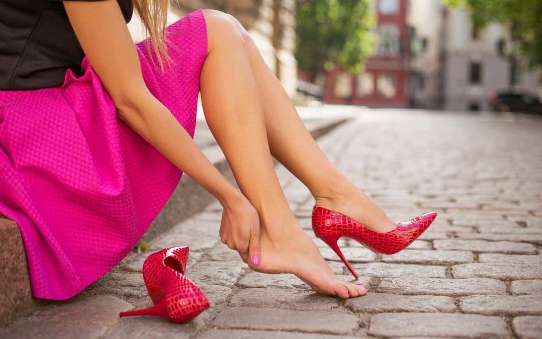

 Although many people try to treat their low back pain on their own first, one of the fastest and best ways to treat it is through custom orthotics, which actually optimize the performance of your entire body, not just your feet and lower back.
Over-the-counter orthotics do not provide proper support and can even cause more damage to the body.
Chronic lower back pain is not a normal thing that has to do with age or lifestyle.
Although many people try to treat their low back pain on their own first, one of the fastest and best ways to treat it is through custom orthotics, which actually optimize the performance of your entire body, not just your feet and lower back.
Over-the-counter orthotics do not provide proper support and can even cause more damage to the body.
Chronic lower back pain is not a normal thing that has to do with age or lifestyle.