If you are experiencing any of these situations, then your hippocampus might be lowered than usual.
The Hippocampus
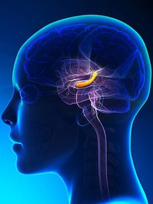
In the brain, there is an S-shaped structured located in the inner folds in the temporal lobe called the hippocampus. The hippocampus is a complex brain structure that has a layer of densely packed neurons, and its primary function involves how humans learn and how their memory works. The hippocampus is part of the limbic system as well since it works the feeling and reacting function in the body. The limbic system is situated at the edge of the cortex and includes the hypothalamus and the amygdala.
These structures help controls the body�s different functions like the endocrine system and the �fight or flight� reaction response. With the hippocampus helping humans process what information they are learning, this structure can retrieve two kinds of memories that are important; they are declarative memories and spatial relationship memories.
Declarative memories: These are memories that are related to facts and events a person experience. It includes examples like how to memorize speeches or line in a play that a person is doing.
Spatial relationship memories: These memories involve pathways or routes that a person must learn. An example of this is transportation drivers like cab drivers, bus drivers, and truckers who have to learn the routes in the places they are going to. So they use spatial memory and practice their routes many times until they have it in their memories. The spatial relationship memories are stored on the right side of the hippocampus.
Sadly though, the hippocampus can be damaged by neurological diseases like Alzheimer�s disease and PTSD (Post-Traumatic Stress Disorder). When it is damaged, a variety of conditions can affect the hippocampus�s ability to do its job for the brain, thus making the individual suffer from retaining information.
Hippocampus Conditions
Several conditions can cause problems to the body when the hippocampus is damaged. This is known as hippocampus atrophy, where the neurons and neuronal volume in the hippocampal that is a loss.
Alzheimer�s Disease
Alzheimer�s disease is when an individual begins to lose their memory. When the hippocampus is damaged, it can cause a dissociation between the cortexes and leads to information registration failure. Studies show that when Alzheimer�s disease is progressing, the hippocampus will lose its volume, and it will become harder for an individual to function in their daily lives.
Epilepsy
When a person has epilepsy, it might be due to a damaged hippocampus. Research shows that around 50 to 75% of patients with this disease may have hippocampal sclerosis, and in case they have died, they have medial temporal lobe epilepsy. More research states that the mechanics of hippocampal sclerosis in epilepsy can be related to the development of inflammation on the uncontrolled local hippocampus and blood-brain barrier damage.
Hypertension
When the hippocampus is damaged, hypertension can happen to a person. Hypertension is another name for high blood pressure, and it can lead to severe health complications to the body. Even though the causes of hypertension are still unknown, the risk factors from hypertension can include:
Environmental factors like stress or a lack of exercise
Hormone activity
Blood plasma
Studies show that hypertension and other risk factors are being increasingly viewed as a putative factor that is leading to hippocampal atrophy.
Cushing�s Disease
Cushing�s disease or Cushing syndrome is when the body is exposed to high levels of cortisol for a long time. Studies show that when there is a loss of cellular volume to the corticosteroid�s levels in the body and it could be responsible. When there is too much cortisol being produced in the body, it is one of the signs of Cushing syndrome. Some of the other signs include:
Weight gain
Fatty tissue deposits around the midsection, face, upper back and between the shoulders
Pink or purple stretch marks
Thinning, fragile skin that bruises easily
Slow healing cuts, insect bites and infections
Acne
Muscle weakness
Cognitive difficulties
Loss of emotional control
Since stress does play a role in the endocrine system and the neurological system, there are nearly 80 years of research on how much focus has been on the various levels of the HPA (hypothalamic-pituitary-adrenal) axis and the hormones it produces. It shows that glucocorticoids as the mediators for the stress effects on the hippocampus and being the contributing factor for stress-associated psychopathologies.
Conclusion
The hippocampus is located in the temporal lobe of the brain. This S-shaped structure can be easily damaged due to stress and other neurological factors that can affect the entire body and its systems. When harmful factors affect the hippocampus, it can lead the hormones that are producing to become imbalanced and cause dysfunction. Some products are here to make sure that the endocrine system is functioning properly and supporting the metabolic system, the gastrointestinal system, as well as making sure the hormones are balanced.
The scope of our information is limited to chiropractic, musculoskeletal, and nervous health issues or functional medicine articles, topics, and discussions. We use functional health protocols to treat injuries or disorders of the musculoskeletal system. Our office has made a reasonable attempt to provide supportive citations and has identified the relevant research study or studies supporting our posts. We also make copies of supporting research studies available to the board and or the public upon request. To further discuss the subject matter above, please feel free to ask Dr. Alex Jimenez or contact us at 915-850-0900.
References:
Anand, Kuljeet Singh, and Vikas Dhikav. �Hippocampus in Health and Disease: An Overview.� Annals of Indian Academy of Neurology, Medknow Publications & Media Pvt Ltd, Oct. 2012, www.ncbi.nlm.nih.gov/pmc/articles/PMC3548359/.
Dresden, Danielle. �Hippocampus: Function, Size, and Problems.� Medical News Today, MediLexicon International, 7 Dec. 2017, www.medicalnewstoday.com/articles/313295.php.
Felman, Adam. �Hypertension: Causes, Symptoms, and Treatments.� Medical News Today, MediLexicon International, 22 July 2019, www.medicalnewstoday.com/articles/150109.php.
Kim, Eun Joo, et al. �Stress Effects on the Hippocampus: a Critical Review.� Learning & Memory (Cold Spring Harbor, N.Y.), Cold Spring Harbor Laboratory Press, 18 Aug. 2015, www.ncbi.nlm.nih.gov/pmc/articles/PMC4561403/.
Team, Mayo Clinic. �Cushing Syndrome.� Mayo Clinic, Mayo Foundation for Medical Education and Research, 30 May 2019, www.mayoclinic.org/diseases-conditions/cushing-syndrome/symptoms-causes/syc-20351310.
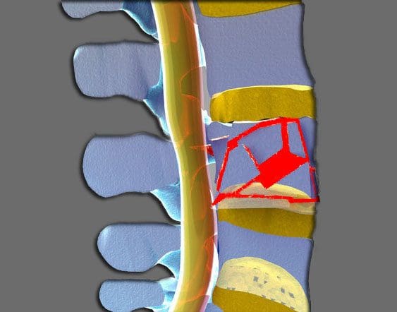
A burst fracture describes an injury to the spine where the vertebrae get compressed severely. These types of injuries occur from severe trauma, like an automobile accident or a serious fall, sports injury, work injury. These injuries entail a great deal of force into the spine, so much so that a vertebra can get crushed.
When crushed in the front of the spine, a wedge-shaped fracture occurs and is known as a compression fracture.
But if the vertebral body gets crushed in all directions this is known as a burst fracture.
The term burst means that the vertebral body spreads out in all directions.
Severe Injury
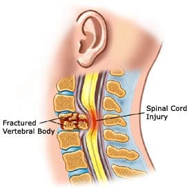
This is a much more severe injury than a compression fracture. With the bones crushed and possible rough jagged edges, if they spread out the spinal cord has a high probability of being injured. The fragments can bruise the spinal cord causing paralysis or partial neurologic injury. The spine becomes far less stable than from a compression fracture.
Nerve Injury
Neurologic injuries from a burst fracture can range from no injury to paralysis. This depends on the amount of force present at the time of the injury and how much the spinal canal is compromised.
A greater amount of force equals more bony fragments that can be forced into the spinal canal and cause higher loss of spinal cord function.
This can cause loss of:
Strength
Sensation
Reflexes below the injury
With an incomplete spinal cord injury, partial paralysis or partial reflex loss occurs.
With a mild burst fracture, only short-term symptoms could be present and no neurologic injury.
Intense Pain
Burst fractures can cause intense pain and the pain is right where the trauma took place.
But pain can also present in the legs and feet depending on how the spinal nerves were affected, shifted or pinched. Patients complain of an electric tingling or shooting type sensation in their legs with spinal cord compression. With a burst fracture, individuals are unable to walk right after the trauma. But the pain percentage present is severe enough that they know not to try and walk.
Diagnosis
If at the sight of the accident the patient says that they have severe back pain should not be in a seated flexed position. They need to be kept lying flat and transported in a flat position.
If they stand or sit with a burst fracture, it can increase the possibility of a neurologic injury.
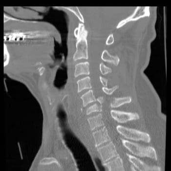
Burst fractures require immediate medical attention from an orthopedic or neurosurgeon. The patient is taken to an emergency room and x-rays, CT scans are gathered.
The diagnosis of a burst fracture is typically made with x-rays and a CT scan.
Sometimes, an MRI will be ordered to assess the amount of:
Soft tissue trauma
Bleeding
Ligament injury
The CT scan and x-rays allow the doctor to determine the level of the fracture, and if it is a:
Compression fracture
Burst fracture
Fracture-dislocation
This will determine how much the spinal canal has been compromised and if its angulation or angle has taken an abnormal bend or curve. These factors all contribute to the development of an optimal treatment plan.
The physical exam will document:
Spinal deformity and Angulation of the spine
Tenderness of the spine where the fracture is located
Neurologic exam
Neurologic exam should include testing:
Muscle strength
Sensation
Reflexes of the lower extremities
Testing of bowel and bladder control
Treatment & Recovery
A stable burst fracture can be treated without surgery.
A stable burst fracture falls into these parameters:
There is no neurologic injury
The angulation is less than 20 degrees
The amount of spinal canal compromise is less than 50%
With this type of treatment, a brace along with physical therapy/chiropractic can have excellent results.
A turtle or clamshell brace TLSO�(Thoracic Lumbar Sacral Orthosis) is a body cast used in the treatment of a burst fracture.
The brace is worn for eight to twelve weeks for adequate and optimal healing.
There are times when a fracture thought stable and treated can start to angulate. This may require surgery. However, all burst fractures require some form of treatment.
Recovery
Nonsurgical treatment patients stay in the hospital for one or two days while their brace is fit.
X-rays are done in the standing position to make sure the spine stays stable.
Pain medications are prescribed for three to four weeks
Non-narcotic medications can begin after the final week
When the brace is removed, physical therapy and chiropractic are instituted to help return strength to the core and lower extremities.
Surgical patients will remain in the hospital for three to five days.
They will be fitted with a brace after the incisions present less pain and are recovered from the surgery.
They can walk within one or two days with the help of a physical therapist.
X-rays are taken to follow the position of the spine and see how the healing is progressing.
Chiropractic/Physical therapy is implemented to help with core strength and lower extremity strength.
Recovery time depends on the severity of the neurologic injury.
Patients that don’t have a neurologic injury can make a full recovery with return to most activities.
Patients with partial neurological injuries can also expect to fully recover.
Unfortunately, with permanent neurologic injury, recovery can be limited.
But treatment for burst fractures today is superior to what they were years ago, especially with spine specialists and specific spinal procedures.
Chiropractic Rehab
Chiropractic is not a treatment for fractures but is a treatment for subluxations and rehabilitation with these types of fractures. Once a fracture has stabilized and healed properly, a chiropractic evaluation can rule out any lingering subluxation, herniation, and joint restriction. The adjustments are safe and effective in establishing optimal function to a subluxated joint.
Chiropractic Treatment For Car Accident Injuries El Paso, Texas
We focus on what works for you. We also strive to create fitness and better the body through researched methods and total wellness programs. These programs are natural and use the body�s own ability to achieve goals of improvement.
NCBI Resources
Chiropractors can help alleviate some of the long-term and immediate concerns associated with bone fractures. A chiropractor can help with compression techniques which are beneficial in maintaining the bone in place for healing. A chiropractor may also advocate wellness techniques, such as a healthy diet that will optimize the body�s ability to restore its original health and wellness. Chiropractors may also educate a patient on a variety of exercises and stretches to reduce the likelihood of complications and which, if done properly and at fixed intervals, will promote quicker recovery.
Q: Can back pain be hereditary and run in the family? I’m 24 and have chronic low back pain. But I found out that my mother, grandmother, and brother also have back pain. None of us were diagnosed with any type of spine condition. So I’m wondering if there is a hereditary/gene link or if it’s just a coincidence? El Paso, TX.
A: Back pain is a very common problem, and it is not uncommon that you and family members have back pain. Every year, 13 million people visit a chiropractor for chronic back pain. I can’t say without further study and research of your family whether the chronic low back pain is directly associated with your genes. But there are studies that support the connection between back pain and genetics.
Research in the past was difficult to rule out environmental factors like stress, smoking, and diet as the only cause of back pain. But today, there is evidence that shows genetics do have a role in back pain.
Doctors are finding that chronic back pain does show a significant genetic hereditary link. Specifically the development of low spine degenerative disc disease, which is a disc-related condition associated with normal wear and tear.
In fact, several twin sibling studies and genetic marker studies have researched this connection. Below are a couple of studies that stand out in the correlation between back pain and genetics.
Back Pain & Hereditary Genes Studies
A spinal study, which began in 1991, was a multidisciplinary, multinational research project on the cause of disc degeneration. The most significant findings were that there was a substantial influence of genetic/hereditary influence in lumbar disc degeneration.
It identified the specific genes that are associated with disc degeneration. However, environmental factors, work, sports, injuries, etc are also part of the condition, what the study found is that there is a connection of disc degeneration through genetic influences.
A study on genetics and lumbar disc disease found evidence that back pain could run in families. Specifically, lumbar disc disease could be inherited. The severity of the disease could not be determined or a patients’ response to various treatments. But it does suggest that genetics do play a role. Other findings include:
People that have lumbar disc disease were more likely to have family members with the disease.
The risk of inheriting the disease increased in both close and distant relatives.
Research is still ongoing to identify the exact genes that influence disc degeneration and back pain.
And as you said that you or your family have not been diagnosed with a spine condition,� this could be something to talk to your doctor or chiropractor about, along with creating a back pain treatment plan. For now, you can do physical therapy, chiropractic massage, CrossFit rehabilitation, etc to reduce and prevent back pain. Hopefully, it is a coincidence, but if it is genetics, not to worry as there is a treatment plan for that as well. But do not wait to make an appointment and let the pain get any worse.
Our team has taken great pride in bringing our families and injured patients only clinically proved treatment protocols. �By teaching complete holistic wellness as a lifestyle, we also change not only our patient�s lives but their families as well.� We do this so that we may reach as many El Pasoans who need us, no matter the affordability issues.
How to eliminate Back Pain naturally | (2020) Foot Levelers |El Paso, Tx
NCBI Resources
Lumbar spine disc herniation is a well-known type of injury that often causes impairing low back pain, however, it can also compress the nerve roots in the area and generate radicular pain and other symptoms along the lower extremities, such as altered sensations and muscle weakness.
Do you experience bloating after eating a meal? While many people may not experience this symptom, it’s important to understand that any amount of bloating is generally abnormal and it can be a sign of gut inflammation. If you regularly experience bloating, or you have irritable bowel syndrome (IBS), there�s a chance that you may have small intestinal bacterial overgrowth (SIBO). In the following article, we will discuss the top 10 red flags of SIBO. �
Contents
What is Small Intestinal Bacterial Overgrowth (SIBO)?
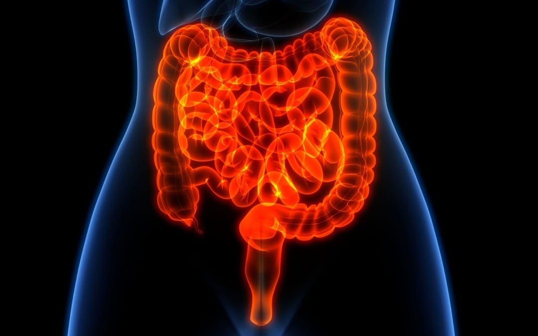
Small intestinal bacterial overgrowth (SIBO) is a digestive health issue that happens when there is excess bacteria in the small intestine. The bacteria in the gastrointestinal (GI) tract plays a fundamental role in the immune system and overall health and wellness. As a matter of fact, research studies have shown that the gut microbiome has tens of trillions of microorganisms, including more than 1,000 different species of bacteria with over 3 million genes. �
Most of the gut bacteria are found in the large intestine and colon, where they ultimately help break down food, synthesize vitamins or minerals, and eliminate waste. However, if the healthy bacteria commonly found in the large intestine and colon start to grow excessively in the small intestine, SIBO can occur. Small intestinal bacterial overgrowth can also be caused by an excess growth of “healthy” bacteria found in the small intestine itself. �
With SIBO, the excess bacteria may start to consume the undigested food in the small intestine, causing it to ferment and produce hydrogen. Moreover, hydrogen can “feed” single-celled microorganisms commonly found in the small intestine, known as archaea, which may then produce methane. Patients with SIBO have increased levels of hydrogen and/or methane in their digestive system. This formula can cause a variety of digestive health issues. �
Furthermore, patients with SIBO may also develop a variety of symptoms depending on which type of gas is predominantly produced in their gut. Hydrogen-dominant SIBO, by way of instance, generally causes diarrhea while methane-dominant SIBO, by way of instance, generally causes constipation. Small intestinal bacterial overgrowth doesn’t simply cause a variety of digestive health issues, SIBO can also cause a wide array of symptoms, including: �
10 Red Flags You May Have SIBO
Gas, bloating, and/or diarrhea
Abdominal pain, discomfort, and/or cramping
Constipation
Irritable bowel syndrome (IBS) or inflammatory bowel disease (IBD)
Vitamin and mineral deficiencies (vitamins A, B12, D, and E)
Fat malabsorption (pale, bulky, and malodorous stools)
Rosacea and/or skin rashes
Leaky gut or intestinal permeability
Because there are many symptoms that can show that you may have small intestinal bacterial overgrowth, occasionally showing none of the red flags listed above, SIBO may frequently go undiagnosed. Approximately 6 to 15 percent of “healthy”, asymptomatic people, and about 80 percent of people with irritable bowel syndrome (IBS), may actually be suffering from SIBO. If you experience any of the previous symptoms, make sure to see a doctor immediately. �
What Causes Small Intestinal Bacterial Overgrowth (SIBO)?
When enzymes start to break down the food we eat, our gastrointestinal (GI) tract depends on the proper function of nerves, muscles, and neurotransmitters to move the food accordingly throughout our digestive system, from the stomach to the small intestine and to the colon. In a healthy gut, bacteria pass through the gastrointestinal (GI) tract together with the food we eat into the colon. Symptoms and health issues may start when this process is affected. �
Damaged or injured nerves and/or muscles in the gut can ultimately cause leftover bacteria to stay longer in the small intestine, increasing the risk of SIBO. By way of instance, diabetes mellitus and scleroderma are two health issues that can both affect the muscles in the gut, causing SIBO to develop. �
Physical obstructions in the gut, such as scarring from surgeries or Crohn�s disease, can also cause excess bacteria to grow in the small intestine. Diverticuli, tiny pouches that can develop in the wall of the small intestine, can also start to collect bacteria instead of passing it to the colon. �
Drugs and/or medications that can affect or interrupt our healthy gut microbiome. This can include antibiotics, acid-blocking medicine, and steroids. In addition, it’s essential to mention that one of the most common causes of small intestinal bacterial overgrowth (SIBO) is a poor diet that is high in sugar, refined carbohydrates, and alcohol. If you suspect you may have SIBO, make sure you talk to a healthcare professional for diagnosis and treatment. �
Small intestinal bacterial overgrowth (SIBO) is a serious health issue which usually occurs because of an underlying chronic health issue. Several common symptoms may ultimately help determine the presence of SIBO. Several red flags may ultimately suggest the presence of small intestinal bacterial overgrowth but, because some people may not experience any symptoms, it can often go undiagnosed. Proper diagnosis is fundamental. SIBO, or small intestinal bacterial overgrowth is treatable. Patients should contact a healthcare professional immediately if they suspect they have SIBO so that they can begin treatment right away. – Dr. Alex Jimenez D.C., C.C.S.T. Insight
Neurotransmitter Assessment Form
The following Neurotransmitter Assessment Form can be filled out and presented to Dr. Alex Jimenez. The following symptoms listed on this form are not intended to be utilized as a diagnosis of any type of disease, condition, or any other type of health issue. �
Do you experience bloating after eating a meal? While many people may not experience this symptom, it’s important to understand that any amount of bloating is generally abnormal and it can be a sign of gut inflammation. If you regularly experience bloating, or you have irritable bowel syndrome (IBS), there�s a chance that you may have small intestinal bacterial overgrowth (SIBO). In the article above, we discussed the top 10 red flags of SIBO.
The scope of our information is limited to chiropractic, musculoskeletal, and nervous health issues or functional medicine articles, topics, and discussions. We use functional health protocols to treat injuries or disorders of the musculoskeletal system. Our office has made a reasonable attempt to provide supportive citations and has identified the relevant research study or studies supporting our posts. We also make copies of supporting research studies available to the board and or the public upon request. To further discuss the subject matter above, please feel free to ask Dr. Alex Jimenez or contact us at 915-850-0900.�
Curated by Dr. Alex Jimenez �
References:
Myers, Amy. �10 Signs You Have Small Intestinal Bacterial Overgrowth (SIBO).� Amy Myers MD, 12 Nov. 2019, www.amymyersmd.com/2018/04/10-signs-small-intestinal-bacterial-overgrowth-sibo/.
Additional Topic Discussion: Chronic Pain
Sudden pain is a natural response of the nervous system which helps to demonstrate possible injury. By way of instance, pain signals travel from an injured region through the nerves and spinal cord to the brain. Pain is generally less severe as the injury heals, however, chronic pain is different than the average type of pain. With chronic pain, the human body will continue sending pain signals to the brain, regardless if the injury has healed. Chronic pain can last for several weeks to even several years. Chronic pain can tremendously affect a patient’s mobility and it can reduce flexibility, strength, and endurance. �
Neural Zoomer Plus for Neurological Disease
�
Dr. Alex Jimenez utilizes a series of tests to help evaluate neurological diseases. The Neural ZoomerTM Plus is an array of neurological autoantibodies which offers specific antibody-to-antigen recognition. The Vibrant Neural ZoomerTM Plus is designed to assess an individual�s reactivity to 48 neurological antigens with connections to a variety of neurologically related diseases. The Vibrant Neural ZoomerTM Plus aims to reduce neurological conditions by empowering patients and physicians with a vital resource for early risk detection and an enhanced focus on personalized primary prevention. �
Food Sensitivity for the IgG & IgA Immune Response
�
Dr. Alex Jimenez utilizes a series of tests to help evaluate health issues associated with food sensitivities. The Food Sensitivity ZoomerTM is an array of 180 commonly consumed food antigens that offers very specific antibody-to-antigen recognition. This panel measures an individual�s IgG and IgA sensitivity to food antigens. Being able to test IgA antibodies provides additional information to foods that may be causing mucosal damage. Additionally, this test is ideal for patients who might be suffering from delayed reactions to certain foods. Utilizing an antibody-based food sensitivity test can help prioritize the necessary foods to eliminate and create a customized diet plan around the patient�s specific needs. �
Gut Zoomer for Small Intestinal Bacterial Overgrowth (SIBO)
Dr. Alex Jimenez utilizes a series of tests to help evaluate gut health associated with small intestinal bacterial overgrowth (SIBO). The Vibrant Gut ZoomerTM offers a report that includes dietary recommendations and other natural supplementation like prebiotics, probiotics, and polyphenols. The gut microbiome is mainly found in the large intestine and it has more than 1000 species of bacteria that play a fundamental role in the human body, from shaping the immune system and affecting the metabolism of nutrients to strengthening the intestinal mucosal barrier (gut-barrier). It is essential to understand how the number of bacteria that symbiotically live in the human gastrointestinal (GI) tract influences gut health because imbalances in the gut microbiome may ultimately lead to gastrointestinal (GI) tract symptoms, skin conditions, autoimmune disorders, immune system imbalances, and multiple inflammatory disorders. �
Formulas for Methylation Support
� XYMOGEN�s Exclusive Professional Formulas are available through select licensed health care professionals. The internet sale and discounting of XYMOGEN formulas are strictly prohibited.
Proudly,�Dr. Alexander Jimenez makes XYMOGEN formulas available only to patients under our care.
Please call our office in order for us to assign a doctor consultation for immediate access.
If you are a patient of Injury Medical & Chiropractic�Clinic, you may inquire about XYMOGEN by calling 915-850-0900.
�
�
For your convenience and review of the XYMOGEN products please review the following link. *XYMOGEN-Catalog-Download �
* All of the above XYMOGEN policies remain strictly in force. �
Do you have difficulty digesting protein-rich foods? Do you have difficulty digesting starch-rich foods? Do you have difficulty digesting fatty or greasy foods? Do you experience abdominal distention after meals? Do you have abdominal pain and inflammation? If so, you may be having SIBO symptoms. �
Small intestinal bacterial overgrowth (SIBO) is a gastrointestinal (GI) tract health issue that can become a persistent problem if it’s not managed accordingly, especially if it’s ultimately left untreated. For many people suffering from chronic gas, bloating, constipation, and/or diarrhea, they may have also already had a diagnosis of irritable bowel syndrome (IBS). However, research studies have shown that one of the main causes of IBS may be SIBO. �
SIBO is a digestive health issue where there are too many bacteria in the small intestine. Bacterial overgrowth can also cause IBS. Although there are many treatment options for SIBO, one of the most important treatments for SIBO is doing everything we can to help keep SIBO from coming back. The purpose of the following article is to discuss how understanding the migrating motor complex (MMC) can help treat small intestinal bacterial overgrowth (SIBO). �
Contents
What is the Migrating Motor Complex?
The migrating motor complex (MMC) refers to the collection of electrical waves that occur in the gut. The MMC helps regulate several important functions of the gut, such as sweeping out the stuff we no longer need in there and moving it down to the colon where it can then be excreted by the human body. �
Phases of the Migrating Motor Complex
The MMC is how the digestive system eliminates waste from the human body. The MMC cycle includes four phases, including:� �
The first phase is a period of calmness that lasts 45 to 60 minutes where rare action potentials and contractions occur.
The second phase is a period of about 30 minutes where peristaltic contractions occur and gradually increase in frequency. Peristalsis starts in the stomach and continues throughout the small intestine.
The third phase lasts 5 to 15 minutes and it’s made-up of rapid, evenly spaced out peristaltic contractions. The pylorus stays open during these peristaltic contractions which allow many indigestible materials to pass into the small intestine.
The fourth and final phase is a period of transition between the contractions from the third phase and the inactivity from the first phase.
Gastric, biliary, and pancreatic secretion increases during the MMC to further with the digestion process as well as to help decrease bacteria in the gastrointestinal (GI) tract. Healthcare professionals believe that motilin, the enteric hormone, regulates the MMC. Because eating food can interrupt the MMC, fasting between meals is important to help complete the four phases. Moreover, the well-known �growling” sounds you generally hear when you are hungry may be the migrating motor complex performing its job functions accordingly, such as cleaning your bowels of waste and excessive bacteria. �
Migrating Motor Complex (MMC) Health Issues
If the migrating motor complex (MMC) isn’t working properly, the foods we consume may ultimately remain in the stomach and small intestine longer than what is generally considered to be healthy, which can make us feel a heaviness after eating or it can make us feel too-full, even if you’ve only had a small meal. Furthermore, a slow MMC can also cause bacteria to stay in the gastrointestinal (GI) tract for too long, which can also lead to SIBO. � Approximately 70 percent of people with SIBO also have MMC health issues. Research studies have shown that reduced MMC function may be associated with excess methane and/or hydrogen gasses produced by the excess bacteria in the gut. SIBO can also increase inflammation and intestinal permeability. �
Other research studies have shown that utilizing acid-reducing medications or an H. pylori infection can affect MMC function. Lack of exercise, grazing, and constipation can also affect MMC. Stress can also affect MMC function. Finally, thyroid problems and adrenal fatigue can also affect MMC function. �
Research studies have shown that people with IBS can frequently have decreased MMC function although researchers still don’t understand how these changes occur. Several researchers believe that food poisoning and other bacterial infections can affect the gut microbiome which then changes how the gut microbiome signals the MMC to start and stop. Eating inflammatory foods or foods that you�re sensitive and/or allergic to can also cause nerve damage in the gut. Subsequently, these damaged nerves then can�t properly signal the MMC to function accordingly, leading to SIBO and other health issues. �
Small intestinal bacterial overgrowth (SIBO) is a serious health issue which usually occurs because of an underlying chronic health issue. Several common symptoms may ultimately help determine the presence of SIBO. In addition, research studies have demonstrated that poor migrating motor complex (MMC) function, or the collection of electrical waves that help regulate several important functions of the gut, can ultimately cause SIBO and other digestive system health issues if left untreated. SIBO, or small intestinal bacterial overgrowth is treatable. Patients should contact a healthcare professional immediately if they suspect they have SIBO so that they can begin treatment right away. – Dr. Alex Jimenez D.C., C.C.S.T. Insight
The following Neurotransmitter Assessment Form can be filled out and presented to Dr. Alex Jimenez. The following symptoms listed on this form are not intended to be utilized as a diagnosis of any type of disease, condition, or any other type of health issue. �
Do you have difficulty digesting protein-rich foods? Do you have difficulty digesting starch-rich foods? Do you have difficulty digesting fatty or greasy foods? Do you experience abdominal distention after meals? Do you have abdominal pain and inflammation? If so, you may be having SIBO symptoms. �
Small intestinal bacterial overgrowth (SIBO) is a gastrointestinal (GI) tract health issue that can become a persistent problem if it’s not managed accordingly, especially if it’s ultimately left untreated. For many people suffering from chronic gas, bloating, constipation, and/or diarrhea, they may have also already had a diagnosis of irritable bowel syndrome (IBS). However, research studies have shown that one of the main causes of IBS may be SIBO. �
SIBO is a digestive health issue where there are too many bacteria in the small intestine. Bacterial overgrowth can also cause IBS. Although there are many treatment options for SIBO, one of the most important treatments for SIBO is doing everything we can to help keep SIBO from coming back. The purpose of the article above was to discuss how understanding the migrating motor complex (MMC) can help treat small intestinal bacterial overgrowth (SIBO).
The scope of our information is limited to chiropractic, musculoskeletal, and nervous health issues or functional medicine articles, topics, and discussions. We use functional health protocols to treat injuries or disorders of the musculoskeletal system. Our office has made a reasonable attempt to provide supportive citations and has identified the relevant research study or studies supporting our posts. We also make copies of supporting research studies available to the board and or the public upon request. To further discuss the subject matter above, please feel free to ask Dr. Alex Jimenez or contact us at 915-850-0900.�
Curated by Dr. Alex Jimenez �
References:
Albina, Victoria. �SIBO Begone: 5 Easy Ways to Keep Your SIBO From Coming Back.� Victoria Albina, Victoria Albina, 26 Mar. 2019, victoriaalbina.com/sibo/.
Brisson, John. �Migrating Motor Complex (MMC) and Digestive Health.� Fix Your Gut, Fix Your Gut, 13 Dec. 2014, www.fixyourgut.com/mmc-digestive-health/.
Additional Topic Discussion: Chronic Pain
Sudden pain is a natural response of the nervous system which helps to demonstrate possible injury. By way of instance, pain signals travel from an injured region through the nerves and spinal cord to the brain. Pain is generally less severe as the injury heals, however, chronic pain is different than the average type of pain. With chronic pain, the human body will continue sending pain signals to the brain, regardless if the injury has healed. Chronic pain can last for several weeks to even several years. Chronic pain can tremendously affect a patient’s mobility and it can reduce flexibility, strength, and endurance. �
Neural Zoomer Plus for Neurological Disease
Dr. Alex Jimenez utilizes a series of tests to help evaluate neurological diseases. The Neural ZoomerTM Plus is an array of neurological autoantibodies which offers specific antibody-to-antigen recognition. The Vibrant Neural ZoomerTM Plus is designed to assess an individual�s reactivity to 48 neurological antigens with connections to a variety of neurologically related diseases. The Vibrant Neural ZoomerTM Plus aims to reduce neurological conditions by empowering patients and physicians with a vital resource for early risk detection and an enhanced focus on personalized primary prevention. �
Food Sensitivity for the IgG & IgA Immune Response
Dr. Alex Jimenez utilizes a series of tests to help evaluate health issues associated with food sensitivities. The Food Sensitivity ZoomerTM is an array of 180 commonly consumed food antigens that offers very specific antibody-to-antigen recognition. This panel measures an individual�s IgG and IgA sensitivity to food antigens. Being able to test IgA antibodies provides additional information to foods that may be causing mucosal damage. Additionally, this test is ideal for patients who might be suffering from delayed reactions to certain foods. Utilizing an antibody-based food sensitivity test can help prioritize the necessary foods to eliminate and create a customized diet plan around the patient�s specific needs. �
Gut Zoomer for Small Intestinal Bacterial Overgrowth (SIBO)
�
Dr. Alex Jimenez utilizes a series of tests to help evaluate gut health associated with small intestinal bacterial overgrowth (SIBO). The Vibrant Gut ZoomerTM offers a report that includes dietary recommendations and other natural supplementation like prebiotics, probiotics, and polyphenols. The gut microbiome is mainly found in the large intestine and it has more than 1000 species of bacteria that play a fundamental role in the human body, from shaping the immune system and affecting the metabolism of nutrients to strengthening the intestinal mucosal barrier (gut-barrier). It is essential to understand how the number of bacteria that symbiotically live in the human gastrointestinal (GI) tract influences gut health because imbalances in the gut microbiome may ultimately lead to gastrointestinal (GI) tract symptoms, skin conditions, autoimmune disorders, immune system imbalances, and multiple inflammatory disorders. �
Formulas for Methylation Support
� XYMOGEN�s Exclusive Professional Formulas are available through select licensed health care professionals. The internet sale and discounting of XYMOGEN formulas are strictly prohibited.
Proudly,�Dr. Alexander Jimenez makes XYMOGEN formulas available only to patients under our care.
Please call our office in order for us to assign a doctor consultation for immediate access.
If you are a patient of Injury Medical & Chiropractic�Clinic, you may inquire about XYMOGEN by calling 915-850-0900.
�
For your convenience and review of the XYMOGEN products please review the following link. *XYMOGEN-Catalog-Download �
* All of the above XYMOGEN policies remain strictly in force. �
For most individuals experiencing symptoms,� health care providers will run a few tests. For the most part, these tests come back normal or inconclusive. The patient is usually relieved, but not satisfied as they are still experiencing symptoms. The truth of the matter is, most tests practitioners run on patients are basic.
What Does That Mean?
It means that they are checking your levels and ruling out issues based on standard testing, but they are not diving deep into the cause of the symptom itself. Most individuals have a family history of one or more autoimmune diseases. An autoimmune disease is when the body misidentifies its own cells as a foreign body leading them to attack. These diseases can be triggered at any point in one’s life.
So What Do I Do Now?
Due to family history,� an ANA test can be run. Most of the time for patients still experiencing symptoms after the standard tests came back negative, the ANA comes back positive. However, a positive test does not always mean answers. This test can provide useful information but does not include a definitive answer as to what kind of autoimmunity a patient may have.
Using A Functional Approach
In modern/traditional medicine, most practitioners will suggest the patient is fine and that there is nothing to treat until they have been diagnosed. However, by using a more naturopathic and holistic approach, integrative practitioners can take these symptoms and use them to the patient’s advantage to help avoid a full-blown diagnosis.� The main reason this is effective is due to the fact that individuals do not just wake up one day with a new disease, but rather there are steps progressing in the background that eventually build up to a diagnosis if not treated.
Uncovering the underlying issue and using this stage in someone’s life as an opportunity to improve their quality of living is what functional medicine revolves around. By combining symptoms, previous lab results, and the patients declining quality of life, testing that relates to triggers of autoimmunity can be conducted. These tests will provide insightful information allowing the practitioner to not only treat the symptom but more importantly, to treat the cause.
Testing
There are multiple factors, including environmental that cause an autoimmune response to start to express. There are certain markers in the body that will shift before the onset of the autoimmune disease in which the environmental triggers will be shown.
Many labs are equipt to test for these markers and use top of the line technology. Some tests that evaluate these triggers that contribute to the progression of autoimmunity are:
The Gut Zoomerfrom Vibrant Wellness:The Gut Zoomer provides information and patient potential risks for intestinal permeability, IBS & IBD, SIBO, celiac, MS, obesity, diabetes, nutrition, viruses, fungal or yeast species, worm species, bile acids, SCFAs and more
The Food Sensitivity Panel from Vibrant Wellness:This test from Vibrant Wellness recognizes the specific antibody-to-antigen responses in commonly ingested foods. This panel tests for IgG and IgA sensitivity to the food antigens. This test is beneficial so patients do not have to an elimination diet, but rather have the test remove the guesswork and let results tell them what foods cause their body inflammation.
Dietary Antigen Test Plus from Dunwoody Labs:��This specific test from Dunwoody Labs allows the health care provider to see if there is an increased antibody response to food. Often times, the antibodies this screens for attack the body’s tissue leading to more autoimmune symptoms. This test looks at 4 separate antibody types IgE, IgG4, Total IgG, IgA, and complement.
Oxidative Stress Test from Dunwoody Labs:If the body is under large amounts of stress, this will cause an increase in the activity of T-Cells, thus keeping the body out of balance.
GI Microbial Assay Plus (GI-MAP� ) from Diagnostic Solutions: This is a stool test that not only analyzes but also evaluates the DNA of the actual organisms that are living in the gut. This allows the health care providers to see what is impacting health such as, mucus metabolism, methane production, T-Cells, and inflammatory LPS.�
Why Would I Want / Need This?
Preventative medicine provides the ability to keep up with, if not improve a patient’s quality of life while decreasing or avoiding the hard prescription medicine! By treating the underlying cause and not just masking the symptom, it allows individuals to truly feel better.
The number of individuals diagnosed with a disease that could have been prevented should earlier steps have been taken, is on the rise while their quality of life is declining. Feeling good should not be a delicacy, but rather a normal standard. With the use of integrative medicine, many grandparents will be able to play hide and seek and tag with their grandchildren, parents will be able to get through the day without as much fatigue and headaches, and children will be able to play, focus, and learn with fewer belly aches no matter the hour of the day. Integrative medicine not only utilizes tests to guide patients in the right direction but also gives patients tools and further educates them on exercise and nutrition components that will help prevent inflammation and stimulate good bacteria in their bodies to grow. – Kenna Vaughn, Senior Health Coach
*The scope of our information is limited to chiropractic, musculoskeletal, and nervous health issues or functional medicine articles, topics, and discussions. We use functional health protocols to treat injuries or disorders of the musculoskeletal system. Our office has made a reasonable attempt to provide supportive citations and has identified the relevant research study or studies supporting our posts. We also make copies of supporting research studies available to the board and or the public upon request. To further discuss the subject matter above, please feel free to ask Dr. Alex Jimenez or contact us at 915-850-0900.
Resources:
�Basic Oxidative Stress.� Dunwoody Labs, 20 July 2018, www.dunwoodylabs.com/index.php/ox-stress/.
Burdette, Cheryl. �Is There Such Thing as Being Pre-Autoimmune.� 3 Dec. 2019.
�588 Dietary Antigen A, G, E, and C.� Dunwoody Labs, 17 Jan. 2019, www.dunwoodylabs.com/index.php/dietary-antigen-and-environmental-allergen-exposure-profiles/.
You might not think it, but weight lifting and spine strengthening exercises can help reduce back pain. Remember the point of this type of weight lifting is not to build up the muscles like a bodybuilder but is to develop:
Core strength
Spine strength
Body strength
The muscles in the back keep the spine moving and functioning properly. When the spine or abdominal muscles are weak this creates a higher probability of a back strain or injury. Having strong, healthy spine muscles are important because they function in maintaining correct posture, which in some cases, causes chronic back pain because of poor posture.
If�only one part of the body is strengthened like the back is not enough. Therefore strengthening the rest of the body is a must. These include the body’s core and leg muscles. Total body strength will reduce back pain and can help perform regular activities, like lifting heavy objects much easier, with more confidence and with a lesser probability of injury.
Contents
Spine strengthening exercises benefits
Most important reasons are they:
Prevents future back injuries
Stabilizes the spine
Helps the spine move properly
Help maintain correct posture
Increases muscle tone
Teaches correct body mechanics
Helps build bone this is especially beneficial for those with osteoporosis or at risk of developing it
A personal trainer or sports chiropractor can help start a spine strengthening regimen. They will teach:
Simple
Specific
Strengthening
Weight lifting exercises.
A physical therapist can also develop a custom weight lifting/strength�exercise�program for optimal spine health and for reducing pain.
Most workout regimens incorporate a combination of weight lifting with actual weights/exercise machines and strengthening exercises/calisthenics with the body’s weight as the resistance for maintaining a healthy strong spine.
Here are a few weight lifting and back strengthening exercises that can help decrease and prevent back pain.
Talk to a doctor or chiropractor before beginning any exercise program. Remember to listen to your body and stop right away if there is something off.
Push-ups
Push-ups help strengthen the:
Back
Chest
Arms
Core muscles
Your own body weight is the resistance.
To do this:
Position the body in a straight line from head to toe, the face looking down.
Hands should be wider than shoulder-distance apart. Walk the hands out so they are slightly higher than the shoulders
Keep the balance on toes and hands, with a straight back, lower the body to the floor by slowly bending the elbows until at a 90-degree angle.
Push up using arm upper back, and chest muscles.
Do 3 sets of 10 every day. As the strength increases do more reps.
Chest Flyes
Chest flies are excellent for building muscle in the:
Upper back
Chest
Dumbbells or a weight machine can be used for this exercise. To do this:
Lie on the floor with the knees bent and the feet flat on the ground.
Extend the arms out to either side of the body, and let them rest on the floor.
With a dumbbell in each hand, raise the dumbbells until they meet at the top at the same time, and keep a slight bend in the elbows.
Lower the hands to the ground, and repeat.
Do this exercise 15 times 3 times a week. With added strength add more reps.
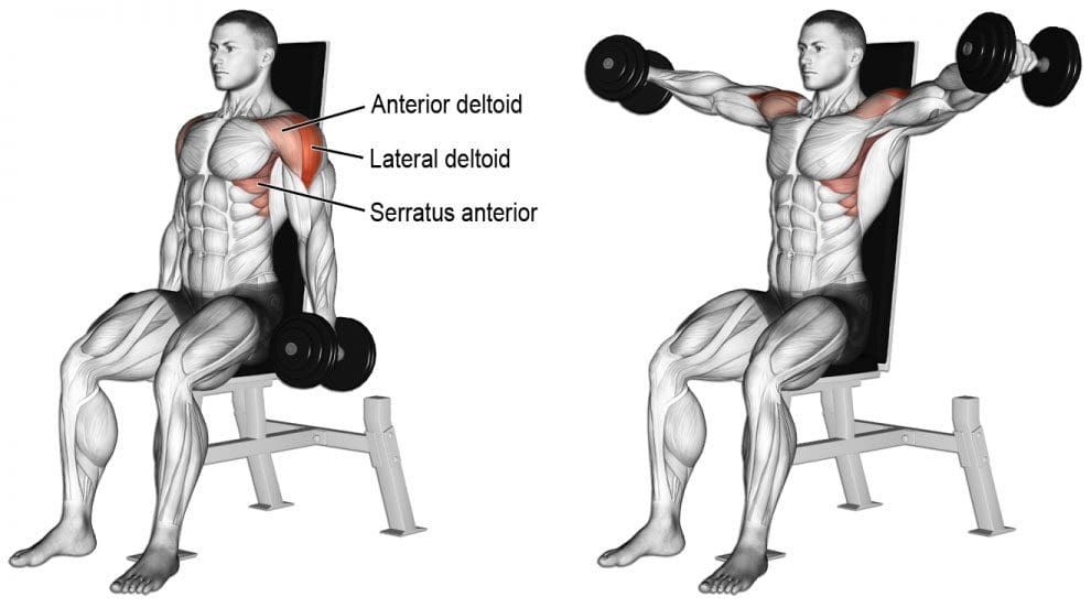
Lateral Raises
Lateral raises help strengthen the entire back. All that is needed is a set of dumbbells. To do this move:
Stand or sit with the feet equal hip-distance apart. Arms are at the side.
With a dumbbell in each hand and a slight bend in the elbows, raise the arms to the side until shoulder height. Keep the core engaged during the movement.
Once at shoulder height, slowly lower the dumbbells, and repeat.
Repeat this exercise 15 times 3 times a week. As strength increases add more reps.
These exercises should be performed slowly with a gradual build-up to more complex movements and adding more weight. Remember to breathe naturally. If you hold your breath during exercise, it can cause tension in the�muscles, which can worsen any pain or create new injuries.�Before adding weight or new spine strengthening exercises, talk to a doctor about exercising with back pain. They will let you know if there are certain movements or positions that should be avoided.
Weight lifting exercises done incorrectly can lead to more back pain and added injuries. If there is any pain while doing these exercises, stop and call a doctor, chiropractor or physical therapist right away.
As El Paso�s Chiropractic Rehabilitation Clinic & Integrated Medicine Center,�we passionately are focused on treating patients after frustrating injuries and chronic pain syndromes. We focus on improving your ability through flexibility, mobility and agility programs tailored for all age groups and disabilities.
Back Pain Chiropractic Care | El Paso, Tx
NCBI Resources
It can be tempting to not exercise with a spinal condition. But remember that if there is no movement at all, you could make the pain worse. Knowing what your body can handle and sticking to a workable schedule, these healthy steps will relieve you and help with back pain.
Do you frequently eat processed foods that are bagged or boxed? Do you frequently eat fried foods? Do you have difficulty digesting foods? Do you experience constipation or inconsistent bowel movements? Do you have increased bloating or gas? If so, you may be experiencing SIBO symptoms. �
Small intestinal bacterial overgrowth (SIBO) is a serious health issue that happens when bacteria that generally grow in one region of the digestive system, such as the colon, grow in the small intestine, ultimately affecting the gastrointestinal (GI) tract. If left untreated, SIBO can commonly cause pain, discomfort, diarrhea, and malnutrition (because of the loss of nutrients), among other symptoms.�Proper nutrition can help decrease these harmful bacteria. �
Following the SIBO diet together with antibiotics can also help speed up recovery and ultimately help reduce uncomfortable symptoms. The purpose of the article below is to describe the benefits of following the SIBO diet as well as what foods you should and shouldn’t eat to help improve SIBO symptoms. �
Contents
Understanding the SIBO Diet
The SIBO diet involves gradually eliminating several types of foods in an attempt to help reduce inflammation in the gastrointestinal (GI) tract and help decrease bacterial overgrowth in the small intestine. In a variety of instances, the gradual elimination of sugars alone can help improve SIBO symptoms. �
Healthcare professionals recommend including a diet that is low in FODMAPs, or carbohydrates that can be difficult to digest and can become fermented by gut bacteria in the colon. When the digestive system is unable to break down carbs, these can sit in the gut and can cause SIBO symptoms, such as bloating and diarrhea. With SIBO, the bacterial overgrowth in the small intestine may ultimately start to ferment carbs too soon, causing a variety of symptoms. �
Foods You Should Eat for SIBO
As we will discuss further below, the list of foods you shouldn’t eat when you have small intestinal bacterial overgrowth (SIBO) can be considered restrictive, however, there are still several foods you can enjoy while following the SIBO diet. The SIBO diet includes foods that are high in fiber and low in sugar. �
Moreover, several foods can have low amounts of FODMAPs in smaller servings but these should still be limited or avoided because larger servings may increase the overall number of FODMAPs. Furthermore, several recommended types of foods for a SIBO, as well as a low FODMAP, diet include:� �
oatmeal
unsweetened cereal (with low FODMAP grains)
gluten-free crackers
rice or gluten-free noodles
quinoa
seeds
peanuts
several types of fruits, such as strawberries, blueberries, grapes, oranges
leafy greens
broccoli (heads only, less than 3/4 cup)
olives
potatoes
carrots
pumpkin
spaghetti squash and summer squashes
eggs
fish
meat
Foods You Shouldn’t Eat with SIBO
According to research studies, the low FODMAP diet has been demonstrated to safely and effectively help treat irritable bowel syndrome (IBS) and its associated symptoms. Patients with IBS also commonly have SIBO. Reducing or eliminating foods that are high in FODMAPs can improve digestive health. �
When reducing or eliminating FODMAPs as a part of the SIBO diet, healthcare professionals suggest focusing on the main categories, including: �
fructose, basic sugars frequently found in fruits and several types of vegetables as well as in honey and agave nectar,
fructans, a sugar substance or chemical found in many gluten products, fruits, several vegetables, and prebiotics,
polyols, sugar alcohol commonly utilized as a sweetener,
galactans, a substance or chemical found in several types of legumes, and
lactose, a sugar molecule frequently found in many dairy products.
Several types of foods which you may want to consider completely eliminating from your diet that has higher amounts of FODMAPs include: �
honey
agave nectar
high-fructose corn syrup
soda and other types of soft beverages
dried fruits
apples
asparagus
artichokes
peas
cauliflower
butternut squash
garlic
onions
beans
sweetened cereals
grains
barley
rye
flavored yogurt
ice cream
sausage
Evidence Findings of the SIBO Diet
Healthcare professionals utilize antibiotics as the main treatment approach for small intestinal bacterial overgrowth (SIBO) symptoms. However, research studies have demonstrated that dietary changes, such as limiting sugars and lactose, may also ultimately help reduce SIBO. The SIBO diet can be utilized together with probiotics and antibiotics. A 2010 research study also determined that probiotics can also help reduce SIBO symptoms. According to the research study, drinking more water while on the SIBO diet can also help reduce pain, discomfort, and inflammation. Make sure to talk to your doctor before making any dietary modifications or implementing a new treatment option. In addition, discuss all of the benefits and risks with your doctor or dietitian. �
The SIBO diet is a nutrition plan which temporarily eliminates high FODMAP foods while including a variety of low-FODMAP foods to help decrease bacterial overgrowth in the small intestine. The SIBO diet generally lasts anywhere between 2 to 6 weeks. Although it has been demonstrated to be a safe and effective treatment approach, the SIBO diet treats symptoms while it may not necessarily treat the underlying condition or disease. Conventional treatment options for SIBO shouldn�t be ignored. Talk to a healthcare professional before involving diet changes to any treatment plan. It�s also fundamental to mention that you should ultimately bring FODMAPs back into your normal diet when your SIBO symptoms improve. This can help prevent healthy gut bacteria loss. If your symptoms begin to worsen after implementing the SIBO or low-FODMAP diet, make sure to seek immediate medical attention. �
Small intestinal bacterial overgrowth (SIBO) is a serious health issue which usually occurs because of an underlying chronic health issue. Several common symptoms may ultimately help determine the presence of SIBO. Additionally, if the patient has a chronic condition or disease, such as celiac disease or Crohn’s disease, they should talk to a healthcare professional to develop a treatment plan, such as the SIBO diet. SIBO, or small intestinal bacterial overgrowth is treatable. If left untreated, this gastrointestinal (GI) tract problem can also cause dehydration and malnutrition. Patients should contact a healthcare professional immediately if they suspect they have SIBO so that they can begin treatment right away. – Dr. Alex Jimenez D.C., C.C.S.T. Insight
Neurotransmitter Assessment Form
�
The following Neurotransmitter Assessment Form can be filled out and presented to Dr. Alex Jimenez. The following symptoms listed on this form are not intended to be utilized as a diagnosis of any type of disease, condition, or any other type of health issue. �
Do you frequently eat processed foods that are bagged or boxed? Do you frequently eat fried foods? Do you have difficulty digesting foods? Do you experience constipation or inconsistent bowel movements? Do you have increased bloating or gas? If so, you may be experiencing SIBO symptoms. �
Small intestinal bacterial overgrowth (SIBO) is a serious health issue that happens when bacteria that generally grow in one region of the digestive system, such as the colon, grow in the small intestine, ultimately affecting the gastrointestinal (GI) tract. If left untreated, SIBO can commonly cause pain, discomfort, diarrhea, and malnutrition (because of the loss of nutrients), among other symptoms. Proper nutrition can help decrease these harmful bacteria. �
Following the SIBO diet together with antibiotics can also help speed up recovery and ultimately help reduce uncomfortable symptoms. The purpose of the article above was to describe the benefits of following the SIBO diet as well as what foods you should and shouldn’t eat to help improve SIBO symptoms. �
The scope of our information is limited to chiropractic, musculoskeletal, and nervous health issues or functional medicine articles, topics, and discussions. We use functional health protocols to treat injuries or disorders of the musculoskeletal system. Our office has made a reasonable attempt to provide supportive citations and has identified the relevant research study or studies supporting our posts. We also make copies of supporting research studies available to the board and or the public upon request. To further discuss the subject matter above, please feel free to ask Dr. Alex Jimenez or contact us at 915-850-0900.�
Curated by Dr. Alex Jimenez �
References:
Anthony, Kiara. �SIBO Diet 101: What You Should and Shouldn’t Eat.� Edited by Natalie Butler, Healthline, Healthline, 16 Aug. 2018, www.healthline.com/health/sibo-diet.
Additional Topic Discussion: Chronic Pain
Sudden pain is a natural response of the nervous system which helps to demonstrate possible injury. By way of instance, pain signals travel from an injured region through the nerves and spinal cord to the brain. Pain is generally less severe as the injury heals, however, chronic pain is different than the average type of pain. With chronic pain, the human body will continue sending pain signals to the brain, regardless if the injury has healed. Chronic pain can last for several weeks to even several years. Chronic pain can tremendously affect a patient’s mobility and it can reduce flexibility, strength, and endurance. �
Neural Zoomer Plus for Neurological Disease
�
Dr. Alex Jimenez utilizes a series of tests to help evaluate neurological diseases. The Neural ZoomerTM Plus is an array of neurological autoantibodies which offers specific antibody-to-antigen recognition. The Vibrant Neural ZoomerTM Plus is designed to assess an individual�s reactivity to 48 neurological antigens with connections to a variety of neurologically related diseases. The Vibrant Neural ZoomerTM Plus aims to reduce neurological conditions by empowering patients and physicians with a vital resource for early risk detection and an enhanced focus on personalized primary prevention. �
Food Sensitivity for the IgG & IgA Immune Response
�
Dr. Alex Jimenez utilizes a series of tests to help evaluate health issues associated with food sensitivities. The Food Sensitivity ZoomerTM is an array of 180 commonly consumed food antigens that offers very specific antibody-to-antigen recognition. This panel measures an individual�s IgG and IgA sensitivity to food antigens. Being able to test IgA antibodies provides additional information to foods that may be causing mucosal damage. Additionally, this test is ideal for patients who might be suffering from delayed reactions to certain foods. Utilizing an antibody-based food sensitivity test can help prioritize the necessary foods to eliminate and create a customized diet plan around the patient�s specific needs. �
Gut Zoomer for Small Intestinal Bacterial Overgrowth (SIBO)
Dr. Alex Jimenez utilizes a series of tests to help evaluate gut health associated with small intestinal bacterial overgrowth (SIBO). The Vibrant Gut ZoomerTM offers a report that includes dietary recommendations and other natural supplementation like prebiotics, probiotics, and polyphenols. The gut microbiome is mainly found in the large intestine and it has more than 1000 species of bacteria that play a fundamental role in the human body, from shaping the immune system and affecting the metabolism of nutrients to strengthening the intestinal mucosal barrier (gut-barrier). It is essential to understand how the number of bacteria that symbiotically live in the human gastrointestinal (GI) tract influences gut health because imbalances in the gut microbiome may ultimately lead to gastrointestinal (GI) tract symptoms, skin conditions, autoimmune disorders, immune system imbalances, and multiple inflammatory disorders. �
Formulas for Methylation Support
� XYMOGEN�s Exclusive Professional Formulas are available through select licensed health care professionals. The internet sale and discounting of XYMOGEN formulas are strictly prohibited.
� Proudly,�Dr. Alexander Jimenez makes XYMOGEN formulas available only to patients under our care.
� Please call our office in order for us to assign a doctor consultation for immediate access.
� If you are a patient of Injury Medical & Chiropractic�Clinic, you may inquire about XYMOGEN by calling 915-850-0900.
�
�
For your convenience and review of the XYMOGEN products please review the following link. *XYMOGEN-Catalog-Download �
* All of the above XYMOGEN policies remain strictly in force. �
Stomach pain, burning, or aching 1-4 hours after eating?
If you are experiencing any of these situations, then you might be experiencing low glycine levels in your body.
Glycine
Glycine is a vital amino acid that is beneficial to the body. It helps support the gastrointestinal system, the neurological system, the musculoskeletal system, and the body�s metabolism from harmful factors that can cause the human body to malfunction. Glycine is even a neurotransmitter and can help increase glutathione in the brain by providing anti-inflammatory effects. Glycine provides a sweet taste when it is consumed in the body. Even though glycine has a crucial role in the human body, the amino acid has received little to no attention until recently.
The fantastic thing about glycine is that it is a “non-essential” amino acid. What this means is that the body can make glycine by itself and distribute it to the necessary systems that need glycine. This is different from the “essential” amino acids since some nutrients and vitamins must come from the food diet themselves. When there is a mild deficiency of glycine, it is not harmful to the body; however, when there is a severe shortage of glycine, it can lead to immune response failure, slow body growth, and abnormal nutrient metabolism.
Glycine for The Brain
Since glycine is a neurotransmitter for the brain, it composes both excitatory and inhibitory capacities. For the excitatory capacity function, glycine serves as the antagonist for NMDA receptors for the brain. For inhibitory capacities, glycine helps increase the neurotransmitter serotonin. Studies show that serotonin is the ancestor of melatonin. When the levels are increased due to glycine supplements, the beneficial factors it causes can help reduce insomnia and provide better sleep quality.
Glycine on Sleep
Despite being a massive impact on serotonin, glycine has been known to be used as a therapeutic option for individuals to improve their sleep quality as a novel and safe approach.� Studies show that glycine can increase the blood flow to extremities by reducing the body�s core temperature, which is an important signal to initiate sleep. When this happens, glycine can increase a person�s time when they are in REM sleep, providing the person to have a good night’s sleep. Further research shows that glycine can inhibit the stimulatory orexin neurons that are responsible for arousal and energy homeostasis, which is critical and can induce non-REM sleep or even night waking.
Glycine�s Cognitive Effects
There are many beneficial cognitive effects that glycine has to offer. Research shows that the beneficial effect of glycine can help improve episodic memory in young and middle-aged adults. This can be beneficial for patients who have schizophrenia, Parkinson’s disease, and Huntington’s disease. Additional research also shows that patients who have Alzheimer’s disease use glycine as energy production for their brains.
Glycine Osteoprotective Effects
There is even more information on glycine supplementation, providing estrogen-like osteoprotective effects for menopausal women. The research shows that glycine helps decreased weight gain and providing an increase in vaginal weight gain caused by ovariectomy. Many healthcare providers recommend their female patients that have menopause to use glycine in their diet.
More Glycine Effects
Since glycine is a non-essential amino acid and a neurotransmitter for the central nervous system, this supplement can help supple muscle, bone, and connective tissue with collagen. Glycine has a small R group, which forms a triple helix structure that makes up of tropocollagen. In the body, 33% of all collagen is composed of glycine. Collagen levels in the body can decrease naturally due to anyone getting old. When this happens, inflammatory symptoms like arthritis occur. Researchers hypothesize that supplemental doses of glycine can strengthen joints and prevent reactive arthritis by blunting cytokine release by increasing chloride influx in the body.
Glycine is one of the three amino acids that can help aid the production of glutathione for the human body. Since this amino acid is a scavenger antioxidant, it will oppose proinflammatory signals from hydrogen peroxide. Studies show that glycine supplementation plays a vital role in balancing the redox reactions caused by metabolic syndrome in the human body and protecting patients from oxidative damage. There is even more research as a study stated that glycine has a vital role in the metabolism and nutrition of mammals and humans. Since glycine protects from inflammation and has fantastic health benefits for the body, when there are decreased levels of glycine in the body, it can be linked to metabolism-related disorders like type 2 diabetes and fatty liver disease.
Glycine provides cytoprotective effects on the liver and gastrointestinal tract by conjugating bile acids. This is crucial because glycine plays a role in helping lipids to be digested and lipid-soluble vitamins to be absorbed in the body. In alcohol-induced hyperlipidemia, studies have shown that glycine can reduce alcohol levels in the bloodstream while also retaining the membrane integrity by reducing lipid levels. Glycine can even protect the stomach and intestines from damages caused by gastrointestinal disorders. Since glycine can maintain enterocyte integrity and prevent apoptosis, its anti-inflammatory effects can fight oxidative stress. They can provide the requirements to the intestines and the gut in the body.
Conclusion
Glycine is a vital amino acid that provides anti-inflammatory properties for not only the body’s metabolism but also helps the gastrointestinal system. With more and upcoming research about glycine, it is essential for this amino acid to continue to provide outstanding effects to the human body and to make sure that it functions properly. When harmful factors start entering the body, or there is a glycine deficiency, it can cause the body to malfunction. So incorporating glycine-rich foods in the daily diet can help alleviate the symptoms gradually. Some products are beneficial for the body since they help support the immune system and make sure the body is functioning.
The scope of our information is limited to chiropractic, musculoskeletal, and nervous health issues or functional medicine articles, topics, and discussions. We use functional health protocols to treat injuries or disorders of the musculoskeletal system. Our office has made a reasonable attempt to provide supportive citations and has identified the relevant research study or studies supporting our posts. We also make copies of supporting research studies available to the board and or the public upon request. To further discuss the subject matter above, please feel free to ask Dr. Alex Jimenez or contact us at 915-850-0900.
References:
Bannai, Makoto, et al. �Oral Administration of Glycine Increases Extracellular Serotonin but Not Dopamine in the Prefrontal Cortex of Rats.� Wiley Online Library, John Wiley & Sons, Ltd (10.1111), 17 Mar. 2011, onlinelibrary.wiley.com/doi/full/10.1111/j.1440-1819.2010.02181.x.
D�az-Flores, Margarita, et al. �Oral Supplementation with Glycine Reduces Oxidative Stress in Patients with Metabolic Syndrome, Improving Their Systolic Blood Pressure.� Canadian Journal of Physiology and Pharmacology, U.S. National Library of Medicine, Oct. 2013, www.ncbi.nlm.nih.gov/pubmed/24144057.
File, S E, et al. �Beneficial Effects of Glycine (Bioglycin) on Memory and Attention in Young and Middle-Aged Adults.� Journal of Clinical Psychopharmacology, U.S. National Library of Medicine, Dec. 1999, www.ncbi.nlm.nih.gov/pubmed/10587285.
Griffin, Jeddidiah WD, and Patrick C Bradshaw. “Amino Acid Catabolism in Alzheimer’s Disease Brain: Friend or Foe?” Oxidative Medicine and Cellular Longevity, Hindawi Publishing Corporation, 2017, www.ncbi.nlm.nih.gov/pmc/articles/PMC5316456/.
Kawai, Nobuhiro, et al. �The Sleep-Promoting and Hypothermic Effects of Glycine Are Mediated by NMDA Receptors in the Suprachiasmatic Nucleus.� Neuropsychopharmacology: Official Publication of the American College of Neuropsychopharmacology, Nature Publishing Group, May 2015, www.ncbi.nlm.nih.gov/pmc/articles/PMC4397399/.
Kim, Min-Ho, et al. “Estrogen-like Osteoprotective Effects of Glycine in Vitro and in Vivo Models of Menopause.” Amino Acids, U.S. National Library of Medicine, Mar. 2016, www.ncbi.nlm.nih.gov/pubmed/26563333.
Li, X et al. “Dietary Glycine Prevents Peptidoglycan Polysaccharide-Induced Reactive Arthritis in the Rat: Role for Glycine-Gated Chloride Channel.” Infection and Immunity, American Society for Microbiology, Sept. 2001, www.ncbi.nlm.nih.gov/pmc/articles/PMC98707/.
McCarty, Mark F, et al. �Dietary Glycine Is Rate-Limiting for Glutathione Synthesis and May Have Broad Potential for Health Protection.� The Ochsner Journal, The Academic Division of Ochsner Clinic Foundation, 2018, www.ncbi.nlm.nih.gov/pmc/articles/PMC5855430/.
Razak, Meerza Abdul, et al. �Multifarious Beneficial Effect of Nonessential Amino Acid, Glycine: A Review.� Oxidative Medicine and Cellular Longevity, Hindawi, 2017, www.ncbi.nlm.nih.gov/pmc/articles/PMC5350494/.
Ross, Krista Anderson. �Glycine: Another Tool for the Hormone and Sleep Balancing Kit.� Doctor’s Data Specialty Testing Clinical Laboratory, 3 Dec. 2019, www.doctorsdata.com/resources/uploads/newsletters/Glycine’s-Role-in-Sleep-and-Hormone-Balancing.html.
Q: Dr. Jimenez, I have a question about degenerative disc disease, and that is can it cause nerve pain in the feet? El Paso, TX.
A: Yes in certain cases, degenerative disc disease (DDD) can cause nerve problems with the feet.
Degenerative disc disease, specifically of the lower spine, can cause nerve pain to radiate and spread into the feet from the spine.
Contents
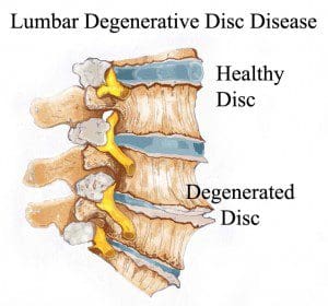
Degenerative disc disease
Is a spine condition that’s caused by everyday wear and tear of the spine can lead to significant loss of disc height.
A disc can also herniate and push on the nerves that flow into the feet, and cause:
Pain
Tingling
Numbness
The wear and tear of the spine combined with a herniated disc can pinch the nerves that go to the feet.
There is a condition known as foraminal spinal stenosis when the spinal foramen or the small opening between the bones of the spine begins to narrow and tighten so the nerves get compressed and do not allow signals and chemicals to flow properly.
This could explain the pain or other symptoms in the foot.
DDD can cause nerve problems in the feet, back and chronic back pain is the most common symptom of degenerative disc disease.
Common degenerative disc disease symptoms
The pain increases when:
Sit down for a long time
Bending
Lifting
Twisting
The pain decreases with:
Walking
Running
Lying down
Continually changing body positions
It’s important to remember that nerve issues with the feet are not always caused by spinal conditions, this could include
Depending on how severe the nerve problem is, there are a number of treatment options. These range from:
Chiropractic care
Physical therapy
Over-the-counter medication
Talk to a doctor or a chiropractor about the right treatment option for foot nerve pain. Whether it is caused by degenerative disc disease or some other condition.
These can help maintain a healthy weight, as being overweight puts extra pressure on the spine that leads to increased back pain. Consult with a registered dietitian or health coach to discuss the best foods for your diet.
Exercise
A training regimen that incorporates aerobic, strength and flexibility exercises that can help manage degenerative disc symptoms.
A personal trainer or sports chiropractor that has experience rehabilitating patients with spine conditions. They can teach exercises that can help relieve pain and other symptoms.
They can develop a custom physical therapy program
Regular chiropractic and physical therapy can help restore proper body mechanics, proper posture, as well as, avoiding positions that cause pain.
Surgery
Surgery is considered a last resort or if the condition is progressing and getting worse. If surgery is needed, your doctor will discuss the best treatment option for your condition.
Difference Foot Orthotics Make to *REDUCE FOOT PAIN* & Correct Posture | El Paso, TX.
NCBI Resources
Chiropractic focuses on restoring and maintaining overall health and wellness. Through the use of spinal adjustments and manual manipulations, a chiropractor can re-align the spine, and improve a patient�s strength, mobility, and flexibility.
IFM's Find A Practitioner tool is the largest referral network in Functional Medicine, created to help patients locate Functional Medicine practitioners anywhere in the world. IFM Certified Practitioners are listed first in the search results, given their extensive education in Functional Medicine


 Glycine is a