Tomatoes are low-calorie and nutrient-dense, what health benefits can individuals gain from their consumption?
Tomato Benefits
All varieties of tomatoes offer nutrients, including potassium and vitamin C, making them part of a balanced diet.
Raw tomatoes contain vitamin C, which brightens skin and fights inflammation.
Cooking tomatoes releases more antioxidants which are vital in small quantities such as lycopene, for maintaining heart health and preventing certain cancers.
Other benefits contribute to heart, prostate, and cognitive/brain health.
Various tomato recipes and products can offer a balance of nutrients. Variety is key and this applies to all fruits and vegetables. Try them raw, cooked, and steamed, as the different methods can offer different benefits.
Cooked and Raw Tomatoes
Tomatoes are low in calories and rich in nutrients. A raw, medium-sized tomato contains roughly 22 calories and less than 1 gram of fat. It is low sodium and low glycemic, with just 6 milligrams of sodium and 3 grams of sugar. They are an excellent source of hydration as a raw tomato contains about half a cup of water.
Tomatoes contain several essential vitamins and minerals that support the immune system and the bones and blood.
Antioxidants help combat free radicals and unstable molecules that damage the body’s cells. (Edward J. Collins, et al., 2022)
Antioxidants like lycopene, lutein, and zeaxanthin, are better absorbed with cooked tomatoes.
Raw tomatoes contain small amounts of vitamins A and K, fluoride, folate, and beta-carotene.
Heart Health
Tomatoes provide a healthy serving of potassium.
Potassium and sodium are both vital for heart function.
Potassium is essential for relaxing the blood vessels.
One medium tomato contains around the same amount as a banana.
The heart needs these electrolytes to contract and expand.
Most individuals with high blood pressure can benefit from high potassium, fiber, and lycopene levels.
Studies have linked lycopene to lower heart disease risk and mortality. (Bo Song, et al., 2017)
Exercise Recovery
Electrolytes are essential for basic cell function.
Potassium, sodium, magnesium, and fluoride can help decrease muscle soreness and exercise fatigue after physical activity or workouts.
The anti-inflammatory properties come from the vitamin C.
Eating tomatoes before or after physical activity can help replenish magnesium which is essential for muscle contraction. (Edward J. Collins, et al., 2022)
Protection Against Dementia
Potassium provides power to the heart and has a role in body nerve function.
One recent study found that individuals who consumed more potassium and less sodium had improved cognitive function. (Xiaona Na, et al., 2022)
Another study analyzed how carotenoids/antioxidants that affect the color of vegetables affect long-term brain health.
Researchers found that individuals with increased blood levels of lutein and zeaxanthin, which are both present in cooked tomatoes had a lower rate of dementia. (May A. Beydoun, et al., 2022)
Lutein and zeaxanthin are also known for protecting eye health as the body ages.
Help Prevent Prostate Cancer
Cooking tomatoes compromises the vitamin C content, but increases the availability of several antioxidants that can protect against cancer growth.
Especially for men, lycopene is beneficial to help reduce prostate-related issues.
Studies have found that men who eat tomatoes, including raw, sauce, and on pizza have a lower risk of developing prostate cancer due to the total amount of lycopene absorbed, which is optimized in cooked tomatoes. (Joe L. Rowles 3rd, et al., 2018)
Lycopene and other plant pigments/carotenoids are believed to protect against cancer because of their antioxidant properties. (Edward J. Collins, et al., 2022)
Lycopene and other antioxidants in tomatoes can also benefit male fertility by improving sperm count and sperm motility. (Yu Yamamoto, et al., 2017)
Balance Blood Sugar
Tomatoes can help manage blood sugar in individuals with diabetes.
They have fiber that helps regulate blood sugar and bowel movements.
Fiber naturally slows digestion to keep the body fuller and longer and does not negatively impact blood sugar levels.
Collins, E. J., Bowyer, C., Tsouza, A., & Chopra, M. (2022). Tomatoes: An Extensive Review of the Associated Health Impacts of Tomatoes and Factors That Can Affect Their Cultivation. Biology, 11(2), 239. https://doi.org/10.3390/biology11020239
Song, B., Liu, K., Gao, Y., Zhao, L., Fang, H., Li, Y., Pei, L., & Xu, Y. (2017). Lycopene and risk of cardiovascular diseases: A meta-analysis of observational studies. Molecular nutrition & food research, 61(9), 10.1002/mnfr.201601009. https://doi.org/10.1002/mnfr.201601009
Na X, Xi M, Zhou Y, et al. Association of dietary sodium, potassium, sodium/potassium, and salt with objective and subjective cognitive function among the elderly in China: a prospective cohort study. (2022). Glob Transit. 4:28-39. doi:10.1016/j.glt.2022.10.002
Beydoun, M. A., Beydoun, H. A., Fanelli-Kuczmarski, M. T., Weiss, J., Hossain, S., Canas, J. A., Evans, M. K., & Zonderman, A. B. (2022). Association of Serum Antioxidant Vitamins and Carotenoids With Incident Alzheimer Disease and All-Cause Dementia Among US Adults. Neurology, 98(21), e2150–e2162. https://doi.org/10.1212/WNL.0000000000200289
Rowles, J. L., 3rd, Ranard, K. M., Applegate, C. C., Jeon, S., An, R., & Erdman, J. W., Jr (2018). Processed and raw tomato consumption and risk of prostate cancer: a systematic review and dose-response meta-analysis. Prostate cancer and prostatic diseases, 21(3), 319–336. https://doi.org/10.1038/s41391-017-0005-x
Yamamoto, Y., Aizawa, K., Mieno, M., Karamatsu, M., Hirano, Y., Furui, K., Miyashita, T., Yamazaki, K., Inakuma, T., Sato, I., Suganuma, H., & Iwamoto, T. (2017). The effects of tomato juice on male infertility. Asia Pacific journal of clinical nutrition, 26(1), 65–71. https://doi.org/10.6133/apjcn.102015.17
Quagliani, D., & Felt-Gunderson, P. (2016). Closing America’s Fiber Intake Gap: Communication Strategies From a Food and Fiber Summit. American journal of lifestyle medicine, 11(1), 80–85. https://doi.org/10.1177/1559827615588079
Can healthcare professionals help individuals with spinal pain by incorporating non-surgical spinal decompression to restore mobility?
Introduction
Many individuals don’t realize that putting unwanted pressure on their spines can lead to chronic pain within their spinal discs that is affecting their spinal mobility. This usually happens with demanding jobs requiring individuals to carry heavy objects, step wrong, or be physically inactive, which causes the surrounding back muscles to be overstretched and leads to referred pain that affects the upper and lower body portions. This can cause individuals to go to their primary doctors to get treated for back pain. This leads to them missing out on their busy work schedules and paying a high price to get treated. Back pain correlating with spinal issues can be a huge problem and make them feel miserable. Fortunately, numerous clinical options are cost-effective and personalized to many individuals dealing with spinal pain that is causing them to find the relief they deserve. Today’s article focuses on why spinal pain affects many people and how spinal decompression can help reduce spinal pain and restore spinal mobility. Coincidentally, we communicate with certified medical providers who incorporate our patients’ information to provide various treatment plans to reduce spinal pain affecting their backs. We also inform them that there are non-surgical options to reduce the pain-like symptoms associated with spinal issues in the body. We encourage our patients to ask amazing educational questions to our associated medical providers about their symptoms correlating with body pain in a safe and positive environment. Dr. Alex Jimenez, D.C., incorporates this information as an academic service. Disclaimer
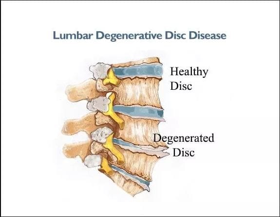
Why Spinal Pain Is Affecting Many People?
Have you often experienced pain from your back muscles that seem to ache after bending down constantly to pick up objects? Do you or your loved ones feel muscle stiffness in the back and experience numbness in your upper or lower body portions? Or are you experiencing temporary relief after stretching your back muscles, only for the pain to return? Many individuals with back pain never realize that their pain is within their spinal column. Since the spine is an S-curve shape with three different regions in the body, the spinal discs within each spinal segment can become compressed and become misaligned over time. This causes degenerative changes within the spine and can cause the three different spinal regions to develop pain-like issues in the body. When several environmental factors start to be the causes of degeneration of the spinal discs, it can affect the spinal structure. It can become a strong influence affecting their function, predisposing the disc to injuries. (Choi, 2009) At the same time, this can cause a significant impact when getting treated due to its high cost and can start normal age-related changes that cause pathophysiological issues to the vertebral body. (Gallucci et al., 2005)
When many individuals are dealing with spinal pain associated with herniated discs, it can not only cause discomfort but also mimic other musculoskeletal disorders that can cause radiating pain to different locations in the body. (Deyo et al., 1990) This, in turn, causes individuals to suffer constantly and research various treatments to reduce the pain they are experiencing. When spinal pain affects most individuals, many will seek cost-effective therapies to ease the pain they are experiencing and to be mindful of the daily habits they adopt over time and correct them.
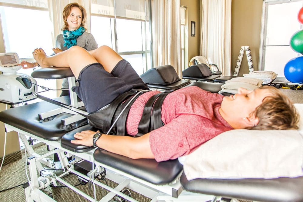
Spinal Decompression In-Depth- Video
Do you often feel constant muscle aches and pains in your body that are your general areas of complaint? Do you feel your muscles pull uncomfortably after lifting or carrying a heavy object? Or do you feel constant stress in your neck, shoulders, or back? When many individuals are dealing with general pain, they often assume that it is just back pain when it could be a spinal issue that can be the root cause of the pain they are experiencing. When this happens, many individuals opt for non-surgical treatments due to its cost-effectiveness and how it can be personalized depending on the severity of the pain. One of the non-surgical treatments is spinal decompression/traction therapy. The video above gives an in-depth look at how spinal decompression can help reduce spinal pain associated with low back pain. Spinal pain can increase with age and be provoked by extreme lumbar extension, so incorporating spinal decompression can help reduce pain in the upper and lower extremities. (Katz et al., 2022)
How Spinal Decompression Can Reduce Spinal Pain
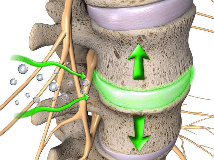
When individuals develop spinal issues, spinal decompression can help restore the spine to its original position and help the body naturally heal itself. When something is out of place within the spine, it is important to naturally restore it to its proper place to allow the affected muscles to heal. (Cyriax, 1950) Spinal decompression uses gentle traction to pull the spinal joints to let the spinal disc back in its original position and help increase fluid intake back in the spine. When people start incorporating spinal decompression into their health and wellness routine, they can reduce their spinal pain after a few consecutive treatments.
Spinal Decompression Restoring Spinal Mobility
Spinal decompression can also be incorporated with other non-surgical treatments to restore spinal mobility. When pain specialists utilize spinal decompression within their practices, they can help treat various musculoskeletal conditions, including spinal disorders, to allow the individual to regain spinal mobility. (Pettman, 2007) At the same time, pain specialists can use mechanical and manual manipulation to reduce the pain the individual feels. When spinal decompression starts to use gentle traction on the spine, it can help minimize radical pain correlated with nerve entrapment, create negative pressure within the spinal sections, and relieve musculoskeletal disorders causing pain. (Daniel, 2007) When people start thinking more about their health and wellness to reduce their pain, spinal decompression can be the answer through a personalized plan and can help many individuals find the relief they deserve.
Daniel, D. M. (2007). Non-surgical spinal decompression therapy: does the scientific literature support efficacy claims made in the advertising media? Chiropr Osteopat, 15, 7. https://doi.org/10.1186/1746-1340-15-7
Gallucci, M., Puglielli, E., Splendiani, A., Pistoia, F., & Spacca, G. (2005). Degenerative disorders of the spine. Eur Radiol, 15(3), 591-598. https://doi.org/10.1007/s00330-004-2618-4
Katz, J. N., Zimmerman, Z. E., Mass, H., & Makhni, M. C. (2022). Diagnosis and Management of Lumbar Spinal Stenosis: A Review. JAMA, 327(17), 1688-1699. https://doi.org/10.1001/jama.2022.5921
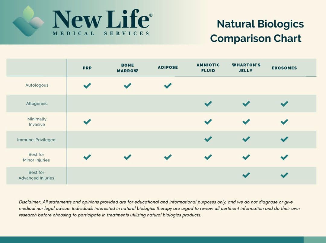
As the body grows older the ability to live life to the fullest can be difficult. Can using natural biologics help enhance the body’s natural ability to heal?
Natural Biologics
Though sometimes a necessary treatment option, surgical procedures can be the first line of treatment introduced to patients. Natural biologics is a less invasive alternative that can eliminate hospitalizations and expedite recovery. (Riham Mohamed Aly, 2020)
What Are They?
The body is born with components to initiate healing and recovery. These components include:
Cells
Cytokines
Proteins
Collagens
Elastin
Hyaluronic acid
At the time of birth, these components are in abundance but decrease as the body ages. This is why children recover from injuries quicker than adults. Recovery for adults can be slower from a decrease in these natural healing components. The objective of natural biologics treatments is to increase the healing components by reintroducing the body’s own components – autologous – or by bringing in new components – allogeneic – from a donor. (National Institutes of Health 2016) Choosing between the two options depends on an individual’s age and health, as those who are older or in poor physical health may experience complications from inferior component amounts.
Healing components derived from donor sources can show more promise, as treatments are usually acquired from discarded birth tissues at delivery.
Birth tissues are rich in healing components, containing the most abundant collection of natural healing elements.
It’s important to note that there is no harm to the mother or the baby from the obtained tissue products.
Platelet-rich plasma is cultivated by drawing an individual’s blood and spinning it in a centrifuge to separate the plasma.
The resulting liquid is reinjected into the injured area to generate a healing environment.
This form of natural biologics is effective for individuals with minor injuries that can be repaired easily.
This process is not as effective for older individuals who already have a reduction in natural healing components.
Lifestyle factors such as smoking, unhealthy diet, and alcohol/substance abuse can decrease the effectiveness of PRP treatments.
Bone Marrow Aspirate
This is an invasive, painful process that begins by putting a patient under anesthesia and drilling into the bone to extract the marrow. (American Cancer Society, 2023)
Like PRP, success depends on the individual’s age, health, and lifestyle.
Invasive procedures like this have a higher probability of infection and require a long-term recovery period.
Adipose-Derived Stem Cells
Adipose tissue/fat treatments are collected through a procedure that resembles the process of liposuction.
The procedure is done under general anesthesia and is an invasive process.
The treatment’s success depends on the individual’s health, age, and lifestyle.
There is more risk of infection when choosing this procedure and a long-term recovery period.
Allogeneic Treatment
Donor-based regenerative cells.
Amniotic Fluid Therapy
Amniotic fluid contains various growth factors, cytokines, and anti-inflammatory proteins that may promote tissue repair, reduce inflammation, and stimulate cellular regeneration. (Petra Klemmt. 2012)
Collected at the time of birth, this therapy is an ideal treatment for individuals who have sustained injuries that affect day-to-day functionality.
Physicians and clinicians are utilizing amniotic fluid therapy to treat many conditions, from orthopedic to wound care.
Amniotic fluid is collected at the time of birth and is abundant with increased healing components compared to autologous sources.
Amniotic fluid is immune-privileged (limits or suppresses immune response) and the risk of rejection is rare.
These therapies are usually done in a physician’s office with minimal downtime after treatment.
Wharton’s Jelly
Wharton’s jelly is derived from the umbilical cord at the time of birth and is primarily composed of a gel substance made up of hyaluronic acid and a network of collagen fibers.
Believed to contain a population of mesenchymal stem cells that have the capacity to differentiate into various cell types, and other secreted growth factors and cytokines. (F. Gao, et al., 2016)
It is considered the most valuable source to enhance the healing of various tissues, including bone, cartilage, skin, and nerve tissue.
It is immune-privileged with little risk of rejection and minimal if any, recovery time after an in-office treatment.
Exosomes
Exosomes are small, membrane-bound vesicles that play a role in intercellular communication within the body. (Carl Randall Harrell, et al., 2019)
They contain a variety of bioactive molecules, including proteins, lipids, nucleic acids (like RNA), and signaling molecules.
They serve as vehicles for transferring the signaling molecules from one cell to another, allowing cells to influence the behavior and function of neighboring or distant cells.
They can be collected or isolated from various biological fluids and cell cultures through specialized techniques but are most robust when collected at birth.
The exosomes within the umbilical cord are utilized for tissue repair and regeneration, signaling the cells to promote:
Proliferation – increase in the number of cells through cell division.
Differentiation – the transformation of unspecialized cells into specialized cells.
Tissue healing in damaged or injured areas.
Exosomes from the umbilical cord are immune-privileged with minimal risk of rejection.
Treatments are ideal for increasing cell communication and initiating repair when paired with another source of allogeneic therapy like amniotic fluid or Wharton’s Jelly.
Choosing which natural biologics therapy is the best is different for everyone. When selecting a treatment, it is essential for individuals to consult their primary healthcare provider to determine which application will have optimal results.
Is Motion Key To Healing?
References
Aly R. M. (2020). Current state of stem cell-based therapies: an overview. Stem cell investigation, 7, 8. https://doi.org/10.21037/sci-2020-001
Mazini, L., Rochette, L., Admou, B., Amal, S., & Malka, G. (2020). Hopes and Limits of Adipose-Derived Stem Cells (ADSCs) and Mesenchymal Stem Cells (MSCs) in Wound Healing. International journal of molecular sciences, 21(4), 1306. https://doi.org/10.3390/ijms21041306
Klemmt P. (2012). Application of amniotic fluid stem cells in basic science and tissue regeneration. Organogenesis, 8(3), 76. https://doi.org/10.4161/org.23023
Sabapathy, V., Sundaram, B., V M, S., Mankuzhy, P., & Kumar, S. (2014). Human Wharton’s Jelly Mesenchymal Stem Cells plasticity augments scar-free skin wound healing with hair growth. PloS one, 9(4), e93726. https://doi.org/10.1371/journal.pone.0093726
Gao, F., Chiu, S. M., Motan, D. A., Zhang, Z., Chen, L., Ji, H. L., Tse, H. F., Fu, Q. L., & Lian, Q. (2016). Mesenchymal stem cells and immunomodulation: current status and future prospects. Cell death & disease, 7(1), e2062. https://doi.org/10.1038/cddis.2015.327
Harrell, C. R., Jovicic, N., Djonov, V., Arsenijevic, N., & Volarevic, V. (2019). Mesenchymal Stem Cell-Derived Exosomes and Other Extracellular Vesicles as New Remedies in the Therapy of Inflammatory Diseases. Cells, 8(12), 1605. https://doi.org/10.3390/cells8121605
When individuals experience a neuromusculoskeletal injury strain, can following basic pulled muscle treatment protocols help in healing and a full recovery?
Pulled Muscle Treatment
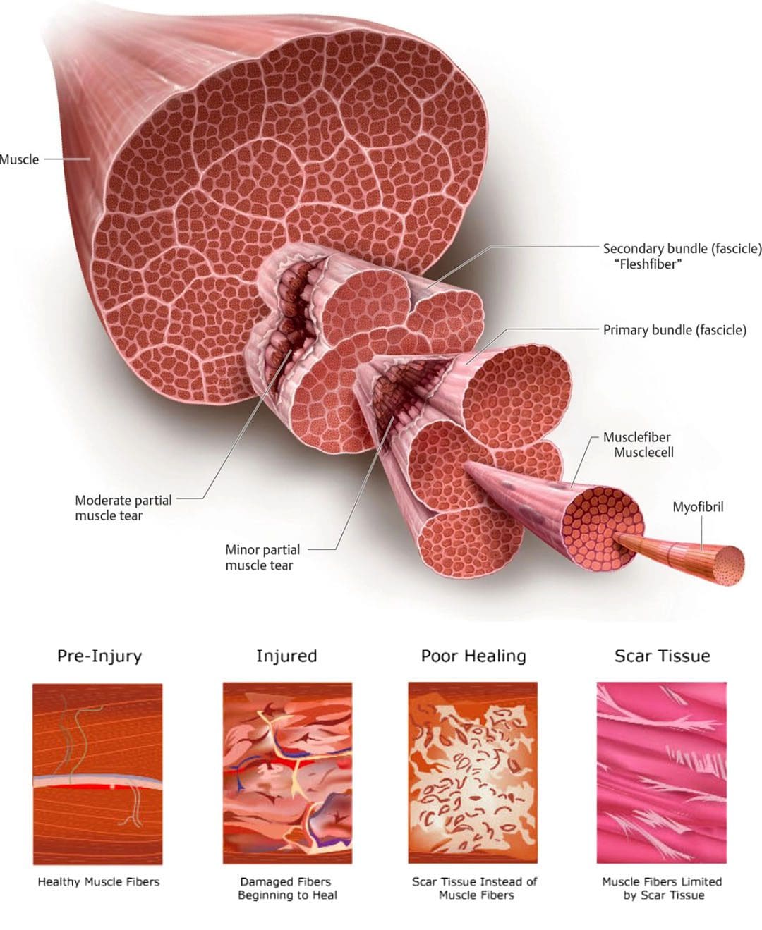
A pulled muscle or muscle strain occurs when a muscle is stretched beyond its ability resulting in discomfort symptoms and mobility issues. Microscopic tears can occur within the muscle fibers potentially worsening the injury. This type of injury usually causes mild to severe pain, bruising, and immobility, and nerve injuries can develop as well. Common muscle strains include:
Pulled hamstrings
Groin strains
Pulled abdominal muscles
Calf strains
Pulled muscle treatment requires patience to promote proper healing and restoration of optimal function.
Individuals need to focus on the different stages of healing.
Gradually increase activity levels as the body allows to prevent stiffness and atrophy which can cause complications.
Symptoms
The usual symptoms of this type of injury include:
Pain
Limited mobility
Muscle spasms
Swelling
Bruising
Often individuals will feel a sudden grabbing or tearing sensation and are then unable to continue the activity.
Can limit the ability to perform certain activities.
May have moderate swelling and bruising.
Grade III
Severe injury that can cause significant pain.
Muscle spasms.
Swelling.
Significant bruising.
Basic Treatment Protocols
Most pulled muscle strain injuries heal with simple treatment. Following the right steps can ensure an expedited recovery. In the early stages after the injury, there is a balance between doing too much or not enough. The amount of activity an individual will be able to do, and the time required for recovery depends on the severity of the injury. Here are some guidelines in the right direction.
Rest
Rest is recommended for the early recovery stage.
Depending on the severity of the injury this could last from one to five days.
Immobilization is usually not necessary, and not moving at all can lead to muscle and joint stiffness.
To avoid injuries make sure the muscles are not over-exerted.
Gradually increase activity levels when starting an exercise program to build endurance.
Properly Warming Up
Warming up before taking on physical activities will help loosen the muscles and prevent injuries.
Beginning work or exercise with stiff muscles can lead to an increased chance of strain.
Studies have shown that temperature can influence the stiffness of a muscle. (K. W. Ranatunga. 2018)
Maintaining body and muscle warmth helps prevent injury and re-injury.
Injuries and Chiropractic: The Road To Recovery
References
Hospital for Special Surgery, Muscle Strain: What You Need to Know About Pulled Muscles.
Kary J. M. (2010). Diagnosis and management of quadriceps strains and contusions. Current reviews in musculoskeletal medicine, 3(1-4), 26–31. https://doi.org/10.1007/s12178-010-9064-5
Malanga, G. A., Yan, N., & Stark, J. (2015). Mechanisms and efficacy of heat and cold therapies for musculoskeletal injury. Postgraduate medicine, 127(1), 57–65. https://doi.org/10.1080/00325481.2015.992719
Mair, S. D., Seaber, A. V., Glisson, R. R., & Garrett, W. E., Jr (1996). The role of fatigue in susceptibility to acute muscle strain injury. The American journal of sports medicine, 24(2), 137–143. https://doi.org/10.1177/036354659602400203
Ranatunga K. W. (2018). Temperature Effects on Force and Actin⁻Myosin Interaction in Muscle: A Look Back on Some Experimental Findings. International journal of molecular sciences, 19(5), 1538. https://doi.org/10.3390/ijms19051538
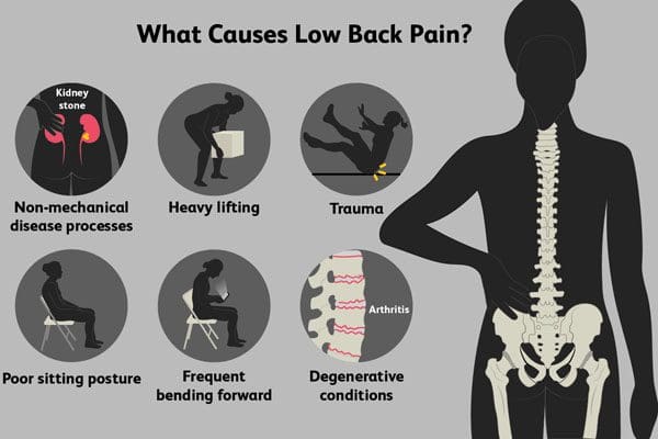
Can healthcare professionals provide the best non-surgical therapeutic options for individuals with chronic low back pain?
Introduction
Chronic low back pain can happen to numerous individuals, affecting their daily routine and making them miss out on important life events. With the ever-changing world, many individuals, especially working individuals, will experience chronic low back pain at some point due to unbearable stress that seems to affect the surrounding muscles that protect the lumbar spine. This causes many individuals to overstretch or shorten the muscles that are contributing to lower back pain, which can be the causing factor in the development of lower back pain. At the same time, when individuals suffer from low back pain, it can be imposed as a grave economic cost to society. (Pai & Sundaram, 2004) This, in turn, causes many individuals to miss out on work and be financially burdened as the cost of chronic low back pain treatment is high. However, numerous therapeutic options are cost-effective, safe, and effective in reducing chronic low back pain. Today’s post looks at the effects of chronic low back pain and how many individuals can look at various non-surgical options that many individuals can utilize to reduce chronic low back pain. Coincidentally, we communicate with certified medical providers who incorporate our patients’ information to provide various treatment plans to reduce chronic low back pain. We also inform them that there are non-surgical options to reduce the pain-like symptoms associated with the factors that cause chronic lower back pain. We encourage our patients to ask amazing educational questions to our associated medical providers about their symptoms correlating with body pain in a safe and positive environment. Dr. Alex Jimenez, D.C., incorporates this information as an academic service. Disclaimer
The Effects Of Chronic Low Back Pain
Have you been dealing with chronic pain that flares up in your lower back after a hard workday? Do you feel muscle aches or pains that don’t relieve itself after a day of rest? Or do you and your loved ones take any medication to temporarily relieve your back pain, only to have it come back after a few hours? Many people with chronic low back pain will feel symptoms of stiffness, muscle aches, and radiating pain traveling to their lower extremities. When chronic low back pain is associated with musculoskeletal conditions, it can impact their daily routine. To that point, musculoskeletal disorders correlating with chronic low back pain can encompass a spectrum of conditions and increase naturally over time. (Woolf & Pfleger, 2003) When many individuals are dealing with chronic low back pain, it can become a socio-economic burden that leads to disability. (Andersson, 1999) However, there are numerous options for individuals with chronic lower back pain who can find the relief they need to reduce its effects and will be able to get back to their daily routine.
Understanding Long-Lasting Injuries- Video
Chronic low back pain is when back pain that lasts longer than a few weeks and is one of the most common problems many people experience. When finding relief for chronic low back pain, many individuals will try home remedies to alleviate the pain. However, it can temporarily relieve the issue and mask the symptoms. When individuals see their primary doctor for chronic low back pain, many will seek a personalized plan to reduce chronic low back pain and its associated symptoms. When relieving chronic low back pain, comprehensive pain management treatments often rely on physical therapy, multidisciplinary approaches, and non-surgical options to reduce chronic low back pain. (Grabois, 2005) When understanding how the individual has chronic low back pain, it is important to identify the causes and how it can cause lifelong injuries that can develop into disability. When primary doctors start to utilize non-surgical treatments in their practices, many individuals can find the benefits of non-surgical treatments as they are cost-effective, safe, and gentle on the spine and lumbar region and can be personalized with associated medical providers to reduce pain-like symptoms correlating with chronic low back pain. Check out the video above to learn more about how non-surgical treatments can help reduce chronic low back pain and help revitalize a person’s body through a personalized treatment plan.
Non-Surgical Options For Chronic Low Back Pain
When treating chronic low back pain, non-surgical treatments effectively relieve pain and restore mobility to the back. Non-surgical treatments can be customized to the individual’s pain severity while being cost-effective. When individuals are evaluated for chronic low back pain, they are provided with many healthcare providers to reduce the pain-like symptoms caused by chronic low back pain. (Atlas & Deyo, 2001) Many individuals will incorporate various treatment options like:
Exercises
Spinal Decompression
Chiropractic care
Massage Therapy
Acupuncture
Many of these treatments are non-surgical and incorporate various mechanical and manual manipulation techniques to stretch and strengthen the weak back muscles, elongate the spine through realignment, and help restore movement while reducing symptoms in the lower extremities. When individuals incorporate non-surgical treatments consecutively, they will have a positive experience and feel better in the long run. (Koes et al., 1996)
Koes, B. W., Assendelft, W. J., van der Heijden, G. J., & Bouter, L. M. (1996). Spinal manipulation for low back pain. An updated systematic review of randomized clinical trials. Spine (Phila Pa 1976), 21(24), 2860-2871; discussion 2872-2863. https://doi.org/10.1097/00007632-199612150-00013
For individuals about to engage in physical activity or exercise, how does warming up the body help prepare for the work ahead?
Central Nervous System Activation
A proper warm-up before physical activity or working out prepares the mind and body to reduce risks of injury, mentally and physically transition to physical activity work, and enhance performance. A well-designed warm-up also primes the central nervous system/CNS for activity. The central nervous system transmits messages to the muscles to prepare them for action. Central nervous system activation increases motor neuron recruitment and engages the sympathetic nervous system so the body can better handle the physical stressors. The process may seem complex, but priming the nervous system is as simple as warming up with light aerobic activity before getting into more explosive movements.
CNS
The CNS consists of the brain and spinal cord. This central communication system uses another part of the nervous system known as the peripheral nervous system or PNS to transmit and receive messages throughout the body. The PNS is connected to the entire body and the brain and spinal cord (CNS).
Nerves run throughout the body, receiving signals from the CNS to the muscles, fibers, and organs, transmitting various information back to the brain. (Berkeley University. N.D.)
There are two types of systems within the peripheral nervous system – somatic and autonomic.
Somatic nervous system actions are those controlled by the person through voluntary actions like choosing to pick something up.
Properly preparing the body for an intense strength training session or other physical activity needs the correct messages to be sent through the autonomic nervous system.
Parasympathetic and Sympathetic States
The autonomic nervous system consists of two subcategories, which are parasympathetic and sympathetic.
The sympathetic nervous system helps the body get ready to face stress which includes physical stress. (R. Bankenahally, H. Krovvidi. 2016)
The fight, flight, or freeze response describes the sympathetic nervous system’s aspect.
The parasympathetic nervous system is responsible for relaxation and de-stressing.
Individuals are recommended to perform a few calming movements and actions after a workout to return the body to a parasympathetic state. This can be:
Activating the CNS can increase performance and prevent injuries. The process wakes up and alerts the body for the activity. Individuals are recommended before beginning a training session, to communicate to the body about the physical stress it is about to endure and to prepare for the work ahead. This is a concept known as post-activation potentiation/PAP. (Anthony J Blazevich, Nicolas Babault. 2019) PAP helps increase force and power production, which enhances physical performance.
Whenever an individual trains, the brain adapts and learns what the body is doing and the purpose of the training.
Muscle memory describes this interaction.
Individuals who have started up a new strength training routine or after an extended break report feeling awkward for the first few sessions, or even weeks, depending on their experience. (David C Hughes, Stian Ellefsen, Keith Baar, 2018)
However, after a few sessions, the body is more adept at performing the movements and ready to increase resistance, repetitions, or both.
This has to do with the neural drive and muscle memory than it has to do with true potential physical abilities. (Simon Walker. 2021)
The first step is a general warm-up that should use large muscle groups and be of low intensity so as not to exhaust the body before beginning the actual training. General warm-up benefits central nervous system activation and the entire body include: (Pedro P. Neves, et al., 2021) (D C. Andrade, et al., 2015)
Increases blood circulation.
Assists the release of oxygen from hemoglobin and myoglobin.
Warms the muscles, so they contract more effectively.
Increases nerve impulse speed.
Increases nutrient delivery.
Lowers joints’ resistance through increased synovial fluid/joint lubrication.
Increases joint range of motion.
Improves joint resiliency.
Removes metabolic waste quicker.
Reduces risk of injury.
A general warm-up can be simple as any aerobic activity will work. This can include:
Performing bodyweight movements – light jumping jacks or jogging in place.
Treadmill
Rowing machine
Stair climber
Elliptical trainer
It is recommended to use the rating perceived exertion scale/RPE to determine the general warm-up effort. An exertion rating of between 5 to 6 is equivalent to moderate walking or a slow jog. Individuals should be able to speak clearly without taking a pause.
Try this strategy before the next workout to see increased performance and reduced injury risks.
Blazevich, A. J., & Babault, N. (2019). Post-activation Potentiation Versus Post-activation Performance Enhancement in Humans: Historical Perspective, Underlying Mechanisms, and Current Issues. Frontiers in physiology, 10, 1359. https://doi.org/10.3389/fphys.2019.01359
Hughes, D. C., Ellefsen, S., & Baar, K. (2018). Adaptations to Endurance and Strength Training. Cold Spring Harbor perspectives in medicine, 8(6), a029769. https://doi.org/10.1101/cshperspect.a029769
Walker S. (2021). Evidence of resistance training-induced neural adaptation in older adults. Experimental gerontology, 151, 111408. https://doi.org/10.1016/j.exger.2021.111408
Andrade, D. C., Henriquez-Olguín, C., Beltrán, A. R., Ramírez, M. A., Labarca, C., Cornejo, M., Álvarez, C., & Ramírez-Campillo, R. (2015). Effects of general, specific, and combined warm-up on explosive muscular performance. Biology of sport, 32(2), 123–128. https://doi.org/10.5604/20831862.1140426
Can healthcare professionals implement an inclusive and positive approach for gender affirming healthcare for non-binary individuals?
Introduction
When it comes to many individuals looking for the right healthcare options for their ailments and general well-being, it can be scary and challenging to some, including many individuals within the LGBTQ+ community. Many individuals need to research when finding positive and safe healthcare facilities that listen to what the person is dealing with when getting a routine check-up or their ailments treated. Within the LGBTQ+ community, many individuals do find it difficult to express what is affecting their bodies due to past traumas of not being seen or heard due to their identities, pronouns, and orientation. This can cause numerous barriers between them and their primary doctor, leading to a negative experience. However, when medical professionals provide a positive, safe environment, listen to the person’s ailments, and be non-judgmental to their patients, they can open the doors to improving inclusive healthcare wellness within the LGBTQ+ community. Today’s article focuses on one identity within the LGBTQ+ community, known as non-binary, and how inclusive healthcare can be optimized while benefitting many individuals dealing with general aches, pains, and conditions within their bodies. Coincidentally, we communicate with certified medical providers who incorporate our patients’ information to provide a safe and positive experience in inclusive healthcare. We also inform them that there are non-surgical options to reduce the effects of general aches and pain while restoring their quality of life. We encourage our patients to ask amazing educational questions to our associated medical providers about their symptoms correlating with body pain in a safe and positive environment. Dr. Alex Jimenez, D.C., incorporates this information as an academic service. Disclaimer
What Is Non-Binary Gender?
The term non-binary is used within the LGBTQ+ community to describe a person who doesn’t identify as a male or female within the gender identity spectrum. Non-binary individuals can even fall under various gender identities that make them who they are. These can include:
Genderqueer: An individual who doesn’t follow the traditional gender norm.
Agender: An individual who doesn’t identify with any gender.
Genderfluid: An individual whose gender identity is not fixed or can change over time.
Intergender: An individual who identifies as a combination of male and female.
Androgynous: An individual whose gender expression combines masculine and feminine traits.
Gender Non-Conforming: An individual who doesn’t conform to society’s expectation of gender identity.
Transgender: An individual whose gender identity is different from their assigned gender at birth.
When it comes to non-binary binary individuals looking for healthcare treatment for their ailments, it can be a bit of a challenge as many individuals who identify as non-binary within the LGBTQ+ community have to deal with the socio-economic impact when getting treatment, which can lead to unnecessary stress when going in for a routine check-up or getting their ailments treated. (Burgwal et al., 2019) When this happens, it can lead to a negative experience for the individual and make them feel inferior. However, when healthcare professionals take the time to be properly trained, use the correct pronouns, and create an inclusive, positive, and safe space for individuals who identify as non-binary, it can open the doors to creating more of an inclusive awareness and lead to more appropriate care for the LGBTQ+ community. (Tellier, 2019)
Optimizing Your Wellness- Video
Do you or your loved ones are dealing with consistent pain in their bodies that makes it difficult to function? Do you feel stress in different body locations that correlate with musculoskeletal disorders? Or do your ailments seem to be affecting your daily routine? More often than not, in today’s ever-changing world, many individuals are researching safe and inclusive healthcare treatments to reduce their ailments. It is an important aspect to many individuals within the LGBTQ+ community, as finding the appropriate care they need can be stressful. Many healthcare professionals must provide the best possible healthcare and interventions within the LGBTQ+ community to understand the health disparities that they are experiencing. (Rattay, 2019) When healthcare professionals create a negative experience with their patients within the LGBTQ+ community, it can cause them to develop socio-economic stressors that can overlap with their pre-existing condition, creating barriers. When disparities are associated with socio-economic stressors, it can lead to poor mental health. (Baptiste-Roberts et al., 2017) When this happens, it can lead to coping mechanisms and resilience that can correlate with serious implications for the person’s overall health and well-being. However, all is not lost, as many healthcare professionals are integrating into safe, affordable, and positive healthcare spaces for individuals who identify as non-binary. We here at Injury Medical Chiropractic and Functional Medicine Clinic will work on reducing the effects of health disparities while raising awareness to continuously improve positive and inclusive experiences for non-binary individuals seeking inclusive healthcare. Check out the video above to learn more about optimizing wellness to improve your health and well-being.
How To Optimize Non-Binary Inclusive Healthcare?
When it comes to inclusive health care for non-binary individuals within the LGBTQ+ community, many healthcare providers must honor the individual’s gender identity while creating a positive and trusting relationship to reduce the ailments they are experiencing. By making a safe and positive experience for their patients, LGBTQ+ individuals will start to address to their doctors what issues they are experiencing, and it allows the doctor to come up with a personalized health care plan that is catered to them while improving their health outcomes. (Gahagan & Subirana-Malaret, 2018) At the same time, being an advocate and systemically improving, including gender-affirming care, can lead to positive results and benefit LGBTQ+ individuals. (Bhatt et al., 2022)
References
Baptiste-Roberts, K., Oranuba, E., Werts, N., & Edwards, L. V. (2017). Addressing Health Care Disparities Among Sexual Minorities. Obstet Gynecol Clin North Am, 44(1), 71-80. https://doi.org/10.1016/j.ogc.2016.11.003
Bhatt, N., Cannella, J., & Gentile, J. P. (2022). Gender-affirming Care for Transgender Patients. Innov Clin Neurosci, 19(4-6), 23-32. https://www.ncbi.nlm.nih.gov/pubmed/35958971
Burgwal, A., Gvianishvili, N., Hard, V., Kata, J., Garcia Nieto, I., Orre, C., Smiley, A., Vidic, J., & Motmans, J. (2019). Health disparities between binary and non binary trans people: A community-driven survey. Int J Transgend, 20(2-3), 218-229. https://doi.org/10.1080/15532739.2019.1629370
Gahagan, J., & Subirana-Malaret, M. (2018). Improving pathways to primary health care among LGBTQ populations and health care providers: key findings from Nova Scotia, Canada. Int J Equity Health, 17(1), 76. https://doi.org/10.1186/s12939-018-0786-0
Rattay, K. T. (2019). Improved Data Collection for Our LGBTQ Population is Needed to Improve Health Care and Reduce Health Disparities. Dela J Public Health, 5(3), 24-26. https://doi.org/10.32481/djph.2019.06.007
Tellier, P.-P. (2019). Improving health access for gender diverse children, youth, and emerging adults? Clinical Child Psychology and Psychiatry, 24(2), 193-198. https://doi.org/10.1177/1359104518808624
IFM's Find A Practitioner tool is the largest referral network in Functional Medicine, created to help patients locate Functional Medicine practitioners anywhere in the world. IFM Certified Practitioners are listed first in the search results, given their extensive education in Functional Medicine