For individuals who are getting older, can increasing bone strength help prevent fractures and optimize bone health?
Bone Strength
Bone strength is important, as a fractured hip can be serious for older individuals. A study found that for individuals in their 60s who had a hip fracture, 6.5% of women and 9.4% of men died within a year. Among individuals in their 80s, 13.1% of women and 19.6% of men died within a year. (Dimet-Wiley, et al., 2022)
Increasing bone strength can help prevent various issues. A small increase in bone mineral density has been shown to help reduce the risk of fractures, especially hip fractures. A decades-long study found that just a 3% increase in bone strength helps lower the chance of breaking a hip. Researchers enrolled two groups of individuals aged 60 and older, one in 1989 and the second in 1999.
The bone mineral density of each subject’s femoral neck joint at the top of the thigh bone near the hip was measured.
They then followed the subjects for years to see who experienced hip fractures.
While the bone mineral density of the second group was only 3% higher than the first group, these subjects experienced a 46% reduction in hip fractures. (Tran, T. et al., 2023)
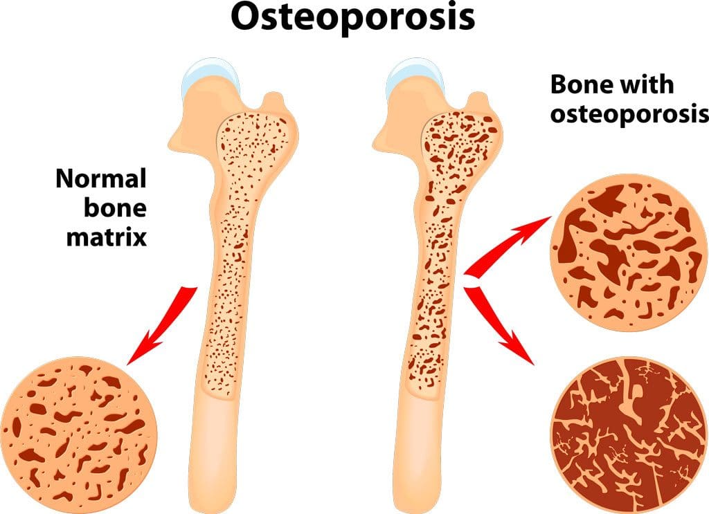
Bone Loss
Bone loss is progressive in men and women and increases as the body ages. Osteoporosis is a condition in which bone tissue deteriorates. (Department of Health and Human Services Office of Disease Prevention and Health Promotion. 2020) Bones constantly break down and reform as a normal remodeling process. If the balance of this process is impaired, osteoporosis develops, resulting in more bone breakdown than formation. While men and women experience bone loss, it’s more common in females. (National Institute of Arthritis and Musculoskeletal Diseases. 2022) Menopause is a risk factor because of the decline of estrogen (National Library of Medicine, Medline Plus, 2022). Estrogen reinforces bone strength by protecting against bone breakdown; with estrogen loss, bone breakdown increases. However, anyone of any age or background can experience bone loss due to the following:
While some loss of bone strength is common, several strategies exist to maintain bone health. Exercise, specifically weight-bearing activities, can increase bone strength. When bones and muscles are used to hold a position against gravity, this mechanically stresses the bone, causing it to reform stronger. Movement and physical exercise as medicine and the forces transmitted through the bones generate mechanical signals that tell the cells to increase bone formation relative to breakdown. Exercises focusing on posture, balance, gait, and coordination are recommended for individuals with osteoporosis to strengthen the core, quadriceps, and hip flexors. Different types of exercises can include:
Walking to strengthen the spine and hips.
Walking outside or on a treadmill provides more loading force to the bone.
Planks and push-ups can strengthen the forearm and wrist bones.
Holding a water bottle in each hand and lifting up and down 10 times together or alternating a few times a day.
Side leg lifts can strengthen the hip and forearm bones simultaneously.
Weight training provides the bones with a workout by having them support a weight load.
Any exercise therapy program should be designed by a healthcare provider, physical therapist, and trainer according to the individual’s condition and appropriate for them.
Diet
What goes into the body definitely affects bone health. Calcium and vitamin D are key to bone building, but both are needed as vitamin D is needed to absorb the calcium ingested. Calcium can be found in:
Dairy
Dairy products and non-dairy alternatives are fortified with calcium.
Leafy greens.
Beans.
Almonds.
The recommended daily calcium intake for adults over 50 is 1,200 milligrams.
Vitamin D can come from:
Sunlight
Fish.
Mushrooms.
Fortified milk.
Supplements.
The recommended daily vitamin D intake for adults aged 70 is 15 micrograms and 20 micrograms for individuals over 70.
Studies have found that increasing calcium and vitamin D intake with supplements can help maintain bone health. Talk to a healthcare provider about whether supplements could be beneficial.
Hormone Therapy
Females also naturally produce testosterone, which promotes bone formation. As levels drop with age and negatively impact bone strength, hormone therapy could be recommended. Declining testosterone levels start with women in their 20s and men in their 30s. The typical drop in women is 1% to 3% yearly before menopause and stabilizes somewhat afterward. Female patients at risk of bone loss may be prescribed testosterone in various forms that continuously emit the hormone. The dosage is low, so patients do not experience unwanted hair growth or skin changes. Combined with estrogen, testosterone effectively increases bone growth in female patients. Not everyone is a candidate for hormone therapy, like individuals with a history of breast cancer, heart disease, blood clots, or liver disease. (National Library of Medicine. Medline Plus, 2019)
Making small adjustments can optimize bone health and overall well-being
At Injury Medical Chiropractic and Functional Medicine Clinic, we passionately focus on treating patients’ injuries and chronic pain syndromes to create personalized care plans that improve ability through flexibility, mobility, and agility programs tailored to the individual. Using an integrated approach, our goal is to relieve pain naturally by restoring health and function to the body through Functional Medicine, Acupuncture, Electro-Acupuncture, and Sports Medicine protocols. If the individual needs other treatment, they will be referred to a clinic or physician best suited for them, as Dr. Jimenez has teamed up with the top surgeons, clinical specialists, medical researchers, and premier rehabilitation providers to provide the most effective clinical treatments. We focus on what works for you and strive to better the body through researched methods and total wellness programs.
Chiropractic Care: Movement Medicine
References
Dimet-Wiley, A., Golovko, G., & Watowich, S. J. (2022). One-Year Postfracture Mortality Rate in Older Adults With Hip Fractures Relative to Other Lower Extremity Fractures: Retrospective Cohort Study. JMIR aging, 5(1), e32683. doi.org/10.2196/32683
Tran, T. S., Ho-Le, T. P., Bliuc, D., Center, J. R., Blank, R. D., & Nguyen, T. V. (2023). Prevention of Hip Fractures: Trade-off between Minor Benefits to Individuals and Large Benefits to the Community. Journal of bone and mineral research : the official journal of the American Society for Bone and Mineral Research, 38(11), 1594–1602. doi.org/10.1002/jbmr.4907
Can incorporating various yoga poses help reduce neck tension and provide pain relief for individuals dealing with neck pain?
Introduction
Within the hustling and bustling of modern life, it is common for many individuals to carry stress in their bodies. When the body deals with everyday stressors, tension, discomfort, and pain can often manifest in the upper and lower portions of the body. When the body’s upper and lower portions deal with these issues, they can cause overlapping risk profiles in the musculoskeletal system. One of the most common musculoskeletal issues is neck pain. It can cause many problems to the cervical portion of the spine and cause the surrounding muscles to become tense and in pain from the stress of everyday responsibilities. Luckily, there are numerous ways to reduce stress from the neck and help relax the affected muscles from discomfort, including yoga. In today’s article, we will look at how neck pain affects the upper body, the benefits of yoga for neck pain, and various yoga poses to reduce the overlapping effects of neck pain. We discuss with certified medical providers who consolidate our patients’ information to assess how neck pain is correlated with everyday stressors that affect the upper body. We also inform and guide patients on how yoga and the various poses can benefit the body and provide pain relief to the surrounding muscles. We also encourage our patients to ask their associated medical providers many intricate and important questions about incorporating yoga into their daily routine to reduce muscle tension and provide clarity to their bodies. Dr. Jimenez, D.C., includes this information as an academic service. Disclaimer.
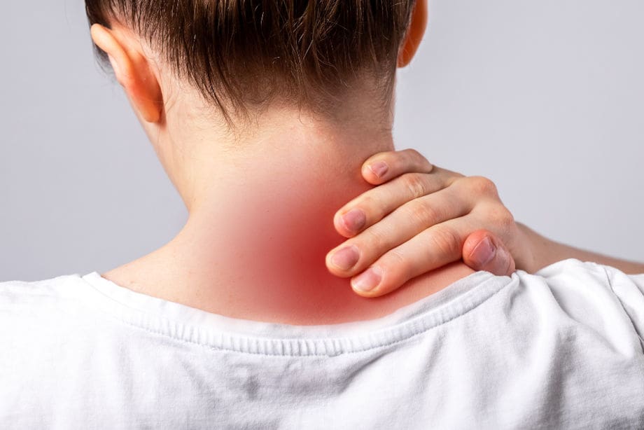
How Does Neck Pain Affect The Upper Body?
Do you feel discomfort or pain in your neck and shoulders after a long, hard workday? Do you notice you hunched more than usual when doing your daily routine? Or do you see yourself developing a hunched posture from looking at the computer screen or phone for an extended period? Many of these normal motions are often correlated with the upper body, especially in the neck and shoulder regions, which causes neck pain. As one of the most common problems affecting many people worldwide, neck pain is a multifactorial disease with numerous risk factors contributing to its development. (Kazeminasab et al., 2022) Like back pain, neck pain can have acute and chronic stages depending on the severity and environmental factors leading to its development. The various muscles, ligaments, and tissues surrounding the neck and shoulders keep the neck stable and mobile. When many individuals overuse these muscles in the neck and shoulders repetitively, it can increase neck pain in the upper body in adulthood. (Ben Ayed et al., 2019)
When acute neck pain turns chronic, it can cause the individual to be in constant discomfort, pain, and misery, so they start to look for various solutions to reduce the correlating symptoms when speaking to their primary doctors. When many individuals begin to explain to their doctors what their daily routine looks like, many doctors will start to assess and formulate a plan that focuses on any specific description of any injuries, including potential mechanisms, inciting and relieving factors, and pain patterns they have encountered throughout the day to come up with a personalized treatment plan to not only reduce neck pain but also provide relief to tension and discomfort to the body. (Childress & Stuek, 2020)
The Science of Motion- Video
The Benefits Of Yoga For Neck Pain
Many primary doctors will work with associated medical providers to develop a personalized plan to relieve neck pain and its associated symptoms in many individuals. Many of these customized treatment plans include spinal manipulation, acupuncture, massage, decompression therapy, and therapeutic exercises. One of the therapeutic exercises that many individuals have utilized is yoga. Yoga is a holistic practice encompassing breathing control, meditation, and various poses to stretch and strengthen the affected upper muscles. Yoga is excellent for reducing neck pain and helping with upper cervical spine mobility, stretching the neck musculature to help the individual improve mobility and flexibility. (Raja et al., 2021) Additionally, the effects of yoga and its many poses can reduce tension, give clarity to the mind, and allow the nutrients and oxygen to the musculo-articular system to naturally heal the body itself. (Gandolfi et al., 2023)
Yoga Poses For Neck Pain
At the same time, many individuals with sedentary jobs that correlate to neck pain have implemented yoga as part of their routine. Yoga improves their range of joint motion and cognitive function and helps relieve musculoskeletal discomfort in the neck and shoulder regions. (Thanasilungkoon et al., 2023) Below are some of the various yoga poses that can help reduce the pain-like symptoms of neck pain and ease the surrounding muscles.
Seated Neck Stretches
For seated neck stretches, this yoga pose helps stretch and release the neck muscles that carry tension and stress in the cervical region of the body.
In a seated upright position, turn the head to the right and gently lift the chin.
You should feel a stretch along the left side of the neck and shoulders.
Hold the position for three to five breaths and repeat on the left side.
Camel Pose
For the camel pose, this yoga pose helps strengthen the front neck muscles while easing tension on the shoulders and back of the neck.
You can kneel on a yoga mat by keeping your knees and feet hip-distance apart while keeping the pelvis neutral.
Lift the chest while arching your back and pressing the pelvis slightly forward.
Bring the fingertips to the heels or yoga blocks beside the ankles.
Focus on drawing the chin close to the neck while pressing the feet to the mat.
Hold the position for three to five breaths before releasing and lifting the sternum to rise back up.
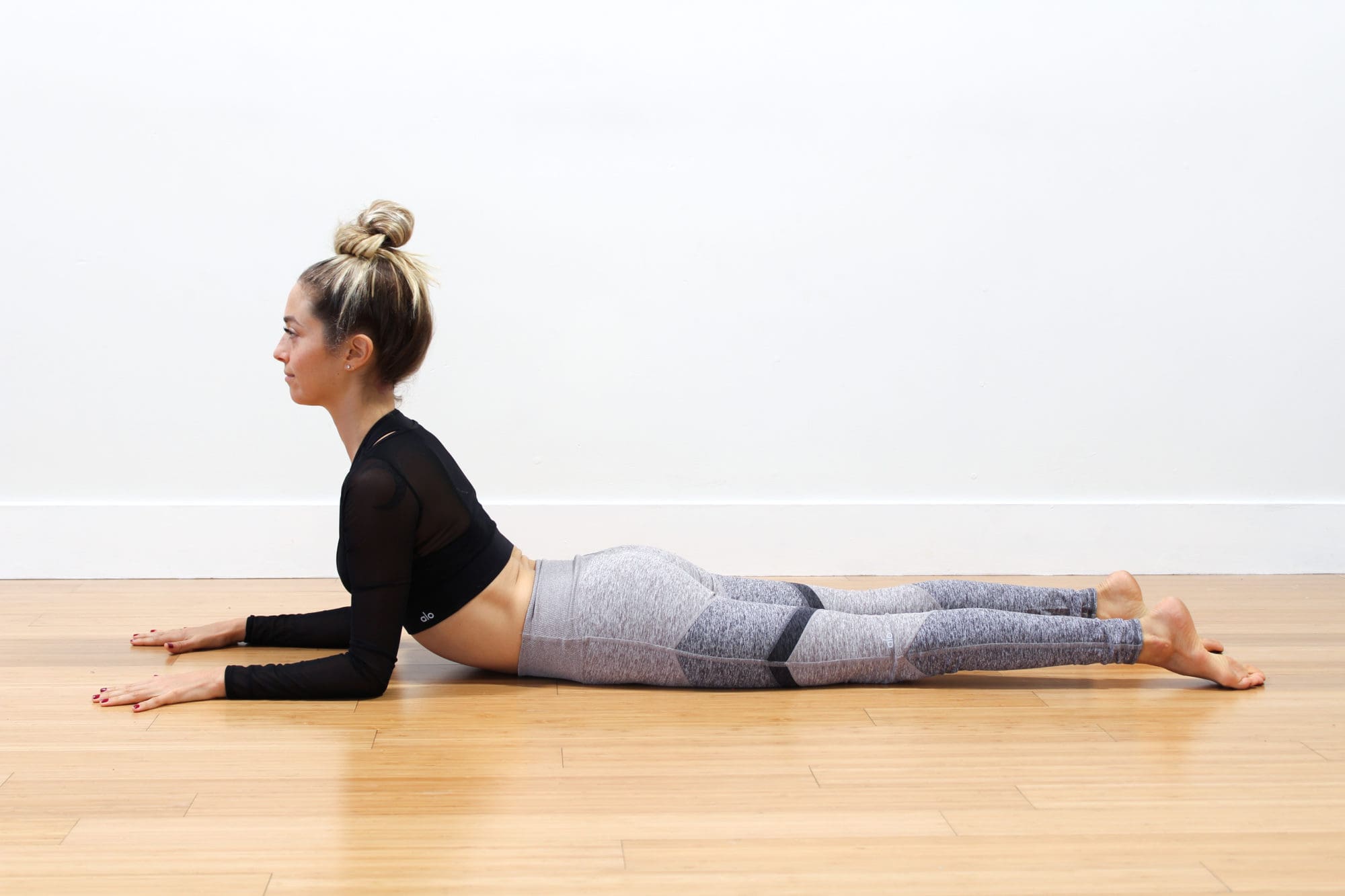
Sphinx Pose
The sphinx pose allows you to lengthen and strengthen the spine while stretching the shoulders and releasing tension.
On a yoga mat, lie on your stomach with the elbows under the shoulders.
Press your palms and forearms on the mat and tighten the lower half to support you as you lift your upper torso and head.
Keep looking straight ahead as you are being mindful of lengthening the spine.
Hold this position for three to five breaths.
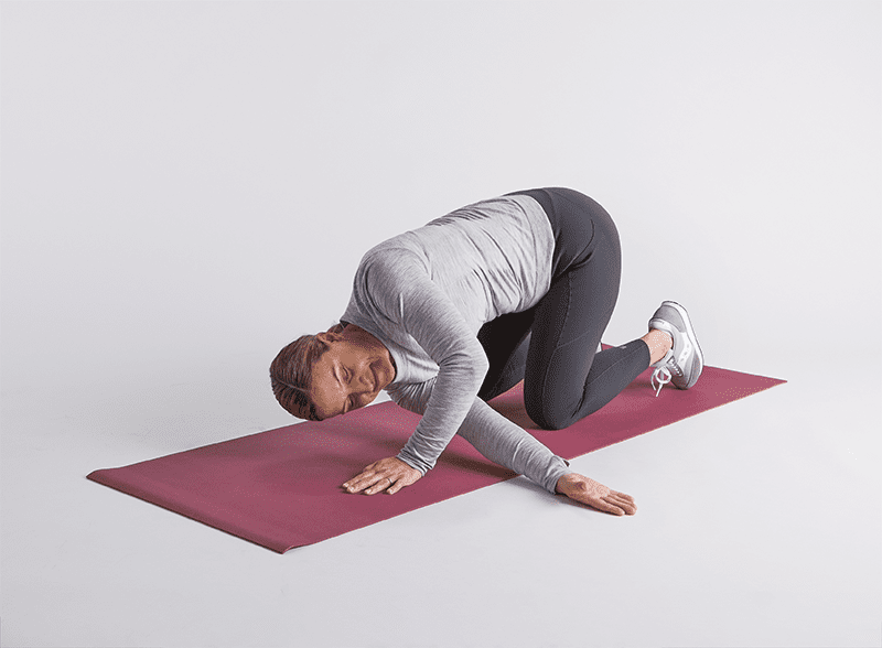
Thread The Needle Pose
The thread-the-needle pose helps release tension stored in the neck, shoulders, and back.
On a yoga mat, start in an all-fours position with the wrist under the shoulders and the knees under the hips.
Lift the right hand and move it to the left along the floor with the palm facing up.
Hold the position for three to five breaths for thirty seconds and release.
Return to the all-fours position and repeat to the left side.
Conclusion
Overall, incorporating yoga as part of a daily routine can provide beneficial results in reducing neck pain and its associated comorbidities. Yoga does not require hours of practice or even contorting into various poses, as just a few minutes of gentle stretching and mindful breathing each day can provide positive results. When people start to utilize yoga as part of their daily activities, they will notice their posture improving, their minds clearer than ever, and live a happier, healthier life without dealing with neck pain.
References
Ben Ayed, H., Yaich, S., Trigui, M., Ben Hmida, M., Ben Jemaa, M., Ammar, A., Jedidi, J., Karray, R., Feki, H., Mejdoub, Y., Kassis, M., & Damak, J. (2019). Prevalence, Risk Factors and Outcomes of Neck, Shoulders and Low-Back Pain in Secondary-School Children. J Res Health Sci, 19(1), e00440. www.ncbi.nlm.nih.gov/pubmed/31133629
Childress, M. A., & Stuek, S. J. (2020). Neck Pain: Initial Evaluation and Management. American Family Physician, 102(3), 150-156. www.ncbi.nlm.nih.gov/pubmed/32735440
Gandolfi, M. G., Zamparini, F., Spinelli, A., & Prati, C. (2023). Asana for Neck, Shoulders, and Wrists to Prevent Musculoskeletal Disorders among Dental Professionals: In-Office Yoga Protocol. J Funct Morphol Kinesiol, 8(1). doi.org/10.3390/jfmk8010026
Kazeminasab, S., Nejadghaderi, S. A., Amiri, P., Pourfathi, H., Araj-Khodaei, M., Sullman, M. J. M., Kolahi, A. A., & Safiri, S. (2022). Neck pain: global epidemiology, trends and risk factors. BMC Musculoskelet Disord, 23(1), 26. doi.org/10.1186/s12891-021-04957-4
Raja, G. P., Bhat, N. S., Fernandez-de-Las-Penas, C., Gangavelli, R., Davis, F., Shankar, R., & Prabhu, A. (2021). Effectiveness of deep cervical fascial manipulation and yoga postures on pain, function, and oculomotor control in patients with mechanical neck pain: study protocol of a pragmatic, parallel-group, randomized, controlled trial. Trials, 22(1), 574. doi.org/10.1186/s13063-021-05533-w
Thanasilungkoon, B., Niempoog, S., Sriyakul, K., Tungsukruthai, P., Kamalashiran, C., & Kietinun, S. (2023). The Efficacy of Ruesi Dadton and Yoga on Reducing Neck and Shoulder Pain in Office Workers. Int J Exerc Sci, 16(7), 1113-1130. www.ncbi.nlm.nih.gov/pubmed/38287934
Individuals suffering from a jammed finger: Can knowing the signs and symptoms of a finger that is not broken or dislocated allow for at-home treatment and when to see a healthcare provider?
Jammed Finger Injury
A jammed finger, also known as a sprained finger, is a common injury when the tip of a finger is forcefully pushed toward the hand, causing the joint to become compressed. This can cause pain and swelling in one or more fingers or finger joints and cause ligaments to stretch, sprain, or tear. (American Society for Surgery of the Hand. 2015) A jammed finger can often heal with icing, resting, and taping. This is often enough to allow it to heal in a week or two if no fractures or dislocations are present. (Carruthers, K. H. et al., 2016) While painful, it should be able to move. However, if the finger cannot wiggle, it may be broken or dislocated and require X-rays, as a broken finger or joint dislocation can take months to heal.
Treatment
Treatment consists of icing, testing, taping, resting, seeing a chiropractor or osteopath, and progressive regular use to regain strength and ability.
Ice
The first step is icing the injury and keeping it elevated.
Use an ice pack or a bag of frozen vegetables wrapped in a towel.
Ice the finger in 15-minute intervals.
Take the ice off and wait until the finger returns to its normal temperature before re-icing.
Do not ice a jammed finger for over three 15-minute intervals in one hour.
Try To Move The Affected Finger
If the jammed finger does not move easily or the pain gets worse when trying to move it, you need to see a healthcare provider and have an X-ray to check for a bone fracture or dislocation. (American Society for Surgery of the Hand. 2015)
Try to move the finger slightly after swelling, and the pain subsides.
If the injury is mild, the finger should move with little discomfort for a short time.
Tape and Rest
If the jammed finger is not broken or dislocated, it can be taped to the finger next to it to keep it from moving, known as buddy taping. (Won S. H. et al., 2014)
Medical-grade tape and gauze between the fingers should be used to prevent blisters and moisture while healing.
A healthcare provider may suggest a finger splint to keep the jammed finger lined up with the other fingers.
A splint can also help prevent a jammed finger from re-injury.
Resting and Healing
A jammed finger must be kept still to heal at first, but eventually, it needs to move and flex to build strength and flexibility.
Targeted physical therapy exercises can be helpful for recovery.
A primary care provider might be able to refer a physical therapist to ensure the finger has a healthy range of motion and circulation as it heals.
A chiropractor or osteopath can also provide recommendations for helping rehabilitate the finger, hand, and arm to normal function.
Easing The Finger Back to Normal
Depending on the extent of the injury, the finger and hand can be sore and swollen for a few days or weeks.
It can take some time to start feeling normal.
Once the healing process begins, individuals will want to return to using it normally.
Avoiding using a jammed finger will cause it to lose strength, which can, over time, further weaken it and increase the risk of re-injury.
If the pain and swelling persist, see a healthcare provider to get it checked for a possible fracture, dislocation, or other complication as soon as possible, as these injuries are harder to treat if the individual waits too long. (University of Utah Health, 2021)
At Injury Medical Chiropractic and Functional Medicine Clinic, we passionately focus on treating patients’ injuries and chronic pain syndromes and improving ability through flexibility, mobility, and agility programs tailored to the individual. Our providers use an integrated approach to create personalized care plans that include Functional Medicine, Acupuncture, Electro-Acupuncture, and Sports Medicine protocols. Our goal is to relieve pain naturally by restoring health and function to the body. If the individual needs other treatment, they will be referred to a clinic or physician best suited for them. Dr. Jimenez has teamed up with the top surgeons, clinical specialists, medical researchers, and premier rehabilitation providers to provide the most effective clinical treatments.
Carruthers, K. H., Skie, M., & Jain, M. (2016). Jam Injuries of the Finger: Diagnosis and Management of Injuries to the Interphalangeal Joints Across Multiple Sports and Levels of Experience. Sports health, 8(5), 469–478. doi.org/10.1177/1941738116658643
Won, S. H., Lee, S., Chung, C. Y., Lee, K. M., Sung, K. H., Kim, T. G., Choi, Y., Lee, S. H., Kwon, D. G., Ha, J. H., Lee, S. Y., & Park, M. S. (2014). Buddy taping: is it a safe method for treatment of finger and toe injuries?. Clinics in orthopedic surgery, 6(1), 26–31. doi.org/10.4055/cios.2014.6.1.26
How do healthcare professionals in a chiropractic clinic provide a clinical approach to preventing medical errors for individuals in pain?
Introduction
Medical errors resulted in 44,000–98,000 hospitalized American deaths annually, and many more caused catastrophic injuries. (Kohn et al., 2000) This was more than the number of people who died annually from AIDS, breast cancer, and auto accidents at the time. According to later research, the actual number of deaths may be closer to 400,000, placing medical errors as the third most common cause of death in the US. Frequently, these mistakes are not the product of medical professionals who are inherently bad; rather, they are the outcome of systemic issues with the health care system, such as inconsistent provider practice patterns, disjointed insurance networks, underutilization or absence of safety protocols, and uncoordinated care. Today’s article looks at the clinical approach to preventing a medical error in a clinical setting. We discuss associated medical providers specializing in various pretreatments to aid individuals suffering from chronic issues. We also guide our patients by allowing them to ask their associated medical providers very important and intricate questions. Dr. Alex Jimenez, DC, only utilizes this information as an educational service. Disclaimer
Defining Medical Errors
Determining what medical error is the most crucial step in any conversation about preventing medical errors. You might assume this is a very easy chore, but that is only until you delve into the vast array of terminology utilized. Many terms are used synonymously (sometimes mistakenly) since some terminology is interchangeable, and occasionally, the meaning of a term depends on the specialty being discussed.
Even though the healthcare sector stated that patient safety and eliminating or reducing medical errors were priorities, Grober and Bohnen noted as recently as 2005 that they had fallen short in one crucial area: determining the definition of “perhaps the most fundamental question… What is a medical error? A medical error is a failure to complete a planned action in a medical setting. (Grober & Bohnen, 2005) However, none of the terms that one would often identify expressly with a medical error—patients, healthcare, or any other element—are mentioned in this description. Despite this, the definition offers a solid framework for further development. As you can see, that specific definition consists of two parts:
An execution error: A failure to complete a planned action as intended.
A planning error: is a technique that, even with perfect execution, does not produce the desired results.
The concepts of faults of execution and planning errors are insufficient if we are to define a medical error adequately. These may occur anywhere, not only at a medical establishment. The component of medical management must be added. This brings up the idea of unfavorable occurrences, known as adverse events. The most common definition of an adverse event is unintentional harm to patients brought about by medical therapy rather than their underlying disease. This definition has gained international acceptance in one way or another. For example, in Australia, the term incidents are defined as in which harm resulted in a person receiving health care. These consist of infections, injury-causing falls, and issues with prescription drugs and medical equipment. Certain unfavorable occurrences might be avoidable.
Common Types of Medical Errors
The only issue with this notion is that not all negative things happen accidentally or intentionally. Because the patient may ultimately benefit, an expected but tolerated adverse event may occur. During chemotherapy, nausea and hair loss are two examples. In this instance, refusing the recommended treatment would be the only sensible approach to prevent the unpleasant consequence. We thus arrive at the concept of preventable and non-preventable adverse occurrences as we further refine our definition. It isn’t easy to categorize a choice to tolerate one impact when it is determined that a favorable effect will occur simultaneously. But purpose alone isn’t necessarily an excuse. (Patient Safety Network, 2016, para.3) Another example of a planned mistake would be a right foot amputation due to a tumor on the left hand, which would be accepting a known and predicted unfavorable event in the hopes of a beneficial consequence where none has ever arisen before. There is no evidence to support the anticipation of a positive outcome.
Medical errors that cause harm to the patient are typically the focus of our research. Nonetheless, medical mistakes can and do occur when a patient is not harmed. The occurrence of near misses could provide invaluable data when planning how to reduce medical errors in a healthcare facility. Still, the frequency of these events compared to the frequency clinicians report them needs to be investigated. Near misses are medical errors that could have caused harm but did not to the patient, even if the patient is doing well. (Martinez et al., 2017) Why would you acknowledge something that could potentially result in legal action? Consider the scenario where a nurse, for whatever reason, had just been looking at photographs of different medications and was about to provide a medication. Maybe something lingers in her memory, and she decides that’s not how a specific medication looks. Upon checking, she found that the incorrect medicines had been administered. After checking all the paperwork, she fixes the mistake and gives the patient the right prescription. Would it be possible to avoid an error in the future if the administration record included photographs of the proper medication? It is easy to forget that there was a mistake and a chance for harm. That fact remains true regardless of whether we were fortunate enough to find it in time or suffer any negative consequences.
Errors of Outcomes & Process
We need complete data to develop solutions that improve patient safety and decrease medical errors. At the very least, when the patient is in a medical facility, everything that can be done to prevent harm and put them in danger should be reported. Many doctors have determined that using the phrases errors and adverse events was more comprehensive and suitable after reviewing mistakes and adverse events in health care and discussing their strengths and weaknesses in 2003. This combined definition would increase data gathering, including mistakes, close calls, near misses, andactive and latent errors. Additionally, the term adverse events includes terms that usually imply patient harm, such as medical injury and iatrogenic injury. The only thing that remains is determining whether a review board is a suitable body to handle the separation of preventable and non-preventable adverse events.
A sentinel event is an occurrence where reporting to the Joint Commission is required. The Joint Commission states that a sentinel event is an unexpected occurrence involving a serious physical or psychological injury. (“Sentinel Events,” 2004, p.35) There isn’t a choice, as it needs to be documented. Most healthcare facilities, however, do keep their records outlining sentinel incidents and what to do in the event of one to guarantee that the Joint Commission standards are met. This is one of those situations when it’s better to be safe than sorry. Since “serious” is a relative concept, there may be some wriggle room when defending a coworker or an employer. On the other hand, reporting a sentinel event incorrectly is better than failing to report a sentinel event. Failing to disclose can have serious consequences, including career termination.
When considering medical errors, people frequently make the mistake of focusing just on prescription errors. Medication errors are undoubtedly frequent and involve many of the same procedural flaws as other medical errors. Breakdowns in communication, mistakes made during prescription or dispensing, and many other things are possible. But we would be gravely misjudging the issue if we assumed that drug errors are the only cause of harm to a patient. One major challenge in classifying the different medical errors is determining whether to classify the error based on the procedure involved or the consequence. It is acceptable to examine those classifications here, given numerous attempts have been made to develop working definitions that incorporate both the process and the outcome, many of which are based on Lucian Leape’s work from the 1990s.
Enhance Your Lifestyle Today- Video
Analyzing & Preventing Medical Errors
Operative and nonoperative were the two main categories of adverse events that Leape and his colleagues distinguished in this study. (Leape et al., 1991) Operative problems included wound infections, surgical failures, non-technical issues, late complications, and technical difficulties. Nonoperative: headings such as medication-related, misdiagnosed, mistreated, procedure-related, fall, fracture, postpartum, anesthesia-related, neonatal, and a catch-all heading of the system were included under this category of adverse occurrences. Leape also classified errors by pointing out the point of process breakdown. He also categorized these into five headings, which include:
System
Performance
Drug Treatment
Diagnostic
Preventative
Many process faults fall under more than one topic, yet they all help to pinpoint the exact cause of the issue. If more than one physician was engaged in determining the precise areas that need improvement, then additional questioning might be required.
Technically, a medical error can be made by any staff member at a hospital. It is not limited to medical professionals like physicians and nurses. An administrator may unlatch a door, or a cleaning crew member could leave a chemical within a child’s grasp. What matters more than the identity of the perpetrator of the mistake is the reason behind it. What before it? And how can we make sure that doesn’t occur again? After gathering all the above data and much more, it’s time to figure out how to prevent similar errors. As for sentinel events, the Joint Commission has mandated since 1997 that all of these incidents undergo a procedure called Root Cause Analysis (RCA). However, using this procedure for incidents that need to be reported to outside parties would need to be corrected.
What Is A Root Cause Analysis?
RCAs “captured the details as well as the big picture perspective.” They make evaluating systems easier, analyzing whether remedial action is necessary, and tracking trends. (Williams, 2001) What precisely is an RCA, though? By examining the events that led to the error, an RCA can focus on events and processes rather than reviewing or placing blame on specific people. (AHRQ,2017) This is why it is so crucial. An RCA frequently makes use of a tool called the Five Whys. This is the process of continuously asking yourself “why” after you believe you have determined the cause of an issue.
The reason it’s called the “five whys” is because, while five is an excellent starting point, you should always question why until you identify the underlying cause of the problem. Asking why repeatedly could reveal many process faults at different stages, but you should keep asking why about every aspect of the issue until you run out of other things that could be adjusted to provide a desirable result. However, different tools besides this one can be utilized in a root cause investigation. Numerous others exist. RCAs must be multidisciplinary and consistent and involve all parties involved in the error to avoid misunderstandings or inaccurate reporting of occurrences.
Conclusion
Medical errors in healthcare institutions are frequent and mostly unreported events that seriously threaten patients’ health. Up to a quarter of a million individuals are thought to pass away each year as a result of medical blunders. These statistics are unacceptable in a time when patient safety is supposedly the top priority, but not much is being done to alter practices. If medical errors are accurately defined and the root cause of the problem is found without assigning blame to specific staff members, this is unnecessary. Essential changes can be made when fundamental causes of system or process faults are correctly identified. A consistent, multidisciplinary approach to root cause analysis that uses frameworks like the five whys to delve down until all issues and defects are revealed is a helpful tool. Although it is now necessary for the wake of sentinel events, the Root Cause Analysis may and should be applied to all mistake causes, including near misses.
Kohn, L. T., Corrigan, J., Donaldson, M. S., & Institute of Medicine (U.S.). Committee on Quality of Health Care in America. (2000). To err is human : building a safer health system. National Academy Press. books.nap.edu/books/0309068371/html/index.html
Leape, L. L., Brennan, T. A., Laird, N., Lawthers, A. G., Localio, A. R., Barnes, B. A., Hebert, L., Newhouse, J. P., Weiler, P. C., & Hiatt, H. (1991). The nature of adverse events in hospitalized patients. Results of the Harvard Medical Practice Study II. N Engl J Med, 324(6), 377-384. doi.org/10.1056/NEJM199102073240605
Martinez, W., Lehmann, L. S., Hu, Y. Y., Desai, S. P., & Shapiro, J. (2017). Processes for Identifying and Reviewing Adverse Events and Near Misses at an Academic Medical Center. Jt Comm J Qual Patient Saf, 43(1), 5-15. doi.org/10.1016/j.jcjq.2016.11.001
For individuals who are dealing with constant constipation due to medications, stress, or lack of fiber, can walking exercise help encourage regular bowel movements?
Walking For Constipation Assistance
Constipation is a common condition. Too much sitting, medications, stress, or not getting enough fiber can result in infrequent bowel movements. Lifestyle adjustments can regulate most cases. One of the most effective ways is to incorporate regular moderate-vigorous exercise, encouraging the bowel muscles to contract naturally (Huang, R., et al., 2014). This includes jogging, yoga, water aerobics, and power or brisk walking for constipation alleviation.
The Research
A study analyzed middle-aged obese women who had chronic constipation over a 12-week period. (Tantawy, S. A., et al., 2017)
The first group walked on a treadmill 3 times a week for 60 minutes.
The second group did not engage in any physical activity.
The first group had greater improvement in their constipation symptoms and quality of life assessments.
A gut bacteria imbalance is also linked to constipation issues. Another study focused on the effect of brisk walking versus exercises that strengthened core muscles like planks on intestinal microbiota composition. (Morita, E., et al., 2019) The results showed that aerobic exercises like power/brisk walking can help increase intestinal Bacteroides, an essential part of healthy gut bacteria. Studies have shown a positive effect when individuals engage in at least 20 minutes of brisk walking daily. (Morita, E., et al., 2019)
Exercise Can Help Decrease Colon Cancer Risks
Physical activity can be a significant protective factor in decreasing colon cancer. (National Cancer Institute. 2023) Some estimate the risk reduction to be 50%, and exercise can even help prevent recurrence after a colon cancer diagnosis, also 50% in some studies for patients with stage II or stage III colon cancer. (Schoenberg M. H. 2016)
The best effects were obtained through moderate-intensity exercise, such as power/brisk walking, about six hours per week.
Mortality was reduced by 23% in individuals who were physically active for at least 20 minutes several times a week.
Inactive colon cancer patients who began exercising after their diagnosis had significantly improved outcomes than individuals who remained sedentary, showing that it is never too late to start exercising.(Schoenberg M. H. 2016)
The most active patients had the best outcomes.
Exercise-Related Diarrhea Prevention
Some runners and walkers experience an overly active colon, resulting in exercise-related diarrhea or loose stools, known as runner’s trots. Up to 50% of endurance athletes experience gastrointestinal problems during intense physical activity. (de Oliveira, E. P. et al., 2014) Prevention steps that can be taken include.
Not eating within two hours of exercising.
Avoid caffeine and warm fluids before exercising.
If sensitive to lactose, avoid milk products or use Lactase.
Ensure the body is well-hydrated before exercise.
Hydrating during exercise.
If exercising in the morning:
Drink about 2.5 cups of fluids or a sports drink before bed.
Drink about 2.5 cups of fluids after waking up.
Drink another 1.5 – 2.5 cups of fluids 20-30 minutes before exercising.
Drink 12-16 fluid ounces every 5-15 minutes during exercise.
If exercising for over 90 minutes:
Drink a 12 – 16 fluid-ounce solution containing 30-60 grams of carbohydrates, sodium, potassium, and magnesium every 5-15 minutes.
Professional Help
Periodic constipation may resolve with lifestyle adjustments like increased fiber intake, physical activity, and fluids. Individuals who are experiencing bloody stools or hematochezia, have recently lost 10 pounds or more, have iron deficiency anemia, have positive fecal occult/hidden blood tests, or have a family history of colon cancer need to see a healthcare provider or specialist to perform specific diagnostic tests to ensure there aren’t any underlying issues or serious conditions. (Jamshed, N. et al., 2011) Before engaging in walking for constipation assistance, individuals should consult their healthcare provider to see if it is safe for them.
At Injury Medical Chiropractic and Functional Medicine Clinic, our areas of practice include Wellness & Nutrition, Chronic Pain, Personal Injury, Auto Accident Care, Work Injuries, Back Injury, Low Back Pain, Neck Pain, Migraine Headaches, Sports Injuries, Severe Sciatica, Scoliosis, Complex Herniated Discs, Fibromyalgia, Chronic Pain, Complex Injuries, Stress Management, Functional Medicine Treatments, and in-scope care protocols. We focus on what works for you to achieve improvement goals and create an improved body through research methods and total wellness programs. If other treatment is needed, individuals will be referred to a clinic or physician best suited to their injury, condition, and/or ailment.
Poop Testing: What? Why? and How?
References
Huang, R., Ho, S. Y., Lo, W. S., & Lam, T. H. (2014). Physical activity and constipation in Hong Kong adolescents. PloS one, 9(2), e90193. doi.org/10.1371/journal.pone.0090193
Tantawy, S. A., Kamel, D. M., Abdelbasset, W. K., & Elgohary, H. M. (2017). Effects of a proposed physical activity and diet control to manage constipation in middle-aged obese women. Diabetes, metabolic syndrome and obesity : targets and therapy, 10, 513–519. doi.org/10.2147/DMSO.S140250
Morita, E., Yokoyama, H., Imai, D., Takeda, R., Ota, A., Kawai, E., Hisada, T., Emoto, M., Suzuki, Y., & Okazaki, K. (2019). Aerobic Exercise Training with Brisk Walking Increases Intestinal Bacteroides in Healthy Elderly Women. Nutrients, 11(4), 868. doi.org/10.3390/nu11040868
Schoenberg M. H. (2016). Physical Activity and Nutrition in Primary and Tertiary Prevention of Colorectal Cancer. Visceral medicine, 32(3), 199–204. doi.org/10.1159/000446492
de Oliveira, E. P., Burini, R. C., & Jeukendrup, A. (2014). Gastrointestinal complaints during exercise: prevalence, etiology, and nutritional recommendations. Sports medicine (Auckland, N.Z.), 44 Suppl 1(Suppl 1), S79–S85. doi.org/10.1007/s40279-014-0153-2
Jamshed, N., Lee, Z. E., & Olden, K. W. (2011). Diagnostic approach to chronic constipation in adults. American family physician, 84(3), 299–306.
For individuals looking to improve their fitness health, can a fitness assessment test identify potential areas and help evaluate overall health and physical status?
Fitness Assessment
A fitness test, also known as a fitness assessment, helps evaluate an individual’s overall and physical health. It comprises a series of exercises to design an appropriate exercise program for general health and fitness. (National Strength and Conditioning Association. 2017) Fitness assessment testing benefits include:
Identifying areas that need improvement.
Assisting professionals in understanding what types of exercise are safest and most effective.
Helping measure fitness progress over time.
Allowing for an individualized plan that can help prevent injuries and maintain the body’s overall health.
An assessment can comprise a wide range of tests, including:
Body composition tests.
Cardiovascular stress tests.
Endurance tests.
Range of motion tests.
They are meant to ensure the individual won’t be at risk of injury and provide the trainer with the insights needed to establish clear and effective fitness goals. Individuals who wonder whether fitness testing would benefit them should consult their healthcare provider.
General Health
Before starting a fitness program, it is important to inform the trainer of individual medical history and get the necessary approval from a primary healthcare provider. (Harvard Health Publishing. Harvard Medical School. 2012) Fitness specialists usually use one or more screening tools to help determine individual baseline health.
This may include obtaining vital sign measurements like height and weight, resting heart rate/RHR, and resting blood pressure/RBP. Many trainers will also use a physical activity readiness questionnaire/PAR-Q comprising questions about general health. (National Academy of Sports Medicine. 2020) Among the questions, individuals may be asked about the medications being taken, any problems with dizziness or pain, or medical conditions that may impair their ability to exercise.
Body Composition
Body composition describes total body weight components, including muscles, bones, and fat. The most common methods for estimating body composition include:
Bioelectrical Impedance Analysis – BIA
During BIA, electrical signals are sent from electrodes through the soles of the feet to the abdomen to estimate body composition. (Doylestown Health. 2024)
These measurements use calipers to estimate the amount of body fat in a fold of skin.
Cardiovascular Endurance
Cardiovascular endurance testing, also known as stress testing, measures how efficiently the heart and lungs work to supply oxygen and energy to the body during physical activity. (UC Davis Health, 2024) The three most common tests used include:
12-minute Run Tests
Twelve-minute run tests are performed on a treadmill, and an individual’s pre-exercise heart and respiration rates are compared with post-exercise heart and respiration rates.
Exercise Stress
Exercise stress testing is performed on a treadmill or stationary bike.
It involves using a heart monitor and blood pressure cuff to measure vital signs during exercise.
VO2 Max Testing
Performed on a treadmill or stationary bike.
V02 max testing uses a breathing device to measure the maximum rate of oxygen consumption during physical activity (UC Davis Health, 2024)
Some trainers will incorporate exercises like sit-ups or push-ups to measure response to specific exercises.
These baseline results can be used later to see if health and fitness levels have improved.
Strength and Endurance
Muscle endurance testing measures the length of time a muscle group can contract and release before it fatigues. Strength testing measures the maximal amount of force a muscle group can exert. (American Council on Exercise, Jiminez C., 2018) The exercises used include:
The push-up test.
Core strength and stability test.
Sometimes, a trainer will use a metronome to measure how long the individual can keep up with the rhythm. The results are then compared to individuals of the same age group and sex to establish a baseline level. Strength and endurance tests are valuable as they help the trainer spot which muscle groups are stronger, vulnerable, and need focused attention. (Heyward, V. H., Gibson, A. L. 2014).
Flexibility
Measuring the flexibility of joints is vital in determining whether individuals have postural imbalances, foot instability, or limitations in range of motion. (Pate R, Oria M, Pillsbury L, 2012)
Shoulder Flexibility
Shoulder flexibility testing evaluates the flexibility and mobility of the shoulder joint.
It is performed by using one hand to reach behind the neck, between the shoulders, and the other hand to reach behind the back, toward the shoulders, to measure how far apart the hands are. (Baumgartner TA, PhD, Jackson AS, PhD et al., 2015)
Fitness assessment testing has various benefits. It can help trainers design a personalized workout program, help individuals identify fitness areas that need improvement, measure progress, and add intensity and endurance to their routine, which can help prevent injuries and help maintain overall health. We focus on what works for you and strive to better the body through researched methods and total wellness programs. These natural programs use the body’s ability to achieve improvement goals. Ask a healthcare professional or fitness professional for guidance if you need advice.
Pate R, Oria M, Pillsbury L, (Eds). (2012). Health-related fitness measures for youth: Flexibility. In R. Pate, M. Oria, & L. Pillsbury (Eds.), Fitness Measures and Health Outcomes in Youth. doi.org/10.17226/13483
Can understanding the body’s hinge joints and how they operate help with mobility and flexibility problems and manage conditions for individuals with difficulty fully bending or extending their fingers, toes, elbows, ankles, or knees?
Hinge Joints
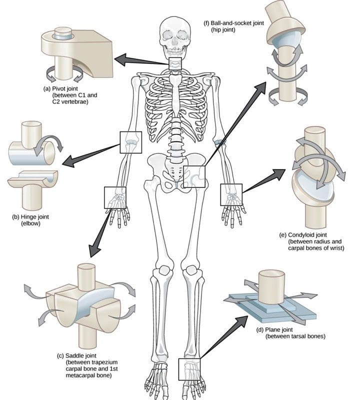
A joint forms where one bone connects to another, allowing motion. Different types of joints differ in structure and movement depending on their location. These include hinge, ball and socket, planar, pivot, saddle, and ellipsoid joints. (Boundless. General Biology, N.D.) Hinge joints are synovial joints that move through one plane of motion: flexion and extension. Hinge joints are found in the fingers, elbows, knees, ankles, and toes and control movement for various functions. Injuries, osteoarthritis, and autoimmune conditions can affect hinge joints. Rest, medication, ice, and physical therapy can help alleviate pain, improve strength and range of motion, and help manage conditions.
Anatomy
A joint is formed by the joining of two or more bones. The human body has three main classifications of joints, categorized by the degree to which they can move. These include: (Boundless. General Biology, N.D.)
Synarthroses
These are fixed, immovable joints.
Formed by two or more bones.
Amphiarthroses
Also known as cartilaginous joints.
A fibrocartilage disc separates the bones that form the joints.
These movable joints allow for a slight degree of movement.
Diarthroses
Also known as synovial joints.
These are the most common freely mobile joints that allow movement in multiple directions.
The bones that form the joints are lined with articular cartilage and enclosed in a joint capsule filled with synovial fluid that allows for smooth motion.
Synovial joints are classified into different types depending on differences in structure and the number of motion planes they allow. A hinge joint is a synovial joint that allows movement in one plane of motion, similar to a door hinge that moves forward and backward. Within the joint, the end of one bone is typically convex/pointed outward, with the other concave/rounded inward to allow the ends to fit smoothly. Because hinge joints only move through one plane of movement, they tend to be more stable than other synovial joints. (Boundless. General Biology, N.D.) Hinge joints include:
The finger and toe joints – allow the fingers and toes to bend and extend.
The elbow joint – allows the elbow to bend and extend.
The knee joint – allows the knee to bend and extend.
The talocrural joint of the ankle – allows the ankle to move up/dorsiflexion and down/plantarflexion.
Hinge joints allow the limbs, fingers, and toes to extend away and bend toward the body. This movement is essential for activities of daily living, such as showering, getting dressed, eating, walking, standing up, and sitting down.
Conditions
Osteoarthritis and inflammatory forms of arthritis can affect any joint (Arthritis Foundation. N.D.) Autoimmune inflammatory forms of arthritis, including rheumatoid and psoriatic arthritis, can cause the body to attack its own joints. These commonly affect the knees and fingers, resulting in swelling, stiffness, and pain. (Kamata, M., Tada, Y. 2020) Gout is an inflammatory form of arthritis that develops from elevated levels of uric acid in the blood and most commonly affects the hinge joint of the big toe. Other conditions that affect hinge joints include:
Injuries to the cartilage within the joints or ligaments that stabilize the outside of the joints.
Ligament sprains or tears can result from jammed fingers or toes, rolled ankles, twisting injuries, and direct impact on the knee.
These injuries can also affect the meniscus, the tough cartilage within the knee joint that helps cushion and absorb shock.
Rehabilitation
Conditions that affect hinge joints often cause inflammation and swelling, resulting in pain and limited mobility.
After an injury or during an inflammatory condition flare-up, limiting active movement and resting the affected joint can reduce increased stress and pain.
Applying ice can decrease inflammation and swelling.
Once the pain and swelling start to subside, physical and/or occupational therapy can help rehabilitate the affected areas.
A therapist will provide stretches and exercises to help improve the joint range of motion and strengthen the supporting muscles.
For individuals experiencing hinge joint pain from an autoimmune condition, biologic medications to decrease the body’s autoimmune activity are administered through infusions delivered every several weeks or months. (Kamata, M., Tada, Y. 2020)
Cortisone injections may also be used to decrease inflammation.
At Injury Medical Chiropractic and Functional Medicine Clinic, we passionately focus on treating patients’ injuries and chronic pain syndromes and improving ability through flexibility, mobility, and agility programs tailored to the individual. Our providers use an integrated approach to create personalized care plans that include Functional Medicine, Acupuncture, Electro-Acupuncture, and Sports Medicine protocols. Our goal is to relieve pain naturally by restoring health and function to the body. If the individual needs other treatment, they will be referred to a clinic or physician best suited for them. Dr. Jimenez has teamed up with the top surgeons, clinical specialists, medical researchers, and premier rehabilitation providers to provide the most effective clinical treatments.
Kamata, M., & Tada, Y. (2020). Efficacy and Safety of Biologics for Psoriasis and Psoriatic Arthritis and Their Impact on Comorbidities: A Literature Review. International journal of molecular sciences, 21(5), 1690. doi.org/10.3390/ijms21051690
IFM's Find A Practitioner tool is the largest referral network in Functional Medicine, created to help patients locate Functional Medicine practitioners anywhere in the world. IFM Certified Practitioners are listed first in the search results, given their extensive education in Functional Medicine