Misalignment of the spine can occur due to various factors, causing stress and joint shifting. The spine plays an important role by supporting the body’s weight and maintaining stability, consisting of vertebrae, facet joints, spinal nerves and cord, and intervertebral discs. The surrounding muscles, tissues, and ligaments protect the spinal cord from damage. However, the spine may develop chronic conditions due to axial load pressure, affecting the body. Fortunately, non-surgical and non-invasive treatments can realign the spine and naturally heal the body. This article discusses spinal subluxation and its symptoms, along with the effectiveness of spinal decompression in alleviating subluxation. We utilize and incorporate valuable information about our patients to certified medical providers using non-surgical therapies like spinal decompression to alleviate pain-like symptoms associated with spinal subluxation. We encourage referring patients to associated medical providers based on their findings while supporting that education is a remarkable tool to ask our providers essential questions at the patient’s request. Dr. Jimenez, D.C., comprises this information as an educational service. Disclaimer
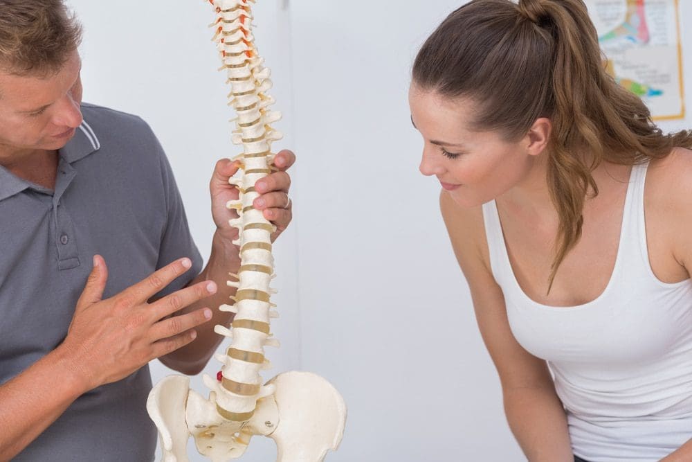
What Is Spinal Subluxation?
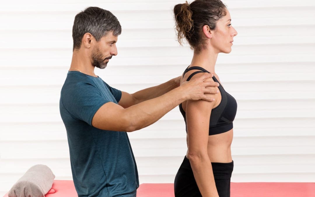
Are you experiencing muscle tightness in your neck, back, or shoulders? Do you feel pain radiating down your arms or legs? Or are you experiencing muscle aches in different parts of your body? These issues may be caused by spinal subluxation, which research shows can occur in the cervical, thoracic, and lumbar spine sections. A spinal subluxation can be caused by traumatic injuries or normal factors that cause the spinal vertebrae to shift out of alignment. This can cause a lot of discomfort. Studies also reveal that spinal subluxation can interfere with neuron communication between the brain and the rest of the body, leading to unwanted symptoms that affect the functioning of the nervous and organ systems and overall health.
Symptoms Associated With Spinal Subluxation
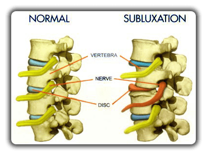
A spinal subluxation happens when the spine shifts out of alignment due to traumatic or normal factors. According to Dr. Eric Kaplan, D.C, FIAMA, and Dr. Perry Bard, D.C., in their book “The Ultimate Spinal Decompression,” biomechanical instability can cause the surrounding muscles and joints to destabilize or increase antagonist coactivation to stabilize the body. Simple movements like bending, twisting, or turning can cause the surrounding muscles to overstretch and make the body feel unstable. Research studies mentioned that displacement in any part of the spinal skeletal frame could press against the surrounding nerves, which can cause neuron signals to be hardwired and create too much or too little communication with the surrounding muscles and joints. Other symptoms associated with spinal subluxation include:
Muscle tightness around the back
Pain and discomfort
Headaches
Limited mobility
Tingling sensations
Digestive and respiratory issues
Low energy
Thoracic Spine Pain- Video
Do you experience pain or discomfort when twisting, turning, or bending? Have you felt muscle aches, pain, or tenderness in your back, or do you feel unsteady when walking? These symptoms may be caused by spinal misalignment or subluxation. Subluxation occurs as pressure compresses spinal discs, causing vertebrae to shift from their normal position. A subluxation can occur in different spine sections, resulting in overlapping risks. This causes pain in various body parts, known as referred pain. Fortunately, non-surgical treatments like chiropractic care and spinal decompression can reduce the effects of subluxation, realign the spine, and promote natural healing for muscles, ligaments, and joints. The video above explains thoracic spine pain symptoms and how manual and mechanical manipulation can alleviate pain-like symptoms, rehydrate spinal discs, and kick-start the body’s natural healing process.
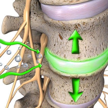
If you suffer from muscle pain associated with spinal subluxation, you can alleviate the associated pain symptoms in several ways. One option is spinal decompression, a non-surgical treatment shown to effectively reduce residual pain and disability, improve range of motion, and modulate neural mechanical sensitivity, as research studies mentioned. Through gentle spine stretching, spinal decompression helps realign the body and allows spinal discs to return to their original position. This, in turn, will enable nutrients, fluids, and oxygenated blood to rehydrate the discs and promote natural healing. For added benefits, spinal decompression can be combined with additional treatments, such as physical therapy and chiropractic care. Best of all, it is a safe and non-invasive treatment allowing individuals to be more mindful of how they move their bodies.
Conclusion
Spinal misalignment or subluxation can occur over time due to traumatic injuries or normal factors. This can cause the spinal vertebrae to shift out of alignment, leading to referred muscle pain and chronic issues that can eventually result in disability. However, non-surgical and non-invasive treatments like spinal decompression use mechanical traction to gently stretch the spine and realign it, releasing the body’s natural healing process. Additionally, non-surgical treatments like spinal decompression help individuals be more mindful of their bodies and prevent new injuries from occurring. Combining spinal decompression with other therapies can promote health and wellness in many individuals.
References
Kaplan, E., & Bard, P. (2023). The Ultimate Spinal Decompression. JETLAUNCH.
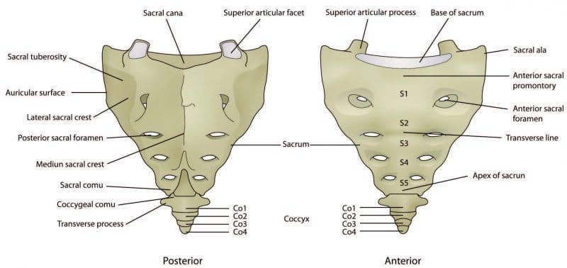
The sacrum and coccyx are part of the vertebral spinal column and could contribute to low back pain. They are not like the other bones in the spinal column. The sacrum, also known as the sacral vertebra, sacral spine, and S1 is a large, flat triangular-shaped bone that is between the hip bones and below the last lumbar vertebra known as L5. The coccyx, known as the tailbone, is positioned below the sacrum.
The sacrum and coccyx are made up of smaller bones that fuse and grow into a solid bone mass by the age of 30. The sacrum is composed of 5 fused vertebrae known as S1-S5 and 3 to 5 smaller bones that fuse creating the coccyx. Both are weight-bearing bones and are integral to walking, standing, and sitting functions.
Sacrum and the Lumbosacral Spine
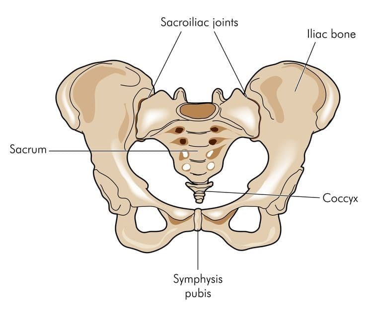
The sacrum forms the back of the pelvis. Along with the coccyx and the two sacroiliac joints make up the pelvic girdle. S1 is at the top of the sacrum and connects to the last lumbar vertebrae L5. Together they create the lumbosacral spine. Where they join forms the lumbosacral curves known as lumbar lordosis and lumbar kyphosis.
The curvature works to support the upper body, weight/force distribution maintains spinal balance and flexibility. Lordosis is the inward curve of the spine, but too much can cause swayback that can be associated with spondylolisthesis. Loss of this curve can cause spinal imbalance and can lead to Flatback syndrome.
Kyphosis is the outward curve of the spine. The location of the sacrum at the intersection of the spine and pelvis means it has an important role in the movement of the low back and hips. The sacrum�s joints help to bear weight and help stabilize the spinal column along with the ligaments, tendons, and muscles help support/stabilize joint movement.
Lumbosacral joint
Joint L5 and S1connect the lumbar spine to the sacrum. The pressure at this meeting point can be massive as the curve of the spine shifts from the lordotic forward curve to a kyphotic backward curve. The L5-S1 region bears weight, absorbs, and distributes the upper body�s weight when moving and resting. Disc herniation and spondylolisthesis are more common at L5-S1 for this reason.
Sacroiliac joints
The sacroiliac joints connect the sacrum to the left and right sides of the pelvis. The range of movement of the sacroiliac joints is minimal compared to other joints like the knees. However, the joints are essential for walking, standing, and stabilization of the hips. Sacroiliitis and sacroiliac joint dysfunction are two spinal disorders related to the joints. Other spinal disorders related to the sacral spine include:
The coccyx commonly known as the tailbone is just below the sacrum. It is smaller than the sacrum and has an important weight-bearing function. It helps supports weight while sitting.An example is leaning back while sitting. This motion and position increase the pressure/weight on the coccyx. An injury in this area can cause tailbone pain. Inflammation of the coccyx�s connective tissue that results in tailbone pain that gets worse when sitting is a common symptom. A traumatic event like a fall or auto accident that causes a tailbone fracture can also cause this pain.
Sacral and Coccygeal Nerves
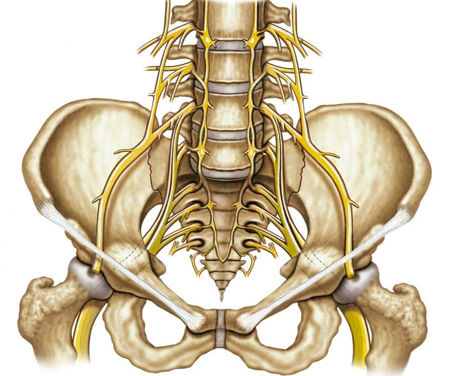
The spinal cord ends at L1-L2, which branches out into the cauda equina, which is a bundle of nerves that looks like a horse’s tail. In the sacrum, there are sacral nerves known as the sacral plexus. Plexus means a network of nerve structures. The sacral and lumbar plexus compose the lumbosacral plexus. This is where the sciatic nerve, which is the largest nerve in the sacral plexusconverges into the band. Sciatic nerve compression causes a combination of symptoms known as sciatica. It is very well known for causing low back and leg pain.
The coccygeal nerve serves the tailbone. There are five sacral nerves numbered S1 through S5 and are part of the spinal cord.
S1 supports groin and hip function
S2 the back of the thighs
S3 the middle of the buttock area
S4 and S5 the anus and vagina
Injury or trauma to the sacral spine can cause mild stress fractures to severe bone fractures. These fractures can cause sacral nerve compression and intense pain. Symptoms include:
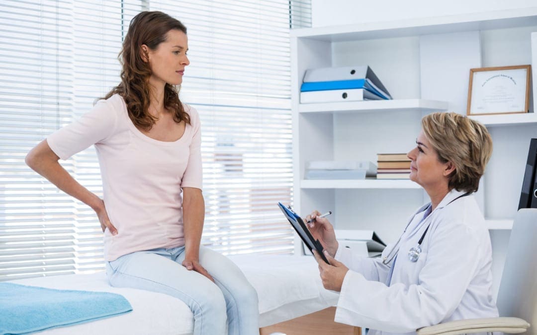
A doctor or chiropractor, physical therapist are excellent sources for information to help prevent sacrum and coccyx pain. These medical professionals will utilize a patient’s medical history, recommend lifestyle changes and injury prevention guidelines.
If at risk of developing osteoporosis then a bone mineral density test could be recommended.
Proper posture must be maintained. Avoid slouching as this places added pressure on the lumbosacral spine and the sacroiliac joints.
Proper body mechanics when engaging in any activity needs to be observed.
Use legstrength to lift objects.
Avoid twisting while lifting or holding heavy objects, as this can cause sprain, strain, or serious injury of the lower spine.
Put on the seat belt. Auto accidents are a major cause of spine trauma. Exercise restraint when driving or riding in any vehicle even a golf cart.
Sciatica Pain Relief
Dr. Alex Jimenez�s Blog Post Disclaimer
The scope of our information is limited to chiropractic, musculoskeletal, physical medicines, wellness, and sensitive health issues and/or functional medicine articles, topics, and discussions. We use functional health & wellness protocols to treat and support care for injuries or disorders of the musculoskeletal system. Our posts, topics, subjects, and insights cover clinical matters, issues, and topics that relate and support directly or indirectly our clinical scope of practice.*
Our office has made a reasonable attempt to provide supportive citations and has identified the relevant research study or studies supporting our posts. We also make copies of supporting research studies available to the board and or the public upon request. We understand that we cover matters that require an additional explanation as to how it may assist in a particular care plan or treatment protocol; therefore, to further discuss the subject matter above, please feel free to ask Dr. Alex Jimenez or contact us at 915-850-0900. The provider(s) Licensed in Texas& New Mexico*
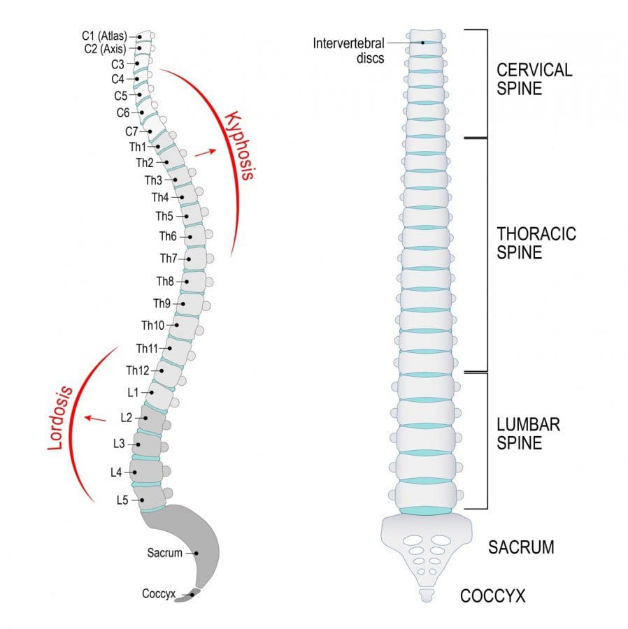
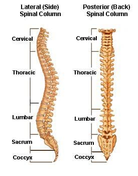
The spinal/vertebral column extends from the skull to the pelvis and consists of individual bones known as vertebrae. It is what holds the body upright, allows the body to bend, twist, and is the conduit for major nerves running from the brain to the rest of the body. The vertebrae are grouped into four regions. They are the:
�
SpinalTerminology
Number of Vertebrae
Area of Body
Abbreviation
Cervical
7
Neck
C1-C7
Thoracic
12
Chest
T1-T12
Lumbar
5-6
Low back
L1-L5
Sacrum
5 fused vertebrae
Pelvis
S1-S5
Coccyx
3
Tailbone
None
�
Cervical Vertebrae
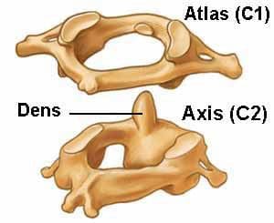
The cervical spine breaks down into two parts. The upper cervical C1 and C2, and the lower cervical C3 through C7. The C1 vertebrae are known as the Atlas and the C2 the Axis. The Occipital Bone is a flat bone that forms the back of the head.
�
Atlas
The Atlas is the first cervical vertebra and is abbreviated as C1. This vertebra supports the skull. It appears different from the other spinal vertebrae, as it resembles a ring and is made up of two masses joined at the front and back by the anterior and posterior arches. �
�
Axis
The Axis is the second cervical vertebra and is abbreviated C2. It is a tooth-like process that projects upward. It is referred to as the odontoid process or dens, which is Latin for tooth. It provides a kind of pivot and collar that allows the head along with the atlas to rotate.
�
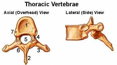
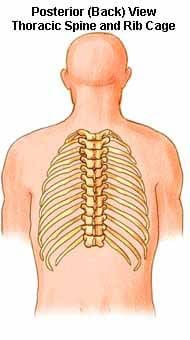
Thoracic Vertebrae
The thoracic vertebrae become larger from T1 through T12. What makes the thoracic spine unique is that it is the only vertebrae that support the ribs and is made up of pedicles, spinous processes, and large neural passageways that help reduce nerve compression. Unfortunately, not everyone has a large intervertebral foramen, which can cause compression. �
�
Vertebral Body
Spinous Process
Transverse Facet
Pedicle
Foramen
Lamina
Superior Facet
The thoracic vertebrae are attached to the ribs. However, at T11 and T12, the ribs are not attached and are called floating ribs. The region of the spine’s range of motion is limited because of the rib/vertebrae attachments and the long spinous processes. �
Lumbar Vertebrae
The lumbar vertebrae increase in size from L1 through L5. These are the vertebrae that take the body’s weight along with any loading force that can create biomechanical stress. The pedicles are longer and wider than the thoracic spine pedicles, and the spinous processes are horizontal and more square. The neural passageway is large but nerve root compression is very common due to disc herniation from poor posture, prolonged sitting, improper lifting, etc. �
�
Vertebrae’s Purpose
The vertebrae range in size with the cervical region being the smallest. The lumbar low back region is the largest. The vertebral bodies of the spinal column are what bear the weight. The body’s upper weight is dispersed through the spine to the sacrum and pelvis. Thee natural curves in the spine provide resistance, flexibility by distributing the body’s weight, and axial loads/forces sustained when in motion. Vertebrae are made up of many elements critical to the overall function of the spine. This includes the intervertebral discs and facet joints. Functions of the spinal/vertebral column include: �
Protection
Spinal Cord Internal Organs
Attachment
Ligaments Muscles Tendons
Support Structure
Head Shoulders Chest Connect Upper and Lower body Balance
The sacrum is located behind the pelvis. It consists of five bones that are abbreviated S1 through S5. They are fused together in a triangular shape. The sacrum fits between the hipbones and connects the spine to the pelvis. The last vertebra L5 moves with the sacrum. Right below are five more bones that are also fused together and they form the Coccyx or tailbone.
�
Intervertebral Discs
The intervertebral discs make up a quarter of the spinal/vertebral column’s length. There are no discs between the Atlas, Axis, and Coccyx. Discs are not connected to the body’s vascular system and so depend on the endplates to disperse essential minerals and nutrients. The cartilaginous layers keep the discs in place. They are fibrocartilaginous cushions that function as the spine/body’s shock absorbers. They protect the vertebrae, brain, nerves, etc. There is some vertebral motion that the discs allow but individual disc movement is limited. Significant motion is possible when the discs work together. �
�
Annulus Fibrosus and Nucleus Pulposus
Intervertebral discs are made up of an annulus fibrosus and a nucleus pulposus. The annulus fibrosus is a strong radial structure made up of lamellae. Concentric sheets of collagen fibers connect to the endplates. These sheets are positioned at various angles. The annulus fibrosus encapsulates the nucleus pulposus. �
�
Both are made up of water, collagen, and proteoglycans. However, the larger amount of water and proteoglycans are in the nucleus pulposus. Proteoglycan molecules are essential because they attract and retain water. The nucleus pulposus consists of a hydrated gel-like substance that resists compression. The amount of water in the nucleus changes throughout the day. This depends on the activity or non-activity. All in all proper care and maintenance of the spinal/vertebral column is vital to general health and overall well-being.
Car Accident Rehabilitation Chiropractor
�
Dr. Alex Jimenez�s Blog Post Disclaimer
The scope of our information is limited to chiropractic, musculoskeletal, physical medicines, wellness, and sensitive health issues and/or functional medicine articles, topics, and discussions. We use functional health & wellness protocols to treat and support care for injuries or disorders of the musculoskeletal system. Our posts, topics, subjects, and insights cover clinical matters, issues, and topics that relate and support directly or indirectly our clinical scope of practice.*
Our office has made a reasonable attempt to provide supportive citations and has identified the relevant research study or studies supporting our posts. We also make copies of supporting research studies available to the board and or the public upon request. We understand that we cover matters that require an additional explanation as to how it may assist in a particular care plan or treatment protocol; therefore, to further discuss the subject matter above, please feel free to ask Dr. Alex Jimenez or contact us at 915-850-0900. The provider(s) Licensed in Texas& New Mexico*
IFM's Find A Practitioner tool is the largest referral network in Functional Medicine, created to help patients locate Functional Medicine practitioners anywhere in the world. IFM Certified Practitioners are listed first in the search results, given their extensive education in Functional Medicine