Can a knee brace relieve discomfort, provide support, and expedite recovery for individuals recovering from an injury or surgery?
Knee Brace
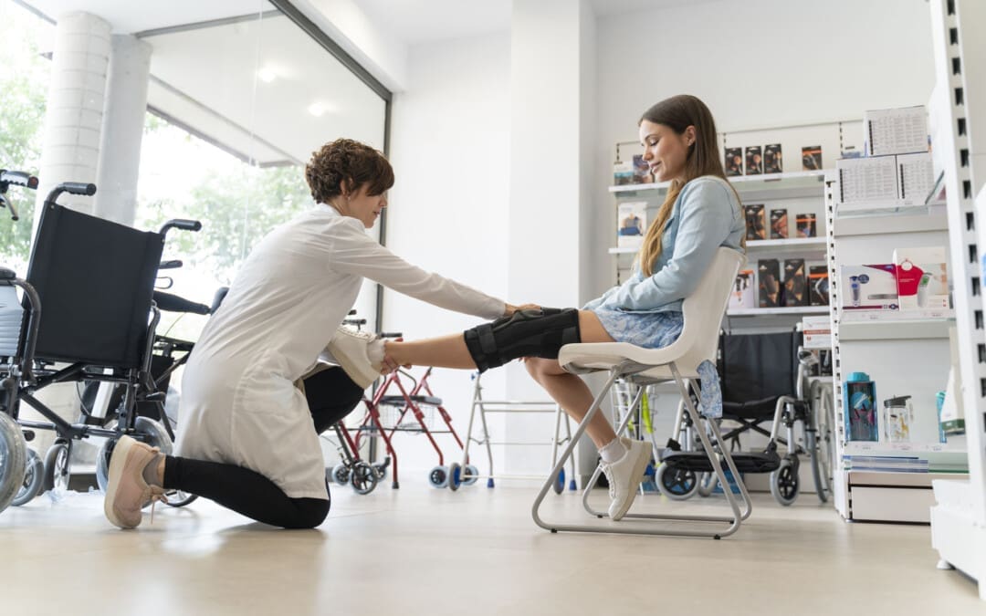
A knee brace is a medical device that supports and stabilizes the knee joint to help with pain and recovery after an injury or surgery. Many knee braces are made of various materials and offer a range of support levels. A healthcare provider or physical therapist can recommend the appropriate one for your condition and suggest the best one. Ask a healthcare provider if you’re unsure, as wearing a knee brace correctly and for the recommended time is important for healing. They are generally safe. However, individuals with health conditions such as poor circulation should be cautious when using them and consult their healthcare provider.
What They Do
The knee joint comprises bones, cartilage, ligaments, tendons, and muscles. A knee brace stabilizes these structures, preventing them from moving too much or too fast. Some braces redistribute the knee joint’s weight, decreasing the force the knee absorbs. (American Academy of Family Physicians, 2020)
Conditions
A knee brace is used after surgery to aid in healing and following an injury. This can be:
Knee braces differ in function and support level. Some stabilize the knee, while others completely immobilize the joint. A healthcare provider and/or physical therapist will explain what support is needed and how to use it. They can also check the brace’s fit and determine if adjustments or a different size are required.
Most Commonly Used
Prophylactic Brace
This is a protective knee pad that shields the kneecap from direct impact.
A knee unloader is typically used to control discomfort due to inflammatory conditions like tendonitis and osteoarthritis.
Functional
This brace limits motion in the joint after an injury or prevents dislocation.
Bledsoe Brace
This brace has straps to wrap around the thigh and shin and support brackets on the inside and outside of the knee joint.
A small mechanism locks the knee into full extension or allows the knee to bend a specific amount.
Knee Immobilizer
A knee immobilizer keeps the knee in one position.
It is a long cloth brace that runs the length of the shin and thigh.
Knee Brace vs Knee Support
A knee support or sleeve is usually a tight-fitting fabric garment. It provides compression to help reduce swelling and discomfort. A knee brace offers more support and can also be set to limit mobility.
Wearing The Brace
Individuals may need to wear a knee brace all day or only when performing specific tasks and operations. It depends on the individual and the condition the brace is being used for. Some may only need to wear a knee brace during certain activities or a flare-up of pain. (Mayo Clinic, 2022) Wearing a brace for unnecessarily long periods can cause skin abrasion, joint stiffness, and muscle atrophy. (American Academy of Family Physicians, 2020) Conversely, neglecting to wear it can cause more susceptibility to injury or extend and or impair healing time. Ask a healthcare provider when you should and should not wear the brace. This could be when:
Sitting
Walking
Driving
Sleeping
Stretching
Contraindications
Some medical conditions can make an individual susceptible to injury and adverse effects from wearing a knee brace. These include: (Holden, M. A. et al., 2021)
Poor circulation
Superficial wounds on the knee
Psoriasis
Eczema
Arterial insufficiency
Severe varicose veins
A history of thrombophlebitis
Injury Medical Chiropractic and Functional Medicine Clinic
If you have one of these conditions, a healthcare provider will decide if a knee brace is safe. Injury Medical Chiropractic and Functional Medicine Clinic works with primary healthcare providers and specialists to build optimal health and wellness solutions. We focus on what works for you to relieve pain, restore function, prevent injury, and help mitigate issues through adjustments that help the body realign itself. They can also work with other medical professionals to integrate a treatment plan to resolve musculoskeletal problems.
Best Knee Injury Chiropractor
References
American Academy of Family Physicians. (2020). Knee Bracing: What Works? https://familydoctor.org/knee-bracing-what-works/
Sprouse, R. A., McLaughlin, A. M., & Harris, G. D. (2018). Braces and Splints for Common Musculoskeletal Conditions. American family physician, 98(10), 570–576.
American Academy of Pediatrics. (2019). Knee pain: how to choose the right knee brace for your child. https://www.healthychildren.org/English/health-issues/injuries-emergencies/sports-injuries/Pages/Knee-Pain-and-braces.aspx
Mayo Clinic. (2022). To brace or not to brace: What’s the best answer? https://www.mayoclinichealthsystem.org/hometown-health/speaking-of-health/to-brace-or-not-to-brace#:~:text=If%20you%20have%20early%20onset%2C%20mild%20arthritis,below%20the%20knee%20for%20compression%20and%20comfort.
Holden, M. A., Callaghan, M., Felson, D., Birrell, F., Nicholls, E., Jowett, S., Kigozi, J., McBeth, J., Borrelli, B., Jinks, C., Foster, N. E., Dziedzic, K., Mallen, C., Ingram, C., Sutton, A., Lawton, S., Halliday, N., Hartshorne, L., Williams, H., Browell, R., … Peat, G. (2021). Clinical and cost-effectiveness of bracing in symptomatic knee osteoarthritis management: protocol for a multicentre, primary care, randomised, parallel-group, superiority trial. BMJ open, 11(3), e048196. https://doi.org/10.1136/bmjopen-2020-048196
Can knowing about wrist sprains—their types, symptoms, causes, and diagnoses—help develop an effective treatment program?
Wrist Sprain
Wrist sprains are injuries that affect ligaments that attach bone to bone. They occur after a fall from work overuse, house tasks, during sports activities, or with other direct trauma. Symptoms of a wrist sprain include:
Pain
Swelling
Bruising
Decreased range of motion
Weakness
Tingling
The injury affects the ligaments and soft tissue structures connecting bone to bone. Mild wrist sprains typically heal within a few weeks; most heal without complications in six to 12 weeks. (National Health Service, 2020) However, severe injuries can require surgery, physical therapy, and months to recover fully.
This joint is between the radius and three small bones in the base of the hand.
The scaphoid
The triquetrum
The lunate
Ulnocarpal
This joint is between the ulna and the articular disc and cushions it from the carpal bones, the lunate, and the triquetrum.
Wrist sprains can affect any of these joints but more commonly affect the ligament between the scaphoid and lunate bone or the triangular fibrocartilage complex/TFCC on the pinky side of the wrist.
Sports include skateboarding, gymnastics, basketball, snowboarding, hockey, and contact sports.
Diagnosis
A healthcare provider will diagnose a wrist sprain based on symptoms and injury causes. X-rays are the first imaging to rule out fractures. Other tests can include:
Magnetic resonance imaging – MRI
Computed tomography – CT scan
Arthrogram -X-rays with contrast dye
Treatment
Nonsteroidal anti-inflammatory drugs, such as Aleve, Advil, Motrin, and aspirin, can treat pain and inflammation. The severity of the wrist sprain determines whether additional treatment is needed. Sprains should initially be treated with the RICE protocol (American Academy of Orthopaedic Surgeons, 2024)
Rest
Minimize using the injured wrist for at least two days.
Wear a splint for support.
Avoid sudden movements.
Avoid placing too much pressure on the wrist.
Ice
Cold packs are recommended several times daily for 20 minutes to decrease pain and swelling.
Compression
Wrap the wrist with an elastic bandage or Kinesio tape to help reduce swelling.
Elevation
To decrease swelling, use pillows to elevate the wrist as much as possible above the level of your heart.
Grade 1 sprains usually heal with basic care within a week or two.
Individuals may need the brace for a week or more.
A healthcare provider may also recommend stretching exercises to overcome stiffness and regain mobility. (American Academy of Orthopaedic Surgeons, 2024) Physical therapy, occupational therapy, or treatment by a certified hand therapist can also reduce pain and improve range of motion and strength.
Treatment for grade 3 sprains often requires surgery. Grade 3 sprains, including avulsion fractures, often require a six-week cast for bones to heal. In some cases, the bones might also need a screw or temporary wires to hold them in the proper position. (Vannabouathong, C. et al., 2018) Severe wrist sprains may also require surgery to repair the injured ligament. If the original ligament cannot be repaired, a piece of the tendon can be used to reconstruct it. (American Society for Surgery of the Hand, 2020)
Healing Time
Mild to moderate sprains usually recover within a few weeks without long-term complications. (American Society for Surgery of the Hand, 2018) The prognosis for severe wrist sprains improves with early diagnosis and treatment. After surgery, ligaments usually heal within eight to 12 weeks but can take six to 12 months for function to return to normal. (American Academy of Orthopaedic Surgeons, 2024)
Injury Medical Chiropractic and Functional Medicine Clinic
Injury Medical Chiropractic and Functional Medicine Clinic works with primary healthcare providers and specialists to build optimal health and wellness solutions. We focus on what works for you to relieve pain, restore function, prevent injury, and help mitigate issues through adjustments that help the body realign itself. They can also work with other medical professionals to integrate a treatment plan to resolve musculoskeletal problems.
The Path to Healing Personal Injury
References
National Health Service. (2020). Advice after spraining your wrist. https://www.ruh.nhs.uk/patients/patient_information/ORT_057_Advice_after_a_wrist_sprain.pdf
American Academy of Orthopaedic Surgeons. (2024). Wrist sprains. https://orthoinfo.aaos.org/en/diseases–conditions/wrist-sprains
Mass General Brigham. (2025). Wrist sprains. https://www.massgeneralbrigham.org/en/patient-care/services-and-specialties/sports-medicine/conditions/hand-arm/wrist-sprain
American Society for Surgery of the Hand. (2017). Anatomy 101: Wrist joints. https://www.assh.org/handcare/blog/anatomy-101-wrist-joints
National Library of Medicine. (2021). Wrist injuries and disorders. Retrieved from https://medlineplus.gov/wristinjuriesanddisorders.html
American Society for Surgery of the Hand. (2018). Sprained wrist. https://www.assh.org/handcare/condition/sprained-wrist
Vannabouathong, C., Ayeni, O. R., & Bhandari, M. (2018). A Narrative Review on Avulsion Fractures of the Upper and Lower Limbs. Clinical medicine insights. Arthritis and musculoskeletal disorders, 11, 1179544118809050. https://doi.org/10.1177/1179544118809050
American Society for Surgery of the Hand. (2020). Scapholunate torn ligament. https://www.assh.org/handcare/condition/scapholunate-torn-ligament
Can incorporating an exercise program like the Alfredson Protocol help athletes and individuals who have hurt their Achilles tendon find pain relief and healing so they can return to regular physical activities?
Exercise Protocol Achilles Tendonitis
Achilles tendonitis occurs when the tendon at the back of the ankle gets injured. It is common in runners. For individuals who have Achilles tendonitis, walking and running can be painful. You might have to stop engaging in exercise and physical activities like sports. Depending on your job, having the condition may make working harder. Here are a few of the signs and symptoms of the condition:
Pain in the back of the lower leg, just above the heel.
Pain with running, jumping, or pointing the toes.
A small lump on the Achilles tendon just above the heel.
The first line of treatment is to rest and ice the tendon. Anti-inflammatory medications can help reduce pain. (American Academy of Orthopaedic Surgeons, 2022) Physical therapy can include strengthening exercises, ultrasound heat therapy, and deep massage. Exercises stretching the nearby muscles will help gradually increase the stress the tendon can handle, eventually reducing inflammation and swelling. Stretching and flexibility exercises will help an Achilles tendon heal. (University of Michigan, 2023)
The only way to determine if an individual has injured their Achilles tendon is to see a doctor. If the injury is Achilles tendonitis, a physical therapist may be recommended. A physical therapist can train individuals on the Alfredson protocol, an exercise protocol program for those with Achilles tendonitis (tendinopathy) that research has shown is helpful for those with the condition. The therapist will train on how to exercise to strengthen the tendon. The exercises stretch the Achilles tendon to help it handle forces and stress, known as eccentric loading. (Stevens M., & Tan C. W. 2014)
Inflammation
Tendonitis is inflammation of a tendon. However, studies have shown that the tendon might not be inflamed in those with the condition. When an area of the body is inflamed, inflammatory cells are present. Individuals usually feel pain in the inflamed area. For those with Achilles tendonitis, the tendon will present with pain, but not necessarily because the tendon is inflamed. Under a microscope, researchers examined tissue from the tendons of those with Achilles tendonitis. They did not find inflammatory cells in the tissue. (Stevens M., & Tan C. W. 2014) This means that although individuals felt pain, they were not inflamed. If there are no inflammatory cells in the tendon, this could explain why those with Achilles tendonitis often do not find relief from the anti-inflammatory treatment of non-steroidal anti-inflammatory drugs (NSAIDs). Studies have shown that gentle exercise protocols for the tendon are more helpful. However, researchers are not sure why these exercises are so beneficial. (O’Neill S., Watson P. J., & Barry S. 2015)
Eccentric Exercise
A chiropractic physical therapy team can help individuals heal the injury with eccentric loading exercises. Eccentric loading exercises work the muscles and tendons to help them get stronger. Once healing has begun, they can help strengthen the tendon. Individuals start slowly with easy exercises and then work up to harder ones. They will have the patient lengthen or stretch out the muscle. As the patient moves, the muscles and tendons contract or shorten. The Alfredson protocol consists of eccentric loading exercises for the Achilles and the muscles that support it.
Alfredson Protocol
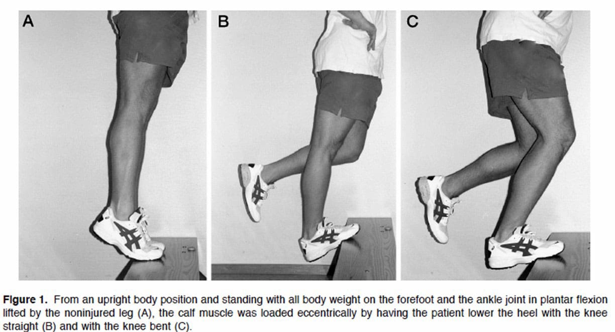
Before exercising, talk to a doctor or physical therapist to know if it’s safe. How to do the Alfredson protocol:
First, stand on a small step or curb.
Stand with the balls of your feet on the edge.
Your heels should hang over the edge.
Hold onto something for balance.
Keep the knees straight.
This will load a muscle part of the Achilles tendon called the gastrocnemius.
Using both feet, lift the heels and rise onto the balls of the feet.
Keep the foot with the painful Achilles tendon on the step.
Lift the non-injured foot off the step.
Slowly lower down using the injured ankle.
The heel should move towards the floor.
The ball of the foot should remain in contact with the edge of the step.
Return the non-injured foot to the step.
Repeat the exercise.
Do three sets of 15 reps with the knees straight. Then, do the Alfredson protocol again with the knees slightly bent. This will work a muscle called the soleus, which connects to the gastrocnemius. Perform three sets of 15 repetitions. Perform both exercises twice a day. This could be in the morning and the evening. The Alfredson protocol is most beneficial when done for about 12 weeks. (Stevens M., & Tan C. W. 2014)
Injury Medical Chiropractic and Functional Medicine Clinic
The Alfredson exercise protocol can be done at home with a step or raised platform to put the feet on safely. Individuals should consider working with a personal trainer to ensure safety and get the most out of the workouts. Injury Medical Chiropractic and Functional Medicine Clinic works with primary healthcare providers and specialists to build optimal health and wellness solutions. We focus on what works for you to relieve pain, restore function, prevent injury, and help mitigate issues through adjustments that help the body realign itself. They can also work with other medical professionals to integrate a treatment plan to resolve musculoskeletal problems.
American Academy of Orthopaedic Surgeons. OrthoInfo. (2022). Achilles Tendinitis. https://orthoinfo.aaos.org/en/diseases–conditions/achilles-tendinitis/
University of Michigan. (2023). Achilles Tendon Injury: Physical Therapy and Rehab. https://www.uofmhealth.org/health-library/tr2261
Stevens, M., & Tan, C. W. (2014). Effectiveness of the Alfredson protocol compared with a lower repetition-volume protocol for midportion Achilles tendinopathy: a randomized controlled trial. The Journal of orthopaedic and sports physical therapy, 44(2), 59–67. https://doi.org/10.2519/jospt.2014.4720
O’Neill, S., Watson, P. J., & Barry, S. (2015). WHY ARE ECCENTRIC EXERCISES EFFECTIVE FOR ACHILLES TENDINOPATHY?. International journal of sports physical therapy, 10(4), 552–562.
Can Pilates exercise movements be performed in bed for individuals recovering from illness or injury?
Bed Pilates
Pilates exercises can be practiced in bed. Joseph Pilates’ exercises and equipment, such as his patented V-shape bed, were designed to help rehabilitate injured individuals who had to stay on or near a bed. After a healthy night’s sleep, Pilates exercises dynamically stimulate circulation and the nervous system. They can also be used to calm the mind and body before going to bed.
Those with health concerns should check with their healthcare provider before starting any exercise program to ensure safety.
Beginners are recommended to learn the Pilates principles and movement fundamentals.
Here are a few Pilates mat exercises adapted for individuals who need or prefer to exercise in bed. A firm mattress is recommended, as a soft mattress will mess up the correct form, making the exercise ineffective.
Spine Twist
This exercise helps improve the flexibility of the spine and core, making it easier to move around, and supports healthy posture. (Geremia J. M. et al., 2015) Pilates exercises like the spine twist have been found to reduce low back pain and disability. (Notarnicola A. et al., 2014) To perform:
Sit up in bed, torso straight, abs engaged, and inhale.
Exhale as you turn your head and shoulders to the right.
Keep your torso straight, and imagine growing taller through the turn.
Inhale as you return to the starting position.
Exhale and turn to the other side.
Repeat five times on each side.
Tendon Stretch
The tendon stretch helps by improving flexibility in the hamstrings and calves. (Chinnavan E., Gopaladhas S., & Kaikondan P. 2015) To perform:
Sit up straight, legs straight out.
Bring heels together and use a towel to pull your feet towards you while pushing your heels away.
Hold for three seconds.
Next, without the towel, point your toes away.
Hold for another three seconds.
Do ten reps.
Double-Leg Stretch
The double-leg stretch is an intermediate exercise that works the abdominals and the core. If this move feels difficult, start with one leg at a time. Another option is to keep the knees slightly bent instead of extending the legs fully. To perform:
Bring both knees to your chest and, with your hands, press down on the ankles to stretch the lower back.
Pull in the abdomen.
Exhale and extend the arms up and the leg or legs as far out in front as possible.
Hold the position for ten seconds and then release.
Do ten reps.
Pelvic Curl
The pelvic curl builds strength in the lower back and core. To perform:
Bend knees and place feet on the bed hip-distance apart.
Curl your pelvis, squeeze the glutes, and lift your body.
A hip-opening exercise, or frog, can be done while lying down or sitting in bed. Hip openers help keep the spine and hips flexible and in alignment. To perform:
Bring the soles of your feet together and as close to the torso as possible.
Let the knees open up as far as possible, and that feels comfortable.
While in the stretch, breathe in and out as deep as possible.
If sitting, place hands on ankles and use forearms to push down on the knees for added stretch.
Injury Medical Chiropractic and Functional Medicine Clinic
Chiropractic care aims to help individuals improve movement with less pain due to condition, after injury, or surgery. A chiropractic therapy team can assess your condition and develop a customized treatment plan to expedite pain relief and improve mobility. Injury Medical Chiropractic and Functional Medicine Clinic works with primary healthcare providers and specialists to build optimal health and wellness solutions. We focus on what works for you to relieve pain, restore function, prevent injury, and help mitigate issues through adjustments that help the body realign itself. They can also work with other medical professionals to integrate a treatment plan to resolve musculoskeletal problems.
Home Exercises for Pain Relief
References
Geremia, J. M., Iskiewicz, M. M., Marschner, R. A., Lehnen, T. E., & Lehnen, A. M. (2015). Effect of a physical training program using the Pilates method on flexibility in elderly subjects. Age (Dordrecht, Netherlands), 37(6), 119. https://doi.org/10.1007/s11357-015-9856-z
Notarnicola, A., Fischetti, F., Maccagnano, G., Comes, R., Tafuri, S., & Moretti, B. (2014). Daily pilates exercise or inactivity for patients with low back pain: a clinical prospective observational study. European journal of physical and rehabilitation medicine, 50(1), 59–66.
Chinnavan, E., Gopaladhas, S., & Kaikondan, P. (2015). Effectiveness of pilates training in improving hamstring flexibility of football players. Bangladesh Journal of Medical Science, 14(3), 265–269. https://doi.org/10.3329/bjms.v14i3.16322
Athletic individuals must train regularly, eat healthy, and rest properly to recover and perform their best. Is sleep different for athletes?
Athletes and Sleep
Physical activity is an important component of a healthy lifestyle. Regular exercise increases longevity and can also reduce the risk of anxiety and depression and improve sleep (Centers for Disease Control and Prevention, 2024). When one area is lacking for athletes, overall performance can suffer. Evidence shows that more or extended sleep can benefit athletes and their recovery and performance. (Bird, Stephen P. 2013) Recommendations for athletes range between seven and nine hours nightly, and elite athletes are encouraged to get at least nine hours of sleep nightly and to treat sleep as much as athletic training and diet.
Sleep is essential for overall health and well-being for both athletes and non-athletes. Everyone needs sleep to feel restored and function their best daily. (Richard J. Schwab, 2024) Other physical benefits include:
Cardiovascular Recovery
This allows the heart to rest and cells and tissue to be repaired. (MedlinePlus, 2017) This can help the body recover after physical exertion. As an individual progresses through the stages of sleep, the changes in heart rate and breathing throughout the night promote cardiovascular health (National Heart, Lung, and Blood Institute, 2011)
Illness Prevention
The proper amount of sleep helps the body recover from illness. During sleep, the body produces cytokines/hormones that help the immune system fight off infections. These therapeutic effects are important for an athlete’s recovery and performance.
Lack of Sleep Affects Performance
Poor quality and quantity of sleep can lead to several negative effects. Sleep deprivation reduces the ability to react quickly and think clearly. A lack of sleep also increases irritability and risk for anxiety and depression. Sleep-deprived individuals are more likely to make poor decisions and take unnecessary risks. From a physical standpoint, a lack of sleep increases the risk for medical concerns, including type 2 diabetes, high blood pressure, kidney disease, and stroke. When athletes do not receive adequate sleep, it can:
Inhibit Ability
In a study of sleep-deprived male team athletes, average and total sprint times decreased. (Skein, M. et al., 2011)
Decrease Accuracy
In a study, male and female sleep-deprived tennis players had decreased serve accuracy by up to 53% compared to performance after normal sleep. (Reyner L. A. & Horne J. A. 2013)
Cause Quicker Exhaustion
A study of male runners and volleyball players found that both athletes exhausted faster after sleep deprivation. (Azboy O. & Kaygisiz Z. 2009)
Decrease Reaction Time
A study found that lack of sleep adversely affected reaction time in a group of male collegiate athletes. (Taheri M. & Arabameri E. 2012)
Difficulty Learning and Decision Making
A lack of sleep negatively impacts cognitive skills and functions.
Athletes can become distracted, and decisions like passing the ball or going for the smash can be difficult or made too late.
Increases Risk of Injury
Research on middle—and high-school athletes showed that chronic lack of sleep was associated with increased rates of injury. (Milewski M. D. et al., 2014)
Increases The Risk of Illness or Immunosuppression
Poor sleep habits are associated with lower resistance to illness like a cold. (Prather A. A. et al., 2015)
Athletic Sleep Hygiene
Common components to sleep well include:
Avoid alcohol and Caffeine
Before bedtime, these can interrupt sleep or lead to more disturbed sleep.
Have a Wind-Down Routine
Activities such as reading, bathing, or meditating can help the body relax and get ready for sleep.
Reduce Stressors
Not only do mental stressors affect sleep quality, but they also impact performance overall.
Create an Optimal Sleep Environment
A sleeping space should be dark and cool with little to no noise.
The environment should be used only for sleep and sex.
No Electronics Before Bed
This includes TVs, cell phones, and computers.
The blue light that these devices emit can affect circadian rhythm.
Don’t Stay Awake In Bed
If you can’t fall asleep after 20 minutes of trying, get out of bed.
Do a quiet activity in another space until you feel sleepy.
Avoid Overtraining
Keep a consistent training schedule so as not to overexert yourself.
Quick Naps
Keep naps brief. Naps should be longer than an hour and not after 3 p.m.
Injury Medical Chiropractic and Functional Medicine Clinic
The right bed and mattress contribute to overall health and can improve one’s quality of life. Doctor Alexander Jimenez, DC, at Injury Medical Chiropractic and Functional Medicine Clinic, says a healthy mattress can improve sleep, reduce pain, increase energy levels, and elevate mood. Injury Medical Chiropractic and Functional Medicine Clinic works with primary healthcare providers and specialists to develop an optimal health and wellness solution. We focus on what works for you to relieve pain, restore function, prevent injury, and help mitigate the pain through spinal adjustments that help the body realign itself. They can also work with other medical professionals to integrate a treatment plan to resolve musculoskeletal issues.
Lumbar Spine Injuries In Athletes
References
Centers for Disease Control and Prevention. (2024). Benefits of Physical Activity. Retrieved from https://www.cdc.gov/physical-activity-basics/benefits/?CDC_AAref_Val=https://www.cdc.gov/physicalactivity/basics/pa-health/index.htm
Bird, Stephen P. PhD. (2013). Sleep, Recovery, and Athletic Performance: A Brief Review and Recommendations. Strength and Conditioning Journal, 35(5), 43-47. https://doi.org/DOI: 10.1519/SSC.0b013e3182a62e2f
Schwab, R. J. (2024). Overview of Sleep. Merck Manual Consumer Version. https://www.merckmanuals.com/home/brain-spinal-cord-and-nerve-disorders/sleep-disorders/overview-of-sleep
National Library of Medicine. MedlinePlus. (2017). Healthy Sleep Also called: Sleep Hygeine. Retrieved from https://medlineplus.gov/healthysleep.html
National Heart, Lung, and Blood Institute. (2011). Your guide to healthy sleep. Retrieved from https://www.nhlbi.nih.gov/resources/your-guide-healthy-sleep
Skein, M., Duffield, R., Edge, J., Short, M. J., & Mündel, T. (2011). Intermittent-sprint performance and muscle glycogen after 30 h of sleep deprivation. Medicine and science in sports and exercise, 43(7), 1301–1311. https://doi.org/10.1249/MSS.0b013e31820abc5a
Reyner, L. A., & Horne, J. A. (2013). Sleep restriction and serving accuracy in performance tennis players, and effects of caffeine. Physiology & behavior, 120, 93–96. https://doi.org/10.1016/j.physbeh.2013.07.002
Azboy, O., & Kaygisiz, Z. (2009). Effects of sleep deprivation on cardiorespiratory functions of the runners and volleyball players during rest and exercise. Acta physiologica Hungarica, 96(1), 29–36. https://doi.org/10.1556/APhysiol.96.2009.1.3
Taheri, M., & Arabameri, E. (2012). The effect of sleep deprivation on choice reaction time and anaerobic power of college student athletes. Asian journal of sports medicine, 3(1), 15–20. https://doi.org/10.5812/asjsm.34719
Milewski, M. D., Skaggs, D. L., Bishop, G. A., Pace, J. L., Ibrahim, D. A., Wren, T. A., & Barzdukas, A. (2014). Chronic lack of sleep is associated with increased sports injuries in adolescent athletes. Journal of pediatric orthopedics, 34(2), 129–133. https://doi.org/10.1097/BPO.0000000000000151
Prather, A. A., Janicki-Deverts, D., Hall, M. H., & Cohen, S. (2015). Behaviorally Assessed Sleep and Susceptibility to the Common Cold. Sleep, 38(9), 1353–1359. https://doi.org/10.5665/sleep.4968
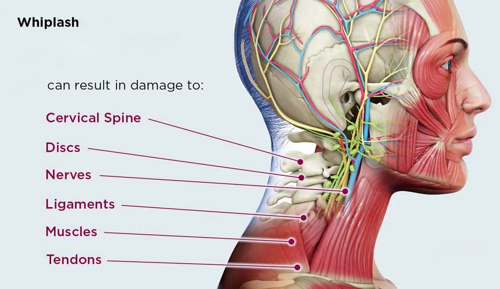
Experiencing a whiplash injury can be disorienting and painful. Can recognizing the signs of more serious complications, seeking timely medical intervention, and following a structured whiplash rehabilitation plan help individuals find relief and restore function and quality of life?
Whiplash Rehabilitation
For individuals who have recently been in an automobile accident, once the initial neck pain and stiffness have passed, it’s crucial to recognize potential delayed symptoms, such as nerve damage. While many recover quickly, some may experience prolonged symptoms requiring more aggressive treatment and management.
Neck Injury
Whiplash injuries result from sudden, forceful whipping back and forth of the head, commonly occurring in rear-end vehicle collisions, and are one of the most common neck injuries. Other potential causes include: (Johns Hopkins Medicine, 2024)
Following a healthcare provider’s recommendations regarding medication use, including dosage and duration, is critical to managing symptoms while effectively minimizing potential side effects.
Rehabilitation Steps
To determine the appropriate treatment whiplash rehabilitation plan, a healthcare provider will assess how much the pain affects daily life, including mental health and the ability to do regular activities (American Academy of Physical Medicine and Rehabilitation, 2024). X-rays or other imaging tests will be done to determine how serious the damage to the neck or spine is. Resting and icing the injured area are recommended to relieve inflammation. A provider may recommend slowly increasing neck movements several times daily and continuing with normal daily activities, as exercise will help maintain flexibility. Not moving the neck may prolong pain, stiffness, and healing. Physical therapy may be recommended if symptoms continue for over one or two weeks. (American Academy of Physical Medicine and Rehabilitation, 2024)
Symptoms last longer than the healthcare provider estimated
Other symptoms develop, like weakness, numbness, or a sensation of pins and needles.
Long-Term Injury Side Effects
Individuals in whiplash rehabilitation usually recover in a few weeks to months, but some may have longer-lasting pain as the injury can cause nerve damage. (Fundaun J. et al., 2022) Discuss any new or worsening symptoms with a healthcare provider for guidance. (Johns Hopkins Medicine, 2024)
Over-the-counter pain relievers, such as acetaminophen or NSAIDs.
Muscle relaxants for tightness and tension.
Prescription pain medications.
If pain becomes chronic, antidepressants such as serotonin and norepinephrine reuptake inhibitors may be prescribed. These medications can help manage pain and improve sleep quality. (Ferreira G. E. et al., 2023) For severe inflammation and pain, corticosteroid injections directly into the affected area can provide relief. Healthcare providers administer these injections, offering long-lasting effects. (Harvard Health Publishing, 2015)
Injury Medical Chiropractic and Functional Medicine Clinic
Navigating a whiplash injury can be challenging, but understanding the whiplash rehabilitation process can significantly aid in recovery. A healthcare provider can determine the most effective treatment strategies. It is important to talk with a healthcare provider to determine the cause and extent of the injury to provide individualized patient education regarding treatment. This can include physical therapy, rest, health coaching, medication, and surgery, which may be recommended in certain cases. Overcoming these limitations is possible. Injury Medical Chiropractic and Functional Medicine Clinic works with primary healthcare providers and specialists to develop an optimal health and wellness solution. We focus on what works for you to relieve pain, restore function, prevent injury, and help mitigate the pain through spinal adjustments that help the body realign itself. They can also work with other medical professionals to integrate a treatment plan to resolve musculoskeletal issues.
American Academy of Physical Medicine and Rehabilitation. (2024). Cervical whiplash. https://www.aapmr.org/about-physiatry/conditions-treatments/musculoskeletal-medicine/cervical-whiplash
Rush University Medical Center. (2014). 5 facts about whiplash. https://www.rush.edu/news/5-facts-about-whiplash
National Library of Medicine. MedlinePlus. (2024). Whiplash. Retrieved from https://medlineplus.gov/ency/imagepages/9853.htm
National Health Service NHS. (2023). Whiplash. https://www.nhs.uk/conditions/whiplash/
University of Rochester Medical Center. (2024). Whiplash injury. https://www.urmc.rochester.edu/encyclopedia/content.aspx?contenttypeid=85&contentid=p01388
National Library of Medicine. MedlinePlus. (2020). Pregabalin. Retrieved from https://medlineplus.gov/druginfo/meds/a605045.html
Fundaun, J., Kolski, M., Baskozos, G., Dilley, A., Sterling, M., & Schmid, A. B. (2022). Nerve pathology and neuropathic pain after whiplash injury: a systematic review and meta-analysis. Pain, 163(7), e789–e811. https://doi.org/10.1097/j.pain.0000000000002509
National Institute of Arthritis and Musculoskeletal and Skin Diseases. (2023). Back pain: diagnosis, treatment, and steps to take. Retrieved from https://www.niams.nih.gov/health-topics/back-pain/diagnosis-treatment-and-steps-to-take
Ferreira, G. E., Abdel-Shaheed, C., Underwood, M., Finnerup, N. B., Day, R. O., McLachlan, A., Eldabe, S., Zadro, J. R., & Maher, C. G. (2023). Efficacy, safety, and tolerability of antidepressants for pain in adults: overview of systematic reviews. BMJ (Clinical research ed.), 380, e072415. https://doi.org/10.1136/bmj-2022-072415
Harvard Health Publishing. (2015). New recommendations aim to improve safety of pain-relieving spinal steroid injections. Harvard Health Publishing
Harvard Medical School. https://www.health.harvard.edu/blog/new-recommendations-aim-to-improve-safety-of-pain-relieving-spinal-steroid-injections-201505077991
Can understanding what knee tests are used help a healthcare provider diagnose the cause of individuals experiencing knee pain?
Knee Pain Tests
A knee examination is the first step in determining the cause of knee pain. Different knee tests may be performed during the exam to help the healthcare provider find the cause and develop an optimal treatment plan. These tests evaluate knee function and range of motion and look for conditions and injuries such as arthritis, meniscus tears, ACL tears, other ligament injuries, and kneecap issues.
Checking If There is Fluid in the Knee
Many individuals know if their knee is swollen, as they can see or feel the swelling. However, if there is excess fluid in the knee joint, the healthcare provider may compress the joint to feel for excess fluid. Fluid is often visible above the kneecap and can be compressed in this area. Fluid may also be detected in the back of the knee, referred to as a Baker’s cyst if the fluid has collected into a cluster. (Frush T. J., & Noyes F. R. 2015)
Arthritis Tests
Certain characteristic findings can detect knee arthritis:
Crepitus
Crepitus is the sensation when rough cartilage or exposed bone is rubbing when the knee is bent. (Lo G. H. et al., 2018)
The examiner will feel and listen for grinding as the knee is bent back and forth.
Deformity
As knee cartilage wears away, the knees can become progressively knock-kneed or bow-legged.
Limited Motion
If arthritis, bone spurs, and swelling prevent normal mobility, the knee’s range of motion often becomes limited.
Torn Meniscus Tests
Tests used to determine if there is a meniscus tear include:
Joint Line Tenderness
Joint line tenderness is a non-specific test in which the area of the meniscus is felt. It is considered a positive test when there is pain in this area.
McMurray’s test
This test is performed with the patient lying flat. The examiner bends the knee and rotates the shin bone.
This test is performed with the patient squatting.
The test is performed with the leg fully externally rotated or internally rotated, depending on whether the lateral or medial meniscus is being tested.
A click is heard or felt over the area of the tear.
ACL Tear Tests
These knee pain tests are for an anterior cruciate ligament (ACL) tear:
Lachman Test
The Lachman test is one of the most reliable to diagnose an ACL tear.
With the knee slightly bent, the examiner stabilizes the thigh while pulling the shin forward.
The shin shifts too far forward with a torn ACL.
Anterior Drawer Test
This test is performed with the patient lying flat.
The knee is bent 90 degrees, and then the shin is pulled forward to check the stability of the ACL.
Pivot Shift Test
The pivot shift test can be difficult, especially if the patient is experiencing discomfort and cannot relax the knee.
This test places stress on the knee joint and assesses the rotational stability of the ACL.
Other Ligament Injuries
For a suspected injury to other ligaments, including the posterior cruciate ligament (PCL), medial collateral ligament (MCL), and lateral collateral ligament (LCL), the following tests may be used:
Posterior Drawer Test
The posterior drawer is performed similarly to the anterior drawer test, in which the patient lies flat.
The knee is bent 90 degrees; the shin is pushed backward to check stability and function and detect if the posterior cruciate ligament (PCL) has been injured.
Collateral Ligament Stability
Side-to-side stability of the knee detects problems with the MCL and LCL.
The shin is shifted to each side, with the patient lying flat and the knee slightly bent.
The LCL or MCL damage causes the knee to open up too much, a condition known as varus (LCL) or valgus (MCL) instability. (Ohori T. et al., 2017)
Kneecap Tests
Tests for kneecap issues include:
Patellar Grind
In this test, also called Clarke’s sign, the patient lies on their back with the leg extended.
The examiner pushes the kneecap down to reproduce the knee pain while the patient flexes the thigh muscles.
Damaged cartilage can cause a grinding sensation/crepitus.
Patellar Tenderness
The examiner can slightly lift the kneecap and place direct pressure on parts of the underside.
The examiner looks for regions of sensitivity or pain.
Patellar Apprehension
This test indicates an unstable kneecap.
The examiner places pressure on the kneecap in a certain direction, and the patient may feel like the kneecap is going to pop out.
Injury Medical Chiropractic and Functional Medicine Clinic
Knee pain tests typically check the range of motion, discomfort symptoms, and sounds that could indicate a specific type of knee injury. Injury Medical Chiropractic and Functional Medicine Clinic works with primary healthcare providers and specialists to develop an optimal health and wellness solution. We focus on what works for you to relieve pain, restore function, and prevent injury. Regarding musculoskeletal pain, specialists like chiropractors, acupuncturists, and massage therapists can help mitigate the pain through spinal adjustments that help the body realign itself. They can also work with other medical professionals to integrate a treatment plan to resolve musculoskeletal issues.
Overcoming an ACL Injury
References
Frush, T. J., & Noyes, F. R. (2015). Baker’s Cyst: Diagnostic and Surgical Considerations. Sports health, 7(4), 359–365. https://doi.org/10.1177/1941738113520130
Lo, G. H., Strayhorn, M. T., Driban, J. B., Price, L. L., Eaton, C. B., & Mcalindon, T. E. (2018). Subjective Crepitus as a Risk Factor for Incident Symptomatic Knee Osteoarthritis: Data From the Osteoarthritis Initiative. Arthritis care & research, 70(1), 53–60. https://doi.org/10.1002/acr.23246
Gupta, Y., Mahara, D., & Lamichhane, A. (2016). McMurray’s Test and Joint Line Tenderness for Medial Meniscus Tear: Are They Accurate?. Ethiopian journal of health sciences, 26(6), 567–572. https://doi.org/10.4314/ejhs.v26i6.10
Ohori, T., Mae, T., Shino, K., Tachibana, Y., Fujie, H., Yoshikawa, H., & Nakata, K. (2017). Varus-valgus instability in the anterior cruciate ligament-deficient knee: effect of posterior tibial load. Journal of experimental orthopaedics, 4(1), 24. https://doi.org/10.1186/s40634-017-0087-3
IFM's Find A Practitioner tool is the largest referral network in Functional Medicine, created to help patients locate Functional Medicine practitioners anywhere in the world. IFM Certified Practitioners are listed first in the search results, given their extensive education in Functional Medicine