For individuals with lower back pain when walking, could they have injured a muscle or have an underlying condition affecting the joints, ligaments, or nerves?
Back Pain When Walking
Lower back pain when walking can occur for a variety of reasons. It can result from poor posture, injuries, muscle fatigue, or an underlying condition. Specific injuries, like muscle strains, can affect the ligaments in the spine and cause pain. Muscles that have not been used often can result in fatigue and pain. Specific health conditions, such as degenerative disc disease, spinal stenosis, herniated discs, sciatica, or even sacroiliac joint dysfunction, can cause lower back pain. Factors like overexertion or improper gait can exacerbate it.
Muscle Issues
Muscle strains, ligament sprains, or fatigue can cause lower back pain when walking. Pain can occur after walking or come on gradually from wear and tear without an apparent cause, as follows (American Association of Neurological Surgeons, 2024)
Strains
Occur when the fibers in the back are overstretched or torn.
Pain from muscle strains is usually worse during activity and better when resting.
Sprains
It occurs when the ligaments that connect bone to bone become detached.
If the muscles do not adequately support the spine, the spinal joints absorb more pressure, which can lead to injury to the spinal ligaments.
Fatigue
It can happen from overexertion and lead to lower back pain when walking.
It could occur when walking longer than the body is used to, on uneven surfaces that make the muscles work harder to help maintain balance, or climbing hills that cause you to lean forward while walking.
Degenerative Disc Disease
Between each vertebra is a disc that provides cushioning between the bones.
As the disc wears down, surrounding muscles, ligaments, joints, and nerves in the spine absorb more pressure, causing damage.
Degenerative disc disease is a wear-and-tear condition that becomes more common as individuals age and is a common cause of lower back pain.
Healthcare providers recommend walking as a low-impact activity for individuals with the disease. However, if the condition is more severe, individuals could experience pain from this exercise, especially when walking on hard surfaces. (Hospital for Special Surgery, 2024)
Sciatica
Sciatica pain occurs when a nerve exiting the spine in the lower back becomes compressed or pinched. It is a common symptom of a herniated disc, in which a disc moves out of place and puts pressure on nearby nerves. In addition to lower back pain, sciatica can cause pain in the hip, the back of the thigh, and down the leg. Sciatica can also cause: (American Academy of Orthopaedic Surgeons, 2021)
Numbness
Tingling
Muscle cramps
Leg muscle weakness
Healthcare providers often recommend walking as a safe form of physical activity for individuals with sciatica. However, individuals should avoid twisting or bending forward. (American Academy of Orthopaedic Surgeons, 2021) To decrease pain, avoid walking on uneven surfaces or uphill.
Lumbar Spinal Stenosis
Spinal stenosis is a wear-and-tear condition that often affects the lumbar spine/five vertebrae in the lower back.
It causes the space surrounding the spinal cord to narrow.
Lumbar spinal stenosis typically causes pain when standing upright, including when walking.
Many with this condition find that leaning slightly forward helps to reduce the pain by opening up the compressed areas.
Hyperlordosis
Lordosis describes the normal curve in the spine in the lower back. However, when this curve is exaggerated, it causes hyperlordosis or swayback. (American Academy of Orthopaedic Surgeons, 2020) Hyperlordosis affects the range of motion, putting abnormal pressure on the muscles, ligaments, and joints. It also reduces the spine’s ability to absorb shock correctly when walking, leading to pain. (Cedars-Sinai, 2025)
Prevention Strategies
Individuals can take steps to reduce their risk of lower back pain when walking, even if they’ve been diagnosed with a condition that can potentially cause this symptom. Walking can decrease chronic low back pain for some. (Suh J. H. et al., 2019) As with any new exercise program, check with a healthcare provider to ensure that walking for exercise is appropriate for the injury, condition, or disease and is safe. Recommended tips: (Harvard Health Publishing, 2015)
Wear shoes made for walking.
Perform gentle lower back stretches before walking.
Start slowly by walking for a few minutes, then gradually increase the time.
Walk on a smooth surface, such as a sidewalk or athletic track, or indoors, such as in a shopping center or mall.
Warm up and cool down by walking slowly at the beginning and end of the walk.
This allows the back and leg muscles to warm up before exercise and recover afterward.
Walk at a slow to moderate pace/speed that allows one to converse.
Standing up straight while walking or standing upright reduces pressure on the lower back.
Alternative Exercise
If there is still back pain when walking, it might not be an appropriate exercise for the individual and/or how their condition presents symptoms. Alternate activities can include: (Hospital for Special Surgery, 2023)
Elliptical Trainer
This exercise keeps the feet in contact with the pedals, putting less shock-absorbing pressure on the spine than walking.
Recumbent Biking
will keep the back upright, which is recommended if there is more pain when bending forward.
Upright Stationary Biking
This is recommended if the back pain improves when bending forward.
Walking In A Pool
This activity provides benefits while reducing pressure on the spine.
To target different muscles, try walking laps in waist-deep water in multiple directions (forward, backward, and side to side).
Water Aerobics
This activity provides cardiovascular health benefits with decreased pressure on the back.
Injury Medical Chiropractic & Functional Medicine Clinic
See a physical therapist for a personalized exercise program to reduce back pain and appropriate for your condition. Injury Medical Chiropractic and Functional Medicine Clinic works with primary healthcare providers and specialists to develop an optimal health and wellness solution. We focus on what works for you to relieve pain, restore function, and prevent injury. Regarding musculoskeletal pain, specialists like chiropractors, acupuncturists, and massage therapists can help mitigate the pain through spinal adjustments that help the body realign itself. They can also work with other medical professionals to integrate a treatment plan to resolve musculoskeletal issues.
Beyond Adjustments: Chiropractic and Integrative Healthcare
References
American Association of Neurological Surgeons. (2024). Low back strain and sprain. https://www.aans.org/patients/conditions-treatments/low-back-strain-and-sprain/
Hospital for Special Surgery. (2024). Degenerative disc disease. https://www.hss.edu/condition-list_degenerative-disc-disease.asp
American Academy of Orthopaedic Surgeons. (2021). Sciatica. https://orthoinfo.aaos.org/en/diseases–conditions/sciatica
American Academy of Orthpaedic Surgeons. (2021). Lumbar spinal stenosis. https://orthoinfo.aaos.org/en/diseases–conditions/lumbar-spinal-stenosis/
American Academy of Orthopaedic Surgeons. (2020). Spine basics. https://orthoinfo.aaos.org/en/diseases–conditions/spine-basics/
Suh, J. H., Kim, H., Jung, G. P., Ko, J. Y., & Ryu, J. S. (2019). The effect of lumbar stabilization and walking exercises on chronic low back pain: A randomized controlled trial. Medicine, 98(26), e16173. https://doi.org/10.1097/MD.0000000000016173
Harvard Health Publishing. (2015). 5 tips for getting started with a walking program. https://www.health.harvard.edu/exercise-and-fitness/get-started
Hospital for Special Surgery. (2023). Best types of exercise for back pain. https://www.hss.edu/article_best-exercise-lower-back-pain.asp
Are there benefits to back cracking, risks, and how can it be done safely?
Back Cracking
Back cracking is intentionally applying pressure or twisting movements, producing a popping or cracking sound in the spine. Back cracking involves stretching or extending the spine. In most cases, it is considered safe when done gently as it can provide temporary relief from back pain and stiffness by:
Stretching the ligaments and muscles around the spine
Releasing gas bubbles that may be causing pressure
Improving joint mobility
Mechanism of Action
Cracking your back creates small gas bubbles in the synovial fluid (the lubricating fluid in the joints).
These bubbles form when the pressure in the joints is suddenly released, causing a popping or cracking sound.
It is generally safe, but there are certain conditions under which individuals should avoid cracking their backs.
Popping Sound
Research has used a new type of magnetic resonance imaging (MRI), cine MRI, to study the noise source. Cine MRI produces moving images.
This study using this MRI found that the formation of bubbles makes a popping sound.
The popping sound does not come from the popping of bubbles in the synovial fluid, as previously believed. (Kawchuk G. N. et al., 2015)
When someone cracks their back, the force pulls the bones of the joint apart, causing the pressure within the joint to drop and form a bubble, which eventually dissipates. (Kawchuk G. N. et al., 2015)
Crepitus
Crepitus is the medical term for cracking or popping noise from joints.
It is not a condition or disease but can be a symptom of one.
Other terms include clicking or crunching.
Is It Safe To Perform Daily?
Back cracking once a day is generally considered safe. But if it causes pain or swelling, then stop and contact a healthcare provider. If someone feels the need to crack their back more throughout the day, it could be a sign that they need to see a professional chiropractic healthcare provider. (AICA Orthopedics, 2022) Individuals may crack their backs to address certain conditions or to relieve various discomfort symptoms that can include: (National Center for Complementary and Integrative Health, 2025)
Headache
Neck pain
Lower back pain
Sciatica
Individuals may often experience mild side effects like headache, stiffness, or pain. These side effects tend to resolve within a day. Though back cracking can provide temporary relief for some conditions, some serious side effects like neurological problems or strokes have been reported. (National Center for Complementary and Integrative Health, 2025)
Rotate the upper body to the right side and press against the right knee with the left elbow.
Hold the stretch for 30 seconds and come back to the center.
Repeat on the other side.
Knee to Chest
Lie flat on the ground.
Lift one leg and bring the knee to the chest, pulling the knee in with your hands.
Hold for five seconds.
Repeat with the other leg.
Several back-cracking assistive devices, such as poles and wheels, are available. Talk to a healthcare provider to determine the right type and ensure it is safe for you and your condition or injury.
Individuals Who Should Avoid Back Cracking
Back cracking can cause additional stress or damage to the joints in those with back injuries or other conditions. Individuals with these conditions should avoid back cracking (AICA Orthopedics, 2022)
Numbness or tingling of the arms or legs.
Osteoporosis
Spinal cancer
Spinal abnormalities
Individuals who have a high stroke risk.
A Professional Back Adjustment
A chiropractor is a healthcare provider who specializes in spine and spinal adjustments. They adjust the spine and other areas of the body to correct misalignment problems, reduce and relieve pain, and allow the body to recover independently. (National Library of Medicine. MedlinePlus, 2023) The chiropractor will take a health history to learn about previous injuries and conditions. Then, they will evaluate the patient and determine the best course of action. Although a chiropractor performs spinal adjustments, they may also incorporate other treatments, including: (National Library of Medicine. MedlinePlus, 2023)
Injury Medical Chiropractic & Functional Medicine Clinic
Injury Medical Chiropractic and Functional Medicine Clinic works with primary healthcare providers and specialists to develop an optimal health and wellness solution. We focus on what works for you to relieve pain, restore function, and prevent injury. Regarding musculoskeletal pain, specialists like chiropractors, acupuncturists, and massage therapists can help mitigate the pain through spinal adjustments that help the body realign itself. They can also work with other medical professionals to integrate a treatment plan to resolve musculoskeletal issues.
Chiropractic Secrets
References
Kawchuk, G. N., Fryer, J., Jaremko, J. L., Zeng, H., Rowe, L., & Thompson, R. (2015). Real-time visualization of joint cavitation. PloS one, 10(4), e0119470. https://doi.org/10.1371/journal.pone.0119470
AICA Orthopedics. (2022). Is cracking your back bad? https://aica.com/is-cracking-your-back-bad/
National Center for Complementary and Integrative Health. (2025). Spinal manipulation: what you need to know. Retrieved from https://www.nccih.nih.gov/health/spinal-manipulation-what-you-need-to-know
American Academy of Orthopedic Surgeons. (2022). Spine conditioning program. https://orthoinfo.aaos.org/en/recovery/spine-conditioning-program/
National Library of Medicine. MedlinePlus. (2023). Chiropractic. Retrieved from https://medlineplus.gov/chiropractic.html
Learn about kyphosis, its causes and symptoms, and treatment approaches to alleviate discomfort and improve posture.
What Is Kyphosis?
How frequently have you seen that after spending too much time sitting down, your posture has become more hunched? Do you have neck and shoulder strains that are momentarily relieved by stretching? Or do you experience shoulder and back discomfort and tension as a result of bad posture? Frequently, people have experienced musculoskeletal problems that may impact the neck, shoulders, and back—the three most frequent parts of the body. A spinal disorder called kyphosis may result from prolonged hunching. An increase in the forward curve of the spine that affects the thoracic location is known as kyphosis, and it may be brought on by degenerative alterations in the intervertebral discs. (Lam & Mukhdomi, 2025) The development of a hunchback or rounded upper back is a symptom of kyphosis that may impact posture and general musculoskeletal health. Depending on how severe the kyphosis is, the symptoms might vary from severe stiffness and pain to trouble breathing. Finding different treatment choices for this spinal ailment may also be made easier for many people by having a better grasp of the environmental variables that contribute to its development and the symptoms that are connected with it.
Environmental Factors Contributing to Kyphosis
Kyphosis may occur as a result of many environmental factors. This is because a lot of individuals engage in physically demanding activities that put a lot of strain on the spine. On the other hand, kyphosis may occur as a result of the spine’s gradual aging process. Among the environmental elements that cause kyphosis are:
Bad Posture
Living a Sedentary Lifestyle
Overweight Items & Inappropriate Lifting
Osteoporosis
Conditions & Injuries of the Spine
Kyphosis Symptoms & Its Effects on the Musculoskeletal System
Because it may impact both the cervical and thoracic regions of the spine, kyphosis can result in a number of musculoskeletal issues.When kyphosis begins to damage the cervical region, it may result in referred neck pain, which puts more pressure on the soft tissues in the back. This forces the head to cope with a mechanical imbalance, which puts strain on the muscles and creates weariness. (Ogura and others, 2021) At the same time, when kyphosis begins to impact the thoracic spine, other risk factors include poor bone density and dysfunction in the lower extremities in older persons, which may restrict movement. (Lorbergs and others, 2017). Other musculoskeletal problems linked to kyphosis include:
Stiffness & Pain in the Upper Back
Minimal Flexibility & Mobility
Referred pain
Weakness and Muscle Fatigue
Digestive & Breathing Problems
Understanding Long-Lasting Injuries- Video
Treatment Approaches For Kyphosis
If the spinal curvature has become much worse, many people have chosen to undergo surgery to manage the symptoms of kyphosis. Nonetheless, a lot of individuals have chosen nonsurgical treatments since they are less expensive and noninvasive. In order to improve a person’s posture and lessen the difficulties associated with kyphosis, nonsurgical therapies may take many different forms. (Jenkins et al., 2021) When individuals begin using nonsurgical methods to lessen cervical kyphosis, their mobility and postural control will significantly improve. (Oakley and others, 2024)
Chiropractic Care & More
For those with kyphosis, chiropractic adjustments are a non-invasive treatment option that may help reduce pain and realign the spine. The goal of chiropractic therapy is to stretch and strengthen weak, tense muscles while realigning the spine to its natural position. By increasing the range of motion in the neck, chiropractic therapy may alleviate overlapping risk profiles, such as headaches and kyphotic neck discomfort. (Norton and others, 2022) Additionally, in order to avoid a slouched posture, chiropractors may create a personalized treatment plan for those with kyphosis that offers substantial relief from the neck to the back. (Fortner and others, 2017). For those with kyphosis, chiropractic adjustments may provide the following advantages:
Adjustments to the spine may help realign the vertebrae, improve posture, and lessen excessive curvature.
Postural Training: To improve support, chiropractors may provide workouts that build stronger core and back muscles.
Pain management: Spinal decompression methods and manual treatment may ease tense muscles and lessen transferred pain.
Increased Flexibility and Mobility: Mobility exercises and stretching help increase range of motion and avoid stiffness.
Additional Treatments For Kyphosis
In addition to chiropractic treatment, alternative kyphosis management techniques may enhance spinal stability and stop the progression of spine curvature. Among these extra treatments are;
Physical Therapy.
Supporting Postural Ergonomics
Final Thoughts
Although kyphosis may cause pain and suffering, quality of life can be greatly improved with early diagnosis and appropriate therapy. Proactively treating kyphosis may result in improved posture, less discomfort, and more mobility, whether via physical therapy, chiropractic adjustments, or lifestyle changes.
Injury Medical Chiropractic & Functional Medicine Clinic
We associate with certified medical providers who implement the importance of the causes and symptoms of kyphosis. While asking important questions to our associated medical providers, we advise patients to integrate small changes into their daily routine to reduce the effects of kyphosis from affecting the cervical and thoracic areas. Dr. Alex Jimenez, D.C., envisions this information as an academic service. Disclaimer.
References
Fortner, M. O., Oakley, P. A., & Harrison, D. E. (2017). Treating ‘slouchy’ (hyperkyphosis) posture with chiropractic biophysics((R)): a case report utilizing a multimodal mirror image((R)) rehabilitation program. Journal of Physical Therapy Science, 29(8), 1475-1480. https://doi.org/10.1589/jpts.29.1475
Jenkins, H. J., Downie, A. S., Fernandez, M., & Hancock, M. J. (2021). Decreasing thoracic hyperkyphosis – Which treatments are most effective? A systematic literature review and meta-analysis. Musculoskelet Sci Pract, 56, 102438. https://doi.org/10.1016/j.msksp.2021.102438
Lorbergs, A. L., Murabito, J. M., Jarraya, M., Guermazi, A., Allaire, B. T., Yang, L., Kiel, D. P., Cupples, L. A., Bouxsein, M. L., Travison, T. G., & Samelson, E. J. (2017). Thoracic Kyphosis and Physical Function: The Framingham Study. J Am Geriatr Soc, 65(10), 2257-2264. https://doi.org/10.1111/jgs.15038
Norton, T. C., Oakley, P. A., & Harrison, D. E. (2022). Improving the cervical lordosis relieves neck pain and chronic headaches in a pediatric: a Chiropractic Biophysics((R)) (CBP((R))) case report with a 17-month follow-up. Journal of Physical Therapy Science, 34(1), 71-75. https://doi.org/10.1589/jpts.34.71
Oakley, P. A., Gage, W. H., Harrison, D. E., & Mochizuki, G. (2024). Non-surgical reduction in thoracolumbar kyphosis and sagittal vertical axis corresponding with improved sensorimotor control in an older adult with spinal deformity: a Chiropractic Biophysics((R)) case report. Journal of Physical Therapy Science, 36(11), 756-764. https://doi.org/10.1589/jpts.36.756
Ogura, Y., Dimar, J. R., Djurasovic, M., & Carreon, L. Y. (2021). Etiology and treatment of cervical kyphosis: state of the art review-a narrative review. J Spine Surg, 7(3), 422-433. https://doi.org/10.21037/jss-21-54
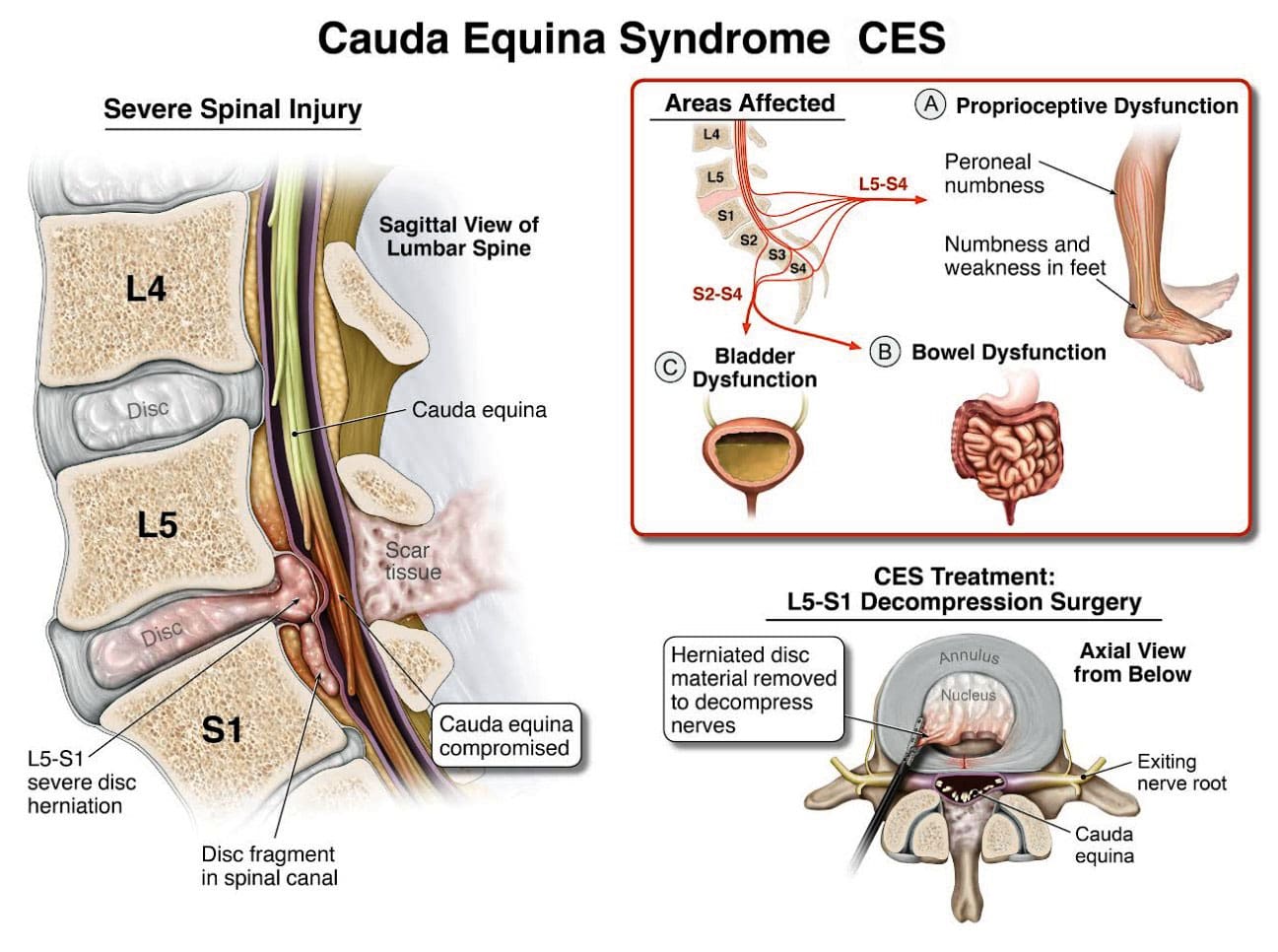
Cauda equina syndrome/CES is a rare condition in which the nerves in the lower back are compressed. It can include sciatica as one of its symptoms. Individuals presenting with symptoms that could be CES are advised to see a healthcare provider as soon as possible, as delaying treatment can lead to permanent damage.
Cauda Equina Syndrome
A cluster of nerve roots called the cauda equina, Latin for horse’s tail, sends and receives messages to the legs, bladder, and other body parts. Cauda equina syndrome is a rare condition in which nerve roots in the lower spinal cord are compressed. This compresses the nerves and disrupts motor and sensory function in the bladder and lower extremities. The most common cause is a ruptured or herniated disc in the lumbar area. This usually occurs when a severe disc herniation compresses the nerve bundle at the base of the spinal cord, causing significant neurological dysfunction like bladder/bowel issues and numbness in the saddle area. If found early, it is treated with surgery within 24 to 48 hours of symptom onset.
This is characterized by symptoms that include unusual urinary sensation, loss of desire to urinate, poor urinary stream, and having to strain to urinate. (Gardner A., Gardner E., & Morley T. 2011)
Pott’s paralysis is a neurological complication of tuberculosis (TB) of the spine.
TB is a bacterial infection that usually affects the lungs but can spread to the spine.
Iatrogenic Side Effects
Injuries or illnesses that result from medical or surgical treatment
Spinal Lesions or Malignant Tumors
A spinal lesion refers to any abnormal growth or damage within the spine.
It can include benign (noncancerous) and malignant (cancerous) tumors.
A malignant tumor is a cancerous growth within the spine; essentially, a malignant tumor is a type of spinal lesion with the potential to spread to other parts of the body.
Spinal Infection, Inflammation, Hemorrhage, or Fracture
A spinal infection refers to a bacterial, fungal, or viral infection that occurs within the bones of the spine (vertebrae) or the surrounding tissues, potentially causing pain, inflammation, and, in severe cases, neurological complications like weakness or paralysis;
Spinal inflammation is a general term for swelling or irritation within the spinal column.
Spinal hemorrhage” indicates bleeding within the spinal canal.
A spinal fracture refers to a break in one or more of the vertebrae in the spine.
Spinal Arteriovenous Malformations (AVMs)
A spinal arteriovenous malformation (AVM) is a rare condition in which the arteries and veins in the spinal cord tangle abnormally.
This can damage the spinal cord over time.
Complications from Lumbar Surgery
Lumbar surgery can have several complications, including infections, blood clots, nerve damage, and spinal fluid leaks.
Spinal Anesthesia
Spinal anesthesia is a regional anesthesia that blocks pain and sensation in the lower body.
It involves injecting a local anesthetic medication into the subarachnoid space surrounding the spinal cord.
The exact cause is not fully understood, but it can involve direct nerve root injury from the needle, inflammation caused by the anesthetic, or a spinal hematoma compressing the nerve roots.
Infection of the tissues (meninges) that cover the cauda equina and spinal cord.
An abscess pressing on the cauda equina.
Diagnosis
Diagnosis requires a medical history of symptoms, general health, activity level, and a physical exam to assess strength, reflexes, sensation, stability, alignment, and motion. (American Association of Neurological Surgeons, 2024) Testing includes:
X-ray or computerized tomography (CT) imaging is enhanced by the injection of contrast material into the cerebrospinal fluid spaces, which can show displacement of the spinal cord or spinal nerves.
Specialized Nerve Testing
This could be nerve conduction velocity tests and testing electrical activity in muscles or electromyography.
Treatment
The extent of urinary problems can determine treatment protocols. A CES diagnosis is usually followed by emergency surgery within 24 to 48 hours to relieve compression of the nerves. Moving quickly is essential to prevent permanent complications such as nerve damage, incontinence, or leg paralysis. (American Association of Neurological Surgeons, 2024)
Depending on the cause, corticosteroids also may be prescribed to reduce swelling.
Antibiotics may be needed if an infection is responsible for CES.
For situations in which a tumor is the cause, surgery to remove it may be necessary, followed by chemotherapy and/or radiation.
The outcome with CES-I during surgery is generally favorable.
Those whose CES has deteriorated to CES-R tend to have a less favorable prognosis.
Post Surgery Therapy
After surgery, CES can be challenging to deal with. If bladder function has been impaired, recovery of control can take time.
Frequent urinary infections are also a potential complication.
Loss of bladder or bowel control can be psychologically distressing, impacting social life, work, and relationships.
Sexual dysfunction can also occur, contributing to relationship difficulties or depression.
Therapy with a mental health professional may be recommended. When damage is permanent, it will be important to include family and friends in the adjustment to living with a chronic condition. Psychological counseling and/or a support group can be helpful. Other specialists who can help include: (American Academy of Orthopaedic Surgeons, 2024)
Occupational therapist
Physical therapist
Physiotherapist
Sex therapist
Social worker
Injury Medical Chiropractic and Functional Medicine Clinic
Injury Medical Chiropractic and Functional Medicine Clinic works with primary healthcare providers and specialists to build optimal health and wellness solutions. We focus on what works for you to relieve pain, restore function, prevent injury, and mitigate issues through adjustments that help the body realign itself. The clinic can also work with other medical professionals to integrate a treatment plan to resolve musculoskeletal problems.
Disc Herniation
References
American Association of Neurological Surgeons. (2024). Cauda Equina Syndrome. https://www.aans.org/patients/conditions-treatments/cauda-equina-syndrome/
Gardner, A., Gardner, E., & Morley, T. (2011). Cauda equina syndrome: a review of the current clinical and medico-legal position. European Spine Journal: official publication of the European Spine Society, the European Spinal Deformity Society, and the European Section of the Cervical Spine Research Society, 20(5), 690–697. https://doi.org/10.1007/s00586-010-1668-3
Fairbank, J., & Mallen, C. (2014). Cauda equina syndrome: implications for primary care. The British journal of general practice: the journal of the Royal College of General Practitioners, 64(619), 67–68. https://doi.org/10.3399/bjgp14X676988
American Academy of Orthopaedic Surgeons. (2024). Cauda equina syndrome. https://orthoinfo.aaos.org/en/diseases–conditions/cauda-equina-syndrome
Could incorporating standing lumbar flexion exercise into a daily routine help decrease pain and improve overall spinal mobility for individuals with low back pain?
Standing Lower Back Flexion Exercise
A chiropractic physical therapy team visit can help determine which exercises are best for an individual’s injury or condition and teach them what to stop doing if they have low back pain. Exercise and proper posture can decrease discomfort and improve mobility for individuals with low back pain. (Suh, J. H. et al., 2019) Sometimes, exercises that bend backward are recommended, while other times, flexion or forward bending movements are the best way to manage lower back pain. Many find the standing Williams lumbar flexion exercises maneuver helpful for low back pain. (Amila A, Syapitri H, Sembiring E. 2021)
Benefits
Individuals with certain diagnoses may benefit from spinal flexion. These diagnoses include:
Be sure to speak with a healthcare provider to understand the diagnosis and low back symptoms, and work with a physical therapist to be sure that forward flexion of the spine is the correct exercise for your back.
When To Avoid Lumbar Flexion
Some should avoid excessive forward bending, which could cause further damage or injury to the spine. Reasons to avoid flexion include:
Neurological signs such as difficulty urinating or controlling bowel movements (Howell E. R. 2012)
Before starting this or any other exercise program for your spine, check with a healthcare provider or physical therapist.
How to Perform
Gradually progressing with other gentle lumbar flexion exercises before full-standing lumbar flexion is recommended. These include performing a week or two of lumbar flexion lying down, followed by a couple weeks of lumbar flexion seated. Once these exercises are easy to perform and pain-free, progress with lumbar flexion standing postures.To perform, follow these steps:
Stand with your feet shoulder-width apart.
Slowly bend forward by sliding your hands down the front of your thighs.
Reach down as far as possible and let your lower back bend forward.
Grab your ankles and gently pull into more forward flexion to increase the backstretch.
Hold the end position for a second or two, then slowly return to the starting position.
As you exercise, be sure to monitor changes in symptoms. Pain worsening in the back or traveling down your leg indicates that you should stop the exercise (Spine-health, 2017). If the pain decreases in your leg or centralizes to your back, continue the exercise. Standing lumbar flexion can be repeated for 10 repetitions a couple of times daily. It can help decrease low back or leg pain symptoms and stretch tight hamstrings and back muscles. (Montefiore Pediatric Orthopedic and Scoliosis Center, 2003)
Injury Medical Chiropractic and Functional Medicine Clinic
Exercise can also prevent future lower back problems. Standing back flexion, postural correction, regular physical activity, and exercise are tools for keeping the spine healthy. Injury Medical Chiropractic and Functional Medicine Clinic works with primary healthcare providers and specialists to build optimal health and wellness solutions. We focus on what works for you to relieve pain, restore function, prevent injury, and help mitigate issues through adjustments that help the body realign itself. They can also work with other medical professionals to integrate a treatment plan to resolve musculoskeletal problems.
What Causes Disc Herniation?
References
Suh, J. H., Kim, H., Jung, G. P., Ko, J. Y., & Ryu, J. S. (2019). The effect of lumbar stabilization and walking exercises on chronic low back pain: A randomized controlled trial. Medicine, 98(26), e16173. https://doi.org/10.1097/MD.0000000000016173
Amila A, Syapitri H, Sembiring E. (2021). The effect of William Flexion Exercise on reducing pain intensity for elderly with low back pain. Int J Nurs Health Serv., 4(1), 28-36. https://doi.org/https://doi.org/10.35654/ijnhs.v4i1.374
Lurie, J., & Tomkins-Lane, C. (2016). Management of lumbar spinal stenosis. BMJ (Clinical research ed.), 352, h6234. https://doi.org/10.1136/bmj.h6234
Sfeir, J. G., Drake, M. T., Sonawane, V. J., & Sinaki, M. (2018). Vertebral compression fractures associated with yoga: a case series. European journal of physical and rehabilitation medicine, 54(6), 947–951. https://doi.org/10.23736/S1973-9087.18.05034-7
Howell E. R. (2012). Conservative management of a 31 year old male with left sided low back and leg pain: a case report. The Journal of the Canadian Chiropractic Association, 56(3), 225–232.
Spine-health. (2017). Exercise with lower back pain: Should you work through the pain? Spine-health
Knowledge from Veritas. https://www.spine-health.com/blog/exercising-lower-back-pain-should-you-work-through-pain
Montefiore Pediatric Orthopedic and Scoliosis Center. Center, M. P. O. a. S. (2003). Low Back Strain. https://www.cham.org/File%20Library/Global%20Navigation/Expertise%20And%20Programs/Pediatric%20Expertise/Orthopedics/Monte-LOW-BACK-STRAIN-WITH-EXERCISES.pdf
Can individuals who sit for long hours daily prevent tight neck and shoulder muscles by improving their posture, regularly stretching, and massaging their trapezius muscles?
Trapezius Self Massage
The trapezius muscle is a triangle-shaped muscle in the upper back that starts at the base of the neck, spans the length of the upper shoulders, and extends into the middle back. This muscle’s main function is stabilizing and moving the scapula/shoulder blade. The trapezius also helps to move the head, neck, arms, shoulders, and torso, stabilizes the spine, and plays an important role in posture. Physical and mental stress can tighten the trapezius muscle, leading to neck and shoulder pain. Learning to perform a trapezius self-massage can ease tension and provide pain relief. (Domingo A. R. et al., 2017)
Anatomy
The trapezius consists of three parts in three different areas of the back. The bottom of the skull, across the shoulders, and down to the mid back. A trapezius self-massage focuses on the upper portion of the traps. This part is located at the top of the shoulders. To find the upper trapezius, cross one arm in front of your body so that you can place the palm on top of the other shoulder.
For a trapezius self-massage, you need to know that there are two areas where your upper traps start and where the muscle connects to a bone. The first point is on the bottom of the skull, close to the center of the back of the skull. Start there with your fingers and trace the muscle down the back of the neck to where the shoulders widen. If you get lost, You can walk your fingers up or down the muscle on either side to relocate its origin at the base of the skull, the vertebra at the base of your neck that sticks out. This is C-7, another of the upper trapezius’s origin sites. (University of Washington Department of Radiology, 2025)
Massage Technique
Massage oil is optional but can hydrate the skin during a massage. You can perform the trapezius self-massage using your hands.
Start at the Base of The Neck
Choose one shoulder to work at a time.
Raise the arm on the opposite side of your body.
Reach this arm across your body and fold it around your neck so that your fingers rest at the back base of your neck.
Apply a decent amount of pressure to the muscle while moving your fingers in a circular motion.
The action is similar to kneading dough.
Massage this area at the base of your neck for about 30 seconds to start.
If this part of your muscle is sore, you can massage it longer.
Slowly Work Out Towards The End of The Shoulder
Once you have spent about 30 seconds massaging the muscle at the base of the neck, work your way out toward the end of your shoulder.
In close increments, in your fingers across the trapezius muscle, spending at least 30 seconds at each point.
Follow the muscle until you reach the end of the shoulder.
Apply enough pressure, and use slow, rhythmic movements so that you feel relief.
If the pressure is not relieving or makes you wince, it’s too much.
Repeat as Needed
Repeat each side two to three times before switching to the other shoulder. After massaging, you may notice a certain trapezius area is particularly sore or tense. Zero in on those areas a little longer. Remember to relax throughout the trapezius self-massage. This is an opportunity to learn where tension is in your neck and shoulders and how to apply pressure to relieve it. This knowledge can also help you be mindful throughout your day, whether sitting, doing chores, or other physical activities. If you notice scrunching or slouching, massage the trapezius and remind yourself to keep your shoulders relaxed.
Benefits
Tension and tightness in the trapezius muscle are common, particularly among individuals who work in an office, do manual labor, or deal with a lot of stress. (Marker R. J. Campeau S., & Maluf K. S. 2017) Trapezius strains are a common overuse injury that is more likely to happen when the muscle is tight. (Salavati M. et al., 2017) The injury can cause unhealthy posture to avoid the pain. This poor posture will place more stress on the muscles, leading to a cycle of poor posture and chronic pain. A trapezius self-massage can benefit in many ways, including:
Improved blood circulation
Better quality of sleep
Improved posture
Improved range of motion
Decreased swelling
Faster recovery after workouts
Reduced risk of injury
Seeing a Healthcare Provider
Like any other muscle in the body, the trapezius can be injured and requires special treatment to recover. Sometimes, the neck or shoulder pain may not come from the trapezius muscle. Consider seeing a healthcare provider if you have pain in your neck or shoulder that doesn’t get better within a week or two, especially if it isn’t responding to at-home treatment. Regardless of how long you have been experiencing pain or stiffness, contact a healthcare provider if it prevents you from getting adequate sleep or interfering with daily activities. Reasons to see a healthcare provider immediately for neck or shoulder pain include: (Mount Sinai, 2025)
There is sudden pressure or pain in the left shoulder, which can sometimes signal a heart attack.
A fall or accident resulted in pain, swelling, or problems moving the neck or arm.
If there is shoulder pain, a fever, swelling, or redness.
The skin on the shoulder area appears discolored.
It’s important to take regular breaks to stretch and move your muscles. You can release tension in the trapezius by doing shoulder shrugs throughout the day and stretching regularly. When the trapezius feels tight or sore, give yourself a massage.
Injury Medical Chiropractic and Functional Medicine Clinic
Injury Medical Chiropractic and Functional Medicine Clinic works with primary healthcare providers and specialists to build optimal health and wellness solutions. We focus on what works for you to relieve pain, restore function, prevent injury, and help mitigate issues through adjustments that help the body realign itself. They can also work with other medical professionals to integrate a treatment plan to resolve musculoskeletal problems.
Whiplash Chiropractic Massage Therapy
References
Domingo, A. R., Diek, M., Goble, K. M., Maluf, K. S., Goble, D. J., & Baweja, H. S. (2017). Short-duration therapeutic massage reduces postural upper trapezius muscle activity. Neuroreport, 28(2), 108–110. https://doi.org/10.1097/WNR.0000000000000718
University of Washington Department of Radiology. (2025). Trapezius. https://rad.washington.edu/muscle-atlas/trapezius/
Marker, R. J., Campeau, S., & Maluf, K. S. (2017). Psychosocial stress alters the strength of reticulospinal input to the human upper trapezius. Journal of Neurophysiology, 117(1), 457–466. https://doi.org/10.1152/jn.00448.2016
Salavati, M., Akhbari, B., Ebrahimi Takamjani, I., Ezzati, K., & Haghighatkhah, H. (2017). Reliability of the Upper Trapezius Muscle and Fascia Thickness and Strain Ratio Measures by Ultrasonography and Sonoelastography in Participants With Myofascial Pain Syndrome. Journal of Chiropractic Medicine, 16(4), 316–323. https://doi.org/10.1016/j.jcm.2017.06.003
Mount Sinai. (2025). Shoulder pain. https://www.mountsinai.org/health-library/symptoms/shoulder-pain
Can individuals managing facet arthropathy treat the condition with over-the-counter pain relievers, prescription muscle relaxers, exercise, and chiropractic spinal manipulation?
Facet Arthropathy
Facet arthropathy, or facet osteoarthritis, is arthritis that affects the facet joints in the spine. It causes pain and stiffness due to cartilage degeneration within these joints, often resulting from wear and tear associated with aging. Essentially, it occurs when the small joints in the back of the spine become arthritic and rub against each other painfully.
It affects the bony protrusions, called facet joints, that connect the spine’s bones.
Symptoms include neck and back pain that can worsen with standing, bending, or twisting.
Facet arthropathy is diagnosed using X-rays and other imaging studies.
Severe cases may require surgery.
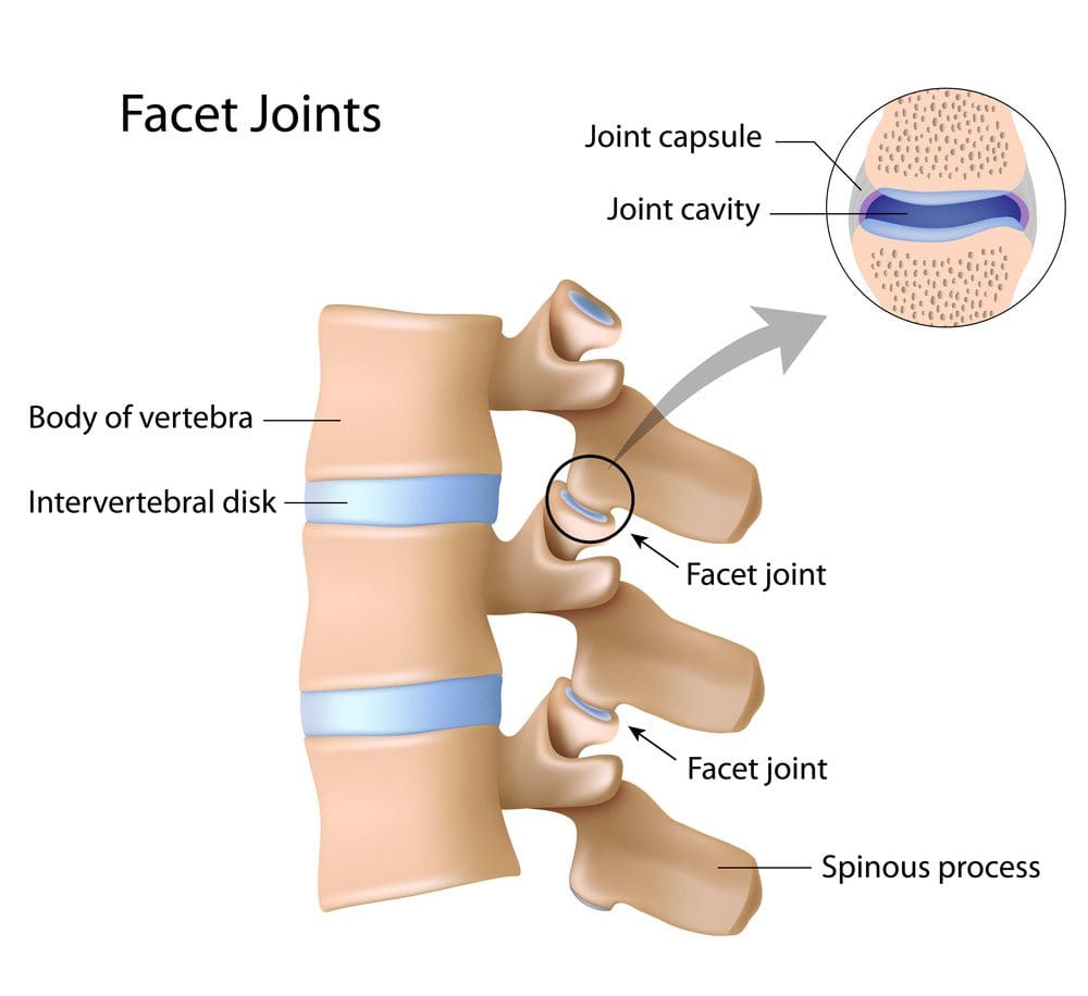
Facet Joints
Twenty-four vertebrae form the spine, with two facet joints between each. Facet joints are small joints located at the back of each vertebra in the spine. They allow movement and stability, help maintain the alignment of the spinal bones/vertebrae, and limit excessive motion. The joints and the cushioning intervertebral disc form a three-joint complex between each vertebra.
The three-joint complex allows the spine to move, including bending, rotating, and extending.
Synovial fluid lubricates the joints so they can move.
The intervertebral disc provides flexibility and dissipates compressive loads.
The facet joints stabilize the spine by constraining rotation and bending.
Symptoms
Arthropathy refers to any disease affecting a joint, including arthritis. Osteoarthritis, also known as arthrosis, is a specific type of arthropathy. It is a non-inflammatory, degenerative arthritis. Pain is the main symptom that is typically worse in the morning when awakening, and in the evening, the pain can also get worse when twisting or bending backward. The symptoms can vary based on the part of the affected spine. Low back pain is the most common, a condition referred to as lumbar facet arthropathy because it affects the lumbar spine of the lower back. (Perolat R. et al., 2018) Common Symptoms include:
Muscle spasms or cramps.
Pain that may come in periodic flare-ups
Pain that worsens with standing or inactivity.
Dull pain on both sides of the spine.
Aching pain on both sides of the spine.
Pain in the lower back, buttocks, shoulders, or back of the skull
Radiating pain to the buttocks and legs.
Pain that improves with sitting, leaning forward, or changing positions.
Pins-and-needles sensations in the hands or feet.
Clicking sounds when moving the spine.
Catching sensations when moving the spine.
Muscle weakness.
Causes
Facet arthropathy causes progressive damage to the spine. Spinal osteoarthritis, aka spondylosis, is the most common cause, but it can also occur with a severe form of spinal arthritis known as ankylosing spondylitis. It is primarily due to age-related wear and tear, but injuries or repetitive stress on the spine can also cause it. Arthritis in the facet joints can develop due to:
Aging-related wear and tear
Disc problems
A previous back injury
Torn ligaments
Spinal fractures
Deterioration of facet joints can also cause bony overgrowths called osteophytes or bone spurs, which can cause radiating pain and restrict the spine’s range of motion.
Degeneration
The facet joints and intervertebral discs degenerate due to age-related wear and tear.
The cartilage in the facet joints can dry out, crack, and wear down.
The joint capsule and synovial membrane can inflame or tear, affecting synovial fluid production.
The loss of cartilage can lead to hypermobility, and the joint can stiffen over time.
Diagnosis
Imaging studies are important to the diagnosis. Several types confirm the diagnosis and also characterize the nature and severity of the condition:
X-rays provide a plain, black-and-white image of the spinal column.
CT scan composites multiple X-rays to create a three-dimensional image of the spinal column.
MRI uses magnetic and radio waves to generate images of soft tissues like ligaments and cartilage.
To confirm the diagnosis, a diagnostic block, which is a small amount of local anesthetic, is injected into a facet joint. The needle placement is directed either with an ultrasound or a CT scan. Facet arthroplasty is confirmed if the injection provides immediate relief (American Academy of Orthopaedic Surgeons, 2022). The healthcare provider will want to exclude other possible causes as part of the differential diagnosis. Conditions that mimic facet arthropathy include:
Herniated disc
Psoriatic arthritis
Reactive arthritis
Spinal gout
Spinal compression fracture
Treatment
The treatment varies depending on the location and severity of the condition. Generally, conservative treatments are used before more invasive procedures are considered.
Lifestyle Changes
Initially, a healthcare provider may recommend rest and avoiding aggravating movements, including any activity that involves bending or twisting.
Activities that take the weight off the facet joint, such as sitting, leaning forward, or changing positions, may help ease the pain.
Patients may also be advised to adjust their sleep positions to take the pressure off facet joints.
Options included curling up on your side or lying on your back with the knees supported with pillows.
Medications
If a diagnostic block is used, a patient may not need medications immediately. However, as the anesthetic starts to wear off, the patient may be prescribed over-the-counter or prescription pain relievers based on the severity of the pain. These can include:
Analgesics like Tylenol
Nonsteroidal anti-inflammatory drugs like Advil or Aleve
Muscle relaxants like Lloresal for acute back pain
Antidepressants like Cymbalta for chronic back pain
Physical Therapy
Physical therapy is a major part of the treatment of lower back pain. The treatment plan will include personalized exercises to strengthen the core muscles and avoid stress on the spine. Examples include:
Knee-to-chest stretches, hugging your knees for 30 to 60 seconds.
Walking 10 to 20 minutes per day.
Aquatic therapy to alleviate pressure on the spine.
Surgery
If conservative measures don’t work or provide sufficient relief, a healthcare provider may recommend specialist procedures or surgeries that include:
Lumbar intra-articular injections deliver an anesthetic or corticosteroid into the spine for longer-lasting pain relief.
Sinuvertebral nerve ablation destroys spinal nerves with a strong electrical current.
Extracorporeal shockwave therapy ESWT delivers low- or high-energy electrical pulses to help ease pain.
Spinal fusion surgery involves fusing two or more vertebrae to eliminate movement and pain in the facet joints.
Facet rhizotomy is a surgical procedure used to sever one of the nerves supplying the facet joint.
Stem cell regeneration is an experimental procedure in which stem cells are harvested and injected into damaged joints to restore function.
Injury Medical Chiropractic and Functional Medicine Clinic
Injury Medical Chiropractic and Functional Medicine Clinic works with primary healthcare providers and specialists to build optimal health and wellness solutions. We focus on what works for you to relieve pain, restore function, prevent injury, and help mitigate issues through adjustments that help the body realign itself. They can also work with other medical professionals to integrate a treatment plan to resolve musculoskeletal problems.
Facet Syndrome Pain Treatment
References
Perolat, R., Kastler, A., Nicot, B., Pellat, J. M., Tahon, F., Attye, A., Heck, O., Boubagra, K., Grand, S., & Krainik, A. (2018). Facet joint syndrome: from diagnosis to interventional management. Insights into imaging, 9(5), 773–789. https://doi.org/10.1007/s13244-018-0638-x
American Academy of Orthopaedic Surgeons. (2022). Spinal injections. https://orthoinfo.aaos.org/en/treatment/spinal-injections/
IFM's Find A Practitioner tool is the largest referral network in Functional Medicine, created to help patients locate Functional Medicine practitioners anywhere in the world. IFM Certified Practitioners are listed first in the search results, given their extensive education in Functional Medicine