Neck pain is one of the most common sources of pain and chronic pain worldwide. According to the International Association for the Study of Pain, each year, around 30% to 50% of the general population experiences neck pain and approximately 15% will, at some point in their lives, have chronic neck pain. Women seem to experience it more often than men, and it is most prevalent at around middle age. Neck pain can be debilitating, impacting a person home life as well as their work performance. It can also trigger migraines and limit the range of motion. Understanding the cervical spine is integral in understanding how to manage pain in that area.
What is the Cervical Spine?
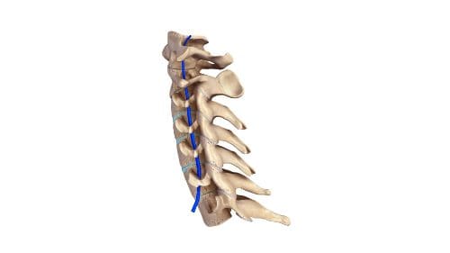
Seven vertebrae make up the cervical spine: C1 through C7. They protect the spinal cord and are part of the system that makes up the neck.
C1 is located at the base of the skull and C7 sits at the beginning of the thoracic spine. While C1 is the smallest vertebrae, each subsequent one is slightly larger as you move down the spine. This is necessary because the farther down the spine, the more weight it must bear.
The vertebrae C3 through C6 are called �typical vertebrae.� Like other vertebrae in the spine, they have a similar construction. The top vertebrae, C1 and C2, are �atypical vertebrae.� Their construction is somewhat different from typical vertebrae due to their specialized function and location.
The atlas, C1, is the only vertebrae that have more of a ring shape than a shape resembling a vertebra. It is what connects the skull to the spine and is responsible for about half of the head�s backward and forward range of motion.
The axis, C2, is the second vertebra and has a unique construction that connects it to C1 at the atlanto-axial joint. It is responsible for around half of the head�s rotation. The vertebra prominens, C7, is much larger than the vertebrae that sit above it and its shape is different to facilitate its connection to T1, at the beginning of the thoracic spine.
Neck Pain
The cervical spine has several critical functions. It houses the spinal cord and protects it, supports the head and facilitates its movement, and facilitates the flow of blood to the brain.
The human head is around 10 to 13 pounds and the cervical spine, along with an intricate network of muscles, tendons, and ligaments support it. This is what also allows flexibility to the head so that it can move up and down, backward and forwards, rotational, and side bending. This job alone puts a great deal of stress on the neck and can lead to neck pain. Common causes of neck pain include:
Whiplash (whipping the head forwards and then backward very suddenly)
Degenerative disc disease
Pinched Nerve
Age-related conditions
Spinal stenosis
Sleeping in certain positions
Neck strain
Osteoarthritis
Keeping the neck in one spot too long, such as looking down at a mobile device
Herniated disc
Neck injury
Fibromyalgia
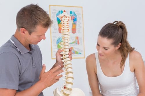
Chiropractic Care for the Cervical Spine
A chiropractor will typically treat a patient with neck pain using cervical spinal manipulation, cervical spinal mobilization, or a combination of the two techniques. Cervical spinal manipulation is what most people think of regarding chiropractic treatment. It involves short, quick thrusts that focus on a single joint at a time, so that range of motion is returned to that area. Cervical spinal mobilization is a gentler, lower impact adjustment that does not use as much force but does move the joint to its correct position.
Other treatments the chiropractor may employ include the application of cold or heat, massage, and exercises to strengthen and stretch the neck. The doctor will carefully consider the patient, their lifestyle, habits, and current level of fitness then create a plan that is tailored specifically for them that will help them manage their pain and return flexibility and range of motion as quickly as possible.
Back pain can be debilitating. A patient can find they have trouble moving or engaging in regular activities like lifting their children or even walking. Pain in the mid to upper back can be caused by a variety of issues, and it can have a significant impact on a person�s quality of life. Many people see chiropractors to get relief from their back pain, but there are some things that chiropractic patients should know so that they can get the most out of their treatments.
What is the Thoracic Spine?
Twelve vertebrae make up the thoracic spine which is located just above the lumbar spine and just below the cervical spine. It is often referred to as the upper back. This part of the spine has several essential functions. The ribs connect with this portion of the spine, and it also is responsible for protecting the spinal cord.
The thoracic spine also differs from the lumbar spine and cervical spine. Instead of curving inward (lordosis) as those areas do, it curves outward (kyphosis). This provides the freedom of movement that allows a person to bend forward and touch their toes. It does not allow for much bending backward; that typically comes from the lower back.
Many nerves extend from the thoracic spine. They control organ function for the major organs, including:
T1 to T4
Heart
Esophagus
Upper body muscles
Lungs
Larynx
Part of the arms
Trachea
Esophagus
T5 to T10
Gallbladder
Diaphragm
Small intestine
Appendix
Liver
Kidneys
Suprarenal gland
Stomach
Spleen
Adrenal gland
Pancreas
T11 to T12
Small intestines
Mid to upper body muscles
Lymph circulation
Colon
Solar plexus
Uterus
Mid to Upper Back Pain
Pain in the thoracic area of the spine is often caused by muscle strain, overuse, and injury to the discs, ligaments, and muscles that surround the spine and support it. Poor posture can also cause pain in that area. It is also very common for myofascial pain to affect the connective tissue of` muscle groups and individual muscles. These problems can occur due to a variety of causes:
Slouching or slumping while standing or sitting
Getting in a car accident where the patient is lurched forward or jolted
Lifting something that is too heavy
Yard work
Getting struck or hit in the back
Playing sports
Osteoarthritis can also occur in this area. It is caused by torn cartilage brought about by the everyday wear and teas and even the simple process of aging. Fractured vertebrae can also cause back pain in the thoracic area, as can a herniated disc, and a spine that is oddly shaped or misshapen. Degenerative disc disease and spinal stenosis can also be culprits.
Chiropractic Care for the Thoracic Spine
The goal of the chiropractor treating a patient for thoracic back pain will usually focus on reducing the pain and inflammation in the area. The treatments may include:
Spinal adjustments
Specialized exercise recommendations
Ergonomic training
Distraction
Heat or ice
Traction
Electrical stimulation
The chiropractor may also recommend nutritional supplements like proteolytic enzymes to aid in managing the swelling and pain that may be caused by disc herniation and some other back injuries. They may also recommend dietary changes or weight loss to help the patient manage their pain.
Chiropractic is a safe, effective, non-invasive treatment for mid to upper back pain. Many patients experience results immediately which is another draw for people. Most patients with back problems will be advised to maintain regular chiropractic visits to manage the pain and keep it at bay effectively.
If you are considering going upright in your workplace or workspace, you are not alone. Companies large and small are recognizing the benefits of this healthy, spine-friendly way of working and they are incorporating it into their employees� workstations. It places the body in an optimal position, between standing and sitting to provide an ergonomic solution to working at a desk that saves space too. Even home offices are getting in on the movement. These case studies tell the stories of four companies that incorporated upright workspace technology for their organizations.
Shape Up
Rhode Island-based start-up company, ShapeUp, is a health and technology-centered small business with just employees. It manages the design and implementation of socially activated wellness programs in the workplace. They were looking for furniture that was high quality and sturdy enough to withstand a workforce that was very active. At the same time, it needed to promote good health to remain consistent with the company�s health-oriented ideals.
Their first step moving in that direction was to purchase several community upright workstations. This would allow employees to get upright at various points during the day. The feedback from employees was so great that upright workstations were placed in each employee�s work area.� They reported reduced back pain and increased energy, attributing it to the simple act of going upright.
FLUX
FLUX, based in San Francisco, is a small tech company with fewer than 50 employees. The venture-backed start-up created software that �reimagines sustainable building design.�
In 2012, Nicholas Chim, the company�s founder, began searching for body-friendly workstations that would help keep his energy level up and help him maintain his focus. He purchased an upright station for himself to�use in his work area. Many of the employees expressed great interest in this new workstation. Once, Chim came home from a business trip and found that one of the employees had taken over his upright station.
It was then that Chim realized he needed to purchase upright stations for all of his employees if he was going to keep them happy and healthy. He now offers upright workstations to all of his employees; all they have to do is request it.
Katie Rowe Mitchell
Katie Rowe Mitchell has a home office where she runs her start-up, Unfold Yoga + Wellness with her friend and partner Nicole Elipas Doherty. The company brings meditation practices and accessible yoga to organizations as a wellness measure for the companies� employees. She left a�longtime corporate job that left her feeling physically uncomfortable, overstressed, and overworked due, in part, to her sedentary work style.
She recognized the link between yoga and having more energy and better focus so she left her corporate job to start her own company that would bring yoga to be stressed out workers. In her own home office, Katie wanted a more active work style, and an upright workstation was the answer. It keeps her engaged in mind, body, and spirit. She has a newfound sense of freedom that sitting behind a desk for hours every day did not provide. Going upright opened a whole new world for Katie.
Wikimedia Foundation
Tech non-profit Wikimedia Foundation is based in San Francisco and has 200 employees. It powers several collaboratively edited projects including Wikimedia. When the company decided to redesign their office space, they decided that they wanted to create a work environment that empowered and encouraged employees to work together. They chose a dynamic environment with an open floor plan � and they included several upright stations. These workstations were grouped so that all of the employees would have an opportunity to use the stations at different times. The standing desks also proved to be space saving and took up less room in the work area than traditional desks and chairs.
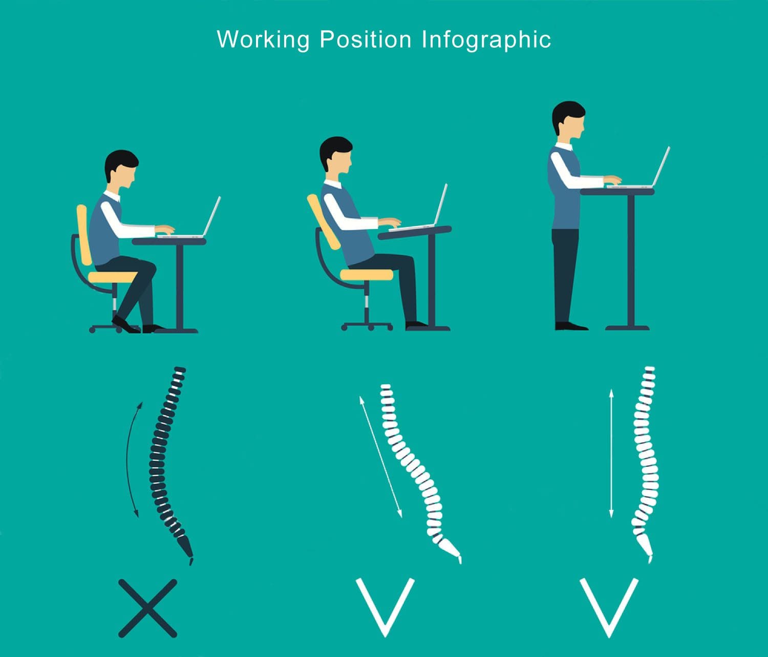
The workstation is one of the most damaging places you can spend your day when it comes to your spine. Office chairs are not designed to promote good posture or spinal health while desks and computer monitors are notorious for being too low or too high. The result can cause pain in your neck and back, headaches, and a variety of other conditions.� A stability ball could be the answer.
However, if you have a job that requires you to sit at a desk for an extended period, what can you do? Are you stuck with an achy, stiff neck and back because your workstation doesn�t promote a healthy posture? You don�t have to suffer; you can work healthier and smarter. Using an exercise ball as your chair is a great way to combat the painful and even detrimental effects of the traditional desk and chair.
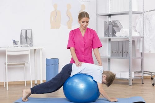
Stability Ball as an Office Chair
A stability ball, also known as a Swiss ball, exercise ball, or physioball, is a large, inflatable ball used for training. A stability ball helps to increase pillar strength, improve stability and have better balance. It is large, making it high enough that it can easily be used as a desk chair.
At least one company has combined the fitness benefits of a stability ball with some of the convenient features of an office chair (wheels, lumbar support, etc.). Gaiam Balance Ball Chairs are stability balls that are intended to be used as chairs. The ball needs to be inflated before use and may need to be reinflated from time to time. It also has a 300-pound weight capacity. It is a somewhat pricier alternative to the plain stability ball.
How Sitting on a Stability Ball Benefits your Spine
There are at least three outstanding benefits you can enjoy by using a stability ball as your chair. Try it for just 30 days and see the difference for yourself. In that time you will see:
Your core muscles are toned. As you balance on the stability ball, it forces you to engage your core muscles including those in your low back, abdominal, and pelvic floor. It will keep your muscles engaged for extended periods of time but also encourage you to move for little extra core work. This, in turn, will help to keep your spine correctly aligned and stabilized.
Your back pain is relieved. Sitting on your stability ball improves your circulation, encouraging blood flow throughout your body. An office chair, on the other hand, does just the opposite. This is helpful in relieving pain. It keeps your spine aligned which also helps with any back pain you may experience. This is in part to the core strength you develop, but also because you are less likely to slouch or sit in a position that puts a strain on your back.
You have better posture.�A better-aligned spine naturally leads to better posture. Sitting on the ball works your core, strengthening those muscles so that your spine is supported, resulting in better posture. You will find that you sit up straighter and over time you will walk taller. Better posture is perfect for your spine, making it more flexible and stronger.
It should be noted that it isn�t healthy to sit in any position for too long. Stand up and move about every hour or so. While the stability ball causes you to change positions throughout the day, you also need full body movement, which includes standing, stretching and walking.
There is no denying that water is an integral part of good health. Dehydration can cause problems with skin, digestion, and organ function. It can cause leg and foot cramps and impair cognitive processes. Staying well hydrated is vital to overall wellness. Because water is part of every cell in the body, and when we don�t drink enough water, the body suffers.
Good spinal health begins with proper hydration. The spine is constructed in such a way that dehydration can cause limited mobility, decreased flexibility, and pain. It can make the backbone to age faster than it should which impacts the entire body. As the natural functions begin to break down the body suffers, and it isn�t long and depression and anxiety often set in. The spine depends heavily on hydration.
Overview of the Spine
The spine of made up of vertebrae, a row of bones that sit on top of each other, connected by small joints. A disc sits between each vertebra, cushioning it and acting as a shock absorber. It allows the spine to flex, bend, and move about without the bones rubbing together.
Each disc has a fluid center (nucleus pulposis) that is surrounded by a flexible, sturdy ring. The ring contains a gel-like substance while the center of the disc is comprised of water. The outer ring protects the center, and the center protects the vertebrae, acting as a cushion for the bones.
If the fluid center does not have adequate water, it cannot do its job, and the spine begins to experience problems. Aging makes it more difficult for the discs to rehydrate and a sedentary lifestyle also complicates the process. It just cannot work without proper hydration. A healthy spine starts with adequate hydration.
The Benefits of Water for the Spine
From the time you get up in the morning, you are putting pressure on your spine and subsequently, the discs that lie between each vertebra. As you move the discs are compressed by the spine, and the water that is inside is squeezed out.
Even upright activities like standing, sitting, or walking can cause pressure on the discs as gravity causes compression in the spine. When the discs do not have enough water, it results in limited mobility, pain, and an increased risk of back injury.
If you don�t drink enough water, your body becomes dehydrated and is unable to replenish the water that the discs so desperately need. You may not even notice the typical signs of dehydration such as a headache and lethargy, but also lower levels of dehydration can cause severe problems in the body, especially if it is prolonged. Soda and similar beverages do not provide adequate water to the body.
How to Properly Hydrate the Body
Water is the best way to hydrate the body, but it isn�t the only way. Foods like watermelon, lettuce, spinach, and soups are excellent sources of hydration. H2O, of course, is the best way, but herbal teas are also good.
Drinks with caffeine are not as effective since the caffeine can have a diuretic effect. Traditionally, people have been told to drink eight glasses of water a day, and that is good advice. However, studies indicate that proper hydration can occur with an intake of just 30 to 50 ounces of water a day.
If you have constant or frequent back pain, the answer could be as close as your kitchen faucet. Dehydration could be the source of your back pain and immobility.
Water also affects the way the cerebrospinal fluid works and moves in the body. When the body is dehydrated, it doesn�t move as it should, and brain function, reflexes, and cognitive processing could be impacted. Don�t chance it. If your problems are caused by something as simple as not drinking enough water, that is something you can change today. Drink up! Your body will thank you.
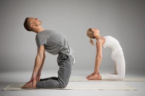
Yoga has long been touted as a healing activity that extends beyond its known fitness benefits. It can help alleviate depression, relieve stress, and decrease anxiety as well as tighten and tone the body.
A 2016 study by Yoga Alliance and Yoga Journal shows that the more than 20 million people who practice yoga spend more than $10 billion on related classes and products. Chiropractors have picked up on the benefits of yoga and are recommending it to their patients. Why, because it helps to improve flexibility as well as spinal health.
Chiropractic and the spine�s role in the body.
Chiropractic is primarily intended to align the spine and balance the body. The spine is the primary support for the body. It houses most of the central nervous system and provides pathways for neural impulses to move throughout the body.
When the spine is out of alignment, it can affect how the central nervous system functions. It can also affect flexibility and even overall mobility as well as cause pain and stiffness.
Chiropractic treatment brings the spine back into alignment. It helps to balance the body and treat injuries as well as help with the changes the aging body experiences.
One of the best-known uses for chiropractic is to treat pain. It is a medication free, non-invasive treatment for chronic pain, sports-related injuries, and even automobile accidents. Many patients had reported finding relief with chiropractic when nothing else worked.
Combining yoga with chiropractic increases the effectiveness of the treatment while strengthening the body and making it better able to respond to it. It is the perfect complement to chiropractic care, and many patients are discovering tremendous health benefits from this winning, healthy combination.
What is Yoga?
In its purest form, it is an ascetic and spiritual discipline that comes from Hindu culture. It involves simple meditation, conscious breathing or breath control, and performing certain body postures.
While an ancient practice long used for spiritual and emotional healing as well as physical wellness, yoga has been adopted in western culture and widely accepted as a form of fitness as well as a therapeutic practice used for relaxation and overall good health. It focuses on inward healing for outward results.
People who regularly practice find they are more centered, handle stress better, and aren�t as likely to experience depression and anxiety. They also are more flexible, have better mobility, and have stronger leaner bodies.
What happens when you combine yoga and chiropractic care?
Because yoga helps to lower blood pressure, decrease stress, and strengthen the body�s core, it is perfect therapy for the chiropractic patient. While chiropractic care is working to align the spine and balance the body, yoga is helping to strengthen the muscles surrounding the spine, providing better support. The numerous health benefits, particularly regarding blood flow and relaxation help to increase chiropractic�s effectiveness as a pain management tool.
Patients who combine yoga and chiropractic will also often find that they see the effects of both treatments much faster than they would if they were only doing one or the other. Both chiropractic and yoga help with balance, flexibility, and mobility, but they come at it from somewhat different approaches. The benefit of this is that it provides a more balanced, well-rounded treatment in these areas as one supports the other.
Yoga also tones and tightens the body, preparing it for much more profound levels of healing, cleansing and releasing the body of tensions that may have been held�in for years. It also works to stretch muscles that have been restricted for years, or even for the patient�s entire life. It prepares the body to accept the full benefits of chiropractic and respond faster and more thoroughly.
Imaging diagnostics of the spine consist from radiographies to computed tomography scanning, or CT scans, in which CT is utilized in conjunction with myelography and most recently with magnetic resonance imaging, or MRI. These imaging diagnostics are being used to determine the presence of abnormalities of the spine, scoliosis, spondylolysis and spondylolisthesis. The following article describes various imaging modalities and their application in the evaluation of common spinal disorders described.
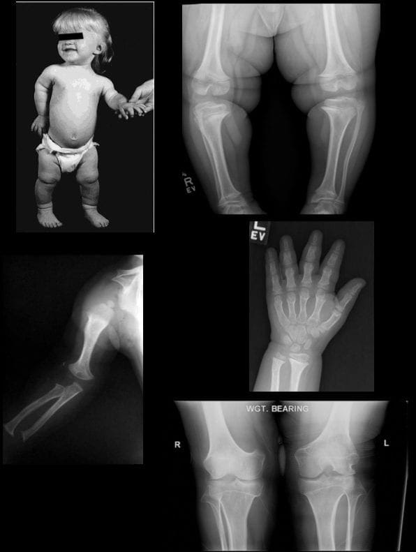
Achondroplasia
Achondroplasia is the most common cause of rhizomelic (root/proximal) short-limb dwarfism. Patients are of normal intelligence.�
It shows multiple distinct radiographic abnormalities affecting long bones, pelvis, skull, and hands.
Vertebral column changes may present with significant clinical and neurological abnormalities.�
Achondroplasia is an autosomal dominant disorder with about 80% of cases from a random new mutation. Advanced paternal age is often linked. Achondroplasia results from a mutation in the fibroblast growth factor gene (FGFR3) which causes abnormal cartilage formation.
All bones formed by endochondral ossification are affected.
Bones that form by intra-membranous ossification are not normal.
Thus, skull vault, iliac wings develop normally vs. the base of the skull, some facial bones, vertebral column, and most tubular bones are abnormal.
�
Dx: is usually made at birth with many features becoming apparent during the first few years of life.
Radiography plays an important part of clinical diagnosis.
Typical features include: shortening and widening of tubular bones, metaphyseal flaring, Trident hand with short, broad metacarpals and proximal and middle phalanges. Longer Fibular, Tibial bowing, markedly short humeri often with dislocated Radial head and elbow flexion deformity.
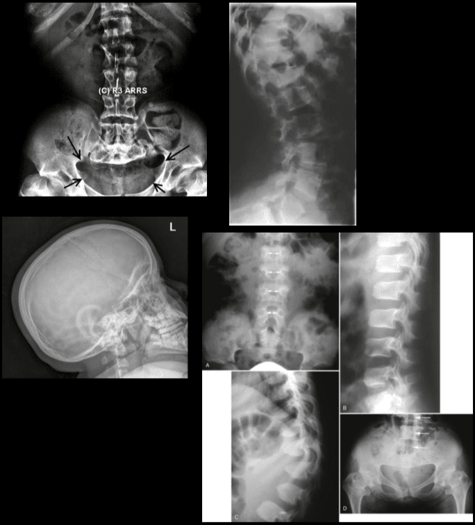
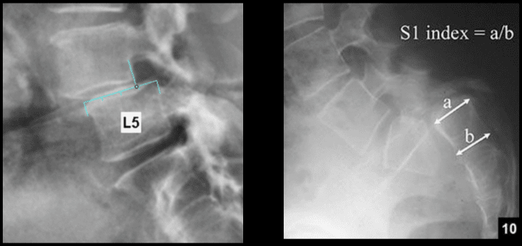
Spine: characteristic narrowing of L1-L5 interpedicular distance on AP views. Lateral view shows shortening of pedicles and vertebral bodies, �bullet shaped vertebrae� can be a characteristic feature. Early degenerative changes and canal narrowing occur. The horizontal sacral inclination is an important feature.
Pelvis is broad and short with characteristic �champagne glass� pelvis appearance.
Femoral heads are hypoplastic, but hip arthrosis is normally not observed even in older patients likely due to reduced leverage and lightweight (50kg) of patients.
Management of Achondroplasia
Recombinant human growth hormone (GH)�is currently being used to augment the height of patients with achondroplasia.
Most complications of Achondroplasia are related to the spine: vertebral canal stenosis, thoracolumbar kyphosis, narrowed foramen magnum and others.
Laminectomy extending to pedicles/lateral recess with foraminotomies and discectomies can be performed.
Cervical manipulations are contraindicated.
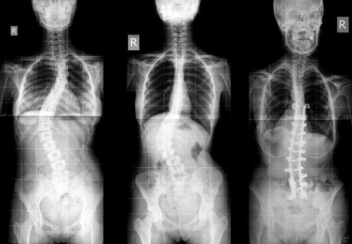
Imaging diagnostics play a fundamental role in the diagnosis the of scoliosis, an abnormality of the spine which is believed to occur due to an underlying health issue, although most cases of scoliosis are idiopathic. More over, radiographies, CT scans, and MRI, among others, can help monitor the changes of the deformity of the spine associated with this spinal manifestation. Chiropractors can provide imaging diagnostics to patients with scoliosis before proceeding with treatment.�
Dr. Alex Jimenez D.C., C.C.S.T.
�
Scoliosis
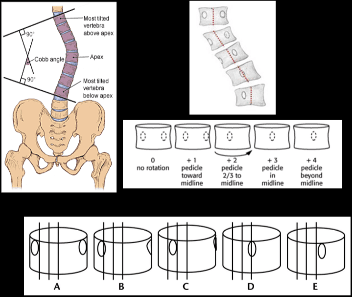
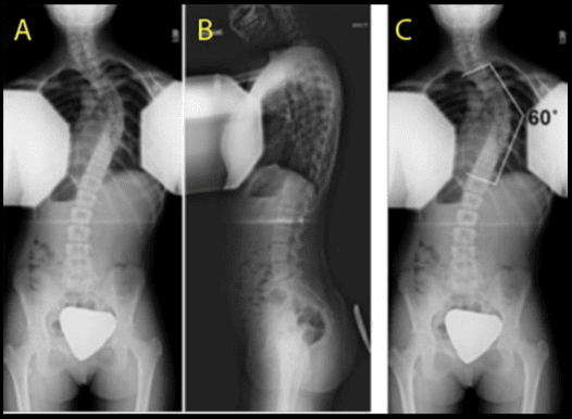
Scoliosis is defined as the abnormal lateral curvature of the spine >10-degree when examined by Cobb�s method of mensuration.
Scoliosis can be described as postural and structural.
Postural scoliosis is not fixed and can be improved by lateral flexion to the side of the convexity.
Structural scoliosis has multiple causes ranging from: ? Idiopathic (>80%) ? Congenital (wedge or hemivertebra, blocked vertebra, Marfan syndrome, skeletal dysplasias) ? Neuropathic (neurofibromatosis, neurological conditions like tethered cord, spinal dysraphism, etc.) ? Scoliosis d/t Spinal neoplasms ? Post-traumatic etc.
Idiopathic scoliosis is the most common type (>80%).
Idiopathic scoliosis can be of 3-types ( infantile, juvenile, adolescent).
Idiopathic adolescent scoliosis if patients >10y.o.
Infantile scoliosis if <3 y.o. M>F.
Juvenile scoliosis if >3 but <10-y.o.
Idiopathic Adolescent scoliosis is the most common with F:M 7:1 (adolescent girls are at particular risk).
Etiology: unknown thought to be the result of some disturbance of proprioceptive control of the spine and spinal musculature, other hypotheses exist.
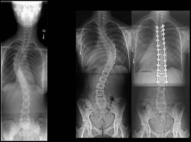
Most seen in the thoracic region and most commonly convex to the right.
Dx: full spine radiography with gonadal and breast shielding (preferably PA views to protect breast tissue).
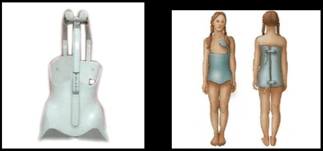
� Curves that are 50-degrees or greater and rapidly progressing will require operative intervention to prevent severe deformity of the thorax & ribs leading to cardiopulmonary abnormalities. � �? If curvature is < 20-degree, no treatment is required (observation). � �? For curves that are >20-40-degrees bracing may be used (orthosis).
Milwaukee (metal) brace (left).
Boston brace polypropylene lined with polyethylene (right) often preferred because it can be worn under clothing.
Bracing wearing is required for 24-hours for the duration of the treatment.
Note Cobb�s method of mensuration to record spinal curvature. It has some limitations: 2D imaging, not able to estimate rotation, etc.
Cobb�s method is still a standard evaluation performed in Scoliosis studies.
Nash-Moe method: determines pedicle rotation in scoliosis.
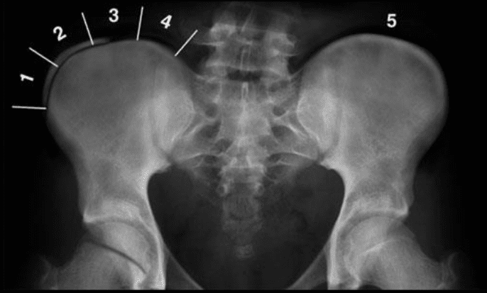
Risser index is used to estimate spinal skeletal maturity.
Iliac growth apophysis appears at ASIS (F- 14, M-16) and progresses medially and expected to be closed in 2-3-years (Risser 5).
Scoliosis progression ends at Risser 4 in females & Risser 5 in males.
During radiographic evaluation of scoliosis, it is crucial to report if Risser growth apophysis remains open or closed.
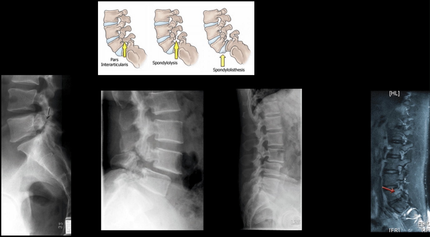
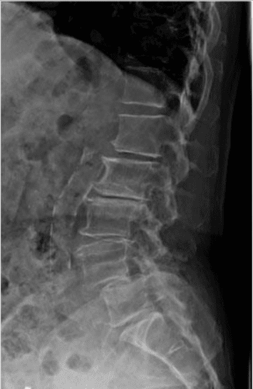
Spondylolysis and spondylolisthesis are health issues which can result in back pain. Spondylolysis is believed to be caused by repeated microtrauma leading to stress fractures in the pars interarticularis. Patients with bilateral pars defects can develop spondylolisthesis, where the degree of slippage of the adjacent vertebrae can progress gradually over time. Patients with suspected spondylolysis and spondylolisthesis may initially be evaluated with pain radiography. Chiropractic care can also help provide imaging diagnostics for these health issues.
Dr. Alex Jimenez D.C., C.C.S.T.
�
Spondylolysis & Spondylolisthesis
Spondylolysis defect in pars interarticularis or osseous bridge between superior and inferior articular processes.
Pathology stress fracture of the pars, believed to be after repeated microtrauma on extensions Men > Women, affects 5% of the general population especially in athletic adolescents.
Clinically postulated that adolescent back pain cases may be related to this process.
Typically spondylolysis remains asymptomatic.
Spondylolysis can be present with or w/o spondylolisthesis.
Spondylolysis is found in 90% at L5 with the remaining 10% in L4.
Can be uni or bilateral.
In 65%�of�cases, spondylolysis is associated with spondylolisthesis.
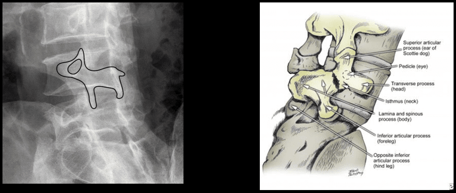
Radiographic Features: break in the Scotty dog collar around the neck on oblique lumbar views.
Radiography has low sensitivity compared to SPECT. SPECT is associated with ionizing radiation, and MRI is currently a preferred method of imaging diagnosis.
MRI can help to show reactive marrow edema next to pars defect or w/o defect so-called pending or potential to develop spondylolysis.
Types of Spondylolisthesis
Type 1 – Dysplastic, rare and found in congenital dysplastic malformation of the sacrum allowing anterior displacement of L5 on S1. Often no pars defect.
Type 2 – Isthmic, most common, often the result of a stress fracture.
Type 3 – Degenerative from the remodeling of articular processes.
Type 4 – Traumatic in an acute posterior arch fracture.
Type 5 – Pathologic due to bone disease locally or generalized.
Grading of spondylolisthesis is based on the Myereding Classification. This classification refers to the overhanging part of the superior body in relation to anterior-posterior part of the inferior body.
Grade 1 – 0-25% anterior slip
Grade 2 – 26-50%
Grade 3 – 51%-75%
Grade 4 – 76-100%
Grade 5 – >100% spondyloptosis
Note degenerative spondylolisthesis at L4 and retrolisthesis at L2, L3.
This abnormality develops due to degeneration of facets and disc with decreased local stability.
Rarely progresses beyond Grade 2.
Must be recognized in the imaging report.
Contributes to vertebral canal stenosis.
Canal stenosis is better delineated by cross-sectional imaging.
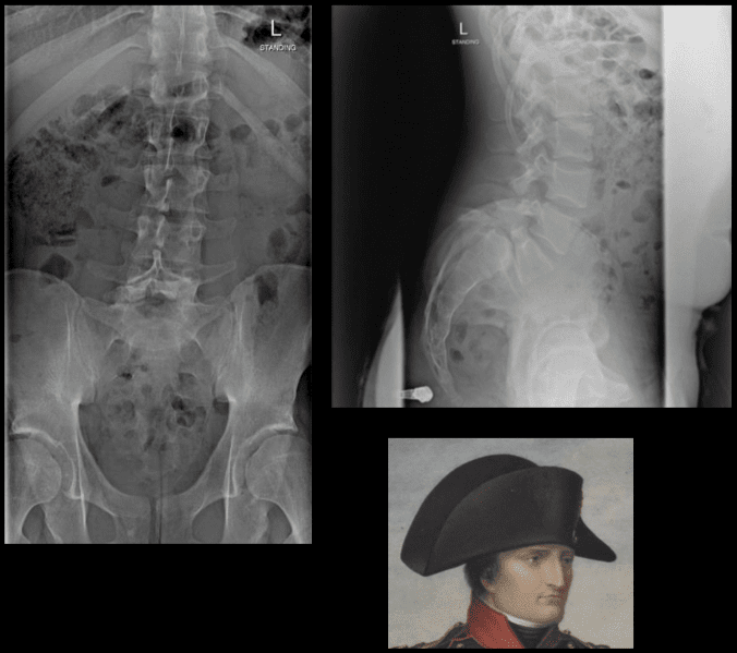
The inverted Napoleon hat sign -�seen on the frontal lumbar/pelvic radiographs at L5-S1.
Represents bilateral spondylolysis with marked anterolisthesis of L5 on S1 often with spondyloptosis and marked exaggeration of the normal lordosis.
Spondylolysis resulting in this degree of spondylolisthesis is more often congenital and/or traumatic in origin and less often degenerative.
The “brim” of the hat is formed by the downward rotation of the transverse processes, and the “dome” of the hat is formed by the body of L5.
In conclusion,�imaging diagnostics for the spine are recommended for patients with specific abnormalities of the spine, however, their increased use can help determine�their best treatment option. Understanding the abnormalities of the spine described above can help healthcare professionals and patients create a treatment program to improve their symptoms. The scope of our information is limited to chiropractic as well as to spinal injuries and conditions. To discuss the subject matter, please feel free to ask Dr. Jimenez or contact us at�915-850-0900�.
Curated by Dr. Alex Jimenez
Additional Topics: Acute Back Pain
Back pain�is one of the most prevalent causes of disability and missed days at work worldwide. Back pain attributes to the second most common reason for doctor office visits, outnumbered only by upper-respiratory infections. Approximately 80 percent of the population will experience back pain at least once throughout their life. The spine is a complex structure made up of bones, joints, ligaments, and muscles, among other soft tissues. Because of this, injuries and/or aggravated conditions, such as�herniated discs, can eventually lead to symptoms of back pain. Sports injuries or automobile accident injuries are often the most frequent cause of back pain, however, sometimes the simplest of movements can have painful results. Fortunately, alternative treatment options, such as chiropractic care, can help ease back pain through the use of spinal adjustments and manual manipulations, ultimately improving pain relief.
IFM's Find A Practitioner tool is the largest referral network in Functional Medicine, created to help patients locate Functional Medicine practitioners anywhere in the world. IFM Certified Practitioners are listed first in the search results, given their extensive education in Functional Medicine