What is a bone density test, how is it performed, and what do the results mean?
Bone Density Test
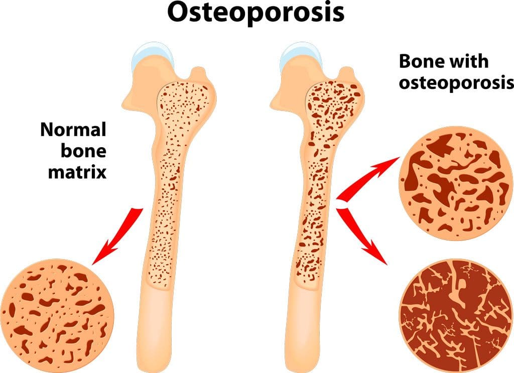
A bone density test examines bone mass, which indicates overall bone strength. Assessing bone density or mass is necessary for diagnosing osteopenia or osteoporosis, conditions that increase the risk of broken bones. The scan is performed through dual-energy X-ray absorptiometry (DEXA), which examines the thickness of the bones. Results from DEXA scans are compared to standardized values to determine whether bone density is lower than normal and whether osteopenia or osteoporosis is present.
Examination
The procedure examines bone density, or bone mass. The bones’ density, or mass, is an overall indicator of bone strength. The greater the bone density, the thicker and stronger the bones are. The test is used to diagnose osteoporosis, a condition characterized by brittle bones at risk of breaking due to significantly low bone density. A bone density test can also diagnose osteopenia, a condition characterized by lower than normal bone mass that can lead to osteoporosis. (National Institute of Arthritis and Musculoskeletal and Skin Diseases, 2025) It is recommended that all women aged 65 and older and all men aged 70 and older have a bone density scan to screen for bone loss to help prevent fractures. (Kling J. M., Clarke B. L., & Sandhu N. P. 2014)
Bone density scans can establish a baseline level of bone density and track changes over time.
For individuals with osteoporosis or osteopenia, a bone density scan can help track how well their bones respond to treatment.
During a DEXA scan, the patient will lie on their back on a table with their legs elevated on a padded platform.
An X-ray scanner will pass over the spine and hips while another scans beneath.
While the scan takes place, the patient will be asked to hold very still to obtain an accurate image.
The scan will obtain bone density readings from the spine and hip, the two most commonly fractured bones, and generally takes less than 30 minutes.
Results
A DEXA scan measures bone density in grams per centimeter squared (g/cm²). This number indicates how densely bone cells are packed together in a specific area of bone. This bone density reading is then compared to a standardized value to determine if bone density is within a normal range or lower than average.
Between minus 1.0 and minus 2.5: Low bone density (osteopenia)
Equal to minus 2.5 or below: Osteoporosis
Bone density values are reported as a Z score for women who have not undergone menopause and men under 50 years old.
Z scores are compared to bone density levels of individuals of the same age and sex.
A Z score of minus 2.0 or lower indicates low bone density, which can be caused by factors other than aging, such as medication side effects, nutritional deficiencies, or thyroid problems.
Arthritis Diagnosis
Because a DEXA scan only measures the thickness of bones, it doesn’t work to diagnose arthritis. An X-ray of the affected joint is currently the most accurate way to diagnose arthritis. The Kellgren-Lawrence classification system categorizes the extent of arthritis based on the severity of joint damage seen on an X-ray. According to this system, arthritis can be classified as: (Kohn M. D., Sassoon A. A., & Fernando N. D. 2016)
Grade 1 (minor)
Minimal or no joint space narrowing, with possible bone spur formation.
Grade 2 (mild)
Possible joint space narrowing, with definite bone spur formation.
Grade 3 (moderate)
Definite joint space narrowing, moderate bone spur formation, mild sclerosis (abnormal thickening of bone), and possible deformation of bone ends.
Grade 4 (severe)
Severe joint space narrowing, large bone spur formation, marked sclerosis, and definite deformation of bone ends.
Injury Medical Chiropractic & Functional Medicine Clinic
Exercise can be incredibly beneficial for improving bone density, joint mobility, and the strength of surrounding muscles, which support and protect joints and bones. Talk to a healthcare provider to learn what interventions and available treatment options would be the most effective. Injury Medical Chiropractic and Functional Medicine Clinic works with primary healthcare providers and specialists to develop an optimal health and wellness solution. We focus on what works for you to relieve pain, restore function, and prevent injury. Regarding musculoskeletal pain, specialists like chiropractors, acupuncturists, and massage therapists can help mitigate the pain through spinal adjustments that help the body realign itself. They can also work with other medical professionals to integrate a treatment plan to resolve musculoskeletal issues.
Osteoporosis
References
National Institute of Arthritis and Musculoskeletal and Skin Diseases. (2025). Bone mineral density tests: what the numbers mean. Retrieved from https://www.niams.nih.gov/health-topics/bone-mineral-density-tests-what-numbers-mean
Kling, J. M., Clarke, B. L., & Sandhu, N. P. (2014). Osteoporosis prevention, screening, and treatment: a review. Journal of women’s health (2002), 23(7), 563–572. https://doi.org/10.1089/jwh.2013.4611
Kohn, M. D., Sassoon, A. A., & Fernando, N. D. (2016). Classifications in Brief: Kellgren-Lawrence Classification of Osteoarthritis. Clinical orthopaedics and related research, 474(8), 1886–1893. https://doi.org/10.1007/s11999-016-4732-4
Diagnosing ankylosing spondylitis usually involves multiple tests. When doctors order blood tests to diagnose ankylosing spondylitis, an individual is experiencing worsening symptoms in their back and joints. Often, a blood test diagnosis means the doctor is looking for evidence of anything else that could be causing the symptoms. However, blood tests by themselves cannot definitively diagnose ankylosing spondylitis, but when combined with imaging and assessment, they can provide important clues that point to the answers.
Ankylosing Spondylitis Blood Test Diagnosis
Ankylosing spondylitis is arthritis that primarily affects the spine and hips. It can be difficult to diagnose as no single test can provide thorough information for a definitive diagnosis. A combination of diagnostic tests are utilized, including a physical exam, imaging, and blood tests. Doctors are not only looking for results that will point to ankylosing spondylitis, but they are looking for any results that might point away from the spondylitis results that might provide a different explanation for symptoms.
Physical Exam
The diagnostic process will begin with the individual’s medical history, family history, and physical exam. During the exam, the doctor will ask questions to help rule out other conditions:
How long have symptoms been presenting?
Do symptoms get better with rest or exercise?
Are the symptoms getting worse or staying the same?
Are the symptoms worse at a particular time of day?
The doctor will check for limitations in mobility and palpate tender areas. Many conditions can cause similar symptoms, so the doctor will check to see if the pain or lack of mobility is consistent with ankylosing spondylitis. The feature sign of ankylosing spondylitis is pain and stiffness in the sacroiliac joints. The sacroiliac joints are located in the lower back, where the base of the spine and pelvis meet. The doctor will look at other spinal conditions and symptoms:
Back pain symptoms caused by – injuries, posture patterns, and/or sleeping positions.
The HLA-B27 gene corresponds with ankylosing spondylitis; if an individual has it, one of their parents has it.
Imaging
X-rays often serve as the first step to a diagnosis.
As the disease progresses, new small bones form between the vertebrae, eventually fusing them.
X-rays work best at mapping the disease progression than the initial diagnosis.
An MRI provides clearer images in the early stages as smaller details are visible.
Blood Tests
Blood tests can help rule out other conditions and check for signs of inflammation, providing supportive evidence along with the results of imaging tests. It typically only takes about a day or two to get the results. The doctor may order one of the following blood tests:
Antinuclear antibodies, or ANA, go after the proteins in the cell’s nucleus, telling the body its cells are the enemy.
This activates an immune response that the body fights to eliminate.
A study determined that ANA is found in 19% of individuals suffering from ankylosing spondylitis and is higher in women than men.
Combined with other tests, the presence of ANA provides another clue to a diagnosis.
Gut Health
The gut microbiome plays an important role in triggering the development of ankylosing spondylitis and its treatment.
Tests to determine the gut’s health can give a doctor a complete picture of what is happening inside the body.
Blood test diagnoses for ankylosing spondylitis and other inflammatory conditions rely heavily on piecing together different tests alongside clinical exams and imaging.
Causes, Symptoms, Diagnosis, and Treatment
References
Cardoneanu, Anca, et al. “Characteristics of the intestinal microbiome in ankylosing spondylitis.” Experimental and therapeutic medicine vol. 22,1 (2021): 676. doi:10.3892/etm.2021.10108
Prohaska, E et al. “Antinukleäre Antikörper bei Spondylitis ankylosans (Morbus Bechterew)” [Antinuclear antibodies in ankylosing spondylitis (author’s transl)]. Wiener klinische Wochenschrift vol. 92,24 (1980): 876-9.
Sheehan, Nicholas J. “The ramifications of HLA-B27.” Journal of the Royal Society of Medicine vol. 97,1 (2004): 10-4. doi:10.1177/014107680409700102
Wenker KJ, Quint JM. Ankylosing Spondylitis. [Updated 2022 Apr 9]. In: StatPearls [Internet]. Treasure Island (FL): StatPearls Publishing; 2022 Jan-. Available from: https://www.ncbi.nlm.nih.gov/books/NBK470173/
Xu, Yong-Yue, et al. “Role of the gut microbiome in ankylosing spondylitis: an analysis of studies in the literature.” Discovery medicine vol. 22,123 (2016): 361-370.
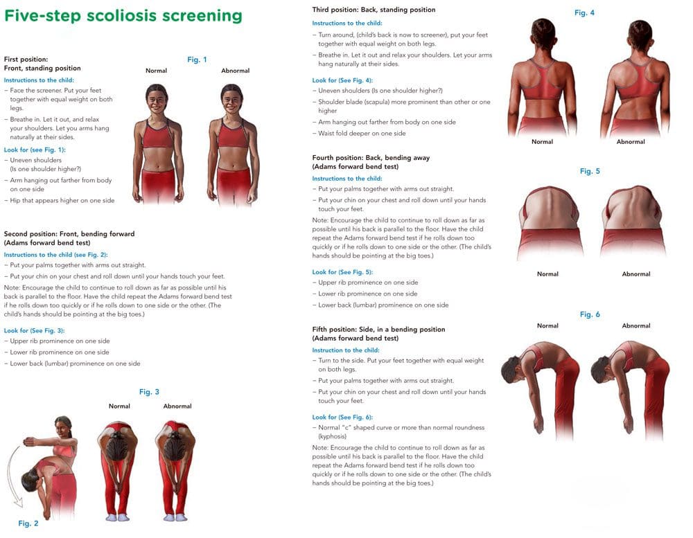
The Adams forward bend test is a simple screening method that can help with scoliosis diagnosis and help in developing a treatment plan. The exam is named after the English physician William Adams. As part of an examination, a doctor or chiropractor will look for an abnormal side-to-side bend in the spine.
Scoliosis Diagnosis
The Adams forward-bend test can help determine if there are indicators for scoliosis.
It is not an official diagnosis, but the results can be used as a starting point.
The Adams test will reveal signs of scoliosis and/or other potential deformities like:
Uneven shoulders
Uneven hips
Lack of symmetry between the vertebrae or the shoulder blades.
The head does not line up with a rib hump or the pelvis.
Detection of Other Spinal Issues
The test can also be used to find spinal curvature issues and conditions like:
Kyphosis or hunchback, where the upper back is bent forward.
Scheuermann’s disease is a form of kyphosis where the thoracic vertebrae can grow unevenly during a growth spurt and cause the vertebrae to develop into a wedge-like shape.
The Adams test by itself is not enough to confirm scoliosis.
A standing X-ray with Cobb angle measurements above 10 degrees is required for diagnosing scoliosis.
The Cobb angle determines which vertebrae are tilted the most.
The higher the angle, the more severe the condition and the more probable it will produce symptoms.
Computed tomography or CT and magnetic resonance imaging or MRI scans can also be used.
Forward Bend Test
References
Glavaš, Josipa et al. “The role of school medicine in the early detection and management of adolescent idiopathic scoliosis.” Wiener klinische Wochenschrift, 1–9. 4 Oct. 2022, doi:10.1007/s00508-022-02092-1
Grossman, T W et al. “An evaluation of the Adams forward bend test and the scoliometer in a scoliosis school screening setting.” Journal of pediatric orthopedics vol. 15,4 (1995): 535-8. doi:10.1097/01241398-199507000-00025
Letts, M et al. “Computerized ultrasonic digitization in the measurement of spinal curvature.” Spine vol. 13,10 (1988): 1106-10. doi:10.1097/00007632-198810000-00009
Senkoylu, Alpaslan, et al. “A simple method for assessing rotational flexibility in adolescent idiopathic scoliosis: modified Adam’s forward bending test.” Spine deformity vol. 9,2 (2021): 333-339. doi:10.1007/s43390-020-00221-2
It is estimated that scoliosis affects anywhere from 2 to 3 percent of children and adults in the United States. That is roughly six to nine million people. While it seems to develop most commonly within specific age ranges for boys and girls, it can also develop in infancy. Every year, approximately 30,000 children are fitted with a scoliosis back brace while 38,000 people have spinal fusion surgery to correct the problem. Scoliosis screenings can have tremendous benefits by identifying both risk factors for scoliosis and allowing for early treatment.
The earlier you detect scoliosis, the easier it is to treat.
Scoliosis typically develops in childhood. For girls, it usually occurs between 7 and 14 years of age. Boys develop it a little later, between 6 and 16 years of age.
Getting a scoliosis screening each year during these critical age ranges allow doctors to identify the condition early and begin treating it before it gets serious. Advanced scoliosis can require extensive treatments, bracing, and even surgery.
Chiropractic has been shown to help scoliosis, as do stretching, special exercises, and physical therapy. There are spinal adjustments that chiropractors do that are specific to the treatment of scoliosis.
When addressing the condition early on, the Cobb angle can be stopped from progressing and even reduced so that the spine has a more natural curve. Non-surgical treatments tend to be much more effective in the earlier stages of scoliosis, so early detection and early diagnosis are critical.
Identifying high-risk cases early can address current issues and prevent future ones.
Chiropractors can identify certain scoliosis risk factors in children before the condition even develops. A scoliosis screening allows them to spot tension in a child�s spinal cord � a common sign that they will develop scoliosis.
When parents are aware that their child is in a high-risk category for developing scoliosis, they can take proactive measures with home monitoring for the signs of scoliosis as well as keeping up with the course of recommended screenings. They will know to look for the signs and can address them quickly so that treatment can be started at the earliest possible time.
Help researchers and doctors become more effective in treating scoliosis.
The early stages and development of scoliosis are still shrouded in mystery for researchers and doctors. While there have been great strides made in better understanding the condition, there is still much left to learn.
There have been many studies that have aided doctors in identifying high-risk children and making early stage diagnoses, such as how the�angle of the ankle and foot are linked to scoliosis. However, screening, diagnosis, and treatment are vital to maintaining the flow of data for more studies to be conducted and more research to be done.
More mainstream screenings mean�identifying more cases of scoliosis at the early stages. This would have a two-prong effect on research. It would provide more data to be reviewed and studied, and it would increase interest in the condition as more cases of early stage scoliosis is found. This would further spur research.
Avoid the �waiting game� of seeing if scoliosis will progress.
Any parent who has had to wait for the results of a test or to see if a condition will develop or worsen knows well the anxiety of playing that waiting game. A family is usually the first person to discover scoliosis in a child.
While they may suspect a problem, or know that a problem exists, they may take a �wait and see� approach in getting treatment. If the curve worsens they may eventually seek treatment, but the constant nagging of not knowing if the curve will get worse � and the anxiety it produces � can impact not only the parents� peace of mind�but the child�s as well.
Scoliosis screenings provide peace of mind and monitor the child�s development so that if their scoliosis does progress or become a problem it can be addressed in the quickest, most efficient way possible.
IFM's Find A Practitioner tool is the largest referral network in Functional Medicine, created to help patients locate Functional Medicine practitioners anywhere in the world. IFM Certified Practitioners are listed first in the search results, given their extensive education in Functional Medicine