For individuals experiencing musculoskeletal issues and pain symptoms, can learning about biomechanics and how it applies to movement, physical training, and performance, help in injury treatment and prevention?
Biomechanics
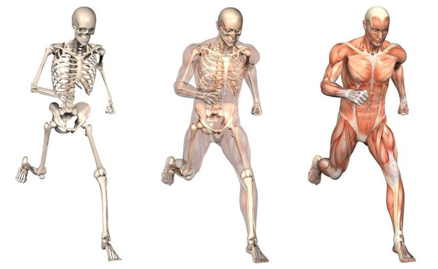
Biomechanics studies all life forms and their mechanical workings. Many think of biomechanics in sports and athletic performance, but biomechanics helps create and improve technologies, equipment, and injury rehabilitation techniques. (Tung-Wu Lu, Chu-Fen Chang 2012) Scientists, sports medicine doctors, physiotherapists, chiropractors, and conditioning specialists utilize biomechanics to help develop training protocols and techniques to improve therapy outcomes.
Body Movement
Biomechanics studies the movement of the body, including how muscles, bones, tendons, and ligaments work together, especially when movement is not optimal or correct. It is part of the larger field of kinesiology, specifically focusing on motion mechanics and analysis of how all the individual parts of the body work together to make up athletic and normal movements. (José M Vilar et al., 2013) Biomechanics includes:
Structure of bones and muscles.
Movement ability.
Mechanics of blood circulation, renal function, and other functions.
The study of forces and the effects of these forces on the tissues, fluid, or materials used for diagnosis, treatment, or research. (Jose I. Priego-Quesada 2021)
Sports
Sports biomechanics studies motion in exercising, training, and sports, which incorporates physics and the laws of mechanics. For example, the biomechanics of a specific exercise looks at:
Body position.
Movement of the feet, hips, knees, back, shoulders, and arms.
Knowing the correct movement patterns helps make the most of the exercise while preventing injuries, correcting form mistakes, informing training protocols, and increasing positive results. Understanding how the body moves and why it moves the way it does helps medical professionals prevent and treat injuries, alleviate pain symptoms, and improve performance.
Equipment
Biomechanics is used in the development of physical and sports equipment to improve performance. For example, a shoe can be designed for optimal performance for a skateboarder, long-distance runner, or soccer player. Playing surfaces are also studied for this purpose, such as how the surface stiffness of artificial turf affects athletic performance. (Jose I. Priego-Quesada 2021)
Individuals
Biomechanics can analyze an individual’s movements for more effective movement during training and games.
For example, an individual’s running gait or swing can be filmed with recommendations on what to change to improve.
Injuries
The science studies the causes, treatment, and prevention of neuromusculoskeletal injuries.
The research can analyze the forces that cause injuries and provide information for medical professionals on how to reduce the risk of injury.
Training
Biomechanics studies sports techniques and training systems to develop ways to improve efficiency.
This can include research on positioning, release, follow-through, etc.
It can analyze and help design new training techniques based on the mechanical demands of the sport, aimed at resulting in better performance.
For example, muscle activation is measured in cycling using electromyography and kinematics, which helps researchers analyze factors like posture, components, or exercise intensity that affect activation. (Jose I. Priego-Quesada 2021)
Motions
In biomechanics, the body’s motions are referred to from anatomical positioning:
Standing upright, with the gaze straight ahead
Arms at the sides
Palms facing forward
Feet spaced slightly apart, toes forward.
The three anatomical planes include:
Sagittal – median – Dividing the body into right and left halves is the sagittal/median plane. Flexion and extension occur in the sagittal plane.
Frontal – The frontal plane divides the body into front and back sides but also includes abduction, or moving a limb away from the center, and adduction, or moving a limb towards the center in the frontal plane.
Transverse – horizontal. – The upper and lower parts of the body are divided by the transverse/horizontal plane. Rotating movements occur here. (American Council on Exercise 2017)
Moving the body in all three planes occurs with daily activity. This is why performing exercises in each plane of motion to build strength, function, and stability is recommended.
Tools
Various tools are used to study biomechanics. Studies are usually performed using a device known as electromyography or EMG sensors. Sensors are placed on the skin and measure the amount and degree of muscle fiber activation in certain muscles during test exercises. EMGs can help:
Researchers understand which exercises are more effective than others.
Therapists know whether patients’ muscles are properly operating and functioning.
Dynamometers are another tool that helps measure muscle strength.
They measure the force output generated during muscle contractions to see if the muscles are sufficiently strong.
They are used to measure grip strength, which can be an indicator of overall strength, health, and longevity. (Li Huang et al., 2022)
Beyond Adjustments: Chiropractic and Integrative Healthcare
References
Lu, T. W., & Chang, C. F. (2012). Biomechanics of human movement and its clinical applications. The Kaohsiung journal of medical sciences, 28(2 Suppl), S13–S25. https://doi.org/10.1016/j.kjms.2011.08.004
Vilar, J. M., Miró, F., Rivero, M. A., & Spinella, G. (2013). Biomechanics. BioMed research international, 2013, 271543. https://doi.org/10.1155/2013/271543
Priego-Quesada J. I. (2021). Exercise Biomechanics and Physiology. Life (Basel, Switzerland), 11(2), 159. https://doi.org/10.3390/life11020159
American Council on Exercise. Makeba Edwards. (2017). Planes of Motion Explained (Exercise Science, Issue. https://www.acefitness.org/fitness-certifications/ace-answers/exam-preparation-blog/2863/the-planes-of-motion-explained/
Huang, L., Liu, Y., Lin, T., Hou, L., Song, Q., Ge, N., & Yue, J. (2022). Reliability and validity of two hand dynamometers when used by community-dwelling adults aged over 50 years. BMC geriatrics, 22(1), 580. https://doi.org/10.1186/s12877-022-03270-6
Knee injuries can present in physically active individuals that lift weights. Can understanding the types of weightlifting knee injuries help in prevention?
Weightlifting Knee Injuries
Weight training is very safe for the knees as regular weight training can improve knee strength and prevent injury as long as the correct form is followed. For Individuals with knee injuries from other activities, incorrect weight-training exercises could worsen the injury. (Ulrika Aasa et al., 2017) As well as, sudden twisting movements, poor alignment, and pre-existing injuries can increase the risk of worsening or creating further injuries. (Hagen Hartmann et al, 2013) The body and the knees are designed to support vertical forces on the joints.
Common Injuries
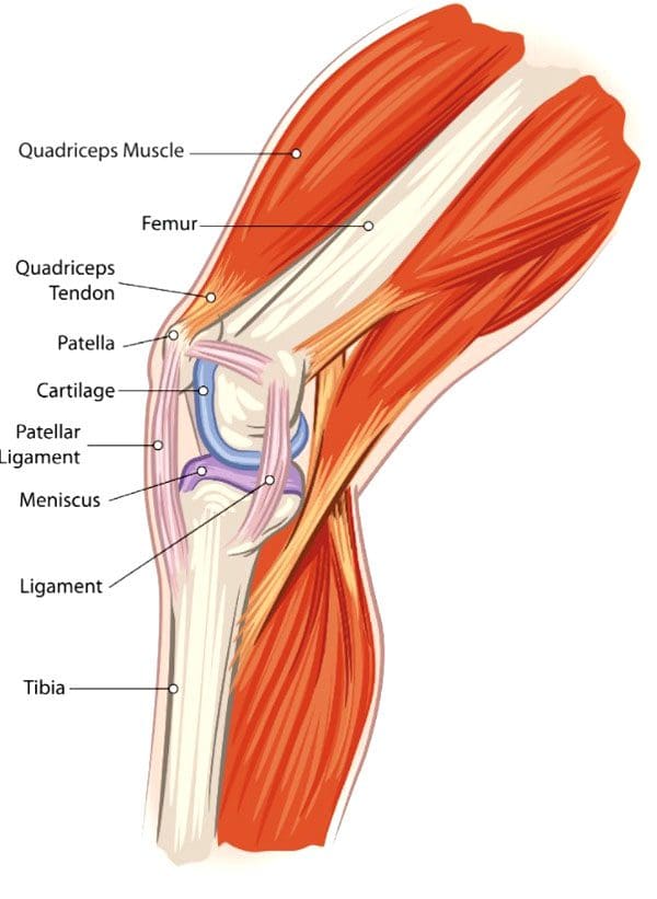
Weightlifting knee injuries occur as the knee joints endure a wide range of stresses and strains. In weight training, the ligaments that attach to the complex bone system of the knee joint can be damaged by incorrect movements, overloading the weight, and increasing the weight too soon. These injuries can result in pain, swelling, and immobility that can range from minor to severe, from a sprain or a slight tear to a complete tear in serious cases.
Anterior Cruciate Ligament – ACL – Injury
This ligament attaches the thigh’s femur bone to the lower leg’s shin bone/tibia and controls excessive rotation or extension of the knee joint. (American Academy of Family Physicians. 2024)
Anterior means front.
ACL injuries are seen mostly in athletes but can happen to anybody.
Severe damage to the ACL usually means surgical reconstruction and up to 12 months of rehabilitation.
When weightlifting, try to avoid twisting knee movements, intentionally or accidentally, under excessive load.
Posterior Cruciate Ligament – PCL – Injury
The PCL connects the femur and tibia at different points to the ACL.
It controls any backward motion of the tibia at the joint.
Injuries occur most with high-impact forces as a result of accidents and sometimes in activities where forceful trauma to the knee occurs.
Medial Collateral Ligament – MCL – Injury
This ligament maintains the knee from bending too far to the inside/medially.
Injuries mostly occur from impact to the outside of the knee or from accidental bodyweight force on the leg that bends at an unusual angle.
Lateral Collateral Ligament – LCL – Injury
This ligament connects the smaller bone of the lower leg/fibula to the femur.
It is opposite to the MCL.
It maintains excessive outward movement.
LCL injuries occur when a force pushes the knee out.
Cartilage Injury
Cartilage prevents bones from rubbing together and cushions impact forces.
Knee menisci are cartilage that cushions the knee joints inside and outside.
Other types of cartilage protect the thigh and shin bones.
When cartilage gets torn or damaged, surgery may be required.
Tendonitis
Aggravated and overused knee tendons can lead to weightlifting knee injuries.
A related injury known as iliotibial band syndrome/ITB causes pain to the outside of the knee, usually in runners, but it can occur from overuse.
Rest, stretching, physical therapy, and anti-inflammatory medication are a common treatment plan.
The condition causes the cartilage to deteriorate and bones to rub together, resulting in pain and stiffness.
Prevention
Individuals can minimize their risk of weightlifting knee injuries and pain by following their doctor’s and personal trainers’ recommendations.
Individuals with an existing knee injury should follow their doctor’s or physical therapist’s recommendations.
A knee sleeve can keep the muscles and joints secure, providing protection and support.
Stretching the leg and knee muscles can maintain joint flexibility.
Avoid sudden lateral movements.
Possible recommendations can include:
Avoiding Certain Exercises
Isolation exercises like leg curls, standing, or on a bench, as well as using the leg extension machine, can stress the knee.
Deep Squat Training
Research shows that the deep squat can protect against lower leg injury if the knee is healthy. However, this is when done with proper technique, under expert supervision, and with a gradual progressive load. (Hagen Hartmann et al, 2013)
Individuals should talk to their doctor before beginning a new exercise routine. A personal trainer can provide training in learning the proper technique and weightlifting form.
How I Tore my ACL Part 2
References
Aasa, U., Svartholm, I., Andersson, F., & Berglund, L. (2017). Injuries among weightlifters and powerlifters: a systematic review. British journal of sports medicine, 51(4), 211–219. https://doi.org/10.1136/bjsports-2016-096037
Hartmann, H., Wirth, K., & Klusemann, M. (2013). Analysis of the load on the knee joint and vertebral column with changes in squatting depth and weight load. Sports medicine (Auckland, N.Z.), 43(10), 993–1008. https://doi.org/10.1007/s40279-013-0073-6
American Academy of Family Physicians. ACL injury. (2024). ACL injury (Diseases and Conditions, Issue. https://familydoctor.org/condition/acl-injuries/
Mellinger, S., & Neurohr, G. A. (2019). Evidence based treatment options for common knee injuries in runners. Annals of translational medicine, 7(Suppl 7), S249. https://doi.org/10.21037/atm.2019.04.08
Driban, J. B., Hootman, J. M., Sitler, M. R., Harris, K. P., & Cattano, N. M. (2017). Is Participation in Certain Sports Associated With Knee Osteoarthritis? A Systematic Review. Journal of athletic training, 52(6), 497–506. https://doi.org/10.4085/1062-6050-50.2.08
For individuals experiencing lower back pain can understanding the anatomy and function of the multifidus muscle help in injury prevention and in the development of a highly effective treatment plan?
Multifidus Muscle
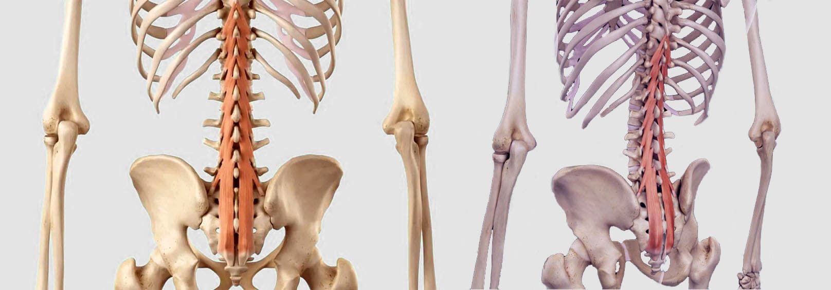
The multifidus muscles are long and narrow on either side of the spinal column, which helps stabilize the lower region of the spine or lumbar spine. (Maryse Fortin, Luciana Gazzi Macedo 2013) Sitting too much, practicing unhealthy postures, and lack of movement can progress to the multifidus muscle weakening or atrophy, which can lead to spinal instability, vertebral compression, and back pain. (Paul W. Hodges, Lieven Danneels 2019)
Anatomy
Known as the deep layer, it is the innermost layer of the three muscle layers of the back and controls the movement of the spine. The other two layers, known as the intrinsic and superficial, are responsible for the thoracic cage/rib cage and shoulder movement. (Anouk Agten et al., 2020) The multifidus has attachment points at:
The thoracic spine of the middle back.
The lumbar spine of the lower back.
The iliac spine – the base of the wing-shaped iliac bone of the pelvis.
Sacrum – series of bones at the base of the spine connected to the tailbone.
When standing or moving, the multifidus muscle works with the transversus abdominus and pelvic floor muscles to stabilize the lumbar spine. (Christine Lynders 2019)
Muscle Function
The main function is to stabilize the lower back, but it also helps extend the lower spine whenever reaching or stretching. (Jennifer Padwal et al., 2020) Because the muscle has numerous attachment points and is serviced by a specific branch of nerves known as the posterior rami, it allows each vertebra to work individually and more efficiently.
The multifidus muscle works with two other deep muscle groups to stabilize and move the spine. (Jeffrey J Hebert et al., 2015)
The rotatores muscle enables unilateral rotation, turning from side to side, and bilateral extension or bending backward and forward.
The semispinalis muscle above the multifidus allows extension and rotation of the head, neck, and upper back.
The multifidus muscle ensures spinal strength because it has more attachment points to the spine than the other layers, which reduces spinal flexibility and rotation but increases strength and stability. (Anouk Agten et al., 2020)
Lower Back Pain
A weak multifidus muscle destabilizes the spine and provides less support to the vertebra. This adds pressure on muscles and connective tissues between and adjacent to the spinal column, increasing the risk of lower back pain symptoms. (Paul W. Hodges, Lieven Danneels 2019) The loss of muscle strength and stability can cause atrophy or wasting away. This can cause compression and other back problems. (Paul W. Hodges et al., 2015) Back problems associated with multifidus muscle deterioration include (Paul W. Hodges, Lieven Danneels 2019)
Herniated discs – also bulging or slipped discs.
Nerve entrapment or compression pinched nerve.
Sciatica
Referred pain – nerve pain originating from the spine felt in other areas.
Osteoarthritis – wear-and-tear arthritis
Spinal osteophytes – bone spurs
Weak abdominal or pelvic floor muscles can compromise the core, increasing the risk of chronic lower back pain and injury.
Individuals are recommended to consult a physical therapist and chiropractor who can help develop the appropriate treatment, rehabilitation, and strengthening plan based on age, injury, underlying conditions, and physical abilities.
Can Core Exercises Help with Back Pain?
References
Fortin, M., & Macedo, L. G. (2013). Multifidus and paraspinal muscle group cross-sectional areas of patients with low back pain and control patients: a systematic review with a focus on blinding. Physical therapy, 93(7), 873–888. https://doi.org/10.2522/ptj.20120457
Hodges, P. W., & Danneels, L. (2019). Changes in Structure and Function of the Back Muscles in Low Back Pain: Different Time Points, Observations, and Mechanisms. The Journal of orthopaedic and sports physical therapy, 49(6), 464–476. https://doi.org/10.2519/jospt.2019.8827
Agten, A., Stevens, S., Verbrugghe, J., Eijnde, B. O., Timmermans, A., & Vandenabeele, F. (2020). The lumbar multifidus is characterised by larger type I muscle fibres compared to the erector spinae. Anatomy & cell biology, 53(2), 143–150. https://doi.org/10.5115/acb.20.009
Lynders C. (2019). The Critical Role of Development of the Transversus Abdominis in the Prevention and Treatment of Low Back Pain. HSS journal : the musculoskeletal journal of Hospital for Special Surgery, 15(3), 214–220. https://doi.org/10.1007/s11420-019-09717-8
Padwal, J., Berry, D. B., Hubbard, J. C., Zlomislic, V., Allen, R. T., Garfin, S. R., Ward, S. R., & Shahidi, B. (2020). Regional differences between superficial and deep lumbar multifidus in patients with chronic lumbar spine pathology. BMC musculoskeletal disorders, 21(1), 764. https://doi.org/10.1186/s12891-020-03791-4
Hebert, J. J., Koppenhaver, S. L., Teyhen, D. S., Walker, B. F., & Fritz, J. M. (2015). The evaluation of lumbar multifidus muscle function via palpation: reliability and validity of a new clinical test. The spine journal : official journal of the North American Spine Society, 15(6), 1196–1202. https://doi.org/10.1016/j.spinee.2013.08.056
Hodges, P. W., James, G., Blomster, L., Hall, L., Schmid, A., Shu, C., Little, C., & Melrose, J. (2015). Multifidus Muscle Changes After Back Injury Are Characterized by Structural Remodeling of Muscle, Adipose and Connective Tissue, but Not Muscle Atrophy: Molecular and Morphological Evidence. Spine, 40(14), 1057–1071. https://doi.org/10.1097/BRS.0000000000000972
For individuals suffering from back pain, can knowing basic chiropractic terminology help in understanding diagnosis and treatment plan development?
Chiropractic Terminology
The chiropractic principle is that a properly aligned spine positively affects an individual’s overall health. One of the main aspects of chiropractic care is applying calculated force to the spinal joints to restore correct spinal alignment. Chiropractic terminology describes specific types of techniques and care.
General Subluxation
A subluxation can mean different things for various doctors. In general, a subluxation is a significant structural displacement or an incomplete or partial dislocation of a joint or organ.
To medical doctors, a subluxation refers to a partial dislocation of a vertebrae.
This is a serious condition, usually brought on by trauma, that can result in a spinal cord injury, paralysis, and/or death.
X-rays show a conventional subluxation as an obvious disconnect between the vertebrae.
Chiropractic Subluxation
The chiropractic interpretation is more subtle and refers to the misalignment of adjacent spinal vertebrae.
Subluxation in this context refers to position changes in the joints and soft tissues of the spine.
Vertebral misalignment is believed to lead to pain and abnormal intervertebral joint motion.
This difference between the serious subluxation medical condition and the chiropractic version may cause individuals to dismiss seeking back pain treatments.
Motion Segment
Chiropractors and surgeons use it as a technical term.
Motion segment refers to two adjacent vertebrae and the intervertebral disc between them.
This is the area chiropractors assess and adjust.
Adjustment
The chiropractor performs a spinal manual adjustment to realign joint subluxations.
Adjustments involve applying force to motion segments to bring them back into a centered alignment.
The goal for adjustments and realigning the vertebrae includes:
Spinal manipulation is a technique used by chiropractors to provide relief for musculoskeletal pain related to the back and neck. Manipulation provides mild to moderate relief and works as well as some conventional treatments like pain-relieving medications. (Sidney M. Rubinstein et al., 2012)
Spinal manipulation is divided into grades of mobilization.
Depending on their training, practitioners of various medical disciplines may be licensed to perform grade 1 to grade 4 mobilizations.
Only physical therapists, osteopathic physicians, and chiropractors are licensed to perform grade 5 mobilizations, which are high-velocity thrust techniques.
Most massage therapists, athletic trainers, and personal trainers are not licensed to perform spinal manipulations.
Based on a systematic review, the effectiveness of these treatments found that there is quality evidence that manipulation and mobilization can help reduce pain and improve function for individuals with chronic low back pain, with manipulation appearing to produce a more profound effect than mobilization. Both therapies are safe, with multimodal treatments potentially being an effective option. (Ian D. Coulter et al., 2018)
As with any treatment, results vary from person to person and with different chiropractors. There are also potential risks with spinal manipulation. Though rare, cervical, carotid, and vertebral artery dissections have occurred with cervical/neck manipulation. (Kelly A. Kennell et al., 2017) Individuals with osteoporosis may be advised to avoid chiropractic adjustments or manipulation because of the increased risk of injury. (James M. Whedon et al., 2015)
Many individuals choose chiropractic treatment for a variety of conditions. Understanding chiropractic terminology and reasoning allows individuals to ask questions as they discuss their symptoms to develop a personalized treatment plan and restore function and wellness.
What Causes Disc Herniation?
References
Henderson C. N. (2012). The basis for spinal manipulation: chiropractic perspective of indications and theory. Journal of electromyography and kinesiology : official journal of the International Society of Electrophysiological Kinesiology, 22(5), 632–642. https://doi.org/10.1016/j.jelekin.2012.03.008
Blanchette, M. A., Stochkendahl, M. J., Borges Da Silva, R., Boruff, J., Harrison, P., & Bussières, A. (2016). Effectiveness and Economic Evaluation of Chiropractic Care for the Treatment of Low Back Pain: A Systematic Review of Pragmatic Studies. PloS one, 11(8), e0160037. https://doi.org/10.1371/journal.pone.0160037
Rubinstein, S. M., Terwee, C. B., Assendelft, W. J., de Boer, M. R., & van Tulder, M. W. (2012). Spinal manipulative therapy for acute low-back pain. The Cochrane database of systematic reviews, 2012(9), CD008880. https://doi.org/10.1002/14651858.CD008880.pub2
Coulter, I. D., Crawford, C., Hurwitz, E. L., Vernon, H., Khorsan, R., Suttorp Booth, M., & Herman, P. M. (2018). Manipulation and mobilization for treating chronic low back pain: a systematic review and meta-analysis. The spine journal : official journal of the North American Spine Society, 18(5), 866–879. https://doi.org/10.1016/j.spinee.2018.01.013
Kennell, K. A., Daghfal, M. M., Patel, S. G., DeSanto, J. R., Waterman, G. S., & Bertino, R. E. (2017). Cervical artery dissection related to chiropractic manipulation: One institution’s experience. The Journal of family practice, 66(9), 556–562.
Whedon, J. M., Mackenzie, T. A., Phillips, R. B., & Lurie, J. D. (2015). Risk of traumatic injury associated with chiropractic spinal manipulation in Medicare Part B beneficiaries aged 66 to 99 years. Spine, 40(4), 264–270. https://doi.org/10.1097/BRS.0000000000000725
For individuals looking to improve their diet, can knowing the different salt types help in food preparation and health?
Salt Types
Salt brings out the natural flavor of foods and can be used as a preservative. Salt types come in various colors and textures for cooking, flavor, and health. Some are considered healthier compared to regular table salt, like pink Himalayan salt and different sea salts. Some individuals prefer them because most go through less processing and can have more trace minerals like magnesium and potassium. However, all salts are healthy in moderation, as sodium is a necessary part of a balanced diet. Although essential for the body, sodium can be harmful when too much is consumed. A study examining consumer-grade pink Himalayan sea salts available in Australia determined that to receive the additional health benefits of the minerals from this type of salt, individuals must consume so much that it elevates the amount of sodium in the body to dangerous levels. (Flavia Fayet-Moore et al., 2020)
Salt
Salt is a mineral made from the combined elements:
Sodium – Na
Chlorine -Cl
Together, they form crystallized sodium chloride NaCl.
The majority of salt production comes from evaporated seawater and salt mines. Many salts used in food preparation are iodized. Iodine is added to various refined salt products to help meet nutritional requirements. Iodine intake levels that fall below the recommended values could result in a deficiency and develop goiter. Goiter is associated with hypothyroidism. (Angela M. Leung et al., 2021) Lack of iodine can also have adverse effects on growth and development. (National Institutes of Health Office of Dietary Supplements. 2023)
Essential for Health
Salt sustains life and optimal bodily function. Sodium and chlorine are important elements that maintain:
Cellular balance
Circulation
Blood sugar levels
Sodium is a mineral and an electrolyte. Common electrolytes include potassium, calcium, and bicarbonate. Without adequate sodium levels, the brain cannot send the necessary impulses to the rest of the body to function properly. However, consuming too much salt can cause health issues.
Higher salt intake in individuals who are sensitive to salt can increase blood pressure.
Doctors usually recommend that individuals with hypertension reduce sodium intake or follow a low-sodium diet.
Elevated sodium levels also cause water retention – considered a protective response as the body works to regulate serum sodium levels concentration in the blood to maintain balance.
If levels are too high, a condition known as hypernatremia can develop, which can cause:
Excessive thirst
Vomiting
Infrequent urination
Diarrhea
Sodium levels that are too low can lead to hyponatremia, which can cause:
Though there are different types of salt, they all contain roughly the same amount of sodium.
Types
The average sodium intake by adults is around 3,393mg per day, ranging between 2,000–5,000mg. Guidelines recommend a maximum intake of 2,300mg per day. (U.S. Department of Health and Human Services and U.S. Department of Agriculture. 2020) Whether from unhealthy dietary choices like processed foods or incorrect knowledge of sodium content when cooking, an American Heart Association survey showed that more than half of respondents inaccurately stated that sea salt had a lower sodium content than table salt. (American Heart Association. 2024)
Refined – Table Salt
Refined/iodized salt is finely granulated and commonly used in cooking. This type is highly refined to remove impurities and eliminate trace minerals often found in specialty salts. Because the salt is finely ground, anti-caking agents are added to ensure the salt doesn’t clump. Some table salts also have added sugar and other additives.
Refined table salt is about 97–99% sodium chloride (NaCl).
Iodine is added to prevent iodine deficiency.
Individuals trying to reduce sodium intake but meet iodine levels can do so with foods like eggs, dairy products, and fish.
Kosher
Kosher salt is coarse and flakey and can add a crunchy texture to dishes and drinks. Pure kosher salt does not contain additives like anti-caking agents and iodine. The size of the salt crystals is ideal for drawing out moisture.
Per teaspoon, kosher salt generally has less sodium than 1 teaspoon of table salt.
Because it has a coarser grain, less salt fits in the measuring spoon.
Sea Salt
Sea salt is produced from evaporated seawater and comes as fine grains or large crystals. Examples include:
Black Sea
Celtic
French – fleur de sel
Hawaiian sea salt
Sea salt can have trace amounts of minerals like iron, potassium, and zinc, which can produce different flavors in cooking but no additional health benefits with normal consumption. Some sea salts may also contain trace amounts of microplastics. However, research indicates these amounts are too low to warrant public health concerns. (Ali Karami et al., 2017)
Himalayan Pink Salt
Himalayan pink salt is mined in the red salt range in Pakistan, the second-largest salt mine in the world, and in the Andes mountains of Peru. Trace amounts of iron oxide make the salt pink. It is typically used at the end of cooking to add flavor and a crunch. Himalayan salt is popular for its health benefits and mineral properties. However, using Himalayan salt over other types has no known health advantages. Researchers concluded that the potential health benefits provided by the higher nutrient content would be counteracted by the large amount of sodium that would need to be consumed. (Flavia Fayet-Moore et al., 2020)
Substitutes
Salt substitutes contain some or all sodium and potassium, magnesium, or other minerals. Substitutes can be half sodium chloride and half potassium chloride. Monosodium glutamate/MSG can also be used as an alternative. A study found that substituting salt with MSG is safe and comparable to salt flavor. (Jeremia Halim et al., 2020) Individuals often use substitutes on a sodium-restricted diet but should check with their doctor before using these products, especially if they have kidney conditions.
Body In Balance – Chiropractic+Fitness+Nutrition
References
Fayet-Moore, F., Wibisono, C., Carr, P., Duve, E., Petocz, P., Lancaster, G., McMillan, J., Marshall, S., & Blumfield, M. (2020). An Analysis of the Mineral Composition of Pink Salt Available in Australia. Foods (Basel, Switzerland), 9(10), 1490. https://doi.org/10.3390/foods9101490
Leung, A. M., Braverman, L. E., & Pearce, E. N. (2012). History of U.S. iodine fortification and supplementation. Nutrients, 4(11), 1740–1746. https://doi.org/10.3390/nu4111740
National Institutes of Health Office of Dietary Supplements. (2023). Iodine: Fact Sheet for Professionals. Retrieved from https://ods.od.nih.gov/factsheets/Iodine-HealthProfessional/
U.S. National Library of Medicine. MedlinePlus. (2022). Sodium blood test. Retrieved from https://medlineplus.gov/lab-tests/sodium-blood-test/
U.S. Department of Agriculture. FoodData Central. (2020). Salt. Retrieved from https://fdc.nal.usda.gov/fdc-app.html#/food-details/1112305/nutrients
U.S. Department of Health and Human Services and U.S. Department of Agriculture. (2020). 2020–2025 Dietary Guidelines for Americans. Retrieved from https://www.dietaryguidelines.gov/sites/default/files/2020-12/Dietary_Guidelines_for_Americans_2020-2025.pdf
American Heart Association. (2024). Sea Salt vs. Table Salt (Healthy Living, Issue. https://www.heart.org/en/healthy-living/healthy-eating/eat-smart/sodium/sea-salt-vs-table-salt
Karami, A., Golieskardi, A., Keong Choo, C., Larat, V., Galloway, T. S., & Salamatinia, B. (2017). The presence of microplastics in commercial salts from different countries. Scientific reports, 7, 46173. https://doi.org/10.1038/srep46173
Halim, J., Bouzari, A., Felder, D., & Guinard, J. X. (2020). The Salt Flip: Sensory mitigation of salt (and sodium) reduction with monosodium glutamate (MSG) in “Better-for-You” foods. Journal of food science, 85(9), 2902–2914. https://doi.org/10.1111/1750-3841.15354
For individuals who have decided to start exercising for fitness and health, walking is a great place to start. Can planning a walking exercise schedule help individuals maintain a fitness routine and improve endurance and speed quicker?
Walking Exercise Planning Schedule
While any amount of walking benefits health, individuals can increase the benefits by walking more per week or by increasing the pace. Brisk walking for 30 minutes per day, totaling 150 minutes per week, is recommended by health experts to decrease risks for heart disease, stroke, diabetes, and other conditions. (Centers for Disease Control and Prevention. 2022)
Individuals with ongoing health conditions should talk to their doctor before starting any new exercise program.
Beginners are encouraged to focus on using proper walking posture and technique to steadily improve strength and endurance.
The increased duration or intensity can help if weight loss is a goal.
Improving diet is also necessary for the best results.
Individuals can build healthy walking habits by tracking walks.
Schedule
Checklist
Individuals can walk outdoors, indoors, or on a treadmill.
Wear proper athletic shoes and clothing.
Check walking posture.
Walk at an easy pace for a couple of minutes before picking up speed.
First Week
An example of what a walking exercise schedule can look like, but it’s advised to consult a professional trainer to develop a personalized fitness plan.
Start with a 15-minute walk at an easy pace.
Walk five days the first week.
Building a healthy habit is the goal, so consistency is important.
Spread out rest days, like making days 3 and 6 rest days.
Weekly goal – 60 to 75 minutes
Second Week
Add five minutes, so the walk time increases gradually.
Or, individuals can extend more on some days, followed by a rest day.
Weekly goal – 80 to 100 minutes
Third Week
Add five more minutes with each session, so the walk increases to 25 minutes.
Weekly goal – 100 to 125 minutes
Fourth Week
Add another five minutes to increase the walk to 30 minutes.
Weekly goal – 120 to 150 minutes
Individuals who find any week to be difficult are suggested to repeat that week instead of adding time until they are able to progress naturally. Once able to walk for 30 minutes at a time comfortably, individuals are ready for a variety of different walking exercise workouts to add intensity and endurance. A weekly walking plan can include:
Longer walks
Higher-intensity walks
Speed-building walks
Beginner Walking Speed
An individual’s objective should be brisk walking to achieve a moderate-intensity workout. This is the intensity that is associated with the most health benefits.
If the speed is slower and the heart rate is lower during the initial weeks, this is normal.
The first goal is to walk for 30 to 60 minutes a day without injury.
Adding speed and intensity gradually.
Staying consistent in regularly walking before trying to walk faster and longer.
Using proper walking posture and arm motion will help in faster walking.
To reduce the risk of injury, gradually increase the length of the walk or pace, only changing one component at a time.
Individuals may consider joining a walking group or club to have others to walk with and an incentive to maintain regular walking.
Home Exercises for Pain Relief
References
Centers for Disease Control and Prevention. (2022). How Much Physical Activity Do Adults Need? Retrieved from https://www.cdc.gov/physicalactivity/basics/adults/index.htm
Centers for Disease Control and Prevention. (2022). Measuring Physical Activity Intensity. Retrieved from https://www.cdc.gov/physicalactivity/basics/measuring/index.html
Centers for Disease Control and Prevention. (2022). Target Heart Rate and Estimated Maximum Heart Rate. Retrieved from https://www.cdc.gov/physicalactivity/basics/measuring/heartrate.htm
Mahmod, S. R., Narayanan, L. T., & Supriyanto, E. (2018). Effects of incremental cardiorespiratory exercise on the speech rate and the estimated exercise intensity using the counting talk test. Journal of physical therapy science, 30(7), 933–937. https://doi.org/10.1589/jpts.30.933
During a fall individuals tend to automatically outstretch their hands to help break a fall, which can slam onto the ground causing a falling onto an outstretched hand or FOOSH injury. Should individuals get checked by a healthcare provider if they believe there is no injury?
FOOSH Injuries
Falling down usually results in minor injuries. A FOOSH injury occurs when falling down and trying to break the fall by reaching out with the hand/s. This can result in an upper extremity injury like a sprain or a fracture. But sometimes, falling on one’s hands can lead to serious injuries and/or create future musculoskeletal issues. Individuals who have fallen or suffered a FOOSH injury should consult their healthcare provider and then a physical therapist or chiropractor to safely develop a treatment plan to rehabilitate, strengthen, and expedite recovery.
After The Injury
For individuals who have fallen down and landed on their hand, wrist, or arm, here are a few things to ensure the proper care for the injury, including:
Follow the R.I.C.E. protocol for acute injuries
Visit a healthcare provider or local emergency clinic
Contact a physical therapist
A FOOSH injury could be or become serious, so to avoid letting small issues become big problems, get examined by a musculoskeletal specialist. The healthcare provider will obtain an imaging scan of the injured and surrounding areas. They will perform a physical examination to determine the type of injury, like a sprain or muscle strain. Not getting appropriate medical treatment after a fall can result in chronic pain and loss of function. (J. Chiu, S. N. Robinovitch. 1998)
Common Injuries
A FOOSH injury can injure different areas. These usually involve the wrist and hand, but the elbow or shoulder can also be injured. Common injuries include:
Colles’ fracture
A wrist fracture where the end of the arm bone is displaced backward.
Smith’s fracture
A wrist fracture, similar to a Colles’ fracture, is where the end of the arm bone is displaced towards the front of the wrist.
Boxer’s fracture
A fracture of the small bones in the hand.
Typically, it occurs after punching something, but it can happen from falling on an outstretched fist.
Elbow dislocation or fracture
The elbow can pop out of the joint or can break a bone in the elbow.
Collarbone fracture
The force from falling with the hands and arms outstretched can travel up to the collarbone, causing a fracture.
Proximal humeral fracture
Falling onto an outstretched hand injury can cause the arm bone to get jammed into the shoulder, causing a proximal humeral fracture.
Shoulder dislocation
The shoulder can pop out of the joint.
This can cause a rotator cuff tear or labrum injury.
Regardless of the injury, individuals should visit a healthcare provider to evaluate the damage. If the injury is serious, the practitioner can make an accurate or differential diagnosis and develop a treatment plan. (William R. VanWye et al., 2016)
Physical Therapy
Individuals can benefit from physical therapy to help recover and return to their previous level of function. Physical therapy varies depending on the specific injury, but generally, a physical therapist can help individuals return to function after a fall on an outstretched hand. (William R. VanWye et al., 2016) Common treatments can include:
Treatments and modalities to decrease pain, inflammation, and swelling.
Instruction on how to wear an arm sling properly.
Exercises and stretches to improve the range of motion, strength, and functional mobility.
Balance exercises.
Scar tissue management if surgery was necessary.
The therapy team will ensure the proper treatment is utilized to quickly and safely return to normal activities.
Chiropractic Care For Healing After Trauma
References
Chiu, J., & Robinovitch, S. N. (1998). Prediction of upper extremity impact forces during falls on the outstretched hand. Journal of biomechanics, 31(12), 1169–1176. https://doi.org/10.1016/s0021-9290(98)00137-7
VanWye, W. R., Hoover, D. L., & Willgruber, S. (2016). Physical therapist screening and differential diagnosis for traumatic-onset elbow pain: A case report. Physiotherapy theory and practice, 32(7), 556–565. https://doi.org/10.1080/09593985.2016.1219798
IFM's Find A Practitioner tool is the largest referral network in Functional Medicine, created to help patients locate Functional Medicine practitioners anywhere in the world. IFM Certified Practitioners are listed first in the search results, given their extensive education in Functional Medicine