“Various problems with the sacrum make up or contribute to a significant portion of lower back problems. Can understanding the anatomy and function help prevent and treat back injuries?”
The Sacrum
The sacrum is a bone shaped like an upside-down triangle located at the base of the spine that helps support the upper body when sitting or standing and provides pelvic girdle flexibility during childbirth. It comprises five vertebrae that fuse during adulthood and connect to the pelvis. This bone takes and endures all of the body’s pressure and stress from everyday activities and movements.
Formation
Humans are born with four to six sacral vertebrae. However, fusion does not take place in all sacral vertebrae simultaneously:
Fusion starts with the S1 and S2.
As the individual gets older, the overall shape of the sacrum begins to solidify, and the vertebrae fuse into a single structure.
The process usually starts in the mid-teens and finishes in the early to mid-twenties.
It is believed to start earlier in females than males.
The sacrum in a female is wider and shorter and has a more curved top or the pelvic inlet.
The male sacrum is longer, narrower, and flatter.
Structure
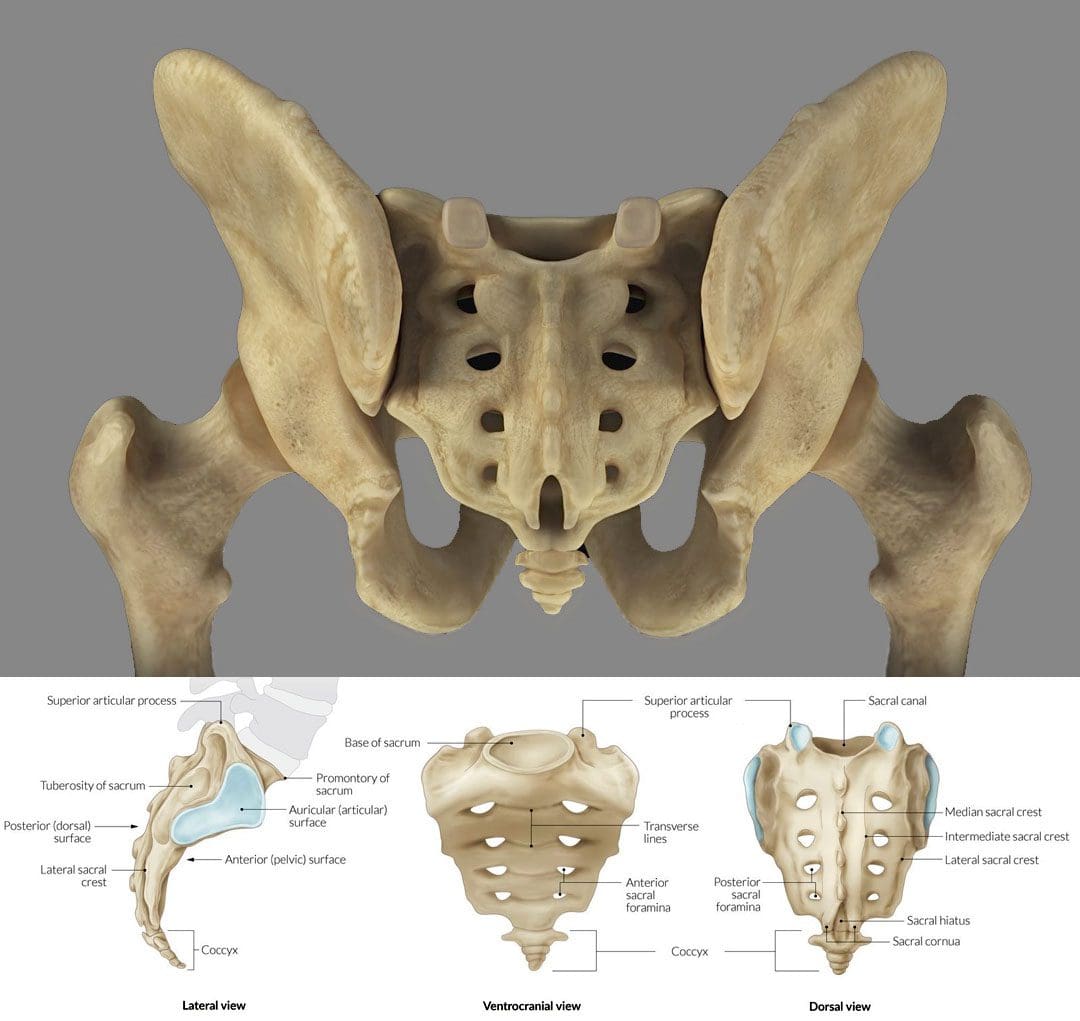
The sacrum is an irregular bone that makes up the back/posterior third of the pelvic girdle. There is a ridge across the front/anterior portion of the S1 vertebra known as the sacral promontory. Small holes/foramen on both sides of the sacrum are left over after the vertebrae fuse together. Depending on the number of vertebrae, there can be three to five foramen on each side, though there are usually four. (E. Nastoulis, et al., 2019)
Each anterior foramen is typically wider than the posterior or dorsal/backside foramen.
Each sacral foramina/plural of foramen provides a channel for the sacral nerves and blood vessels.
Small ridges develop between each of the fused vertebrae, known as transverse ridges or lines.
The top of the sacrum is called the base and is connected to the largest and lowest of the lumbar vertebrae – L5.
The bottom is connected to the tailbone/coccyx, known as the apex.
The sacral canal is hollow, runs from the base to the apex, and serves as a channel at the end of the spinal cord.
The sides of the sacrum connect to the right and left hip/iliac bones. The attachment point is the auricular surface.
Right behind the auricular surface is the sacral tuberosity, which serves as an attachment area for the ligaments that hold the pelvic girdle together.
Location
The sacrum is at the level of the lower back, just above the intergluteal cleft or where the buttocks split. The cleft starts at around the level of the tailbone or coccyx. The sacrum is curved forward and ends at the coccyx, with the curvature being more pronounced in females than males. It connects to the L5 lumbar vertebra by way of the lumbosacral joint. The disc between these two vertebrae is a common source of low back pain.
On either side of the lumbosacral joint are wing-like structures known as the sacral ala, which connect to the iliac bones and form the top of the sacroiliac joint.
These wings provide stability and strength for walking and standing.
Anatomical Variations
The most common anatomical variation applies to the number of vertebrae. The most common is five, but anomalies have been documented, including individuals with four or six sacral vertebrae. (E. Nastoulis, et al., 2019)
Other variations involve the sacrum’s surface and curvature, where the curvature differs widely between individuals.
In some cases, the first and second vertebrae do not fuse and remain separately articulated.
Failure of the canal to completely close during formation is a condition known as spina bifida.
Function
Studies on the sacrum are ongoing, but some proven functions include:
It serves as an anchor point for the spinal column to attach to the pelvis.
It provides stability for the body’s core.
It acts as a platform for the spinal column to rest on when sitting.
It facilitates childbirth, providing pelvic girdle flexibility.
It supports upper body weight when sitting or standing.
It provides extra stability for walking, balance, and mobility.
Conditions
The sacrum can be a main source or focal point for lower back pain. It is estimated that 28% of men and 31.6% of women aged 18 years or older have experienced low back pain in the past three months. (Centers for Disease Control and Prevention. 2020) Conditions that can cause sacrum pain symptoms include.
Sacroiliitis
This is a common condition of sacroiliac/SI joint inflammation.
A doctor only makes the diagnosis when all other possible causes of pain have been ruled out, known as a diagnosis of exclusion.
About half of all chordomas form in the sacrum, but the tumors can also develop elsewhere in the vertebral column or at the base of the skull. (National Library of Medicine. 2015)
Spina Bifida
Individuals can be born with conditions that affect the sacrum.
Spina bifida is a congenital condition that can arise from the malformation of the sacral canal.
Unlocking the Secrets of Inflammation
References
Gruss, L. T., & Schmitt, D. (2015). The evolution of the human pelvis: changing adaptations to bipedalism, obstetrics and thermoregulation. Philosophical transactions of the Royal Society of London. Series B, Biological sciences, 370(1663), 20140063. https://doi.org/10.1098/rstb.2014.0063
Nastoulis, E., Karakasi, M. V., Pavlidis, P., Thomaidis, V., & Fiska, A. (2019). Anatomy and clinical significance of sacral variations: a systematic review. Folia morphologica, 78(4), 651–667. https://doi.org/10.5603/FM.a2019.0040
Barros, G., McGrath, L., & Gelfenbeyn, M. (2019). Sacroiliac Joint Dysfunction in Patients With Low Back Pain. Federal practitioner : for the health care professionals of the VA, DoD, and PHS, 36(8), 370–375.
Can combining chiropractic treatment with the common therapies of medication, exercise, and/or physical therapy help relieve sciatic endometriosis pain symptoms?
Sciatic Endometriosis
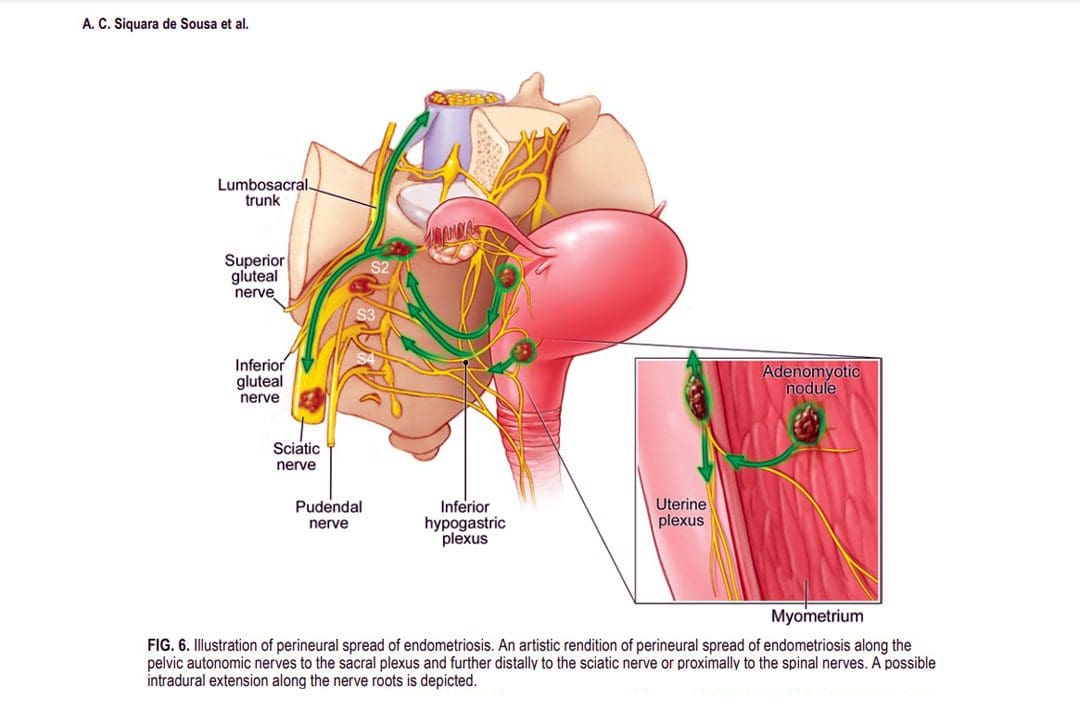
Sciatic endometriosis is a condition in which endometrial cells (tissue that resembles the lining of the uterus) grow outside of the uterine lining and compress the sciatic nerve. This places stress and pressure on the nerve causing back, pelvic, hip, and leg pain, especially before and during the menstrual cycle. It can also cause pain, irregular periods, and infertility. (The American College of Obstetricians and Gynecologists. 2021)
These areas of endometrial tissue growth are also known as lesions or implants.
Women with sciatic endometriosis often experience leg pain and weakness around the time of their menstrual cycle. (Lena Marie Seegers, et al., 2023)
Sciatic endometriosis can also cause pain when urinating, during a bowel movement, during sex, and fatigue, and irregular vaginal bleeding.
The abnormal growth may be caused by higher-than-normal levels of estrogen.
Researchers believe that endometriosis is related to retrograde menstruation, which causes menstrual blood to flow back into the pelvis instead of out through the vagina. (World Health Organization. 2023)
Sometimes, the cells grow in the area of the pelvis right above the sciatic nerve. (Adaiah Yahaya, et al., 2021)
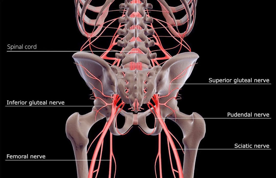
The sciatic nerve is the longest nerve in the body and travels down the back of each leg. (Johns Hopkins Medicine. 2023)
When endometrial lesions place pressure on the sciatic nerve, they can cause irritation and inflammation leading to severe pelvic pain, which makes it harder to conceive. (Liang Yanchun, et al., 2019)
Symptoms
Some women with endometriosis experience no symptoms or misinterpret the symptoms as typical premenstrual syndrome/PMS signs. The most common signs and symptoms of sciatic endometriosis include:
Difficulty walking or standing.
Loss of sensation, muscle weakness, and reflex alteration.
Limping.
Balance problems.
Bloating and nausea.
Constipation or diarrhea before or after a period.
Painful, heavy, and/or irregular periods.
Bleeding between periods.
Pain during sex, urination, and bowel movements.
Pain in the stomach, pelvis, lower back, hips, and buttocks. (MedlinePlus. 2022)
Weakness, numbness, tingling, burning, or dull aching sensations in the back of one or both legs.
Endometriosis, including sciatic endometriosis, typically cannot be diagnosed with a pelvic examination or ultrasound by themselves. A healthcare provider may need to perform a biopsy using laparoscopy and discuss menstrual cycles, symptoms, and medical history.
The laparoscopy procedure involves making tiny incisions and taking a tissue sample with tools attached to a thin tube with a camera. (MedlinePlus. 2022)
Imaging tests, like magnetic resonance imaging/MRI, and computed tomography/CT scans, can help provide essential information about the location and size of any endometrial lesions. (The American College of Obstetricians and Gynecologists. 2021)
Treatment
Symptoms can sometimes be temporarily relieved with over-the-counter/OTC pain relievers. Depending on the condition and severity a healthcare provider may prescribe hormonal treatment to prevent new endometrial implants from growing. These can include:
Hormonal birth control.
Progestin – a synthetic form of progesterone.
Gonadotropin-releasing hormone – GnRH agonists.
If pain persists or worsens, individuals may need to undergo surgery to remove the tissue.
The American College of Obstetricians and Gynecologists. Endometriosis.
Seegers, L. M., DeFaria Yeh, D., Yonetsu, T., Sugiyama, T., Minami, Y., Soeda, T., Araki, M., Nakajima, A., Yuki, H., Kinoshita, D., Suzuki, K., Niida, T., Lee, H., McNulty, I., Nakamura, S., Kakuta, T., Fuster, V., & Jang, I. K. (2023). Sex Differences in Coronary Atherosclerotic Phenotype and Healing Pattern on Optical Coherence Tomography Imaging. Circulation. Cardiovascular imaging, 16(8), e015227. https://doi.org/10.1161/CIRCIMAGING.123.015227
World Health Organization. Endometriosis.
Yahaya, A., Chauhan, G., Idowu, A., Sumathi, V., Botchu, R., & Evans, S. (2021). Carcinoma arising within sciatic nerve endometriosis: a case report. Journal of surgical case reports, 2021(12), rjab512. https://doi.org/10.1093/jscr/rjab512
Johns Hopkins Medicine. Sciatica.
Yanchun, L., Yunhe, Z., Meng, X., Shuqin, C., Qingtang, Z., & Shuzhong, Y. (2019). Removal of an endometrioma passing through the left greater sciatic foramen using a concomitant laparoscopic and transgluteal approach: case report. BMC women’s health, 19(1), 95. https://doi.org/10.1186/s12905-019-0796-0
MedlinePlus. Endometriosis.
Center for Endometriosis Care. Sciatic endometriosis.
Chen, S., Xie, W., Strong, J. A., Jiang, J., & Zhang, J. M. (2016). Sciatic endometriosis induces mechanical hypersensitivity, segmental nerve damage, and robust local inflammation in rats. European journal of pain (London, England), 20(7), 1044–1057. https://doi.org/10.1002/ejp.827
Siquara de Sousa, A. C., Capek, S., Howe, B. M., Jentoft, M. E., Amrami, K. K., & Spinner, R. J. (2015). Magnetic resonance imaging evidence for perineural spread of endometriosis to the lumbosacral plexus: report of 2 cases. Neurosurgical focus, 39(3), E15. https://doi.org/10.3171/2015.6.FOCUS15208
Can chiropractic treatment protocols diagnose what’s causing head pressure in individuals, and provide effective treatment?
Head Pressure
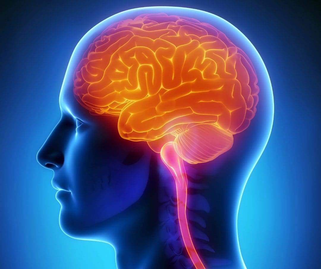
Head pressure can have various causes and symptoms that affect different areas depending on whether the cause is a headache, allergies, injury, illness, or disease. The location of the pressure or pain can help a doctor of chiropractic determine the cause.
The underlying factor is usually not life-threatening, but the pressure that has built can be the result of serious conditions like a head injury or brain tumor.
Chiropractic care, which includes a combination of spinal manipulation, active and passive exercises, and massage, is often used for headache management and prevention. (Moore Craig, et al., 2018)
Chiropractic therapy is often sought out for tension and cervicogenic headaches, migraines, and each responds differently to the treatment.
The Head
The head is made up of a complex system of lobes, sinuses/channels, blood vessels, nerves, and ventricles. (Thau L, et al., 2022)
The pressure of these systems is regulated and any disruption to this balance can be noticeable.
Diagnosis can be difficult to figure out what is causing discomfort or head pressure.
Pain, pressure, irritability, and nausea are all symptoms that can occur with headaches. (Rizzoli P, Mullally W. 2017)
Head pressure can also occur only at times when standing up, bending down to pick up an object, or otherwise changing posture in some way that blood pressure is affected.
Chiropractic Treatment
The Injury Medical team will develop a personalized treatment plan to help relieve pressure symptoms through a multidisciplinary approach that can include. (Moore Craig, et al., 2018)
Moore, C., Leaver, A., Sibbritt, D., & Adams, J. (2018). The management of common recurrent headaches by chiropractors: a descriptive analysis of a nationally representative survey. BMC neurology, 18(1), 171. https://doi.org/10.1186/s12883-018-1173-6
Schizodimos, T., Soulountsi, V., Iasonidou, C., & Kapravelos, N. (2020). An overview of the management of intracranial hypertension in the intensive care unit. Journal of Anesthesia, 34(5), 741–757. https://doi.org/10.1007/s00540-020-02795-7
Wall M. (2017). Update on Idiopathic Intracranial Hypertension. Neurologic Clinics, 35(1), 45–57. https://doi.org/10.1016/j.ncl.2016.08.004
A nerve becomes pinched/compressed when added pressure is placed on it by surrounding structures that can include muscles, bones, ligaments, tendons, or a combination. This injures and damages the nerve causing function problems and symptoms and sensations in that area or other parts of the body that are supplied by that nerve. Medical practitioners refer to this as nerve compression or entrapment. Although compressed nerves are more commonly associated with the neck, arms, hands, elbows, and lower back, any nerve in the body can experience irritation, spasms, inflammation, and compression. The causes and treatment of a compressed nerve in the knee.
Compressed Nerve In The Knee
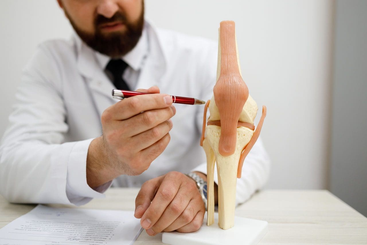
There’s only one nerve that goes through the knee that has an increased risk of getting compressed. It’s a branch of the sciatic nerve called the peroneal nerve. The nerve goes around the outside of the knee before traveling down the outside of the lower leg. At the bottom of the knee, it lies between the bone and skin, making it vulnerable to irritation or compression by anything that can put pressure on the outside of the knee.
Causes
Traumatic injuries over time can lead to pressure on the nerve from inside the knee. Common causes of a compressed nerve in the knee include:
Frequently Crossing Legs
Compression by the opposite knee, while the legs are crossed is the most common cause.
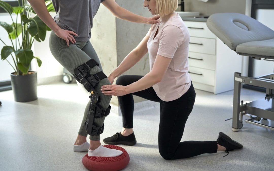
Knee Brace
A too-tight or strong brace can compress the leg and nerve.
Thigh-High Compression Stockings
Designed to maintain pressure on the legs, if too tight these stockings can compress the nerve.
Squatting Posture For Long Periods
This position places pressure on the side of the knee.
Fractures
A fracture of the large lower leg bone/tibia or sometimes the small bone/fibula near the knee can entrap the nerve.
Lower Leg Cast
The portion of the cast around the knee can be tight and compress the nerve.
Tell the doctor if a cast or brace feels tight or is causing numbness or pain in the leg.
Knee-High Boots
The top of a boot can land right below the knee and be too tight pinching the nerve.
Knee Ligament Injury
The nerve can become compressed due to bleeding or inflammation from an injured ligament.
Knee Surgery Complications
This is rare, but the nerve can inadvertently get pinched during knee replacement surgery or an arthroscopic procedure.
Prolonged Bed Rest
When lying down the legs tend to rotate outward and the knees flex.
In this position, the mattress can place pressure on the nerve.
Tumors or Cysts
Tumors or cysts can develop right on top or next to a nerve irritating and compressing the area.
Abdominal or Gynecologic Surgery
The equipment used to keep the legs rotated outward and the knees flexed for gynecologic and abdominal surgeries can compress the nerve.
Symptoms
The peroneal nerve supplies sensation and movement to the outside of the lower leg and the top of the foot. When compressed, it becomes inflamed, which causes the symptoms of a compressed nerve. Usually, only the lining/myelin sheath around the nerve is what gets injured. However, when the nerve gets damaged, the symptoms are similar but more severe. Common symptoms include:
Weakness that limits the ability to lift the foot toward the leg aka dorsiflexion.
This causes dragging the foot when walking.
The ability to turn the foot outward and extend the big toe is also affected.
Symptoms can be felt on the outside of the lower leg and on the top of the foot and include:
Tingling or pins and needles sensations.
Numbness.
Loss of sensation.
Pain.
Burning.
For individuals that have had a pinched nerve for two or more weeks, the muscles supplied by the nerve can begin to waste away or atrophy.
Symptoms can be intermittent or continuous depending on the cause.
The other common cause is a pinched nerve in the lumbar/lower spine.
When this is the cause, sensations, and pain will present in the lower back or the back and outside of the thigh.
Diagnosis
A doctor will look at medical history and perform an examination to make a diagnosis, determine the cause, and lay out a personalized treatment plan. The nerve in the knee can be felt as it travels around the top of the tibia, so a doctor may tap on it. If there is shooting pain down the leg, a pinched nerve may be present. Tests a doctor may order can include:
Knee X-ray
Shows any bone fractures or abnormal masses.
Knee MRI
Can confirm the diagnosis
Shows masses within the nerve.
Shows details of fractures or other problems in the bones.
Electromyogram – EMG
Tests electrical activity in the muscles.
Nerve Conduction Test
Tests the signal speed of the nerve.
Treatment
Treatment is aimed at reducing pain and improving mobility.
Over-the-Counter Pain Medication
OTC medication can reduce inflammation and improve symptoms short term.
Ice and Heat
Applying either heat or ice for 15 to 20 minutes at a time can provide relief from the symptoms.
An ice pack can make symptoms worse if it adds more pressure on the nerve.
Chiropractic and Physical Therapy
Chiropractic and physical therapy can release the compressed nerve, realign the structures, strengthen the muscles, and provide gait training.
Orthotic Boot
If walking gait is affected because the foot cannot bend, an orthotic boot can help.
This is a support that maintains the foot in a neutral position to walk normally.
Corticosteroid Injection
A corticosteroid injection can reduce inflammation and relieve pressure on the nerve.
Surgery
The nerve can suffer permanent damage if it has been pinched for a long time.
If that happens, surgery cannot repair the damage.
A doctor can perform surgery to correct a fracture, tumor, or other invasive problem causing a compressed nerve.
If conservative treatment doesn’t work, a peroneal nerve decompression procedure can be done to remove the pressure.
If surgery is needed, symptoms can disappear immediately, but it takes around four months to recover and rehabilitate.
Injury Rehabilitation
References
Krych, Aaron J et al. “Is peroneal nerve injury associated with worse function after knee dislocation?.” Clinical orthopedics and related research vol. 472,9 (2014): 2630-6. doi:10.1007/s11999-014-3542-9
Lezak B, Massel DH, Varacallo M. Peroneal Nerve Injury. [Updated 2022 Nov 14]. In: StatPearls [Internet]. Treasure Island (FL): StatPearls Publishing; 2023 Jan-. Available from: https://www.ncbi.nlm.nih.gov/books/NBK549859/
Soltani Mohammadi, Sussan, et al. “Comparing the squatting position and traditional sitting position for ease of spinal needle placement: a randomized clinical trial.” Anesthesiology and pain medicine vol. 4,2 e13969. 5 Apr. 2014, doi:10.5812/aapm.13969
Stanitski, C L. “Rehabilitation following knee injury.” Clinics in sports medicine vol. 4,3 (1985): 495-511.
Xu, Lin, et al. Zhongguo gu Shang = China Journal of Orthopedics and Traumatology vol. 33,11 (2020): 1071-5. doi:10.12200/j.issn.1003-0034.2020.11.017
Yacub, Jennifer N et al. “Nerve injury in patients after hip and knee arthroplasties and knee arthroscopy.” American Journal of physical medicine & Rehabilitation vol. 88,8 (2009): 635-41; quiz 642-4, 691. doi:10.1097/PHM.0b013e3181ae0c9d
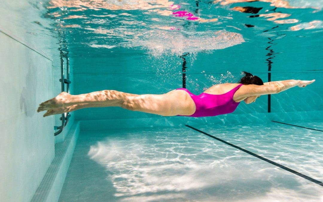
Individuals dealing with or managing chronic back pain should incorporate swimming exercises. Swimming is a low-impact aerobic conditioning exercise that’s easy on the back and healthy for the spine. When an individual struggles with back problems, they may be tempted to rest and avoid physical activity/exercise. Total rest is not recommended as it can cause the muscles that support the back to weaken or atrophy. When the muscles weaken, they cannot stabilize the spine or body correctly, which causes conditions to worsen or contribute to new injuries. Starting swimming exercises can expand the spine, relieve painful pressure or strain on the back and strengthen the muscles for spinal health.
Starting Swimming Exercises
Swimming does not impact the spine and other musculoskeletal structures because the water suspends the body.
Swimming is a full-body, low-impact exercise which is excellent for individuals of all ages and all body shapes and sizes.
Talk to a healthcare professional about any questions or concerns about how swimming may impact your body.
Swimming benefits include stress relief, a strengthened musculoskeletal system, and support in heart health.
Swimming for Back Problems
Relaxes The Nervous System
Tense muscles can cause or contribute to back problems and pain symptoms and aggravate spinal conditions.
Swimming exercises release endorphins to relax the nervous system and tense muscles.
Relieves Pressure on Joints
The water lightens the body relieving pressure on the joints and muscles.
Builds Muscle to Support the Spine
The resistance and movement strengthen the whole body with the joints and spine supported.
Swimming engages muscles not always used, specifically those needed to improve spinal stability.
Exercises for Back Relief
Checking with a physician before exercising is recommended, especially if starting a new exercise routine. When you meet with the Injury Medical Chiropractic and Functional Medicine Cline team, we can determine if starting swimming exercises would benefit you. Once cleared, here are some swimming exercises that could help bring relief:
Walking
Walking around the pool means movement that the body needs to heal and build muscle without aggravating symptoms.
Aerobics
Water aerobics is perfect for working on cardiovascular health needed to build strength.
Increase mobility and flexibility.
Swimming Laps
Start slow when swimming laps, maybe only twice a week at first.
The different types of strokes work various muscles in the hips, chest, and back.
Treading water is a great way to get the body used to the movements.
A swim coach can provide tips on the proper technique and form.
Swim Exercise Tools and Accessories
Proper swimming equipment can make the exercise sessions much more enjoyable.
Swim Cap
Swim caps protect the hair from the water’s elements and keep hair from blocking the view.
Goggles
Goggles protect the eyes and help to see better underwater.
Look for a comfortable pair that doesn’t leak.
Sun protection and clothing
A day in the sun and water increases the risk of exposure to UV rays.
Waterproof Headphones
For listening to music or podcasts while swimming.
Kickboard
Many pools can provide kickboards that swimmers can borrow during their time there.
Lean the upper body on the board and kick, focusing on lower body movements.
Pull Buoy
Pull buoys help focus on the upper body and arm work.
It is placed between the upper thighs to help the legs float as the individual pulls with their arms.
It is recommended to take some lessons to learn how the body moves through the water. Once a basic understanding of balance and buoyancy is met, individuals can propel through the water more efficiently.
Sciatica Secrets Revealed
References
Bartels, Else Marie, et al. “Aquatic exercise for the treatment of knee and hip osteoarthritis.” The Cochrane Database of systematic reviews vol. 3,3 CD005523. 23 Mar. 2016, doi:10.1002/14651858.CD005523.pub3
Cole, A J et al. “Spine pain: aquatic rehabilitation strategies.” Journal of Back and musculoskeletal rehabilitation vol. 4,4 (1994): 273-86. doi:10.3233/BMR-1994-4407
Ferrell, M C. “The spine in swimming.” Clinics in sports medicine vol. 18,2 (1999): 389-93, viii. doi:10.1016/s0278-5919(05)70153-8
Su, Yanlin, et al. “Swimming as Treatment for Osteoporosis: A Systematic Review and Meta-analysis.” BioMed research international vol. 2020 6210201. 15 May. 2020, doi:10.1155/2020/6210201
Wirth, Klaus, et al. “Strength Training in Swimming.” International Journal of environmental research and public health vol. 19,9 5369. 28 Apr. 2022, doi:10.3390/ijerph19095369
The nervous system communicates with the entire body and reacts to internal and external changes using electrical and chemical impulses to send and receive messages. Messages travel/synapse from one neuron to another using specialized chemicals known as neurotransmitters. Paresthesia refers to sensations of numbness, tingling, prickling, skin crawling, itching, or burning, usually in the arms, hands, legs, and/or feet, but can affect other areas of the body. Chiropractic care, massage therapy, decompression therapy, and functional medicine can relieve tissue and nerve compression, improve flexibility, range of motion, and mobility, and strengthen the muscles surrounding the affected nerve to maintain optimal health and prevent worsening or further injury.
Paresthesia
The sensation comes on without warning and is usually painless and described as tingling or numbness. There are different causes of paresthesia, including:
Some individuals have chronic or long-term paresthesia, which can be a sign of a more serious nerve injury or condition. Added physical stress can cause surrounding tissues to irritate or entangle the nerve leading to building pressure. This pressure causes paresthesia in the area interrupting circulation and function. A pinched nerve can happen anywhere in the body, like the neck, shoulder, wrist, back, and face.
A herniated disc in the lower spine can cause back pain and paresthesia in the leg or foot on the affected side.
Carpal tunnel syndrome is a pinched nerve in the wrist that causes numbness and tingling in the hand and fingers.
Pinched nerve symptoms can be intermittent or constant.
Usually, a temporary sensation is caused when pressure is placed on the affected nerve.
Once that pressure is relieved, the discomfort goes away.
Individuals with an Increased Risk
Overuse Injury
Individuals with jobs or hobbies requiring repetitive motions are at a higher risk for nerve compression, paresthesia, or injury.
Anyone can get a pinched nerve, and most individuals will experience paresthesia at some point.
Weight and water gain can cause swelling and increase pressure on nerves.
Thyroid Disease
This puts individuals at risk for carpal tunnel syndrome.
Rheumatoid arthritis
This causes inflammation, which can also compress nerves in the joints.
Diagnosis
To diagnose paresthesis, a doctor will look at the individual’s medical history and ask questions about the symptoms. They will perform a physical examination and, depending on the findings, may recommend tests that can include:
Nerve Conduction Study
This measures how fast nerve impulses travel in the muscles.
Electromyography – EMG
To look at the electrical activity of how nerves and muscles interact.
Magnetic Resonance Imaging – MRI
This looks at the different areas of the body in high definition.
Ultrasound
Used to produce images, this can be applied to the smaller areas to look for nerve compression or damage.
Chiropractic
Treatment options depend on the cause of the paresthesia. Body misalignments can cause nerve interference that can lead to health problems such as migraines, or can disrupt nerve communication and block proper circulation. Chiropractic care focuses on treating the nervous system and is a safe and effective method for treating nerve problems that cause discomfort and sensations. After a thorough examination of problem areas, massage, decompression, and chiropractic adjustments will:
Bova, Joseph, and Adam Sergent. “Chiropractic management of a 24-year-old woman with idiopathic, intermittent right-sided hemiparesthesia.” Journal of chiropractic medicine vol. 13,4 (2014): 282-6. doi:10.1016/j.jcm.2014.08.002
Christensen, Kim D, and Kirsten Buswell. “Chiropractic outcomes for managing radiculopathy in a hospital setting: a retrospective review of 162 patients.” Journal of chiropractic medicine vol. 7,3 (2008): 115-25. doi:10.1016/j.jcm.2008.05.001
Freihofer, H P Jr. “Parästhesien” [Paresthesia]. Schweizerische Monatsschrift fur Zahnheilkunde = Revue mensuelle suisse d’odonto-stomatologie vol. 89,2 (1979): 124-5.
Karne, Sampada Swapneel, and Nilima Sudhakar Bhalerao. “Carpal Tunnel Syndrome in Hypothyroidism.” Journal of Clinical and diagnostic research: JCDR vol. 10,2 (2016): OC36-8. doi:10.7860/JCDR/2016/16464.7316
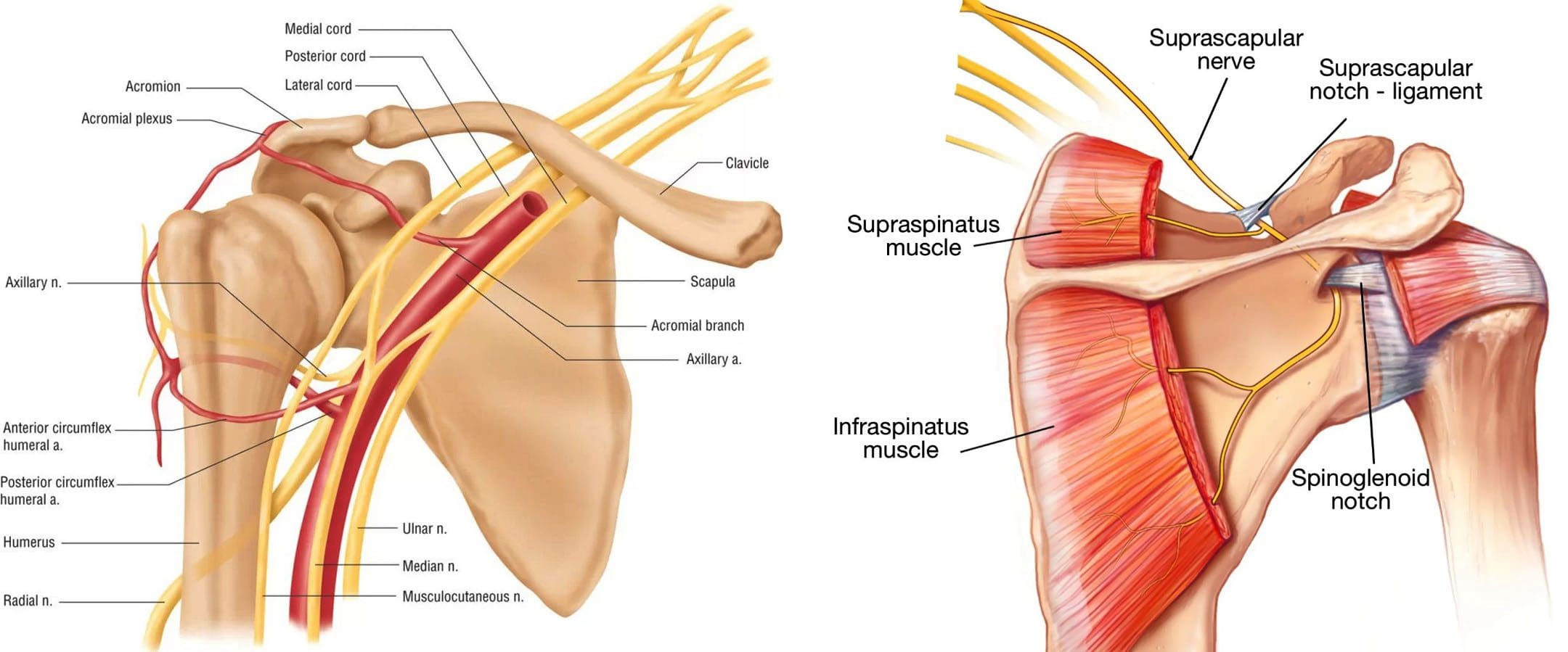
An acute injury or changes to the upper body over time can cause a compressed/pinched nerve in the shoulder. A pinched nerve in the shoulder happens when a muscle, ligament, tendon, or bone irritates or presses on a nerve exiting the neck. Shoulder nerve pain can develop from various sources, such as overuse work injuries, sports injuries, household chores, tendinitis, arthritis, torn cartilage, and other medical conditions, and injuries can contribute to symptoms. Chiropractors are highly qualified to treat pinched nerves. They are trained in whole-body realignment and rehabilitation techniques that find the root source and relieve pressure on compressed nerves.
Shoulder Nerve Pain
The shoulder joint is one of the most complex joints because of its wide range of motion. It is used so frequently that repetitive motion strain is common, often leading to injury. It is usually because of the continued use combined with an unhealed strain/injury that leads to shoulder nerve injury or when surrounding tissues like cartilage or tendons irritate or compress the nerves.
Pinched nerves also occur when a nerve root in the neck is damaged through wear and tear or an acute injury.
Individuals 50 years and older are likely to experience pinched nerves because of degeneration in the cervical spine and/or arthritis.
A nerve can become pinched when bone spurs form around the spinal discs.
Bone spurs are formations of bone that grow when discs weaken with age.
Bone spurs grow around the discs putting pressure on the nerve root.
Symptoms
Compressed Pinched Nerve/Cervical Radiculopathy
Pain sensations in the shoulder.
Tingling and/or pins and needles in fingers or hand.
Weakness in shoulder and arm muscles.
Symptoms have been known to overlap with shoulder arthritis, frozen shoulder, swimmer’s shoulder, or rotator cuff tears, so it’s always best to consult a chiropractor to understand possible causes. Other conditions with symptoms to compare:
Shoulder Arthritis
Stiffness in the joint.
Aching inside the shoulder.
Grinding when moving the joint.
Frozen Shoulder/Adhesive Capsulitis
Stiffness in the joint.
Pain in one shoulder.
Decreased range of motion and movement.
Swimmer’s Shoulder/Impingement
Pain and discomfort in the shoulder.
Weakness in the surrounding area.
Stiffness or tightness in the joint.
Impeded range of motion.
Rotator Cuff Tears
Pain and discomfort symptoms when moving the shoulder.
Weakness in the arm.
Deep aching sensations along the top and side of the joint.
Chiropractic Treatment
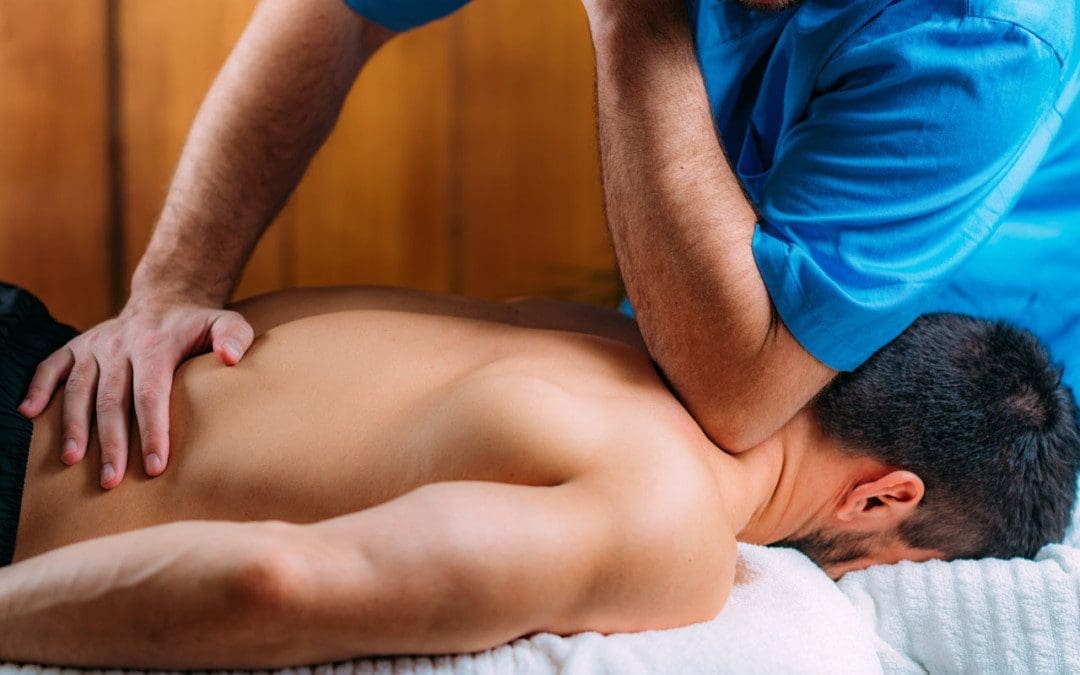
Chiropractors are experts on the neuromusculoskeletal system. First, a thorough medical examination will be conducted, including health history and regular activities, to understand the nature of the symptoms. Depending on the type of injury, tests, and exams may be needed to help diagnose and pinpoint the cause. Then the chiropractor will develop a personalized treatment plan. The objective is to relieve pressure and tension on the nerves and relax the muscles. In addition to adjusting the joint or other impacted areas, the therapy team will provide at-home exercises and stretches to maintain the adjustments and expedite healing.
Chiropractic Rehab
References
Kokkalis, Zinon T et al. “Nerve Injuries around the Shoulder.” Journal of long-term effects of medical implants vol. 27,1 (2017): 13-20. doi:10.1615/JLongTermEffMedImplants.2017019545
Leider, Joseph D et al. “Treatment of suprascapular nerve entrapment syndrome.” Orthopedic reviews vol. 13,2 25554. 11 Jul. 2021, doi:10.52965/001c.25554
Matzkin, Elizabeth, et al. “Swimmer’s Shoulder: Painful Shoulder in the Competitive Swimmer.” The Journal of the American Academy of Orthopaedic Surgeons vol. 24,8 (2016): 527-36. doi:10.5435/JAAOS-D-15-00313
Neviaser, Andrew S, and Jo A Hannafin. “Adhesive capsulitis: a review of current treatment.” The American Journal of sports medicine vol. 38,11 (2010): 2346-56. doi:10.1177/0363546509348048
Safran, Marc R. “Nerve injury about the shoulder in athletes, part 1: suprascapular nerve and axillary nerve.” The American Journal of sports medicine vol. 32,3 (2004): 803-19. doi:10.1177/0363546504264582
Strakowski, Jeffrey A, and Christopher J Visco. “Diagnostic and therapeutic musculoskeletal ultrasound applications of the shoulder.” Muscle & Nerve vol. 60,1 (2019): 1-6. doi:10.1002/mus.26505
IFM's Find A Practitioner tool is the largest referral network in Functional Medicine, created to help patients locate Functional Medicine practitioners anywhere in the world. IFM Certified Practitioners are listed first in the search results, given their extensive education in Functional Medicine