Can understanding the anatomy and function of the long thoracic nerve help individuals make informed healthcare decisions after an injury to the nerve?
Long Thoracic Nerve
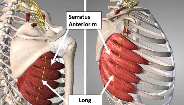
Also referred to as the posterior thoracic nerve, the long thoracic nerve/LTN is a thin superficial nerve that runs from the cervical spine to the chest wall side of the trunk. It supplies motor function to the thorax’s serratus anterior muscle, helping stabilize the shoulder blade. Injury to this nerve can cause limited or abnormal shoulder and shoulder blade motion, including difficulty raising the arm during overhead reaching.
Anatomy
The long thoracic nerve originates from the ventral rami of cervical nerves C5, C6, and C7. (Waxenbaum JA, Reddy V, Bordoni B. 2023) In some individuals, the root from C7 is absent; in others, a small nerve root branches from C8. The nerve roots from C5 and C6 go through the medial scalene muscle to join the C7 nerve. It travels behind the brachial plexus axillary artery and vein and courses down the lateral side of the thorax. The long thoracic nerve terminates at the lower portion of the serratus anterior muscle, sending small nerve tendrils to each muscle’s projections, which attach to the ribs. Because the long thoracic nerve is located on the lateral side of the chest, it is vulnerable to injury during sports or surgical procedures. The nerve also has a smaller diameter than other cervical and brachial plexus nerves, which increases its potential for injury.
Function
The long thoracic nerve attaches to the underside of the shoulder blade and inserts as muscular slips into the ribs. It supplies motor function to the serratus anterior muscle, essential for normal shoulder motion. When it contracts, it pulls the shoulder blade against the ribs and thorax, helping to move and stabilize the arm as it moves forward and up during shoulder motions. Injury to the long thoracic nerve causes a condition called scapular winging. This occurs when the serratus anterior muscle becomes weakened or paralyzed after injury. (Lung K, St Lucia K, Lui F. 2024)
Susceptibility to Injury
The LTN is relatively unprotected and can be damaged by several things, including:
Heavy backpacks
Sports
Activities the body is not used to, like digging
Using crutches
Conditions
Injury to the long thoracic nerve may occur as a result of trauma, lifting heavy weights above the shoulder, or a surgical procedure. Surgical procedures that may place the nerve at risk for injury may include: (Lung K, St Lucia K, Lui F. 2024)
Axillary lymph node dissection
Improperly placed intercostal drains
Chest tube placements
Mastectomy
Thoracotomy
The long thoracic nerve is protected during these procedures by the surgeon and proper surgical technique, but occasionally, difficulties arise during surgery, and the nerve may become injured. Individuals may also have an anatomical variance that places their nerves in varying positions. The surgeon may not see it and accidentally injure their nerves during surgery.
The superficial long thoracic nerve may also be injured during sports or trauma to the trunk. A blow to the side or a sudden overhead stretch to the shoulder may be enough to damage the nerve, paralyzing the serratus anterior muscle.
Weakness or paralysis of the serratus anterior muscle will result in a winged scapula. To test for this:
Stand about two feet from a wall, facing it.
Place both hands on the wall and gently push against it.
If one of the shoulder blades sticks out abnormally, it could be a winged scapula.
Have a family member or friend stand behind you and check the shoulder blade position.
If you suspect a winged scapula, visit a physician who can assess the condition and determine if there is a long thoracic nerve injury.
Winging the scapula may result in difficulty lifting the arm overhead. The serratus anterior muscle works with other scapular stabilizers, such as the upper trapezius and levator scapula, to properly position the shoulder blade when lifting the arm. Failure of the serratus to stabilize the shoulder blade may make lifting the arm impossible.
Clinical examination is usually used to diagnose a long thoracic nerve injury. X-rays and MRIs cannot show the nerve injury directly, although an MRI can show some secondary signs to help confirm the diagnosis. An electromyographic or EMG test may also be performed to examine the function of the long thoracic nerve.
Treatment and Rehabilitation
Treatment for LTN pain and reduced movement may include:
Rest
Heat or ice
Anti-inflammatory pain medication
Neck support or a pillow
Avoiding strenuous activity and driving
If the long thoracic nerve is severely injured and the serratus anterior is completely paralyzed, the best course of action is to be active and monitor the condition. Full recovery of arm function can take one to two years. If permanent nerve injury has occurred, surgery may be an option to restore shoulder motion and function. Several different kinds of surgery can be used to address winged scapula. (Vetter M. et al., 2017)
One involves transferring the pectoralis major tendon to the scapula (Vetter M. et al., 2017) so it functions as the serratus.
Often, the tendon has to be lengthened, which may be done using part of the hamstring tendon.
After surgery, individuals will likely wear a sling on their arm for a few weeks, and then gentle range-of-motion exercises will be initiated.
After eight to ten weeks, gentle progressive strengthening of the new tendon can begin.
Full shoulder motion and strength recovery are expected six to 12 months after surgery.
Lie on your back and raise both arms toward the ceiling.
Make a fist and punch up toward the ceiling.
Ensure the motion is steady and deliberate, and keep the elbow straight.
Hold the position for three seconds, then slowly lower the arm to the starting position.
Perform 10 to 15 repetitions.
Holding a small dumbbell in your hands can make the exercise more challenging.
Pushup
Lie on your stomach and place your hands flat on the ground by your shoulders as if you were going to perform a pushup.
Perform a pushup and press further, allowing the shoulder blades to wrap around the thorax.
Hold this position for three seconds, and slowly release.
Perform 10 to 15 reps.
If this is too difficult, perform the pushup against a wall to reduce the effect of gravity on the exercise.
Scapular Winging in Depth
References
Waxenbaum, J. A., Reddy, V., & Bordoni, B. (2024). Anatomy, Head and Neck: Cervical Nerves. In StatPearls. https://www.ncbi.nlm.nih.gov/pubmed/30844163
Lung, K., St Lucia, K., & Lui, F. (2024). Anatomy, Thorax, Serratus Anterior Muscles. In StatPearls. https://www.ncbi.nlm.nih.gov/pubmed/30285352
Vetter, M., Charran, O., Yilmaz, E., Edwards, B., Muhleman, M. A., Oskouian, R. J., Tubbs, R. S., & Loukas, M. (2017). Winged Scapula: A Comprehensive Review of Surgical Treatment. Cureus, 9(12), e1923. https://doi.org/10.7759/cureus.1923
Berthold, J. B., Burg, T. M., & Nussbaum, R. P. (2017). Long Thoracic Nerve Injury Caused by Overhead Weight Lifting Leading to Scapular Dyskinesis and Medial Scapular Winging. The Journal of the American Osteopathic Association, 117(2), 133–137. https://doi.org/10.7556/jaoa.2017.025
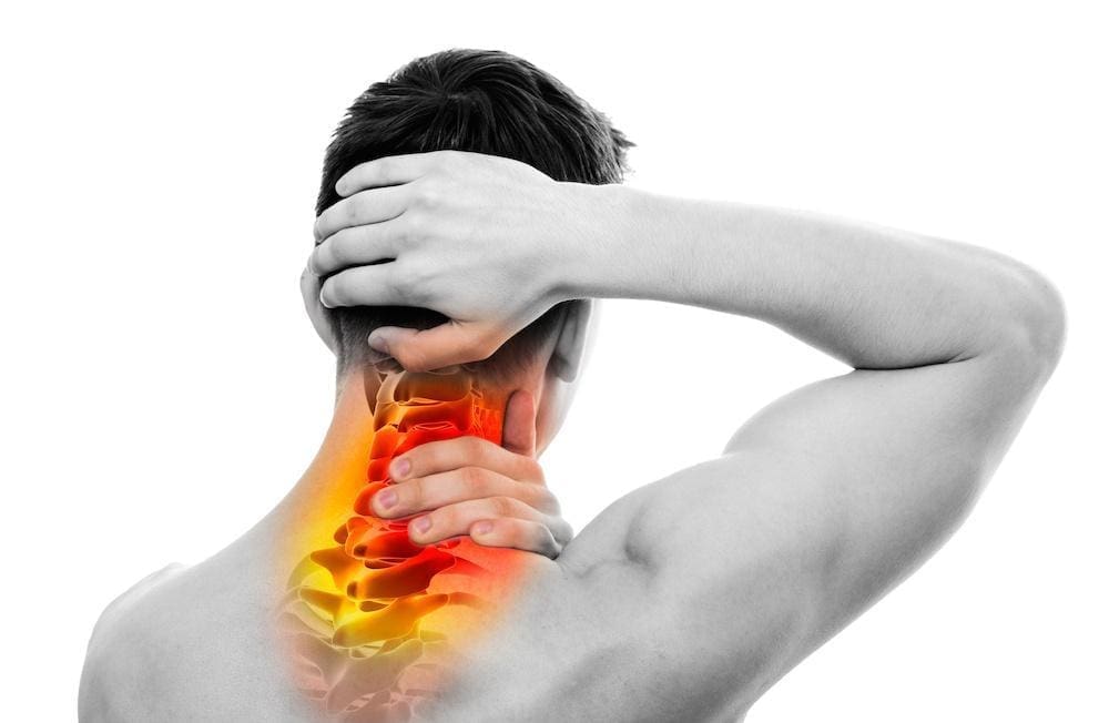
The neck is an extremely flexible part of the upper body that allows the head to move without causing pain or discomfort. It is part of the musculoskeletal system‘s cervical spinal region, which supports the spinal column and is surrounded by various muscles, tissues, and ligaments that protect the spinal cord. However, poor posture, spending too much time hunched over a computer, or looking down at our cellphones can cause neck muscles to become overstretched, leading to compression of the cervical spinal discs. This can cause the cervical discs to bulge or herniate, aggravating the spinal cord and causing neck pain and other associated conditions. This post will discuss how cervical disc compression affects neck pain and how decompression surgery and spinal decompression can help alleviate this condition. We work with certified medical providers who use our patients’ valuable information to treat individuals dealing with cervical disc compression affecting their necks and causes mobility issues. We encourage patients to ask essential questions and seek education from our associated medical providers about their condition. Dr. Jimenez, D.C., provides this information as an educational service. Disclaimer
What Is Cervical Disc Compression?
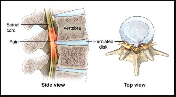
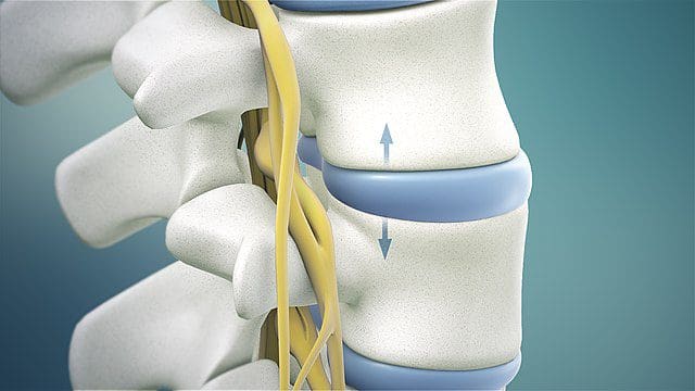
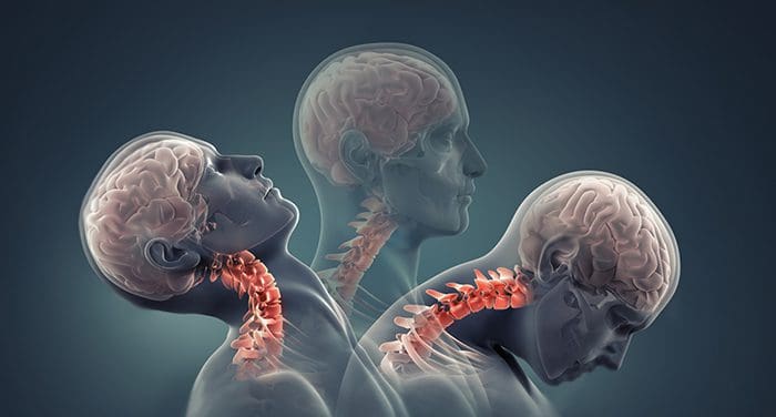
Have you been experiencing neck pain or muscle aches in your shoulders? Do you feel numbness or tingling running down your arms and fingers? These symptoms may be signs of cervical disc compression. The cervical spinal discs act as shock absorbers for the spine, preventing unwanted pressure and mobility issues. Research studies revealed that age-related degenerative properties like dehydration could cause herniated and compressed cervical discs, leading to posterior disc protrusion into the spinal cord. Trauma can also cause extreme hyperflexion or hyperextension of the posterior neck muscles, resulting in various neck symptoms. Additional research studies stated cervical disc displacement could cause compression or impingement on spinal nerve roots, leading to inflammation and neck pain.
How Is It Associated With Neck Pain?
When the spinal cord and nerve roots in the cervical region are affected by cervical disc compression, the pain can be dull or sharp, depending on how it affects many individuals. According to research studies, many people are unaware that repetitive normal factors or traumatic forces can cause a challenge in determining the origin of the pain from symptomatic or asymptomatic disc compression. Additional research studies mentioned that cervical disc compression could cause upper and lower extremities abnormalities, such as loss of deep tendon reflexes in the arms and legs, loss of motor function in the hands and feet, muscle weakness, headaches, and gait imbalances. However, various treatments can alleviate the pain-like symptoms associated with cervical disc compression and help the body’s natural healing process.
From Inflammation To Healing-Video
Are you experiencing inflammation and pain in your neck? Do you notice a tingling or numb sensation in your hands or feet? Or do you feel stiffness in your shoulders or neck? These symptoms can be caused by compressed cervical discs, which many people are unaware of. The compression of cervical discs is a common source of neck pain and can even cause referred pain in the upper and lower extremities. Repetitive motions to the neck can cause the posterior neck muscles to overstretch and lead to pain. Normal or traumatic factors can also lead to neck pain associated with cervical disc compression, resulting in disc herniation. Fortunately, non-surgical therapies like chiropractic care and spinal decompression can help alleviate the symptoms of pain, discomfort, and inflammation caused by cervical disc compression. Check out the video above for more information on these treatments.
Posterior Cervical Disc Decompression Surgery
If you experience cervical compression on your neck, it can lead to persistent neck pain and discomfort if left untreated. Many people opt for posterior cervical disc decompression surgery to alleviate the effects of disc herniation. According to “The Ultimate Spinal Decompression” by Dr. Perry Bard, D.C., and Dr. Eric Kaplan, D.C., FIAMA, cervical disc herniation can sometimes affect the back of the neck and cause persistent pain. In such cases, decompression surgery is often performed. During the procedure, a small incision is made at the back of the neck, and a portion of the damaged disc is removed to ease the irritated nerve. This brings relief to the individual suffering from neck pain.
Non-Surgical Decompression For Compressed Cervical Disc
If you’re not interested in surgery for cervical disc compression, consider non-surgical spinal decompression instead. Studies have shown that spinal decompression is a safe, non-invasive treatment involving gentle cervical spine traction to reposition the herniated disc. This treatment can also help rehydrate the spinal disc by bringing in nutrients and oxygenated blood to promote natural healing. Additionally, spinal decompression can alleviate any remaining symptoms of neck pain.
Conclusion
The neck is a highly flexible area that enables smooth head movement without discomfort or pain. However, it is also a part of the musculoskeletal cervical region that can be prone to injuries. Compression of the disc due to normal or traumatic factors can result in herniation, causing pain if left untreated. Fortunately, several treatments are available to alleviate neck pain caused by cervical compression and make the neck mobile again.
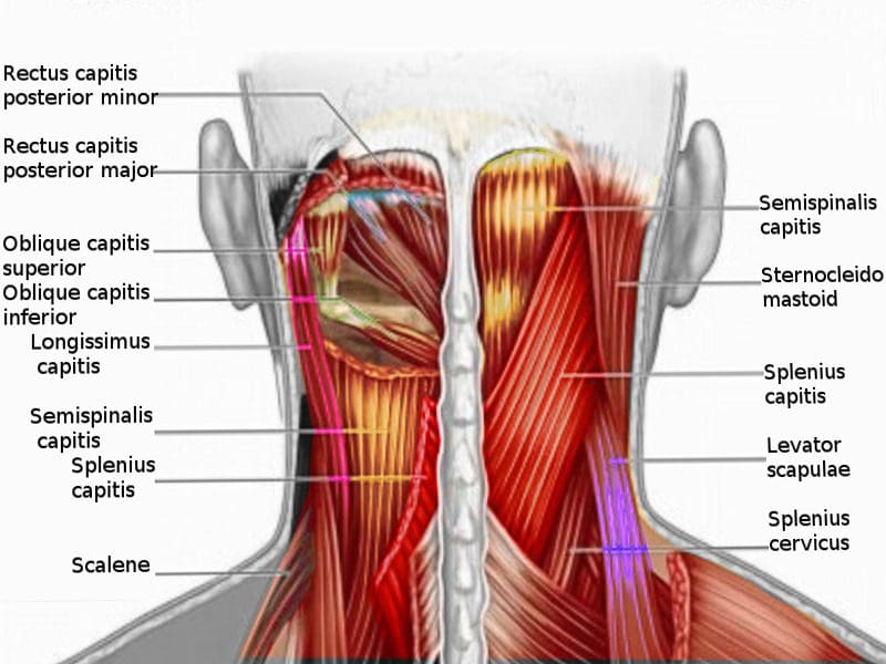
The cervical spine has a casual relationship with the central nervous system as the brain and spinal cord works together to send neuron signals through the nerve pathways spread throughout the entire body. The cervical spine is part of the neck, where ligaments and muscles encompass the cervical joints to stabilize the head, neck, and shoulders. The neck has various muscles that support the head and can succumb to various injuries that can cause muscle strain and other associated symptoms that can affect the upper extremities of the body. Today’s article looks at the posterior cervical muscles, how trigger points affect the posterior cervical muscles, and how non-invasive treatments can help manage myofascial cervical pain. We refer patients to certified providers who specialize in musculoskeletal treatments to aid individuals suffering from myofascial trigger pain associated with the neck affecting the posterior cervical muscles. We also guide our patients by referring them to our associated medical providers based on their examination when appropriate. We ensure to find that education is the solution to asking our providers insightful questions. Dr. Jimenez DC observes this information as an educational service only. Disclaimer
What Are The Posterior Cervical Muscles?
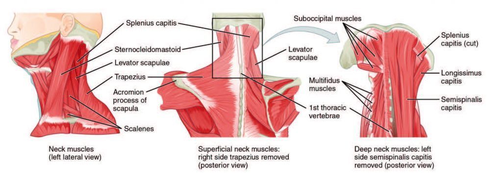
Have you been experiencing random headaches that affect your day? Do you feel any tension in your neck? Or do you experience stiffness and limited mobility when turning your neck? Most of these symptoms are associated with trigger pain affecting the posterior cervical muscle on the neck. The posterior cervical muscles function in the neck’s anatomic region while protecting the cervical spine. The muscles in the cervical spine form a triangle on the neck while providing functionality and movement to the neck, the head, shoulder, and upper back. Some of the muscles that work together with the cervical spine include:
Levator scapulae
SCM (Sternocleidomastiod)
Trapezius
Erector spinae
Deep cervical flexors
Suboccipitals
Semispinalis
Splenius
These muscles and soft tissues provide stability to the neck and work together with the tendons and ligaments to rotate, extend, and retract the head, neck, shoulders, and upper back. To that point, if the neck muscles become strained, it can lead to various issues that can cause pain to the neck and cervical spine.
How Trigger Points Affect The Posterior Cervical Muscles
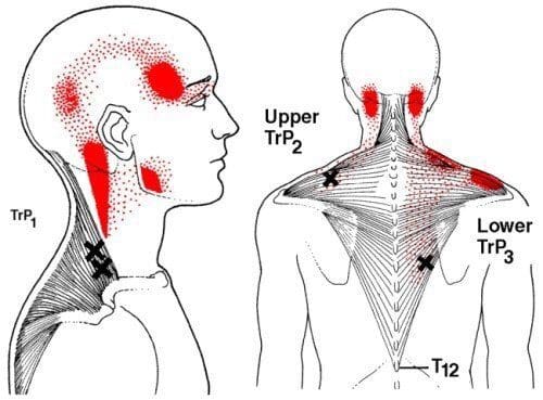
Studies reveal that a musculoskeletal disorder known as cervical myofascial pain causes the surrounding muscles in the neck and shoulder regions to become tender to the touch and evoke pain in different areas of the body. When the posterior cervical muscle has become strained from various issues like poor posture, over-exercising, getting involved in an auto accident, or have suffered from a degenerative condition, it can cause the muscles to be overused, hypersensitive, and stiff while potentially being at risk of developing trigger points along the neck and shoulder muscles. Trigger points are a bit problematic as they are associated with referred pain and can be either latent or active as they create tiny nodules along the taut muscle fibers in the area. Research studies reveal that referred pain from cervical myofascial pain can be elicited by active trigger points on the affected muscle. To that point, the active trigger points can mimic other pain symptoms along the upper extremities, making diagnosing myofascial pain challenging. Thankfully there are ways to manage trigger point pain associated with the posterior cervical muscles along the neck and cervical spine.
Exercises For Neck & Shoulder Pain-Video
Have you been experiencing stiffness along the sides of your neck or on your shoulders? Do headaches make it impossible to get through an entire day? Or have you been feeling tightness along your jawline? Many people with neck pain and its associated symptoms could risk developing myofascial trigger pain along the posterior cervical muscles. Having myofascial trigger pain associated with the posterior cervical muscle and affecting the neck and upper extremities can cause many people to be miserable; however, there are many ways that people can utilize to relieve the pain along the posterior cervical muscles and manage myofascial cervical pain associated with the neck. The video above provides various neck and shoulder pain exercises correlating with myofascial trigger points. To that point, finding non-invasive available treatments to manage myofascial cervical pain can help reduce the effects of pain along the neck and posterior cervical muscles.
Non-Invasive Treatments To Manage Myofascial Cervical Pain
Many people suffer from neck pain associated with myofascial cervical pain; many factors can contribute to developing tiny nodules along the posterior muscles. Studies reveal that activities, whether work-related or for entertainment, can yield repetitive stress on the muscle groups that cause chronic tension in the muscle fibers to form trigger points. To that point, myofascial trigger pain causes the taut muscle bands to be hyper-sensitive and affect the muscle area. Many people suffering from myofascial trigger pain can utilize non-invasive treatments to manage myofascial pain associated with the posterior cervical muscle. Some of the treatments that help manage myofascial cervical pain include:
Acupuncture
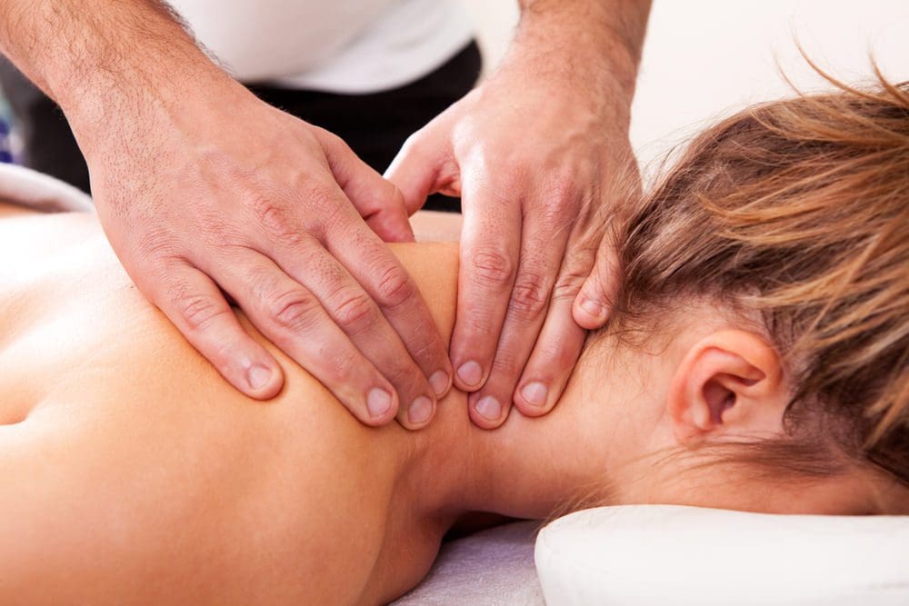
Trigger point release therapy
Chiropractic care
Heat therapy
Exercise/stretching
Many of these treatments help release the trigger points from the affected muscles and help prevent them from re-occurring on the affected muscles along the upper extremities.
Conclusion
The neck has many muscles, ligaments, and tissues that help support the upper extremities of the body. When issues begin to affect the functionality of the neck muscles, it can lead to the development of myofascial trigger pain in the posterior cervical muscles along the neck to cause painful symptoms. This can lead to overlapping chronic issues like stiffness and tenderness in the upper extremities. Non-invasive treatments can help alleviate the pain and manage the symptoms caused by myofascial cervical pain in the upper extremities of the body. This allows the affected muscle to return to its functionality and prevents future issues from affecting the body.
References
Alghadir, Ahmad H, et al. “Efficacy of Combination Therapies on Neck Pain and Muscle Tenderness in Male Patients with Upper Trapezius Active Myofascial Trigger Points.” BioMed Research International, Hindawi, 10 Mar. 2020, https://www.ncbi.nlm.nih.gov/pmc/articles/PMC7085833/.
Fernández-de-Las-Peñas, César, et al. “The Role of Myofascial Trigger Points in Musculoskeletal Pain Syndromes of the Head and Neck.” Current Pain and Headache Reports, U.S. National Library of Medicine, Oct. 2007, https://pubmed.ncbi.nlm.nih.gov/17894927/.
Stathakios, James, and Michael A Carron. “Anatomy, Head and Neck, Posterior Cervical Region.” In: StatPearls [Internet]. Treasure Island (FL), StatPearls Publishing, 27 July 2021, https://www.ncbi.nlm.nih.gov/books/NBK551521/.
Touma, Jeffrey, et al. “Cervical Myofascial Pain.” In: StatPearls [Internet]. Treasure Island (FL), StatPearls Publishing, 4 July 2022, https://www.ncbi.nlm.nih.gov/books/NBK507825/.
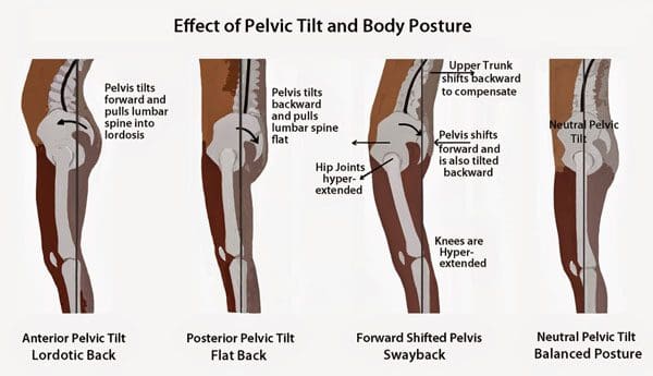
Few individuals realize they have problems with their feet. Flat feet can cause anterior or posterior pelvic tilt. This is a condition where the pelvis tilts too far forward or too far backward. Individuals can go through their entire life without realizing they have some type of foot issue. This can cause poor support in the lumbar spine generating back problems and chronic illness. The problem with foot issues is that it can set-up a chain of physiological deficiencies. This can include:
Many accept these health issues as part of their normal physiology. However, flat-feet and other related musculoskeletal issues can be identified and corrected with proper support. Custom FootOrthotics for the foot issue/s and chiropractic alignment for the whole-body. Based on the assessment of the spinal curvature and posture of the individual, a chiropractor will be able to determine to what degree the pelvic tilt and foot problem/s need to be corrected.
Foot Orthotic Support
How orthotics function to promote proper posture and alleviate back pain works like this.
First, immediate support to the feet that are weakened, unbalanced, and lack physiological strength is provided. This generates proper posture and balance all the way up the spine.
Second, the feet are able to remodel themselves over time as they become accustomed to the new support.
Pelvic Tilt Identification
Chiropractors utilize orthotics when correcting anterior or posterior pelvic tilt. This is because the conditions are the direct result of a poor/weakened foundation. When there is little or no natural arch of the foot there is no support for the:
The lower body needs this support from these various muscle groups with many in the lower back. This causes strain on these muscles and causes pelvic and potential spinal misalignment. Depending on the progress of the tilt/misalignment and posture, will aid a chiropractor to determine the best treatment approach.
Rebuilding
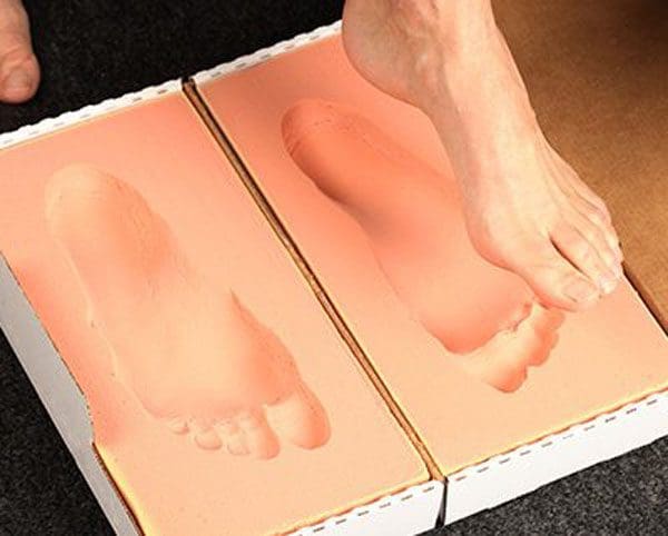
Chiropractic adjustments will be able to restore the lumbar support that was weakened or lost from foot issues. A chiropractor will go to the source or root of the problem. Foot orthotics can be a primary tool for re-training the feet for proper support. These orthotics are custom modeled to an individual�s feet.
This promotes spinal and pelvic wellness combined with the right amount of support required for each foot. Over time, and as the feet remodel themselves the custom orthotics can also be remodeled allowing for continuous improvement. With time it�s possible for many to accomplish the restoration of the body's musculoskeletal health. These include:
Strengthened foot arches
Stress and muscle tension alleviation
A re-trained proper lower back support
Restoration of proper spinal curvature
Correct and retrain anterior/posterior pelvic tilting habits
Custom Foot Orthotics
Foot orthotics require no effort. An individual just puts them in their footwear and done. The orthotics do the work for the individual when standing, walking, or running. If diagnosed with a pelvic tilt condition or are unsure if there is a tilt, contact a chiropractor about the benefits of foot orthotics.
Benefits of Functional Foot Orthotics
Dr. Alex Jimenez�s Blog Post Disclaimer
The scope of our information is limited to chiropractic, musculoskeletal, physical medicines, wellness, and sensitive health issues and/or functional medicine articles, topics, and discussions. We use functional health & wellness protocols to treat and support care for injuries or disorders of the musculoskeletal system. Our posts, topics, subjects, and insights cover clinical matters, issues, and topics that relate and support directly or indirectly our clinical scope of practice.*
Our office has made a reasonable attempt to provide supportive citations and has identified the relevant research study or studies supporting our posts. We also make copies of supporting research studies available to the board and or the public upon request. We understand that we cover matters that require an additional explanation as to how it may assist in a particular care plan or treatment protocol; therefore, to further discuss the subject matter above, please feel free to ask Dr. Alex Jimenez or contact us at 915-850-0900. The provider(s) Licensed in Texas& New Mexico*
References
Betsch, Marcel, et al. �Influence of foot positions on the spine and pelvis.� Arthritis care & research�vol. 63,12 (2011): 1758-65. doi:10.1002/acr.20601
Axial neck pain is also known as uncomplicated neck pain, whiplash, and cervical/neck strain. They refer to pain and discomfort running along the back or posterior of the neck. Axial is defined as forming or around an axis. This type of pain stays around the neck and immediate surrounding structures. It does not spread/radiate out to the arms, hands, fingers, and other areas of the body. Axial neck pain differs from two other neck conditions. They are:
Cervical radiculopathy describes irritation or compression/pinching of the nerve as it exits the spinal cord. The nerves of the cervical spine are known as the peripheral nerves. They are responsible for relaying signals to and from the brain to specific areas of the arms and hands. The signals sent from the brain are for muscle movement, while signals going to the brain are for sensation.
When one of these nerve/s gets irritated, inflamed, or injured, it can result in:
Muscle pain
Weakness
Numbness
Tingling sensation
Burning pain
Other types of abnormal sensations in the arms, hands, or fingers.
Cervical myelopathy describes compression of the spinal cord itself. The spinal cord is the information highway/pipeline to all parts of the body. There is a range of symptoms that can include:
Same symptoms as cervical radiculopathy
Balance problems
Coordination problems
Loss of fine motor skills
Bowel and bladder incontinence
Axial neck pain
Axial neck pain is a quite common type of neck pain. It affects around 10% of the population. However, the majority of these cases do not involve severe symptoms that limit daily activity.
Symptoms
Pain in the back of the neck is the primary and most common symptom. Sometimes the pain travels to the base of the skull, shoulder, or shoulder blade. Other symptoms include:
Neck stiffness
Headaches
Localized muscle pain
Warmth
Tingling
Developmental Risk factors
Poor posture, lack of ergonomics, and muscle weakness increase the chances of developing axial neck pain. Risk factors for development include:
Age
Trauma - Auto accident, sports, personal, work injury
Headaches
Depression
Chronic neck pain
Sleep problems
Diagnosis
Based on symptoms and physical exam findings are how a diagnosis is usually achieved. A doctor will typically order an x-ray, CT, or MRI of the cervical spine. This calls for an immediate visit to a hospital/clinic for evaluation. There could be severe symptoms that could indicate something more dangerous, causing pain like infection, cancer, or fracture. These symptoms include:
Prior trauma/injury from a fall, automobile accident, sports, work injury
Fever
Weight loss
Night sweats
Constant night pain
Rheumatic conditions/diseases that cause neck pain can include morning stiffness and immobility that gets better as the day progresses. If symptoms continue for more than 6 weeks, imaging of the spine could be recommended, especially for individuals that have had previous neck or spine surgery or if it could be cervical radiculopathy or myelopathy.
Treatment
There is a wide range of treatment options. Surgery is rarely required except for severe cases. Returning to normal activities almost right away is one of the most important things to prevent the pain from becoming chronic. First-line treatments typically begin with:
Physical therapy
Chiropractic
Stretching routine
Strengthening exercises
Acetaminophen
Anti-inflammatory medication/s
Muscle relaxants are sometimes prescribed
If a cervical spine fracture has been diagnosed, a neck brace could be recommended for short-term use. A soft collar could be utilized if the pain is severe, but a doctor usually discontinues use after 3 days. Other non-invasive treatment options include:
Invasive treatments like injections, nerve ablation, and surgery are rarely required. But if necessary, it can be beneficial for those cases.
Causes
A variety of the neck's anatomical structures can contribute to the pain. Common causes include:
Poor posture
Age
Degeneration
Ergonomics
Injury to muscles or ligaments
Arthritis
All of these can affect vertebral bodies, discs, and facet joints. Shoulder arthritis or a rotator cuff tear can imitate axial neck pain. Dysfunction of the temporomandibular jaw joint or the blood vessels of the neck can cause axial pain, but it is rare.
Prognosis
Symptoms are usually alleviated within 4-6 weeks from when the pain started. Pain that continues beyond this should encourage a visit to a chiropractic physician.
Posterior tibial tendon dysfunction is a very common ankle and foot problem that occurs when there is a tear or inflammation in the posterior tibial tendon � the tendon that is at the back of the ankle and is the key player in stabilizing the foot. The inflammation or tear affects the integrity of the tendon, weakening it so that is no longer provides the support or stability of the arch of the foot. This results in what is commonly known as flatfoot.
While this condition rarely requires surgery, there is pain, sometimes severe, swelling, and impaired mobility associated with it. Chiropractic for flatfoot has been found to be very effective for most patients, helping them heal faster and manage their pain.
What Causes Posterior Tibial Tendon Dysfunction?
The most common causes of flatfoot are overuse and injury. Falls are common culprits, causing injury to the tendon so that it tears or becomes inflamed.
Overtraining, particularly in high impact exercise or sports like basketball, dancing, soccer, and high impact aerobics can get tears due to the repetitive motion and constant pressure on the foot. Once the tendon is torn or inflamed, the arch begins to collapse until eventually the foot is flat.
This condition occurs more often in women than men. Also, people over 40 seem to be more prone to posterior tibial tendon dysfunction, although it can occur in younger people who overtrain or have any of the other risk factors including diabetes, obesity, and hypertension.
What Are The Symptoms Of Posterior Tibial Tendon Dysfunction?
Pain is the first and most common indicator of flatfoot. It typically is located where the tendon lies; along the inner portion of the ankle and foot. Sometimes swelling may be present.
The patient will also notice that the pain increases with activity. High impact or high intensity activities can be excruciating and quite difficult. It can get to the point where standing or walking for extended periods of time are very painful.
In advanced stages, the pain may shift to the outer portion of the ankle. As the arch collapses, the movement may cause the heel bone to shift outward. This, in turn, puts pressure on the outer ankle bone.
How Is Posterior Tibial Tendon Dysfunction Treated?
The treatment for flatfoot depends on the severity of the condition. Most patients can avoid surgery, but it is still a long healing process.
Typically, the patient will be advised to rest and ice the area, switch to low impact exercise, and take nonsteroidal anti-inflammatory medication for the pain. In moderate to severe cases, the patient may be put in a walking boot or short leg cast for 6 to 8 weeks in order to immobilize the foot and ankle, letting the tendon rest and heal. The downside of this is that the other muscles around the ankle will also �rest� and atrophy as a result.
Shoe inserts, or orthotics, as well as braces are also common treatments for flatfoot. However, any type of immobilization of the ankle and foot is usually accompanied by physical therapy either while the brace is worn or after a cast has been removed.
In more severe cases, the doctor may recommend a steroid injection directly into the tendon although there is an increased risk of the tendon rupturing. Surgery is usually a last resort and it is much more complex. Patients who undergo surgery for flatfoot rarely regain all of the mobility they had prior to the operation.
Chiropractic For Posterior Tibial Tendon Dysfunction
There has been a great deal of success in using chiropractic to treat flatfoot. The chiropractor will typically recommend rest and ice as well as chiropractic manipulations to help bring the ankle, foot, and leg back into alignment to counteract the weakened tendon. Often the ankle will lose mobility as the arch collapses and the bones in the foot and ankle shift. This, in turn, causes the arch to collapse even more.
By using chiropractic techniques to bring the ankle back into alignment, thus restoring the forward glide of the joint, they can alleviate the pain and help heal the condition. They may also recommend an air brace to stabilize the ankle when the patient will be doing a lot of walking as well as advise on lifestyle changes such as weight loss and a healthy diet. Often patients who have tried working with medical doctors to cure their flatfoot but were unsuccessful, were finally able to gain relief and improvement when they started working with their chiropractor.
Injury Medical Clinic: Ankle Sprain Chiropractic Treatment
The importance of Magnetic Resonance Imaging to evaluate the integrity of the lumbar posterior ligament complex post trauma.
Abstract: Posterior ligamentous complex(PLC), consisting of the supraspinous ligament, interspinous ligament, ligamentum flavum, and the facet joint capsules is thought to contribute significantly to the stability of the lumbar spine. There has been much debate on whether Magnetic Resonance Imaging(MRI) is specific and sensitive in diagnosing pathology to the PLC. The objective is to determine the necessity of MRI imaging for evaluating the integrity of the lumbar posterior ligament complex post trauma.
Key Words: Magnetic Resonance Imaging(MRI), interspinous ligament, posterior ligament complex, low back pain, ligament laxity, electromyography, impairment rating
A 41-year-old male, presented to my office for an examination with complaints of low back pain with numbness, tingling and weakness into the left lower extremity after he was the restraint driver in a motor vehicle collision approximately three and a half months� post trauma.�He�rated the pain as a�3/10 on a visual analog scale with 10/10 being the worst and the pain and noted the pain as being�present most of the time.� He stated that he was on pain killers daily and this helped manage his daily activities. Without pain killers his pain levels are rated 8/10 being present most of the time. The pain killers stated by the patient are Oxycodone and Naproxen. He�reported that the pain would be aggravated by activities which required excessive standing, repetitive bending, and lifting. He further noted that in the morning the pain was increased and his left leg would be numb and weak for about the first hour.
The patient stated that his care to date had been managed by a pain management clinic and that he had minimal improvement with treatment which has included physical therapy and massage therapy. He reported the pain clinic next recommended steroid injections which he refused. He states there has been was no imaging ordered and that an Electromyography(EMG) had been performed. He was told the test was negative for pathology.
Prior History: No significant medical history was reported. Clinical Findings:�The patient is 6�0� and weighs 210 lbs.
Physical Exam Findings:
Cervical Spine:
Cervical spine range of motion is full and unrestricted. Maximum cervical compression is negative. Motor and other regional sensory exam are unremarkable at this time.
Thoracic Spine:
Palpation of the thoracic spine region reveals taught and tender fibers in the area of the bilateral upper and mid thoracic musculature. Thoracic spine range of motion is restricted in flexion, extension, bilateral lateral flexion, and bilateral rotation. Regional motor and sensory exam are unremarkable at this time.
Lumbar Spine:
Palpation of the lumbosacral spine region reveals taught and tender fibers in the area of the lumbar paraspinal musculature. Lumbar spine range of motion is limited in flexion, extension, bilateral lateral flexion and bilateral rotation. Extension restriction is due to pain and spasm. Straight leg raise causes pain at approximately 50 degrees when testing either side in the left low back. There is no radicular symptomatology down the leg. Kemp�s maneuver recreates pain in the L4 region on the left. No radicular symptoms are noted. The patient is able to heel and toe walk. Regional motor and sensory exam is unremarkable at this time other than L4, L5 and S1 dermatomes having decreased sensation with light touch.
Muscle testing of the upper and lower extremities was tested at a 5/5 with the exception of the left quadricep tested at a 4/5.� The patient�s deep tendon reflexes of the upper and lower extremities were tested including triceps, biceps, brachioradialis, patella, and Achilles and all were tested at 2+ bilaterally except the left patellar reflex was 1+.
RANGES OF MOTION EVALUATION
All range of motions are based on the�American Medical Association�s Guides to the Evaluation of Permanent Impairment, 5th�Edition1�and performed by a dual inclinometer for the lumbar spine.
�� Range of Motion������Normal�������� Examination�������� % Deficit
Flexion
60
48
20
Extension
25
12
52
Left Lateral Flexion
25
16
36
Right Lateral Flexion
25
18
28
An MRI was ordered to rule out gross pathology.
Imaging:
�
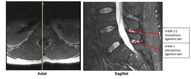
A lumbar MRI reveals;
1)��� Mild disc bulges at T11-T12, T12-L1, L1-L2 and L5-S1
2)��� Low disc signals indicative of disc desiccation at T11-T12, T12-L1, L1-L2, L2-L3, L3-L4 and L4-L5
3)��� Retrolisthesis of 2mm at L3-L4
4)��� Mild ligamentous hypertrophy at L1-L2, L2-L3, L3-L4, L4-L5 and L5-S1
5)��� L4-L5 has a Grade 1-2 tear of the interspinous ligament with mild inflammation
6)��� L5-S1 has a Grade 1 interspinous ligament tear with mild inflammation
After reviewing the MRI I ordered lumbar x-rays to rule out ligament laxity.
X-RAY STUDIES
Lumbar x-rays reveal the following:
1)��� Left lateral tilt
2)��� Retrolisthesis at L1 of 3mm
3)��� Retrolisthesis at L2 of 3mm
4)��� Combined excessive translation of 4mm of L1 during flexion-extension
5)��� Combined excessive translation of 4mm of L2 during flexion-extension
6)��� Excessive translation of L3 in extension posteriorly of 2.5mm
7)��� Decreased disc space at L5-S1
Chiropractic care was initiated. The patient was placed on an initial care plan of 2-3x/week for 3 months and then a recommended break in care for one month so the patient could be evaluated for permanency while he was not care dependent.
At maximum medical improvement, he had continued low back pain rated 4/10, continued numbness and tingling into his left leg and left quadricep weakness rated 4/5. He does not need pain killers for pain management anymore. He continues chiropractic care every two weeks to manage his symptoms.
Conclusion:
In this specific case, pathology to the posterior ligament complex diagnosed on MRI lead to the x-ray finding of excessive translation at L1-L2 and L2-L3. The patient was given a permanent impairment rating of 22% based on my interpretation of the American Medical Association�s Guides to the Evaluation of Permanent Impairment, 5th�Edition1. The interspinous ligament tears at the L4-L5 and L5-S1 level would not have been diagnosed without the MRI.
There has been much debate on whether MRI imaging has a role in evaluating lumbar PLC. MRI is a powerful diagnostic tool that can provide important clinical information regarding the condition of the PLC. Useful sequences for spinal MRI in trauma include sagittal and axial T1-weighted images, T2-weighted FSE, fat-saturated T2-weighted FSE, and STIR sequences to highlight bone edema.2�Ligamentous injuries are best identified on T2-weighted images with fat saturation because the ligaments are thin and bonded on either side by fat, which can appear as hyperintense on both T1 and T2 images.3�T1-weighted images are inadequate in isolation for identifying ligamentous injuries.4�
The diagnostic accuracy for MRI was reported for both supraspinous ligament and interspinous ligament injury with a sensitivity of 89.4% and 98.5%, respectively, and a specificity of 92.3% and 87.2% in 35 patients.5
For patients with persistent symptoms after trauma an MRI may be indicated to evaluate posterior ligamentous complex integrity.
Competing Interests:� There are no competing interests in the writing of this case report.
De-Identification: All of the patient�s data has been removed from this case.
References:
1. Cocchiarella L., Anderson G. Guides to the Evaluation of Permanent Impairment, 5th Edition, Chicago IL, 2001 AMA Press.
2. Cohen, W.A., Giauque, A.P., Hallam, D.K., Linnau, K.F. and Mann, F.A., 2003. Evidence-based approach to use of MR imaging in acute spinal trauma.�European journal of radiology,�48(1), pp.49-60.
3. Terk, M.R., Hume-Neal, M., Fraipont, M., Ahmadi, J. and Colletti, P.M., 1997. Injury of the posterior ligament complex in patients with acute spinal trauma: evaluation by MR imaging.�AJR. American journal of roentgenology,�168(6), pp.1481-1486.
4. Saifuddin, A., Green, R. and White, J., 2003. Magnetic resonance imaging of the cervical ligaments in the absence of trauma.�Spine,�28(15), pp.1686-1691.
5. Haba H, Taneichi H, Kotani Y, et al. Diagnostic accuracy of magnetic resonance imaging for detecting posterior ligamentous complex injury associated with thoracic and lumbar fractures.�J Neurosurg. 2003; 99(1 Suppl):20-26.
IFM's Find A Practitioner tool is the largest referral network in Functional Medicine, created to help patients locate Functional Medicine practitioners anywhere in the world. IFM Certified Practitioners are listed first in the search results, given their extensive education in Functional Medicine