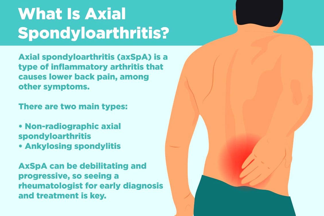
Non-radiographic axial spondyloarthritis or nr-axSpA and non-radiographic ankylosing spondylitis/AS are related. However, non-radiographic axial spondyloarthritis can present AS symptoms with active inflammation of the spine and sacroiliac/SI joints, causing back and hip pain but does not reveal joint damage on X-rays or MRIs. Injury Medical Chiropractic and Functional Medicine Clinic can explain what it means to have non-radiographic axial spondyloarthritis, how it can be managed, and what to do to prevent it from turning into ankylosing spondylitis.
Non-Radiographic Axial Spondyloarthritis
Non-radiographic axial spondyloarthritis means there are early AS symptoms but have not developed enough joint inflammation or damage to show up on an X-ray or other form of imaging. Early evidence of joint inflammation includes blurring of the joint edges and localized regions of joint erosion. It can be difficult for physicians to see these subtle changes on an x-ray.
Ankylosing Spondylitis
Ankylosing spondylitis, or AS, is a form of inflammatory arthritis that affects joints in the spine and elsewhere.
It is a chronic, inflammatory, autoimmune disease.
Medical research is still ongoing to determine the exact cause, but a genetic component is believed to be contributing factor.
Around 85% of individuals with ankylosing spondylitis have inherited the HLA-B27 gene, which is associated with multiple autoimmune conditions.
In the early stages, individuals will present lower back pain around the sacroiliac joints or the joints that connect the spine to the pelvis.
Later stages have more obvious X-ray findings, like the fusing of the sacroiliac joints and the lower spine that takes place over time.
Joint inflammation can progress, causing permanent joint damage and spine rigidity.
Most individuals with the condition can manage their symptoms with NSAIDs, chiropractic care, physical and massage therapy, and range of motion exercises.
Stage 1
There is no evidence of spinal inflammation on x-rays.
MRI provides more detailed images of bones and may reveal bone marrow edema or accumulation of fluid in the structures of the spinal bones and joints.
Individuals with non-radiographic axial spondyloarthritis, you are here.
Stage 2
There is visible inflammation of the spinal joints on the x-ray.
The sacroiliac joints between the spine and the pelvis are the most affected.
Stage 3
Chronic inflammation of the joints has caused bone loss and permanent joint damage, resulting in spine rigidity.
Symptoms of Non-Radiographic Axial Spondyloarthritis
There are differences between back pain associated with muscle strain and arthritis. Back pain symptoms include:
Starts to present before age 40.
It has a gradual onset and can go unnoticed for years.
Improves with movement or activity.
Eases up throughout the day.
Starts up in the evening when resting.
Other symptoms include:
Joint stiffness
Swollen fingers
Heel pain
Bilateral buttock discomfort and pain
Slowing Progression
Progression from non-radiographic axial spondyloarthritis to ankylosing spondylitis occurs in 10% – 20% of individuals over a two-year period. Progression factors include genetics, gender, degree of joint damage, and level of inflammatory markers at the time of diagnosis.
Early diagnosis and treatment can slow the progression before significant joint damage with anti-inflammatory therapy, rheumatological therapy, and targeted exercise.
Work with a specialist like an orthopedic spine specialist and rheumatologist that understands the disorder and is up to date on the most recent treatment modalities.
Individuals with non-radiographic axial spondyloarthritis should expect to have serial X-rays to gauge the progression of the disease.
Staying healthy and active is recommended to slow the progression of nr-AxSpA and AS.
Recent medical advances and lifestyle adjustments can slow the progression in most cases.
axSpA
References
Six tips for living well with ankylosing spondylitis. Available at https://www.mayoclinic.org/diseases-conditions/ankylosing-spondylitis/in-depth/6-tips-for-living-well-with-ankylosing-spondylitis/art-20478753. Accessed 11/07/2022.
Ankylosing spondylitis. Mayo Clinic. Available at https://www.mayoclinic.org/diseases-conditions/ankylosing-spondylitis/symptoms-causes/syc-20354808. Accessed 11/05/2022.
D. J. Pradeep, A. Keat, K. Gaffney, Predicting outcome in ankylosing spondylitis, Rheumatology, Volume 47, Issue 7, July 2008, Pages 942–945, https://doi.org/10.1093/rheumatology/ken195
Kucybała, Iwona, et al. “Radiologic approach to axial spondyloarthritis: where are we now and where are we heading?.” Rheumatology international vol. 38,10 (2018): 1753-1762. doi:10.1007/s00296-018-4130-1
Michelena, Xabier, López-Medina, Clementina, and Helena Marzo-Ortega. “Non-radiographic versus radiographic axSpA: what’s in a name?”.” National Center for Biotechnology Information. October 14, 2020. https://doi.org/10.1093/rheumatology/keaa422
Swift D. Ankylosing spondylitis: disease progression varies widely. Medpage Today. Accessed 11/05/2022.Available at https://www.medpagetoday.com/rheumatology/arthritis/49096
Dr. Alex Jimenez, D.C., presents an overview of hormonal dysfunction can affect the various hormones in the body that can increase cortisol levels in this 3-part series. This presentation provides valuable information to many individuals dealing with hormonal dysfunction and how to know the signs and utilize holistic approaches toward their health wellness. Part 1 will look at the overview of hormonal dysfunction. Part 3 will look at various holistic treatments that are available for hormonal dysfunction. We refer patients to certified providers incorporating various hormone therapies to ensure optimal health. We encourage and appreciate each patient by referring them to associated medical providers based on their diagnosis when it is appropriate. We understand that education is an excellent way when asking our providers intricated questions at the patient’s request and understanding. Dr. Alex Jimenez, D.C., only utilizes this information as an educational service. Disclaimer
An Overview Of Hormonal Expression
Dr. Alex Jimenez, D.C., presents: So the classic teaching about hormones is that they’re produced in an endocrine gland and then transported to the cells where the body will utilize them. But things are a little bit more complex. So every cell contains the genes necessary for hormone expression and functionality in the body.
And we know this because when de-differentiated from the late stage, unfortunately, cancer cells uncover gene expression. Those cells make hormones in inappropriate locations and at inconvenient times. So that means that any cell can make any hormones in the body. Still, the hormonal expression of certain genes indicates the presence of certain enzymes, which determines what hormones will reproduce. So, for example, you need to have the right precursors and enzymes. So in a reproductive female, the granulosa cells, the luteinize, and the corpus luteum are produced in the body. The granulosa cells are follicles, while the corpus odium is after ovulation. And these cells start the formation of the steroid hormones in response to FSH and LH. So FSH and LH come from the pituitary glands and stimulate the granulosa cells to start making estrogen. Steroid production will begin if the messaging from FSH and LH gets to the part of the cell that makes estrogen. This allows the body to regulate hormonal production and keep it functional. When issues disrupt hormonal regulation, the body can over or underproduce hormonal production, leading to metabolic issues associated with muscle and joint pain.
So this is a complex process, and there are many areas where this can go wrong. So you can have the right amounts of the hormone, but you’re not getting hormone production. So the messages must get into the cell first, and FSH and LH are too large to get into the cellular structure. So, they have to activate a membrane-based enzyme called adenylate cyclase to produce cyclic AMP to enter the cellular network and start hormonal formation in the body. This is the P, or the production of hormones. So by thinking about cellular membrane health, if a doctor does an essential fatty acid analysis, the patients may be very low in omega-3s; therefore, their cell membranes are more rigid and affect the body’s hormonal process. When patients are not taking their omega-3s, it could cause the inflammatory cytokines to cause more joint pain issues when various factors affect the body internally. Since inflammation can be good or bad in the body, it can cause hormonal dysfunction when they attack healthy cellular structures. That will affect this hormonal production process.
Hormonal Production Process In The Body
Dr. Alex Jimenez, D.C., presents: Once made, estradiol goes directly into the blood and is not stored, but it’s bound to SHBG and albumin. And SHBG is changed by obesity and insulin associated with hypothyroidism. So when women are obese or hyperinsulinemic will have less SHBG or hypothyroidism to transport the estrogen to the cells. This tells the body that the hormones are no longer FSH or LH and cyclic AMP, but this is estrogen. And so, estradiol is sensitive since estradiol has to go in and have a cytoplasmic receptor. So the estrogen receptor is in the cytoplasm. After it binds to the receptor, it goes into the nucleus, and it’s transcribed and then goes back out and translated to allow the body to make proteins that cause cell proliferation. Estrogen is a proliferative hormone. And once it’s acted on the cell after proliferation, it’s degraded in the cell with heat shock proteins or released back into the circulation in the body’s system.
Let’s talk about some of the basics of biochemistry because the steroidogenic pathway n the body has different ways to reduce carbons in the body. The body’s system could convert it into estrone or estriol, with less estrogen in circulation. And then estradiol, estrone, estriol, everything is eliminated through the detoxification pathways. So in the Living Matrix, healthy detoxification and estrogen metabolism pathways help the body stay functional. When the body goes through detoxification, it helps reduce the issues by figuring out what is causing the problems affecting the body’s system and slowing introducing or taking away the pain that can allow normal hormonal regulation. When the body reduces excessive carbon, it can lower cholesterol from reaching dangerous levels.
Cholesterol & Hormones
Dr. Alex Jimenez, D.C., presents: So it all starts with cholesterol, and the body can’t make enough cholesterol, so we consume food to allow it to enter the body and begin to biotransform into LDL (low-density lipoprotein) to initiate steroid hormone synthesis. So we need LDL in the body to decrease the carbons since it is a union directional. When it comes to hormone deficiency cases, sometimes women can come in with extremely low LDLs because they’re on statins, underweight, or over-athletic; these are connections and pattern recognition. In a female’s body, the ovaries on the left and right side of the female reproduction system produce all three sex steroids: estrogens, progesterones, and androgens, for optimal body function. They are different than the male’s reproduction system, which includes the testicles, due to the other enzymes. They differ from the testes in many ways due to additional hormonal output in the body, which is different than adrenal hormones. For example, if the body can’t make aldosterone or cortisol in the endocrine glands, they will be shunted to make sex hormones. And since each body is diverse, male or female, some hormones can’t make glucocorticoids or mineral corticoids.
So we must think about mitochondrial health for hormone production, which is critical for producing hormones. For expecting mothers, it allows the formation of pregnancy alone in the mitochondria. So mitochondrial health energy is related to hormone health, which causes cholesterol uptake by the mitochondria in the adrenal glands and ovaries that can stimulate ACTH. So the mitochondria can make hormones as we go while enabling the enzymes in the female’s body to induce pregnenolone formation. However, getting the cholesterol to the inner membrane and LDL to the inner membrane to the mitochondria is the rate-limiting step in all steroid hormone synthesis. Now it can develop and go into two different pathways in the body. It can create DHEA, or it can include progesterone when cholesterol is forming pregnenolone and can look diagrammatically in test results.
DHEA & Hormones
Dr. Alex Jimenez, D.C., presents: So if the mitochondria can enable the body’s pregnenolone to form DHEA or progesterone, let’s start with going over things with developing progesterone since it’s hydroxylated. It creates an enzyme called 17 hydroxy progesterone, the immediate precursor of the androgens and estrogens. So the 17 hydroxy progesterone will eventually form androgens or estrogens, and androstenedione can develop testosterone, and both can become estrogens by aromatization. So what does this mean? It means you must be concerned about too many androgens being around because they can become estrogen. The best way to remember this is that androstenedione can become estrone, and testosterone can become estradiol. This can cause progesterone to be the precursor to cortisol in the body and can go in two different directions.
So progesterone is also the precursor to aldosterone, which asks what will happen when the body needs cortisol or aldosterone. The body will then produce less progesterone causing it to be shunned in hormonal production and causing cortisol to overproduce. This is known as cortisol steal, and if it is not treated at the moment, it can cause muscle and joint inflammation in the body, leading to various issues that the person is dealing with.
A decreased formation of androgens and estrogens can inhibit the DHEA pathway. So when the body makes more cortisol, it can cause the hormones to develop an estrogen-dominate shape causing the cortisol hormone to stimulate aromatase. To that point, it can lead to breast cancer, fibroids, and endometriosis in the long term. Women can get stress, hot flashes, and decreased libido due to that hormonal decrease in their bodies.
Conclusion
Other issues like stress can cause increased cortisol formation, anxiety, inflammation, simple carbohydrates, et cetera to the body associated with joint and muscle pain. They can also inhibit sex and sex hormone production directly and indirectly. So this is where people have to be concerned if they’re giving DHEA because DHEA can convert itself into sex hormones. So that can be a good thing if you’re dealing with hormonal deficiency. But if you give too much DHEA, you can overproduce hormonal function.
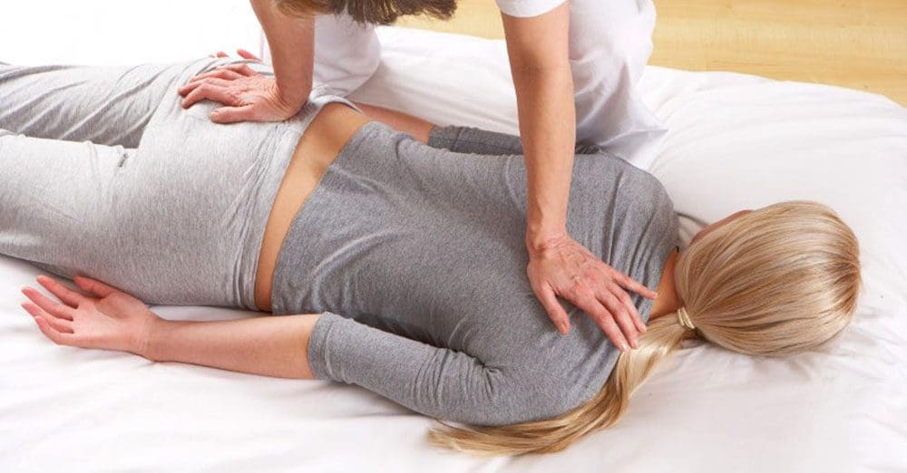
A certified/licensed massage therapist performs chiropractic therapeutic massage under the direction of a chiropractor to soothe tense muscles, release pressure on the nerve/s, break up scar tissue, and stimulate the release of endorphins. Studies have shown that a therapeutic chiropractic massage is as effective as nonsteroidal anti-inflammatory medications for pain relief caused by various musculoskeletal conditions, including sciatica. Although medications can work for a time, they do not deal with the underlying cause of sciatica.
Sciatica Massage
A sciatica massage offers several benefits that include the following:
Improved and increased blood circulation.
Lowered blood pressure.
Release of hormones that help reduce anxiety, depression, and pain.
Relieves body discomfort and soreness by decreasing the activation of pain receptors in the spine and muscles.
Inflammation alleviation.
Muscle stress relief.
Muscle relaxation.
Flare-up relief and prevention.
Improves sleep.
Massage Types
Each type of massage is designed for a different purpose.
Manual Manipulation
Improves mobility and reduces pain.
Stretches tendons, ligaments, and muscles.
Mobilization
Focuses on the musculoskeletal system to improve mobility.
Connective Tissue
A deeper massage that targets connective tissues like ligaments and tendons.
Deep Tissue Massage
Designed to reach the deep layers to realign the muscles and fascia.
Myofascial Massage
Releases trigger points, adhesions, and nerve endings.
Healing Phases
Most cases of sciatica resolve in less than 4 to 6 weeks with conservative treatment. More severe cases with neurologic symptoms can have a longer recovery period.
Phase One
Alleviates discomfort, numbness, tingling, pain, and other symptoms.
The pain is reduced so the chiropractor and therapists can begin to address the cause, although the other symptoms and sensations could still be experienced.
If muscle spasms are present, they will be less frequent and with less intensity.
Phase Two
The individual can focus on functioning within their normal environment.
The chiropractor and therapists examine if they can get up and stand from a chair without problems, get in and out of a car, sit for longer periods, and walk with less discomfort.
A reflex test will be performed to analyze how the healing process is coming along.
If there is significant nerve damage, reflexes could be lessened.
For example, if sciatic inflammation is present, and the Achilles tendon is tapped with a reflex hammer, there could be little or no movement of the calf muscles.
If there is an improvement, there will be a healthy reflex.
The straight leg test will show if the individual can lift their leg with little or no pain.
Phase Three
The final healing phase provides the ability of normal movements and full strength.
This is when the individual can resume normal activities, specifically, those stopped because of the pain.
This can include walking, driving, house chores, sports, or working out.
Contact our office for treatment options customized to the individual and their injury.
Sciatica?
References
Camino Willhuber GO, Piuzzi NS. Straight Leg Raise Test. [Updated 2022 June 22]. In: StatPearls [Internet]. Treasure Island (FL): StatPearls Publishing; 2022 Jan-. Available from: https://www.ncbi.nlm.nih.gov/books/NBK539717/
Chang C, Jeno SH, Varacallo M. Anatomy, Bony Pelvis, and Lower Limb, Piriformis Muscle. [Updated 2022 October 3]. In: StatPearls [Internet]. Treasure Island (FL): StatPearls Publishing; 2022 Jan-. Available from: https://www.ncbi.nlm.nih.gov/books/NBK519497/
Davis D, Maini K, Vasudevan A. Sciatica. [Updated 2022 May 6]. In: StatPearls [Internet]. Treasure Island (FL): StatPearls Publishing; 2022 Jan-. Available from: https://www.ncbi.nlm.nih.gov/books/NBK507908/
Marian Majchrzycki, Piotr Kocur, Tomasz Kotwicki, “Deep Tissue Massage and Nonsteroidal Anti-Inflammatory Drugs for Low Back Pain: A Prospective Randomized Trial,” The Scientific World Journal, vol. 2014, Article ID 287597, 7 pages, 2014. https://doi.org/10.1155/2014/287597
Miller, Kenneth Jeffrey. “Physical assessment of lower extremity radiculopathy and sciatica.” Journal of chiropractic medicine vol. 6,2 (2007): 75-82. doi:10.1016/j.jcme.2007.04.001
The Centralization Phenomenon in Chiropractic Spinal Manipulation of Discogenic Low Back Pain and Sciatica. https://chiro.org/Low_Back_Pain/The_Centralization_Phenomenon.shtml. Accessed October 22, 2022.
After a car accident, you may notice neck pain. It could be a slight soreness you think is nothing but take care of. More than likely, you have whiplash. And that little soreness can turn into a lifetime of chronic neck pain if only treated with pain meds and not treated at the source.
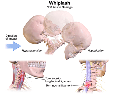
Whiplash trauma, aka neck sprain or neck strain, is an injury to the soft tissues around the neck.
Whiplash can be described as a sudden extension or backward movement of the neck and flexion or forward movement of the neck.
Severe whiplash can also include injury to the following:
Intervertebral joints
Discs
Ligaments
Cervical muscles
Nerve roots
Symptoms of Whiplash
Most people experience neck pain either immediately after the injury or several days later.
Other symptoms of whiplash trauma can include:
Neck stiffness
Injuries to the muscles and ligaments around the neck
Headache and dizziness
Symptoms & possible concussion
Difficulty swallowing and chewing
Hoarseness (possible injury to the esophagus and larynx)
The sensation of burning or prickling
Shoulder pain
Back pain
Diagnosis of Whiplash Trauma
Whiplash trauma usually causes damage to the soft tissues; a doctor will take x-rays of the cervical spine in case of delayed symptoms and rule out other problems or injuries.
Treatment
Fortunately, whiplash is treatable, and most symptoms resolve completely.
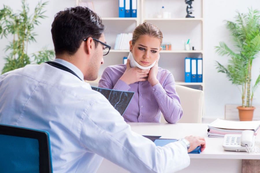
Most often, whiplash is treated with a soft cervical collar.
This collar may need to be worn for 2 to 3 weeks.
Other treatments for individuals with whiplash may include the following:
Heat treatment for relaxing muscle tension and pain
Pain meds such as analgesics and non-steroidal anti-inflammatory
Some people will tell you that whiplash is a made-up injury that people use to get more money in a settlement stemming from an accident. They do not believe it is possible in a low-speed rear-end accident and see it as a legitimate injury claim, mainly because there are no visible marks.
Some insurance experts claim that about a third of whiplash cases are fraudulent, leaving two-thirds of the cases legitimate. Much research also supports the claim that low-speed accidents can indeed cause whiplash, which is very real. Some patients suffer from pain and immobility for the rest of their lives.
NCBI Resources
Chiropractors will use different techniques to relieve the pain of whiplash and help with healing.
Chiropractic Adjustment The chiropractor performs spinal manipulation to move the joints into alignment gently. This will help to align the body to relieve pain and encourage healing.
Muscle Stimulation and Relaxation This involves stretching the affected muscles, relieving tension, and helping them relax. Finger pressure techniques may also be combined with trying to alleviate pain.
McKenzie Exercises These exercises help with disc derangement that whiplash causes. They are first performed in the chiropractor’s office, but the patient can be taught how to do them at home. This helps the patient have some degree of control over their healing.
Each whiplash case is different. A chiropractor will evaluate the patient and determine the appropriate treatment case-by-case basis. The chiropractor will determine the best course of treatment that will relieve your pain and restore your mobility and flexibility.
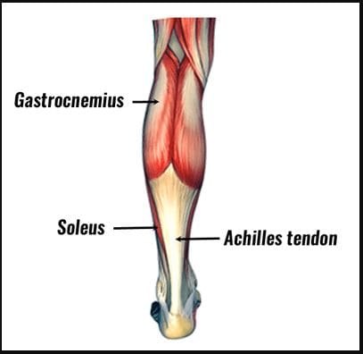
The calves are extremely important to the lower portions of the legs as they allow the individual to move around and help stabilize the entire body. The calves have two muscles: the gastrocnemius and the soleus, which provide plantarflexion and stability at the ankle joint. These two muscles work together to ensure that the upper body’s weight doesn’t lean forward too much to let the individual fall. However, when many people overuse the calf muscles constantly, it can cause the muscle fibers in the lower legs to be overstretched and develop small nodules known as trigger points to cause referred pain to the calves and the ankles that can affect a person’s ability to walk, run, or jump. Today’s article looks at the soleus muscle, how trigger points affect the muscle, and different methods to reduce pain while managing trigger points along the calves. We refer patients to certified providers that incorporate various techniques in the lower body extremities, like lower leg and calve pain therapies correlating to trigger points, to aid many people dealing with pain symptoms along the soleus muscles, causing muscle cramps and issues along the ankles. We encourage and appreciate each patient by referring them to associated medical providers based on their diagnosis when it is appropriate. We understand that education is an excellent way when asking our providers intricated questions at the patient’s request and understanding. Dr. Alex Jimenez, D.C., only utilizes this information as an educational service. Disclaimer
What Is The Soleus Muscle?
Do you feel a cramping sensation in your calves? Do your feet hurt when you are walking, jumping, or running? Or are you experiencing tightness in your calve muscles? When a person is experiencing calf pain in their legs, it could be trigger points causing overlapping issues in the soleus muscles. As stated earlier, the calves have two muscles: the gastrocnemius and the soleus. The gastrocnemius makes up the round shape of the calves and is a complex, superficial muscle. At the same time, the soleus is a large, flat muscle deep within the gastrocnemius and forms the calcaneal tendon (Achilles tendon), which can be mistaken as a nerve for the ankle. Studies reveal that the soleus muscle works together with the gastrocnemius, and these muscles constitute a plantar flexor. The soleus is part of a group of muscles known as antigravity muscles. It acts like a skeletal muscle and helps maintain good posture in the body to prevent the sheer body weight from falling forwards at the ankle when standing.
How Do Trigger Points Affect The Soleus Muscle?
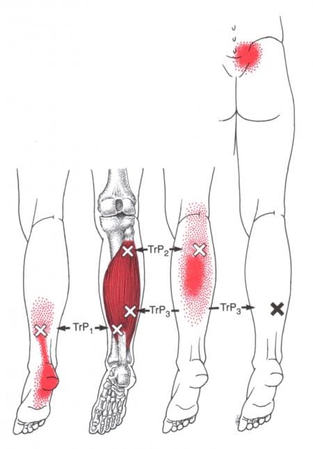
The calves are essential for the movement and mobility functions of the body. When the calve muscles have been overused to repetitive movements or have been dealing with trauma, it can cause the muscle fibers to tear and develop trigger points along the calves causing referred pain to travel down the ankles. Trigger points along the soleus muscle are tricky to diagnose since they cause referred pain to the surrounding muscles. “Myofascial Pain and Dysfunction,” written by Dr. Janet G. Travell, M.D., mention that, unlike the latent trigger points that can affect the gastrocnemius, active trigger points can affect the soleus muscle and cause tenderness referred to the heel. When muscle tenderness affects the heels, it can cause trigger points to mimic chronic conditions like plantar fasciitis. Additional studies reveal that musculoskeletal impairments like trigger points in the soleus muscle could potentially lead to biomechanical changes in the ankle. When trigger points cause hypersensitivity spots in the skeletal muscle taut band, it can restrict the soleus muscle to allow ankle dorsiflexion. Individuals with active soleus trigger points are prone to develop low back pain due to ankle dorsiflexion restriction, leading to them leaning over and lifting objects improperly.
Trigger Point Of The Week: Soleus – Video
Have you been dealing with pain traveling down from your calves to your ankles? Experiencing stiffness around your ankles? Or do you feel like you are about to fall? These pain-like issues are associated with trigger points along the soleus muscles along the calves. The soleus is one of the two muscles (the other is the gastrocnemius) that make up the calves and acts as a skeletal muscle to help maintain good posture in the body. When trigger points affect the soleus muscle, they can cause referred pain to the calves and mimic chronic issues like plantar fasciitis at the ankles. The video above explains where the soleus muscles are located, deep within the gastrocnemius, where the trigger points are causing referred pain to the ankles, and where they are in the soleus muscle fibers in the calves. Even though trigger points can cause referred pain in the affected muscle fibers of the body, they are treatable and can be managed through different methods to help the calves.
Different Methods To Reduce Pain And Manage Trigger Points In The Calves
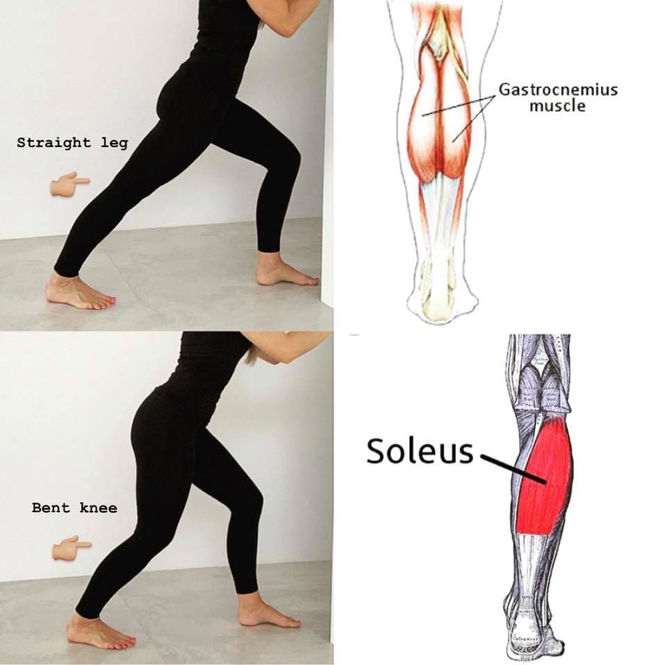
Even though trigger points can affect the soleus muscles and cause referred pain to the ankles, different methods can reduce the pain and help manage trigger points in the calves. Studies reveal that when there are multitudes of therapies that can help manage trigger points, it can help reduce the pain affecting the soleus muscle by optimizing muscle function and preventing the development of chronic pain syndromes. Besides therapies that target trigger points, corrective actions and stretches can help release the pain from the soleus muscle. Incorporating calf stretches, having a correct posture when sitting down, wearing long loose socks while sleeping, and wearing low heels can prevent the soleus muscle from shortening and help manage trigger points from re-occurring in the calves. When people start to take care of their calve muscles, it can help their mobility and stability at the ankles.
Conclusion
As part of the calf muscles and an antigravity muscle, the soleus works with the gastrocnemius to help with stability and plantarflexion to the ankles. This muscle is deep within the gastrocnemius and helps maintain good posture in the body that prevents a person from falling forward at the ankles when standing. When the soleus muscles have been overused, they can develop tiny knots in the muscle fibers known as trigger points that can cause referred pain to the ankles. Trigger points along the soleus muscles can cause overlapping features in the calves by restricting ankle dorsiflexion, causing individuals to develop low back pain associated with leaning forward. However, various therapies can manage trigger points through multiple treatments that can reduce the pain and allow stability back to the calves, enabling the individual to continue walking without feeling pain.
References
Binstead, Justin T, et al. “Anatomy, Bony Pelvis and Lower Limb, Calf – Statpearls – NCBI Bookshelf.” In: StatPearls [Internet]. Treasure Island (FL), StatPearls Publishing, 29 May 2022, https://www.ncbi.nlm.nih.gov/books/NBK459362/.
Travell, J. G., et al. Myofascial Pain and Dysfunction: The Trigger Point Manual: Vol. 2:the Lower Extremities. Williams & Wilkins, 1999.
Valera-Calero, Juan Antonio, et al. “Prediction Model of Soleus Muscle Depth Based on Anthropometric Features: Potential Applications for Dry Needling.” Diagnostics (Basel, Switzerland), U.S. National Library of Medicine, 7 May 2020, https://www.ncbi.nlm.nih.gov/pmc/articles/PMC7277950/.
Various muscles help support the lower legs in the body and allow movement to the host. The lower extremities include the hips, pelvis, thighs, legs, knees, and feet. At the same time, the various muscles help provide support and stability to the upper body’s weight and incorporate mobility and movement for the entire body to go from one location to another. The legs have two sections connected with the knees; the upper portion has the hips and thigh muscles, while the lower legs have the calve muscles, shin muscles, and the Achilles tendon. The calve muscles have two groups of muscles, and when the calve intense exercises, or normal factors have overused muscles may potentially lead to muscle cramps associated with trigger points. Today’s article examines one of the calve muscles known as the gastrocnemius, how the calves are affected by trigger points and corrective actions to prevent muscle cramps in the calves. We refer patients to certified providers that incorporate various techniques in the lower body extremities, like lower leg and calve pain therapies correlating to trigger points, to aid many people dealing with pain symptoms along the gastrocnemius muscles, causing muscle cramps. We encourage and appreciate each patient by referring them to associated medical providers based on their diagnosis when it is appropriate. We understand that education is an excellent way when asking our providers intricated questions at the patient’s request and understanding. Dr. Alex Jimenez, D.C., only utilizes this information as an educational service. Disclaimer
What Are The Gastrocnemius Muscles?
Have you been dealing with walking from one place to another? Do your calves feel hard or tense with the slightest touch or movement? Or are you feeling excruciating pain in your calves that make it hard to move? These pain-like symptoms are indicators of trigger points associated with the calves affecting the gastrocnemius muscles. The calves are mostly referred to as the posterior portion of the lower legs responsible for the foot and ankle plantarflexion while also engaging in activities like running or jumping. The two muscles that make up the calves are the gastrocnemius and the soleus. The gastrocnemius is a complex, superficial muscle fundamental to good posture or walking. This muscle has a casual relationship with the lower body as it affects hip movement and the lumbar area of the spine. The gastrocnemius provides a round shape for the calves to form and narrows down to the ankles, where it forms a tendon.
How Are The Calves Affected By Trigger Point Pain?
Since the gastrocnemius provides the round shape to form the calves when the muscles have become overused or injured in a sports activity, it can cause the individual to have limited mobility. Studies reveal that a tear in the gastrocnemius muscles can implicate lower leg trauma and affect the muscle function to plantar flex the foot at the ankle joint and reduce flexion on the leg to the leg knee joint. When it comes to the development of trigger points along the gastrocnemius muscles that are affecting the calves, according to “Myofascial Pain and Dysfunction,” written by Dr. Janet Travell, M.D., the book states that latent trigger points along the gastrocnemius may cause individuals to complain about calf cramps on the legs, however, when the trigger points are active, the individual is aware of the calf pain and would complain about experiencing pain in the back of their knees. The book also mentioned that trigger points associated with the gastrocnemius muscle could be mistaken for other conditions like plantar foot pain or radiculopathy in the sacroiliac joints. When trigger points affect the calf muscles, it can lead to mobility issues and causes people to be unstable when walking.
Trigger Point Of The Week: Gastrocnemius Muscle- Video
Have you been dealing with calf pain when walking for a short distance? Do your calf muscles ache or tense up when you put slight pressure when stepping down? Or do you feel that your calf muscles are hard when resting? Many of these issues affecting the calves are associated with trigger points affecting the gastrocnemius muscles. The gastrocnemius muscle is one of the muscles that make up the calves in the lower legs. This complex, superficial muscle provides a round shape to the calves and can become overused in various activities that can cause a person to have limited mobility. When the gastrocnemius muscle is overused, it can develop tiny nodules in the muscle fibers known as trigger points that mimic other conditions affecting leg mobility. The video above shows where this complex muscle is in the calves and where the trigger points are located in the muscle fibers. Trigger points along the affected muscle can cause referred pain while mimicking other conditions that can often confuse people about what they are feeling. All is not lost, however, as trigger points are treatable and can be managed through various treatments.
Corrective Actions To Prevent Muscle Cramps On The Calves
When the calf muscles like the gastrocnemius are causing symptoms of pain and muscle cramps due to trigger points, there are various treatments and corrective actions that can prevent muscle cramps from causing more issues in the legs and even help manage trigger points from re-forming in the muscle fibers. Some corrective actions that can help improve calve function are gently flexing the foot at the ankle joint to allow the calf muscles to tract and retract to reduce swelling and pain. Other correction actions that people should incorporate to prevent muscle cramping in the calves when they are sitting is to gently rock in a chair to reduce prolonged immobility to the calves and increase blood flow. Studies reveal that dry needling and other various treatments can help reduce muscle stiffness in the gastrocnemius and improve muscle strength in the calves.
Conclusion
The calves are part of the legs that allow plantarflexion at the ankle joint. Known as the gastrocnemius muscle, it makes up the calves’ round shape. The gastrocnemius muscle is complex and superficial as it forms a rounded shape at the calves and narrows down at the ankles. However, when the muscle has been through various activities and is constantly overstretched, it can affect a person’s walking mobility and develop tiny nodules known as trigger points. Trigger points in the gastrocnemius muscle can cause referred pain in the calve muscles and mimic conditions like plantar foot pain to the feet. Fortunately, various treatments and corrective actions can prevent referred pain from re-occurring in the calves and bring mobility back to the legs so a person can continue to their destination.
References
Albin, S R, et al. “The Effect of Dry Needling on Gastrocnemius Muscle Stiffness and Strength in Participants with Latent Trigger Points.” Journal of Electromyography and Kinesiology : Official Journal of the International Society of Electrophysiological Kinesiology, U.S. National Library of Medicine, 9 Oct. 2020, https://pubmed.ncbi.nlm.nih.gov/33075711/.
Binstead, Justin T, et al. “Anatomy, Bony Pelvis and Lower Limb, Calf.” In: StatPearls [Internet]. Treasure Island (FL), StatPearls Publishing, 29 May 2022, https://www.ncbi.nlm.nih.gov/books/NBK459362/.
Bordoni, Bruno, and Matthew Varacallo. “Anatomy, Bony Pelvis and Lower Limb, Gastrocnemius Muscle.” In: StatPearls [Internet]. Treasure Island (FL), StatPearls Publishing, 26 Apr. 2022, https://www.ncbi.nlm.nih.gov/books/NBK532946/.
Nsitem, Virginia. “Diagnosis and Rehabilitation of Gastrocnemius Muscle Tear: A Case Report.” The Journal of the Canadian Chiropractic Association, U.S. National Library of Medicine, Dec. 2013, https://www.ncbi.nlm.nih.gov/pmc/articles/PMC3845475/.
Travell, J. G., et al. Myofascial Pain and Dysfunction: The Trigger Point Manual: Vol. 2:the Lower Extremities. Williams & Wilkins, 1999.
Back pain after eating is often the result of conditions and/or disorders in other areas of the body that radiate to the back. These problems range from unhealthy posture, digestive issues, bowel problems, ulcers, allergies, etc. This is because the nerves of the back and the abdominal area run through areas of the spine. In addition to the classic symptoms like bloating and gas, individuals can develop symptoms beyond the gut, including sleep problems, fatigue, headaches, urinating problems, muscle aches, pelvic discomfort, and back pain. Chiropractic care and functional medicine can realign the body, alleviate symptoms, and restore function.
Back Discomfort
Back pain after eating can be linked to the body’s digestive process.
Individuals in this group can experience inflammation after consuming specific foods.
Inflammation could worsen existing back problems.
Individuals with food intolerance will have uncomfortable but usually not dangerous symptoms.
Individuals with food allergies can experience life-threatening allergic reactions.
Heartburn
Heartburn results from acid reflux, when stomach contents and acid flow back into the esophagus. Heartburn’s key symptom is a burning sensation in the chest. However, heartburn and indigestion do not directly cause back pain. But for individuals with back problems, heartburn can worsen back discomfort.
GERD
Gastroesophageal reflux disease, or GERD, could cause back pain in individuals with more than two weekly heartburn episodes.
The digestive system condition results from the chronic backflow of stomach acid.
Over time, the powerful acid can inflame the esophageal lining.
A stomach or esophageal ulcer can develop if GERD is not managed.
Pain could be felt in the lower to middle back around the stomach and lower intestines.
Ulcers
Ulcers can also result from a bacterial infection of Helicobacter pylori (or H. pylori).
Long-term use of nonsteroidal anti-inflammatory drugs (or NSAIDs) can also enable ulcer formation.
A peptic ulcer (or open sore) can develop on your stomach’s interior lining.
The small intestine’s upper portion can also be affected.
Peptic ulcers cause burning pain in the stomach. These flare-ups can worsen existing back problems and pain. Spicy foods and stress don’t cause peptic ulcers but can worsen their effects. In severe cases, a peptic ulcer can cause referred back pain felt in another location than where the pain originates. This means pain could be felt in the lower to middle back adjacent to the stomach and lower intestines.
Kidney Infection
Back pain could result from a bacterial kidney infection.
Kidney dysfunction infections, kidney stones, and chronic kidney diseases can be mistaken for general back and side pain.
Other symptoms can include chills, fever, nausea, and vomiting.
Pancreatitis
Pancreatitiscan develop if the pancreas becomes inflamed, brought on by excess alcohol consumption or gallstones. This definitely can cause back discomfort and pain.
Pancreatitis is the inflammation of the pancreas.
After eating, individuals can experience worsening abdominal pain that can refer to the back.
The body’s network of interconnecting sensory nerves causes the pain to be felt in other areas.
Most individuals will experience pain in the upper left or middle abdomen.
When individuals with celiac disease consume foods with gluten, the small intestine gets damaged, and their bodies cannot properly absorb the necessary nutrients.
Besides the back discomfort, there could be a burning sensation during urination or other urinary tract symptoms. Stools could be dark or black, a possible ulcer symptom. To reduce the chances of back discomfort after eating, avoid sugary, spicy, fatty foods or anything that triggers heartburn and reduce alcohol consumption. If you are experiencing frequent episodes of back pain after eating or the pain worsens, contact your physician, healthcare provider, or a chiropractor.
Hormonal Dysfunction In Men
References
Celiac Disease Foundation. (n.d.) “What is celiac disease?” https://celiac.org/about-celiac-disease/what-is-celiac-disease/
Mayo Clinic. (n.d.) “Celiac disease.” https://www.mayoclinic.org/diseases-conditions/celiac-disease/symptoms-causes/syc-20352220#:~:text=Celiac%20disease%2C%20sometimes%20called%20celiac,response%20in%20your%20small%20intestine
Pfizer. (April 25, 2022) “Heartburn, acid reflux, or GERD: what’s the difference?” https://www.pfizer.com/news/articles/heartburn_acid_reflux_or_gerd_what_s_the_difference#:~:text=The%20terms%20acid%20reflux%2C%20heartburn,meals%20or%20when%20lying%20down
Prairie Spine & Pain Institute. (n.d.) “What may cause back pain after eating: symptoms & prevention.” https://prairiespine.com/spine-care/5-things-that-may-cause-back-pain-after-eating-symptoms-and prevention/#:~:text=Exercises%20practiced%20in%20yoga%2C%20Pilates,chi%20may%20be%20particularly%20beneficial.&text=If%20a%20doctor%20cannot%20identify,ice%2C%20and%20taking%20pain%20relievers.
IFM's Find A Practitioner tool is the largest referral network in Functional Medicine, created to help patients locate Functional Medicine practitioners anywhere in the world. IFM Certified Practitioners are listed first in the search results, given their extensive education in Functional Medicine