ClickCease
+1-915-850-0900 [email protected]
Select Page
Chiropractic Relieves Sacroiliac Joint Pain

Chiropractic Relieves Sacroiliac Joint Pain

Chiropractic Relieves: How can a body part you have probably never heard of hurt so BAD? This is a common question we hear from individuals suffering from sacroiliac joint pain.

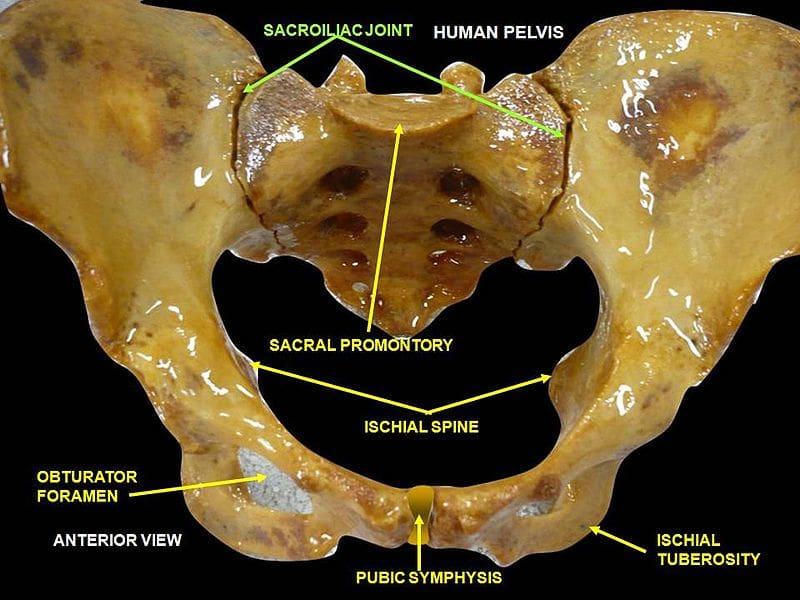
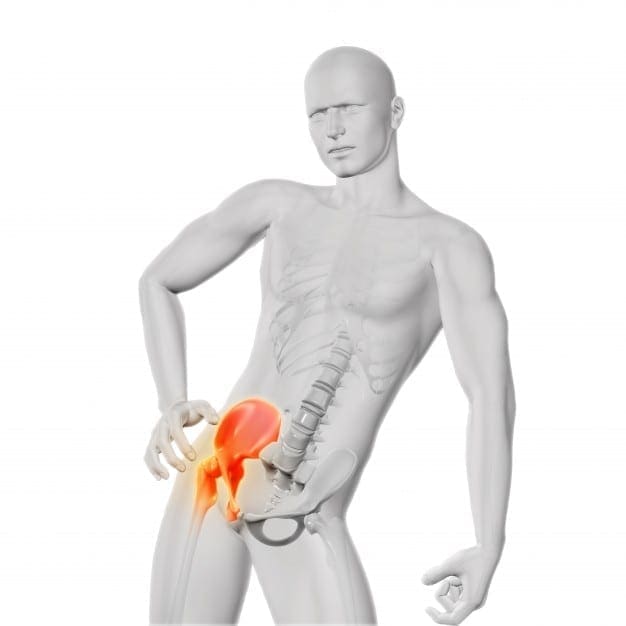
The sacroiliac�joint is formed by the sacrum and the ilium where they meet on either side of the lower back, with the purpose of connecting the spine to the pelvis. This small joint is one of the most durable parts of the human body, and it is responsible for a big job.

chiropractic relieves

The unassuming little sacroiliac joint withstands the pressure of the upper body’s weight pushing down on it, as well as pressure from the pelvis. It’s basically the cushion between the torso and the legs. As such, it handles force from pretty much every angle.

While immensely strong and durable, this joint is not indestructible. Sacroiliac joint pain usually crops up as lower back pain, or pain in the legs or buttocks.

Weakness in these areas may also be present. The typical culprits in causing the sacroiliac joint to exhibit pain are traumatic injuries to the lower back, but more frequently develops over a longer period of time.

Sacroiliac joint pain is often misdiagnosed as soft tissue issues instead of the joint itself. Doctors may rule out other medical conditions before settling on a diagnosis that includes a sacroiliac joint problem.

If you have suffered an injury, a degenerative disease, or otherwise damaged the sacroiliac joint, there are treatments available to help manage pain, promote healing, and lessen the chances of recurrence. Here are a four helpful guidelines to assist in effectively handling sacroiliac joint pain.

chiropractic relieves

Chiropractic Relieves:

First, rest and ice the area. Avoid exaggerated movements of your lower back in order to relieve some of the body’s pressure on the sacroiliac joint. Also apply ice wrapped in a towel periodically to soothe the area and minimize the pain.

A second way to handle sacroiliac pain is with therapeutic massage. Tightness around the joint is a common cause of discomfort and pain. Professional massage serves to loosen and relax the lower back, buttocks, and leg areas, offering relief from pain.

Third, consider chiropractic and seeing a chiropractor. Chiropractic relieves pain, treatment known as adjustments, not only provides great options for pain relief but also helps promote the healing process of this joint.

A chiropractor is specifically trained to guide you through several phases of care. They don�t focus just on pain relief but are primarily interested in helping you fix the problem.

They�re also very well trained in rehabilitation of the spine. This approach will help loosen the muscles surrounding the joint as well as strengthen them. This will decrease the risk of pain returning down the road.

Finally, in very rare cases, doctors will choose to apply an injection to the area to alleviate pain and inflamed tissue. Obviously, the injection won�t fix the problem but may give the patient relief temporarily. Surgery is rarely a viable option.

If you show symptoms of sacroiliac pain, it’s important to see a Doctor of Chiropractic so he or she can perform tests to correctly diagnose your condition. It could very well be another type of lower back problem. Remember chiropractic relieves, so quit suffering and give us a call!

Pregnancy & Chiropractic Care

Migraine Pain Treatment | Dr. Alex Jimenez

Migraine Pain Treatment | Dr. Alex Jimenez

A migraine is characterized as a moderate to severe headache, often accompanied by nausea and sensitivity to light and sound. Nearly 1 in 4 United States households include someone who suffers from migraine. As a matter of fact, migraine is considered to be the 3rd most prevalent condition in the world. Researchers haven’t identified a definitive cause for migraines, however, several factors are believed to trigger the complex headache pain, including a misalignment in the cervical spine. Chiropractic care is a well-known alternative treatment option used to help treat migraine headaches and improve the symptoms. The purpose of the following case study is to demonstrate the effects of chiropractic care on migraine pain management.


A Case of Chronic Migraine Remission After Chiropractic Care


Abstract


  • Objective: To present a case study of migraine sufferer who had a dramatic improvement after chiropractic spinal manipulative therapy (CSMT).
  • Clinical features: The case presented is a 72-year�old woman with a 60-year history of migraine headaches, which included nausea, vomiting, photophobia, and phonophobia.
  • Intervention and outcome: The average frequency of migraine episodes before treatment was 1 to 2 per week, including nausea, vomiting, photophobia, and phonophobia; and the average duration of each episode was 1 to 3 days. The patient was treated with CSMT. She reported all episodes being eliminated after CSMT. The patient was certain there had been no other lifestyle changes that could have contributed to her improvement. She also noted that the use of her medication was reduced by 100%. A 7-year follow-up revealed that the person had still not had a single migraine episode in this period.
  • Conclusion: This case highlights that a subgroup of migraine patients may respond favorably to CSMT. While a case study does not represent significant scientific evidence, in context with other studies conducted, this study suggests that a trial of CSMT should be considered for chronic, nonresponsive migraine headache, especially if migraine patients are nonresponsive to pharmaceuticals or prefer to use other treatment methods.
  • Key indexing terms: Migraine, Chiropractic, Spinal manipulative therapy


Introduction


Migraine remains a common and debilitating condition.[1, 2] It has an estimated incidence of 6% in males and 18% in females.[2] A study in Australia found the cost to industry to be an estimated $750 million.[3] Lipton et al found that migraine is one of the most frequent reasons for consultations with general practitioners, affecting between 12 million and 18 million people each year in the United States.[4] The estimated cost in the United States is $25 billion in lost productivity due to 156 million full-time work days being lost each year.[5] Recent information has suggested that these older figures above are still current, but also underestimated, because of many sufferers not stating their problem because of a perceived poor social stigma.[6]


The Brain Foundation in Australia notes that 23% of households contain at least one migraine sufferer. Nearly all migraine sufferers and 60% of those with tension-type headache experience reductions in social activities and work capacity. The direct and indirect costs of migraine alone would be about $1 billion per annum.[3]


The Headache Classification Committee of the International Headache Society (IHS) defines migraines as having the following: unilateral location, pulsating quality, moderate or severe intensity, and aggravated by routine physical activity. During the headache, the person must also experience nausea and/or vomiting, photophobia, and/or phonophobia.[7] In addition, there is no suggestion either by history or by physical or neurologic examination that the person has a headache listed in groups 5 to 11 of their classification system.[7] Groups 5 to 11 of the classification system include headache associated with head trauma, vascular disorder, nonvascular intracranial disorder, substances or their withdrawal, noncephalic infection, or metabolic disorder, or with disorders of cranium, neck, eyes, nose, sinuses, teeth, mouth, or other facial or cranial structures.


Some confusion relates to the �aura� feature that distinguishes migraine with aura (MA) and migraine without aura (MW). An aura usually consists of homonymous visual disturbances, unilateral paresthesias and/or numbness, unilateral weakness, aphasia, or unclassifiable speech difficulty.[7] Some migraineurs describe the aura as an opaque object, or a zigzag line around a cloud; even cases of tactile hallucinations have been recorded.[8] The new terms MA and MW replace the old terms classic migraine and common migraine, respectively.


The IHS diagnostic criteria for MA (category 1.2) is at least 3 of the following:


  1. One or more fully reversible aura symptoms indicating focal cerebral cortex and/or brain stem dysfunction.
  2. At least 1 aura symptom develops gradually over more than 4 minutes or 2 or more symptoms occurring in succession.
  3. No aura symptom lasts more than 60 minutes.
  4. Headache follows aura with a free interval of less than 60 minutes.


Migraine is often still nonresponsive to treatment.[9] However, several studies have demonstrated statistically significant reduction in migraines after chiropractic spinal manipulative therapy (CSMT).[10-15]


This article will discuss a patient presenting with MW and her response after CSMT. The discussion will also outline specific diagnostic criteria for migraine and other headaches relevant to chiropractors, osteopaths, or other health practitioners.


Case Report


A 72-year�old 61-kg white woman presented with migraine headaches that had commenced in early childhood (approximately 12 years old). The patient could not relate anything to the commencement of her migraines, although she believed there was a family history (father) of the condition. During the history, the patient stated that she suffered regular migraine headaches (1-2 per week) with which she also experienced nausea, vomiting, vertigo, and photophobia. She needed to cease activities to alleviate the symptoms, and she often required acetaminophen and codeine medication (25 mg) or sumatriptan succinate for pain relief. The patient was also taking verapamil (calcium ion antagonist, for essential hypertension), calcitriol (calcium uptake, for osteoporosis), pnuemenium on a daily basis, and carbamazipine (antiepileptic, neurotropic medication) twice daily.


The patient reported that an average episode lasted 1 to 3 days and that she could not perform activities of daily living for a minimum of 12 hours. In addition, a visual analogue scale score for an average episode was 8.5 out of a possible maximum score of 10, corresponding to a description of �terrible� pain. The patient noted that stress or tension would precipitate a migraine and that light and noise aggravated her condition. She described the migraine as a throbbing head pain located in the parietotemporal region and was always left-sided.


The patient had a previous history of a pulmonary embolism (2 years before treatment) and had a partial hysterectomy 4 years before treatment. She also stated she had hypertension that was controlled. She was a widow with 2 children, and she had never smoked. The patient had tried acupuncture, physiotherapy, substantial dental treatment, and numerous other medications; but nothing had changed her migraine pattern. She stated that she had never had previous chiropractic treatment. The patient also stated that she had been treated by a neurologist for �migraines� over many years.


On examination, she was found to have very sensitive suboccipital and upper cervical musculature and decreased range of motion at the joint between the occiput and first cervical vertebra (Occ-C1), coupled with pain on flexion and extension of the cervical spine. She also had significant reduction in thoracic spine motion and a marked increase in her thoracic kyphosis.


Blood pressure testing revealed she was hypertensive (178/94), which the patient reported was an average result (stage 2 hypertension using the Joint National Committee on Prevention, Detection, Evaluation, and Treatment of High Blood Pressure 7 guidelines).


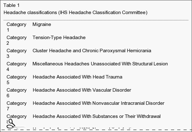
Based on the IHS Headache Classification Committee classification and diagnostic criteria, the patient had an MW�category 1.1, previously called common migraine (Table 1). This appeared secondary to moderate cervical segmental dysfunction with mild to moderate suboccipital and cervical paraspinal myofibrosis.


Table 1 Headache Classifications

Table 1: Headache classifications (IHS Headache Classification Committee)


The patient received CSMT (diversified chiropractic �adjustments�) to her Occ-C1 joint, upper thoracic spine (T2 through T7), and the affected hypertonic musculature. Hypertonic muscles were released through gentle massage and stretching. An initial course of 8 treatments was conducted at a frequency of twice a week for 4 weeks. The treatment program also included recording several features for every migraine episode. This included frequency, visual analogue scores, episode duration, medication, and time before they could return to normal activities.


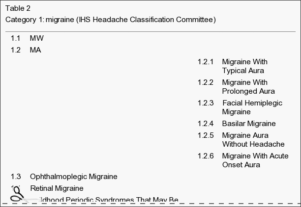
The patient reported a dramatic improvement after her first treatment and noticed a reduction in the intensity of her head and neck pain. This continued with the patient reporting having no migraines in the initial month course of treatment. Further treatment was recommended to increase her range of motion, increase muscle tone, and reduce suboccipital muscle tension. In addition, monitoring of her migraine symptoms was continued. A program of treatment at a frequency of once a week for a further 8 weeks was instigated. After the next phase of treatment, the patient noted much less neck tension, better movement, and no migraine. In addition, she no longer used pain-relieving medication (acetaminophen, codeine, and sumatriptan succinate) and noted that she did not experience nausea, vomiting, photophobia, or phonophobia (Table 2). The patient continued treatment at 2-weekly intervals and stated that, after 6 months, her migraine episodes had disappeared completely. In addition, she was no longer experiencing neck pain. Examination revealed no pain on active neck movement; however, a passive motion restriction at the C1-2 motion segment was still present.


Table 2 Category 1 Migraine

Table 2: Category 1: migraine (IHS Headache Classification Committee)


The patient is currently having treatment every 4 weeks, and she still reports no return of her migraine episodes or neck pain. The patient has now not experienced any migraines for a period of more than 7 years since her last episode, which was immediately before her having her first chiropractic treatment.


Dr Jimenez White Coat

Dr. Alex Jimenez’s Insight

Migraine pain is a debilitating symptom which can be effectively managed with chiropractic care. Chiropractic treatment provides a wide selection of services which can help patients with a variety of injuries and/or conditions, including symptoms of chronic pain, limited range of motion and many other health issues. Chiropractic care can also help control stress associated with migraine. Our staff is determined to treat patients by focusing on the source of the issue rather than temporarily relieving the symptoms using drugs and/or medications. The purpose of the article is to demonstrate evidence-based results on the improvement of migraine using chiropractic care and to educate patients on the best type of treatment for their specific health issues. Chiropractic treatment offers relief from migraine pain as well as overall health and wellness.


Discussion


Case studies do not form high levels of scientific data. However, some cases do present significant findings. For example, cases with long (chronic) and/or severe symptomatology can highlight alternative treatment options. With case studies such as this, there is always a possibility that the symptoms spontaneously resolved, with no effective from the treatment. The case presented highlights a potential alternative treatment option. A 7-year follow-up revealed that the person had still not had a single migraine episode in this period. The patient was certain that there had been no other lifestyle changes that could have contributed to her improvement. She also noted that the migraines had stopped after her first treatment.


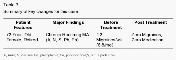
The average frequency of her migraines before treatment was 1 to 2 per week, with episodes that always included nausea, vomiting, photophobia, and phonophobia. In addition, the average duration of each episode was 1 to 3 days before her receiving CSMT. The person also noted that the use of her pain-relieving medication was also reduced by 100% (Table 3).


Table 3 Summary of Key Changes for this Case

Table 3: Summary of key changes for this case


Migraines are a common and debilitating condition; yet because they have an uncertain etiology, the most appropriate treatment regime is often unclear.[16] Previous etiological models described vascular causes of migraine, where episodes seem to be initiated by a decreased blood flow to the cerebrum followed by extracranial vasodilation during the headache phase.[8] However, other etiological models seem connected with vascular changes related to neurologic changes and associated serotonergic disturbances.[9] Therefore, previous treatments have focused on pharmacological modification of blood flow or serotonin antagonist block.[17]


Studies examining the role of the cervical spine to headache (ie, �cervicogenic headache�) have been well described in the literature.[18-30] However, the relation of the cervical spine to migraine is less well documented.[10-15] Previous studies by this author have demonstrated an apparent reduction in migraines after CSMT.[10, 11] In addition, other studies have suggested that CSMT may be an effective intervention for migraine.[14, 15] Although, previous studies have some limitations (inaccurate diagnosis, overlapping symptoms, inadequate control groups), the level of evidence gives support for CSMT in migraine treatment.[11] However, practitioners need to be critically aware of potential overlap of diagnoses when reviewing migraine research or case studies on effectiveness of their treatment.[18-22] This is especially important in comparison of migraine patients who may be suitable for chiropractic manipulative therapy.[23-28]


Between 40% and 66% of patients with migraine, particularly those with severe or frequent migraine attacks, do not seek help from a physician.[29] Among those who do, many do not continue regular physician visits.[30] This may be due to patients’ perceived lack of empathy from the physician and a belief that physicians cannot effectively treat migraine. In a 1999 British survey, 17% of 9770 migraineurs had not consulted a physician because they believed their condition would not be taken seriously; and 8% had not seen a physician because they believed existing migraine medications were ineffective.[30] The most common reason for not seeking a physician’s advice (cited by 76% of patients) was the patients’ belief that they did not need a physician’s opinion to treat their migraine attacks.


The case was presented to assist practitioners making a more informed decision on the treatment of choice for migraines. The outcome of this case is also relevant in relation to other research that concludes that CSMT is a very effective treatment for some people. Practitioners could consider CSMT for migraine based on the following:


  1. Limitation of passive neck movements.
  2. Changes in neck muscle contour, texture, or response to active and passive stretching and contraction.
  3. Abnormal tenderness of the suboccipital area.
  4. Neck pain before or at the onset of the migraine.
  5. Initial response to CSMT.


As with all case reports, results are limited in application to larger populations. Careful clinical decision making should be used when applying these results to other patients and clinical situations.


Conclusion


This case demonstrates that some migraine sufferers may respond well with manual therapies, which includes CSMT. Therefore, migraine patients who have not received a trial of CSMT should be encouraged to consider this treatment and assess any potential response. Where there are no contraindications to CSMT, an initial trial of treatment may be warranted. Following evidence-based medicine guidelines, medical practitioners should discuss CSMT with migraine patients as an option for treatment.[31, 32] Subsequent studies should address this issue and the role that CSMT has in migraine management.


In conclusion, migraine pain is a common condition which affects a large number of the population. Although the cause of migraines is not fully understood, treatment for the complex head pain can ultimately help manage the symptoms. Chiropractic spinal manipulative therapy, or CSMT, may improve migraine in patients and may be a valuable treatment option to consider. However, further research studies are required to demonstrate further results. Information referenced from the National Center for Biotechnology Information (NCBI). The scope of our information is limited to chiropractic as well as to spinal injuries and conditions. To discuss the subject matter, please feel free to ask Dr. Jimenez or contact us at 915-850-0900 .


Curated by Dr. Alex Jimenez


Green-Call-Now-Button-24H-150x150-2-3.png


Additional Topics: Neck Pain


Neck pain is a common complaint which can result due to a variety of injuries and/or conditions. According to statistics, automobile accident injuries and whiplash injuries are some of the most prevalent causes for neck pain among the general population. During an auto accident, the sudden impact from the incident can cause the head and neck to jolt abruptly back-and-forth in any direction, damaging the complex structures surrounding the cervical spine. Trauma to the tendons and ligaments, as well as that of other tissues in the neck, can cause neck pain and radiating symptoms throughout the human body.


blog picture of cartoon paperboy big news


IMPORTANT TOPIC: EXTRA EXTRA: A Healthier You!


OTHER IMPORTANT TOPICS: EXTRA: Sports Injuries? | Vincent Garcia | Patient | El Paso, TX Chiropractor


Blank
References
1. Bigal M.E., Lipton R.B., Stewart W.F. The epidemiology and impact of migraine. Curr Neurol Neurosci Rep. 2004;4(2):98�104. [PubMed]
2. Lipton R.B., Stewart W.F., Diamond M.L., Diamond S., Reed M. Prevalence and burden of migraine in the United States: data from the American Migraine Study 11. Headache. 2001;41:646�657. [PubMed]
3. Alexander L. Migraine in the workplace. Brainwaves. Australian Brain Foundation; Hawthorn, Victoria: 2003. pp. 1�4.
4. Lipton R.B., Bigal M.E. The epidemiology of migraine. Am J Med. 2005;118(Suppl 1):3S�10S. [PubMed]
5. Lipton R.B., Bigal M.E. Migraine: epidemiology, impact, and risk factors for progression. Headache. 2005;45(Suppl 1):S3�S13. [PubMed]
6. Stewart W.F., Lipton R.B. Migraine headache: epidemiology and health care utilization. Cephalalgia. 1993;13(suppl 12):41�46. [PubMed]
7. Headache Classification Committee of the International Headache, Society Classification and diagnostic criteria for headache disorders, cranial neuralgias and facial pain. Cephalgia. 2004;24(Suppl. 1):1�151. [PubMed]
8. Goadsby P.J., Lipton R.B., Ferrari M.D. Migraine�current understanding and treatment. N Engl J Med. 2002;346:257�263. [PMID 11807151] [PubMed]
9. Goadsby P.J. The scientific basis of medication choice in symptomatic migraine treatment. Can J Neurol Sci. 1999;26(suppl 3):S20�S26. [PubMed]
10. Tuchin P.J., Pollard H., Bonello R. A randomized controlled trial of chiropractic spinal manipulative therapy for migraine. J Manipulative Physiol Ther. 2000;23:91�95. [PubMed]
11. Tuchin P.J. The efficacy of chiropractic spinal manipulative therapy (SMT) in the treatment of migraine�a pilot study. Aust Chiropr Osteopath. 1997;6:41�47. [PMC free article] [PubMed]
12. Tuchin P.J., Bonello R. Classic migraine or not classic migraine, that is the question. Aust Chiropr Osteopath. 1996;5:66�74. [PMC free article] [PubMed]
13. Tuchin P.J., Scwafer T., Brookes M. A case study of chronic headaches. Aust Chiropr Osteopath. 1996;5:47�53. [PMC free article] [PubMed]
14. Nelson C.F., Bronfort G., Evans R., Boline P., Goldsmith C., Anderson A.V. The efficacy of spinal manipulation, amitriptyline and the combination of both therapies for the prophylaxis of migraine headache. J Manipulative Physiol Ther. 1998;21:511�519. [PubMed]
15. Parker G.B., Tupling H., Pryor D.S. A controlled trial of cervical manipulation for migraine. Aust NZ J Med. 1978;8:585�593. [PubMed]
16. Dowson A.J., Lipscome S., Sender J. New guidelines for the management of migraine in primary care. Curr Med Res Opin. 2002;18:414�439. [PubMed]
17. Ferrari M.D., Roon K.I., Lipton R.B. Oral triptans (serotonin 5-HT1B/1D agonists) in acute migraine treatment: a meta-analysis of 53 trials. Lancet. 2001;358:1668�1675. [PubMed]
18. Sjasstad O., Saunte C., Hovdahl H., Breivek H., Gronback E. Cervical headache: an hypothesis. Cephalgia. 1983;3:249�256.
19. Vernon H.T. Spinal manipulation and headache of cervical origin. J Manipulative Physiol Ther. 1989;12:455�468. [PubMed]
20. Sjasstad O., Fredricksen T.A., Stolt-Nielsen A. Cervicogenic headache, C2 rhizopathy, and occipital neuralgia: a connection. Cephalgia. 1986;6:189�195. [PubMed]
21. Bogduk N. Cervical causes of headache and dizziness. In: Greive G.P., editor. Modern manual therapy of the vertebral column. 2nd ed. Edinburgh; Churchill Livingstone: 1994. pp. 317�331.
22. Jull G.A. Cervical headache: a review. In: Greive GP, editor. Modern manual therapy of the vertebral column. 2nd ed. Edinburgh; Churchill Livingstone: 1994. pp. 333�346.
23. Boline P.D., Kassak K., Bronfort G. Spinal manipulations vs. amitriptyline for the treatment of chronic tension-type headaches: a randomized clinical trial. J Manipulative Physiol Ther. 1995;18:148�154. [PubMed]
24. Vernon H., Steiman I., Hagino C. Cervicogenic dysfunction in muscle contraction headache and migraine: a descriptive study. J Manipulative Physiol Ther. 1992;15:418�429. [PubMed]
25. Kidd R., Nelson C. Musculoskeletal dysfunction of the neck in migraine and tension headache. Headache. 1993;33:566�569. [PubMed]
26. Whittingham W., Ellis W.S., Molyneux T.P. The effect of manipulation (Toggle recoil technique) for headaches with upper cervical joint dysfunction: a case study. J Manipulative Physiol Ther. 1994;17:369�375. [PubMed]
27. Jull G., Trott P., Potter H., Zito G., Shirley D., Richardson C. A randomized controlled trial of exercise and spinal manipulation for cervicogenic headache. Spine. 2002;27:1835�1843. [PubMed]
28. Bronfort G, Nilsson N, Assendelft WJJ, Bouter L, Goldsmith C, Evans R, et al. Non-invasive physical treatments for chronic headache (a Cochrane review). In: The Cochrane Library Issue 2 2003. Oxford: Update Software.
29. Dowson A., Jagger S. The UK migraine patient survey: quality of life and treatment. Curr Med Res Opin. 1999;15:241�253. [PubMed]
30. Solomon G.D., Price K.L. Burden of migraine: a review of its socioeconomic impact. Pharmacoeconomics. 1997;11(Suppl 1):1�10. [PubMed]
31. Bronfort G., Assendelft W.J.J., Evans R., Haas M., Bouter L. Efficacy of spinal manipulation for chronic headache: a systematic review. J Manipulative Physiol Ther. 2001;24:457�466. [PubMed]
32. Vernon H.T. Spinal manipulation in the management of tension-type migraine and cervicogenic headaches: the state of the evidence. Top Clin Chiropr. 2002;9:14�21.
Close Accordion
Defeat Chronic Pain

Defeat Chronic Pain

Defeat Chronic Pain: If you are one of the estimated 50 to 100 million Americans who struggles with Chronic Pain, you are aware of just how miserable and life-altering it can be. There is not a single area of you life that remains unaffected. You no longer sleep well. Your SEX LIFE is non-existent. Everyday activities have become your own personal �Mount Everest �. You cannot concentrate because the pain IS ALWAYS ON YOUR MIND. It is wearing you out, physically, mentally, and emotionally. It’s sapping your ability to think clearly or make decisions. In short we’re here to defeat chronic pain.

People can see the pain on your face and in your eyes. Chronic Pain and the inability to do the things you love, is making you feel DEPRESSED (not the other way around like your doctor may have suggested). Recent studies have even shown that brains of people suffering with Chronic Pain, show patterns of atrophy that are virtually indistinguishable from what is seen in patients with dementia or ALZHEIMER’S. In fact, a recent study from a prominent Canadian University showed that Chronic Pain causes the brain to degenerate at almost 10 times the rate of someone without pain!

Although Chronic Pain may seem hopeless, there are some things that you can do to help yourself � even though your doctor undoubtedly failed to educate you in this regard. Some of the most basic of these include eating only healthy foods (I recommend a PALEO DIET), taking only WHOLE FOOD SUPPLEMENTS, drinking more WATER, giving up the CIGARETTES, and EXERCISING to the degree that you can (difficult when suffering with Chronic Pain or FIBROMYALGIA).

Although DOING THESE SIMPLE THINGS will certainly help a large percentage who suffer and be able to defeat chronic pain; there is a significant percentage of you whose pain is not greatly diminished by these measures. It is for you that I created this website. But before we move on to treatment of Chronic Pain, you must first understand what Chronic Pain is and how it really works.

Defeat Chronic Pain: It Works Like This

For years, neuro-scientists have known that Chronic Pain can cause brain atrophy (shrinkage) that is indistinguishable from Alzheimer�s or Dementia. More recently, the prestigious Journal of Neuroscience reported research from McGill University showing that, “The longer the individual has had Fibromyalgia, the greater the gray matter loss, with each year of Fibromyalgia being equivalent to 9.5 times the loss in normal aging”. Think about this statement for a moment. Every single year you live with some sort of CHRONIC PAIN SYNDROME (or syndromes as the case may be) is the equivalent of nearly 10 times the brain loss seen in the normal aging process. Re-read this paragraph until the urgency of your situation sinks in!

Although there are several types of pain (the study of Chronic Pain can get extremely complex), we are going to try and keep this as simple as possible. For our purposes, there are two types of Chronic Pain. It has to do with where the pain comes from. Chronic Pain originates in one of the two following areas.

  • The Central Nervous System
  • The Body

As we will discuss shortly, Chronic Pain that arises in the CNS is frequently ‘learned’ pain. Let me explain. In order to learn how to SHOOT FREE THROWS, use chop sticks, PLAY THE PIANO, speak Swahili, you have to practice. Everyone remembers the old adage; Practice makes Perfect. If you stimulate pain pathways in the Brain & Nervous System long enough, or are exposed to enough stressors in your life (CHEMICAL, AUTOIMMUNE, EMOTIONAL, DIETARY, FOOD SENSITIVITIES, PHYSICAL, BACTERIAL, VIRAL, PARASITIC, FUNGAL, MOLD, ELECTROMAGNETIC, etc), you can alter the way your Brain and Central Nervous System function.

Hopefully your pain, even though severe, is still Type II (THE THREE TYPES OF PAIN). As people start losing control of numerous areas of physiology (DIGESTION, HORMONAL, IMMUNITY, BLOOD SUGAR REGULATION, HYPERSENSITIVITY, DYSBIOSIS, etc), the problems ramp up. Over time this pain can (will) become locked into the brain. Although pathological Pain Syndromes arising from a malfunctioning CNS are not the most common causes of Chronic Pain, if this is where you are at, you are going to have to find a way to deal with these underlying issues (FUNCTIONAL NEUROLOGY can be a fantastic starting point). Although I provide information that helps many people help themselves with the severe metabolic and neurological problems, this website is chiefly devoted to defeat chronic Pain that is not locked into the Brain, but is instead originating from the body (Type II Pain).

Defeat Chronic Pain: Nociception

“Simple Nociception” is the most basic type of pain. If someone steps on your toe, it hurts. This is normal, and means that your nervous system is functioning properly. Get the person off your toe, and the pain goes away — almost immediately. Simple. There are several different types of Nociceptive Pain, but the one that we are most concerned about on this website is the one that has to do with ‘deep’ musculoskeletal pain, otherwise known as Deep Somatic Pain (Greek �Soma� = body). Deep Somatic Pain is pain that originates in tissues that are considered to be ‘deep’ in the body. Although we do not always think of many of these tissue types as being deep, this category includes things like LIGAMENTS, TENDONS, MUSCLES, FASCIA, blood vessels, and bones. There are two main types of Nociceptors, chemical and mechanical.

I. Chemical Nociception

The Chemical Nociceptors are stimulated by noxious chemicals. The chief of these are the chemicals we collectively refer to as INFLAMMATION (bear in mind that once Inflammation is involved, we begin moving away from Type I pain and into Type II pain — Nociception is still involved, but so is the Inflammatory Cascade). Inflammation is actually made up of a large group of chemicals manufactured within your body as part of the normal Immune System response. They have names like prostaglandins, leukotrienes, histamines, cytokines, kinins, etc, etc, etc. When these chemicals are out of increased beyond what’s needed for normal tissue repair, the result will be a whole host of health problems —- and Chronic Pain.

Although “SYSTEMIC INFLAMMATION” is at the root of the vast majority of America’s health problems (DIABETES, CANCER, FIBROMYALGIA, THYROID PROBLEMS, ARTHRITIS, HEART DISEASE, and numerous others), you will soon see that even though Inflammation is always involved with the tissues of the “Deep Soma,” it sometimes gets more credit than it deserves. However, you also have to be aware that exposing MICROSCOPIC SCAR TISSUE to chronic inflammation can potentially hyper-sensitize nerves. This hypersensitization makes the nerves within Scar Tissue as much as 1,000 times more pain sensitive than normal (the work of the famous neurologist, DR. CHAN GUNN).

INCREASED TISSUE ACIDITY (usually caused by hypoxia — diminished tissue oxygen levels) is another common form of Chemical Nociception. This frequently occurs as the result of a JUNKY DIET, but is also caused by relentless Mechanical / Neurological / Immune System Dysfunction. It is a big reason that my Decompression Protocols utilize OXYGEN THERAPY extensively.

II. Mechanical Nociception

As you can imagine, Mechanical Dysfunction stimulates the Mechanical Nociceptors. This group of nociceptors (pain receptors) is stimulated by constant mechanical stress in the tissues of the Deep Soma — particularly ligaments, tendons, and fascia. Mechanical tension, mechanical deformation, mechanical pressure, etc are the things that cause Mechanical Nociception, which can in turn, cause pain — chronic, unrelenting, pain. Remove the offending mechanical stressor, and you can oftentimes remove the pain. Sounds simple, doesn�t it? Unfortunately, nothing is ever quite as simple as it initially appears.

Be aware that Nociceptive Pain can actually become Brain-Based over time. This is called ‘Supersensitivity’ and is caused by alterations in the Brain and Central Nervous System that perpetuate the pain cycle (many in the medical community are calling it CENTRALIZATION OR CENTRAL SENSITIZATION. In Mechanical Nociception, even though the injured tissue has, according to all of the medical tests, HEALED, it has healed improperly; i.e. microscopic scar tissue and tissue adhesion — particularly in the FASCIA. I probably do not need to tell you that this can be really really bad news — particularly because it is a significant feature of what I call “CHRONIC PAIN’S PERFECT STORM“.

As nerve function and PROPRIOCEPTION become increasingly fouled up, degenerative arthritis and joint deterioration begin to set in (HERE). Because of involvement in the Brain or Central Nervous System, this kind of pain is often referred to as Neuropathic Pain or Neruogenic Pain. Sometimes people end up with HYPERALGIA (Extreme sensitivity to pain. Stimulus that should cause a little pain, causes extraordinary amounts of pain). Or they end up with ALLODYNIA (Stimulus which do not normally elicit any pain at all, now causes pain). Sometimes these two overlap. Stay with me and you will begin to understand why.

Defeat Chronic Pain: Hypersensitized Nerves Relationship To Injured Or Damaged Fascia

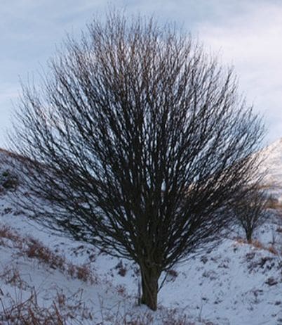
Think of nerve endings as the twigs at the very end of a tree limb. Nerves (just like a tree) begin with a large trunk, which splits / divides into smaller and smaller branches until eventually you arrive at the end � the tiny twig (or nerve ending) at the end of the very smallest branches.

If you have ever seen a �topped� tree, you can understand what happens to nerve endings that are found in microscopic scar tissue. Professional Tree Trimmers cut (or �top�) the largest branches just above where the trunk splits into two or three limbs. What happens to these stubs? Instead of having limbs that continue to branch out and divide into ever-smaller limbs in a normal fashion, you get a stub or stump, that in a short matter of time, swells up and has hundreds of tiny twig-like limbs growing from it. �Topping� stimulates the growth of twigs from the stump. The injured nerves found in microscopic scar tissue act in much the same way.

As the larger nerves that are found in soft tissues are injured, you end up with an inordinate number of immature nerve endings (twigs) growing out of an inflamed nerve �stump�. As you might imagine, extra pain receptors are never a good thing! And because there in Inflammation present, this often leads to Microscopic Scar Tissue, which, even though it is up to 1,000 times more pain-sensitive than normal tissue, cannot be seen with even the most technologically advance imaging techniques such as CT / MRI (HERE). This is a commonly seen phenomenon in Facial Adhesions, and is why even though the people living this nightmare believe that because their pain is so severe that it should make their MRI “Glow Red”, it shows nothing. This tends to lead to deer-in-the-headlight looks when you ask your doctor what might be causing your pain, not to mention accusations of malingering, drug seeking, or attempting to get on Disability.

Defeat Chronic Pain: Nerves Are Like Tree Branches

Uninjured Nerves

defeat chronic pain

Photo by Stephen McCulloch

Injured Nerves

defeat chronic pain

Photo by Linda Bailey


Defeat Chronic Pain: Fascial Adhesions

Microscopic Scar Tissue & Chronic Pain

One of the biggest revelations for many people suffering with Chronic Pain is the absurd numbers of CHRONIC PAIN SYNDROMES brought on by microscopic scarring of the FASCIA. It gets even worse once you realize that this Fascia is the most pain-sensitive tissue in the body —- yet it does not show up on even the most technologically advanced imaging techniques, including MRI. Simply read our “Fascia” page to see why microscopic scarring of this specific “Connective Tissue” is at the root of all sorts of Chronic Pain Cases — not to mention ILL HEALTH.

Destroy Chronic Pain / Doctor Russell Schierling

Medical Inc Teaser

Hips Positioning And MRI Anatomy

Hips Positioning And MRI Anatomy

Hips Positioning & MRI Anatomy

hipsMRI may be requested for:

  • Bone tumor
  • Osteoarthritis
  • Aseptic or avascular necrosis
  • Bursitis
  • Pain

Bones & Cartilage Of The Hips

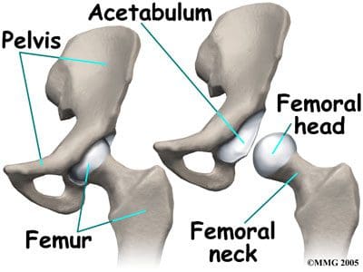
The hips joints join the legs to the trunk of the body, and are formed by the femurs and pelvic bones. The hips are ball-and-socket type joints, where the femoral head (ball) fits into the cup-shaped acetabulum (socket) of the pelvis (Figure 1). When compared to the shoulder, which is also a ball-and-socket joint, the acetabulum is a deeper socket, and encompasses a greater area of the ball, or femoral head. This accommodation is necessary to provide stability for the hip, as it is a major weight-bearing joint, and one of the largest joints in the body. When not weight-bearing, the ball and socket of the hip joint are not perfectly fitted. However, as the hip joint bears more weight, the surface area contact increases, and the joint becomes more stable. When in a standing position, the body�s center of gravity passes through the center of the acetabula. While walking, weight-bearing stresses on the hips can be five times a person�s body weight. Healthy hips can support your weight and allow for pain-free movement. Hip injuries or disease can cause changes that affect your gait, as well as changes that affect the ability of the hips to distribute weight bearing. Abnormal stress is then placed on the joints that are above and below the hips.

hips

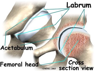
The three fused hips or innominate bones that form the acetabulum include the ilium, pubis, and ischium. The ilium forms the superior aspect, the pubis forms the inferior and anterior aspect, and the ischium forms the inferior and posterior aspect. The depth of the acetabulum socket is further increased by the attached fibrocartilaginous labrum (Figure 2). In addition to providing stability to the hip joint, the labrum allows flexibility and motion. Hip joint stability can be hampered by injuries resulting from playing sports, running, overuse, or falling, as well as by disease or tumor. MRI of the hips may be ordered to assess the joint(s) for internal derangement, fracture, or degenerative joint disease. A blow to the hip joint or a fall can result in dislocation of the hip, or a hip fracture. Osteoporosis or low bone density can also lead to hip fractures. Successful prevention and/or treatment of osteoporosis may be achieved through nutrition (adequate amounts of calcium, vitamin D and phosphorus), exercise, safety measures, and medications.


hips

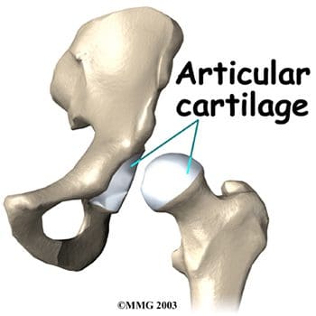
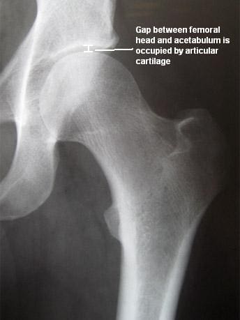
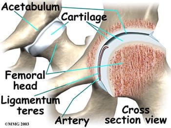
Articular cartilage covers the femoral head and the acetabulum (Figure 3). This cartilage is thin but tough, flexible, smooth and slippery, with a rubbery consistency. It absorbs shock, and allows the bones to move against each other easily and without pain. It is kept lubricated by synovial fluid, which is made in the synovial membrane (joint lining). Synovial fluid is both viscous and sticky. This fluid is what allows us to flex our joints under great pressure without wear. The articular cartilage of the hip is typically about � inch thick, except in the posterior aspect of the hip socket (Figure 4). Here, the cartilage is thicker, as this area absorbs most of the force during walking, running, and jumping. MRI of the hip joint can detect problems involving both the articular cartilage and the fibrocartilaginous ring, or labrum. Cartilage has minimal blood vessels, so it is not good at repairing itself. Fraying, fissuring, and other abnormalities or defects of the cartilage can lead to arthritis in the hip joint. Contrast can be directly injected in the hip joint for a detailed look at the cartilage and labrum.

hipships

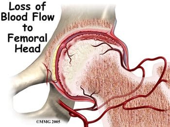
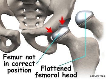
The femurs are the longest bones in the body, with large round heads that rotate and glide within the acetabula of the pelvis. The femoral head is particularly subject to pathologic changes if there is any significant alteration of blood supply (avascular necrosis). The femoral neck connects the head of the femur to the shaft. The neck ends at the greater and lesser trochanters, which are sites of muscle and tendon attachments. A disease characterized by an inadequate blood supply to the femoral head is Legg-Calve-Perthes disease, also known as LCP or simply Perthes disease. This is a degenerative disease of the hip joint that affects children, most commonly seen in boys ages two through twelve. One of the growth plates of the femoral head, the capital femoral epiphysis, is inside the joint capsule of the hip. Blood vessels that feed this epiphysis run along the side of the femoral neck, and are in danger of being torn or �pinched off� if the growth plate is damaged. This can result in a loss of blood supply to the epiphysis, leading to a deformity of the femoral head (Figure 5). The femoral head may become unstable and break easily, which can lead to incorrect healing and deformities of the entire hip joint (Figure 6). Treatment of Perthes disease is centered on the goal of returning the femoral head to a normal shape. Surgical and non-surgical treatments are used, based on the idea of �containment�- holding the femoral head in the acetabulum as much as possible, while still allowing motion of the hip joint for cartilage nutrition and healthy growth of the joint.


hipships

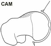
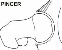
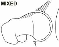
High level athletes and active individuals may be susceptible to a hip condition known as Femoro-Acetabular Impingement, or FAI. FAI is characterized by excessive friction in the hip joint. The femoral head and acetabulum rub abnormally, and can create damage to the articular or labral cartilage. FAI is also associated with labral tears, early hip arthritis, hyperlaxity and low back pain. FAI generally occurs in two forms: Cam and Pincer. The Cam form results in abnormal contact between the femoral head and the socket of the hip because the femoral head and neck relationship is aspherical (Figure 7). Males and those involved in significant contact sports typically display Cam impingement. Pincer impingement occurs when the acetabulum covers too much of the femoral head, resulting in the labral cartilage being pinched between the rim of the socket and the anterior femoral head-neck junction (Figure 8). Pincer impingement may be more common in women. Typically, these two forms exist together, and are labeled as �mixed impingement� (Figure 9).


hipshipships

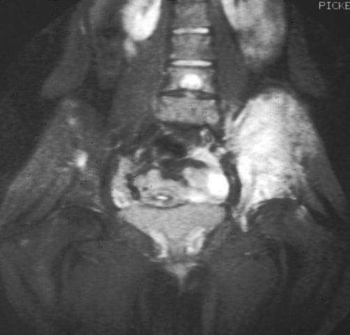
Ewing�s sarcoma is a malignant bone tumor that may affect the pelvis and/or femur, thereby also affecting the stability of the hips. Like Perthes disease, Ewing�s sarcoma is more common in males, typically presenting in childhood or early adulthood. MRI is routinely used in the work-up of these malignant tumors to show bony and soft tissue extent of the tumor, and its relation to nearby anatomic structures (Figure 10). Contrast may be used to help determine the amount of necrosis within the tumor, which aids in determining the response to treatment before surgery.


hips

Figure 10. MRI demonstrating Ewing�s sarcoma.

Ligaments Of The Hips

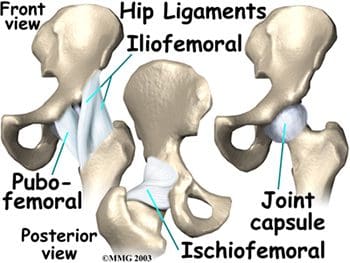
Hip stability is further increased by three strong ligaments that encompass the hip joint and form the joint capsule. These ligaments connect the femoral head to the acetabulum, with names suggestive of the bones they connect. They include the pubofemoral and iliofemoral ligaments anteriorly, and the ischiofemoral ligament posteriorly (Figure 11). The iliofemoral ligament is the strongest ligament in the body. However, sports and overuse can still result in sprains of these sturdy ligaments of the joint capsules of the hips. A smaller ligament, the ligamentum teres, is an intracapsular ligament that connects the tip of the femoral head to the acetabulum (Figure 12). A small artery within this ligament brings some of the blood supply to the femoral head. Damage to the ligamentum teres, and its enclosed artery, can result in avascular necrosis.


hipships

Muscles & Tendons Of The Hips

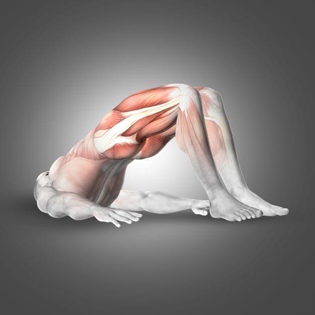
The muscles of the thigh and lower back work together to keep the hip stable, in alignment, and able to move. The hip gains stability because the hip muscles do not attach right at the joint. Hip muscles allow the movements of flexion, extension, abduction, adduction, and medial and lateral rotation. To better understand the functions of the muscles surrounding the hip, they can be divided into groups based on their locations- anterior, posterior, and medial.

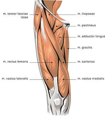
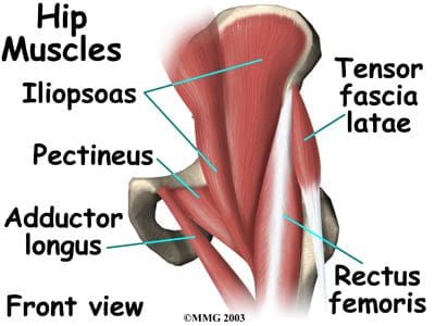
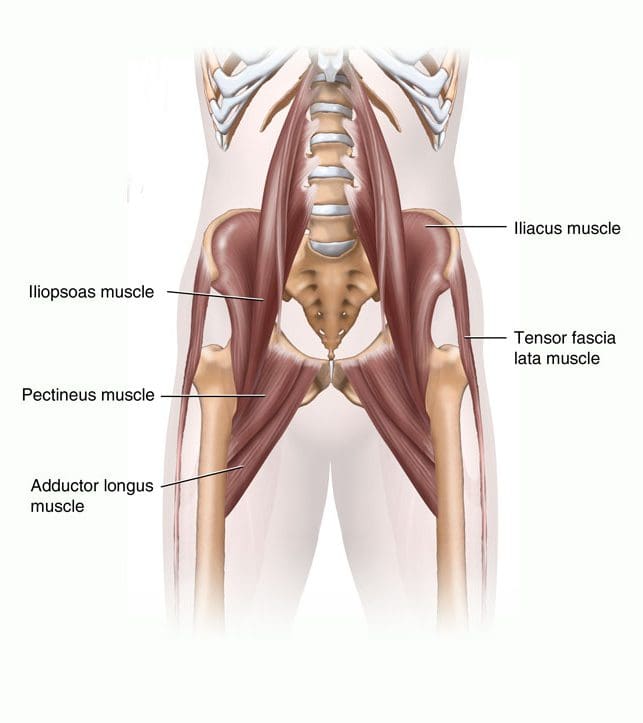
The anterior thigh muscles are the main hip flexors, and are located anterior to the hip joint. Seventy percent of the thigh�s muscle mass is made up of the quadriceps femoris muscle, so named because it arises from four muscle heads- the rectus femoris, vastus medialis, vastus intermedius, and vastus lateralis (Figures 13, 14). The rectus femoris is the only one of the �quad� muscles to cross the hip joint. The sartorius muscle is found anterior to the quadriceps, and also serves as an abductor and lateral rotator of the hip. The most powerful of the anterior thigh hip flexors is the iliopsoas, which originates in the low back and pelvis and attaches at the lesser trochanter.


hipships

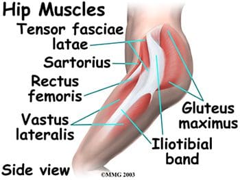
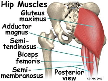
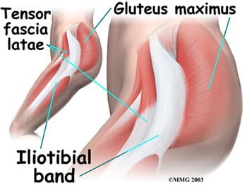
Posterior hip muscles include those of both the thigh and gluteal regions. The posterior thigh muscles are also known as the hamstrings- semimembranosus, semitendinosus, and biceps femoris (Figure 15). These muscles originate at the inferior pelvis, and are the extensors for the hip. They are active in normal walking motions. When the hamstrings are �tight�, they limit hip flexion when the knee joint is extended (bending forward from the waist with knees straight), and can limit lumbar movement, leading to back pain. The gluteal muscles include the gluteus maximus, medius, and minimus, six deep muscles that serve as lateral rotators, and the tensor fasciae latae. The three gluteals and the anterior sartorius muscle are all involved in abduction. The gluteus maximus is the main hip extensor, and is the most superficial of the gluteal muscles. It is involved in running and walking uphill, and assists with normal tone of the iliotibial band, which lies lateral to it. The gluteus medius and minimus both insert at the greater trochanter of the femur. The minimus is the deepest of the three gluteal muscles. Anterior to the gluteus minimus is the tensor fasciae latae muscle. It is a flexor and medial rotator of the hip, originating from the anterior superior iliac spine (ASIS) and inserting on the iliotibial band. The term �tensor fasciae latae� defines this muscle�s job- �muscle that stretches the band on the side�. This muscle helps the iliopsoas, gluteus medius, and gluteus minimus muscles during flexion, abduction and medial rotation of the thigh by making the iliotibial band taut, thereby steadying the trunk and stabilizing the hip (Figure 16). The iliotibial band or tract is not a muscle, but a thickened, fibrous band of deep fascia, or connective tissue. It is found at the lateral aspect of the thigh, and runs from the ilium to the tibia. It encloses the muscles and helps with lateral stabilization of the knee joint, as well as helping to maintain both hip and knee extension. Tightening of the iliotibial (IT) band typically causes more problems at the knee as opposed to the hip, but hip pain can result from the IT band rubbing as it passes over the greater trochanter.

hipships

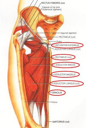
The medial thigh (groin) muscles include five muscles of adduction, and one lateral rotator (Figures 17, 18). The lone lateral rotator is the obturator externus, which covers the external surface of the obturator foramen in the deep upper medial thigh. The adductors include the gracilis, the pectineus, and the adductor brevis, longus and magnus. The gracilis is the longest adductor, extending from the medial inferior aspect of the pubic bone, to the medial aspect of the tibia. The adductor magnus is the most massive of the medial muscles of the thigh.


hipships

The tendons and muscles of the hips are very powerful and create great forces, making them prone to inflammation and irritation. Tendonitis of the hip can result from repetitive movements involving the soft tissues surrounding the hip joint. Overuse of the hip joint in fitness workouts can lead to tendonitis. Tendons lose their elasticity as we age, resulting in swelling and irritation when the tendons are no longer �gliding� on their normal paths. Iliopsoas tendonitis plays a major role in snapping hip syndrome, or dancer�s hip. A snapping sensation when the hip is flexed and extended may be accompanied by an audible snapping or popping noise, as well as pain. This can be both an extra-articular and an intra-articular occurrence. Extra-articular snapping is often found in those patients with a leg length difference (the longer leg is symptomatic), those with tightness of the iliotibial band on the involved side, and those with weak hip abductors and external rotators. Lateral extra-articular snapping can be caused by the iliotibial band, tensor fascia latae or gluteus medius tendon as they slide back and forth across the greater trochanter (Figure 19). If any of these connective tissue bands thickens, they can �catch� on the greater trochanter during the motion of hip extension, thereby creating the �snapping� sensation and sound. Medial extra-articular snapping, which is less common, can occur when the iliopsoas tendon catches on the anterior inferior iliac spine, lesser trochanter, or iliopectineal ridge during hip extension. Intra-articular snapping hip syndrome is similar in many ways to the extra-articular type, but often involves an underlying mechanical problem in the lower extremity, and more intense pain. Intra-articular snapping may be indicative of a torn acetabular labrum, recurrent hip subluxation, a tear of the ligamentum teres, loose bodies, articular cartilage damage, or synovial chondromatosis (cartilage formations in the synovial membrane of the joint). Snapping hip syndrome is usually found in those ages 15-40, often in those in training for the military. It can also affect athletes, especially those involved in dance, gymnastics, soccer, and track and field. These athletes will all be performing repeated hip flexions, which can lead to tendonitis in the hip area. The repetitive motions of those involved in weightlifting and running generally lead to a thickening of the tendons in the hip region, rather than snapping hip syndrome. Prevention, or at least a lessening, of this syndrome may be found with increased stretching of the iliopsosas muscle or the iliotibial band. Surgery is usually not required, unless intra-articular pathology is present.


hips

Figure 19. Hip muscles.

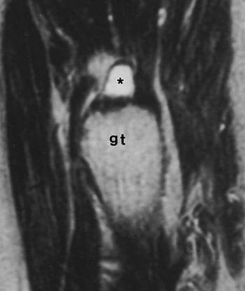
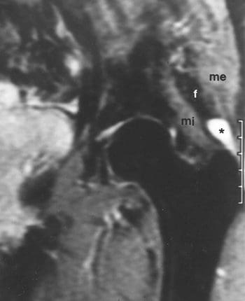
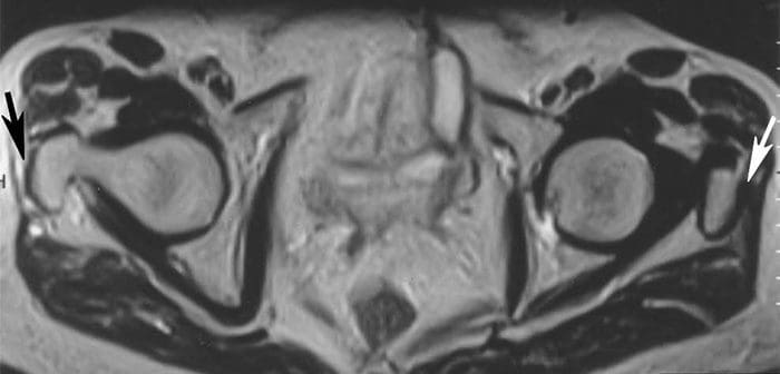
Tendon or muscle strains can occur suddenly, as in sports injuries, or they can develop over time, with symptoms including pain, swelling, muscle spasms, and difficulty moving certain muscles. MRI can be used to detect tendon and muscle tears and strains, as well as bone tumors and infection. MRI has shown good accuracy for the diagnosis of tears of the gluteus medius and gluteus minimus tendons, which are both abductor tendons of the hip. An association was found between these tears and areas of high signal intensity superior or lateral to the greater trochanter on T2-weighted images, tendon elongation in the gluteus medius, and tendon discontinuity (Figure 20). STIR and fat-suppressed T2-weighted coronal images are very sensitive for detection of areas of high signal intensity superior to the greater trochanter. Coronal T1-weighted images demonstrate tendon elongation in the gluteus medius (Figure 21). Axial images may prove superior for localizing involvement to individual abductor tendons and confirming tendon discontinuity (Figure 22). Tears of the abductor tendons may be the leading cause of greater trochanteric pain syndrome.

hips

Figure 20. Sag. T2 shows high signal intensity superior to greater trochanter (gt) corresponding to swollen bursa (*).

hips

Figure 21. Coronal STIR shows high signal intensity superior to greater trochanter in bursa (*) between gluteus medius (me) and gluteus minimus (mi) tendons.

hips

Figure 22. Axial T2 shows high signal intensity corresponding to fluid replacing distal rt. gluteus medius tendon (black arrow); normal left tendon (white arrow).

Nerves Of The Hips

The nerves of the hip supply the various muscles in the hip area. The major nerves include the femoral, obturator, and lateral femoral cutaneous nerves anteriorly, and the large sciatic nerve posteriorly (Figure 23). The femoral nerve innervates the quadriceps femoris and sartorius, and is the sensory nerve to the anterior thigh. Trauma to this nerve usually occurs in the pelvis, as it passes through or near the psoas muscle. The obturator nerve passes along the lateral pelvic wall and through the obturator foramen, then splits into branches that supply the adductor muscle group. This nerve can also be subject to trauma in the pelvis due to its passage through the obturator foramen. The lateral femoral cutaneous nerve is a sensory nerve that travels along the anterolateral aspect of the thigh. It supplies sensation to the skin surface of the thigh. This is the single nerve involved in a painful condition called meralgia paresthetica, which is characterized by tingling, numbness, and burning pain in the outer part of the thigh. Meralgia paresthetica results from focal entrapment of the lateral femoral cutaneous nerve as it passes through the tunnel formed by the lateral attachment of the inguinal ligament and the ASIS. The posterior sciatic nerve passes deep to the gluteus maximus into the posterior thigh, where it innervates the hamstring muscles, on its way down to the lower leg and foot. The sciatic nerve is approximately as big around as the thumb, and is the largest single nerve in the human body. It can be injured in cases of posterior hip dislocation. Pressure on this nerve can cause nerve pain, numbness, tingling and weakness (sciatica symptoms) in the buttocks, leg, or foot, depending on the site of origin of the sciatic nerve compression.

hips

Figure 23. Anterior and posterior views of the nerves of the hip.

Arteries & Veins Of The Hips

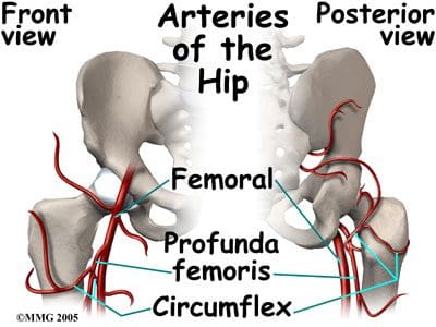
The arterial blood vessels that supply the hips are branches of the internal and external iliacs. The internal iliac artery gives off the superior and inferior gluteals, and the obturator artery. The inferior gluteal flows to the posterior aspect of the hip joint and proximal femur, where it joins a branch of the femoral artery. The obturator artery runs through the obturator foramen, and sends its acetabular branch to the ligamentum teres as part of the blood supply to the femoral head. The external iliac becomes the femoral artery, which has numerous branches that supply the hip and proximal femur. The largest femoral branch is the profunda femoris, which branches superiorly into the medial and lateral circumflex femorals (Figure 24). The circumflex femorals and the inferior gluteal artery contribute to the anastomoses to supply the femoral head, femoral neck, and the hip joint. The medial circumflex also has an acetabular branch to the ligamentum teres. Congenital anomalies in the hip anastomoses, degenerative processes, and trauma can all compromise the blood supply to the hip joint area.

hips

Figure 24. Anterior and posterior views of the arteries of the hip.

Venous flow in the hip and proximal femur typically follows the arterial flow, including the same names for the vessels. The deep veins of the hip and thigh can be the origination of a deep vein thrombosis, which can result in a pulmonary embolus. This can be caused by immobility after hip surgery, sitting in cars or airplanes for extended trips, being overweight, or slow or low blood flow. These blood clots can break off, travel through the larger veins of the thigh and hip, continue through the heart, and become lodged in the smaller vessels of the lung. MRI is being used more frequently to diagnose this very serious condition.

Bursae Of The Hips

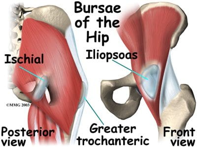
The hip joint is surrounded by bursae, similar to the shoulder. These fluid-filled sacs are lined with a synovial membrane, which produces synovial fluid. Their function is to lessen the friction between tendon and bone, ligament and bone, tendons and ligaments, and between muscles. There may be as many as 20 bursae around the hip. If they become infected or inflamed, the result is a painful condition called bursitis. Common hip bursae that may become inflamed include the greater trochanteric bursa, the iliopsoas bursa, and the ischial bursa (Figure 25). The greater trochanteric bursa is sandwiched between the greater trochanter of the femur, and the muscles and tendons that cross over it. If this bursal sac becomes inflamed, patients experience pain with every step they take, as each step requires the tendon to move over the femur at the hip joint. A tight iliotibial band can also cause irritation of the greater trochanteric bursa. Iliopsoas bursitis can result from irritation of the bursa found between the hip joint and the iliopsoas muscle that passes in front of it. Another common site for bursitis is the ischial bursa, which acts as a lubricating pad between tendons and the ischial tuberosity, which is the bony prominence of the pelvis that you sit on. The ischial bursa acts to prevent destruction of the tendons as they move over the ischial tuberosity. Prolonged sitting can cause ischial bursitis. Inflammation around the ischial tuberosity can irritate the sciatic nerve, and trigger symptoms similar to sciatica. Hip bursitis is seen in runners and athletes in sports that involve excessive running (soccer, football, etc.). It can also be caused by an injury (traumatic bursitis), and is seen in post-op hip replacement and hip surgery patients. Treatment for hip bursitis typically includes rest, anti-inflammatory medications, and ice. It may become necessary to aspirate the bursa, which can be combined with a cortisone injection. MRI may be needed if the diagnosis is unclear, or if the problem does not resolve with normal treatments.

hips

Figure 25. Bursae of the hip.


Axial Scans

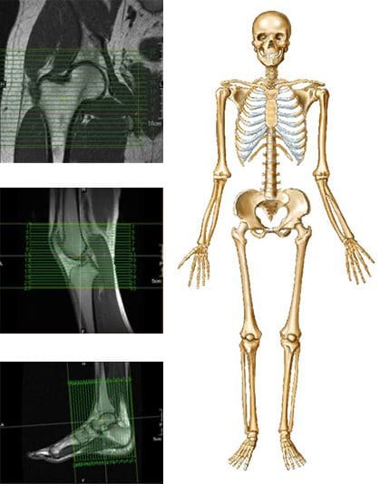
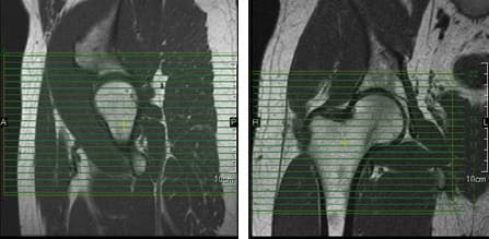
When positioning unilateral axial slices for the hip, a coronal image can be used to ensure inclusion of all pertinent anatomy. The slices should extend superiorly to include the entire femoral head and acetabulum, and inferiorly to include anatomy below the lesser trochanter. The slices should be aligned perpendicular to the shaft of the femur, as seen in the coronal image in Figure 39.

hips

Figure 39. Axial slice setup using sagittal and coronal images.

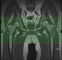
For bilateral axial hip slice setup, parameters may have to be altered to maintain adequate resolution with the larger FOV that is required (Figure 40). The slice group may require angulation to maintain alignment of the femoral heads on the resultant images.

hips

Figure 40. Bilateral axial slice setup using a coronal image.

Coronal Scans

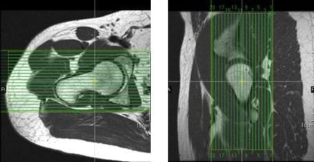
Coronal slices of the hip should cover the area from the posterior margin to the anterior margin of the femoral head. The area from the proximal margin of the femoral shaft to the greater sciatic notch should be included in the image (Figure 41). Slices may be angled so that they are parallel to the femoral neck. Thinner slices may be requested for coronal scanning.

hips

Figure 41. Coronal slice setup using axial and sagittal images.

Sagittal Scans

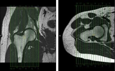
Sagittal slices of the hip should extend past the greater trochanter laterally, and through the acetabulum medially. The slices should be aligned along the long axis of the femur, and perpendicular to the coronal slices, as seen in the coronal image in Figure 42. Two different slice groups will be necessary when performing bilateral sagittal scans.

hips

Figure 42. Sagittal slice setup using coronal and axial images.

Hips Arthrography

MR hip arthrography is often times referred to as the gold standard for assessment of the labrum of the hip. The most clinically significant abnormal findings that result from hip arthrography are labral detachments and tears. Detachment of the labrum, which is more common than a labral tear, can be diagnosed from the appearance of the injected contrast at the acetabular-labral interface (Figure 43). A labral tear can result in injected contrast appearing within the substance of the labrum (Figure 44). Contrast injection is necessary to differentiate torn or detached labra from other pathologic conditions, which may have separate signal intensities. The sensitivity and accuracy for the diagnosis of labral tears and detachment with MR arthrography vs. nonarthrographic MR is 90%. Hip arthrography with MR can also depict intrarticular loose bodies, osteochondral abnormalities, and abnormalities of soft-tissue structures.

Hip arthrography can be performed under fluoro in the x-ray dept., with the patient being moved to the MRI dept. for further imaging, or the entire procedure can be performed in the MRI suite, if MR compatible supplies are available for interventional techniques. The patient should be securely positioned with the hips in internal rotation.

T1-weighted imaging is performed post-contrast to visualize the high signal of the intraarticular contrast. T1 gradient echo sequences offer the benefits of thin sections, elimination of partial volume averaging, and increased detection of small tears. Fatsat sequences are helpful in increasing the contrast between the injected contrast and the adjacent soft tissue. STIR or fatsat T2 sequences performed in the coronal plane may help to detect unsuspected pathologic conditions in the soft tissue and adjacent osseous structures.

Post-contrast axial oblique images have been shown to optimize the detection of the most common sports-related acetabular labral tears, which are anterior or anterosuperior in location. Using a mid-coronal localizer, the axial oblique slices should be prescribed parallel to the long axis of the femoral neck.

hips

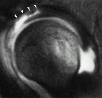
Figure 43. Labral detachment as seen in a fat-suppressed T1-wtd. sag. image; arrowheads indicate involvement of anterior and anterosuperior labrum.

hips

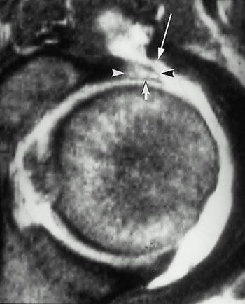
Figure 44. Labral tear as seen in a T1-wtd. image; arrowheads indicate enlarged labrum; short arrow indicates linear intralabral collection of contrast material; long arrow indicates communication between the joint and the iliopsoas bursa.

blank
References:

Kapit, Wynn, and Lawrence M. Elson. The Anatomy Coloring Book. New York: HarperCollins, 1993.

Hip Anatomy, Function, and Common Problems. (Last updated 28July2010). Retrieved from http://healthpages.org/anatomy-function/hip-structure-function-common-problems/

Cluett, J. M.D. (Updated 22May2012). Labral Tear of the Hip Joint. Retrieved from http://orthopedics.about.com/od/hipinjuries/qt/labrum.htm

Hughes, M. D.C. (15July2010). Diseases of the Femur Bone. Retrieved from http://www.livestrong.com/article/175599-diseases-of-the-femur-bone/

A Patient�s Guide to Perthes Disease of the hip. (n.d.). Retrieved from http://www.orthopediatrics.com/docs/Guides/perthes.html

Hip Injuries and Disorders. (Last reviewed 10February2012). Retrieved from http://nlm.nih.gov/medlineplus/hipinjuriesanddisorders.html

Ligament of head of femur. (Updated 20December2011). Retrieved from http://en.wikipedia.org/wiki/Ligament_of_head_of_femur

Ewing�s sarcoma. (Last modified 06January2012). Retrieved from http://en.wikipedia.org/wiki/Ewing%27s_sarcoma

Hip Anatomy. (n.d.). Retrieved from http://www.activemotionphysio.ca/Injuries-Conditions/Hip

Iliotibial Band Friction Syndrome. (n.d.). Retrieved from http://www.physiotherapy-treatment.com/iliotibial-band-friction-syndrome.html

Snapping hip syndrome. (Last modified 09November2011). Retrieved from http://en.wikipedia.org/wiki/Snapping_hip_syndrome

Sekul, E. (Updated 03February2012). Meralgia Paresthetica. Retrieved from http://emedicine.medscape.com/article/1141848-overview

Yeomans, S. D.C. (Updated 07July2010). Sciatic Nerve and Sciatica. Retrieved from http://www.spine-health.com/conditions/sciatica/sciatic-nerve-and-sciatica

Mayo Clinic staff. (26July2011). Meralgia paresthetica. Retrieved from http://www.mayoclinic.com/health/meralgia-paresthetica/DS00914

Deep Vein Thrombosis (DVT)-Blood Clots in the Legs. (n.d.). Retrieved from http://catalog/nucleusinc.com/displaymonograph.php?MID=148

Petersilge, C. M.D. (03May2000). Chronic Adult Hip Pain: MR Arthrography of the Hip. Retrieved from http://radiographics.rsna.org/content/20/suppl_1/S43.full

Acetabular branch of medial circumflex femoral artery. (Last modified 17November2011). Retrieved from http://en.wikipedia.org/wiki/Acetabular_branch_of_medial_circumflex_femoral_artery

Cluett, J. M.D. (Updated 26March2011). Hip Bursitis. Retrieved from http://orthopedics.about.com/cs/hipsurgery/a/hipbursitis.htm

Steinbach, L. M.D., Palmer, W. M.D., Schweitzer, M. M.D. (10June2002). Special Focus Session MR Arthrography. Retrieved from http://radiographics.rsna.org/content/22/5/1223.full

Schueler, S. M.D., Beckett, J.M.D., Gettings, S.M.D. (Last updated 05August2010). Ischial Bursitis/Overview. Retrieved from http://www.freemd.com/ischial-bursitis/overview.htm

Hwang, B., Fredericson, M., Chung, C., Beaulieu, C., Gold, G. (29October2004). MRI Findings of Femoral Diaphyseal Stress Injuries in Athletes. Retrieved from http://www.ajronline.org/content/185/1/166.full.pdf

The Femur (Thigh Bone). (n.d.). Retrieved from http://education.yahoo.com/reference/gray/subjects/subject/59

Norman, W. PhD, DSc. (n.d.). Joints of the Lower Limb. Retrieved from http://home.comcast.net/~wnor/lljoints.htm

Femur. (Last modified 24September2012). Retrieved from http://en.wikipedia.org/wiki/Femur

Wheeless, C. III, M.D. (Last updated 25April2012). Ligaments of Humphrey and Wrisberg. Retrieved from http://wheelessonline.com/ortho/ligaments_of_humphrey_and_wrisberg

Muscle Strains in the Thigh. (Last reviewed August2007). Retrieved from http://orthoinfo.aaos.org/topic.cfm?topic=A00366

Shiel, W. Jr., M.D. (Last reviewed 23July2012). Hamstring Injuries. Retrieved from http://www.medicinenet.com/hamstring_injury/article.htm

Hamstring Muscle Injuries. (Last reviewed July 2009). Retrieved from http://orthoinfo.aaos.org/topic.cfm?topic=a00408

Knee. (Last modified 19September2012). Retrieved from http://en.wikipedia.org/wiki/Knee

DeBerardino, T. M.D. (Updated 30March2012). Quadriceps Injury. Retrieved from http://emedicine.medscape.com/article/91473-overview

Kan, J.H. (n.d.). Osteochondral Abnormalities: Pitfalls, Injuries, and Osteochondritis Dissecans. Retrieved from http://www.arrs.org/shopARRS/products/s11p_sample.pdf

Nerves of the Lower Limb. (Last updated 30March2006). Retrieved from http://download.videohelp.com/vitualis/med/lowrnn.htm

The Adductor Canal. (Last updated 30March2006). Retrieved from http://download.videohelp.com/vitualis/med/addcanal.htm

Nabili, S. M.D. (n.d.). Varicose Veins & Spider Veins. Retrieved from http://www.medicinenet.com/varicose_veins/article.htm

Basic Venous Anatomy. (n.d.). Retrieved from http://vascular-web.com/asp/samples/sample104.asp

Femoral nerve. (Last modified 23September2012). Retrieved from http://en.wikipedia.org/wiki/Femoral_nerve

Peron, S. RDCS. (Last modified 16October2010). Anatomy � Lower Extremity Veins. Retrieved from http://www.vascularultrasound.net/vascular-anatomy/veins/lower-extremity-veins

Medical Multimedia Group, L.L.C. (n.d.). Knee Anatomy. Retrieved from http://www.eorthopod.com/content/knee-anatomy

Knee Joint Anatomy, Function and Problems. (Last updated 06July2010). Retrieved from http://healthpages.org/anatomy-function/knee-joint-structure-function-problems/

Coronary ligament of the knee. (Last modified 09May2010). Retrieved from http://en.wikipedia.org/wiki/Coronary_ligament_of_the_knee

Walker, B. (n.d.). Patellar Tendonitis Treatment � Jumper�s Knee. Retrieved from http://www.thestretchinghandbook.com/archives/patellar-tendonitis.php

Osgood-Schlatter disease. (Last reviewed 12November2010). Retrieved from http://www.ncbi.nlm.nih.gov/pubmedhealth/PMH0002238/

Grelsamer, R. M.D. (n.d.). The Anatomy of the Patella and the Extensor Mechanism. Retrieved from http://kneehippain.com/patient_pain_anatomy.php

Oblique popliteal ligament. (Last modified 24March2012). Retrieved from http://en.wikipedia.org/wiki/Oblique_popliteal_ligament

Shiel, W. Jr., M.D. (Last reviewed 27July2012). Chondromalacia Patella (Patellofemoral Syndrome). Retrieved from http://www.medicinenet.com/patellofemoral_syndrome/article.htm

Knee. (Last modified 19September2012). Retrieved from http://en.wikipedia.org/wiki/Knee

Mosher, T. M.D. (Last updated 11April2011). MRI of Knee Extensor Mechanism Injuries Overview of the Knee Extensor Mechanism. Retrieved from http://emedicine.medscape.com/article/401001-overview

Carroll, J. M.D. (December 2007). Oblique Menisco-meniscal Ligament. Retrieved from http://radsource.us/clinic/0712

DeBerardino, T. M.D. (Last updated 30March2012). Medial Collateral Knee Ligament Injury. Retrieved from http://emedicine.medscape.com/article/89890-overview#a0106

Farr, G. (Last updated 31December2007). Joints and Ligaments of the Lower Limb. Retrieved from http://becomehealthynow.com/article/bodyskeleton/951/

Knee anatomy overview. (02March2008). Retrieved from http://www.kneeguru.co.uk/KNEEnotes/node/741

Dixit, S. M.D., Difiori, J. M.D., Burton, M. M.D., Mines, B. M.D. (15January2007). Management of Patellofemoral Pain Syndrome. Retrieved from http://www.aafp.org/afp/2007/0115/p194.html

Knee Muscles. (Last updated 05September2012). Retrieved from http://www.knee-pain-explained.com/kneemuscles.html

Popliteus muscle. (Last updated 20February2012). Retrieved from http://en.wikipedia.org/wiki/Popliteus_muscle

Kneedoc. (10February2011). Nerves. Retrieved from http://thekneedoc.co.uk/neurovascular/nerves

Wheeless, C. III, M.D. (Last updated 15December2011). Popliteal Artery. Retrieved from http://wheelessonline.com/ortho/popliteal_artery

The Popliteal Artery. (n.d.) Retrieved from http://education.yahoo.com/reference/gray/subjects/subject/159

Knee bursae. (Last updated 09May2012). Retrieved from http://en.wikipedia.org/wiki/Bursae_of_the_knee_joint

Hirji, Z., Hunjun, J., Choudur, H. (02May2011). Imaging of the Bursae. Retrieved from http://www.ncbi.nlm.nih.gov/pmc/articles/PMC3177464/

Kimaya Wellness Limited. (n.d.). Organ>Popliteal Artery. Retrieved from http://kimayahealthcare.com/OrganDetail.aspx?OrganID=103&AboutID=1

Total Vein Care. (Last updated 24February2012). Varicose Vein Anatomy and Function for Patients. Retrieved from http://www.veincare.com/education/

Tibia. (Last updated 01April2012). Retrieved from http://en.wikipedia.org/wiki/Tibia

Norkus,S., Floyd, R. (Published 2001). The Anatomy and Mechanisms of Syndesmotic Ankle Sprains. Retrieved from http://www.ncbi.nlm.nih.gov/pmc/articles/PMC155405/

Soleus muscle. (Last updated 10April2012). Retrieved from http://en.wikipedia.org/wiki/Soleus_muscle

Achilles Tendinitis. (Last reviewed June2010). Retrieved from http://orthoinfo.aaos.org/topic.cfm?topic=A00147

Wheeless, C. III,M.D. (Last updated 11April2012). Sural Nerve. Retrieved from http://wheelessonline.com/ortho/sural_nerve

Medical Multimedia Group, L.L.C. (Last updated 26July2006). Ankle Syndesmosis Injuries. Retrieved from http://www.orthogate.org/patient-education/ankle/ankle-syndesmosis-injuries.html

Cluett, J. M.D. (Last updated 16September2008). Exertional Compartment Syndrome. Retrieved from http://orthopedics.about.com/od/overuseinjuries/a/compartment.htm

Leg Veins (Thigh, Lower Leg) Anatomy, Pictures and Names. (Last updated 21November2010). Retrieved from http://www.healthype.com/leg-veins-thigh-lower-leg-anatomy-pictures-and-names.html

Cluett, J.M.D. (Last updated 6October2009). Stress Fracture. Retrieved from http://orthopedics.about.com/cs/otherfractures/a/stressfracture.htm

Ostlere, S. (1December2004). Imaging the ankle and foot. Retrieved from http://imaging.birjournals.org/content/15/4/242.full

Inverarity, L. D.O. (Last updated 23January2008). Ligaments of the Ankle Joint. Retrieved from http://physicaltherapy.about.com/od/humananatomy/p/ankleligaments.htm

Golano, P., Vega, J., DeLeeuw, P., Malagelada, F.,Manzanares, M., Gotzens, V., van Dijk, C. (Published online 23March2010). Anatomy of the ankle ligaments:a pictorial essay. Retrieved from http://www.ncbi.nlm.nih.gov/pmc/articles/PMC2855022/

Numkarunarunrote, N., Malik, A., Aguiar, R.,Trudell, D., Resnick, D. (11October2006). Retinacula of the Foot and Ankle: MRI with Anatomic Correlation in Cadavers. Retrieved from http://www.ajronline.org/content/188/4/W348.full

Medical Multimedia Group, L.L.C. (n.d.). A Patient�s Guide to Ankle Anatomy. Retrieved from http://www.eorthopod.com/content/ankle-anatomy

The Anterior Tibial Artery. (n.d.). Retrieved from http://education.yahoo.com/reference/gray/subjects/subject/160

Foot and Ankle Anatomy. (Last updated 28July2011). Retrieved from http://northcoastfootcare.com/pages/Foot-and-Ankle-Anatomy.html

Donnelly, L., Betts, J., Fricke, B. (1July2009). Skimboarder�s Toe: Findings on High-Field MRI. Retrieved from http://www.ajronline.org/content/184/5/1481.full

Foot. (Last updated 28August2012). Retrieved from http://en.wikipedia.org/wiki/Foot

Wiley, C. (n.d.). Major Ligaments in the Foot. Retrieved from http://www.ehow.com/list_6601926_major-ligaments-foot.html

Turf Toe: Symptoms, Causes, and Treatments. (Last reviewed 9August2012). Retrieved from http://www.webmd.com/fitness-exercise/turf-toe-symptoms-causes-and-treatments

Cluett, J. M.D. (Last updated 02April2012). Turf Toe. Retrieved from http://orthopedics.about.com/od/toeproblems/p/turftoe.htm

Neurology and the Feet. (n.d.) Retrieved from http://footdoc.ca/www.FootDoc.ca/Website%20Nerves%20Of%20The%20Feet.htm

The Veins of the Lower Extremity, Abdomen, and Pelvis. (n.d.). Retrieved from http://education.yahoo.com/reference/gray/subjects/subject/173

Corley, G., Broderick, B., Nestor, S., Breen, P., Grace, P., Quondamatteo, F., O�Laighin, G. (n.d.). The Anatomy and Physiology of the Venous Foot Pump. Retrieved from http://www.eee.nuigalway.ie/documents/go_anatomy_of_the_plantar_venous_plexus_manuscript.pdf

Morton�s neuroma. (Last modified 8August2012). Retrieved from http://en.wikipedia.org/wiki/Morton%27s_metatarsalgia

Close Accordion
blank
References Anatomy Pics:

Figures 1, 5, 6, 24- http://www.orthopediatrics.com/docs/Guides/perthes.html

Figures 2, 3, 11, 12, 14, 15, 16, 18, 23, 25- http://www.activemotionphysio.ca/Injuries-Conditions/Hip/Hip-Anatomy/a~299/article.html

Figure 4- http://hipkneeclinic.com/images/uploaded/hipanatomy_xray.jpg

Figures 7, 8, 9- http://hipfai.com/

Figure 10- http://en.wikipedia.org/wiki/File:Ewing%27s_sarcoma_MRI_nci-vol-1832-300.jpg

Figure 13- http://www.chiropractic-help.com/Patello-Femoral-Pain-Syndrome.html

Figure 17- http://www.thestretchinghandbook.com/archives/ezine_images/adductor.jpg

Figure 19- http://media.summitmedicalgroup.com/media/db/relayhealth-images/hipanat.jpg

Figures 20-22- http://www.ajronline.org/content/182/1/137.full.pdf+html

Figure 43, 44- http://radiographics.rsna.org/content/20/suppl_1/S43.full

Figure 45- http://www.exploringnature.org/db/detail.php?dbID=24&detID=2768

Figures 46-48- http://www.ajronline.org/content/185/1/166.full.pdf

Figure 49- http://arrs.org/shopARRS/products/s11p_sample.pdf

Figure 50- http://www.thestretchinghandbook.com/archives/medial-collateral-ligament.php

Figures 51, 52- http://www.radsource.us/clinic/0712

Figures 53, 54- http://www.osteo-path.co.uk/BodyMap/Thighs.html

Figure 55- http://www.ncbi.nlm.nih.gov/pmc/articles/PMC1963576/

Figure 56- http://legacy.owensboro.kctcs.edu/gcaplan/anat/Notes/API%20Notes%20M%20%20Peripheral%20Nerves.htm

Figure 57- http://www.keywordpictures.com/keyword/lateral%20cutaneous%20nerve%20of%20thigh/

Figure 58- http://home.comcast.net/~wnor/postthigh.htm

Figure 59- http://becomehealthynow.com/glossary/CONG437.htm

Figure 60- http://fitsweb.uchc.edu/student/selectives/Luzietti/Vascular_pvd.htm

Figure 61- http://www.fashion-res.com/peripheral-vascular-disease-with-stenting-in-the/

Figure 62- http://www.wpclipart.com/medical/anatomy/blood/femoral_artery_and_branches_in_leg.png.html

Figure 63- http://www.globalteleradiologyservices.com/Deep_Vein_Thrombosis_Overview.htm

Figure 64- http://www.vascularultrasound.net/vascular-anatomy/veins/lower-extremity-veins

Figure 82- http://www.jeffersonhospital.org/diseases-conditions/knee-ligament-injury.aspx?disease=658f267f-75ab-4bde-8781-f2730fafa958

Figure 83- http://javierjuan.ifunnyblog.com/anatomybackofknee/

Figure 84- http://www.kneeandshouldersurgery.com/knee-disorders/tibial-osteotomy.html

Figure 85- http://www.disease-picture.com/chondromalacia-patella-physical-therapy/

Figure 86- http://www.eorthopod.com/content/bipartite-patella

Figure 87- http://www.orthogate.org/patient-education/knee/articular-cartilage-problems-of-the-knee.html

Figure 88- http://www.webmd.com/pain-management/knee-pain/menisci-of-the-knee-joint

Figure 89- http://sumerdoc.blogspot.com/2008_07_01_archive.html

Figure 90- http://www.concordortho.com/patient-education/topic-detail-popup.aspx?topicID=55befba2d440dc8e25b85747107b5be0

Figure 91- http://trialx.com/curebyte/2011/08/16/pictures-for-chondromalacia-patella/

Figure 92- http://radiopaedia.org/images/1059

Figure 93- http://radiologycases.blogspot.com/2011/01/osgood-schlatter-disease.html

Figure 94- http://www.physioquestions.com/2010/09/07/knee-injury-acl-part-i/

Figure 95- http://www.jeffersonhospital.org/diseases-conditions/knee-ligament-injury.aspx?disease=4e3fcaf5-0145-43ea-820f-a175e586e3c8

Figures 96, 97- http://radiology.rsna.org/content/213/1/213.full

Figures 98-101- http://appliedradiology.com/Issues/2008/12/Articles/Imaging-the-knee–Ligaments.aspx

Figure 102- http://radiopaedia.org/images/408156

Figure 103- http://aftabphysio.blogspot.com/2010/08/joints-of-lower-limb.html

Figures 104, 105- http://www.radsource.us/clinic/0310

Figure 106- http://nwrunninglab.com/patellar-tendonitis.html

Figure 107- http://www.aafp.org/afp/2007/0115/p194.html

Figure 108- http://www.reboundsportspt.com/blog/tag/knee-pain

Figure 109- http://www.norwellphysicaltherapy.com/Injuries-Conditions/Knee/Knee-Issues/Quadriceps-Tendonitis-of-the-Knee/a~1803/article.html

Figure 110- http://kneeguru.co.uk/KNEEnotes/node/479

Figure 111- http://www.magicalrobot.org/BeingHuman/2010/03/fascia-bones-and-muscles

Figure 112- http://home.comcast.net/~wnor/postthigh.htm

Figures 113, 115, 157-159- http://ipodiatry.blogspot.com/2010/02/anatomy-of-foot-and-ankle_26.html

Figure 114- http://medchrome.com/basic-science/anatomy/the-knee-joint/

Figure 116- http://www.sharecare.com/question/what-are-varicose-veins

Figure 117- http://mendmyknee.com/knee-and-patella-injuries/anatomy-of-the-knee.php

Figures 118-120- http://www.ncbi.nlm.nih.gov/pmc/articles/PMC3177464/

Figure 121- http://www.riversideonline.com/health_reference/Disease-Conditions/DS00448.cfm

Figure 122- http://arthritis.ygoy.com/2011/01/01/what-is-an-arthritis-knee-cyst/

Figure 143- http://usi.edu/science/biology/mkhopper/hopper/BIOL2401/LABUNIT2/LabEx11week6/tibiaFibulaAnswer.htm

Figure 144- http://web.donga.ac.kr/ksyoo/department/education/grossanatomy/doc/html/fibula1.html

Figure 145- http://becomehealthynow.com/popups/ligaments_tib_fib_bh.htm

Figure 146- http://www.parkwayphysiotherapy.ca/article.php?aid=121

Figure 147- http://aidmyankle.com/high-ankle-sprains.php

Figure 148- http://legsonfire.wordpress.com/what-is-compartment-syndrome/

Figures 149, 152- http://www.stepbystepfootcare.ca/anatomy.html

Figures 150, 151- http://www.gla.ac.uk/ibls/US/fab/tutorial/anatomy/jiet.html

Figure 153- http://www.athletictapeinfo.com/?s=tennis+leg

Figure 154- http://radsource.us/clinic/0608

Figure 155- http://www.eorthopod.com/content/achilles-tendon-problems

Figure 156- http://achillesblog.com/assumptiondenied/not-a-rupture/

Figure 181- http://www.orthopaedicclinic.com.sg/ankle/a-patients-guide-to-ankle-anatomy/

Figure 182- http://www.activemotionphysio.ca/article.php?aid=47

Figure 183- http://www.ajronline.org/content/193/3/687.full

Figures 184, 186- http://www.eorthopod.com/content/ankle-anatomy

Figure 185- http://www.crossfitsouthbay.com/physical-therapy/learn-yourself-a-quick-anatomy-reference/ankle/

Figures 187, 227- http://www.activemotionphysio.ca/Injuries-Conditions/Foot/Foot-Anatomy/a~251/article.html

Figure 188- http://inmotiontherapy.com/article.php?aid=124

Figures 189, 190- http://home.comcast.net/~wnor/ankle.htm

Figure 191- http://skillbuilders.patientsites.com/Injuries-Conditions/Ankle/Ankle-Anatomy/a~47/article.html

Figure 192- http://metrosportsmed.patientsites.com/Injuries-Conditions/Foot/Foot-Anatomy/a~251/article.html

Figure 193- http://musc.edu/intrad/AtlasofVascularAnatomy/images/CHAP22FIG30.jpg

Figure 194- http://musc.edu/intrad/AtlasofVascularAnatomy/images/CHAP22FIG31B.jpg

Figure 195- http://veinclinics.com/physicians/appearance-of-vein-disease/

Figure 196- http://mdigradiology.com/services/interventional-services/varicose-veins.php

Figure 216- http://kidport.com/RefLib/Science/HumanBody/SkeletalSystem/Foot.htm

Figure 217- http://www.joint-pain-expert.net/foot-anatomy.html

Figure 218- http://www.thetoedoctor.com/turf-toe-symptoms-and-treatment/

Figures 219, 220- http://radsource.us/clinic/0303

Figure 221- http://www.ajronline.org/content/184/5/1481.full

Figure 222- http://www.answers.com/topic/arches

Figure 223- http://www.mayoclinic.com/health/medical/IM00939

Figure 224- http://radsource.us/clinic/0904

Figure 225- http://www.ortho-worldwide.com/anfobi.html

Figure 226- http://www.coringroup.com/lars_ligaments/patientscaregivers/your_anatomy/foot_and_ankle_anatomy/

Figure 228- http://www.stepbystepfootcare.ca/anatomy.html

Figure 229- http://iupucbio2.iupui.edu/anatomy/images/Chapt11/FG11_18aL.jpg

Figure 230- http://www.ajronline.org/content/184/5/1481.full.pdf

Figure 231- http://metrosportsmed.patientsites.com/Injuries-Conditions/Foot/Foot-Anatomy/a~251/article.html

Figure 232- http://www.painfreefeet.com/nerve-entrapments-of-the-leg-and-foot.html

Figures 233, 234- http://emedicine.medscape.com/article/401417-overview

Figure 235- http://web.squ.edu.om/med-Lib/MED_CD/E_CDs/anesthesia/site/content/v03/030676r00.HTM

Figure 236- http://www.nysora.com/peripheral_nerve_blocks/classic_block_tecniques/3035-ankle_block.html

Figure 237- http://ultrasoundvillage.net/imagelibrary/cases/?id=122&media=464&testyourself=0

Figure 238- http://www.joint-pain-expert.net/foot-anatomy.html

Figure 239- http://jap.physiology.org/content/109/4/1045.full

Figure 240- http://microsurgeon.org/secondtoe

Figure 241- http://elu.sgul.ac.uk/rehash/guest/scorm/406/package/content/common_iliac_veins.htm

Close Accordion
Suffer From Sciatica: Chiropractic Care Reduces Pain, Promotes Healing

Suffer From Sciatica: Chiropractic Care Reduces Pain, Promotes Healing

Suffer Sciatica: Are you experiencing pain along one side of your body from your lower back down through your hip and the back of your leg? If so, you could be suffering from a condition called sciatica.

According to the Mayo Clinic, sciatica can best be described as “most commonly occurring when a herniated disk or a bone spur on the spine compresses part of the nerve. This causes inflammation, pain and often some numbness in the affected leg.”

A variety of issues weigh in on an individual’s likelihood of ending up with sciatica. Most of them deal with increased pressure on the spine.

Suffer Sciatica: Causes

Obesity: carrying too much weight is instrumental in bringing on a number of health related issues. Extra pounds overload the spine, causing damage that results in sciatica.

Improper Lifting: Individuals who frequently twist the bodies and lift heavy loads are more likely to suffer from sciatica. Certain jobs that require these movements are a key cause of the condition.

Sedentary Lifestyle. A person’s job does not have to involve lifting to be responsible for this condition. Sitting for extended periods without stretching or standing puts excess pressure on the spine and can cause sciatica.

Too Many Birthdays. Getting older can affect all of our body’s joints and bones in a negative manner, especially if we never committed to an exercise routing. An individual’s back often deteriorates with age, causing bone spurs and herniated disks that sometimes result in sciatica.

Treatment options for sciatica are varied, and the choice depends on the severity of the condition.

Pain Medication: A common and easy way to treat sciatica is with drug therapy. Anti-inflammatory drugs are frequently used to reduce�the inflammation around the nerve, which is a big contributor of the pain. Over-the-counter pain medicines, as well as codeine, may also help with pain management.

suffer medical diagnosis of a pinched nerve

Acupuncture. Alternative therapies like acupuncture have shown positive results in the treatment of sciatica. If a drug-free treatment option appeals to you, find an experienced acupuncturist in your area and talk to them about treatment options.

Strengthening Exercises. A consistent exercise program strengthens your muscles and helps the body function effectively. Ask your doctor which exercises assist the body with bouncing back from sciatica.

Supplements. Supplying the body with vital vitamins and minerals assists in overall health in general, including improvement from sciatica. Daily doses of supplements such as calcium, magnesium, St. John’s Wort, and Vitamin B12 have shown to treat sciatica effectively.

Chiropractic Care. Chiropractors understand all things spine-related, and work with the body as a whole to help it heal itself. Chiropractic treatment for sciatica works to align the spine and reduce the stress to the lower back. Treatment helps alleviate the underlying causes of the condition, and shows positive results in a short amount of time.

Cortisone Injections. Most of the time, sciatica can be treated by the less invasive measures mentioned above. However, severe bouts of sciatica may require a shot of cortisone directly into the inflamed area. Individuals generally choose this option when other treatments have garnered no relief.

Dealing with sciatica is painful and irritating, as the condition often sidelines the sufferer from daily activities. By knowing the treatment options that are effective in combating both the underlying causes and the pain of sciatica, sufferers can begin a regimen that will help them get back on their feet, pain-free in the shortest period of time possible and no longer have to suffer.

If you are suffering from sciatica and would like to talk to an experienced chiropractor about how to treat the condition, contact us today.

Sciatica

This article is copyrighted by Blogging Chiros LLC for its Doctor of Chiropractic members and may not be copied or duplicated in any manner including printed or electronic media, regardless of whether for a fee or gratis without the prior written permission of Blogging Chiros, LLC.

Tennis Elbow: What Chiropractic Patients Need To Know

Tennis Elbow: What Chiropractic Patients Need To Know

Even if you have never stepped foot onto a court before, you may end up with tennis elbow. Occurring along the muscle that allows extension of the wrist, it is a painful condition that can linger for weeks or months.

Previously, tennis elbow primarily showed up in athletes. Due to the increased interest in physical fitness, tennis elbow is being found in everyday exercisers, as well as people who perform work-related repetitive motion.

Tennis elbow presents several symptoms. Pain will occur on the outside of the elbow an inch or so down from the bony part.

There may also be pain when the individual tries to extend the hand and fingers against resistance. Extreme weakness in the wrist is another symptom.

I Have Been Diagnosed With Tennis Elbow. Now What?

tennis elbow man grabbing elbow

Tennis elbow is often difficult to diagnose, which can delay treatment. A correct diagnosis of tennis elbow is the first step towards being able to treat the condition and rehab the afflicted area. From there, a variety of treatments for tennis elbow are available.

Passive remedies like rest, ice, and arm braces are critical components to healing tennis elbow. Take measures to reduce the movements that aggravate the pain, and use ice at regular intervals to help minimize pain and inflammation.

An arm brace supports and stabilizes the area to promote healing. These remedies assist greatly in treating the condition, especially in the beginning.

Active remedies consist of stretching and strengthening exercises, and are vital aspects of improving the condition. Individuals suffering from tennis elbow should begin an exercise regimen as soon as the pain allows.

Follow a doctor’s recommendation for the rehabilitative program exercises. The goal is to build strength.

An individual dealing with tennis elbow may utilize a variety of medicinal remedies to manage pain and inflammation. Over-the-counter pain relievers and steroid injections are commonly used to treat the condition. Following doctor’s orders when taking medications is strongly recommended.

Untraditional remedies also provide vast improvements in tennis elbow, and these treatments have gained favor in the last few years due to their effectiveness. Regimens of massage therapy and acupuncture work on small areas contributing to the condition, and make significant strides in pain reduction and promote the body’s restorative healing process.

Another remedy that offers strong benefits to treating tennis elbow is chiropractic care. A chiropractor assesses the condition, then lays out a plan to promote healing.

Treatment often includes working to align the bones and treating the surrounding joints so they function at maximum capacity, and can “take up the slack” of the injured area while it heals. Chiropractic care serves the dual purpose of treating the condition directly, and healing the areas around the injury so that the body continues to strengthen and renew.

In a very small number of cases, the only remedy for tennis elbow is surgery. This is considered as the last straw, once all other forms of treatment have been exhausted.

The best way to treat tennis elbow is to avoid it in the first place. Be sure to stretch before exercising, consistently perform strengthening exercises, employ correct techniques and proper equipment during physical activity, and don’t overexert your arms (this goes for your entire body, by the way) during physical activity.

If you are diagnosed with tennis elbow, it’s essential to understand the variety of treatment options available. The best course is often a blend of more than one remedy. Chiropractic care should be part of your healing process, as it helps decrease pain, reduce healing time, and offers a non-medicinal approach to treating the body as a whole.

The Risks Of College Sports

This article is copyrighted by Blogging Chiros LLC for its Doctor of Chiropractic members and may not be copied or duplicated in any manner including printed or electronic media, regardless of whether for a fee or gratis without the prior written permission of Blogging Chiros, LLC.

Fibromyalgia Sufferers Benefit From Chiropractic Care

Fibromyalgia Sufferers Benefit From Chiropractic Care

Fibromyalgia is one of today’s most common chronic pain conditions. According to the American College of Rheumatology, around 1 in 50 Americans currently suffer from fibromyalgia. Unfortunately, the condition is difficult to diagnose, and, being chronic in nature, can linger for several months or even years. Fibromyalgia typically causes pain throughout the body and creates areas so tender that the slightest pressure hurts.

Patients diagnosed with the condition have both traditional and alternative treatment options available to them. Traditional approaches to managing the pain and treating fibromyalgia are anti-inflammatory prescriptions, sleep medications, and muscle relaxers. Patients frequently choose natural remedies instead of or in addition to traditional medication, such as vitamin therapy, acupuncture, and meditation. One of the most common treatments is chiropractic care.

The main ways chiropractic care benefits those who suffer from fibromyalgia are:

Reduces Fibromyalgia Pain Levels

The most difficult issue to deal with for fibromyalgia sufferers is the constant pain, which often affects the entire body for weeks or months at a stretch. Although these patients initially seek care for the pain they�re experiencing, they soon realize that chiropractic also helps restore overall health and aids the body in self-healing.

By adjusting the spine to improve alignment, as well as incorporating some form of soft tissue work, chiropractors can assist fibromyalgia patients in minimizing their painful pressure points. Many who suffer with this condition look for relief with chiropractic treatment options, and experience a decrease in the tender spots.

Increases Range Of Motion

Chiropractors are also able to adjust the body’s joints and help loosen them. For fibromyalgia patients, this can sometimes increase their range of motion and allow them to move more easily.

Sometimes it takes several treatments to see significant results, so it’s important to note that chiropractic treatment of fibromyalgia takes a commitment from the patient. The effects, however, are well worth the time invested.

Boosts Sleep Quality

A common by-product of the pain associated from fibromyalgia is lack of sleep. As anyone knows, being unable to snag several hours sleep can leave you feeling tired, foggy, and irritable.

By working with a chiropractor to loosen their joints and manage their tender points, patients suffering from this condition can enjoy a deeper sleep, and stay asleep longer.

Complements Other Treatments

Sometimes medicines counteract with each other, or mix together and cause side effects. A big advantage of chiropractic care is it can be utilized in tandem with medications or other remedies, whether traditional or natural.

An individual who is diagnosed with this condition should speak to his or her chiropractor about the various ways to treat it, and remember that there is not a one-stop solution. A treatment regimen should be built on a case-by-case basis tailored to the specific needs of that patient.

Empowers The Patient

Individuals who must cope with painful, chronic illnesses can become exasperated with treatment options, and feel as if they have no control over their situations. Unfortunately, these feelings can bring on stress and depression, which only works against getting better. By working with a chiropractor, patients can feel more in charge of their pain management and more optimistic in their recovery.

People who are diagnosed with fibromyalgia need to understand that there are options available to them for pain management, and they may decide to try more than one at a time. Chiropractic care is a valuable tool to treat the symptoms of fibromyalgia, and patients who will commit will see the benefits of less pain, greater mobility, and better sleep. And, perhaps the best benefit of all, they can take control of their circumstances and be a vital part of managing their personal well-being.

Retired Brig. General Rebecca Halstead Shares How Chiropractic Care “Saved Her Life”

If you or a loved one are suffering from fibromyalgia, don�t do it alone. Our chiropractor has a passion for helping sick people get well. So contact us today to schedule an appointment.

This article is copyrighted by Blogging Chiros LLC for its Doctor of Chiropractic members and may not be copied or duplicated in any manner including printed or electronic media, regardless of whether for a fee or gratis without the prior written permission of Blogging Chiros, LLC.

Mastodon