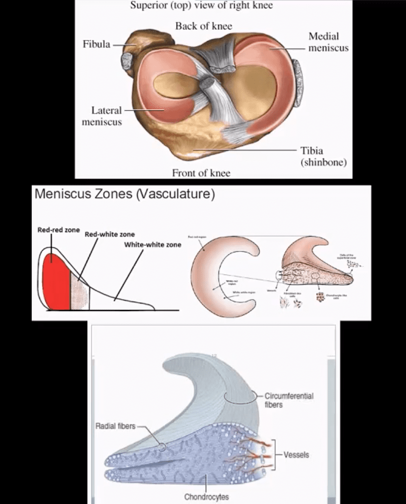
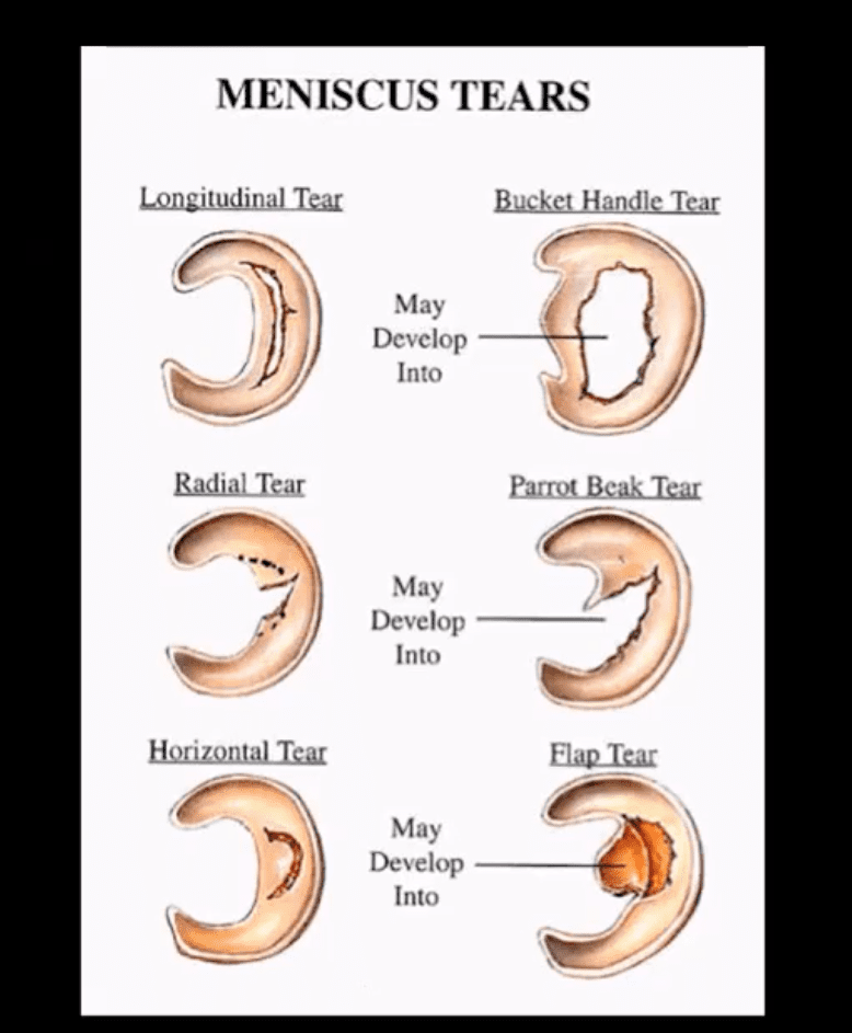
Types, location, and stability of tears are v. important during MRI Dx
Vertical/longitudinal tears especially occur in acute ACL tears. Some longitudinal tears found at the periphery or “red zone” may heal
Bucket handle tear: longitudinal tear in the inner edge that is deep and vertical extending through the long axis and may displace into a notch
Oblique/flap/parrot-beak are complex tears
Radial tear at 90-degree to plateau
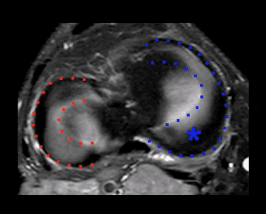
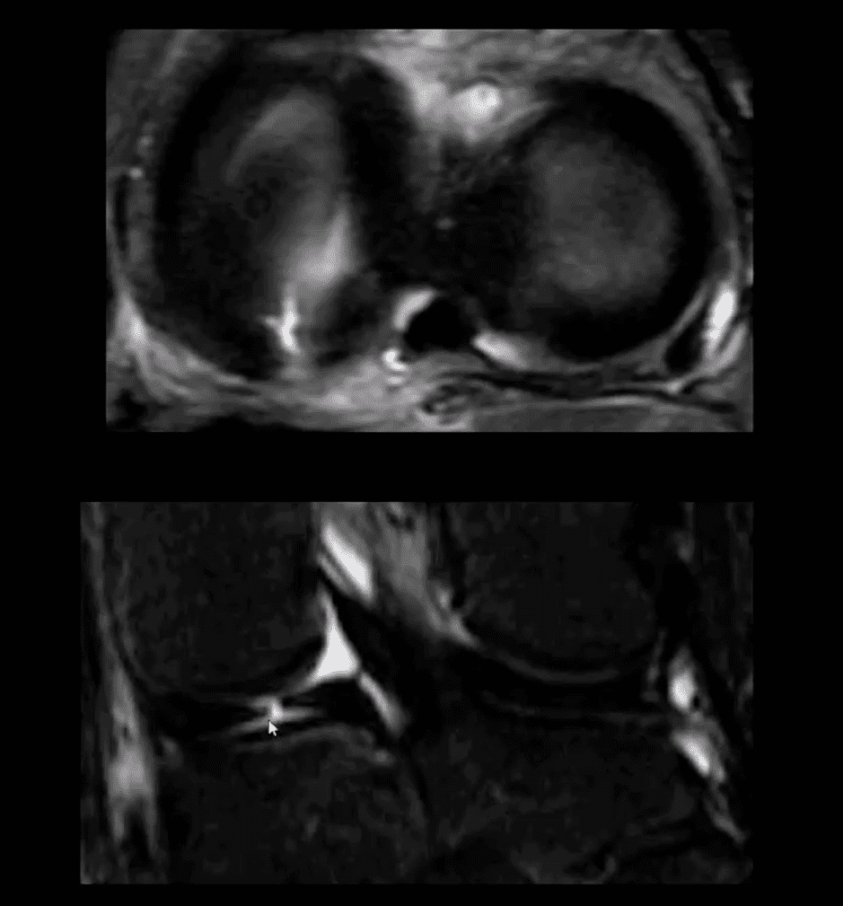
Axial T2
Axial T2 WI fat-sat and coronal STIR slices of the posterior horn of the medial meniscus.
Note a radial tear of the posterior horn of the medial meniscus near the meniscal root. This is potentially an unstable lesion requiring operative care
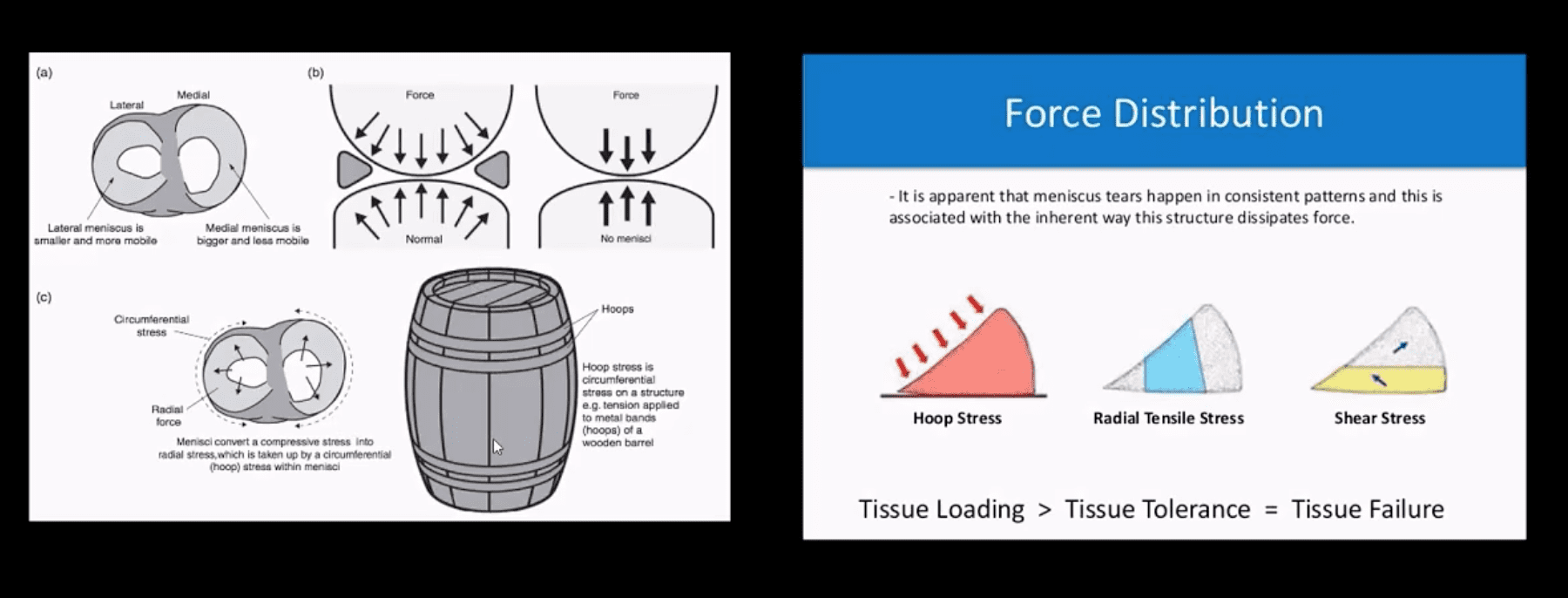
The meniscus, in this case, is unable to provide a “hoop-stress mechanism.”
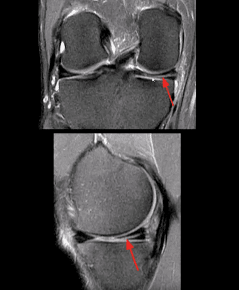
MRI Slices Coronal & Sagittal
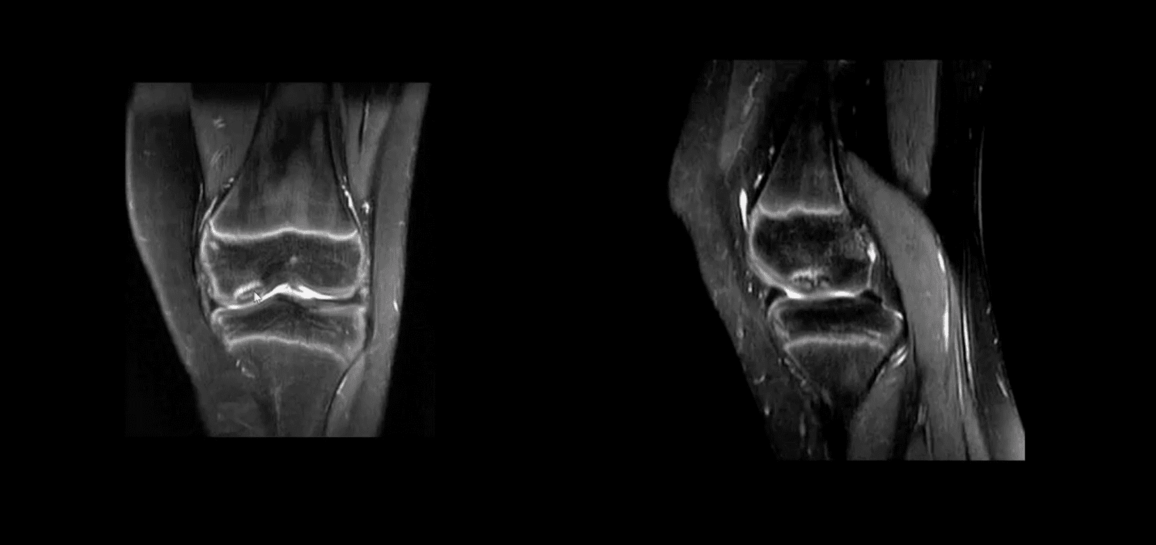
Fat-sat coronal and sagittal proton density MRI slices revealing horizontal (cleavage) tear that is more typical in the aged meniscus
In some cases, when this tear does not contain a radial component, it may partially heal obviating the need for operative care
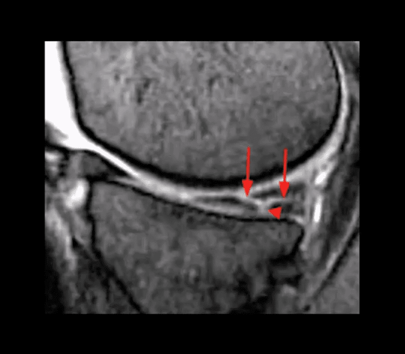
T2 w GRE Sagittal MRI Slice
Complex tear with a horizontal oblique and radial component.
This type of tear is very unstable and in most cases may need operative care
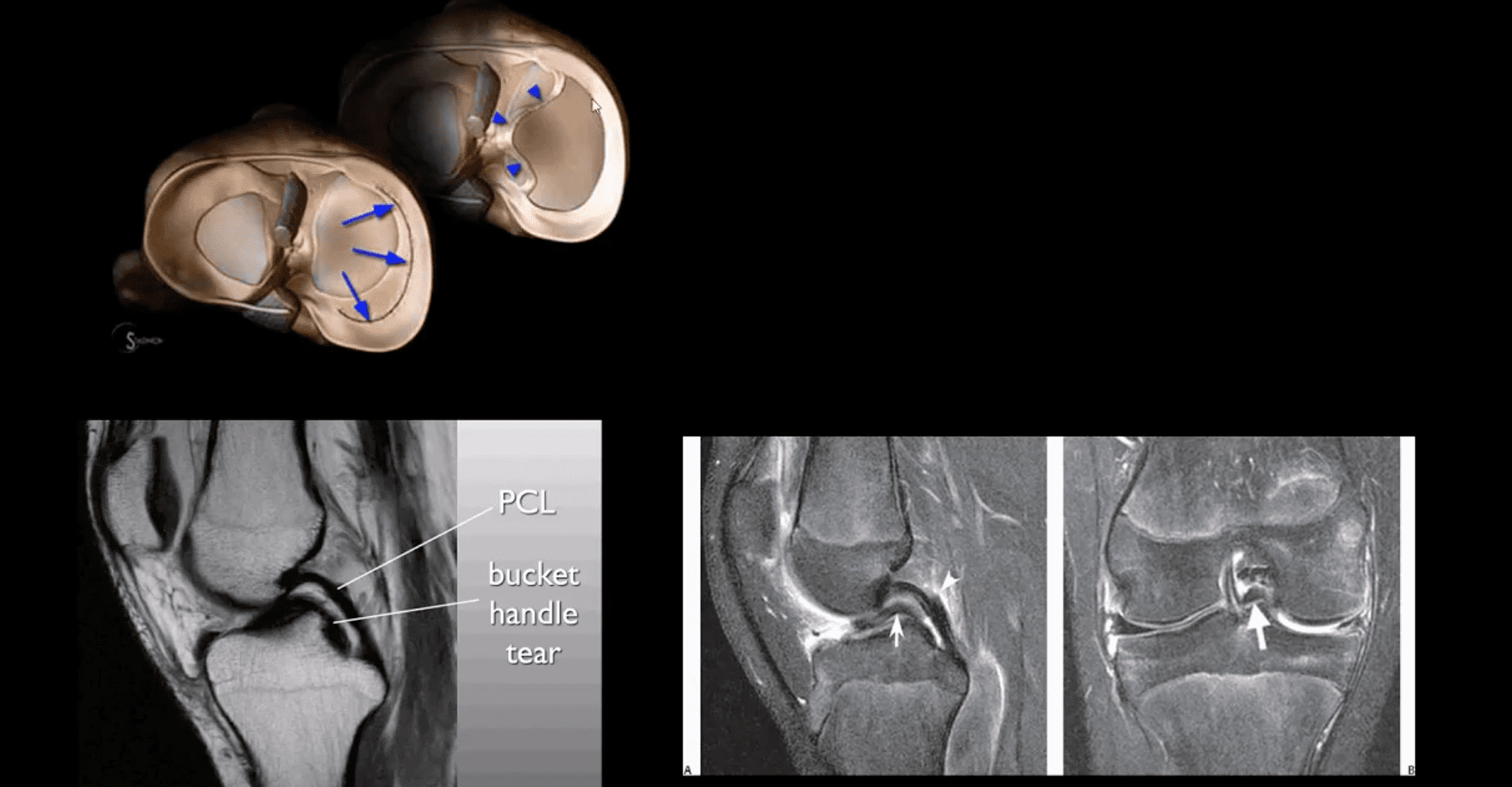
Bucket Handle Tear
Bucket handle tear are m/c in the medial meniscus esp. with acute ACL and MCL tear
MRI signs; double PCL sign on sagittal slices
Absent “bow-tie” sign and others
Most cases require operative care
DDx From Meniscal Degeneration
Occasionally meniscal tears need to be DDx from meniscal degeneration which may also appear bright (high signal) on fluid-sensitive MRI
The simplest rule is that if there is a true meniscal tear aka Grade 3 lesion, it always reaches/extends to the tibial plateau surface
The Role of MSK Ultrasound (US) in Knee Examination
MSK US of the knee permits high resolution and dynamic imaging of primarily superficial anatomy (tendons, bursae, capsular ligaments)
MSK US cannot adequately evaluate cruciate ligaments and the menisci in their entirety
Thus MR imaging remains modality of choice
Potential Pathologies Successfully Evaluated by MSK US
Patellar tendionosis/patellar tendon rupture
Quadriceps tendon tear
Prepatellar bursitis
Infrapatellar bursitis
Pes Anserine bursitis
Popliteal cyst (Baker cyst)
Inflammation/joint effusion with synovial thickening and hyperemia can be imaged with US (e.g., RA) especially with the addition of color power Doppler
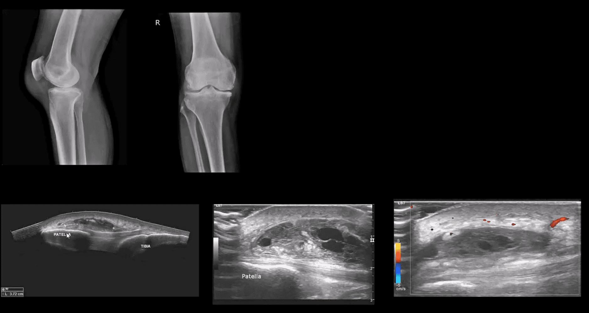
Patient Presented With Atraumatic Knee Pain & Swelling
Radiography revealed sizeable soft tissue density within the superficial pre-patella region along with mild-to-moderate OA
MSK US demonstrated large septated heterogeneous fluid collection with mild positive Doppler activity on the periphery indicating inflammation d/t Dx of Superficial pre-patella bursitis
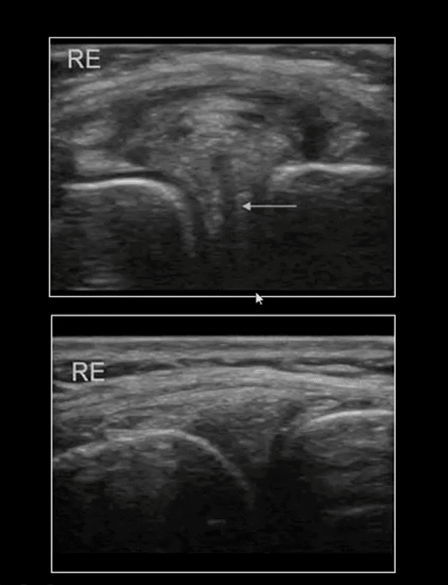
Long Axis US Images
Note normal lateral meniscus and fibers of LCL (above bottom image) compared to
Horizontal degenerative cleavage tear along with protrusion of lateral meniscus and LCL bulging (above top image)
Major limitation: unable to visualize the entire meniscus and the ACL/PCL
MRI referral is suggested
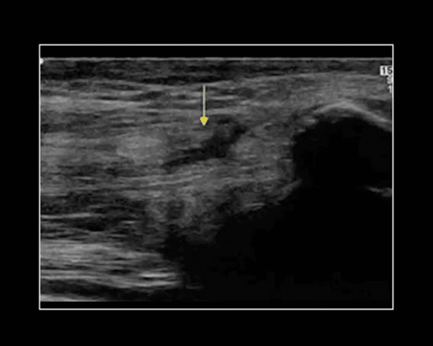
Rupture of Distal Tendon of Quadriceps
Note rupture of distal tendon of the Quadriceps muscle presented as fiber separation and fluid (hypo to anechoic) fluid collection within the substance of the tendon
Advantages of MSK US over MRI to evaluate superficial structures:
Dynamic imaging
Availability
Cost-effective
Patient’s preparation
Disadvantages: limited depth of structures, inability to evaluated bone and cartilage, etc.
Osteochondral Knee Injuries (OI)
osteochondral knee injuries can occur in children 10-15 y.o presented as Osteochondritis Dissecance (OCD) and in mature skeleton m/c following hyperextension and rotation trauma, particularly in ACL tear.
OCD-typically develops from repeated forces in immature bone and affects m/c postero-lateral portion of the medial femoral condyle.
OI in mature bone occurs m/c during ACL tears mainly affecting so-called terminal sulcus of the lateral femoral condyle at the junction of the weight-bearing portion opposed to tibial plateau and the part articulating with the patella
Osteochondral injuries may potentially damage the articular cartilage causing secondary OA. Thus need to be evaluated surgically
Imaging plays an important role and should begin with radiography often followed by MR imaging and orthopedic referral.
OCD Knee
95% associated with some trauma. Other etiology: ischemic bone necrosis especially in adults
Other common location for osteochondral injuries: elbow (capitellum), talus
1st step: radiography may detect osteochondral fragment potentially attached or detached
Location: a posterior-lateral aspect of the medial femoral condyle. Tunnel (intercondylar notch) view is crucial
MRI: modality of choice >90% specificity and sensitivity. Crucial for further management. T1-low signal demarcating line with T2 high signal demarcating line that signifies detachment and unlikely healing. Refer to orthopedic surgeon
Management: stable lesion esp. in younger children>off weight-bearing-heals in 50-75%
Unstable lesion and older child or impending physeal closure>operative fixation.
Many people think of joints, bones, and the, skeletal system when they think of chiropractic, but in fact, the muscles also play an integral part in supporting the body. The muscles are layers and interwoven work to move and stabilize the spine, facilitate the movement of the body�s joints, and aid in respiration. When there is pain within this system, chiropractic can be a very effective treatment. More patients are turning to chiropractic care to treat a variety of painful conditions because it does not use addictive pharmaceuticals with unpleasant side effects; it is completely natural. Chiropractic can also keep patients from requiring surgery in many cases. So when it comes to myofascial pain and trigger points, this form of treatment is often considered optimal.
What is Myofascial Pain?
In simple terms, myofascial pain is simply pain in the muscles. When you break down the word, �myo� means muscle and �fascia� refers to the connective tissue that are interwoven throughout the body.
The pain originates in specific trigger points that are located in the muscles and fascia at various areas of the body. The pain can range in intensity from mild and annoying to severe and debilitating.
What are Trigger Points?
Trigger points are tightened, hypersensitive spots that can be located in any muscle. Different people may have different trigger points. It isn�t like specific lower back pain or neck pain which occur in particular areas of the body. Trigger points can vary from person to person.
When trigger points form, they become nodules or spots that exist in one of the muscle�s taut bands. The patient may experience a variety of symptoms including pain, weakness, burning, tingling, and other symptoms.
What often makes trigger points challenging to locate is that they cause what is known as referred pain. In other words, the person may experience the pain at the exact location of the trigger point, or the pain can be referred to other areas in the body. Referred pain usually has fairly consistent pain patterns so it can be traced to the origin � eventually.
Around 85% of the pain that individuals experience is attributed to myofascial pain. The trigger points determine whether the pain is chronic or acute. It is a condition that is very common.
How do Trigger Points Form?
Trigger points form when the muscle undergoes trauma of some type. The trauma can come from disease, accidents, related work conditions (from persistent, repetitive motion), and sports injuries.
Activities or habits that place a repetitive, long-term strain on the muscles can also cause trigger points. Poor posture, improper ergonomics, and repetitive movements are the most common of these types of activities. Emotional and physical stress are often identified as causes of irritating trigger points.
Benefits of Chiropractic for Myofascial Pain and Trigger Points
Chiropractic care is often a preferred treatment for myofascial pain due to its effectiveness and drug-free approach. Patients who undergo treatment will usually experience a dramatic decrease in their pain level, or it will be eliminated.
They also enjoy increased strength, flexibility, and range of motion. With continued chiropractic care, they will find that they have more endurance for work and recreational activities and even sleep better. It should be noted that sleep disruptions are a common complaint associated with myofascial pain.
Overall, chiropractic can give patients with myofascial pain a better quality of life with decreased incidence of injury. They are often able to lower their pain medication or eliminate it.
Because chiropractic is a whole-body approach, patients learn healthy habits including diet, exercise, and mental wellness. Most of all, they can live with less pain or no pain at all.
Result from valgus or varus stress with or w/o axial loading
Associated with periarticular soft tissues injury
High-stress injury m/c due to jumps falls and axial loading, often with the splitting of the tibial plateau. Men>women. Patients are in their 30s
Low impact or no trauma in patients with osteoporosis d/t insufficiency fractures
Impaction injury is more common with depression of tibial plateau. Women>men. Patients are in their 70s
Lateral Tibial Plateau Fractures More Common
Functional anatomy plays a significant role
60% of weight bearing is by the medial plateau
The medial plateau is more concave
Lateral plateau is slightly higher and more convex. Valgus stress impacts lateral plateau.
Tibial plateau fractures considered intra-articular and prone to delayed healing, non-union, meniscal injury (m/c lateral) ACL tear, secondary OA. Other complications: compartment syndrome, vascular injury.
Management: operative in many cases especially if >3-mm step-off at the plateau
If medial plateau or bicondylar Fxs present, ORIF will be required.
Imaging Plays A Crucial Role
Begins with x-radiography. X-radiography may not reveal the complexity and extent of this injury.
CT scanning w/o contrast will further delineate fracture complexity and pre-operative planning
MR imaging may be considered to evaluate for internal derangement: meniscal, ACL injuries.
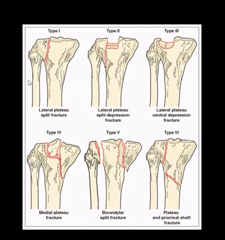
Shatzke classification may help to evaluate the complexity of this injury
Key Diagnostic Sign
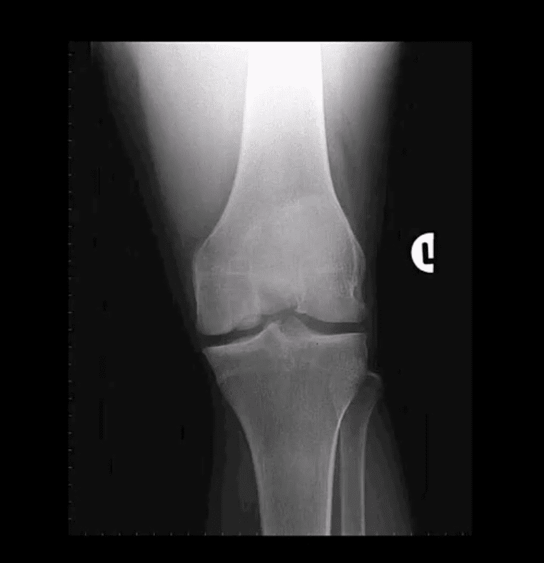
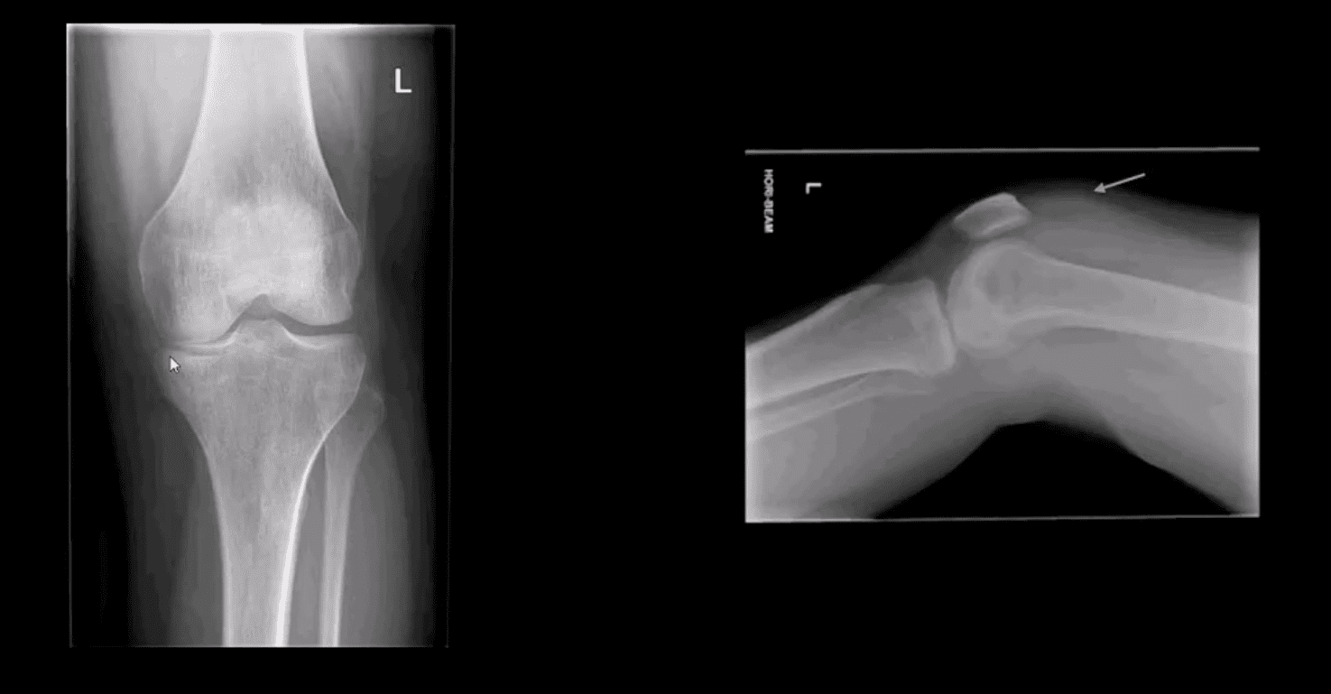
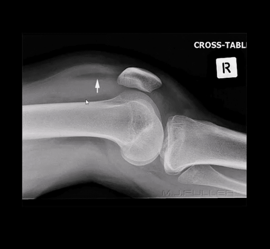
AP and lateral horizontal beam (cross table) left knee radiograph. Note subtle depression of the lateral plateau manifested by the lateral plateau appearing at the same level or lower as the medial. A critical diagnostic sign is the presence of fat-blood-interphase or FBI sign on cross-table lateral (above arrow) indicating intra-articular knee fracture
Lipohemarthorosis aka FBI Sign
Can be detected by radiography, CT or MR imaging
FBI sign is a reliable secondary radiographic sign of intra-articular knee fractures, regardless of how small they are
Mechanism: fracture results with acute hemarthrosis
Hemarthrosis will also occur w/o Fx. However, Fx will result with a fatty marrow being released into the joint cavity. Fat is a less dense medium (lighter) and will appear on the top of the hemorrhage if the patient is held in the supine position for 5-10-minutes before the cross-table radiograph is taken
FBI sign confirms the intra-articular Fx.
ACL/PCL, meniscal tears will not result in FBI sign
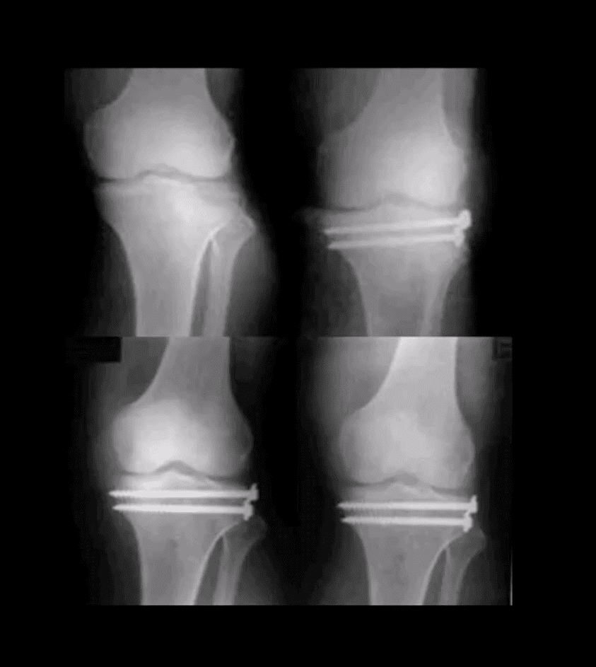
Lateral Tibial Plateau Fx
Lateral tibial plateau Fx that was managed operatively
Most common complication: premature secondary OA
More complex injuries may result in more extensive operative care
Knee Internal Derangement
Acute or chronic injuries of meniscal fibrocartilages and ligamentous restraints
Tears of the ACL and posterior horn of the medial meniscus are the most common
Acute ACL tears, however, often result with a lateral meniscus tear
Acute ACL tear may occur as a combined injury of the ACL, MCL, and medial meniscus
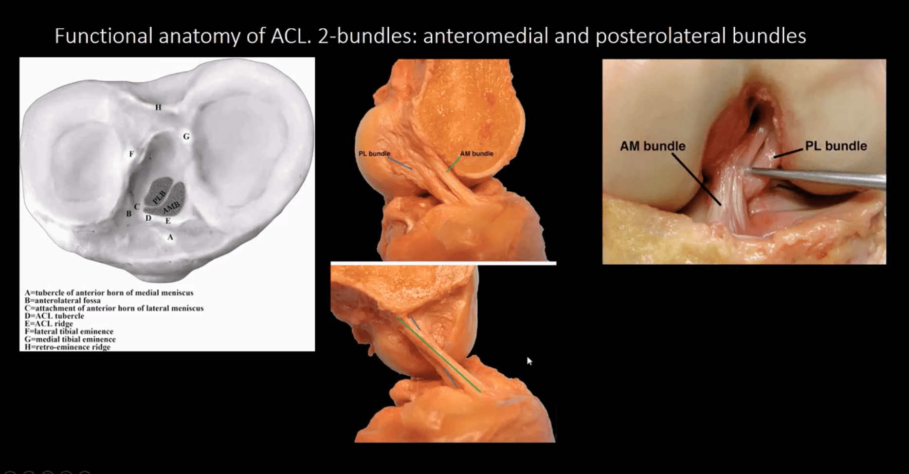
Functional anatomy: ACL prevents anterior displacement of the tibia and secondary varus stress
MCL functions together with ACL in resisting external rotation of the tibia especially when the foot is planted (closed chain position)
MCL is firmly attached to the medial meniscus, explaining the classic triad of ACL, MCL and medial meniscal tear (O’Donahue terrible triad)
Cruciate ligaments (ACL/PCL) are intra-articular but extra-synovial. Less likely to be torn in closed pack position (full extension). When all articular facets of tibia and femur are in full contact, the ACL/PCL are at least tension and stable
When the knee is flexed 20-30-degrees or more ACL is taut and remains unstable
ACL is a significant mechanoreceptor that feeds the info to CNS about the joint position. Thus the majority of previous ACL tears will lead to some degree of knee instability
Functional Anatomy of ACL
Diagnosis of ACL Tear
Diagnosis of ACL tear requires MR imaging
Concerns exist of not only ligamentous injuries but injuries to the articular cartilage and menisci.
Most vendors will perform at least: one T1 WI in coronal or sagittal planes. Sagittal and coronal Proton-density slices to evaluate cartilaginous structures. Fast spin-echo sagittal, axial and coronal T2 fat-saturated or sagittal and coronal STIR images are crucial to demonstrate edema within the substance of knee ligaments
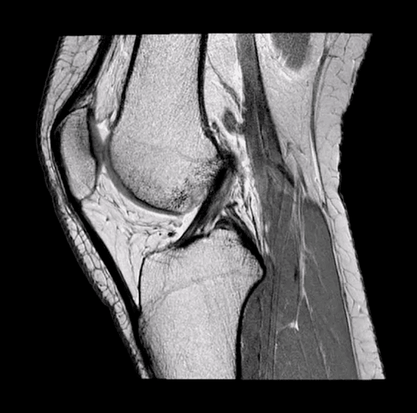
ACL is aligned along the Blumensaat line or oblique line corresponding the intercondylar roof of Femoral condyles. Lack of such alignment by the ACL is significant for ACL tear
Imaging Dx of Internal Derangement
MRI shows 78-100% sensitivity and 78-100% specificity
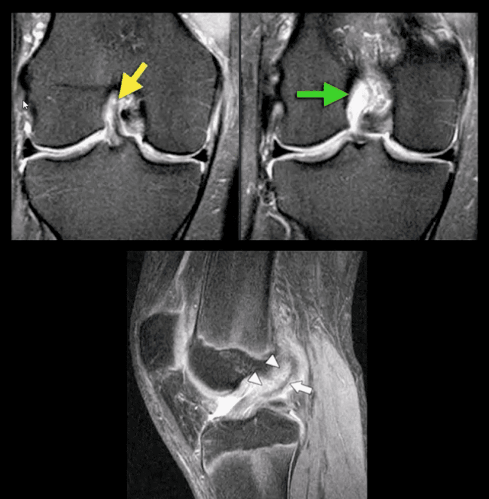
Primary signs of ACL tear: non-visualization of ACL (above green arrow), loss of its axis along the Blumensaat line (above triangle heads), wavy appearance and substance tear (above white arrow) or edema and cloud-like indistinctness (above yellow arrow)
Reliable Secondary Signs of ACL Tear
May be observed on the radiographs and MRI
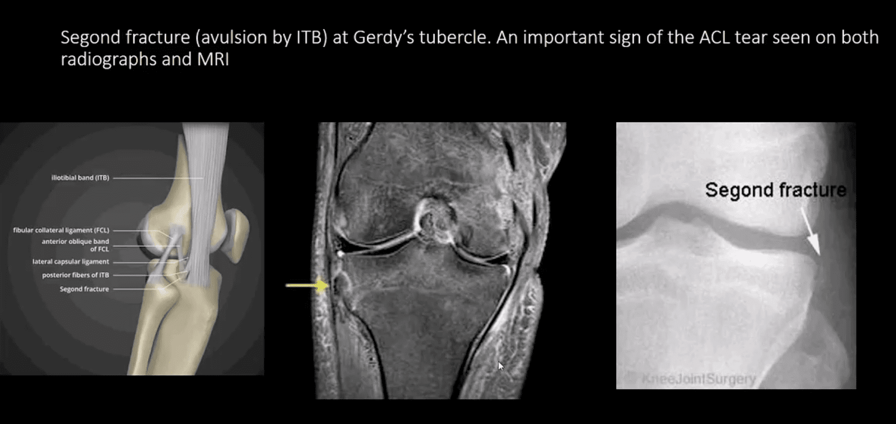
Segond avulsion fracture (80% specificity for ACL tear) (next slide)
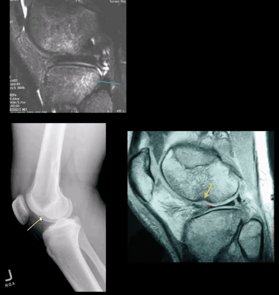
Deep femoral notch sign indicating osteochondral fracture (above bottom images) and
Pivot -shift bone marrow edema in the posterolateral tibial condyle d/t external rotation and often valgus impact by the lateral femoral condyles (above top image)
Segond Fracture (Avulsion by ITB)
Segond fracture at Gerdy’s tubercle. A vital sign of the ACL tear seen on both radiographs and MRI
Management of ACL Tears
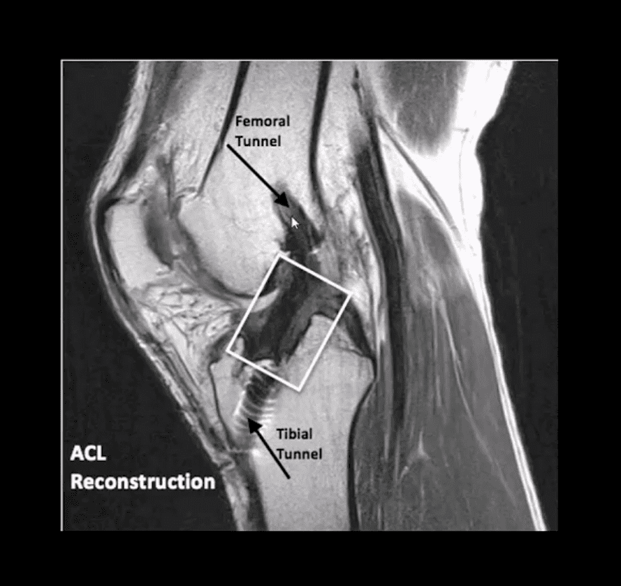
In acute cases, usually operative using cadaveric or autograft (patella ligament or hamstring) ACL reconstruction
Complications: graft tear, instability and premature DJD, joint stiffness d/t lack of postoperative rehab or gaft shortening. More rare, infection, a formation of intraosseous synovial cysts, etc.
Have you ever had a pain in the neck? And your kids or significant other don’t count. If you’ve ever had a stiff, sore neck, then you’ve more than likely experienced cervicalgia. You’re not alone. The American Osteopathic Association estimates that more than 25% of Americans have experienced or chronically experience neck pain. Neck pain is one of the primary causes of chronic pain, ranking number three behind knee pain (number two) and back pain (number one). Chronic pain affects around 65% of people in the United States, ranging in age 18 to 34. They either have experienced it firsthand or care for someone who has recently experienced it. That number increases as the population ages.
It is also worth noting that most doctors prescribe pain medications, but more than 33% of patients with chronic pain won’t take them because they are afraid of becoming addicted.
What is Cervicalgia?
Cervicalgia is a blanket term used to describe neck pain. It can range from a simple crick in the neck to severe pain that prevents you from turning your head.
Knowing the term for the pain, though, does not help when it comes to treatment because treatment lies in the cause of the pain. It can become quite complex because there are so many causes for the pain. Sometimes the cause itself must be eliminated before the treatments for the pain can be effective.
What are the Causes of Cervicalgia?
The causes of cervicalgia are vast and varied. A patient who sits at their desk for too long or sleeps in a poor position can develop neck pain.
Injuries such as sports injuries and whiplash fall at the more severe end of the spectrum. Even simple gravity can be a culprit.
The human head can weigh as much as 10 pounds, sometimes even more, and the neck is tasked with keeping it upright. Just the action of fighting gravity and keeping the head erect for long periods of time (like all day) can cause the neck muscles to become strained and fatigued. This can also cause neck injuries to heal slower because the neck is almost always in use and under consistent stress.
How is Cervicalgia Treated?
Treatment for cervicalgia depends on both the symptoms and the cause. If you have been injured, you should immediately seek medical attention to assess the injury’s severity.
You can apply ice to help reduce inflammation and swelling, but do not delay a medical evaluation. Some neck injuries can be severe, causing severe conditions, including paralysis.
After an assessment, your doctor may prescribe medication such as anti-inflammatories and stronger painkillers. A cervical collar may also be recommended since it allows the neck to rest, which will promote healing.
If the pain is caused by other reasons such as stress, poor posture, or sleeping on the wrong pillow (in other words, you have a crick in your neck), you can use an over-the-counter anti-inflammatory medication, and using a heating pad will help. Massage is also effective.
However, prevention is the best cure. When you know what is causing your cervicalgia, you can take steps to prevent it. Chiropractic can help both in prevent cervicalgia and in treating it.
Chiropractic for Cervicalgia
Chiropractic treatment can help relieve cervicalgia pain for many of the causes, including injury, stress, and misalignment. Depending on the cause, the chiropractor will use specific techniques to treat the root of the problem.
They will bring the body back into alignment, which also helps to prevent the pain of cervicalgia. The most attractive aspect is that it allows for pain management without the use of any medications.
When you get regular chiropractic care, you can reduce your chances of experiencing pain in your neck and back. That is why so many people are choosing chiropractic care for their neck and back pain instead of turning to traditional medicine because it works.
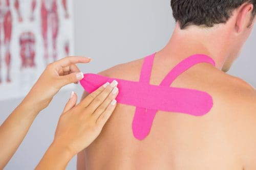
You may have seen professional athletes, dancers, gymnasts, and others who engage in extremely physical activities using a type of tape on various points of their bodies. It is sometimes colored and does not seem to inhibit range of motion. While it looks like tape or maybe a very fancy bandage, it is a highly technical, specialized tape that is used to treat patients of all ages and activity levels. It is called Kinesio tape, and it is often used by chiropractors to help address specific injuries.
What is Kinesio Tape?
Also called Kinesio Tex Tape, Kinesio Tape is a special adhesive tape that has elastic properties. It was developed by Dr. Kenzo Kase, a chiropractor, and acupuncturist, in 1979. It is safe for all ages including pediatric and geriatric patients. The tape is comprised of a 100% cotton fiber strip with medical grade acrylic adhesive. It is soft and gentle, but it works.
The tape is hypoallergenic and latex free, so it is appropriate for a vast audience. It is also water resistant, making it wearable for many activities. When worn, the tape does not limit or inhibit range of motion, and the same tape can be worn for several days without losing its effectiveness.
How does Kinesio Tape Work?
The tape can stimulate or relax muscles, depending on the tension that it puts on the body when applied. When worn, it lifts the skin by microscopic increments which aids in lymphatic drainage.
It also helps to decrease inflammation and swelling which reduces pressure in the area. This allows the blood and lymphatic fluid to flow freer and more effectively in and out of the affected area.
When used correctly, Kinesio Tape can reduce inflammation, promote better circulation, prevent injury, facilitate healing, the re-educate the neuromuscular system. This helps the body return to homeostasis. It can be applied in many different configurations, but often the applications are a single �I,� �Y,� or �X.�
The Kinesio Taping Method
The Kinesio taping method is a systematic, therapeutic technique that offers a two-prong approach of supporting the patient and rehabilitating the condition or affected area. It can alleviate pain reduce swelling, providing relief to the patient. There are specific taping shapes that are specialized to address certain areas of the body as well as certain conditions. While it provides stability and support to the body�s joints and muscles, it does not restrict the range of motion. The technique is designed to address soft tissue injury by manipulating that area and facilitating healing.
What Conditions is Kinesio Taping used to Treat?
Many conditions throughout the body can benefit from Kinesio Taping. Chiropractors use it for:
Lower back strain
Plantar fasciitis
Back strain
Carpal tunnel syndrome
Rotator cuff injury
Ankle Sprains
Whiplash
Herniated disc
Post-surgery edema
Tennis elbow
Pre-surgery edema
Patella tracking
Athletes may also use it for additional support or to prevent injury. Because it relies on the body�s natural healing process, many people find it to be preferred treatment for many painful conditions. When combined with chiropractic care, Kinesio Tape is very effective.
When treating a condition with Kinesio Tape, the chiropractor may use a variety of techniques, depending on the illness or injury. They may use spinal manipulation, massage, and other treatments, combining them with recommendations for lifestyle changes and diet modifications.
The draw for this treatment is that it encourages the body to heal itself, eliminating the need for drugs with their undesired and unpleasant side effects, or more invasive procedures like surgery. Kinesio Taping is safe, natural, and a perfect complement to chiropractic care.
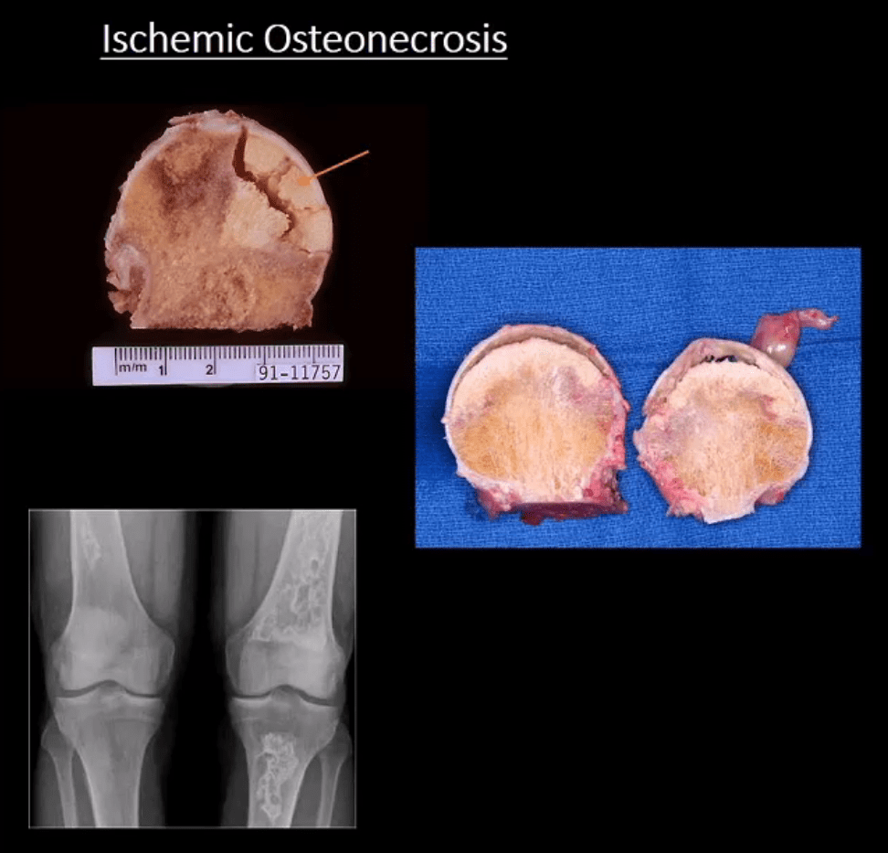
Ischemic Osteonecrosis (More accurate term) aka avascular necrosis AVN: this term describes subarticular (subchondral) bone death
Intramedullary bone infarct: depicts osteonecrosis within the medullary cavity of the bone (above x-ray image)
Causes: m/c: trauma, systemic corticosteroids, diabetes, vasculitis in SLE. The list is long. Other vital causes: Sickle cell disease, Gaucher disease, alcohol, caisson disease, SCFE, LCP, etc.
Pathology: ischemia and bone infarct with resultant devitalized center surrounded by ischemia and edema with normal bone on the outer periphery (MRI double line sign)
Sub-articular necrotic bone eventually collapses and fragments leading to progressive bone and cartilage destruction and rapidly progressing DJD
Early Dx often missed but crucial to prevent severe DJD
M/C Sites
Hips, shoulders, talus, scaphoid bone. Many peripheral idiopathic AVN sites are known by their eponyms (e.g., Kienbock aka AVN of the lunate bone, Preisier aka scaphoid AVN)
Radiography is insensitive to early AVN and may only present as subtle osteopenia
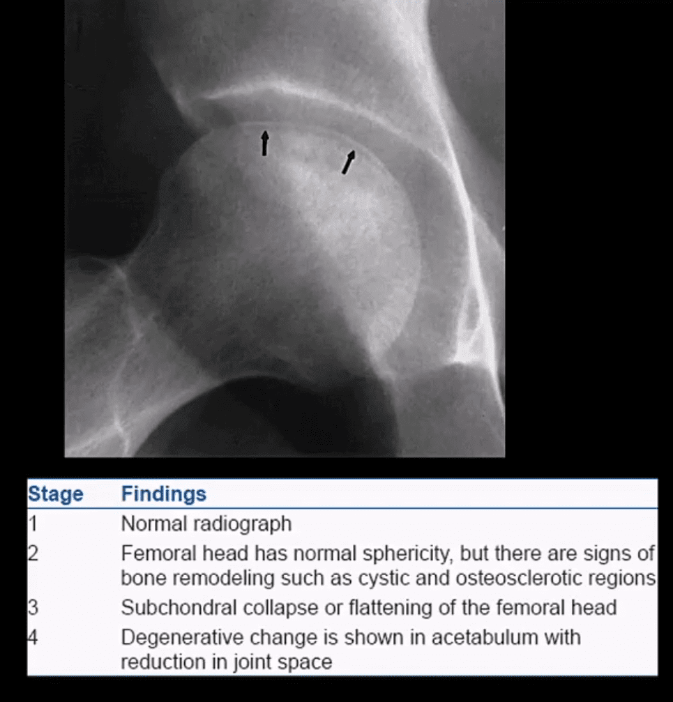
Some of the early appreciable rad features are increased patchy bone sclerosis followed by sub-articular bone collapse or “crescent sign” signifying stage-3 on Ficat classification (above)
Earliest detection and early intervention can be achieved by MRI (most sensitive modality)
If MRI contraindicated or unavailable, 2nd most sensitive modality is radionuclide bone scan (scintigraphy)
X-ray and CT scanning are of equal value
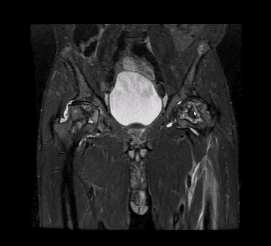
Coronal MRI Slice
Fluid sensitive, sensitive coronal MRI slice revealing bill ischemic osteonecrosis of the femoral head
MRI findings: l
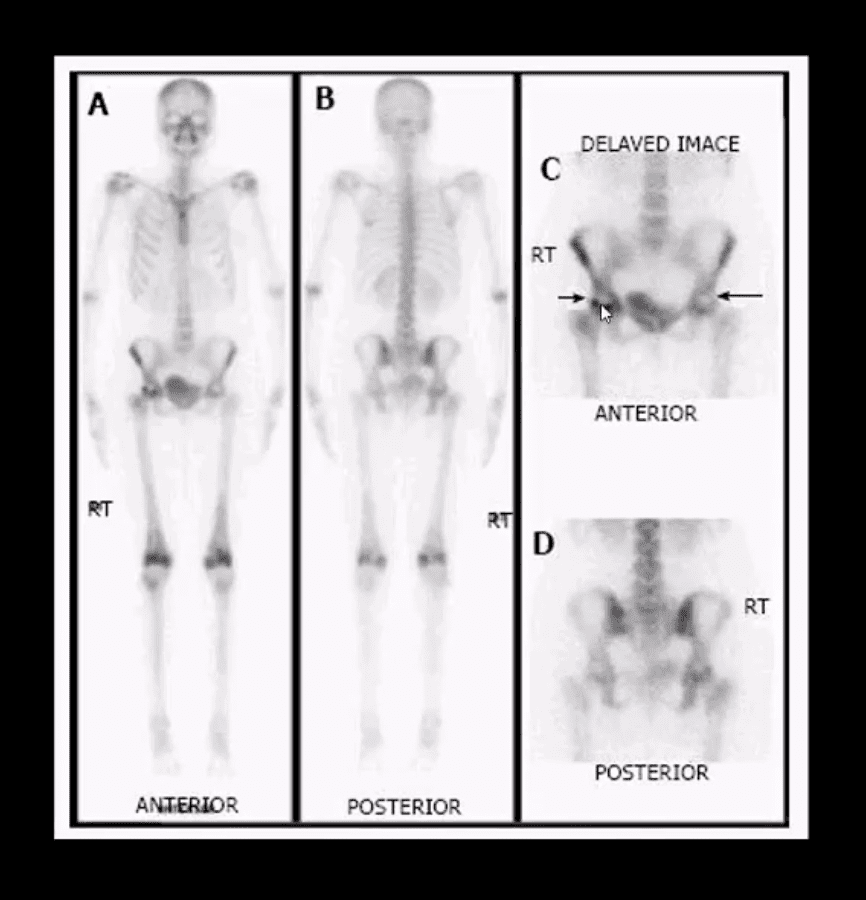
Tc99-MMDP Radionuclide Bone
Bone scan reveals a central area of photopenia (cold spot) d/t necrotic fragment surrounded by increased osteoblastic activity as increased uptake of Tc-99 MDP in the right hip
The patient is a 30-year-old female with breast cancer and chemotherapy treatment who suddenly presented with right hip pain
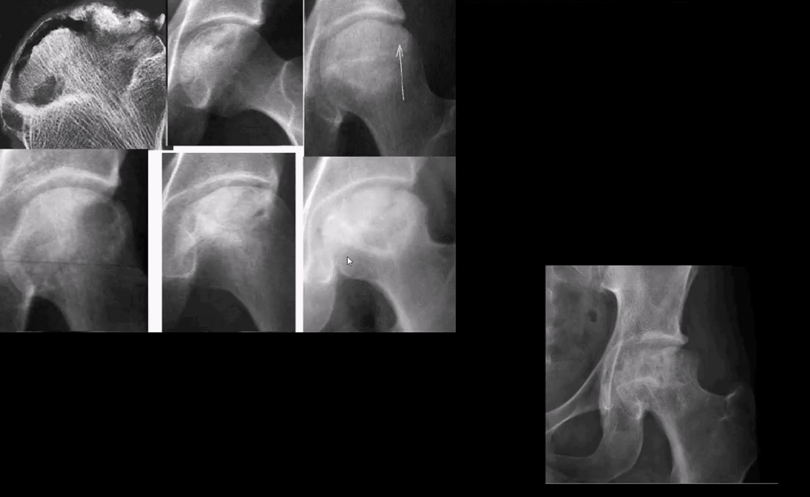
Radiographic Progression of AVN
Later stages present with articular collapse, subarticular cysts, increased patchy sclerosis and complete flattening of the femoral head with resultant severe DJD. Rx: THA
Management
Early imaging Dx with MRI or bone scintigraphy is essential
Referral to the Orthopedic surgeon
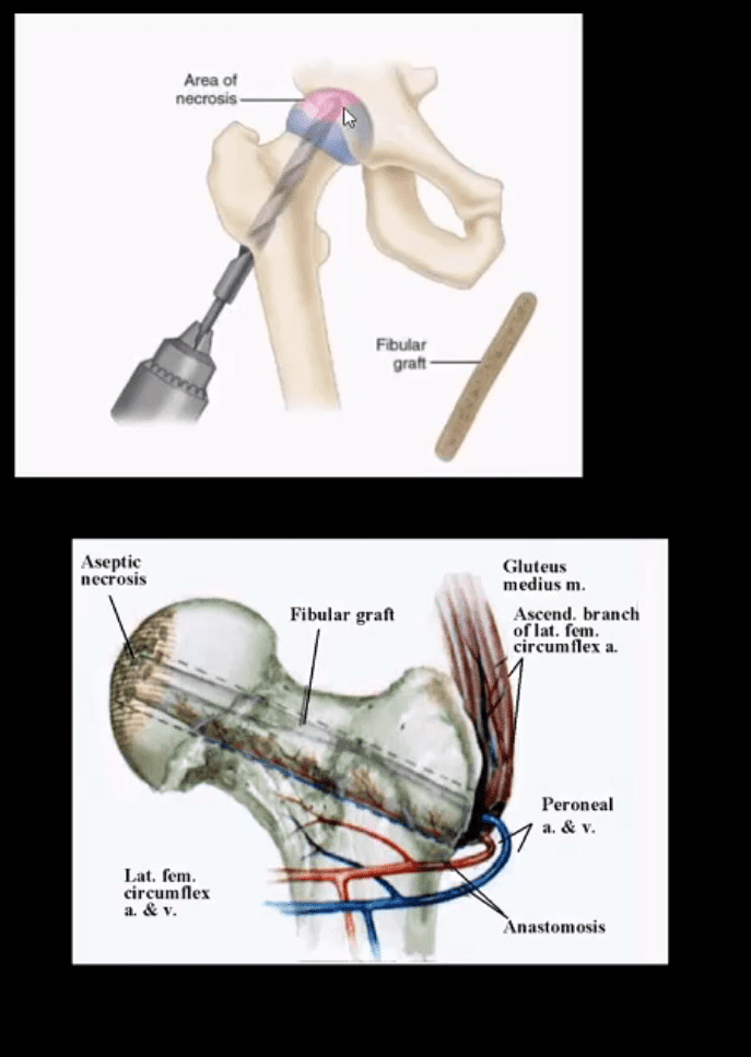
Core decompression (above) can be used to revascularize the affected bone during earlier stages but produces mixed results
Delayed changes of AVN: THA as IN severe DJD cases
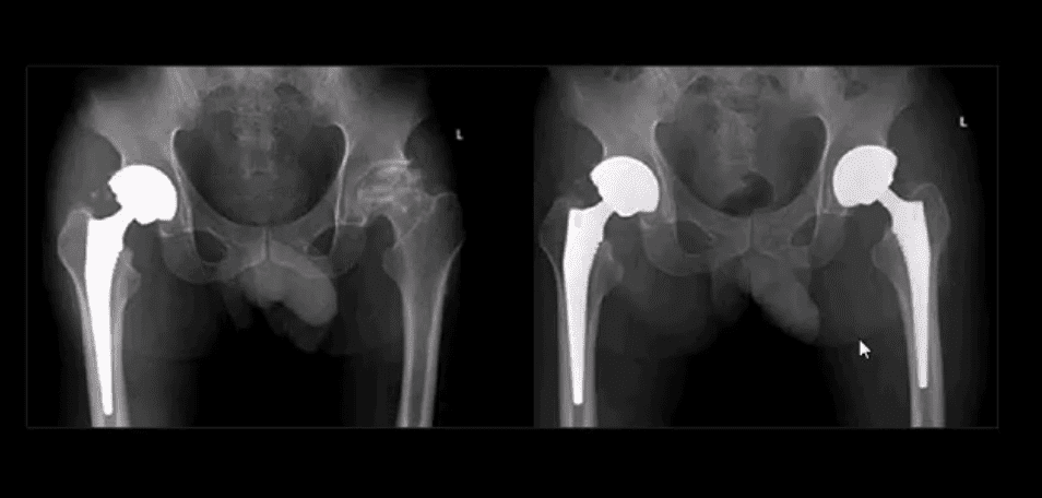
B/L THA
B/L THA in the patient with ischemic osteonecrosis of the right and later left hip
When B/L hip AVN is present, typically consider systemic causes (corticosteroids, diabetes)
Inflammatory Arthritis Affecting the Hip
Consider common systemic inflammatory condition such as RA and AS/EnA
Hip RA may develop in 30% of patients with RA
Key features to DDx inflammatory arthritis vs. DJD is symmetrical/uniform aka concentric joint loss often leading to axial migration and Protrusion Acetabule in advanced cases
Key element between RA vs. AS: the presence of RA bone erosion w/o productive bone changes or enthesitis in AS d/t inflammatory subperiosteal bone proliferation, whiskering/fluffy periostitis (collar-type enthesitis circumferentially affecting head-neck junction)
Dx: Hx, PE, labs: CRP, RH, anti-CCP Ab (RA)
CRP, HLA-B27, RF- (AS)
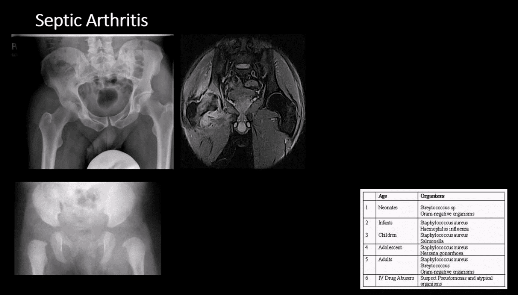
Septic Arthritis
Gonococcal infections, iatrogenic causes, I.V. drug use, and some others
Routes:�hematogenous, adjacent spread, direct inoculation (e.g., iatrogenic)
Clinically: pain and reduced ROM presented as monoarthritis, generalized signs/symptoms. CBC, ESR, CRP changes. ARthrocentesis and culture are crucial
M/C pathogen Staph. Aureus & Neisseria Gonorrhea
1st step: radiography, often unrewarding in the early stage. Later (4-10 days) indistinctness of the white cortical line at the femoral articular epiphysis, loss of joint space, effusion as a widening of the medial joint area (Waldenstrom sign)
MRI – best at early DX: T1, T2, STIR, T1+C may help with early. Early I.V. antibiotics crucial to prevent rapid joint destruction
Slipped Capital Femoral Epiphysis (SCFE)
Important to diagnose but easily missed potentially leading to Ischemic Osteonecrosis of the femoral head aka AVN
Presents typically in overweight children (more often boys), age over eight years. Greater incidence in African-American boys
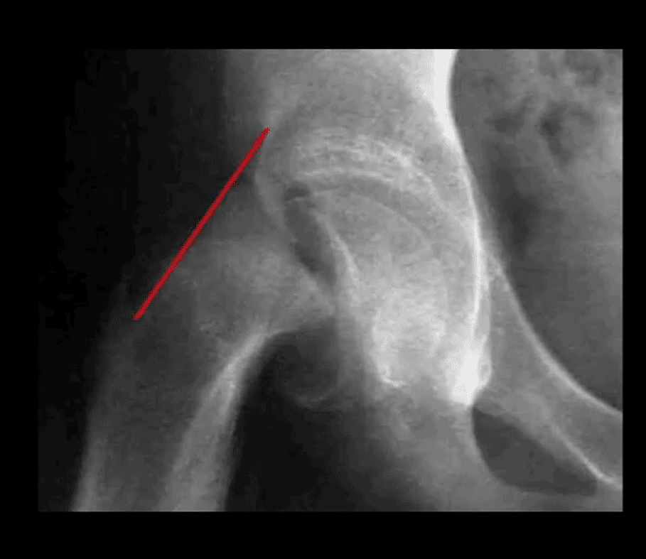
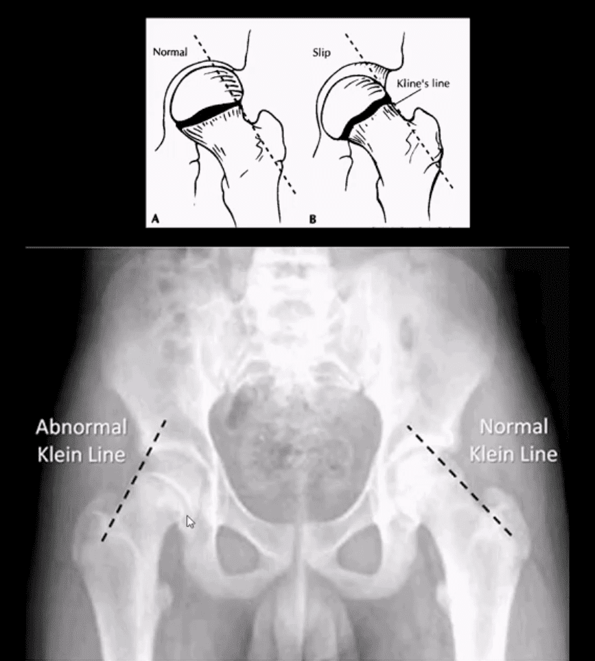
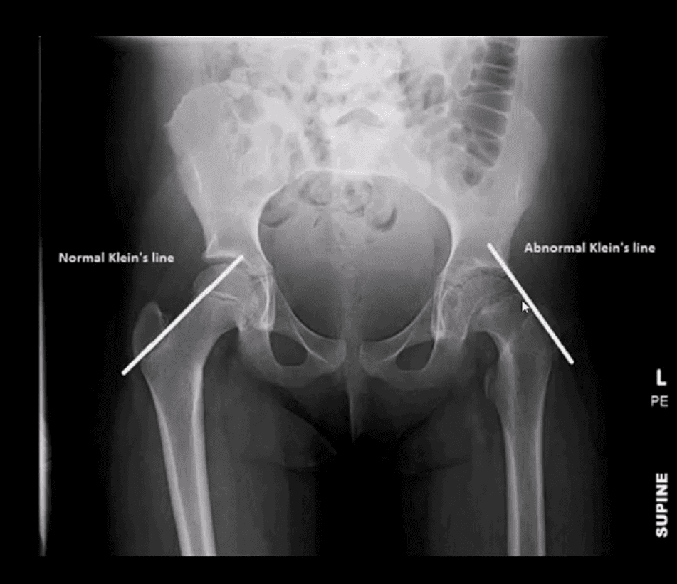
1st step: radiography, especially look for a widened physeal growth plate (so-called pre-slip). Later, slip and disturbed Klein’s line (above image). MRI – best modality for early Dx and early intervention
The frog lateral view often demonstrates the medial slip better than the AP view
Clinically Limping Child or Adolescent
M>F (10-18 years). African-Americans are at greater risk. 20% of cases of SCFE are B/L. Complications: AVN >>DJD
Radiography:�AP pelvis, spot, and frog leg may reveal slippage as Klein line failed to cross through the lateral aspect of the femoral head
Additional features: physis may appear widened
MRI w/o gad, is required for the earliest Dx and prevention of complications (AVN)
Normal and Abnormal Klein Line
Consistent with SCFE. The physis is also widened. Dx: SCFE
Urgent referral to the Pediatric Orthopedic surgeon
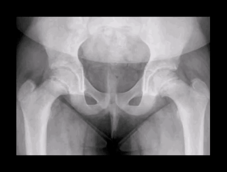
Subtle Changes in Left Hip
Note suspected subtle changes in the left hip that may require MR examination to confirm the Dx
Delay in care may result in major complications
Perthes’ Disease
aka Legg-Calves-Perthes Disease (LCP)
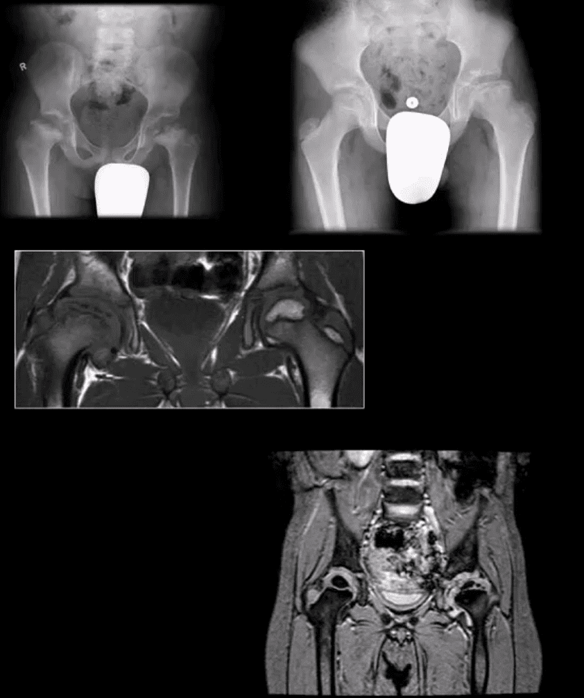
Refers to Osteochondritis of the femoral head with osteonecrosis likely d/t disturbed vascularization of the femoral head
Presents typically in children (more often boys) aged under eight years as atraumatic “limping child.” 15% may have B/L Perthe’s
Imaging steps: 1st step x-radiography, followed by MRI especially in stage 1 (early) w/o x-ray abnormalities
Unspecific signs: joint effusion with Waldenstrome sign+ (>2-mm increase in medial joint space compared to the opposite side). Past approach: Fluoroscopic Arthrography (replaced by MRI)
Pathologic-Radiologic Correlation: in well-established cases, the femoral head characteristically becomes sclerotic, flattened and fragmented due to avascular necrosis (AVN). Later on, an occasional Coxa Magna changes may develop (>10% femoral head enlargement)
Management: symptoms control, bracing. Boys at younger ate show better prognosis d/t more immaturity and better chances of bone/cartilage repair mechanisms. In advanced cases, operative care: osteotomy, hip arthroplasty in adulthood if advanced DJD develops
Common Neoplasms & Other Conditions Affecting Hip/Pelvis
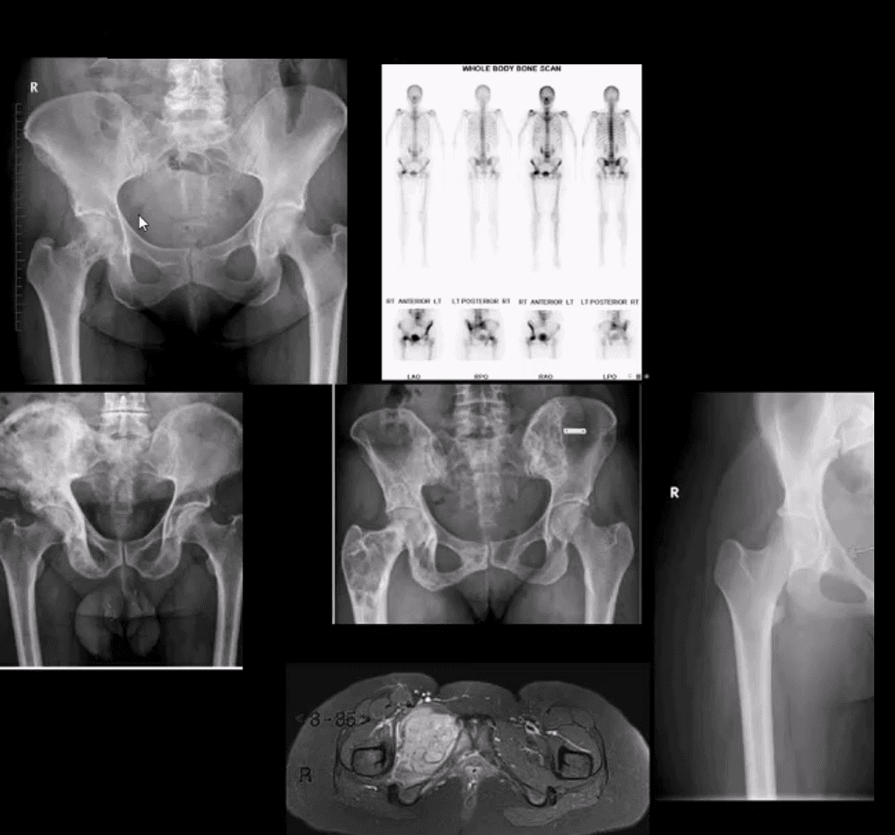
M/C hip & pelvis neoplasms in adults: bone metastasis ( above far left), 2nd m/c Multiple Myeloma (M/C primary bone malignancy in adults). Tips: remember Red Marrow distribution. Less frequent: Chondrosarcoma
Paget’s disease of bone (above-bottom left image) is m/c detected in the pelvis and Femurs
Children and young adults ‘limping child’ benign neoplasms: Fibrous Dysplasia (above middle image), Solitary Bone Cyst (21%), Osteoid Osteoma, Chondroblastoma. Malignant pediatric neoplasms: m/c Ewing Sarcoma (above middle right and bottom images) vs. Osteosarcoma. >2y.o-consider Neuroblastoma
Imaging: 1st step: radiography followed by MRI are most appropriate.
If Mets are suspected: Tc99 bone scintigraphy is most sensitive
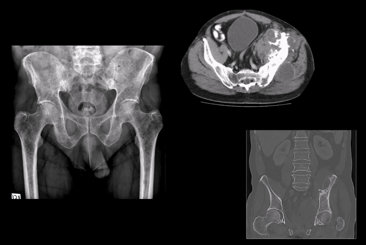
Multiple Myeloma
Multiple Myeloma in a 75-y.o male (AP pelvis view)
Chondrosarcoma in a 60-y.o male (axial and coronal reconstructed CT+C slices in the bone window)
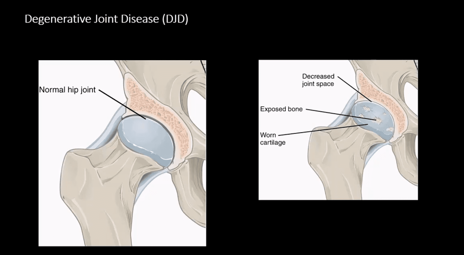
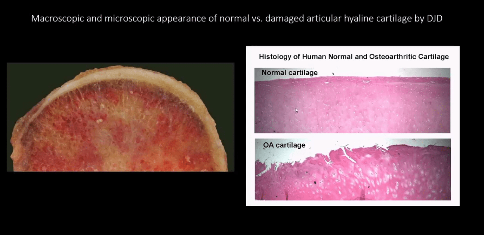
Macroscopic & Microscopic Appearance of Normal vs. Damaged Articular Hyaline Cartilage by DJD
Hip Osteoarthritis (OA) aka Osteoarthrosis
Symptomatic and potentially disabling DJD
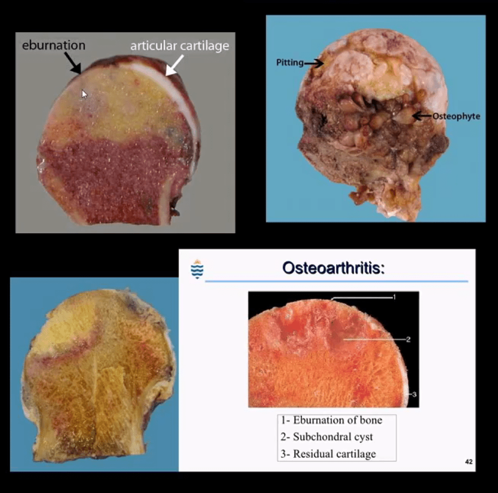
Progressive damage and loss of the articular cartilage causing denudation and eburnation of articular bone
Cystic changes, osteophytes, and gradual joint destruction
Develops d/t repeated joint loading and microtrauma
Obesity, metabolic/genetic factors
Secondary Causes: trauma, FAI syndrome, osteonecrosis, pyrophosphate crystal deposition, previous inflammatory arthritis, Slipped Capital Femoral Epiphysis, Leg-Calves-Perthes disease in children, etc.
Hip OA, 2nd m/c after knee OA. Women>men
88-100 symptomatic cases per 100000
Radiography is the Modality of Choice for the Dx and Grading of DJD
Special imaging is not required unless other complicating factors exist
The acetabular-femoral joint is divided into superior, axial and medial compartments/spaces
Normal joint space at the superior compartment should be 3-4-mm on the AP hip/pelvis view
Understanding the pattern of hip joint narrowing/migration helps with the DDx of DJD vs. Inflammatory arthritis
In DJD, m/c hip narrowing is superior-lateral (non-uniform) vs. inflammatory axial (uniform)
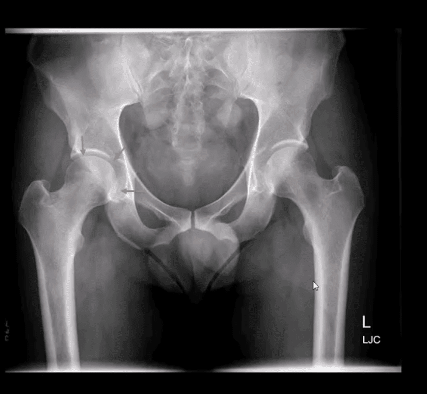
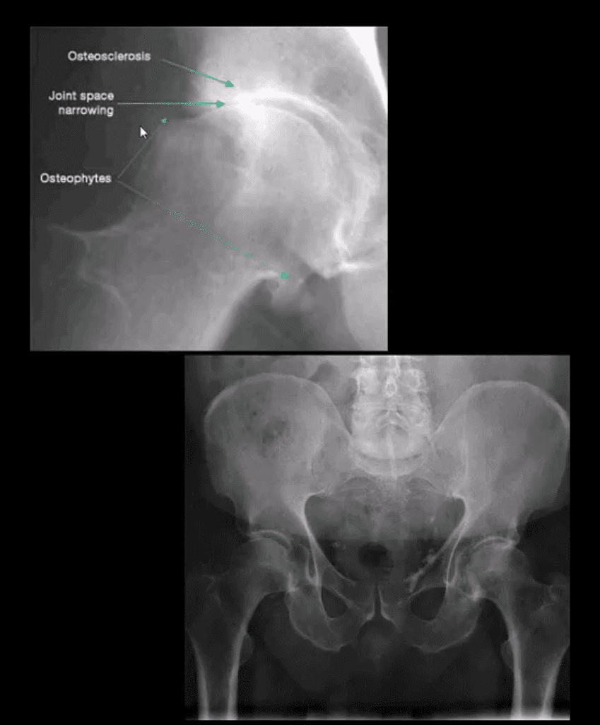
AP Hip Radiograph Demonstrates DJD
With a non-uniform loss of joint space (superior migration), large subcortical cysts and subchondral sclerosis
Radiographic features:
Like with any DJD changes: radiography will reveal L.O.S.S.
L: loss of joint space (non-uniform or asymmetrical)
O: osteophytes aka bony proliferation/spurs
S: Subchondral sclerosis/thickening
S: Subcortical aka subchondral cysts “geodes.”
Hip migration is m/c superior resulting in a “tilt deformity.”
Radiographic Presentation of Hip OA May Vary Depending On Severity
Mild OA: mild reduction of joint space often w/o marked osteophytes and cystic changes
During further changes, collar osteophytes may affect femoral head-neck junction with more significant joint space loss and subchondral bone sclerosis (eburnation)
Cyst formation will often occur along the acetabular and femoral head subarticular/subchondral bone “geodes” and usually filled with joint fluid and some intra-articular gas
Subchondral cysts may occasionally be very large and DDx from neoplasms or infection or other pathology
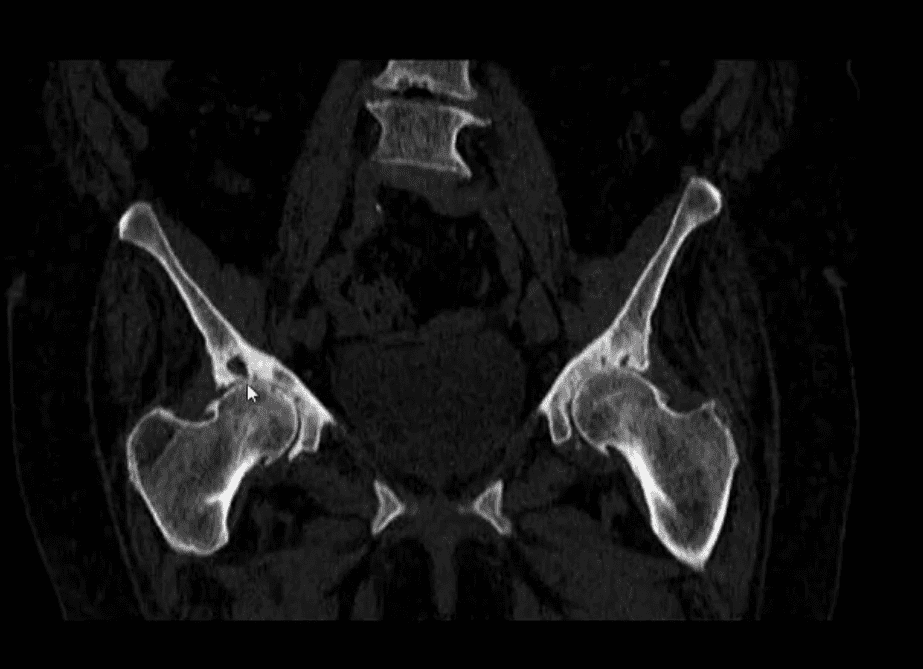
Coronal Reconstructed CT Slices in Bone Window
Note moderate joint narrowing that appears non-uniform
Sub-chondral cysts formation (geodes) are noted along the acetabular and femoral head subchondral bone
Other features include collar osteophytes along head-neck junction
Dx: DJD of moderate intensity
Referral to the Orthopedic surgeon will be helpful for this patient
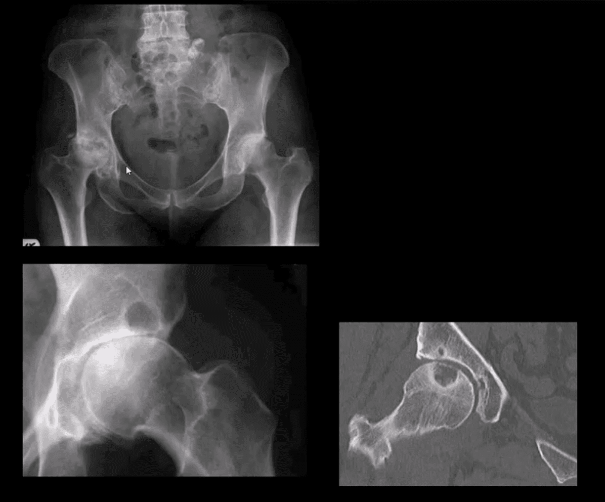
AP Pelvis (below the first image), AP Hip Spot (below the second image) CT Coronal Slice
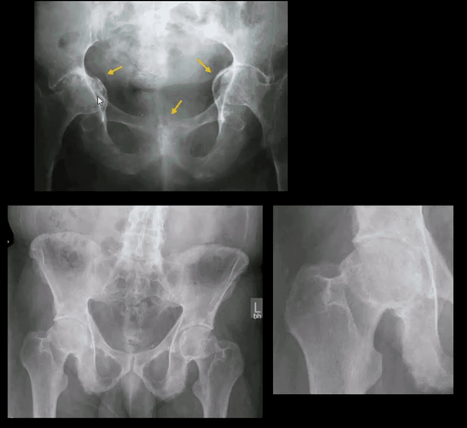
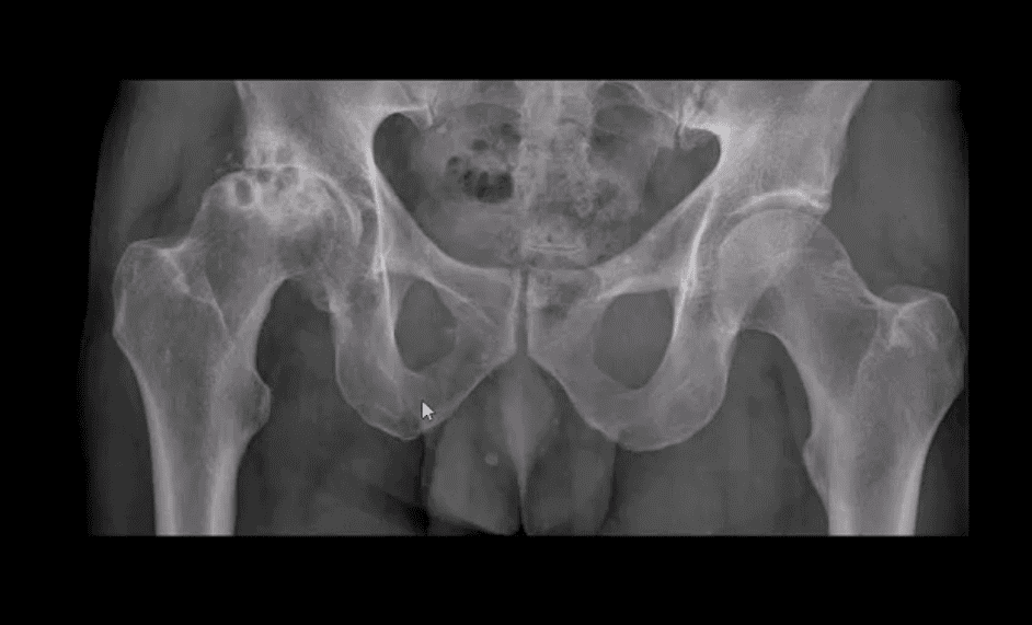
Note multiple subchondral cysts, severe non-uniform joint narrowing (superior-lateral) and subchondral sclerosis with osteophytes
Advanced hip arthrosis
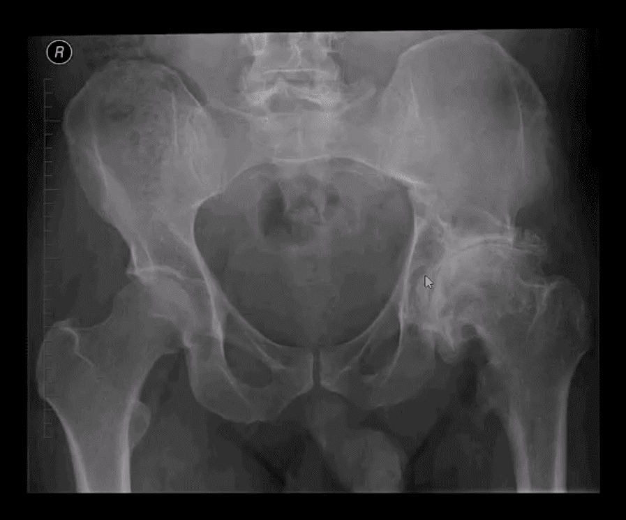
Severe DJD, Left Hip
When reading radiological reports pay particular attention to the grading of hip OA
Most severe (advanced) OA cases require total hip arthroplasty (THA)
Refer your patients to the Orthopedic surgeon for a consultation
Most mild cases are a good candidate for conservative care
Hip Arthroplasty aka Hip Replacement
Can be total or hemiarthroplasty
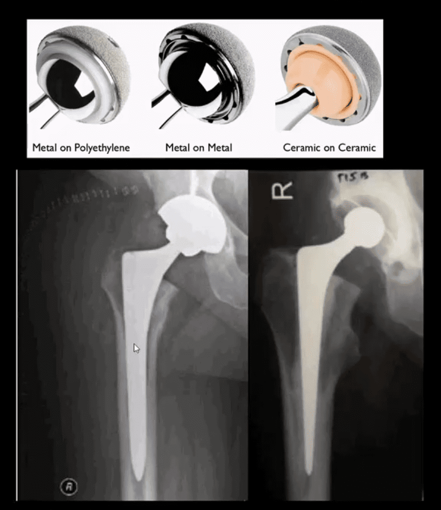
THA can be metal on metal, metal on polyethylene and ceramic on ceramic
A hybrid acetabular component with polyethylene and metal backing is also used (above right image)
THA can be cemented (above right image) and non-cemented (above-left image)
Non-cemented arthroplasty is used on younger patients utilizing porous metallic parts allowing good fusion and bone ingrowth into the prosthesis
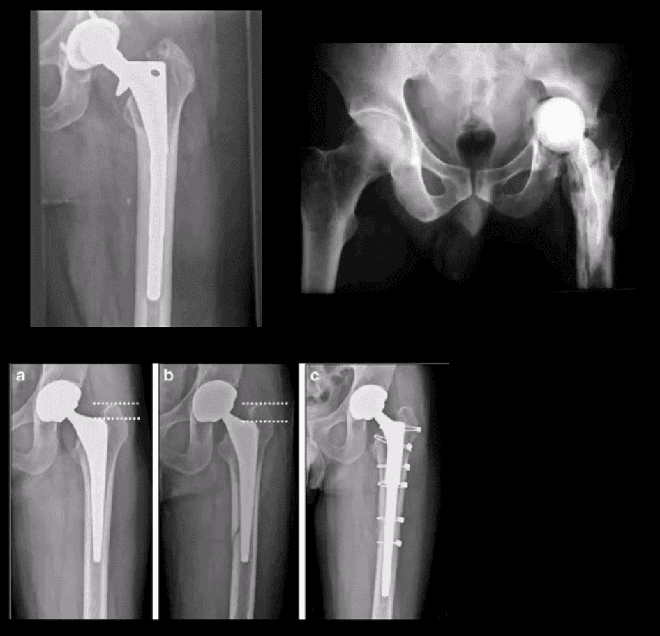
Failed THA May Develop
Most develop within the first year and require revision
Femoral stem may fracture (above left)
Postsurgical infection (above right)
Fracture adjacent to the prosthesis (stress riser)
Particle disease
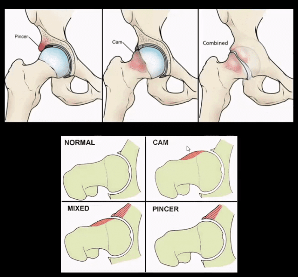
Femoroacetabular Impingement Syndrome
(FAI): abnormality of normal morphology of the hip leading to eventual� cartilage damage and premature DJD
Clinically:�hip/groin pain aggravated by sitting (e.g., hip flexed & externally rotated). Activity related pain on axial loading esp. with hip flexion (e.g., walking uphill)
Pincer-type�acetabulum: > in middle age women potentially� many causes
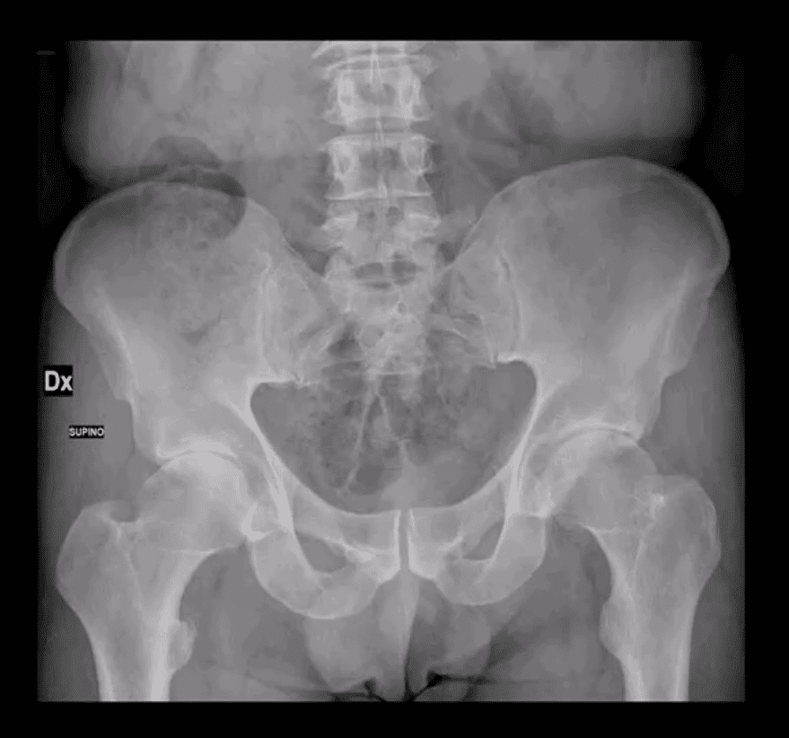
CAM-type deformity:�> in men in 20-50 m/c 30s
Mixed type (pincer-CAM) is most frequent
Up until the 90s, FAI was not well-recognized
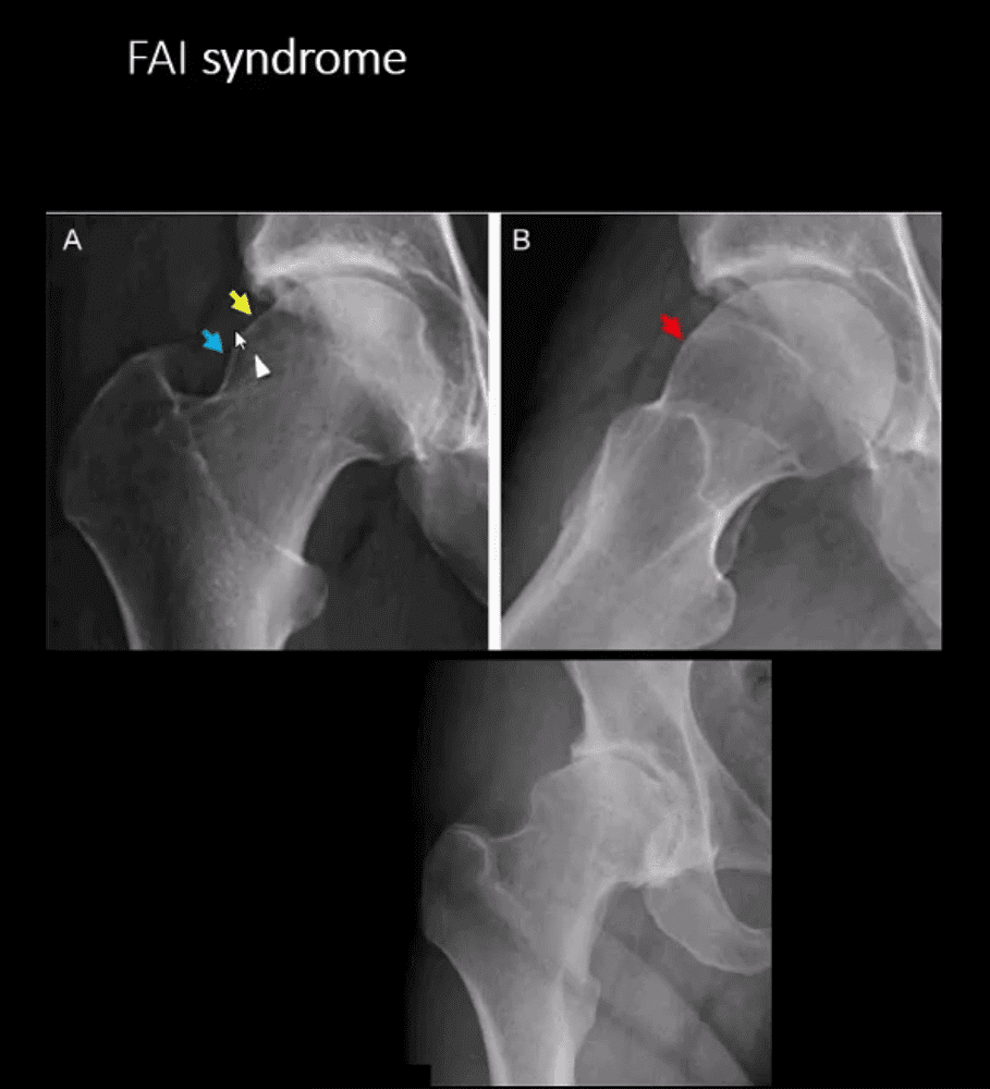
FAI Syndrome
CAM-type FAI syndrome
Radiography can be a reliable Dx tool
X-radiography findings:�osseous bump on the lateral aspect of femoral head-neck junction. Pistol-grip deformity. Loss of normal head sphericity. Associated features: os acetabule, synovial herniation pit (Pit’s pit). Evidence of DJD in advanced cases
MRI and MR arthrography (most accurate Dx of labral tear) can aid the diagnosis of labral tear and other changes of FAI
Referral to the Orthopedic surgeon is necessary to prevent DJD progression and repair labral abnormalities. Late Dx may lead to irreversible changes of DJD
AP Pelvis: B/L CAM-type FAI syndrome
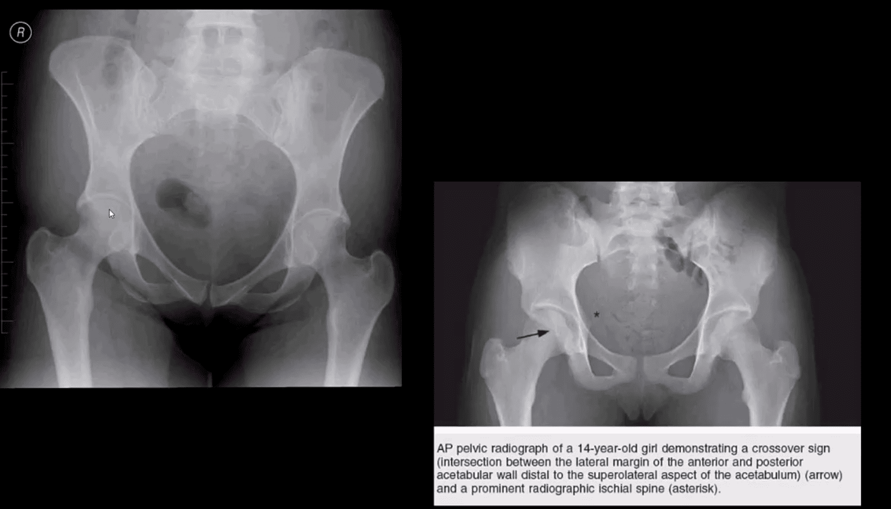
Pincer-Type FAI with Acetabula Over-Coverage
Key radiographic signs: “Cross-over sign” and abnormal center-edge and Alfa-angle evaluation methods
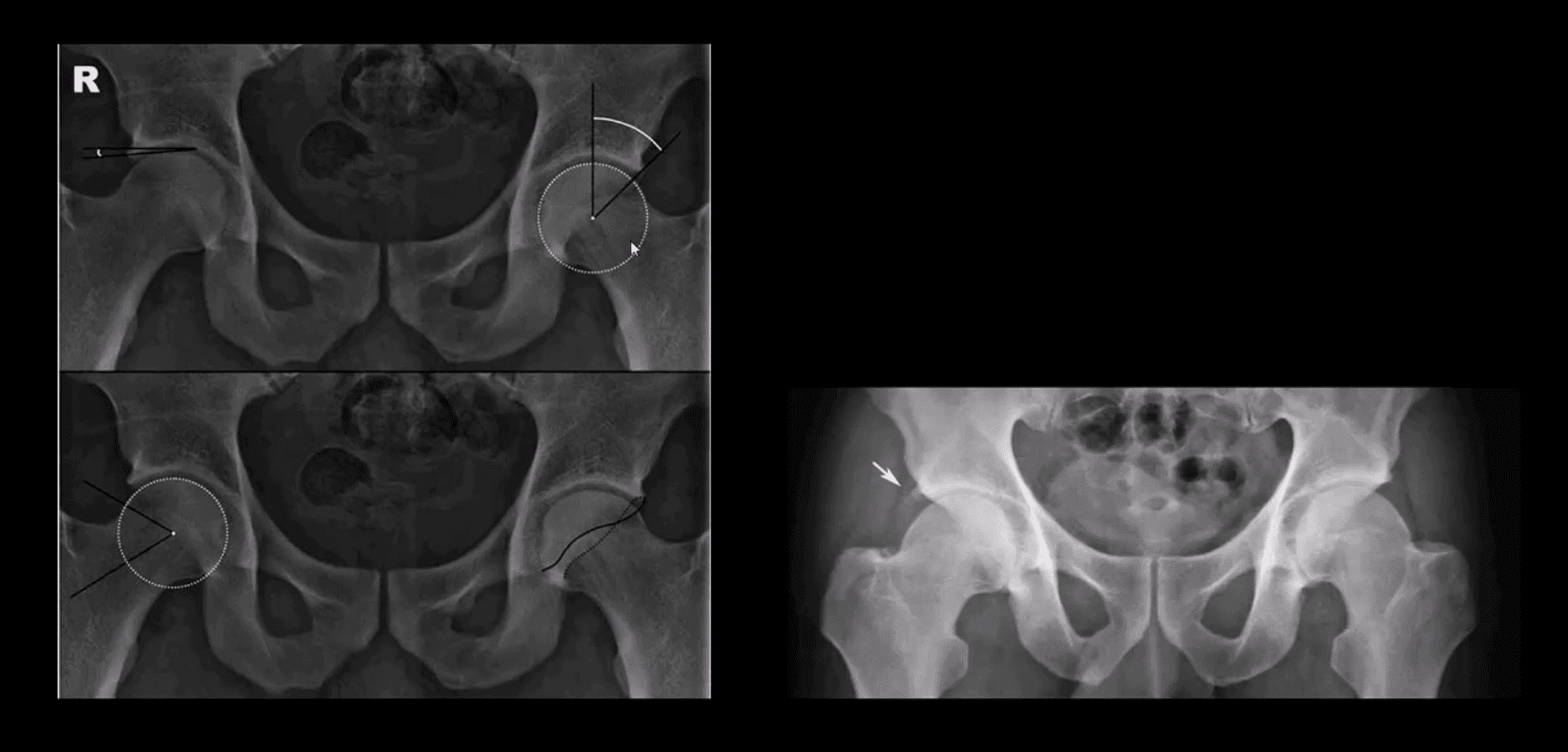
Dx of FAI
Center-edge angle (above the first image) and Alfa-angle (above the second image)
B/L CAM-type FAI with os acetabule�(above right image)
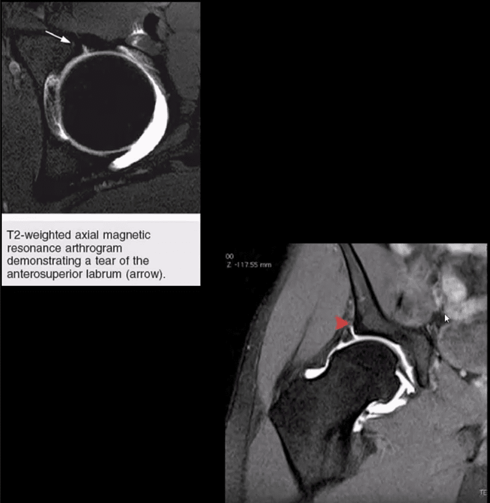
MR Arthrography
Labral tear and CAM-type FAI syndrome on axial (above left) and coronal T2 W (above right) MR arthrography
Note acetabula labral tear. Referral to an orthopedic surgeon is required. For more information:
IFM's Find A Practitioner tool is the largest referral network in Functional Medicine, created to help patients locate Functional Medicine practitioners anywhere in the world. IFM Certified Practitioners are listed first in the search results, given their extensive education in Functional Medicine