Find out why cauliflower is a must-have in your kitchen. From recipes to health benefits, learn all about cauliflower.
Introduction
Throughout their path toward health and wellbeing, many people throughout the globe want to include nutrient-dense meals for energy. Eating a lot of fruits and vegetables is one of the greatest methods for many individuals to do this. In addition to having many vitamins and minerals that the body needs, many fruits and vegetables may help lessen the overlapping pain-like sensations that lead to the development of chronic problems. Problems with the intestines, muscles, and essential organs start to play a part when environmental influences start to become a part of a person’s daily routine, which results in pain-like sensations. In order to facilitate the transition to a healthy lifestyle, many people look for non-surgical therapies that include a personalized treatment plan and nutritional component. One of them is consuming cauliflower, a vegetable. The nutritional benefits of cauliflower, its definition, how it supports the body, and simple dishes to include in a tasty, nutritious diet are all covered in today’s post.
What Is Cauliflower?
Known for being a humble vegetable with its pale appearance, the cauliflower is a powerhouse full of nutrients. It can become the most versatile vegetable to be added to any diet. As part of the Brassicaceae family, cauliflower falls in line with cruciferous vegetables like broccoli, Brussels sprouts, kale, and cabbage that contribute a significant amount of dietary intake to human health due to being low in calories while providing a diverse range of macronutrients. (Zhang et al., 2025) Additionally, cauliflower can come in various colors and be filled with multiple nutrients that benefit the body, including white, green, purple, and orange.
Nutritional Value of Cauliflower
Whether people want to improve their health, lose weight, or add more plant-based variety to their meals, cauliflower deserves a prominent place on their plate. One cup of chopped cauliflower, which is about 107 grams, includes:
Calories: 27
Total Carbohydrates: 5 g
Dietary Fiber: 2.1g
Sugar: 2 g
Protein: 2.1 g
Total Fat: 0.3 g
Saturated Fat: 0.1g
Trans Fat Regulation: 0g
Cholesterol: 0g
Sodium: 32mg
Potassium: 320mg
Vitamin C: 85% of the Daily Value (DV)
Iron: 2% of DV
Vitamin B6: 10% of DV
Magnesium: 4% of DV
Calcium: 2% of DV
Additionally, cauliflower is rich in phytonutrients, particularly glucosinolates and sulforaphane, which have neuroprotective and anticancer agents that can reduce neuroinflammation in the body. (Otoo & Allen, 2023)
Eating Right To Feel Better- Video
How Cauliflower Helps The Body
When it comes to the body, cauliflower can provide various nutrients that the body needs to provide energy and help with normal functioning. Incorporating cauliflower into a healthy diet can help reduce overlapping risk profiles like obesity and hypertension, which can cause individuals to experience pain and discomfort. (Madsen et al., 2023)
Supports Immune Function & Digestive Health
Cauliflower has a high vitamin C content that can help boost the immune defense response by being an antioxidant to protect against cell damage within the body. Since cauliflower is a cruciferous vegetable, it has many phytochemicals, including vitamins, carotenoids, fibers, and other nutrient compounds, to protect the body against reactive oxygen species (ROS) damage. (Ahmed & Ali, 2013) Additionally, with its nutrient components, cauliflower can help support gut health by aiding regular bowel movements. As a cruciferous vegetable, it feeds the beneficial gut bacteria to promote a healthy microbiome balance. This is due to cauliflower containing glucosinolates (GLS), a chemical compound in plants in the Brassicaceae family. GLS, when served in various dishes, contains anti-obesity effects and pro-healthy compounds to benefit the body’s gut system. (Sikorska-Zimny & Beneduce, 2021)
Reduces Inflammation & Oxidative Stress
When inflammation and oxidative stress occur in the body, many people don’t often realize that many environmental factors play a part in their development. While inflammation is the body’s natural response, chronic inflammation can correlate with oxidative stress, causing the body to develop musculoskeletal and gut issues. When oxidative stress is associated with chronic inflammation, it can impact the body’s central nervous system, causing the neuron signals to affect the gut system by increasing bad bacteria in the gastrointestinal system. (Shandilya et al., 2022) Consuming cauliflower in various dishes can help reduce the oxidative damage and inflammation correlated with chronic body issues.
Simple & Delicious Cauliflower Recipes
Since cauliflower has a mild flavor and versatile texture, it can be easily incorporated into numerous dishes that are not only delicious but also nutritious. Below are a few simple and easy recipes that many people can try:
Cauliflower Rice
Cauliflower rice is a low-carb, grain-free substitute for rice
How to make:
Start by grating raw cauliflower with a grater or pulse it in the food processor until it resembles rice grains.
Then, for 5 minutes, add olive oil, garlic, salt, and pepper to the cauliflower grains and sauté in a pan.
You can use various sauces and herbs to enhance the cauliflower rice for extra flavor.
Cauliflower Pizza Crust
Individuals with gluten allergies or sensitivity can make this pizza crust with cauliflower.
How to make:
Start by steaming and mashing a head of cauliflower, transferring it to cheesecloth, and squeezing out the excess water.
Then, add one egg, shredded mozzarella, seasoning, and cauliflower to a mixing bowl and mix. This should resemble dough.
Take the cauliflower dough and shape it into a crust.
Bake at 400°F (200°C) for at least 15 minutes, then remove it from the oven.
Then add your favorite toppings on the cauliflower pizza crust, and bake again.
Cauliflower Buffalo Bites
As a healthier alternative to chicken wings, they can be perfect for your next party.
How to make:
Toss cauliflower florets in flour and spices, and bake at 425°F (220°C) for 20 minutes.
Toss in buffalo sauce and bake for another 10 minutes. Serve with celery and light ranch.
Final Thoughts
More than just a simple white vegetable, cauliflower is a superfood full of nutrients and antioxidants that can be used in a variety of recipes and diets. A nutritious diet that includes cauliflower may provide the body with the vital vitamins, antioxidants, and nutrients it needs to function at its best. This cruciferous vegetable gives the body the energy it needs and may be a great way to add veggies to a nutritious meal.
Injury Medical & Functional Medicine Clinic
We associate with certified medical providers who understand the importance of assessing individuals and incorporating cauliflower into their dietary needs. When asking important questions to our associated medical providers, we advise patients to incorporate numerous vegetables like cauliflower into their foods to provide energy and nutrients to their bodies in their customized treatment plan. Dr. Alex Jimenez, D.C., uses this information as an academic service. Disclaimer.
References
Ahmed, F. A., & Ali, R. F. M. (2013). Bioactive Compounds and Antioxidant Activity of Fresh and Processed White Cauliflower. BioMed Research International, 2013, 1-9. https://doi.org/10.1155/2013/367819
Madsen, H., Sen, A., & Aune, D. (2023). Fruit and vegetable consumption and the risk of hypertension: a systematic review and meta-analysis of prospective studies. Eur J Nutr, 62(5), 1941-1955. https://doi.org/10.1007/s00394-023-03145-5
Otoo, R. A., & Allen, A. R. (2023). Sulforaphane’s Multifaceted Potential: From Neuroprotection to Anticancer Action. Molecules, 28(19), 6902. https://doi.org/10.3390/molecules28196902
Shandilya, S., Kumar, S., Kumar Jha, N., Kumar Kesari, K., & Ruokolainen, J. (2022). Interplay of gut microbiota and oxidative stress: Perspective on neurodegeneration and neuroprotection. J Adv Res, 38, 223-244. https://doi.org/10.1016/j.jare.2021.09.005
Sikorska-Zimny, K., & Beneduce, L. (2021). The Metabolism of Glucosinolates by Gut Microbiota. Nutrients, 13(8), 2750. https://doi.org/10.3390/nu13082750
What is pseudoarthrosis of the cervical and lumbar spine?
Pseudoarthrosis of the cervical and lumbar spine
Individuals may need a spinal fusion to treat a fractured vertebra, scoliosis, or conditions like spinal stenosis, degenerative disc disease, and spondylolisthesis/slipped vertebrae. A spinal fusion reduces pain and stabilizes the spine by limiting movement between vertebrae. Pseudoarthrosis happens when the bones don’t heal after a fracture or bone surgery. When pseudoarthrosis affects the cervical or lumbar spine, it means that two vertebrae did not heal and grow together after spinal surgery to fuse them (spinal fusion). Reasons for a failed spinal fusion include:
Issues with the instruments used to stabilize the bone
Lack of bone growth
The number of vertebrae being fused.
The patient’s health and lifestyle play a role in failed fusions, which can include
Diabetes
Inflammatory health conditions increase the risk
Smoking
Long-term steroid use
In many cases, revision surgery is needed.
Surgery-Related
During a spinal fusion, surgeons insert a bone graft between two vertebrae and then apply spinal fixation hardware (instrumented spinal fusion) that includes:
Plates
Rods
Screws
The bone graft promotes growth between the two bones.
The hardware stabilizes the vertebrae and prevents movement while they fuse and grow together.
The hardware goes inside, or internal fixation.
Although rare, a severe spinal fracture or deformity may need external fixation.
A rigid frame secured outside the body helps to stabilize the bones.
If the fusion fails, it could be caused by one or more of the following surgical issues:
The surgeon must carefully plan and use the right hardware.
The type of hardware used during a spinal fusion may influence bone healing.
The instruments can come loose or break, interfering with the fusion process.
Spinal osteoporosis, having thin, weak bones, can affect fixation.
Even with the optimal surgical preparedness, weak bones significantly increase the chance of the instruments loosening and pseudoarthrosis developing.
Bone Graft
The type of bone graft used may affect the fusion.
For example, in cervical/neck spinal fusions, an autograft, which uses a small piece of bone from the patient’s body, has a higher success rate. (Verla T. et al., 2021)
Other graft options include specialized steel cages that fit between vertebrae and contain bone growth factors.
The surgeon recommends the optimal bone graft for the type of surgery, the number of vertebrae involved, and risk factors.
Risk Factors
The patient’s overall health and lifestyle impact the results of spinal fusion. Smoking increases the risk. (Berman D. et al., 2017)
Nicotine restricts blood circulation, decreases bone density, reduces new bone formation, and delays bone healing. (Hernigou J., & Schuind F., 2019)
The primary sign of pseudoarthrosis is pain in the same area as before the fusion surgery.
If the bones pinch a spinal nerve, one arm may experience pain, tingling, burning, or numbness.
Rarely does a pinched nerve affect both arms.
The pain may return shortly after the procedure.
The pain may develop gradually or not appear for many months.
However, it’s more likely to appear after several months when the individual returns to their usual activities.
Diagnosis
The healthcare provider will learn about symptoms and perform a physical exam to evaluate the back.
They’ll assess mobility and the type of movement that causes pain.
Then, they order diagnostic imaging to see the spine and identify the cause of pain.
Individuals may need a CT scan, MRI, and/or X-rays to evaluate the spinal structures and instrumentation fully.
Treatment
Treatment for pseudoarthrosis will likely start with:
Physical therapy
Pain management – especially in cases where it is important to rule out other sources of back or neck pain.
Medication
Injections
If symptoms don’t improve with conservative care or if there is severe pain, the healthcare provider may recommend revision surgery.
Revision surgery is another procedure to treat complications or correct issues that arise after the initial pseudoarthrosis surgery.
Injury Medical Chiropractic and Functional Medicine Clinic
As a Family Practice Nurse Practitioner, Dr. Jimenez combines advanced medical expertise with chiropractic care to address various conditions. Our clinic integrates Functional Medicine, Acupuncture, Electro-Acupuncture, and Sports Medicine to create customized care plans that promote natural healing, mobility, and long-term wellness. By focusing on flexibility, agility, and strength, we empower patients to thrive, regardless of age or health challenges. At El Paso’s Chiropractic Rehabilitation Clinic & Integrated Medicine Center, we passionately focus on treating patients after injuries and chronic pain syndromes. We focus on improving your ability through flexibility, mobility, and agility programs tailored for all age groups and disabilities. We use in-person and virtual health coaching and comprehensive care plans to ensure every patient’s personalized care and wellness outcomes.
Enhancing Health Together
References
Boonsirikamchai, W., Wilartratsami, S., Ruangchainikom, M., Korwutthikulrangsri, E., Tongsai, S., & Luksanapruksa, P. (2024). Pseudarthrosis risk factors in lumbar fusion: a systematic review and meta-analysis. BMC musculoskeletal disorders, 25(1), 433. https://doi.org/10.1186/s12891-024-07531-w
Verla, T., Xu, D. S., Davis, M. J., Reece, E. M., Kelly, M., Nunez, M., Winocour, S. J., & Ropper, A. E. (2021). Failure in Cervical Spinal Fusion and Current Management Modalities. Seminars in plastic surgery, 35(1), 10–13. https://doi.org/10.1055/s-0041-1722853
Berman, D., Oren, J. H., Bendo, J., & Spivak, J. (2017). The Effect of Smoking on Spinal Fusion. International journal of spine surgery, 11(4), 29. https://doi.org/10.14444/4029
Hernigou, J., & Schuind, F. (2019). Tobacco and bone fractures: A review of the facts and issues that every orthopaedic surgeon should know. Bone & joint research, 8(6), 255–265. https://doi.org/10.1302/2046-3758.86.BJR-2018-0344.R1
Scoliosis Research Society. (2023). Pseudoarthrosis. https://www.srs.org/Patients/Conditions/Pseudoarthrosis
Torres, H. M., Arnold, K. M., Oviedo, M., Westendorf, J. J., & Weaver, S. R. (2023). Inflammatory Processes Affecting Bone Health and Repair. Current osteoporosis reports, 21(6), 842–853. https://doi.org/10.1007/s11914-023-00824-4
Jiao, H., Xiao, E., & Graves, D. T. (2015). Diabetes and Its Effect on Bone and Fracture Healing. Current osteoporosis reports, 13(5), 327–335. https://doi.org/10.1007/s11914-015-0286-8
Many ask, what and when should you eat before, during, and after engaging in Pilates exercises?
Pilates Nutrition Plan
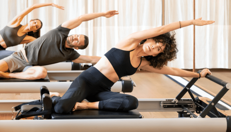
Pilates involves a lot of core work on the mat. However, it does not require a special diet. To fully utilize Pilates nutrition, individuals should consider what foods keep their bodies feeling the most balanced. A balanced nutrition plan can enhance Pilates practice by:
Providing sustained energy
Supporting muscle recovery
Promoting overall well-being
Pre-Pilates
Timing
Eat a light meal or snack 2-3 hours before a class or a smaller snack 30 minutes before.
Food Options
Eat complex carbohydrates like whole grains, fruits, and vegetables for sustained energy.
Avoid
Heavy, greasy, or spicy foods may cause discomfort during exercise.
Foods that will make you gassy or cause queasiness.
During the Workout
Complex carbohydrates and lean proteins, with a little healthy fat, are good pre-Pilates meals, as they sustain energy better than simple carbs or sugary snacks. (American Heart Association, 2024) Suggestions for a pre-Pilates meal.
Individuals can adjust the portion size.
A protein shake that uses fruit can be convenient.
Peanut butter on whole-grain bread offers a quick source of complex carbohydrates and protein.
Yogurt with fruit.
Small portion of oatmeal.
Because Pilates emphasizes using the abdominal muscles, individuals will want to ensure that any food they’ve eaten before their session is fully digested. Try to eat light before, like snacking on a banana or sipping on a smoothie for some carbohydrates for energy. Regardless of what is eaten, nutrition experts recommend waiting two to three hours after eating before exercising.
Maintain Hydration
Drink water and/or eat hydrating fruits throughout the day, especially before and during the workout or class.
After Working Out
Individuals want to ensure their bodies have enough nutrients to strengthen their muscles and replenish their energy. Try a protein green smoothie afterward or a light snack with lean protein like fish or chicken and carbohydrates like whole grains. The diet between workouts will depend on weight loss goals.
Timing
Consume a protein-rich snack or meal within 30-60 minutes after your class to aid muscle recovery.
Food Choices
Focus on protein for muscle repair and carbohydrates to replenish energy stores.
Hydration
Continue to hydrate to replenish fluids lost through perspiration.
While Pilates can complement cardiovascular exercise as part of a fat-burning workout program, health experts advise that losing weight will not come from exercise alone. Individuals will need to work on reducing their calorie intake overall. (Centers for Disease Control and Prevention, 2023)
Injury Medical Chiropractic and Functional Medicine Clinic
As a Family Practice Nurse Practitioner, Dr. Jimenez combines advanced medical expertise with chiropractic care to address various conditions.
Wellness & Nutrition: Personalized plans to optimize health and prevent disease.
Sports Injuries & Orthopedic Care: Treatment for sprains, strains, and complex injuries.
Chronic Pain Management: Non-invasive solutions for fibromyalgia, sciatica, and low back pain.
Personal Injury & Auto Accident Care: Tailored rehabilitation for whiplash, soft tissue injuries, and more.
Functional Medicine: Root-cause analysis for chronic disorders, incorporating nutrition, lifestyle, and environmental factors.
Neuromusculoskeletal Health: Care for neck pain, migraines, herniated discs, and scoliosis.
Our clinic integrates Functional Medicine, Acupuncture, Electro-Acupuncture, and Sports Medicine to create customized care plans that promote natural healing, mobility, and long-term wellness. By focusing on flexibility, agility, and strength, we empower patients to thrive, regardless of age or health challenges. At El Paso’s Chiropractic Rehabilitation Clinic & Integrated Medicine Center, we passionately focus on treating patients after injuries and chronic pain syndromes. We focus on improving your ability through flexibility, mobility, and agility programs tailored for all age groups and disabilities. We use in-person and virtual health coaching and comprehensive care plans to ensure every patient’s personalized care and wellness outcomes.
Home Exercises for Pain Relief
References
American Heart Association. (2024). Food as fuel before, during, and after workouts. https://www.heart.org/en/healthy-living/healthy-eating/eat-smart/nutrition-basics/food-as-fuel-before-during-and-after-workouts
Centers for Disease Control and Prevention. (2023). Tips for Maintaining Healthy Weight. Retrieved from https://www.cdc.gov/healthy-weight-growth/about/tips-for-balancing-food-activity.html?CDC_AAref_Val=https://www.cdc.gov/healthyweight/calories/index.html
Can understanding the mechanics of the throwing motion help to understand why it may cause shoulder pain, the symptoms of a shoulder problem, the diagnosis, and the treatment options available?
Throwing a Ball and Shoulder Pain
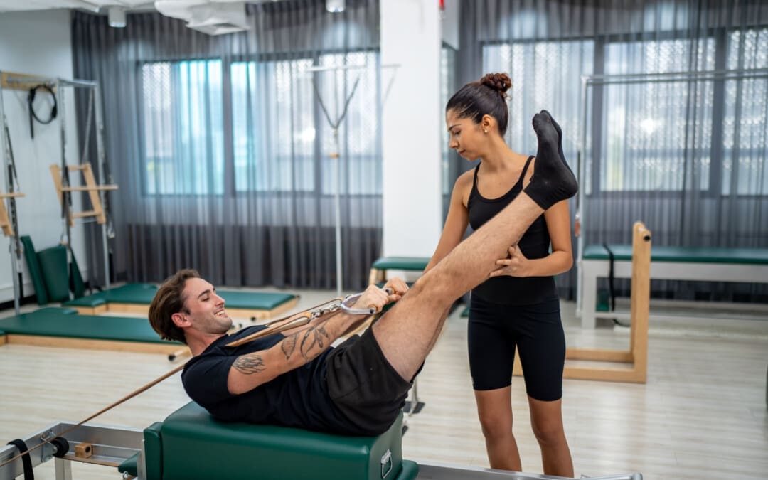
The throwing motion is a complex shoulder movement that requires the mechanics of muscles, tendons, joints, ligaments, and bones. They all must move in a synchronized and stable pattern to move the shoulder joint. When these mechanics are interrupted or altered, inflammation can result in pain symptoms. (Wardell M., Creighton D., & Kovalcik C., 2022)
The labrum stabilizes the ball in the socket of the shoulder.
The shoulder blade rotation coordinates with the arm to ensure mobility. (Itoigawa Y. et al., 2023)
The throwing motion generates high torque and acceleration forces acting on the shoulder joint and the surrounding muscles, ligaments, and tendons.
Causes of Pain
Pain when throwing can come from the:
Shoulder blade
Shoulder joint – cartilage and labrum
Rotator cuff muscles and tendons
Nerves that control the muscles’ function
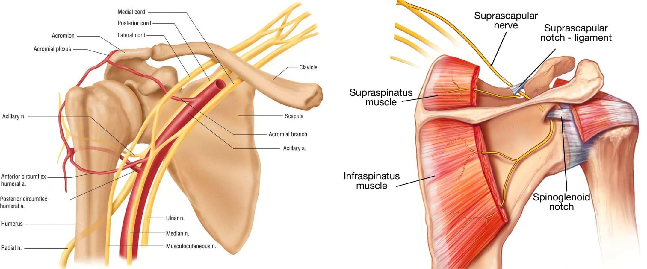
The shoulder blade is attached to the upper back by ligaments, muscles, and tendons. The various muscles and tendons that control the movement of the shoulder blade impact movements. Abnormalities of any area can lead to shoulder dysfunction and pain when throwing. (Wardell M., Creighton D., & Kovalcik C., 2022) The most common is the tightness of the posterior shoulder capsule, causing a loss of normal internal rotation of the shoulder. If this is causing pain, individuals may notice that they can’t reach up as high on the side with the painful shoulder when reaching behind their back.
Symptoms
Whether an athlete or playing catch in the backyard, shoulder function abnormalities can cause significant pain. Some symptoms include.
Aching Pain
Often deep in the shoulder or extending down the upper arm.
Dead Arm
Lack of strength in the throwing motion.
Pain at Night
Pain can awaken you from sleep.
Diagnosis
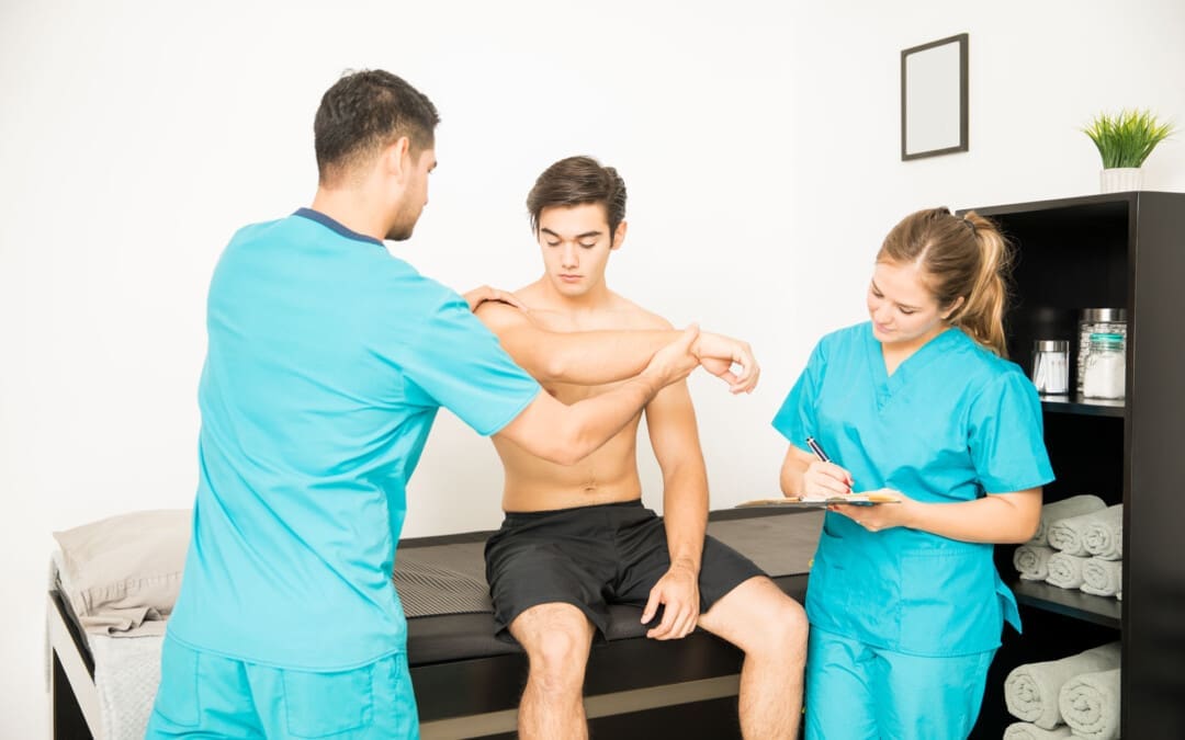
Finding a healthcare provider familiar with sports injuries can be helpful. They can best determine if a structural abnormality needs to be addressed. (American Academy of Orthopaedic Surgeons, 2021)
Treatment
Most can improve with nonsurgical treatments. The earliest phase of treatment is resting the joint and reducing inflammation. Treatments can include:
Ice
Anti-inflammatory medications
Cortisone injection
Once the inflammation has subsided, the source of the discomfort can be addressed.
Physical Therapy
Therapy can include:
A structured shoulder stretching and strengthening program will help.
The physical therapist will focus on scapular mobility when managing shoulder joint problems.
Stretching to improve internal rotation or any other lost motion can help allow a more normal throwing motion.
Strength exercises are often aimed at the rotator cuff, as these muscles initiate proper shoulder movements and stabilize the shoulder joint.
Maintaining flexibility and strength of the periscapular muscles (muscles that attach to the scapula bone) is important to ensure that the scapular movements are coordinated with the throwing motion.
If improvements are not made within three months of therapy, or individuals can’t return to competitive sports within six months. In that case, the individual may need to return to their healthcare provider or see an orthopedic specialist who may recommend surgery. (American Academy of Orthopaedic Surgeons, 2024)
Injury Medical Chiropractic and Functional Medicine Clinic
As a Family Practice Nurse Practitioner, Dr. Jimenez combines advanced medical expertise with chiropractic care to address various conditions.
Wellness & Nutrition: Personalized plans to optimize health and prevent disease.
Chronic Pain Management: Non-invasive solutions for fibromyalgia, sciatica, and low back pain.
Personal Injury & Auto Accident Care: Tailored rehabilitation for whiplash, soft tissue injuries, and more.
Sports Injuries & Orthopedic Care: Treatment for sprains, strains, and complex injuries.
Functional Medicine: Root-cause analysis for chronic disorders, incorporating nutrition, lifestyle, and environmental factors.
Neuromusculoskeletal Health: Care for neck pain, migraines, herniated discs, and scoliosis.
Our clinic integrates Functional Medicine, Acupuncture, Electro-Acupuncture, and Sports Medicine to create customized care plans that promote natural healing, mobility, and long-term wellness. By focusing on flexibility, agility, and strength, we empower patients to thrive, regardless of age or health challenges.
At El Paso’s Chiropractic Rehabilitation Clinic & Integrated Medicine Center, we passionately focus on treating patients after frustrating injuries and chronic pain syndromes. We focus on improving your ability through flexibility, mobility, and agility programs tailored for all age groups and disabilities. We use in-person and virtual health coaching and comprehensive care plans to ensure every patient’s personalized care and wellness outcomes.
Lumbar Spine Injuries in Sports: Chiropractic Healing
References
Wardell, M., Creighton, D., & Kovalcik, C. (2022). Glenohumeral Instability and Arm Pain in Overhead Throwing Athletes: A Correlational Study. International journal of sports physical therapy, 17(7), 1351–1357. https://doi.org/10.26603/001c.39800
Itoigawa, Y., Koga, A., Morikawa, D., Kubota, A., Uehara, H., Maruyama, Y., Takazawa, Y., & Ishijima, M. (2023). Posterior shoulder stiffness was associated with shoulder pain during throwing in college baseball players: assessment of shear wave elastography. European journal of orthopaedic surgery & traumatology: orthopedie traumatologie, 33(4), 1237–1244. https://doi.org/10.1007/s00590-022-03286-z
American Academy of Orthopaedic Surgeons. (2021). Shoulder Injuries in the Throwing Athlete. https://orthoinfo.aaos.org/en/diseases–conditions/shoulder-injuries-in-the-throwing-athlete/
American Academy of Orthopaedic Surgeons. (2024). Shoulder Impingement/Rotator Cuff Tendinitis. https://orthoinfo.aaos.org/en/diseases–conditions/shoulder-impingementrotator-cuff-tendinitis
Should individuals experiencing nerve pain or various sensations get a nerve conduction velocity study to examine nerve health and function?
Nerve Conduction Velocity
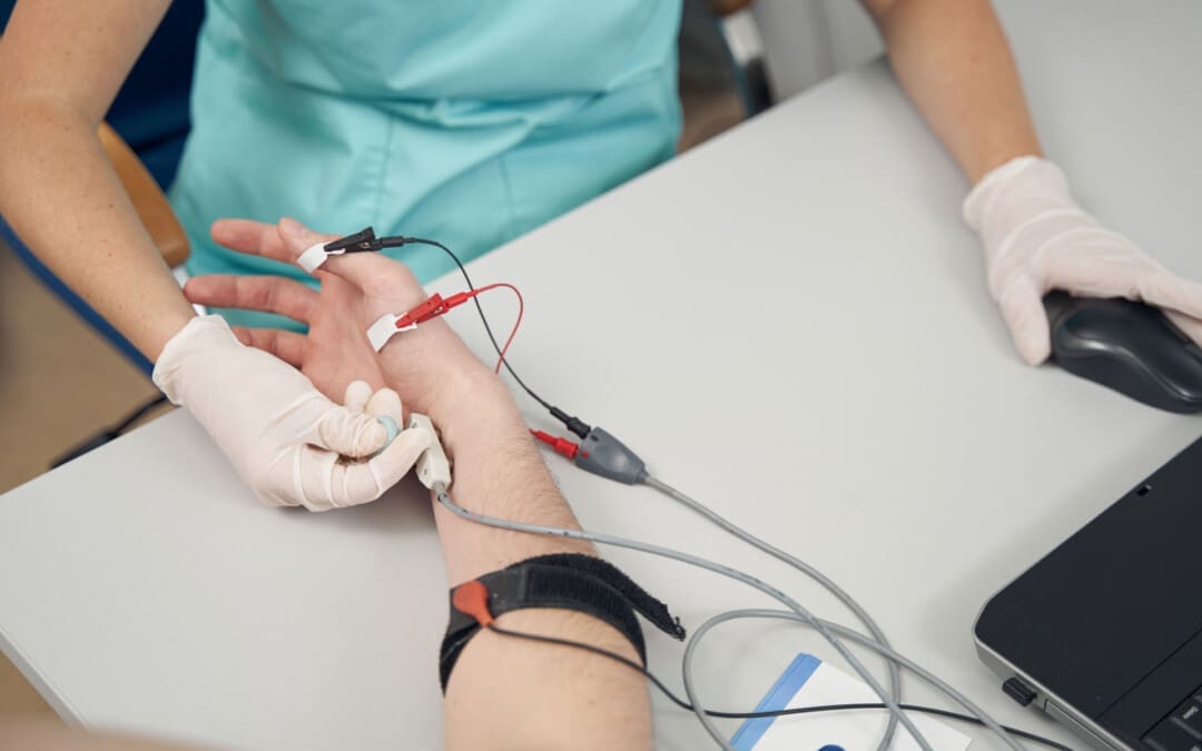
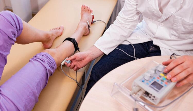
A nerve conduction velocity (NCV) is a noninvasive test that measures the speed and strength of nerve stimulation using electrical probes placed on the skin. It’s used to diagnose nerve damage or disease, often alongside an EMG (electromyogram) to differentiate between nerve and muscle problems. It can also evaluate sensory issues, pain, and weakness of the extremities.
This test involves safe electrical shocks that can be slightly uncomfortable but not painful.
Nerve conduction velocity (NCV) measures the speed at which electrical impulses travel along a nerve fiber, which measures how quickly electrical signals travel through a nerve.
This information indicates nerve health and function.
Electromyography (EMG) is a nerve test that involves placing tiny needles into the muscles.
A slower NCV can indicate nerve injury or dysfunction.
Test Uses
Generally, the test is ordered to assess peripheral nerve diseases, those that connect from the muscles, organs, and skin to the spinal cord or brain. It can help identify the type and location of nerve damage.
Peripheral nerve conditions typically cause pain, sensory loss, tingling, or burning.
Mild weakness and diminished reflexes can be detected during a neurological examination.
Conditions
Nerve conduction studies are performed to help diagnose conditions.
Nerve damage (neuropathy), such as from diabetes, chemotherapy, or autoimmune disorders
Charcot-Marie-Tooth disease
Nerve compression
Many different conditions, including trauma, inflammation, and tumors, can compress one or more nerves.
Radiculopathy
Often described as a pinched nerve, radiculopathy can affect an arm or a leg, causing pain and weakness.
Peripheral Neuropathy
This nerve damage begins in the most distal nerves, those farthest from the center of the body, such as the toes and fingers. It is often due to chronic alcohol misuse, uncontrolled diabetes, nutritional deficits, and inflammatory diseases. (Ferdousi M. et al., 2020)
Carpal Tunnel Syndrome
Commonly caused by inflammatory diseases or overuse of the wrists, such as from assembly line work, carpal tunnel syndrome causes numbness, pain, and weakness of the fingers and hands. (Tada K. et al., 2022)
Ulnar neuropathy
This common condition causes arm pain and sensory changes, usually due to repetitive movements or a prolonged position that causes pressure on the ulnar nerve.
Guillain-Barré syndrome (GBS)
This inflammatory condition causes demyelination, or loss of the insulating covering around nerves, which results in leg weakness.
It begins in the motor nerves, which send signals to muscles in the legs. (Shibuya K. et al., 2022)
The inflammation travels to nerves of the upper body, often affecting the muscles that control breathing.
Respiratory support is necessary until the condition improves.
Chronic Demyelinating Polyneuropathy (CIDP)
This condition is a chronic, recurrent form of GBS that usually affects the legs and causes episodes of weakness.
ICU neuropathy
Metabolic changes, severe illness, and not moving enough can cause nerves to develop a pattern of weakness and sensory loss.
Myasthenia gravis (MG)
This autoimmune condition affects the junction between the nerves and the muscles.
Myasthenia gravis causes drooping eyelids and weakness of the arms and shoulders.
Amyotrophic lateral sclerosis (ALS)
ALS is a serious, degenerative disease affecting the spinal cord’s motor neurons.
Amyotrophic lateral sclerosis progresses rapidly, resulting in substantial weakness of muscles throughout the body.
How it’s Done
Surface electrodes are placed on the skin over nerves, and a small electrical current is applied to stimulate the nerve.
The time it takes for the electrical signal to travel between the electrodes is measured, and this time is used to calculate the NCV.
Values
Normal NCV values are generally between 50 and 70 meters per second. However, these values can vary depending on the nerve and the individual.
NCV Factors
Various factors can influence NCV.
Age
Sex
Medical conditions like diabetes
Interpretation
A slower NCV can indicate nerve damage or demyelination (loss of the myelin sheath, which insulates nerve fibers), while an EMG can help determine if the problem is with the nerve or the muscle.
Results
The results of NCV testing can be used to determine the type, severity, and location of nerve damage. The results will be ready in report form about a week after the test.
The test measures velocity (how fast a nerve transmits signals) and amplitude (how many nerve fibers were activated). (Tavee J. 2019)
The measurements are transmitted to a computer and shown as waves and numerical values.
The values are compared to a standard measurement based on the tested nerve.
The distance between the electrodes.
The person’s age.
Compared to the standard, the NCV results can identify certain patterns of nerve damage. (Tada K. et al., 2022) Outcomes include: (Tavee J. 2019)
If one or more nerves are affected.
If motor nerves (control movement), sensory nerves (transmit sensory signals), or both are affected.
Whether a nerve is blocked or damaged.
The severity of the damage.
The type of nerve damage
Axonal (damage to the nerve itself)
Demyelination (damage to the protective fatty layer around the nerve)
The results can help point to certain diagnoses.
Preparation Before the Test
Individuals will not need to change their diet before having an NCV. However, patients will be asked to avoid lotions or creams on their skin before the test. Individuals who are also having an EMG at the time of their NCV might be asked to stop taking medications or supplements that increase the risk of bleeding and bruising. If a healthcare provider says not to stop taking the medicines for health reasons, the patient might be warned that they could have some bruising after the EMG test.
NCV may advise against getting the test for those with electrical device implants.
Make sure your healthcare providers are aware of your whole medical history.
Injury Medical Chiropractic & Functional Medicine Clinic
Injury Medical Chiropractic and Functional Medicine Clinic works with primary healthcare providers and specialists to develop an optimal health and wellness solution. We focus on what works for you to relieve pain, restore function, and prevent injury. Regarding musculoskeletal pain, specialists like chiropractors, acupuncturists, and massage therapists can help mitigate the pain through spinal adjustments that help the body realign itself. They can also work with other medical professionals to integrate a treatment plan to resolve musculoskeletal issues.
Peripheral Neuropathy and Chiropractic Care
References
Ferdousi, M., Kalteniece, A., Azmi, S., Petropoulos, I. N., Worthington, A., D’Onofrio, L., Dhage, S., Ponirakis, G., Alam, U., Marshall, A., Faber, C. G., Lauria, G., Soran, H., & Malik, R. A. (2020). Corneal confocal microscopy compared with quantitative sensory testing and nerve conduction for diagnosing and stratifying the severity of diabetic peripheral neuropathy. BMJ open diabetes research & care, 8(2), e001801. https://doi.org/10.1136/bmjdrc-2020-001801
Tada, K., Murai, A., Nakamura, Y., Nakade, Y., & Tsuchiya, H. (2022). In Carpal Tunnel Syndrome, Sensory Nerve Conduction Velocities Are Worst in the Middle Finger Than in the Index Finger. Frontiers in Neurology, 13, 851108. https://doi.org/10.3389/fneur.2022.851108
Shibuya, K., Tsuneyama, A., Misawa, S., Suzuki, Y. I., Suichi, T., Kojima, Y., Nakamura, K., Kano, H., Ohtani, R., Aotsuka, Y., Morooka, M., Prado, M., & Kuwabara, S. (2022). Different patterns of sensory nerve involvement in chronic inflammatory demyelinating polyneuropathy subtypes. Muscle & Nerve, 66(2), 131–135. https://doi.org/10.1002/mus.27530
Tavee J. (2019). Nerve conduction studies: Basic concepts. Handbook of Clinical Neurology, 160, 217–224. https://doi.org/10.1016/B978-0-444-64032-1.00014-X
What is a bone density test, how is it performed, and what do the results mean?
Bone Density Test
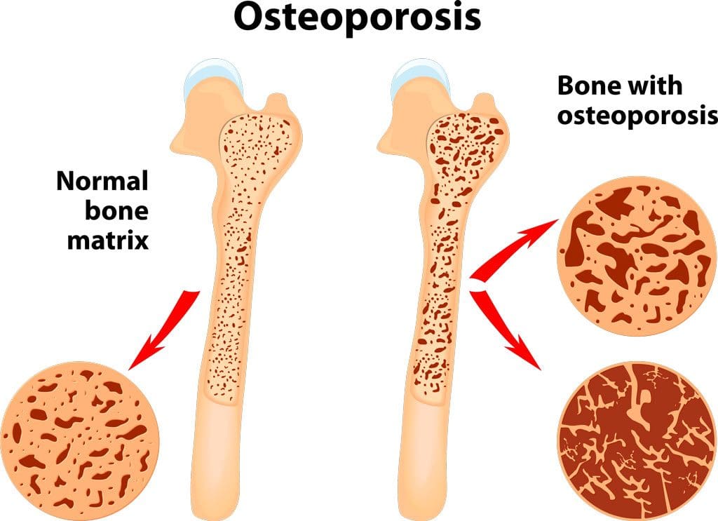
A bone density test examines bone mass, which indicates overall bone strength. Assessing bone density or mass is necessary for diagnosing osteopenia or osteoporosis, conditions that increase the risk of broken bones. The scan is performed through dual-energy X-ray absorptiometry (DEXA), which examines the thickness of the bones. Results from DEXA scans are compared to standardized values to determine whether bone density is lower than normal and whether osteopenia or osteoporosis is present.
Examination
The procedure examines bone density, or bone mass. The bones’ density, or mass, is an overall indicator of bone strength. The greater the bone density, the thicker and stronger the bones are. The test is used to diagnose osteoporosis, a condition characterized by brittle bones at risk of breaking due to significantly low bone density. A bone density test can also diagnose osteopenia, a condition characterized by lower than normal bone mass that can lead to osteoporosis. (National Institute of Arthritis and Musculoskeletal and Skin Diseases, 2025) It is recommended that all women aged 65 and older and all men aged 70 and older have a bone density scan to screen for bone loss to help prevent fractures. (Kling J. M., Clarke B. L., & Sandhu N. P. 2014)
Bone density scans can establish a baseline level of bone density and track changes over time.
For individuals with osteoporosis or osteopenia, a bone density scan can help track how well their bones respond to treatment.
During a DEXA scan, the patient will lie on their back on a table with their legs elevated on a padded platform.
An X-ray scanner will pass over the spine and hips while another scans beneath.
While the scan takes place, the patient will be asked to hold very still to obtain an accurate image.
The scan will obtain bone density readings from the spine and hip, the two most commonly fractured bones, and generally takes less than 30 minutes.
Results
A DEXA scan measures bone density in grams per centimeter squared (g/cm²). This number indicates how densely bone cells are packed together in a specific area of bone. This bone density reading is then compared to a standardized value to determine if bone density is within a normal range or lower than average.
Between minus 1.0 and minus 2.5: Low bone density (osteopenia)
Equal to minus 2.5 or below: Osteoporosis
Bone density values are reported as a Z score for women who have not undergone menopause and men under 50 years old.
Z scores are compared to bone density levels of individuals of the same age and sex.
A Z score of minus 2.0 or lower indicates low bone density, which can be caused by factors other than aging, such as medication side effects, nutritional deficiencies, or thyroid problems.
Arthritis Diagnosis
Because a DEXA scan only measures the thickness of bones, it doesn’t work to diagnose arthritis. An X-ray of the affected joint is currently the most accurate way to diagnose arthritis. The Kellgren-Lawrence classification system categorizes the extent of arthritis based on the severity of joint damage seen on an X-ray. According to this system, arthritis can be classified as: (Kohn M. D., Sassoon A. A., & Fernando N. D. 2016)
Grade 1 (minor)
Minimal or no joint space narrowing, with possible bone spur formation.
Grade 2 (mild)
Possible joint space narrowing, with definite bone spur formation.
Grade 3 (moderate)
Definite joint space narrowing, moderate bone spur formation, mild sclerosis (abnormal thickening of bone), and possible deformation of bone ends.
Grade 4 (severe)
Severe joint space narrowing, large bone spur formation, marked sclerosis, and definite deformation of bone ends.
Injury Medical Chiropractic & Functional Medicine Clinic
Exercise can be incredibly beneficial for improving bone density, joint mobility, and the strength of surrounding muscles, which support and protect joints and bones. Talk to a healthcare provider to learn what interventions and available treatment options would be the most effective. Injury Medical Chiropractic and Functional Medicine Clinic works with primary healthcare providers and specialists to develop an optimal health and wellness solution. We focus on what works for you to relieve pain, restore function, and prevent injury. Regarding musculoskeletal pain, specialists like chiropractors, acupuncturists, and massage therapists can help mitigate the pain through spinal adjustments that help the body realign itself. They can also work with other medical professionals to integrate a treatment plan to resolve musculoskeletal issues.
Osteoporosis
References
National Institute of Arthritis and Musculoskeletal and Skin Diseases. (2025). Bone mineral density tests: what the numbers mean. Retrieved from https://www.niams.nih.gov/health-topics/bone-mineral-density-tests-what-numbers-mean
Kling, J. M., Clarke, B. L., & Sandhu, N. P. (2014). Osteoporosis prevention, screening, and treatment: a review. Journal of women’s health (2002), 23(7), 563–572. https://doi.org/10.1089/jwh.2013.4611
Kohn, M. D., Sassoon, A. A., & Fernando, N. D. (2016). Classifications in Brief: Kellgren-Lawrence Classification of Osteoarthritis. Clinical orthopaedics and related research, 474(8), 1886–1893. https://doi.org/10.1007/s11999-016-4732-4
Can adding fartlek training improve speed and endurance for runners and running enthusiasts?
Running Fartlek Training
Fartlek training, which means speed play in Swedish, is a form of running training that involves alternating between bursts of fast running and slower recovery jogging.
It is a form of interval or speed conditioning.
It involves varying one’s pace throughout their run, alternating between fast treks and slow jogs.
Workouts are unstructured and allow runners to decide the duration and intensity of the fast and slow sections.
Unlike formal interval training, this flexibility offers a new way to improve speed, endurance, mental preparedness, and stamina.
Training Benefits
Traditional interval training uses specific timed or measured segments. Fartleks are more unstructured, with work-rest intervals based on how the body feels. With fartlek training, individuals can experiment with pace and endurance as they run, which helps them tune in to their body and adjust how it performs. Many runners enjoy the training because it involves speed work and is more flexible and less demanding.
A stopwatch is not needed for time intervals.
Training doesn’t have to be done on a track and can be performed on all terrain, such as roads, trails, or hills.
The training stresses the body’s systems, leading to faster speeds and improving anaerobic threshold. (Bacon, A. P. et al., 2013)
Improves the anaerobic threshold and increases the body’s ability to train longer at higher intensities. (Mazurek K. et al., 2016)
This is due to an increased VO2 max, which measures how much oxygen the body can take in and use.
Training includes a higher risk of injury and strain.
Beginners are more prone to shin splints.
The training is demanding, so it should not be done daily.
Workout
The method is to vary brief periods of slightly higher pace into regular runs (Kumar P. 2015). Maintain a faster pace for a short distance or time interval, such as 200 meters or 30 seconds. Intervals can vary throughout the workout, and landmarks such as streetlights or telephone poles can be used to mark segments instead of measuring miles or meters. Once a fast segment is completed, slow to below-normal running cadence until the body fully recovers and breathing returns to normal. Return to running normally and incorporate slightly faster intervals later in the run.
Fartleks should be short because they are intense.
The actual higher-pace portion of the run should last up to 30 seconds.
Gradually add more time to the faster-paced portion as conditioning improves, up to 60 seconds.
Training Example
An example of a 40- to 45-minute fartlek workout suitable for beginners.
10-minute warm-up at a light pace
1 minute on (fast pace)
2 minutes off (easy)
2 minutes on
1 minute off
Repeat the set 3 to 4 times
10-minute cooldown at an easy pace
Remember that beginners should go slow when introducing fartlek training into their workouts. It is more intense and can increase the risk of injuries and strains, such as shin splints. Get help from a running coach or trainer if you are unsure how to incorporate the training into your routine.
Training on a Treadmill
Speed play can be done on a treadmill. The idea is to find ways to create speed variation intervals and help relieve treadmill boredom. Some examples include:
If watching television, use commercials to go into speed intervals.
At the gym, make a game out of the sprints and/or
Speed up during certain parts of songs where you can feel the energy moving you faster.
One precaution is learning to use the treadmill’s buttons to increase and decrease the pace. This can slow you down and disrupt form, so maybe do longer durations for each phase so there is less contact with the control panel.
Injury Medical Chiropractic & Functional Medicine Clinic
Fartlek training is an excellent way to add variety, fun, and interest to running. This speed work can also enhance performance, increase cardiovascular output, and allow individuals to run at higher intensities for longer periods. Injury Medical Chiropractic and Functional Medicine Clinic works with primary healthcare providers and specialists to develop an optimal health and wellness solution. We focus on what works for you to relieve pain, restore function, and prevent injury. We can also work with other medical professionals to integrate a treatment plan to resolve musculoskeletal issues.
Building a Stronger Body
References
Bacon, A. P., Carter, R. E., Ogle, E. A., & Joyner, M. J. (2013). VO2max trainability and high-intensity interval training in humans: a meta-analysis. PloS one, 8(9), e73182. https://doi.org/10.1371/journal.pone.0073182
Mazurek, K., Zmijewski, P., Krawczyk, K., Czajkowska, A., Kęska, A., Kapuściński, P., & Mazurek, T. (2016). High-intensity interval and moderate continuous cycle training in a physical education programme improves health-related fitness in young females. Biology of Sport, 33(2), 139–144. https://doi.org/10.5604/20831862.1198626
Scribbans, T. D., Vecsey, S., Hankinson, P. B., Foster, W. S., & Gurd, B. J. (2016). The Effect of Training Intensity on VO2max in Young Healthy Adults: A Meta-Regression and Meta-Analysis. International journal of exercise science, 9(2), 230–247. https://doi.org/10.70252/HHBR9374
Kumar, P. (2015). Effect of fartlek training for developing endurance ability among athletes. Int J Phys Ed Sports Health., 2(2), 291-293. https://www.kheljournal.com/archives/2015/vol2issue2/PartE/3-3-75-957.pdf
IFM's Find A Practitioner tool is the largest referral network in Functional Medicine, created to help patients locate Functional Medicine practitioners anywhere in the world. IFM Certified Practitioners are listed first in the search results, given their extensive education in Functional Medicine