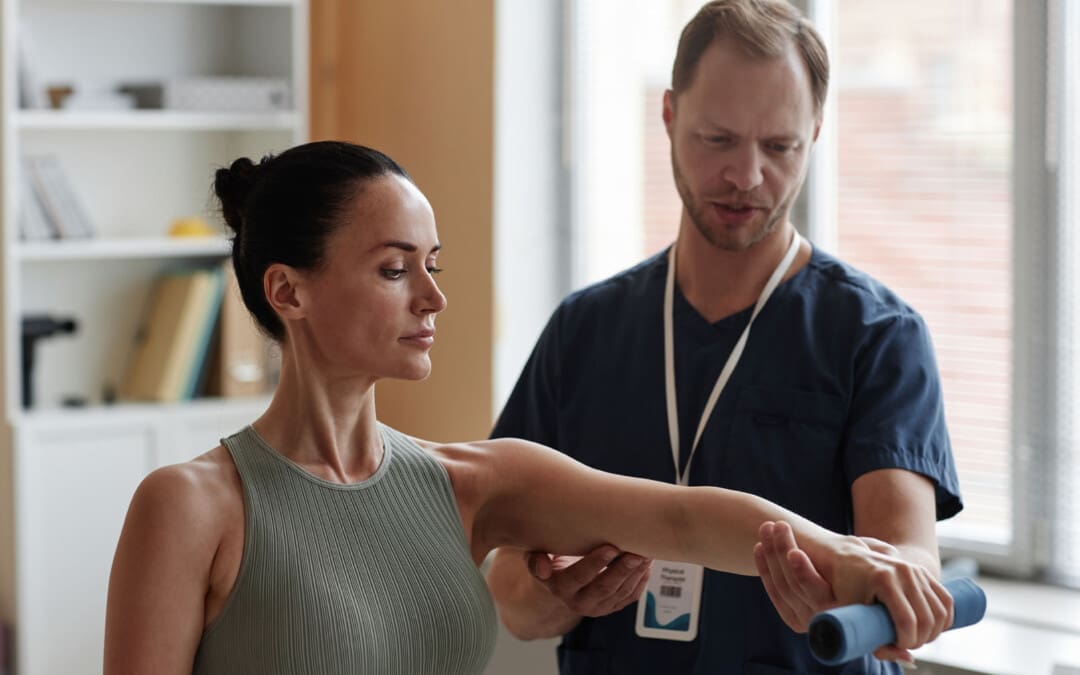
Individuals dealing with symptoms like sudden pain, weakness, and tenderness in the back of the knee could have a hamstring injury. Can knowing the symptoms and performing self-care help bring relief?
Hamstring Pain Behind The Knee
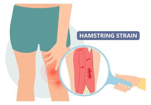
The hamstrings consist of three long muscles that run down the back of the thigh, cross over the back of the knee, and connect to bones in that area. A hamstring injury, such as a strain or tear, tendonitis, or biceps femoris tendinopathy, can cause pain in the back of the knee, difficulty bending the knee, swelling, and bruising. A hamstring strain occurs when the muscle is stretched too far or torn completely. This can happen from sudden, forceful movements or overstretching. Hamstring tendonitis develops over time, usually after a sudden increase in activity, when the hamstring tissue cannot recover from too much loading. Pain is often felt after physical activity and exercise and, in severe cases, during the activity or throughout the day. Biceps femoris tendinopathy can also cause pain in the back of the knee. Strains, tendonitis, bursitis, and muscle tears are all possible explanations for a hamstring injury that leads to pain behind the knee. Discussing pain symptoms with a healthcare provider is recommended, especially if it occurs suddenly during physical activity or exercise. They can help identify the exact cause and offer guidance for rehabilitation, including physical therapy referrals.
Causes and Triggers
Individuals may experience hamstring pain behind the knee when the muscles in that area are overworked, inflamed, or injured, such as from activities like running, walking, dancing, soccer, or basketball. Possible types of injuries and their causes.
A primary cause of muscle strain occurs when the muscle is stretched too far or has to handle a sudden force like sprinting or kicking. (American Academy of Orthopaedic Surgeons, 2021)
Severe Cases
Most causes of pain behind the knee are easily treatable at home with self-care and rest. However, it can be more severe, signaling a blood clot, infection, torn muscle or tendon/ligament. Hamstring knee pain may be serious if any of the following is experienced (American Academy of Orthopaedic Surgeons, 2021)
Sudden pain during physical activity, often during a full stride.
Feeling a pop or sharp pain that causes falling or limping.
Pain that worsens over time and prevents or hinders walking or exercising as normal.
If pain is severe and does not improve with rest and anti-inflammatory medications, evaluation by a healthcare professional is necessary.
Assesses Hamstring Pain
A healthcare provider will ask about symptoms and injury, including what happened when the pain began. They will perform a physical examination, which may include pressing on the back of the thigh to look for swelling, bruising, tenderness, or bunched-up muscles. (American Academy of Orthopaedic Surgeons, 2021) The healthcare provider will ask the patient to perform specific resisted movements, such as the manual muscle test, and measure the range of motion. Diagnostic testing includes an X-ray or MRI to determine the degree of the injury and which soft tissues or bones may be involved.
Self-Care
The first line of treating hamstring knee pain is the RICE protocol, which includes: (Mount Siani, 2024)
Rest
Stop any activity that causes symptoms and pain.
A healthcare provider may recommend crutches or a knee scooter in severe cases.
Ice
Apply cold packs to the swollen or painful area for 20 minutes throughout the day.
Compression
A knee brace, wrap, or bandage that applies gentle pressure to the injured area can help reduce and prevent swelling.
Elevation
Lifting the leg higher than the heart will help reduce swelling and blood accumulation.
Individuals may need to lie on a bed or sofa and elevate their legs with pillows.
Individuals can use at-home pain relievers like acetaminophen or NSAIDs like ibuprofen or naproxen. Over time, and depending on the severity of the injury, a healthcare provider will advise on gentle hamstring stretches and how to ease back into physical activity.
A healthcare provider will advise immobilizing the knee to help with muscle healing, which could involve wearing a knee brace or using crutches.
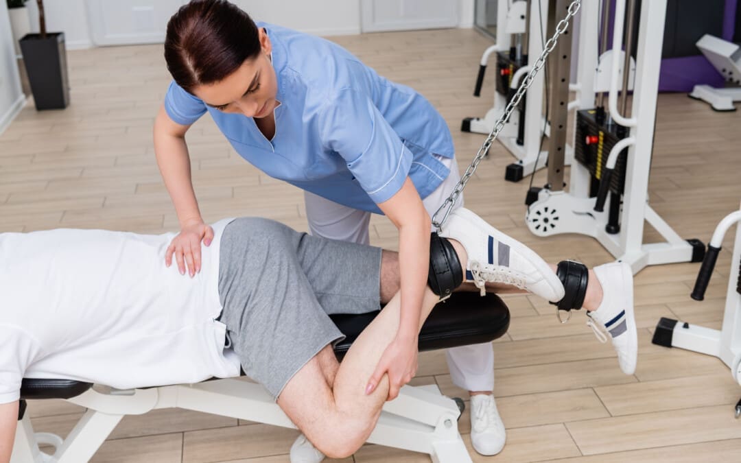
Physical therapy
A healthcare provider may refer the patient to a physical therapist, who will perform a personalized evaluation and prescribe targeted exercises to heal the injury and regain strength, flexibility, and movement.
Surgery
Tendon avulsion injuries are when the hamstring tendon completely tears away from the bone, and surgery is required to reattach the tendon.
Platelet-rich plasma – PRP
Platelet-rich plasma has become an additional treatment for hamstring muscle strain or tendonitis. (Seow D. et al., 2021)
The treatment involves injecting a solution from the patient’s blood into the muscle to heal the injury.
Recovery
Predicting how long a hamstring injury takes to heal and how long the pain will linger depends on the type, location, and severity. The most severe type is the hamstring coming unattached around the knee. This surgical repair and rehabilitation take at least three months before returning to sports and exercise (American Academy of Orthopaedic Surgeons, 2021). Lesser injuries like tendonitis or a mild strain can take less time to heal. However, it’s essential to avoid reinjuring the area so the condition does not become chronic. This includes: (American Academy of Orthopaedic Surgeons, 2021)
Stretching to encourage and maintain flexibility.
Fixing muscle imbalances between the quadriceps and hamstring.
Endurance and conditioning.
Avoiding overuse.
Injury Medical Chiropractic and Functional Medicine Clinic works with primary healthcare providers and specialists to develop personalized treatment programs. We focus on what works for you and use an integrated approach to treating injuries and chronic pain syndromes to improve flexibility, mobility, and agility, relieving pain and helping individuals return to normal activities. If other treatments are needed, Dr. Jimenez has teamed up with top surgeons, clinical specialists, medical researchers, and rehabilitation providers. Our providers use Functional Medicine, Acupuncture, Electro-Acupuncture, and Sports Medicine principles.
Chiropractic Care for Leg Instability
References
National Library of Medicine. (2017). Tendinitis Also called: Tendonitis. Retrieved from https://medlineplus.gov/tendinitis.html
American Academy of Orthopaedic Surgeons. OrthoInfo. (2020). Sprains, strains, and other soft tissue injuries. https://orthoinfo.aaos.org/en/diseases–conditions/sprains-strains-and-other-soft-tissue-injuries/
American Academy of Orthopaedic Surgeons. OrthoInfo. (2021). Hamstring muscle injuries. https://orthoinfo.aaos.org/en/diseases–conditions/hamstring-muscle-injuries/
American Academy of Orthopaedic Surgeons. OrthoInfo. (2021). Pes aserine (knee tendon) bursitis. https://orthoinfo.aaos.org/en/diseases–conditions/pes-anserine-knee-tendon-bursitis/
Mount Siani. (2024). Hamstring strain – aftercare. https://www.mountsinai.org/health-library/selfcare-instructions/hamstring-strain-aftercare
Seow, D., Shimozono, Y., Tengku Yusof, T. N. B., Yasui, Y., Massey, A., & Kennedy, J. G. (2021). Platelet-Rich Plasma Injection for the Treatment of Hamstring Injuries: A Systematic Review and Meta-analysis With Best-Worst Case Analysis. The American journal of sports medicine, 49(2), 529–537. https://doi.org/10.1177/0363546520916729
Individuals with inflammation, pain, and swelling on the tops of their feet or hands could be experiencing extensor tendonitis. What treatment options are available?
Extensor Tendonitis
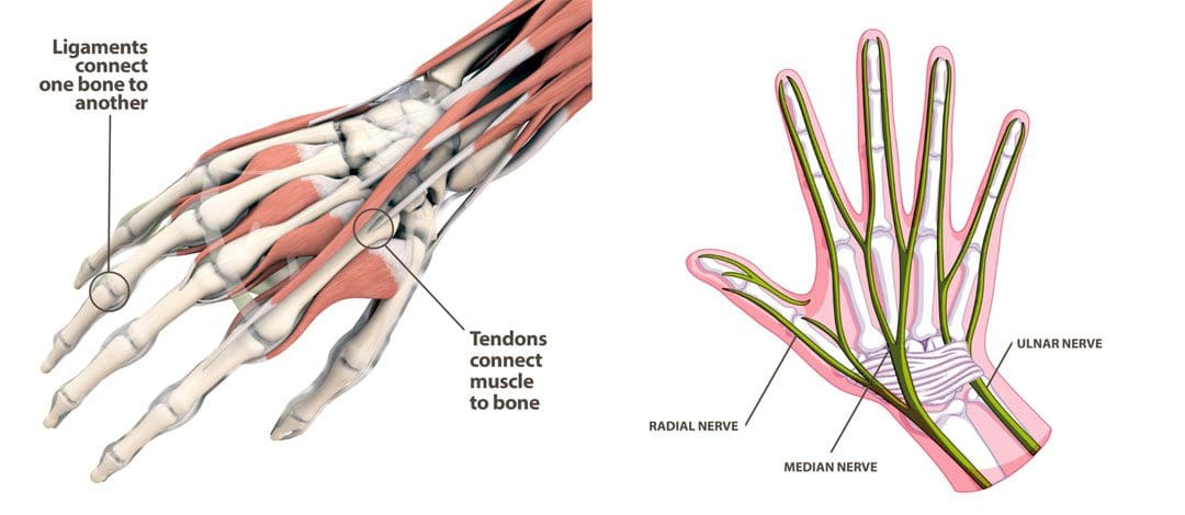
These tendons attach to muscles that straighten the fingers and lift the toes and the top of the foot. Extensor tendonitis is a condition characterized by inflammation of the tendons on the tops of the hands and feet. It often results from overuse of the muscles and from wearing tight shoes. Symptoms tend to worsen with activity and improve with rest. The condition does not usually require surgery; treatments include medications, home remedies/activity modification, and physical therapy.
Types
Tendonitis can develop in any tendon of the extensor muscles. These tendons are long, thin bands of tissue that can be felt on the tops of the hands and feet. The structures attach to muscles on one end and bones of the fingers and toes on the other. The extensor tendons in the hands include: (American Society for Surgery of the Hand, 2024)
The extensor digitorum communis straightens the index, middle, ring, and small fingers.
The extensor digiti minimi straightens the small finger.
The extensor indicis proprius straightens the index finger.
The extensor pollicis longus and extensor pollicis brevis muscles move the thumb into the thumb-up position.
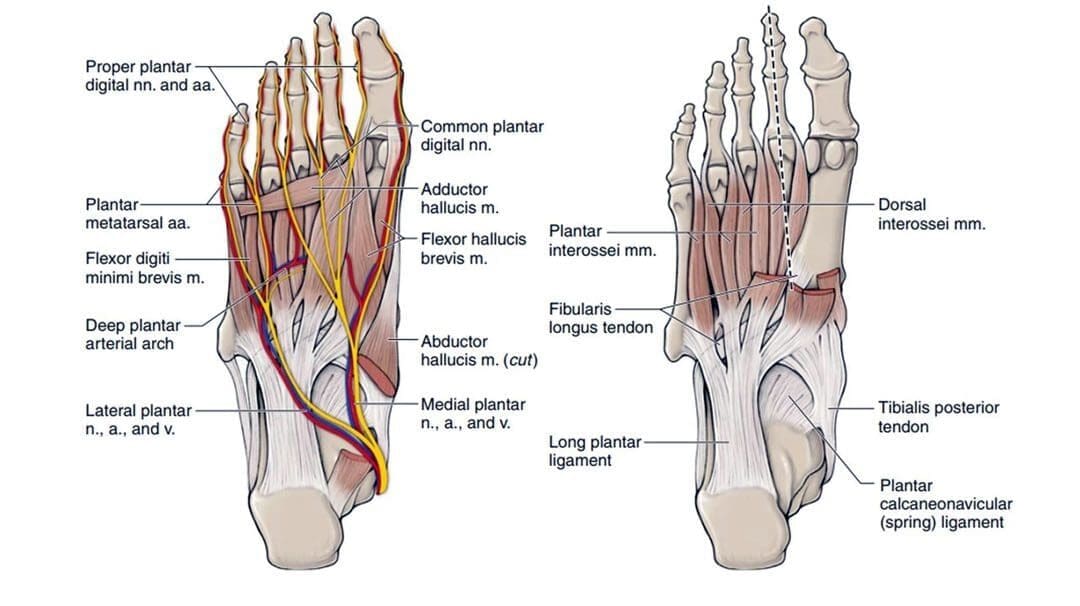
The extensor digitorum longus and extensor digitorum brevis muscles lift the second, third, fourth, and fifth toes.
The extensor hallucis longus lifts the big toe.
Tendonitis vs Tendinopathy
Chronic tendon pain can lead to a condition known as tendinopathy.
Tendonitis results from inflammation.
Tendinopathy is the degeneration/micro-tearing of a tendon that occurs with long-term overuse.
Knowing which condition you are dealing with is important because it requires different treatments.
Symptoms
The primary symptom is pain in the affected tendon/s. Individuals may also experience swelling, and the skin can become red or warm to the touch. Extensor tendonitis can cause pain when using the affected muscles, moving the hand or foot in the opposite direction, and stretching the tendons. Usually, the pain worsens when using the affected muscles and improves with rest.
Causes
Extensor tendonitis in the hands usually results from overuse, which causes inflammation. However, it can also result from trauma, such as falling on the hand or an injury during physical or sports activities. Common activities include: (Hanson Z. C., and Lourie G. M. 2022)
Manual labor work
Typing
Computer mouse use
Weightlifting
Gymnastics
Playing a musical instrument
Extensor tendonitis in the foot can also result from overuse activities like running, especially uphill. However, it can also occur from wearing overly tight or tightly laced shoes for physical activities like running or dancing. Less common causes include: (Arthritis Foundation, N.D.)
Medical conditions like diabetes or arthritis
Medication side effects
Infection
Joint deformities
Treatment
Extensor tendonitis usually improves with conservative treatment, which includes self-care, activity modification, physical therapy, and medication.
Medications
Individuals can treat inflammation with non-steroidal anti-inflammatory drugs NSAIDs like:
Ibuprofen
Naproxen
Aspirin
Acetaminophen can help reduce pain.
In some cases, individuals may need prescription anti-inflammatory medications like corticosteroids or pain relievers for short-term use.
Self-Care and Activity Adjustments
Self-care includes:
Rest and avoid any activities that increase pain symptoms. If you can’t avoid them completely, take frequent breaks to allow the muscles to relax.
Apply ice to the hand or foot several times daily for up to 20 minutes.
Compression wraps should be applied on the foot or hand using an elastic bandage or soft splint to help support the injured tendons and reduce swelling.
Elevate the hand or foot if swollen above the heart level when resting.
Activity Adjustments
Modifying activities can help address the underlying cause/s.
Hand extensor tendonitis can develop from poor positioning.
Setting up an ergonomic workstation can help.
Consult a coach or trainer if the tendonitis is related to sports or exercise.
Individuals might need to adjust their technique or training schedule to decrease pressure on the tendons.
Physical Therapy
Physical therapy is an effective treatment. A therapy team can help determine the condition’s underlying cause and provide a personalized treatment program. Interventions can include:
Pain-reducing treatments like ultrasound, electrical stimulation, electroacupuncture, and laser therapy.
Extensor tendonitis can take weeks or even months to fully heal. Early diagnosis and determining the condition’s underlying cause rather than just treating symptoms are recommended for a faster and optimal recovery. Injury Medical Chiropractic and Functional Medicine Clinic works with primary healthcare providers and specialists to develop personalized treatment programs through an integrated approach to treating injuries and chronic pain syndromes, improving flexibility, mobility, and agility, relieving pain, and helping individuals return to normal activities. Dr. Jimenez has teamed up with top surgeons, clinical specialists, medical researchers, and rehabilitation providers if other treatments are needed.
Move Better, Live Better, with Chiropractic
References
American Society for Surgery of the Hand. (2024). Tendons. https://www.assh.org/handcare/safety/tendons#Finger%20Extensor
Hanson, Z. C., & Lourie, G. M. (2022). Middorsal Wrist Pain in the High-Level Athlete: Causes, Treatment, and Early Return to Play. Orthopaedic journal of sports medicine, 10(4), 23259671221088610. https://doi.org/10.1177/23259671221088610
Arthritis Foundation. Foundation, A. (N.D.). Tendinitis. https://www.arthritis.org/diseases/tendinitis
Bronner, S., Ojofeitimi, S., & Rose, D. (2008). Repair and rehabilitation of extensor hallucis longus and brevis tendon lacerations in a professional dancer. The Journal of orthopaedic and sports physical therapy, 38(6), 362–370. https://doi.org/10.2519/jospt.2008.2749
American Society for Surgery of the Hand. (2014). Extensor tendon injury. https://www.assh.org/handcare/condition/extensor-tendon-injury#:~:text=The%20tendon%20may%20take%20eight%20to%20twelve%20weeks,may%20include%20stitches%20%28for%20cuts%20in%20the%20tendon%29.
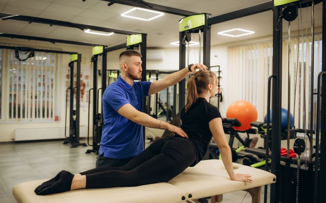
Skeletal muscles attached to the arms, legs, neck, back, and trunk bones are voluntary and consciously controlled. Weakness or inability to control these muscles can signal a health issue like a neuromuscular disorder or electrolyte imbalance. Can recognizing the symptoms can help healthcare providers develop effective treatment programs?
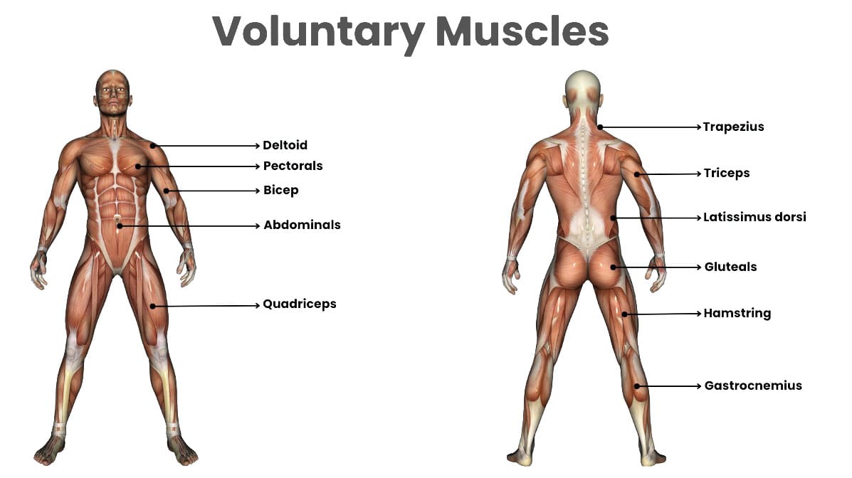
Voluntary Muscles
Voluntary muscles are the skeletal muscles that attach to bones and control movement of the limbs, head, neck, and body under an individual’s conscious control. Skeletal muscles are controlled by neuromuscular signals from the brain that communicate with individual muscle fibers and cause them to contract.
Difference
Voluntary muscles are skeletal muscles that contract and relax under conscious control.
These muscles attach to bones and regulate movement of the body.
Involuntary muscles are not under conscious control.
Involuntary muscles involve automatic internal processes needed for survival, like controlling blood vessels and organs like the heart, lungs, and digestive system.
They contract and relax automatically and receive signals from the autonomic nervous system, which regulates internal bodily functions.
Voluntary
Voluntary muscles are skeletal muscles that comprise 40% of the body’s weight and 50% to 75% of the body’s proteins. These muscles can convert chemical and mechanical energy to cause voluntary muscle contraction and movement. (Trovato F.M. et al., 2016) Skeletal muscle comprises fascicles or bundled units of multiple muscle fibers or muscle cells. Each muscle fiber consists of a cross-banded structure further divided into myofibrils containing thick myosin and thin actin myofilaments, which give the muscle its stripe appearance, and the structure gives the characteristic striated structure. (Trovato F.M. et al., 2016) Muscle contraction occurs when these myofilaments move closer together, stimulated by the release of the neurotransmitter acetylcholine from nerve cells that communicate with the muscle fiber.
Involuntary
The autonomic nervous system controls involuntary muscles, regulating their contraction and relaxation. This system also controls the activity of organs and blood vessels for essential daily functions, including breathing, circulation, digestion, heartbeat regulation, and urination. Most involuntary muscles are composed of smooth muscles. Smooth muscles do not have the striated structure of skeletal muscles and consist of sheets or layers of smooth muscle cells. When the autonomic nervous system stimulates these muscle cells to contract by releasing hormones or other chemical signals, smooth muscle cells shorten through the movement of actin and myosin myofilaments. Involuntary smooth muscles include the blood vessel walls, diaphragm, intestines, and bladder. (Webb R. C. 2003) One exception of an involuntary muscle is the myocardium, or heart muscle. The myocardium comprises a specialized cardiac muscle cell found only in the heart. Cardiac muscle is striated like skeletal muscle but is controlled by the autonomic nervous system and pacemaker cells, causing it to contract automatically and rhythmically.
Weakened Voluntary Muscles
Skeletal muscle diseases, neuromuscular disorders, and other causes can weaken muscles. Neuromuscular or skeletal muscle disorders affect the nerves that send electrical signals to voluntary skeletal muscles to control movement. When the nerves are damaged, communication between the nerves and muscles becomes disrupted. This can result in significant muscle weakness, atrophy, and loss of function. Most neuromuscular disorders are genetic or caused by issues with the immune system. Nerves communicate with muscles through the release of neurotransmitters at the neuromuscular junction, which is the space between a nerve cell and muscle fiber. Neuromuscular disorders can damage the nerve or the neuromuscular junction. Neuromuscular disorder symptoms can include: (Cleveland Clinic, 2023)
Numbness and tingling
Muscle weakness
Muscle twitches, cramps, or spasms
Muscle pain
Muscle atrophy
Decreased coordination
Balance problems
Drooping eyelids and double vision from eye muscle weakness.
Difficulty swallowing due to weakness of the pharynx.
Difficulty breathing due to weakness of the diaphragm.
Common Neuromuscular Disorders
Amyotrophic Lateral Sclerosis – ALS
More commonly known as Lou Gehrig’s disease, it is a genetic disorder that results from hardening of the spinal cord.
It causes damage to the nerves that control muscles and voluntary movement.
Charcot-Marie-Tooth Disease
This is a class of peripheral nerve disorders that cause muscle weakness, atrophy, and loss of sensation, most commonly in the legs and feet.
It is a genetic disorder caused by a gene mutation that damages myelin, or the insulating sheath that surrounds all nerves and supports the conduction of electrical signals.
Multiple Sclerosis – MS
MS causes degeneration of the myelin sheath surrounding nerves, decreasing the impulses along the nerves to muscles.
It can result in muscle weakness, which is often more severe on the dominant side of the body.
There are different forms of MS, but the condition is often progressive and gets worse over time if left untreated.
Muscular Dystrophies
These are genetic diseases characterized by gradual loss of motor function, muscle weakness and atrophy, walking gait problems, progressive respiratory failure, and cardiomyopathy.
There are nine types of muscular dystrophy, all caused by genetic mutations.
Myasthenia Gravis
This is an autoimmune disease that causes inflammation throughout the body.
An autoimmune disease occurs when the immune system attacks healthy cells by mistake.
With myasthenia gravis, the body produces antibodies that attack the receptors for acetylcholine, reducing the body’s ability to contract muscles.
This leads to muscle weakness, atrophy, and fatigue.
Myopathies
These are diseases of muscles that cause muscle weakness and atrophy.
Depending on the type, they may progress and get worse over time.
Electrolyte Imbalances
Muscle weakness can result from altered sodium, potassium, calcium, or magnesium levels.
Always seek immediate medical attention for any sudden, unexplained muscle weakness. Individuals who experience skeletal muscle weakness should discuss the type and duration of symptoms with their doctor, specialist, physical therapist, or chiropractor, as this might be a sign of a medical condition such as a neuromuscular disorder. Working with a chiropractic team can help expedite healing. Injury Medical Chiropractic and Functional Medicine Clinic works with primary healthcare providers and specialists to develop a personalized treatment program through an integrated approach to treat injuries and chronic pain syndromes, improving flexibility, mobility, and agility, relieving pain, and helping individuals return to normal activities. If other treatments are needed, Dr. Jimenez has teamed up with top surgeons, clinical specialists, medical researchers, and rehabilitation providers to provide the most effective treatments.
Chiropractic Massage Therapy
References
Trovato FM, I. M., Conway N, Castrogiovanni P. (2016). Morphological and functional aspects of human skeletal muscle. J Funct Morphol Kinesiol., 1(3), 289-302. https://doi.org/https://doi.org/10.3390/jfmk1030289
Webb R. C. (2003). Smooth muscle contraction and relaxation. Advances in physiology education, 27(1-4), 201–206. https://doi.org/10.1152/advan.00025.2003
Can understanding the causes and symptoms of potential hip tendonitis help healthcare providers diagnose and treat the condition for individuals experiencing pain in the front of the hip with restricted hip flexibility that worsens during movement?
Hip Tendonitis
Hip tendonitis is inflammation of the iliopsoas tendon. It is most commonly caused by overuse of the hip flexors without adequate rest for recovery. The condition can occur when the hip muscles overpower the tendons attached to the hip bone, causing inflammation and irritation. This can lead to pain, tenderness, and mild swelling near the hip joint. Hip tendonitis can be diagnosed with a physical examination, and treatment can include:
Rest
Ice
NSAIDs
Stretching
Physical therapy
Chronic cases may require a cortisone injection into the iliopsoas tendon to decrease inflammation.
Surgical release of the iliopsoas tendon may be recommended to decrease tightness and pain.
There is a high prognosis for a full recovery.
Tendonitis
Inflammation in a muscle’s tendon leads to pain and tenderness that worsens the more the muscle is used. An overuse injury means the tendon becomes repeatedly stressed through repetitive muscle contractions, causing muscle and tendon fibers to micro-tear. If not enough rest is allowed for the micro-tears to heal, a chronic cycle of pain and inflammation develops within the affected tendon. Other tendons that are prone to developing the condition include:
The tendon of the wrist extensors/tennis elbow.
The tendon of the wrist flexors/golfer’s elbow.
The Achilles’ tendon/Achilles tendonitis.
The patellar tendon/jumper’s knee.
The tendons of the thumb/De Quervain’s tenosynovitis.
Bursitis
Bursae are small fluid-filled sacs that help cushion and decrease friction around joints.
Because the iliopsoas tendon overlays bursae, inflammation of the tendon can also cause bursitis or inflammation of the bursae surrounding the tendon.
Tendonitis and bursitis can and often occur together due to overlapping symptoms.
Causes
The iliopsoas originates in the pelvis and vertebrae of the lower spine and attaches to the top of the femur or thigh bone. It allows the hip joint movement that brings the leg closer to the front of the body, like lifting the leg to step up or jump. It also helps keep the torso stable when standing with one or both feet on the ground and rising from a lying position. Hip tendonitis most often results from physical activities that require repeated leg lifting when stepping, running, kicking, or jumping. This can include:
Running
Dancing
Gymnastics
Martial arts
Cycling
Playing soccer
Iliopsoas tendonitis can also occur after hip arthroscopy, a minimally invasive surgical procedure to repair structures inside the hip joint because of altered joint movement and muscle activation patterns after surgery. (Adib F. et al., 2018)
Symptoms
The primary symptoms of hip tendonitis include a soreness or deep ache in the front of the hip that worsens after physical activity and limits the range of motion because of the pain. Other symptoms include:
Tenderness to touch in the front of the hip.
The pain can feel like a dull ache.
Stiffness may also be present.
Hip flexor tightness.
Altered posture, with the pelvis rotated forward and an exaggerated curve in the lower back.
Lower back pain.
Discomfort after prolonged sitting.
Altered walking pattern characterized by shortened steps.
Diagnosis
Hip tendonitis is diagnosed through a physical examination and medical history reviews of individual symptoms.
Individuals may also have an X-ray of their hip performed to examine the joint alignment and determine if a fracture or arthritis is present.
Treatment
Initial treatment involves rest from physical activities, applying ice, and gentle stretching.
Nonsteroidal anti-inflammatory drugs/NSAIDs can ease pain and swelling, decrease inflammation, and reduce muscle spasms.
If chronic pain persists, individuals may receive a cortisone injection into their iliopsoas tendon. (Zhu Z. et al., 2020)
A personalized physical therapy program focusing on hip flexor stretching and strengthening, as well as strengthening the glutes and core, will help expedite an optimal recovery.
Surgery
For cases that do not improve after three months of treatment, surgery to lengthen the iliopsoas tendon, a procedure known as a tenotomy, may be performed. It involves making a small cut into a portion of the tendon, allowing the tendon to increase in length while decreasing tension as it heals back together. A tenotomy temporarily reduces the strength of the iliopsoas; however, this weakness usually resolves within three to six months after surgery. (Anderson C. N. 2016)
Chiropractic Care
Chiropractic care can be an effective treatment because it can help restore proper alignment and motion in the hip, reduce inflammation, and improve muscle and joint function. Treatments may include:
Spinal adjustments to realign the spine and other joints, reducing pressure on nerves and inflammation.
Non-surgical decompression
Manual therapy – massage, trigger point therapy, or spinal manipulation.
Acupuncture
Graston technique
Rehabilitative exercises like stretching, strengthening, and range of motion exercises.
Tendonitis generally has an excellent prognosis for full recovery as long as thorough rest from activities is taken to allow the inflamed tendon to heal. The postsurgical prognosis is positive for chronic and severe cases of iliopsoas tendonitis that require surgery.
Injury Medical Chiropractic and Functional Medicine Clinic works with primary healthcare providers and specialists to develop a customized treatment program through an integrated approach to treating injuries and chronic pain syndromes, improving flexibility, mobility, and agility to relieve pain and help individuals return to normal activities. If other treatments are needed, Dr. Jimenez has teamed up with top surgeons, clinical specialists, medical researchers, and rehabilitation providers to provide the most effective treatments.
Inflammation and Integrative Medicine
References
Adib, F., Johnson, A. J., Hennrikus, W. L., Nasreddine, A., Kocher, M., & Yen, Y. M. (2018). Iliopsoas tendonitis after hip arthroscopy: prevalence, risk factors and treatment algorithm. Journal of hip preservation surgery, 5(4), 362–369. https://doi.org/10.1093/jhps/hny049
Zhu, Z., Zhang, J., Sheng, J., Zhang, C., & Xie, Z. (2020). Low Back Pain Caused by Iliopsoas Tendinopathy Treated with Ultrasound-Guided Local Injection of Anesthetic and Steroid: A Retrospective Study. Journal of pain research, 13, 3023–3029. https://doi.org/10.2147/JPR.S281880
Anderson C. N. (2016). Iliopsoas: Pathology, Diagnosis, and Treatment. Clinics in sports medicine, 35(3), 419–433. https://doi.org/10.1016/j.csm.2016.02.009
Can individuals dealing with musculoskeletal trigger points seek non-surgical treatments to reduce pain in their extremities?
Introduction
The musculoskeletal system has various muscles, tendons, ligaments, and soft tissues that allow the lower and upper extremities to function in multiple tasks that the person is doing. From physical activities to relaxing or just doing errands, the musculoskeletal system has a wonderful relationship with all the various body systems. It helps protect the vital organs from environmental factors and injuries. However, when environmental factors or injuries affect the body, many overlapping risk profiles affect the upper and lower quadrants, thus affecting the muscles and the soft tissues. When the musculoskeletal system starts to feel symptoms of pain and discomfort, it can cause visceral-somatic referred pain in different body locations and cause the development of trigger points in the muscle tissues. This causes the individual to be in excruciating pain and discomfort and is seeking treatment to reduce the pain-like symptoms. Today’s article gives us an understanding of musculoskeletal trigger points and how various non-surgical treatments can alleviate musculoskeletal trigger points in the body. We discuss with certified associated medical providers who consolidate our patients’ information to assess pain-like issues affecting their musculoskeletal system that are correlating to trigger point pain. We also inform and guide patients on various non-surgical treatments and ask their associated medical providers intricate questions to integrate a customized treatment plan to reduce musculoskeletal trigger point pain. Dr. Jimenez, D.C., includes this information as an academic service. Disclaimer.
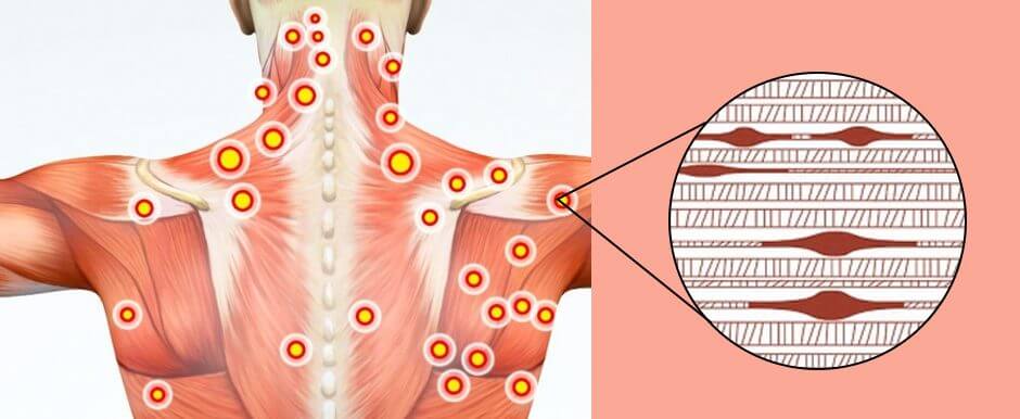
Understanding Musculoskeletal Trigger Points
Do you often experience pain in your legs, arms, hands, and feet throughout the day? How often do you experience symptoms of stiffness and discomfort in your neck, shoulder, or back? Or do you feel tingling and numbing sensations in your hands and feet? More often than not, many people who are experiencing these overlapping pain issues in their musculoskeletal system might have trigger points in their muscle fibers. Trigger points are part of a painful musculoskeletal condition known as myofascial pain syndrome. This painful musculoskeletal condition constitutes a hyperirritable spot within the taut band of the musculoskeletal system, causing pain when being compressed. (Lavelle et al., 2007) When a person is dealing with musculoskeletal trigger points, they will often experience referred pain and discomfort, motor dysfunction, and autonomic issues. This is because when many individuals experience pain in the upper or lower muscle quadrants, they deal with referred pain from the affected muscles. When the affected muscles have abnormal tender muscle regions, it can lead to impaired movements associated with the affected muscles in any joint area. (Macdonald, 1980)
Additionally, musculoskeletal trigger points can be identified as latent or active based on the development of where the pain originates from within the musculoskeletal system. To that point, when environmental factors or injuries develop trigger points, pain-like symptoms like muscle stiffness, dysfunction, and restricted range of motion show up when a pain specialist is assessing a person. (Shah et al., 2015) Fortunately, musculoskeletal trigger points are not difficult to treat once the pain source is located in the musculoskeletal system. This is because non-surgical treatments help manage the pain-like symptoms by inactivating the trigger points and restoring the affected resistant muscles to their full range of motion. (Rubin, 1981)
The Non-Surgical Approach To Wellness-Video
Non-Surgical Treatments For Musculoskeletal Trigger Points
When it comes to treating musculoskeletal trigger points, many individuals seek out various treatments to reduce pain-like symptoms. Since musculoskeletal trigger points can range from mild discomfort to severe pain, it can affect a person’s daily activities and cause them to be miserable. Luckily, musculoskeletal trigger points can be reduced through non-surgical treatments. Non-surgical treatments can vary depending on the pain severity of the trigger points in the musculoskeletal system. At the same time, many individuals can have numerous non-surgical therapies as they are customizable, cost-effective, and personalized for the person’s treatment. Below are some non-surgical treatments that can help alleviate musculoskeletal trigger points.
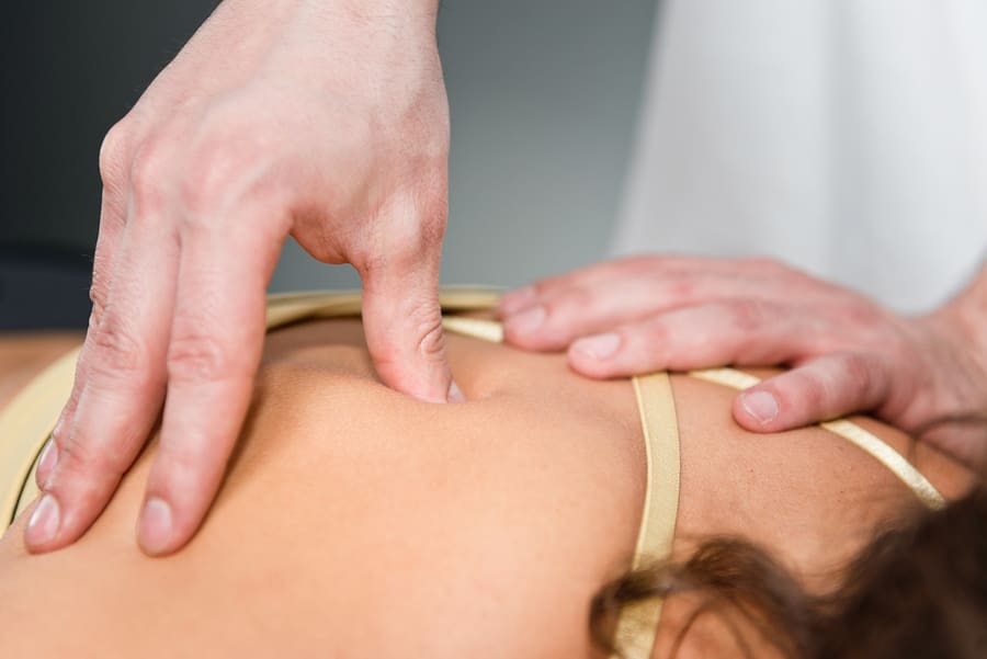
Chiropractic Care
Chiropractic care utilizes mechanical and manual manipulation of the musculoskeletal system and can help reduce the overlapping effects of musculoskeletal trigger points. Chiropractors incorporate various techniques and ischemic pressure to relieve the pain and provide relief. (Vernon & Schneider, 2009) Additionally, chiropractors can locate the trigger points by pressing on the muscle tissue or manipulating the muscle fibers. Chiropractors can also combine massage therapy to relieve trigger points and associated pain symptoms to restore the body to optimal function. This combination can incorporate various techniques to increase blood circulation to the affected muscle, help break down the inflexible scar tissue, and help restore muscle function to the extremities.
Acupuncture
Another form of non-surgical treatment to reduce musculoskeletal trigger points is acupuncture. Acupuncture incorporates solid, thin needles placed on various acupoints in the body by a professional. What acupuncture does is that when the needles are placed in the acupoints of the affected muscle, it can help stimulate the nervous system and help facilitate the body’s natural pain-relieving chemicals to kick-start the healing process. Additionally, when people incorporate acupuncture to reduce musculoskeletal trigger points, the sensory input that is causing them pain is reduced and can provide prolonged relief. (Melzack, 1981)
Lifestyle Adjustments
When it comes to reducing trigger points and combining non-surgical treatments, many individuals dealing with overlapping pain profiles from musculoskeletal trigger points can make lifestyle adjustments to prevent its development. Making small adjustments to a person’s work and living environments can reduce stress from being a co-factor to developing trigger points in the muscle fibers. Other small adjustments like improving posture and employing relaxation techniques like yoga, meditation, or deep breathing exercises can help reduce muscle stress and strain from everyday life. Incorporating non-surgical treatments to reduce and manage musculoskeletal triggers can provide a positive, beneficial result to improve muscle function and allow individuals to live healthier lives.
Melzack, R. (1981). Myofascial trigger points: relation to acupuncture and mechanisms of pain. Archives of Physical Medicine and Rehabilitation, 62(3), 114-117. https://www.ncbi.nlm.nih.gov/pubmed/6972204
Rubin, D. (1981). Myofascial trigger point syndromes: an approach to management. Archives of Physical Medicine and Rehabilitation, 62(3), 107-110. https://www.ncbi.nlm.nih.gov/pubmed/6453568
Shah, J. P., Thaker, N., Heimur, J., Aredo, J. V., Sikdar, S., & Gerber, L. (2015). Myofascial Trigger Points Then and Now: A Historical and Scientific Perspective. PM R, 7(7), 746-761. https://doi.org/10.1016/j.pmrj.2015.01.024
Vernon, H., & Schneider, M. (2009). Chiropractic management of myofascial trigger points and myofascial pain syndrome: a systematic review of the literature. J Manipulative Physiol Ther, 32(1), 14-24. https://doi.org/10.1016/j.jmpt.2008.06.012
For individuals who sit regularly for work and are slumping forward, can strengthening the rhomboid muscles help prevent posture problems and relieve pain?
Rhomboid Muscles
The rhomboids are a group of muscles in the upper back. A rhomboid major and minor muscle on each side of the upper back forms the shoulder girdle, which, along with other muscles, helps maintain the stability of the shoulder and shoulder blade. The rhomboid muscles control:
Pulling
Lifting
Rotating the shoulder blade.
These muscles also contribute to arm movement and enable lifting the arms above the head.
The rhomboid muscles support healthy posture and upper back. (Yoo W. G. 2017)
Sitting for an extended time, slumping forward, overstretching the arm above the body, sleeping on one side, repeated throwing motions, and sports like volleyball can affect the rhomboid muscles and cause pain symptoms.
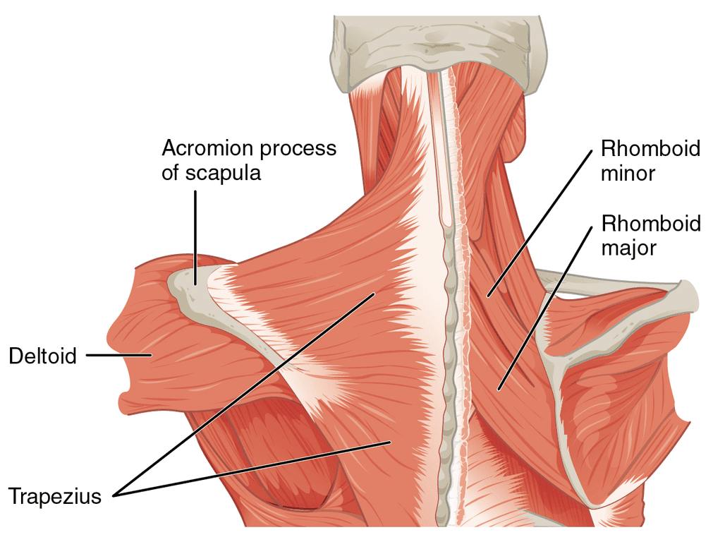
Anatomy
There are two rhomboid muscles. The major originates on the thoracic spine from the second through the fifth vertebrae and inserts on the side of the shoulder blade facing the spine. The minor is superior to the major and inserts on the C7 and T1 vertebrae. The muscles connect between the spine and each of the shoulder blades. When they contract, they pull the shoulder blades together. The muscle fibers run diagonally. They affix the scapula against the torso, allowing a stable base from which the arms can move.
Symptoms
When rhomboid muscles are overused or strained, symptoms can include the following:
Tenderness around the shoulder blade.
Limited range of motion in the shoulder.
Pain around the shoulder blade.
Upper back pain.
Neck pain.
Arm fatigue when performing repetitive overhead movements.
A crunching sound when moving the shoulder.
Weakness in the arm.
Chest pain.
Muscle Building
The action of the rhomboid is to bring the shoulder blades together, lift them or elevate them, as when shrugging, and rotate them so they face downward, away from the head. Bringing the shoulder blades together or scapular retraction builds the rhomboids to support the upper back.
To improve or prevent posture problems or mild, muscle-related upper-back and/or neck pain, 10 to 15 repetitions of scapular retraction performed one to three times every day are targeted exercises that could be recommended to help strengthen the muscles. However, consult a primary care provider, physical therapist, or chiropractor for serious medical conditions that affect posture to develop a personalized exercise program specific to the individual’s condition or injury. Everybody is different, and there is no one-size-fits-all when incorporating exercise to manage back pain. The physical therapy team may recommend other exercises to help manage or reverse any postural issues. (Kim, D. et al., 2015)
Overstretched Muscles
The human body has a unique and challenging relationship with gravity, which creates a downward pull on its structures, including the spine, head, and shoulders. As gravity pulls, the shoulders roll forward, and the chest can sink in. (Harvard Health, 2022). The rhomboid muscles may become overstretched, or the pectoral muscles and soft tissues in front may tighten up and constrict. Strengthening the rhomboids can help release the pectoral muscles.
Forward Head Posture
Unhealthy posture can lead to chronic pain and back problems. (Kripa, S. et al., 2021) Over time, unhealthy posture can also cause a forward head posture. (U.S. National Library of Medicine Clinical Trials, 2020) Forward head posture can lead to soft tissue strain, a kink in the neck, and fatigue in the muscles holding the head up, which can cause chronic neck pain. Maintaining strong extensor muscles in the lumbar and thoracic spine can help prevent back and neck problems as the body ages.
Injury Medical Chiropractic and Functional Medicine Clinic
We passionately focus on treating patients’ injuries and chronic pain syndromes and develop personalized care plans that improve ability through flexibility, mobility, and agility programs tailored to the individual. Using an integrated approach, our areas of chiropractic practice include Wellness & Nutrition, Chronic Pain, Personal Injury, Auto Accident Care, Work Injuries, Back Injury, Low Back Pain, Neck Pain, Migraine Headaches, Sports Injuries, Severe Sciatica, Scoliosis, Complex Herniated Discs, Fibromyalgia, Chronic Pain, Complex Injuries, Stress Management, Functional Medicine Treatments, and in-scope care protocols to relieve pain naturally by restoring health and function to the body through Functional Medicine, Acupuncture, Electro-Acupuncture, and Sports Medicine protocols. If the individual needs other treatment, they will be referred to a clinic or physician best suited for them, as Dr. Jimenez has teamed up with the top surgeons, clinical specialists, medical researchers, and premier rehabilitation providers to provide the most effective clinical treatments. We focus on what works for you and strive to better the body through researched methods and total wellness programs.
Functional Healing
References
Yoo W. G. (2017). Effects of pulling direction on upper trapezius and rhomboid muscle activity. Journal of physical therapy science, 29(6), 1043–1044. https://doi.org/10.1589/jpts.29.1043
Kim, D., Cho, M., Park, Y., & Yang, Y. (2015). Effect of an exercise program for posture correction on musculoskeletal pain. Journal of physical therapy science, 27(6), 1791–1794. https://doi.org/10.1589/jpts.27.1791
Harvard Health. (2022). Is it too late to save your posture? Exercise and Fitness. https://www.health.harvard.edu/exercise-and-fitness/is-it-too-late-to-save-your-posture
Kripa, S., Kaur, H. (2021). Identifying relations between posture and pain in lower back pain patients: a narrative review. Bulletin of Faculty of Physical Therapy, 26. https://doi.org/https://doi.org/10.1186/s43161-021-00052-w
U.S. National Library of Medicine Clinical Trials. (2020). Strengthening and stretching exercise to improve forward head posture and rounded shoulders. Retrieved from https://clinicaltrials.gov/study/NCT04216862
For individuals who are dealing with constant constipation due to medications, stress, or lack of fiber, can walking exercise help encourage regular bowel movements?
Walking For Constipation Assistance
Constipation is a common condition. Too much sitting, medications, stress, or not getting enough fiber can result in infrequent bowel movements. Lifestyle adjustments can regulate most cases. One of the most effective ways is to incorporate regular moderate-vigorous exercise, encouraging the bowel muscles to contract naturally (Huang, R., et al., 2014). This includes jogging, yoga, water aerobics, and power or brisk walking for constipation alleviation.
The Research
A study analyzed middle-aged obese women who had chronic constipation over a 12-week period. (Tantawy, S. A., et al., 2017)
The first group walked on a treadmill 3 times a week for 60 minutes.
The second group did not engage in any physical activity.
The first group had greater improvement in their constipation symptoms and quality of life assessments.
A gut bacteria imbalance is also linked to constipation issues. Another study focused on the effect of brisk walking versus exercises that strengthened core muscles like planks on intestinal microbiota composition. (Morita, E., et al., 2019) The results showed that aerobic exercises like power/brisk walking can help increase intestinal Bacteroides, an essential part of healthy gut bacteria. Studies have shown a positive effect when individuals engage in at least 20 minutes of brisk walking daily. (Morita, E., et al., 2019)
Exercise Can Help Decrease Colon Cancer Risks
Physical activity can be a significant protective factor in decreasing colon cancer. (National Cancer Institute. 2023) Some estimate the risk reduction to be 50%, and exercise can even help prevent recurrence after a colon cancer diagnosis, also 50% in some studies for patients with stage II or stage III colon cancer. (Schoenberg M. H. 2016)
The best effects were obtained through moderate-intensity exercise, such as power/brisk walking, about six hours per week.
Mortality was reduced by 23% in individuals who were physically active for at least 20 minutes several times a week.
Inactive colon cancer patients who began exercising after their diagnosis had significantly improved outcomes than individuals who remained sedentary, showing that it is never too late to start exercising.(Schoenberg M. H. 2016)
The most active patients had the best outcomes.
Exercise-Related Diarrhea Prevention
Some runners and walkers experience an overly active colon, resulting in exercise-related diarrhea or loose stools, known as runner’s trots. Up to 50% of endurance athletes experience gastrointestinal problems during intense physical activity. (de Oliveira, E. P. et al., 2014) Prevention steps that can be taken include.
Not eating within two hours of exercising.
Avoid caffeine and warm fluids before exercising.
If sensitive to lactose, avoid milk products or use Lactase.
Ensure the body is well-hydrated before exercise.
Hydrating during exercise.
If exercising in the morning:
Drink about 2.5 cups of fluids or a sports drink before bed.
Drink about 2.5 cups of fluids after waking up.
Drink another 1.5 – 2.5 cups of fluids 20-30 minutes before exercising.
Drink 12-16 fluid ounces every 5-15 minutes during exercise.
If exercising for over 90 minutes:
Drink a 12 – 16 fluid-ounce solution containing 30-60 grams of carbohydrates, sodium, potassium, and magnesium every 5-15 minutes.
Professional Help
Periodic constipation may resolve with lifestyle adjustments like increased fiber intake, physical activity, and fluids. Individuals who are experiencing bloody stools or hematochezia, have recently lost 10 pounds or more, have iron deficiency anemia, have positive fecal occult/hidden blood tests, or have a family history of colon cancer need to see a healthcare provider or specialist to perform specific diagnostic tests to ensure there aren’t any underlying issues or serious conditions. (Jamshed, N. et al., 2011) Before engaging in walking for constipation assistance, individuals should consult their healthcare provider to see if it is safe for them.
At Injury Medical Chiropractic and Functional Medicine Clinic, our areas of practice include Wellness & Nutrition, Chronic Pain, Personal Injury, Auto Accident Care, Work Injuries, Back Injury, Low Back Pain, Neck Pain, Migraine Headaches, Sports Injuries, Severe Sciatica, Scoliosis, Complex Herniated Discs, Fibromyalgia, Chronic Pain, Complex Injuries, Stress Management, Functional Medicine Treatments, and in-scope care protocols. We focus on what works for you to achieve improvement goals and create an improved body through research methods and total wellness programs. If other treatment is needed, individuals will be referred to a clinic or physician best suited to their injury, condition, and/or ailment.
Poop Testing: What? Why? and How?
References
Huang, R., Ho, S. Y., Lo, W. S., & Lam, T. H. (2014). Physical activity and constipation in Hong Kong adolescents. PloS one, 9(2), e90193. https://doi.org/10.1371/journal.pone.0090193
Tantawy, S. A., Kamel, D. M., Abdelbasset, W. K., & Elgohary, H. M. (2017). Effects of a proposed physical activity and diet control to manage constipation in middle-aged obese women. Diabetes, metabolic syndrome and obesity : targets and therapy, 10, 513–519. https://doi.org/10.2147/DMSO.S140250
Morita, E., Yokoyama, H., Imai, D., Takeda, R., Ota, A., Kawai, E., Hisada, T., Emoto, M., Suzuki, Y., & Okazaki, K. (2019). Aerobic Exercise Training with Brisk Walking Increases Intestinal Bacteroides in Healthy Elderly Women. Nutrients, 11(4), 868. https://doi.org/10.3390/nu11040868
National Cancer Institute. (2023). Colorectal Cancer Prevention (PDQ(R)): Patient Version. In PDQ Cancer Information Summaries. https://www.cancer.gov/types/colorectal/patient/colorectal-prevention-pdq
https://www.ncbi.nlm.nih.gov/pubmed/26389376
Schoenberg M. H. (2016). Physical Activity and Nutrition in Primary and Tertiary Prevention of Colorectal Cancer. Visceral medicine, 32(3), 199–204. https://doi.org/10.1159/000446492
de Oliveira, E. P., Burini, R. C., & Jeukendrup, A. (2014). Gastrointestinal complaints during exercise: prevalence, etiology, and nutritional recommendations. Sports medicine (Auckland, N.Z.), 44 Suppl 1(Suppl 1), S79–S85. https://doi.org/10.1007/s40279-014-0153-2
Jamshed, N., Lee, Z. E., & Olden, K. W. (2011). Diagnostic approach to chronic constipation in adults. American family physician, 84(3), 299–306.
IFM's Find A Practitioner tool is the largest referral network in Functional Medicine, created to help patients locate Functional Medicine practitioners anywhere in the world. IFM Certified Practitioners are listed first in the search results, given their extensive education in Functional Medicine
 Extensor Tendonitis