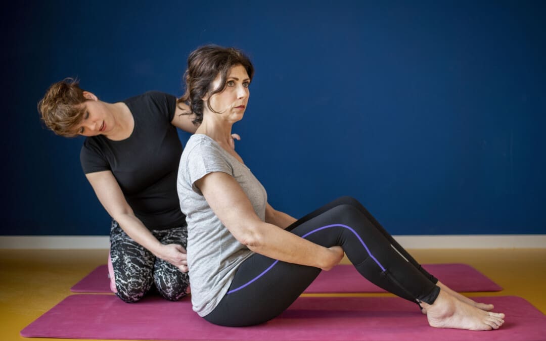
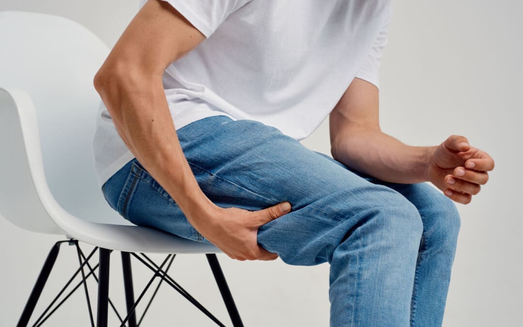
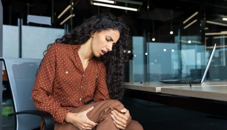
For individuals dealing with or experiencing muscle pain and stiffness, how long does it take to loosen tight muscles?
Length Of Time to Loosen Tight Muscles
Tight muscles are often caused by overuse or strain, combined with muscle soreness, a common symptom of tight muscles. The soreness peaks around the third day and begins to subside, typically resolving within a few days. But if tightness persists or is accompanied by other symptoms like numbness, inability to move, or swelling, it’s important to consult a medical provider. (Spine Medicine and Surgery of Long Island, 2024) However, it can take much longer for individuals who have never stretched and have had tight muscles for years, depending on the severity, injury history, and underlying causes.
Factors Influencing Timeline
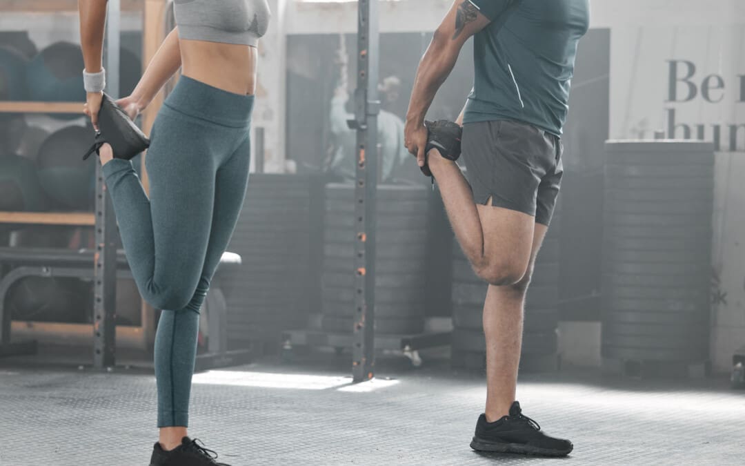
Consistent stretching can take anywhere from a few weeks to a few months to noticeably loosen tight muscles and improve flexibility. The specific time frame depends on factors like the severity of the tightness, underlying causes, and individual consistency with stretching. (Peterson Physical Therapy, 2024)
Severity of Tightness
Muscle knots and significant limitations in range of motion may take longer to resolve than minor stiffness.
Underlying Causes
If tightness is due to a specific injury or condition, addressing that cause is important for lasting and maintaining improvements. (Healthline, 2023)
Individual Factors
Genetics, age, and overall health can influence how quickly muscles adapt to stretching.
Consistency
Regular stretching, ideally daily or several times a week, is essential for feeling progress. (Mayo Clinic, 2023)
Stretching Routine
The length of time can vary based on the starting flexibility level and the specific stretching routine. (Mayo Clinic, 2023) It typically takes several weeks of consistent stretching, at least 3-4 times a week, to notice flexibility improvements. Longer-term changes, beyond the initial feeling of being looser, usually take 8 to 12 weeks.
Longer holds (1-2 minutes) can provide deeper benefits.
Long-term Gains
For substantial and sustained improvements, stretching consistently for several months is recommended. (Mayo Clinic, 2023)
Initial Changes
Individuals may notice small improvements in the first few weeks, especially starting from a more inflexible position.
Influencing Factors and Results
Individual genetics, current flexibility level, and the specific exercises can affect how quickly improvements are seen and felt. (Peterson Physical Therapy, 2024)
What To Expect
Improvements
Within a few weeks, individuals might notice a decrease in the sensation of tightness or increased ease in reaching a stretch. (Peterson Physical Therapy, 2024)
Longer-Term Changes
Significant muscle length and flexibility improvements may take several weeks to months of consistent effort.
Consider professional guidance for specific concerns or limitations. Consult a physical therapist or healthcare provider for personalized recommendations.
The length of time to see results means consistency and patience are important.
Injury Medical Chiropractic and Functional Medicine Clinic
As a Family Practice Nurse Practitioner, Dr. Jimenez combines advanced medical expertise with chiropractic care to address various conditions. Our clinic integrates Functional Medicine, Acupuncture, Electro-Acupuncture, and Sports Medicine to create customized care plans that promote natural healing, mobility, and long-term wellness. By focusing on flexibility, agility, and strength, we empower patients to thrive, regardless of age or health challenges. At El Paso’s Chiropractic Rehabilitation Clinic & Integrated Medicine Center, we passionately focus on treating patients after injuries and chronic pain syndromes. We focus on improving your ability through flexibility, mobility, and agility programs tailored for all age groups and disabilities. We use in-person and virtual health coaching and comprehensive care plans to ensure every patient’s personalized care and wellness outcomes.
Understanding Long-Lasting Injuries
References
Spine Medicine and Surgery of Long Island. (2024). How Long Do Muscle Knots Last? Spine Medicine and Surgery of Long Island. https://www.spinemedli.com/how-long-do-muscle-knots-last/#:~:text=The%20duration%20of%20a%20muscle,chronic%20pain%20if%20left%20untreated.
Peterson Physical Therapy. (2024). How Long Does It Take to Improve Flexibility? https://petersenpt.com/how-long-does-it-take-to-improve-flexibility#:~:text=Over%20the%20years%2C%20I’ve,takes%20to%20become%20more%20flexible.
Healthline. (2023). Everything You Need to Know About Muscle Stiffness. https://www.healthline.com/health/muscle-stiffness
Mayo Clinic. (2023). Stretching: Focus On Flexibility. https://www.mayoclinic.org/healthy-lifestyle/fitness/in-depth/stretching/art-20047931#:~:text=Stretch%20in%20a%20smooth%20movement,hold%20for%20around%2060%20seconds.
Harvard Health Publishing. (2022). Everyday Stretching. https://www.health.harvard.edu/everyday-stretching#:~:text=A%20daily%20regimen%20will%20deliver,or%20three%20times%20a%20week.
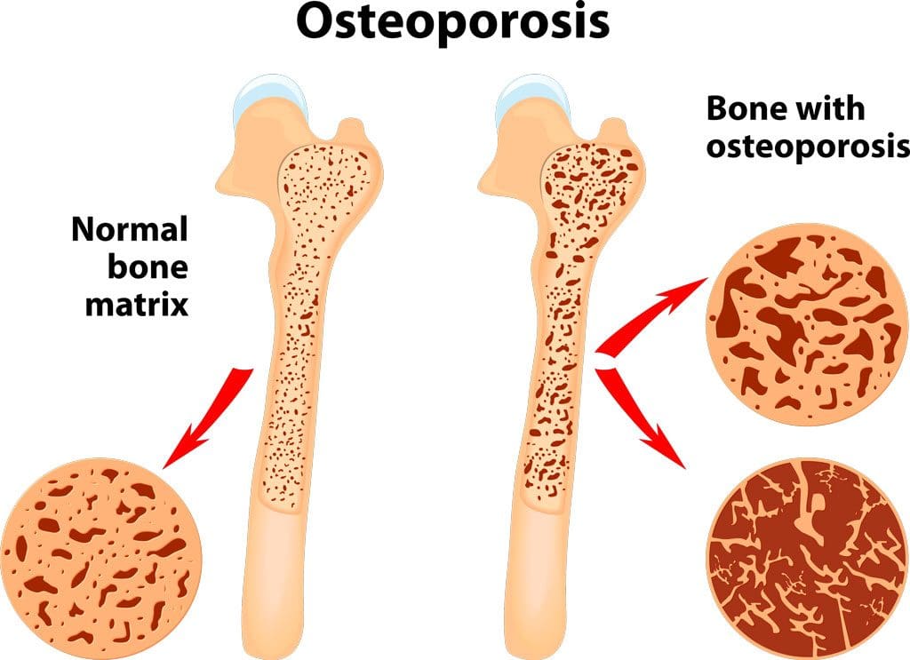
What is a bone density test, how is it performed, and what do the results mean?
Bone Density Test
A bone density test examines bone mass, which indicates overall bone strength. Assessing bone density or mass is necessary for diagnosing osteopenia or osteoporosis, conditions that increase the risk of broken bones. The scan is performed through dual-energy X-ray absorptiometry (DEXA), which examines the thickness of the bones. Results from DEXA scans are compared to standardized values to determine whether bone density is lower than normal and whether osteopenia or osteoporosis is present.
Examination
The procedure examines bone density, or bone mass. The bones’ density, or mass, is an overall indicator of bone strength. The greater the bone density, the thicker and stronger the bones are. The test is used to diagnose osteoporosis, a condition characterized by brittle bones at risk of breaking due to significantly low bone density. A bone density test can also diagnose osteopenia, a condition characterized by lower than normal bone mass that can lead to osteoporosis. (National Institute of Arthritis and Musculoskeletal and Skin Diseases, 2025) It is recommended that all women aged 65 and older and all men aged 70 and older have a bone density scan to screen for bone loss to help prevent fractures. (Kling J. M., Clarke B. L., & Sandhu N. P. 2014)
Bone density scans can establish a baseline level of bone density and track changes over time.
For individuals with osteoporosis or osteopenia, a bone density scan can help track how well their bones respond to treatment.
During a DEXA scan, the patient will lie on their back on a table with their legs elevated on a padded platform.
An X-ray scanner will pass over the spine and hips while another scans beneath.
While the scan takes place, the patient will be asked to hold very still to obtain an accurate image.
The scan will obtain bone density readings from the spine and hip, the two most commonly fractured bones, and generally takes less than 30 minutes.
Results
A DEXA scan measures bone density in grams per centimeter squared (g/cm²). This number indicates how densely bone cells are packed together in a specific area of bone. This bone density reading is then compared to a standardized value to determine if bone density is within a normal range or lower than average.
Between minus 1.0 and minus 2.5: Low bone density (osteopenia)
Equal to minus 2.5 or below: Osteoporosis
Bone density values are reported as a Z score for women who have not undergone menopause and men under 50 years old.
Z scores are compared to bone density levels of individuals of the same age and sex.
A Z score of minus 2.0 or lower indicates low bone density, which can be caused by factors other than aging, such as medication side effects, nutritional deficiencies, or thyroid problems.
Arthritis Diagnosis
Because a DEXA scan only measures the thickness of bones, it doesn’t work to diagnose arthritis. An X-ray of the affected joint is currently the most accurate way to diagnose arthritis. The Kellgren-Lawrence classification system categorizes the extent of arthritis based on the severity of joint damage seen on an X-ray. According to this system, arthritis can be classified as: (Kohn M. D., Sassoon A. A., & Fernando N. D. 2016)
Grade 1 (minor)
Minimal or no joint space narrowing, with possible bone spur formation.
Grade 2 (mild)
Possible joint space narrowing, with definite bone spur formation.
Grade 3 (moderate)
Definite joint space narrowing, moderate bone spur formation, mild sclerosis (abnormal thickening of bone), and possible deformation of bone ends.
Grade 4 (severe)
Severe joint space narrowing, large bone spur formation, marked sclerosis, and definite deformation of bone ends.
Injury Medical Chiropractic & Functional Medicine Clinic
Exercise can be incredibly beneficial for improving bone density, joint mobility, and the strength of surrounding muscles, which support and protect joints and bones. Talk to a healthcare provider to learn what interventions and available treatment options would be the most effective. Injury Medical Chiropractic and Functional Medicine Clinic works with primary healthcare providers and specialists to develop an optimal health and wellness solution. We focus on what works for you to relieve pain, restore function, and prevent injury. Regarding musculoskeletal pain, specialists like chiropractors, acupuncturists, and massage therapists can help mitigate the pain through spinal adjustments that help the body realign itself. They can also work with other medical professionals to integrate a treatment plan to resolve musculoskeletal issues.
Osteoporosis
References
National Institute of Arthritis and Musculoskeletal and Skin Diseases. (2025). Bone mineral density tests: what the numbers mean. Retrieved from https://www.niams.nih.gov/health-topics/bone-mineral-density-tests-what-numbers-mean
Kling, J. M., Clarke, B. L., & Sandhu, N. P. (2014). Osteoporosis prevention, screening, and treatment: a review. Journal of women’s health (2002), 23(7), 563–572. https://doi.org/10.1089/jwh.2013.4611
Kohn, M. D., Sassoon, A. A., & Fernando, N. D. (2016). Classifications in Brief: Kellgren-Lawrence Classification of Osteoarthritis. Clinical orthopaedics and related research, 474(8), 1886–1893. https://doi.org/10.1007/s11999-016-4732-4
Can understanding how leg cramps feel, their causes, and prevention help individuals with treatment options?
Leg Cramp Causes
A leg cramp is an involuntary contraction of the muscles in the leg, typically the calf muscle. It causes a sudden, sharp, and painful tightening of the muscle. They commonly occur from
Dehydration
Muscle overexertion
They can be a symptom of an underlying health condition, such as type 2 diabetes or kidney failure.
They can also be a side effect of certain medications.
Sensation
Leg cramps typically cause sudden, severe pain in the affected muscle. Individuals may also feel a hard knot or twitching of the muscle. The calf muscles are the most common site. (Harvard Health Publishing, 2024)
Causes
The underlying cause of leg cramps isn’t always known. However, work, lifestyle factors, and medical conditions can play a role. Common causes include:
Dialysis – treatment to remove excess fluid from the blood when kidneys fail.
Respiratory diseases of the lungs and airways
Amyotrophic lateral sclerosis (ALS, or Lou Gehrig’s disease, a neurological condition affecting the brain, nerves, and muscles)
Self-Care
Leg cramps often go away on their own after a few minutes. However, other self-care can help. Recommendations: (Harvard Health Publishing, 2024)
Change Body Positions
Moving can help relieve tension and pain.
If the leg cramps happen at night, stand up and take a few steps.
Massage
Gently rubbing the cramped muscle can help it relax.
Stretch
Stretching the cramping muscle can help it relax.
Apply Heat
If the leg cramp lasts more than a few minutes, apply a heating pad or take a warm shower to increase blood circulation to and around the muscle to help it relax.
Treatment
There are no medications specifically for treating leg cramps. However, if symptoms are caused by another medical condition, treating the underlying condition might help reduce cramp frequency. Dehydration or low electrolytes can cause leg cramps; in these cases, drinking water or electrolyte-infused beverages can help. If cramps are related to overexercising, consider reducing the intensity of your activity or exercising in a cooler environment. (Maughan R. J. & Shirreffs S. M. 2019) Magnesium supplements are usually marketed to relieve muscle cramps; they are not proven to alleviate them. (Garrison, S. R. et al., 2020)
Exercises and Stretches
It is important to know which muscle to target when stretching the leg. If the cramp does not resolve after the first stretch attempt, try again.
Place palms flat against the wall at shoulder height.
Step the cramping leg away from the wall around 12 to 18 inches.
Keep your heels flat on the floor.
Keeping the back leg straight, bend the front knee slowly and lean forward until you feel a stretch along the calf.
Hold for 30 to 60 seconds.
Prevention
They can’t always be prevented. However, if they are related to dehydration or other lifestyle factors, individuals may be able to prevent them from occurring or decrease the frequency. Try the following: (Harvard Health Publishing, 2024)
Drink plenty of water.
Stretch the leg muscles before you go to bed.
Try drinking a beverage with electrolytes for longer workouts or activities in hot weather.
Warm up before exercise, such as with a slow jog, before getting into more intense exercises.
Properly cool down after a workout.
Stretch the leg muscles before and after exercising.
Maintain a healthy body weight.
Contact a Healthcare Provider
Leg cramps can sometimes signify something more serious than a Charley horse. See a healthcare provider if any of the following symptoms present (National Library of Medicine, 2020)
Change in skin color, including redness or a deeper tone than normal
Cramps that last more than a few minutes or occur frequently
Muscle weakness
Severe pain from the cramps
Swelling
Warm skin
Injury Medical Chiropractic & Functional Medicine Clinic
Talk to a healthcare provider about leg cramps to learn what interventions would help the most. Injury Medical Chiropractic and Functional Medicine Clinic works with primary healthcare providers and specialists to develop an optimal health and wellness solution. We focus on what works for you to relieve pain, restore function, and prevent injury. Regarding musculoskeletal pain, specialists like chiropractors, acupuncturists, and massage therapists can help mitigate the pain through spinal adjustments that help the body realign itself. They can also work with other medical professionals to integrate a treatment plan to resolve musculoskeletal issues.
Is Motion Key to Healing?
References
Harvard Health Publishing. (2024). How to get rid of muscle cramps in your legs. https://www.health.harvard.edu/pain/how-to-get-rid-of-muscle-cramps-in-your-legs
National Library of Medicine. (2020). Muscle cramps. Retrieved from https://medlineplus.gov/musclecramps.html
Maughan, R. J., & Shirreffs, S. M. (2019). Muscle Cramping During Exercise: Causes, Solutions, and Questions Remaining. Sports Medicine (Auckland, N.Z.), 49(Suppl 2), 115–124. https://doi.org/10.1007/s40279-019-01162-1
Garrison, S. R., Korownyk, C. S., Kolber, M. R., Allan, G. M., Musini, V. M., Sekhon, R. K., & Dugré, N. (2020). Magnesium for skeletal muscle cramps. The Cochrane database of systematic reviews, 9(9), CD009402. https://doi.org/10.1002/14651858.CD009402.pub3
American Academy of Orthopaedic Surgeons. (2018). Knee conditioning program. https://orthoinfo.aaos.org/en/recovery/knee-conditioning-program/
Can stretching quadriceps help relieve stiffness and pain and improve flexibility for individuals with consistently tight quadriceps?
Quadriceps Stretches
Walking, running, biking, and other daily activities can tighten the quadriceps muscles. The quadriceps are four muscles in the front of the thigh that extend the leg and strengthen the knee. Stretching the quadriceps may be a part of a home or gym exercise program or physical therapy treatment to maintain quadricep flexibility. Resting the quadriceps with an exercise program can greatly maximize mobility and prevent injury.
Tight quadriceps may sometimes result from injuries such as patellofemoral stress syndrome or iliotibial band friction syndrome. The quads may also become tight for individuals with spinal stenosis or other related problems with the lower back. (International Sports Sciences Association, 2023) Ely’s test, also known as the Duncan-Ely test, is one way to determine whether your quadriceps are tight. It is a physical examination used to assess the flexibility and potential spasticity of the rectus femoris muscle (a quadriceps muscle) by passively flexing the patient’s knee. Lie on your stomach and try to touch your foot to your buttocks. If you can’t, the rectus femoris, one of the main muscles, may be tight and benefit from quadricep stretches. (Olivencia, O. et al., 2020)
Safety and Precautions
Before trying this or any other exercise program, consult a healthcare provider to ensure exercise is safe for you and your conditions. A professional can help diagnose any overuse injury that might be causing tight quadriceps. Quadriceps stretches will be a little more comfortable after warming up. A few minutes of walking or biking will warm the quadriceps muscles to stretch more easily.
Stretches
To stretch the quadriceps, try the standing, side-lying, and prone quadriceps stretch once fully warmed up and after a workout. Individuals who frequently experience quadriceps tightness should stretch them daily. Incorporate all or some of these stretches into a cool-down or off-day flexibility routine.
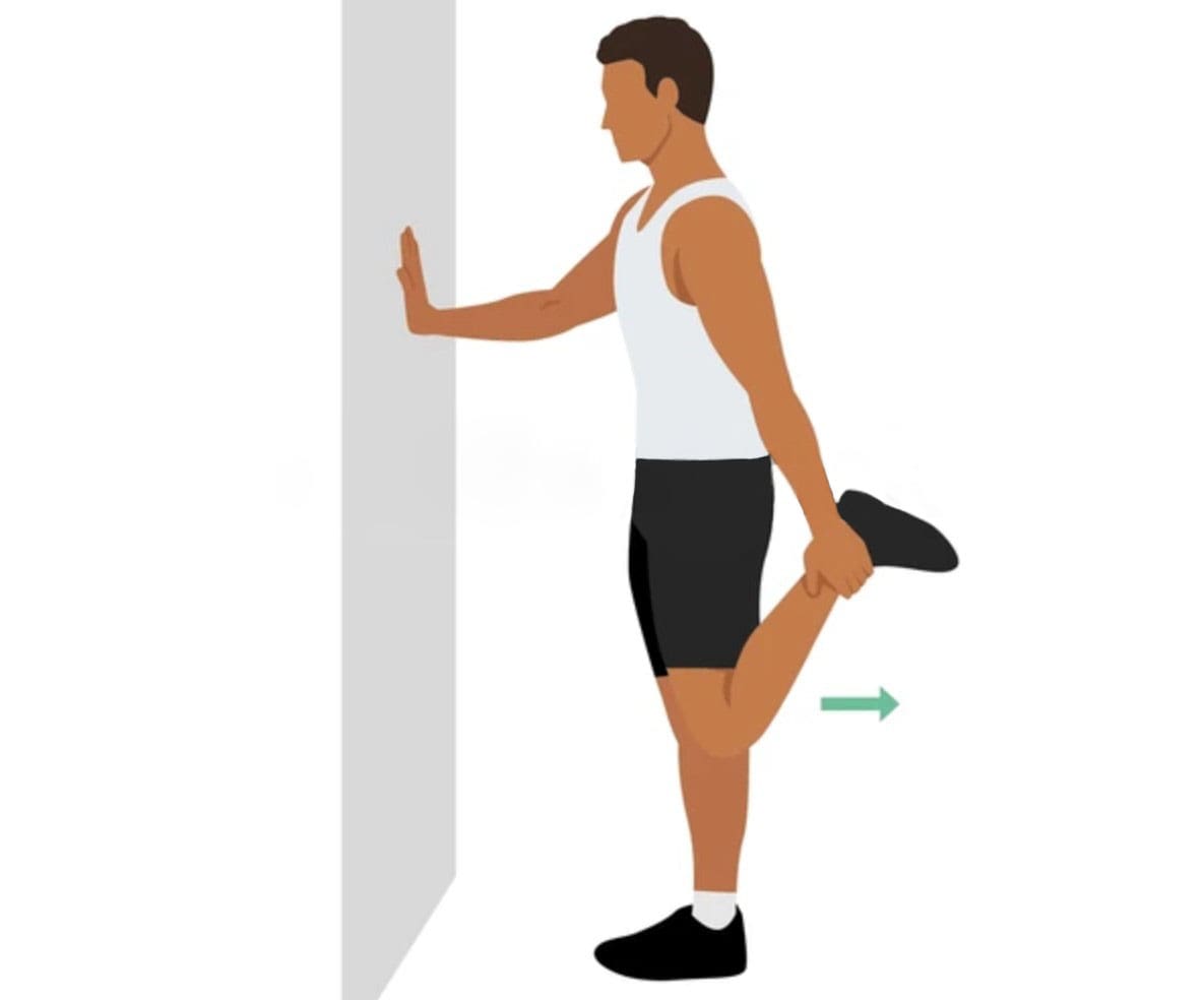
Standing Stretch
The standing quadriceps stretch can be done anywhere in the office, gym, or outside. All you need is a place to stand. Here is how:
While standing, hold onto a countertop or back of a chair to help with balance.
Bend your knee by grasping your ankle.
Move your foot toward your buttocks.
Gently pull on your ankle to bend your knee as far as possible.
Maintain position for 30 seconds.
Return to the standing position.
Repeat the exercise 3 to 5 times with each leg.
Stop stretching if there are any sharp pains.
Side-Lying Stretch
The side-lying quad stretch lengthens the quadriceps. On the floor in a supported position can help focus on the stretch. Here’s how:
Lie on your side.
Bend the knee of your top leg as far as you can, gently pulling with your hand.
Maintain position for 30 seconds.
Return to the starting position.
Repeat the exercise 3 to 5 more times with each leg.
Prone Stretch
Stretch the quadriceps while lying on your stomach. In this position, the floor helps to stabilize the pelvis, minimizing rocking and maximizing the stretch. To do the stretch:
Lie on your stomach.
Bend your knee back as far as you are able.
Grab your ankle to pull your foot toward your buttocks.
Maintain position for 30 seconds.
Return to the starting position.
Repeat the exercise 3 to 5 more times with each leg.
If you have difficulty reaching your ankle, pull the leg up, wrap a towel or strap around the ankle, and use it to pull. This can help stretch the quadriceps effectively even if you cannot reach the ankle easily.
Injury Medical Chiropractic & Functional Medicine Clinic
Consult a healthcare provider or physical therapist to learn the recommended quadriceps stretches or other strengthening exercises. Keeping the quadriceps healthy will help keep the knees moving and maximize functional mobility. Injury Medical Chiropractic and Functional Medicine Clinic works with primary healthcare providers and specialists to develop an optimal health and wellness solution. We focus on what works for you to relieve pain, restore function, and prevent injury. Regarding musculoskeletal pain, specialists like chiropractors, acupuncturists, and massage therapists can help mitigate the pain through spinal adjustments that help the body realign itself. They can also work with other medical professionals to integrate a treatment plan to resolve musculoskeletal issues.
Chiropractic Care For Leg Instability
References
International Sports Sciences Association. (2023). How to Release Tight Quads in 2 Simple Steps. ISSA. https://www.issaonline.com/blog/post/how-to-release-tight-quads-in-2-simple-steps
Olivencia, O., Godinez, G. M., Dages, J., Duda, C., Kaplan, K., Kolber, M. J., Kaplan, & Kolber (2020). THE RELIABILITY AND MINIMAL DETECTABLE CHANGE OF THE ELY AND ACTIVE KNEE EXTENSION TESTS. International journal of sports physical therapy, 15(5), 776–782. https://doi.org/10.26603/ijspt20200776
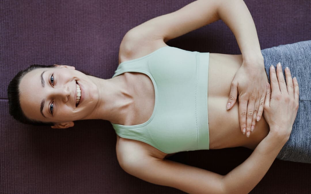
For individuals struggling with constipation, a common digestive issue, could performing abdominal self-massage help bring relief?
Abdominal Self Massage
Constipation refers to having a bowel movement fewer than three times per week. In addition to stress, certain lifestyle issues can lead to constipation, including not getting enough fiber, exercise, and proper hydration. Many also experience constipation while traveling. Abdominal self-massage involves gently massaging the stomach with your hands, either in a circular motion or with strokes, to improve digestion, relieve constipation, and reduce bloating. Performing self-massage on and around the abdomen can help ease constipation in several ways, such as stimulating the muscles, producing bowel movements, and soothing chronic stress. (Sinclair M. 2011)
Massage and Constipation
Abdominal massage can provide several benefits, including:
Stimulates and Improves Digestion
Massage stimulates the muscles and nerves that control digestion, promoting bowel movements and reducing constipation.
Reduces Bloating
Massaging the abdomen may help to reduce bloating and gas by gently moving fluids and gases through the digestive system.
In addition, it can help soften stool, speed up the movement of stool through the gut, and reduce the need to use laxatives. (University of Michigan Medicine, 2021)
Relieves Constipation
Abdominal massage can help with constipation by encouraging bowel movements.
Reduces Pain and Discomfort
Some find that abdominal massage helps to reduce pain and discomfort related to digestive issues.
Massage can help relax tense abdominal muscles and reduce pain associated with conditions like irritable bowel syndrome (IBS).
Improved lymphatic drainage
Massage helps move lymphatic fluid, which carries waste products and toxins away from the abdominal area.
Research
Although massage isn’t a standard treatment for constipation, some research shows it may help restore regularity. A report reviewed several clinical trials focusing on abdominal massage and its use as a treatment for chronic constipation. The results showed that abdominal massage may provide relief by promoting peristalsis, a series of muscle contractions that help move food through the digestive tract. The report also found that massage may help lessen colonic transit time, which is when digested food passes through the colon or last segment of the digestive tract. The report determined that abdominal massage can help alleviate constipation-related pain and discomfort. (Sinclair M. 2011)
In clinical trials, individuals with constipation reported improved quality of life after abdominal self-massage. However, some research suggests that using abdominal massage for constipation relief will not decrease the use of laxatives, the most commonly used treatment. (Lämås K. et al., 2009)
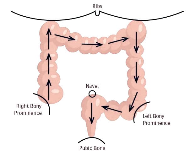
How to Massage
Various massage techniques may help relieve constipation and promote bowel movements. These are typically performed while lying down.
One technique involves placing the palm on the abdomen and making small, circular, clockwise motions around your belly button.
Individuals can also widen these circles so that the massage covers their entire abdomen.
Another technique begins by placing your hand below your breastbone, then gliding that hand down the length of your abdomen in one smooth stroke.
Repeat the movement with the other hand and continue this cycle for a few minutes.
When practicing self-massage, use light and gentle pressure, then gradually increase the pressure.
If you experience pain or tenderness, lighten up and return to a comfortable pressure level.
Try performing massage twice daily, aiming for a 20-minute session. Incorporating deep breathing into each session may also help. Before trying a massage or any home remedy, it is recommended to discuss it with a healthcare provider to see if it’s appropriate and safe. Pregnant women, for example, should avoid any massage on their abdomen. Constipation can sometimes signal an underlying condition that requires medical treatment, such as an underactive thyroid. Other symptoms like abdominal pain may be present, but sometimes constipation may be the only symptom.
Other Remedies
Self-massage alone is unlikely to treat chronic constipation; the goal should be to improve overall digestion to keep the organs functioning properly. To maintain regularity, it’s essential to drink plenty of water daily, eat enough fiber-rich foods, and engage in physical activity. Other alternative treatments include therapies like:
Acupressure
Biofeedback
Probiotics
Before trying self-massage, consult a medical caregiver to ensure it is safe and correct for the individual. Abdominal massage may not help with painful bloating caused by disease, infection, or other reasons.
Injury Medical Chiropractic & Functional Medicine Clinic
Injury Medical Chiropractic and Functional Medicine Clinic works with primary healthcare providers and specialists to develop an optimal health and wellness solution. We focus on what works for you to relieve pain, restore function, and prevent injury. Regarding musculoskeletal pain, specialists like chiropractors, acupuncturists, and massage therapists can help mitigate the pain through spinal adjustments that help the body realign itself. They can also work with other medical professionals to integrate a treatment plan to resolve musculoskeletal issues.
Massage Therapy Rehabilitation
References
Sinclair M. (2011). The use of abdominal massage to treat chronic constipation. Journal of bodywork and movement therapies, 15(4), 436–445. https://doi.org/10.1016/j.jbmt.2010.07.007
University of Michigan Medicine. (2021). Self-abdominal massage. https://www.med.umich.edu/1libr/MBCP/AbdominalSelfmassage.pdf
Lämås, K., Lindholm, L., Stenlund, H., Engström, B., & Jacobsson, C. (2009). Effects of abdominal massage in management of constipation–a randomized controlled trial. International journal of nursing studies, 46(6), 759–767. https://doi.org/10.1016/j.ijnurstu.2009.01.007
For individuals dealing with posture problems causing neck, back, and shoulder pain, can pectoralis minor stretches designed to work these areas be a part of physical therapy or as regular exercises at home?
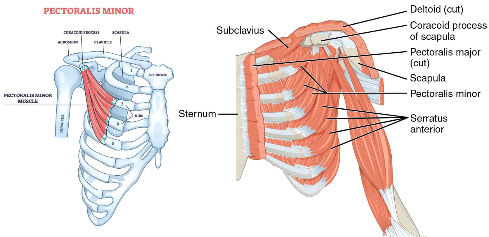
Pectoralis Minor Muscle Stretches
The pectoralis minor is a small, triangular muscle situated deep to the pectoralis major in the anterior chest wall. It originates from the margins of the third to fifth ribs adjacent to the costochondral junction and connects to the coracoid process of the scapula. The pectoralis minor helps with posture, mobility, and shoulder stability and aids breathing. Muscle tightness can cause pain in the chest, shoulder, and neck and a restricted range of motion. Strain and injuries can occur during activities involving overhead movements or forceful pushing. Pectoralis minor stretches are designed to work these muscles that span the ribs and connect to the shoulder to help improve posture and relieve pain and chest weakness. They can help reduce muscle tightness and other conditions like thoracic outlet syndrome. (Kaur U. et al., 2023) (Wagner E. R. et al., 2023) Talk with a healthcare provider Before starting any exercise or stretching program.
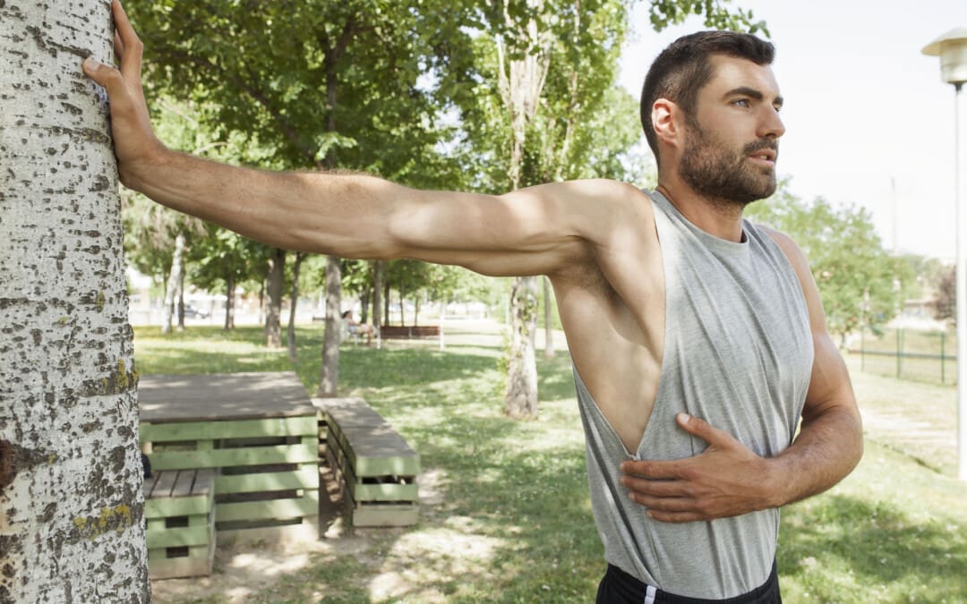
Corner Pectoralis Stretch
A corner pec stretch is similar to a wall push-up, except the emphasis is on staying in a position that lengthens the chest muscles. It’s important to move the whole body as a unit and not bend.
Stand facing a corner with a relaxed, upright posture.
Place your feet so they are parallel, and bend your knees slightly.
Stay as relaxed as possible during the movement to protect your joints.
Keep your gaze forward.
Place your forearms and palms over the walls where two walls connect at a right angle.
With your elbows bent to 90 degrees, move forward into the corner of the wall until you feel a comfortable stretch in the pectorals.
The doorway stretch is similar to the corner stretch. It works the pectoralis major and the minor muscles and helps with mobility. To perform: (Maryland Pain & Wellness Center, 2025)
Stand in a doorway with your feet placed together.
Place the palms and forearms on either side of the doorway.
Your elbows should be even with your shoulders and bend at a 90-degree angle.
Keep your back straight.
Take a step forward, leaning into the doorway.
You should feel the stretch in the muscle.
Repeat the stretch with the other foot.
Exercise and ergonomic changes to your chair or desk height can help improve posture and relieve muscle tightness. (Kaur U. et al., 2023)
T Stretch
The T stretch stretches the front of the chest and is done on the floor, typically with a foam roller placed directly under the spine. To perform: (OrthoCarolina, N.D.)
Lie down on your back with the foam roller aligned to the spine.
Make sure your head and tailbone are supported.
Open your arms straight out like a T.
Hold the position while stretching.
Y Stretch
The Y stretch is similar to the T stretch; both reduce chest muscle tightness and discomfort. To perform: (OrthoCarolina, N.D.)
Use the same foam roll position, lying on your back with the head and tailbone supported and aligned.
Stretch the arms out above your head, placing them into the shape of a Y.
Allow the chest muscles that connect to the arms to relax.
Studies have examined how quickly a prone scapular retraction can help stretch the back and shoulders. Results suggest the exercises must be performed longer before the pectoralis minor is lengthened to improve symptoms. (Dye J., Allyn M., & Frank C. 2024) However, further research is needed.
Health Conditions
Pectoralis minor stretches may be part of a personalized therapy program to improve mobility, posture, and/or breathing and sleep quality with health conditions that include:
Stretching and strengthening exercises can help improve their flexibility and function. Exercises can improve strength and function by standing or lying down, depending on the stretch.
Injury Medical Chiropractic and Functional Medicine Clinic
The pectoralis minor muscles are often overlooked in clinical examinations but can contribute to musculoskeletal pain and dysfunction. A healthcare provider can teach about stretches, how they can help, and whether they are safe for the individual’s injury and/or condition. Injury Medical Chiropractic and Functional Medicine Clinic works with primary healthcare providers and specialists to build optimal health and wellness solutions. Regarding musculoskeletal pain, specialists like chiropractors, acupuncturists, and massage therapists can help mitigate the pain through spinal adjustments that help the body realign itself. The clinic can also work with other medical professionals to integrate a treatment plan to resolve musculoskeletal problems.
Doorway Stretching Routine
References
Kaur, U., Shrestha, D., Hussain, M. A., Dalal, P., Kalita, M., Sharma, V., & Sharma, S. (2023). Prompt Impact of Muscle Energy Technique on Pectoralis Muscle Tightness in Computer Users: A Quasi-Experimental Study. Journal of Lifestyle Medicine, 13(2), 123–128. https://doi.org/10.15280/jlm.2023.13.2.123
Wagner, E. R., Gottschalk, M. B., Ahmed, A. S., Graf, A. R., & Karzon, A. L. (2023). Novel Diagnostic and Treatment Techniques for Neurogenic Thoracic Outlet Syndrome. Techniques in hand & upper extremity surgery, 27(2), 100–114. https://doi.org/10.1097/BTH.0000000000000419
University of North Carolina School of Medicine. (2020). Upper Body Stretching. https://www.med.unc.edu/htcenter/wp-content/uploads/sites/711/2020/04/Upper-Body-Stretching.pdf
Maryland Pain & Wellness Center. (2025). Stretches to Help with Strained Chest Muscles. Maryland Pain & Wellness Center Restoring Hope, Rebuilding Lives. https://www.marylandpainandwellnesscenter.com/blog/stretches-to-help-with-strained-chest muscles#:~:text=With%20your%20knees%20bent%20and,assist%20in%20deepening%20the%20stretch.
OrthoCarolina. (N.D.). Stretching Guide to Ease Tight Muscles. https://www.orthocarolina.com/storage/wysiwyg/stretching_guide_1.pdf
Dye, J., Allyn, M., & Frank, C. (2024). Is there an immediate effect on pectoralis minor length after performing a prone scapular retraction exercise using typical sets and repetitions in pain-free participants? Journal of bodywork and movement therapies, 40, 1014–1019. https://doi.org/10.1016/j.jbmt.2024.07.026
Chankavee, N., Amatachaya, S., Hunsawong, T., Thaweewannakij, T., & Mato, L. (2023). Effects of modified long stick exercise on hyperkyphosis, muscle imbalance, and balance control in elderly community-dwelling women with hyperkyphosis. Journal of back and musculoskeletal rehabilitation, 36(5), 1151–1162. https://doi.org/10.3233/BMR-220350
Liao, Y. X., Saiken, A., Chang, X., Guo, Y. F., Tan, Z., Deng, F., Meng, Q. L., Zhen, H., Li, Y. M., & Fang, B. M. (2025). Associations of fat, bone, and muscle indices with disease severity in patients with obstructive sleep apnea-hypopnea syndrome. Sleep & breathing = Schlaf & Atmung, 29(1), 82. https://doi.org/10.1007/s11325-024-03241-8
Thongchote, K., Chinwaro, U., & Lapmanee, S. (2024). Effects of scapulothoracic exercises on chest mobility, respiratory muscle strength, and pulmonary function in male COPD patients with forward shoulder posture: A randomized controlled trial. F1000Research, 11, 1284. https://doi.org/10.12688/f1000research.126832.2
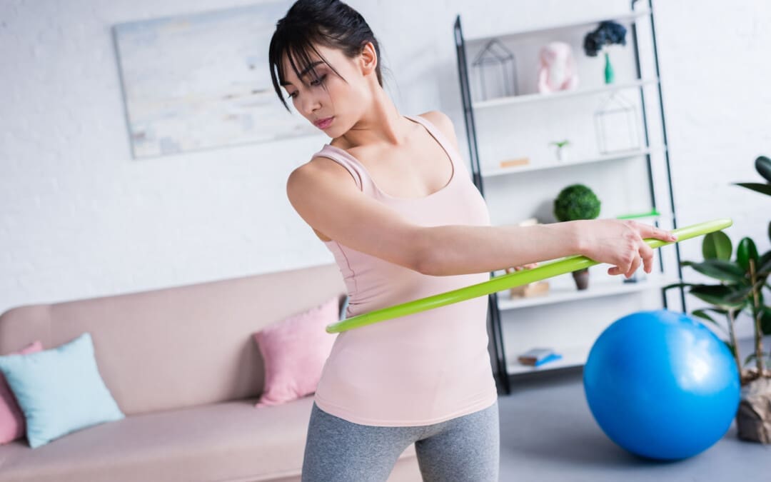
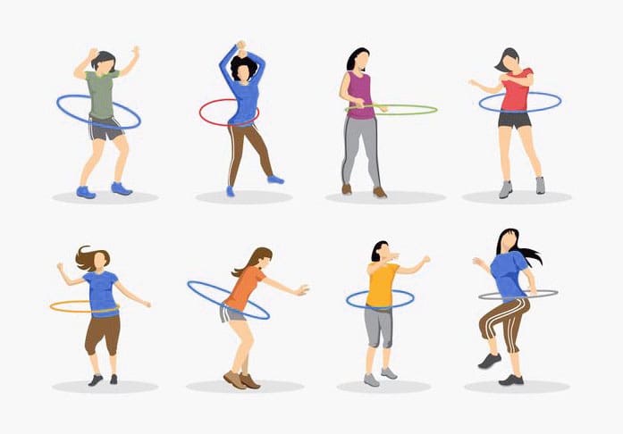
Can hula hooping be an option for individuals and athletes needing an effective, light, fun workout?
Hula Hoop Workout
A hula hoop workout is a low-impact exercise that involves spinning a hula hoop around your body. It can help improve balance, core strength, and aerobic fitness. Hula hooping is a great aerobic exercise that raises the heart rate and engages the whole body. Exercises can be done with a weighted or non-weighted hula hoop. A standard hoop will increase heart rate after about three minutes. A weighted hula hoop can target and build important core and lower body muscles, including the hamstrings, calves, quadriceps, and glutes. Lifting the weighted hula hoop also works the upper body muscles, giving the all-over body workout.
Weighted Hula Hoop
A weighted hula hoop can offer several health benefits.
Burns Calories
According to a research study, hula hooping can burn an average of 200 calories during a 30-minute workout. Researchers found that hooping is comparable in calories burned to boot camp-style fitness classes, kickboxing, and step aerobics. The average heart rate of the study participants was 151 beats per minute, equal to 84% of the age-predicted heart rate maximum. This can result in improved cardiovascular health and muscle conditioning. (American Council on Exercise, 2011)
Helps Build Muscle Mass
A study found that hula hooping increased trunk muscle mass and decreased waist circumference more than walking alone. Participants hula hooped an average of 12.8 minutes daily and walked almost 10,000 steps daily. The results showed more benefits to the core with hula hooping. The body fat percentage in the core region decreased significantly with hula hooping compared to walking. (Lahelma M. et al., 2019)
Lowers LDL Cholesterol
In the same study, researchers found that hula hooping can reduce LDL cholesterol more than walking. The results demonstrated an LDL-lowering effect similar to what resistance training does for cholesterol levels. Hula hooping for 13 minutes daily could benefit anyone with elevated cholesterol levels. (Lahelma M. et al., 2019)
Fun Workout
Weighted hula hooping can help individuals get out of a workout rut if they’re bored with a routine. It is recommended as either a warmup or a full workout.
Allows for Multitasking
If time to work out is limited, you can multitask using a hula hoop, easily add it to an exercise routine, and get moving while speaking on the phone, during breaks, or watching TV.
Benefits
Core strength: The exercise requires core strength to keep the hoop spinning.
Balance: Helps improve balance.
Aerobic fitness: Hooping can be used as a primary cardio routine.
Weight loss: Hooping can help burn calories and contribute to weight loss.
Hula Hoop Workout
Stand with a straight spine and feet shoulder-width apart.
Draw your abdomen in to engage your core.
Place the hoop around your waist, just above your hips.
Hold the hoop with both hands and toss it to one side.
Keep your back straight and move forward and back as fast as you can.
Pulse your hips and feet in a rocking motion.
Keep your arms out to the side or above your head.
Be mindful of posture, which will help you hoop better.
Weighted Workout
A full-weighted hula hoop workout. Start with a warmup for 5 minutes with a light jog or running in place. Then, perform three sets of the following:
Minute 1
50 seconds of a hula hoop halo with the right arm – swinging the hoop around your arm
10 seconds of rest
Minute 2
50 seconds of a hula hoop halo with the left arm
10 seconds of rest
Minute 3
50 seconds around the waist, hula hooping
10 seconds of rest
Minute 4
50 seconds of hula hoop squats, keeping the hoop around the legs and not dropping it
10 seconds of rest
Minute 5
50 seconds of hula hoop sit-ups, keeping the hoop around your legs and not dropping it
10 seconds of rest
Minute 6
0 seconds of hula jumping front to back, keeping the hoop around your legs and not dropping it
10 seconds of rest
How long you hula hoop each day is a matter of personal preference. To gain cardiovascular benefits, it is recommended that adults engage in at least 150 minutes per week of moderate-intensity aerobic activity. Spreading out the exercise throughout the week is preferable. (American Heart Association, 2024)
Injury Medical Chiropractic and Functional Medicine Clinic
Injury Medical Chiropractic and Functional Medicine Clinic works with primary healthcare providers and specialists to build optimal health and wellness solutions. Regarding musculoskeletal pain, specialists like chiropractors, acupuncturists, and massage therapists can help mitigate the pain through spinal adjustments that help the body realign itself. The clinic can also work with other medical professionals to integrate a treatment plan to resolve musculoskeletal problems.
Can Core Exercises Help With Back Pain?
References
American Council on Exercise. (2011). ACE-sponsored research: Hooping—Effective workout or child’s play? https://www.acefitness.org/certifiednewsarticle/1094/ace-sponsored-research-hooping-effective-workout-or-child-s-play/
Lahelma, M., Sädevirta, S., Lallukka-Brück, S., Sevastianova, K., Mustelin, L., Gylling, H., Rockette-Wagner, B., Kriska, A. M., & Yki-Järvinen, H. (2019). Effects of Weighted Hula-Hooping Compared to Walking on Abdominal Fat, Trunk Muscularity, and Metabolic Parameters in Overweight Subjects: A Randomized Controlled Study. Obesity facts, 12(4), 385–396. https://doi.org/10.1159/000500572
American Heart Association. (2024). American Heart Association Recommendations for Physical Activity in Adults and Kids. https://www.heart.org/en/healthy-living/fitness/fitness-basics/aha-recs-for-physical-activity-in-adults
IFM's Find A Practitioner tool is the largest referral network in Functional Medicine, created to help patients locate Functional Medicine practitioners anywhere in the world. IFM Certified Practitioners are listed first in the search results, given their extensive education in Functional Medicine