For individuals going through post surgery, injury rehabilitation, illness and/or chronic condition management, can physical therapy isometric exercises help?
Isometric Exercise
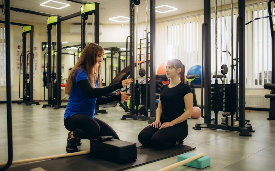
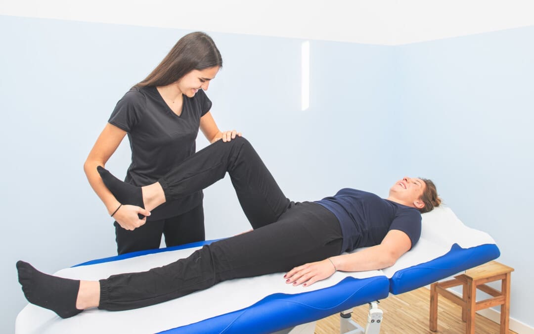
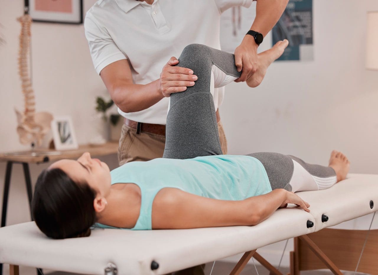
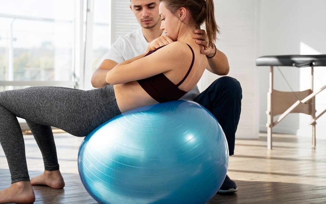
Isometric exercises are used in physical therapy to help build muscle endurance, improve range of motion, relieve pain, and reduce blood pressure more effectively than other types of exercise. Because they don’t involve joint movement, they are a solid starting point for rehabilitation and are suitable for individuals with a limited range of motion. They can be performed by pushing against an immovable object, like a wall, or by having a therapist provide resistance. Examples of isometric exercises include:
A physical therapist/PT may have a patient perform isometric exercises after injury or illness. During an isometric contraction, the muscle does not change in length, and there is no motion around the joint surrounding the muscle/s. (Rhyu H. S. et al., 2015)
When To Use
Isometric muscular contractions can be used at any time during physical rehabilitation and strengthening or a home exercise program and are regularly used with the following (Rhyu H. S. et al., 2015)
Post-surgery
When muscles cannot contract forcefully enough to move the joint it surrounds.
To help increase neuromuscular input to a specific muscle/s.
When injury or condition frailty makes other forms of exercise dangerous and not beneficial.
A healthcare provider or physical therapist should be consulted first if isometrics are used in a rehabilitation program.
Benefits
The benefits of using isometric exercise after injury or surgery may include the following:
No special equipment is necessary to perform isometric exercises.
The ability to safely contract a muscle while protecting a surgical incision or scar tissue.
The muscles can be strengthened in a specific range of motion around a joint. (NikolaidouO. et al., 2017)
A physical therapist can help determine whether isometric exercise benefits the specific condition.
Effectiveness
Isometric exercise is very effective after injury or surgery. However, when a muscle is contracted isometrically, it gains strength in a very small area and with a short range of motion. For example, an isometric shoulder external rotation performed with the arm at the side will only strengthen the rotator cuff muscles in the specific position that the arm is in. (NikolaidouO. et al., 2017).
Strength gains are specific to the joint’s position during the exercise.
Individuals who want to strengthen their gluteal muscles in their hip using isometrics would have to contract their glute muscles in one specific position for several reps.
Once several reps of the exercise in one position have been performed, the individual moves their hip joint into a new position and repeats the gluteal contractions in the new position.
This makes the exercise time-consuming, but it is perfect for injury rehabilitation, preventing and avoiding worsening or further injuries.
How to Perform
To perform isometric exercises, all that is needed is something stable to push against. (Rhyu H. S. et al., 2015) For example, to strengthen the shoulder muscles:
Stand next to a wall and try to lift an arm out to the side.
Allow the hand to press against the wall so no motion occurs at the shoulder joint.
Once pressed against the wall, hold the contraction for 5 to 6 seconds and slowly release it.
Perform 6 to 10 repetitions of the exercise.
This could be one set of completed isometric exercises for the shoulder muscles.
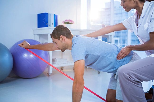
Elastic resistance bands or tubing can also be used to perform isometric exercises. Hold the tubing in a specific position and then move the body away from the anchor point instead of moving the joint. The muscles will contract against the increased resistance of the elastic tubing, and no motion will occur at the joint. A physical therapist can show and train on how to perform isometric exercises with the bands.
Neuromuscular Stimulation
Isometric exercise can strengthen muscles and help improve the neuromuscular recruitment of the muscles being trained. This enhances muscle contraction and expedites gains in muscle recruitment while protecting the joint. Isometric exercise can also be used during physical therapy using neuromuscular electrical stimulation (NMES). (Fouré A. et al., 2014) For example, a PT may use NMES to improve muscular function for individuals who have difficulty contracting their quadriceps after knee surgery and may be instructed to perform isometric quad-setting exercises during the session.
Injury Medical Chiropractic and Functional Medicine Clinic
A physical therapist can use isometric exercises to help individuals injured or have had surgery and are experiencing difficulty with normal functional mobility by improving their strength during recovery. The exercises can safely enhance the function and stability of the muscles and return individuals to the previous level of activity and function. Injury Medical Chiropractic and Functional Medicine Clinic works with primary healthcare providers and specialists to develop an optimal health and wellness solution. We focus on what works for you to relieve pain, restore function, and prevent injury. Regarding musculoskeletal pain, specialists like chiropractors, acupuncturists, and massage therapists can help mitigate the pain through spinal adjustments that help the body realign itself. They can also work with other medical professionals to integrate a treatment plan to resolve musculoskeletal issues.
Personal Injury Rehabilitation
References
Rhyu, H. S., Park, H. K., Park, J. S., & Park, H. S. (2015). The effects of isometric exercise types on pain and muscle activity in patients with low back pain. Journal of Exercise Rehabilitation, 11(4), 211–214. https://doi.org/10.12965/jer.150224
Nikolaidou, O., Migkou, S., & Karampalis, C. (2017). Rehabilitation after Rotator Cuff Repair. The Open Orthopaedics Journal, 11, 154–162. https://doi.org/10.2174/1874325001711010154
Fouré, A., Nosaka, K., Wegrzyk, J., Duhamel, G., Le Troter, A., Boudinet, H., Mattei, J. P., Vilmen, C., Jubeau, M., Bendahan, D., & Gondin, J. (2014). Time course of central and peripheral alterations after isometric neuromuscular electrical stimulation-induced muscle damage. PloS one, 9(9), e107298. https://doi.org/10.1371/journal.pone.0107298
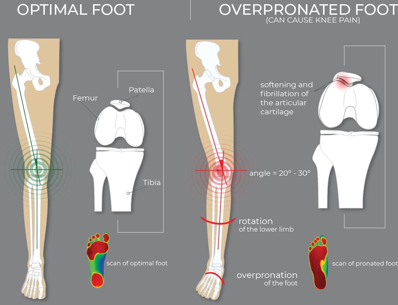
What are the treatment options for individuals dealing with foot overpronation when the foot and ankle move too much downward and inward?
Overpronation
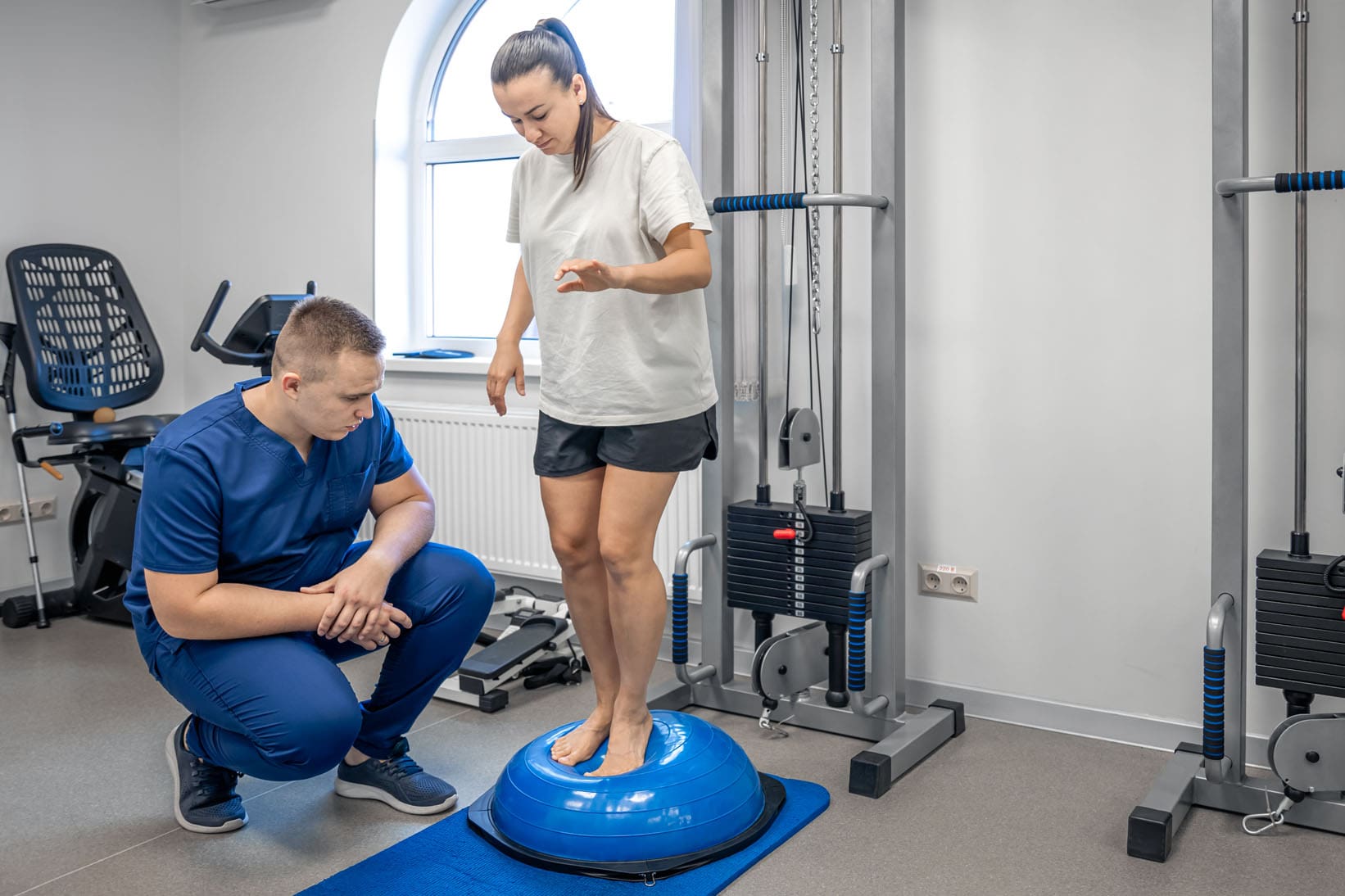
Pronation is the normal foot and ankle movement when taking a step and is usually associated with flat feet. Overpronation is a movement pattern in which the ankle rolls inward and downward, causing the foot’s arch to collapse with each stride. Overpronation can strain the muscles and ligaments in the feet and legs, leading to heel pain, ankle pain, shin splints, and low-back pain. (Pedorthic Association of Canada, 2023) Orthotic inserts for shoes, prescribed stretches, ankle braces, physical therapy, and surgery can all help alleviate the impact of overpronation. (Sánchez-Rodríguez, R. et al., 2020)
Signs and Symptoms
Some individuals with overpronation can have no symptoms at all. (Pedorthic Association of Canada, 2023) while others experience pain or other symptoms in their legs and feet. Overpronation is not a medical condition but a movement pattern that, if left untreated, can increase the risk for certain medical conditions because it strains the feet and leg muscles, joints, and ligaments. (Sánchez-Rodríguez, R. et al., 2020) Certain medical conditions can be a sign of overpronation and include: (Pedorthic Association of Canada, 2023)
Bunions
Heel pain
Plantar fasciitis
Achilles tendon pain
Posterior tibial tendonitis
Shin splints
Knee pain, including patellofemoral pain syndrome
Pain in the iliotibial or IT band
Lower back pain
Arthritis in foot and ankle joints
Stress fractures
Individuals may also experience pain in the midfoot or hips, which can be a symptom of flat feet.
Underpronation
Pronation refers to the normal movement of the foot and ankle while walking. Overpronation and underpronation are both abnormal movement patterns.
Overpronation – when the ankle rolls too much inward and downward.
Underpronation – occurs when an individual’s foot lacks flexibility and moves too little. This condition is called supination and is often associated with a high-arch foot type.
Overpronation can be caused or worsened by flatfeet. However, some individuals have overpronation because their feet and ankles are very flexible, so they tend to move more. Risk factors for flatfeet can also increase the chances of developing overpronation and include:
Age, especially individuals over 40.
Wearing shoes like high heels and shoes with a narrow-toe box.
Women are more prone to overpronate because of the various shoes and high heels worn.
Being overweight
Doing repetitive, impactful movements like running.
Correction and Treatment
Treating overpronation focuses on alleviating strain on muscles in the foot, ankle, and leg to relieve symptoms in the heel, ankle, knees, hips, or back. Common treatments are wearing supportive shoes and/or using foot orthotics. Exercises and stretches are also recommended to maintain flexibility and strength. Surgery is rare, but correcting flat feet that can cause overpronation may be recommended. (Sánchez-Rodríguez, R. et al., 2020) Individuals with overpronation are advised to see a podiatrist who can explain the best treatment options.
Supportive Shoes
The first course of treatment is to wear added supportive footwear. This can include specialized shoes or inserts that support the foot and reduce ankle movement. Individuals are advised to use shoes with firm heel and midfoot support to help prevent disproportionate movement. (Pedorthic Association of Canada, 2023)
Orthotics
A healthcare provider can recommend orthotics for individuals with moderate overpronation. These are meant to support the foot, especially the arch, and reduce overpronation. (Naderi A. Degens H. and Sakinepoor A. 2019) Individuals can purchase orthotics from shoe stores and elsewhere, but those with severe overpronation may need custom orthotics molded to the foot to provide individualized support.
Exercises and Stretches
Exercises and stretches can also help. A study found that exercises targeting the feet, core, and hips helped correct pronation over nine weeks. The exercises included: (Sánchez-Rodríguez, R. et al., 2020)
Toe pickups in which the individuals grab small objects with their toes and move them from one position to another.
Flexing and pointing the toes using a resistance band placed around the toes.
Hip abduction exercises to target the hip and glutes.
Abdominal and oblique muscle exercises to stabilize the torso.
Short-foot exercise raises the foot arch off the ground, drawing the toes toward the heel. (Sulowska I. et al., 2016)
Surgery
Rarely will surgery be needed to treat flat feet and severe overpronation. But if necessary, reconstruction realigns the bones to support the arch better and reduce overpronation. A metal implant is used for flatfeet to stabilize the area. Surgery can also repair torn tendons or other damage contributing to overpronation. (Healthline, 2020)
Injury Medical Chiropractic and Functional Medicine Clinic
Individuals with overpronation but no symptoms don’t necessarily have to see a healthcare provider since this may be the body’s natural movement pattern. But if the feet, legs, hips, or back begin to present with pain and other symptoms, see a healthcare provider who can evaluate gait and recommend treatment options. Injury Medical Chiropractic and Functional Medicine Clinic works with primary healthcare providers and specialists to develop an optimal health and wellness solution. We focus on what works for you to relieve pain, restore function, and prevent injury. Regarding musculoskeletal pain, specialists like chiropractors, acupuncturists, and massage therapists can help mitigate the pain through spinal adjustments that help the body realign itself. They can also work with other medical professionals to integrate a treatment plan to resolve musculoskeletal issues.
Enhance Performance with Functional Foot Orthotics
References
Pedorthic Association of Canada. (2023). Overpronation and Underpronation Correction. https://pedorthic.ca/services/foot-health/pronation/
Sánchez-Rodríguez, R., Valle-Estévez, S., Fraile-García, P. A., Martínez-Nova, A., Gómez-Martín, B., & Escamilla-Martínez, E. (2020). Modification of Pronated Foot Posture after a Program of Therapeutic Exercises. International journal of environmental research and public health, 17(22), 8406. https://doi.org/10.3390/ijerph17228406
Naderi, A., Degens, H., & Sakinepoor, A. (2019). Arch-support foot orthoses normalize dynamic in-shoe foot pressure distribution in medial tibial stress syndrome. European journal of sport science, 19(2), 247–257. https://doi.org/10.1080/17461391.2018.1503337
Sulowska, I., Oleksy, Ł., Mika, A., Bylina, D., & Sołtan, J. (2016). The Influence of Plantar Short Foot Muscle Exercises on Foot Posture and Fundamental Movement Patterns in Long-Distance Runners, a Non-Randomized, Non-Blinded Clinical Trial. PloS one, 11(6), e0157917. https://doi.org/10.1371/journal.pone.0157917
Healthline. (2020). All About Surgery for Flat Feet: Pros and Cons. https://www.healthline.com/health/flat-feet-surgery
Yips are involuntary wrist muscle spasms that affect athletes. They are often associated with golf, baseball, and sports that involve swinging and throwing motions, such as bowling, darts, cricket, and others. Can understanding the information and causes help diagnose and find the right therapy or training?
Yips
Yips are involuntary wrist spasms that athletes experience. The term is also used to refer to performance anxiety without physical spasms. Researchers believe they are caused by muscle overuse that leads to dystonia (a condition that causes muscles to contract involuntarily), and combined with psychological factors like performance anxiety and overthinking, can make them worse. (Beacon Health Systems, 2024)
The most common symptom is muscle spasms, often in the hands and wrists. That’s why it is the most common among athletes who play sports that require precision hand and wrist movements. Yips affect fine motor skills. (Aoyama, T. et al., 2021) In addition to muscle spasms, symptoms can also include: (Beacon Health Systems, 2024)
Twitching
Tremors
Freezing up
Psychological distress
Causes
Healthcare providers, trainers, coaches, and researchers know that psychological and physical factors cause yips. Underlying physical causes include overusing wrist muscles, which leads to dystonia or involuntary muscle movements. Also known as task-specific dystonia, it can also affect individuals who engage in repetitive muscle movements, like factory and assembly line workers, store check-out clerks, musicians, etc. (Clarke P., Sheffield D., and Akehurst S. 2020). Performance anxiety and psychological stress can worsen dystonia. (Aoyama, T. et al., 2021) Athletes can become so focused on their movements that they overthink their actions and perform worse. Individuals who have anxiety, self-consciousness, or stress about a game or performance often find that their involuntary wrist spasms are worse. (Clarke P., Sheffield D. and Akehurst S. 2020)
Increased Risk
Yips are most common in athletes who use their hands and wrists for their sport and are likely to impact more experienced, competing, and older athletes. (Beacon Health Systems, 2024) They are more common in athletes focused on smaller movements or shorter distances. For example, golfers commonly experience involuntary wrist spasms when putting, and baseball players are likelier to experience them when throwing less than 20 meters. (Clarke P., Sheffield D. and Akehurst S. 2020)
Diagnosis
There is no official diagnosis for yips. However, a coach, athletic trainer, sports doctors, and others can observe the pattern of symptoms and behavior and provide an informed diagnosis.
Once trigger/s are identified, they can be addressed. Treatments that can help include: (Beacon Health Systems, 2024)
Alternate Hand Positions
This can provide relief from dystonia and overthinking.
Using Different Equipment or Stabilizers
This allows the immobilization of certain muscles and the activation of different muscles.
Mindfulness
Reducing anxiety and distress can help relax the body.
Practicing mindfulness before games or tournaments can help reduce psychological triggers.
Botox Injections
Botox injections can treat certain types of dystonia.
Sports Psychology
A sports psychologist is a healthcare provider who studies individual athletes’ sports performance and how it affects their minds and skills.
A sports psychologist can help individuals create a program that reduces stress or anxiety around games and performance.
Injury Medical Chiropractic and Functional Medicine Clinic
Yips are common among athletes. It is important to address the physical and psychological components to treat the condition. Talking with a coach or a sports psychologist, other athletes, and supporting staff like trainers can help you find a solution. Injury Medical Chiropractic and Functional Medicine Clinic works with primary healthcare providers and specialists to develop an optimal health and wellness solution. We focus on what works for you to relieve pain, restore function, and prevent injury. Regarding musculoskeletal pain, specialists like chiropractors, acupuncturists, and massage therapists can help mitigate the pain through spinal adjustments that help the body realign itself. They can also work with other medical professionals to integrate a treatment plan to resolve musculoskeletal issues.
Sports Injuries
References
Beacon Health Systems. (2024). Yips. https://www.beaconhealthsystem.org/library/diseases-and-conditions/yips/
Clarke, P., Sheffield, D., & Akehurst, S. (2020). Personality Predictors of Yips and Choking Susceptibility. Frontiers in psychology, 10, 2784. https://doi.org/10.3389/fpsyg.2019.02784
Aoyama, T., Ae, K., Souma, H., Miyata, K., Kajita, K., Kawamura, T., & Iwai, K. (2021). Difference in Personality Traits and Symptom Intensity According to the Trigger-Based Classification of Throwing Yips in Baseball Players. Frontiers in sports and active living, 3, 652792. https://doi.org/10.3389/fspor.2021.652792
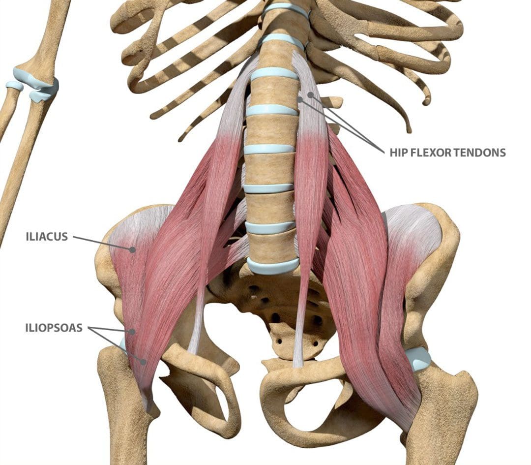
The iliacus muscle is a triangle-shaped muscle in the pelvic bone that flexes and rotates the thigh bone. It works with the other muscles in the hip and thigh to help bend, run, walk, sit, and maintain correct posture. Injuries and common medical conditions can affect its function, causing pain and stiffness. Can physical therapy help?
The Iliacus Muscle
The iliacus is one of the body’s most important hip flexor muscles. The iliacus and surrounding muscles work together to produce the stability and range of motion required for bending, dancing, sitting, and walking.
Anatomy
The iliacus muscle is part of a complex muscle system in the hip and pelvis. Two iliacus muscles on each side of the pelvic bone enable the thigh to flex and rotate. They are innervated by the femoral nerve, which provides movement and sensation to the lower limbs. (Bordoni B. and Varacallo M. 2023) The iliacus muscle sits on the wing-shaped ilium and fits into the curved surface of the ilium, called the iliac fossa. The top of the muscle is attached to the upper wings of the ilium or iliac crest. It extends past the hip joint, which connects to the upper thigh bone/femur at the lesser trochanter protrusion. The iliacus is part of a major trio of muscles called the iliopsoas, including the major psoas and minor psoas muscles. These muscles are also attached to the upper femur but extend upward, connecting to the lumbar/lower spine at several attachment points. The iliopsoas also interact with the quadratus lumborum muscle, the deepest muscle of the lower back that starts at the iliac crest and attaches to the lumbar spine at several points. The quadratus lumborum enables flexion and elevation of the spine, while the iliopsoas enable the flexion and rotation of the hip and thigh.
Functions
The iliacus muscle has many functions that include: (Physiopedia, 2024)
Flexing and rotating the femur.
Helps maintain proper body posture while standing and sitting.
Produces hip movement that enables walking, running, and climbing stairs.
Provides hip flexion – bringing the knee to the chest.
Enables the forward tilt of the pelvis and side-bending.
Conditions
Several conditions can affect the iliacus muscle, specifically from under and/or overuse injuries. These conditions, collectively known as Iliopsoas syndrome, are typically the result of overuse/repetitive strain or injuries. These include:
Iliopsoas tendinopathy – which affects tendons.
Iliopsoas bursitis – which affects cushioning sacs known as bursae.
Iliopsoas syndrome can affect anyone but is common in:
Individuals and athletes who repeatedly use movements that flex the hips.
Track-and-field athletes
Gymnasts
Dancers
Iliopsoas Bursitis
This is the inflammation of the cushioning sac or bursa under the iliacus muscle, which helps the muscle slide over the pelvic bone. Symptoms can range from mild discomfort to pain that radiates through parts of the leg and hips. Runners, skiers, and swimmers are commonly affected, and individuals who regularly have tight hips and individuals with different forms of arthritis can also be affected. Early treatment can prevent the symptoms from worsening. Mild cases can be treated with self-care and stretching to help relieve tightness, rest, ice application, and over-the-counter nonsteroidal anti-inflammatory drugs. In severe cases, treatment options that may be recommended include: (Physiopedia, 2024)
Physical therapy
Assistant walking devices to relieve pressure – for example, a cane.
Corticosteroid steroid injections
Prescription anti-inflammatory medications
Iliopsoas Tendinopathy
Another condition affecting the iliacus muscles is iliopsoas tendinopathy, sometimes called snapping hip syndrome, because individuals can hear an audible snapping sound (Davenport KL. 2019). The condition is often experienced by dancers who repeatedly flex and hyperextend their hips and can result in hip and groin pain that gets worse with kicking or rotation. Treatment of iliopsoas tendinopathy can include:
Retraining muscle imbalances with strengthening and stretching exercises.
If these fail to provide relief, corticosteroid injections may be used. A saline hydro dissection can relieve stress around the tendon by injecting fluids that cushion and release trapped tissues.
Tendon release surgery may be recommended when all other options have failed. The surgical release involves severing the tendon to reduce pain and improve the range of motion.
Rehabilitation
Core muscle strengthening is essential to the rehabilitation of iliacus muscle injuries. The iliopsoas is an integral component of the core group and can benefit from stretching and strengthening exercises (Yogateket, 2019)
Lunge stretches
Straight leg raises
Knee-to-chest stretches
Standing hip flexion with resistance bands
Certain yoga poses can also help and include variations on the bridge pose that encourage hip flexion. (Yoga International, 2024)
Injury Medical Chiropractic and Functional Medicine Clinic
Iliopsoas pain is often felt at the front of the hips, thigh, mid-back, and lower back. Chiropractic care can help with iliacus muscle injuries through:
Evaluation
A chiropractor can evaluate the condition and determine if the iliacus muscle is causing pain.
Treatment plan
A chiropractor can create a personalized treatment plan that may include exercise instructions, manipulation, and other therapies.
Rehabilitation
A chiropractor can create a rehabilitation program to expedite healing.
Injury Medical Chiropractic and Functional Medicine Clinic works with primary healthcare providers and specialists to develop an optimal health and wellness solution. We focus on what works for you to relieve pain, restore function, and prevent injury. Regarding musculoskeletal pain, specialists like chiropractors, acupuncturists, and massage therapists can help mitigate the pain through spinal adjustments that help the body realign itself. They can also work with other medical professionals to integrate a treatment plan to resolve musculoskeletal issues.
Hip Labral Tear and Chiropractic Care
References
Bordoni, B., & Varacallo, M. (2024). Anatomy, Bony Pelvis, and Lower Limb, Iliopsoas Muscle. In StatPearls. https://www.ncbi.nlm.nih.gov/pubmed/30285403
Davenport KL. (2019). The professional dancer’s hip. Performing Arts Medicine, 77-87. https://doi.org/https://doi.org/10.1016/B978-0-323-58182-0.00009-2
Yogateket. Lizette Pompa. (2019). Essential yoga body parts. Hip flexor/psoas and yoga. Yogateket. https://www.yogateket.com/blog/hip-flexor-psoas-and-yoga
The vastus lateralis is a muscle on the outside part of the thigh. Injuries to the muscle include strains, tendinitis, femoral nerve compression, and others. Can rehabilitation like heat and ice, massage, and strength and mobility exercises help individuals return to normal activities and function?
Vastus Lateralis Muscle
The vastus lateralis is the largest of the four quadriceps muscles on the thigh’s outer portion. The vastus lateralis helps extend the knee joint and maintain the knee position when walking or running. The vastus lateralis functions to work with the other quad muscles to help extend the knee joint.
Anatomy
Most muscles are attached to bone points of attachment, called the origin and insertion points. The vastus lateralis origin and insertion points are as follows (Vieira, EPL. 2017)
Origin
The origin is on the upper inter-trochanteric line of the femur or thigh bone.
It also arises from the base of the greater trochanter and the linea aspera, the supracondylar ridge, and the lateral intermuscular septum.
Insertion
From its origin, the muscle courses down the lateral thigh and inserts as part of the lateral quadriceps tendon on the tibial tubercle, an elevated portion of the upper shin.
The muscle is a large, flat structure with different attachments and a flat aponeurosis or sheath of connective tissue on the outer thigh.
The femoral nerve from lower back levels two, three, and four controls or innervates the muscle.
Blood supply to the muscle goes through the lateral circumflex femoral artery of the upper thigh.
Function
The muscle works with the other quadriceps muscles to extend or straighten the knee. The quads are responsible for functional activities like walking, running, climbing stairs, and getting up from a seated position. The vastus lateralis and the iliotibial band, which courses down the lateral thigh next to this muscle, form the lateral wall of the thigh. The vastus lateralis is on the opposite side of the vastus medialis muscle on the inner portion of the thigh. These muscles work together to maintain the appropriate position of the patella/kneecap in the femoral groove of the thigh bone. Malfunctioning these muscles properly can lead to knee pain from patellofemoral stress syndrome. (American Academy of Orthopaedic Surgeons, 2024)
Conditions
Many different injuries and conditions can affect the vastus lateralis and quad muscles, especially in athletes or active individuals. These injuries can cause vastus lateralis pain and other problems. (Timothy J. Von Fange, 2024) Some of the injuries and conditions include:
Patellofemoral Stress Syndrome – PFSS
This occurs when the kneecap tracks improperly in the femoral groove of the knee joint.
This leads to pain and difficulty when walking and running.
Vastus Lateralis Strain
A sudden force on the thigh can cause the quad muscle to be strained.
If the vastus lateralis suffers a pull injury, individuals may have pain, muscle swelling, thigh bruising, and walking difficulties.
Patellar Tendinitis
Irritation of the quad tendon that courses over the kneecap can cause patellar tendinitis.
Femoral Nerve Compression Weakness
The femoral nerve may become pinched or irritated from a herniated disc, lumbar stenosis, or arthritis.
Pain, numbness, tingling, or weakness in the thigh may result.
Iliotibial Band Friction Syndrome
Tight or weak muscles can irritate the IT band, and the vastus lateralis muscle can be affected.
Injury Rehabilitation
Injury to the vastus lateralis or quad muscles can cause pain, swelling of the thigh, or limited walking ability. Various treatments are available to help expedite recovery. A primary healthcare provider may recommend working with a physical therapy team. Self-care techniques can include:
Heat and Ice
Ice may be applied to the lateral thigh the first few days after injury to control pain and decrease swelling and inflammation.
Ice should be applied for 10 to 15 minutes.
Individuals may switch to heat two to three days after to promote circulation and improve tissue mobility.
Heat should be applied for 10 to 15 minutes.
Massage
Massage can help decrease pain and promote circulation.
Massage techniques can improve tissue mobility before stretching to help improve quadriceps motion.
Exercises and Stretching
A physical therapy team will prescribe certain exercises and stretches to help regain strength and range of motion. After an injury, quad stretching can improve the mobility and function of the muscle group.
Prone Towel Quad Stretch
Lie on your stomach and place a towel or strap around the ankle.
Bend the knee up, and gently pull on the towel to bend the knee fully.
A pulling sensation should be felt in the front of the thigh.
Hold the stretch for 30 seconds and release.
Repeat three times.
Half-kneeling Quad and Hip Flexor Stretch
Kneel on one knee.
Slowly move forward until a stretch is felt in the front of the hip and thigh.
Hold this position for 30 seconds.
Relax back to the starting position.
Repeat three times.
Back Exercises
If femoral nerve irritation coming from the lower back is causing thigh pain or weakness, exercises to release the nerve may be helpful and can include:
Prone press-ups
Supine lumbar flexion
Lumbar side glides
The exercises are designed to relieve pressure on the lumbar nerve, and postural correction exercises may be performed to maintain decompression.
Strengthening
Weakness to the vastus laterals and quads may be causing injury, and strengthening exercises may be prescribed during rehabilitation and can include:
Hip-strengthening exercises
Straight leg raises
Leg extension exercises
Lunges
Squats
Strengthening exercises should be done two to four times weekly with appropriate rest between sessions.
Balance exercises and sport-specific plyometric training may be recommended to ensure the quad functions normally.
Most quadriceps and vastus lateralis muscle injuries heal within six to eight weeks.
Recovery may be shorter or longer depending on the nature of the injury.
Injury Medical Chiropractic and Functional Medicine Clinic
By understanding the anatomy and function of the vastus lateralis muscle, a healthcare provider can help individuals understand their specific injury and develop a treatment program to rehabilitate the muscle properly. At Injury Medical Chiropractic and Functional Medicine Clinic, we focus on what works for you and strive to develop fitness and better the body through research methods and total wellness programs. These natural programs use the body’s ability to achieve improvement goals, and athletes can condition themselves to excel in their sport through proper fitness and nutrition. Our providers use an integrated approach to create personalized programs, often including Functional Medicine, Acupuncture, Electro-Acupuncture, and Sports Medicine principles.
Knee Injury Chiropractor
References
Vieira EPL. (2017). Anatomic study of the portions long and oblique of the vastus lateralis and vastus medialis muscles. J Morphol Sci., 28(4), 0-. http://www.jms.periodikos.com.br/article/587cb49f7f8c9d0d058b47a1/pdf/jms-28-4-587cb49f7f8c9d0d058b47a1.pdf
American Academy of Orthopaedic Surgeons. (2024). Patellofemoral pain syndrome. https://orthoinfo.aaos.org/en/diseases–conditions/patellofemoral-pain-syndrome/
Timothy J Von Fange. (2024). Quadriceps muscle and tendon injuries. UpToDate. https://www.uptodate.com/contents/quadriceps-muscle-and-tendon-injuries/print
Ramírez-delaCruz, M., Bravo-Sánchez, A., Esteban-García, P., Jiménez, F., & Abián-Vicén, J. (2022). Effects of Plyometric Training on Lower Body Muscle Architecture, Tendon Structure, Stiffness, and Physical Performance: A Systematic Review and Meta-analysis. Sports medicine – open, 8(1), 40. https://doi.org/10.1186/s40798-022-00431-0
Can individuals incorporate proper posture in their workouts to provide effective results and reduce muscle pain in their bodies?
Introduction
Many people have started participating in various physical activities to improve their health and wellness. Additionally, engaging in different physical activities can provide numerous beneficial properties for the body as it can help strengthen the various muscles, bones, and ligaments that have succumbed to multiple injuries, strains, or conditions that cause many individuals to be in pain. When many people with musculoskeletal conditions start incorporating exercises as part of their treatment, maintaining proper posture while doing each set of stretches or exercises for each muscle can help reduce any unwanted strain or pulls that can cause more harm than good. Today’s post focuses on how maintaining proper posture can help make any workouts more efficient, how adequate posture can provide stability and strength to weak muscles, and how incorporating proper posture in a customized treatment plan can prevent numerous injuries from reoccurring. We discuss with certified associated medical providers who consolidate our patients’ information to assess many individuals to understand the importance of proper posture. We also inform and guide patients while asking their associated medical provider intricate questions to integrate non-surgical treatments to reduce the overlapping symptoms correlating with poor posture and help create a customized treatment plan that utilizes proper posture during exercises. Dr. Jimenez, D.C., includes this information as an academic service. Disclaimer.
Maintaining Posture Is Important For Effective Workouts
How often do you feel muscle strain on your shoulders, neck, and lower back after a strenuous long day? Do you notice that you feel more hunched over that you feel muscle aches and discomfort? Or do you feel discomfort when stretching your muscles that it causes temporary relief? More often than not, when the world has been on its entire feet or sitting down all day from working, school, or commuting to different locations, many individuals will often slip into an unhealthy habit of slouching when relaxing after a hard day. This, in turn, can cause many individuals to develop neck and back issues that often correlate with an increased stress load in the neck and lower back area. (Hansraj, 2014) Hence, postural correction and its beneficial effects on the back and neck are limited. However, when manual and physical therapists incorporate a PSB (postural-structural-biomechanical) model, it can help ascertain the various causes of musculoskeletal conditions and play an important role in clinical assessment and managing multiple muscle pains. (Lederman, 2011)
So why is it important to exercise to maintain proper posture? Well, when many individuals are in a hunched position from looking at their phones or leaning a lot more while being on the computer or driving, itcan cause the muscles in the neck, shoulders, and upper back to be in a static position, causing the muscles to be overused. (Abd El-Azeim et al., 2022) Additionally, say a person is working out to relieve stress, they would have to maintain a proper posture to prevent injuries and use the equipment to achieve muscular activity. When doing an effective workout, many individuals can improve their posture through muscle stretching exercises that can help improve postural alignment, which plays a role in preventing and treating musculoskeletal pain disorders. (Matsutani et al., 2023) At the same time, maintaining proper posture while working out can provide effective, good-quality movement and neutral spinal alignment.(Katzman et al., 2021)
Discovering The Benefits Of Chiropractic Care- Video
Proper Posture Stabilizes Weak Muscles
At the same time, having proper posture can help stabilize weak muscles in the upper and lower body quadrants. This is because environmental factors like obesity, repetitive movements, andexcessive sitting or standing can cause the muscles to be overused and weak over time when a person is not taking a break. This causes strength imbalances between the muscle groups, causing the accessory muscles to take over the main muscle’s job function and causing the spine to compensate and exaggerate the body’s natural curve. Hence why, exercises, especially core exercises, can help distribute the weight of the overbearing load and can help many individuals reduce the forward lean motion while decreasing muscle strain and fatigue on the upper and lower quadrants. Core exercises can help induce muscular contractions while influencing neuromuscular potentiation. (Lyons et al., 2021) This can help strengthen the weak muscles and stabilize the other muscles so the body can be realigned to invoke proper posture. Also, poor posture can correlate with lumbar spine and pelvis instability as the muscles can become weak. (Kim & Yim, 2020) Incorporating stability exercises into the weak muscles can help many individuals improve their posture when performing exercises.
Incorporating Proper Posture In A Customed Treatment Plan
When it comes to musculoskeletal pain conditions, many individuals can seek out pain specialists like chiropractors, acupuncturists, massage therapists to ease the pain in the muscles from an initial visit. Visiting a chiropractic care office or going to a gym and being assigned to a personal trainer can help practice healthy habits in maintaining and incorporating proper posture in a customized treatment plan. A chiropractic team can help mitigate the pain through spinal adjustments that can help the body realign itself and can work with other associated medical professionals to come up with a customer treatment plan that can help relieve muscle pain, improve the body’s flexibility and mobility, resolve musculoskeletal issues and prevent future pain symptoms from reoccurring. Additionally, a physical therapist can work together with a chiropractor to incorporate targeted exercises to help improve posture while stretching and strengthening the targeted muscles. This, in turn, helps with improving neuromuscular improvement to maintain correct posture. When many individuals develop improper posture over time, it can lead to dire consequences, as muscle pain can cause overlapping risk profiles in the body. Making small adjustments to how people sit or stand can help maintain proper posture. That way, the body can realign itself over time, and many can have a pain-free, healthy lifestyle.
References
Abd El-Azeim, A. S., Mahmoud, A. G., Mohamed, M. T., & El-Khateeb, Y. S. (2022). Impact of adding scapular stabilization to postural correctional exercises on symptomatic forward head posture: a randomized controlled trial. Eur J Phys Rehabil Med, 58(5), 757-766. https://doi.org/10.23736/S1973-9087.22.07361-0
Hansraj, K. K. (2014). Assessment of stresses in the cervical spine caused by posture and position of the head. Surg Technol Int, 25, 277-279. https://www.ncbi.nlm.nih.gov/pubmed/25393825
Katzman, W. B., Parimi, N., Gladin, A., Wong, S., & Lane, N. E. (2021). Long-Term Efficacy of Treatment Effects After a Kyphosis Exercise and Posture Training Intervention in Older Community-Dwelling Adults: A Cohort Study. J Geriatr Phys Ther, 44(3), 127-138. https://doi.org/10.1519/JPT.0000000000000262
Kim, B., & Yim, J. (2020). Core Stability and Hip Exercises Improve Physical Function and Activity in Patients with Non-Specific Low Back Pain: A Randomized Controlled Trial. Tohoku J Exp Med, 251(3), 193-206. https://doi.org/10.1620/tjem.251.193
Lederman, E. (2011). The fall of the postural-structural-biomechanical model in manual and physical therapies: exemplified by lower back pain. J Bodyw Mov Ther, 15(2), 131-138. https://doi.org/10.1016/j.jbmt.2011.01.011
Lyons, K. D., Parks, A. G., Dadematthews, O., Zandieh, N., McHenry, P., Games, K. E., Goodlett, M. D., Murrah, W., Roper, J., & Sefton, J. M. (2021). Core and Whole Body Vibration Exercise Influences Muscle Sensitivity and Posture during a Military Foot March. Int J Environ Res Public Health, 18(9). https://doi.org/10.3390/ijerph18094966
Matsutani, L. A., Sousa do Espirito Santo, A., Ciscato, M., Yuan, S. L. K., & Marques, A. P. (2023). Global posture reeducation compared with segmental muscle stretching exercises in the treatment of fibromyalgia: a randomized controlled trial. Trials, 24(1), 384. https://doi.org/10.1186/s13063-023-07422-w
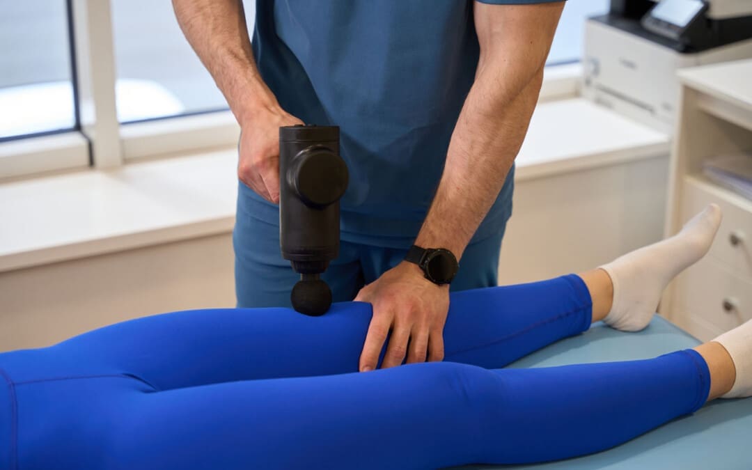
Therapeutic massage guns are great for athletes and fitness enthusiasts. Can daily massage gun use be incorporated into a daily routine?
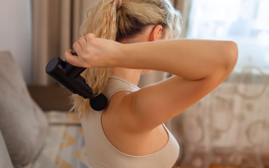
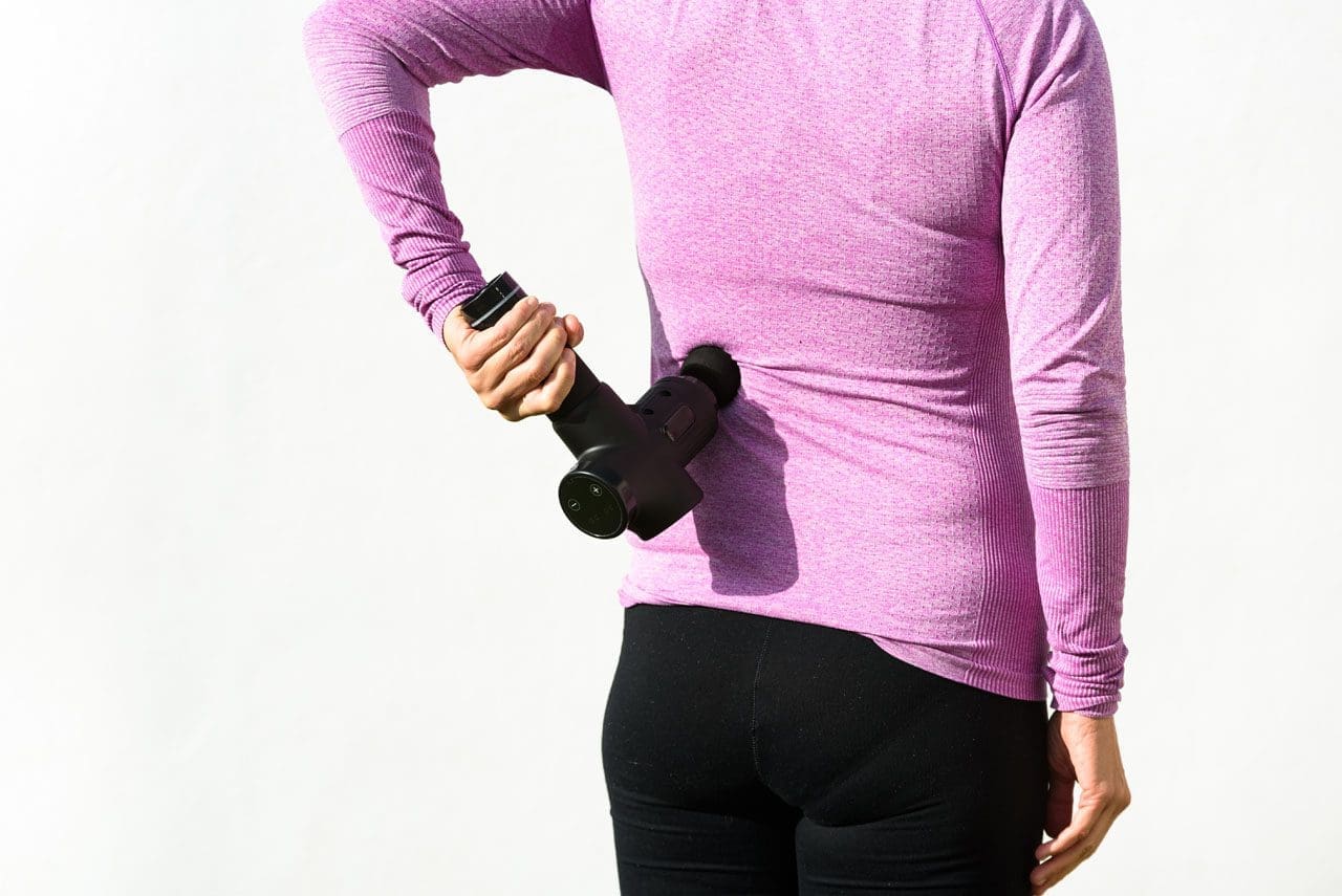
Daily Massage Gun Use
Athletes or individuals participating in recreational sports and exercise may want to consider adding a massage gun to their daily routine. Massage guns create short, repeated bursts of pressure on certain areas of the body. Most massage guns have multiple settings to allow for different preferences and levels of intensity. They can help by warming up the muscles and during recovery. While the benefits are still being researched, daily massage gun therapy can improve the quality of physical activity, exercise, and life.
Benefits
Because massage guns work by softly and repeatedly stimulating or massaging the muscles, they are gentle enough to use daily as long as they are used correctly. They offer several benefits, including increasing physical ability and reducing recovery time. One study found that consistently using a massage gun increased the quality of life in fibromyalgia patients, indicating potential beyond exercise and sports. (Kraft, K., Kanter, S., and Janik, H. 2013) Some of the benefits include:
Decrease Muscle Soreness
Delayed-onset muscle Soreness/DOMS is a feeling of soreness experienced after a workout or intense manual labor. An example is waking up and feeling sore from work or exercising the previous day (National Academy of Sports Medicine, 2024). A study compared the effectiveness of vibration and massage therapy on DOMS. Both were found to be equally effective in preventing delayed onset muscle soreness. (Imtiyaz, S., Veqar, Z., and Shareef, M. Y. 2014)
Help Prevent Muscle Fatigue
Muscle fatigue occurs when the muscles are overworked and cannot function. Reduced muscle performance can lead to shorter workouts and injuries. A study on non-athletic males found that massage therapy helped prevent or reduce muscle fatigue. Researchers also found that different massage gun settings prevented or prolonged the onset of muscle fatigue. (Otadi, K. et al., 2019)
Increase Range of Motion
Range of motion/ROM describes the flexibility of a muscle or joint in the body. When engaging in physical activity or exercise, it is vital to know and consider the range of motion of the joints to ensure proper form and decrease the risk of injury. One study found that massage therapy can improve joint performance by increasing an individual’s range of motion. (Lyu, B. J. et al., 2020) The performance of the joints depends in part on the muscles that expand and contract to move it. Another research study found that a five-minute massage on certain muscle groups improved muscle performance and range of motion. (Konrad, A. et al., 2020)
Increase Muscle Strength and Endurance
Muscle strength and endurance are key components of a healthy musculoskeletal system and are essential to avoid straining or overworking muscles. Muscle strength describes the immediate performance of a muscle or muscle group, and muscle endurance describes how long performance can be sustained. (Lyu, B. J. et al., 2020) Massage therapy has been shown to improve muscle strength and endurance by helping increase immediate performance and the number of repetitions an individual can endure. (García-Sillero, M. et al., 2021)
Improve Agility
Agility is the ability to accelerate, decelerate, stabilize, and quickly change directions while maintaining proper posture. (National Academy of Sports Medicine, 2024) It is a skill that needs to be developed. One study found that engaging in massage therapy before a workout could improve agility. (Lyu, B. J. et al., 2020)
Daily Use
A massage gun is safe to use daily as long as it is used properly. Using a massage device correctly will improve effectiveness and prevent discomfort or injury. Recommended daily usage of a massage gun includes:
Set the massage gun to the lowest setting.
Use a light touch.
Adjust the speed to comfort level, ensuring there is no pain.
Focus the massage gun on an area for 10 to 30 seconds.
Hover it across the muscle or tendon to allow the vibrations to do their job.
Do this over the desired areas as part of a warm-up and cool-down.
Safety
While massage guns are useful for warmups and workout recovery, they should be used cautiously. Individuals should use massage guns over muscles and not on bony or sensitive areas. Individuals should avoid massaging around vital organs and/or inflamed body areas as these areas could cause a jarring or uncomfortable sensation and lead to injury. If the massage gun is causing bruising, sensitivity, or discomfort, stop using it and allow the body to recover before trying again. If pain persists, talk to a healthcare provider or a physical therapist. They can determine what is causing issues, whether a massage gun is being used correctly, and whether it is an appropriate self-care treatment for the individual. There are situations where using a massage gun is not advised. Individuals should talk to a healthcare provider before using the device if they have any of the following:
Have a musculoskeletal condition like osteoporosis or arthritis.
Take blood thinners.
Have anemia.
Have other blood-related conditions.
Are older than 65.
Take several medications.
Have a broken bone or a fracture.
Have varicose veins or a history of deep vein thrombosis.
Using a massage gun can help improve performance and reduce recovery time and can be incorporated into a daily routine.
Injury Medical Chiropractic and Functional Medicine Clinic
Injury Medical Chiropractic and Functional Medicine Clinic treats patients’ injuries and chronic pain syndromes. We focus on improving ability through flexibility, mobility, and agility programs tailored to the individual. We use in-person and virtual health coaching and comprehensive care plans to ensure every patient’s care and wellness outcomes. Our providers use an integrated approach to create customized care plans that include Functional Medicine, Acupuncture, Electro-Acupuncture, and Sports Medicine principles. Our goal is to relieve pain naturally by restoring health and function to the body. Patients who need other treatment will be referred to a clinic or physician best suited for them. Dr. Jimenez has teamed up with the top surgeons, clinical specialists, medical researchers, and premier rehabilitation providers to provide our community with the best clinical treatments.
Enhance Your Lifestyle With Chiropractic!
References
Kraft, K., Kanter, S., & Janik, H. (2013). Safety and effectiveness of vibration massage by deep oscillations: a prospective observational study. Evidence-based complementary and alternative medicine : eCAM, 2013, 679248. https://doi.org/10.1155/2013/679248
National Academy of Sports Medicine. Kaminski, J. (2024). Muscle soreness & DOMS. NASM. https://blog.nasm.org/doms-muscle-sorenes
Imtiyaz, S., Veqar, Z., & Shareef, M. Y. (2014). To Compare the Effect of Vibration Therapy and Massage in Prevention of Delayed Onset Muscle Soreness (DOMS). Journal of clinical and diagnostic research : JCDR, 8(1), 133–136. https://doi.org/10.7860/JCDR/2014/7294.3971
Otadi, K., Ghasemi, M., Jalaie, S., Bagheri, H., Azizian, M., Emamdoost, S., Sarafraz, H., & Sepahvand, M. (2019). A prophylactic effect of local vibration on quadriceps muscle fatigue in non-athletic males: a randomized controlled trial study. Journal of physical therapy science, 31(3), 223–226. https://doi.org/10.1589/jpts.31.223
Lyu, B. J., Lee, C. L., Chang, W. D., & Chang, N. J. (2020). Effects of Vibration Rolling with and without Dynamic Muscle Contraction on Ankle Range of Motion, Proprioception, Muscle Strength and Agility in Young Adults: A Crossover Study. International journal of environmental research and public health, 17(1), 354. https://doi.org/10.3390/ijerph17010354
Konrad, A., Glashüttner, C., Reiner, M. M., Bernsteiner, D., & Tilp, M. (2020). The Acute Effects of a Percussive Massage Treatment with a Hypervolt Device on Plantar Flexor Muscles’ Range of Motion and Performance. Journal of sports science & medicine, 19(4), 690–694.
García-Sillero, M., Jurado-Castro, J. M., Benítez-Porres, J., & Vargas-Molina, S. (2021). Acute Effects of a Percussive Massage Treatment on Movement Velocity during Resistance Training. International journal of environmental research and public health, 18(15), 7726. https://doi.org/10.3390/ijerph18157726
National Academy of Sports Medicine. Miller, K. (2024). Speed, agility, and quickess: SAQ. NASM. https://blog.nasm.org/sports-performance/speed-agility-quickness-saq
IFM's Find A Practitioner tool is the largest referral network in Functional Medicine, created to help patients locate Functional Medicine practitioners anywhere in the world. IFM Certified Practitioners are listed first in the search results, given their extensive education in Functional Medicine