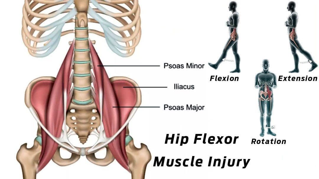
The iliopsoas muscle is a primary hip flexor that assists in the femur’s external rotation and maintains the hip joint’s strength and integrity. It also helps to stabilize the lumbar spine and pelvis. Athletes often overuse these muscles with all the sprinting, jumping, kicking, and changing directions when running, causing strains and/or tears. Repetitive hip flexion can result in chronic degenerative tendon changes. Chiropractic care and physical therapy can assist in the early phases of healing, safely transitioning to rehabilitation, and returning to physical activities.
Iliopsoas Muscle
The hip flexors are the group of muscles, including the iliac and psoas major muscles/iliopsoas and the rectus femoris/quadriceps. One of the largest and thickest muscles in the body, the psoas, extends from the lumbar vertebrae, crosses in front of each hip, and attaches to the inside top of the thigh bone. The muscle works by flexing the hip joint and lifting the upper leg towards the body. These fibers can tear if tension is more than the muscle can bear. An iliopsoas strain occurs when one or more of these hip flexor muscles become overly stretched or begin to tear.
Injury
The injury can occur from sports or everyday physical activities. This leads to inflammation, pain, and scar tissue formation. An iliopsoas injury is commonly caused by sudden movements, including sprinting, kicking, and changing direction fast while running. Individuals participating in any sports, especially cycling, running, dance, tennis, martial arts, and soccer, are more likely to experience this injury. Other contributing factors include:
Muscle tightness
Joint stiffness
Muscle weakness
Inadequate core stability
Not warming up correctly
Improper biomechanics
Decreased fitness and conditioning
Individuals will feel a sudden stinging pain or pulling sensation, usually on the front of the hip, groin, or abdominal area. Other symptoms include:
Healing and recovery depend on the severity of the injury. A minor iliopsoas muscle injury can take around three weeks to recover fully. More serious strains and tears take six to eight weeks before returning to activity, as the tissue needs time to repair before starting rehabilitation.
Chiropractic Rehabilitation and Recovery
The first steps when dealing with this injury should be P.R.I.C.E. protection, rest, ice, compression, and elevation. It is important to rest and seek treatment immediately; if left untreated, the condition could worsen, lead to a chronic condition, and require surgery. A chiropractic treatment and rehabilitation plan will consist of the following:
Soft tissue massage
Joint mobilization
A chiropractor may recommend crutches to keep the weight off the hip.
A brace can help compress and stabilize the hip flexor to expedite healing.
A flexibility and strengthening program will be implemented to target the muscles around the hip.
Core strengthening exercises will improve the stability of the pelvis area to prevent any further overuse problems.
Wearing compression clothing could also be recommended, as the clothing helps maintain muscle temperature.
Labral Tear
References
Dydyk AM, Sapra A. Psoas Syndrome. [Updated 2022 Oct 24]. In: StatPearls [Internet]. Treasure Island (F.L.): StatPearls Publishing; 2022 Jan-. Available from: https://www.ncbi.nlm.nih.gov/books/NBK551701/
Lifshitz, Liran BPt, MSc, PT; Bar Sela, Shlomo BPt MPE; Gal, Noga BPt, MSc; Martin, RobRoy PhD, PT; Fleitman Klar, Michal BPt. Iliopsoas the Hidden Muscle: Anatomy, Diagnosis, and Treatment. Current Sports Medicine Reports 19(6):p 235-243, June 2020. | DOI: 10.1249/JSR.0000000000000723
Rauseo, Carla. “THE REHABILITATION OF A RUNNER WITH ILIOPSOAS TENDINOPATHY USING AN ECCENTRIC-BIASED EXERCISE-A CASE REPORT.” International journal of sports physical therapy vol. 12,7 (2017): 1150-1162. doi:10.26603/ijspt20171150
Rubio, Manolo, et al. “Spontaneous Iliopsoas Tendon Tear: A Rare Cause of Hip Pain in the Elderly.” Geriatric orthopedic surgery & rehabilitation vol. 7,1 (2016): 30-2. doi:10.1177/2151458515627309
The hips in the lower portions of the body allow the legs to move the host from one location to another and provide stability to support the upper body’s weight. The hips will enable the torso to twist and turn without feeling pain. This is due to the various muscles and ligaments surrounding the pelvic bone and hip joint socket that allow the motion to be possible. However, when various injuries or factors start to affect the multiple muscles surrounding the pelvis or there is a chronic condition like osteoarthritis that causes wear and tear on the hip joints can cause underlying symptoms associated with the hips and cause many individuals to have difficulty when moving around. Luckily there are ways to improve hip mobility and the surrounding muscles in the hip and pelvic region of the body. Today’s article looks at the causes of the development of tight hips in the body and how different stretches can release tight hip flexor muscles. We refer our patients to certified providers that incorporate techniques and multiple therapies for many individuals suffering from hip pain and its correlating symptoms that can affect the musculoskeletal system in the hips, legs, and lumbar region of the spine. We encourage and appreciate each patient by referring them to associated medical providers based on their diagnosis when it is appropriate. We understand that education is a fantastic way when asking our providers intricated questions at the patient’s request and understanding. Dr. Jimenez, D.C., only utilizes this information as an educational service. Disclaimer
What Causes The Body To Develop Tight Hips?
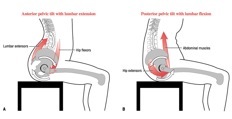
Have you been dealing with hip mobility issues? When you sit, do you feel uncomfortable, and your hip muscles become tight? Or do you have a decreased range of motion when moving your hips? It could correlate with your hips if you have been experiencing muscle pain issues in the lower extremities. The hips help stabilize the upper and lower portions of the body while providing the full leg’s range of motion. When a person begins to sit for long periods or twist their body in a weird position, it can cause the muscles that surround the hips to become shortened. Other issues, like chronic conditions, can play a role in developing tight hip flexors. Studies reveal that various pathologies affecting the hips, lumbar spine, and lower extremities could strongly correlate with restricted hip mobility that can cause harmful effects that can affect the hips. To that point, some of the symptoms associated with tight hip flexors include:
Instability
Hypermobility
Limited range of motion
Reduce muscle strength in the groin
Muscle cramps
Sharp, sudden pain in the hips, pelvis, or groin
Low back pain
Piriformis syndrome
Other research studies mentioned that hypermobility disorders could affect the hip joints. Hypermobility disorders like EDS (Ehlers-Danlos syndrome) could cause micro or macro-trauma on the hip joint and affect the ligaments surrounding the hip joint. To that point, it can cause the hip flexor muscles to become tense and potentially affect how a person moves, which then causes soft tissue injuries and chronic pain.
Hip Flexor Stretches-Video
Do you feel tight along your hips? Do you see yourself hobbling around when walking? Or do you feel aches or strains when stretching? Many of these issues correlate with tight hip flexors that could result from hip pain in the lower extremities. When a person has tight hip flexors, it could be due to them constantly sitting down, causing the hip muscles to be shortened, or chronic conditions that can affect the hip joint and muscles. However, there are various ways to prevent tight hip flexors and regain mobility back to the hips. Studies reveal that stretching combined with core stabilization can help improve the hip’s range of motion while ensuring core endurance exercises can help strengthen the surrounding muscles in the hip area. The video above shows stretches targeting the hip flexor muscles and helps improve hip mobility.
Different Stretches To Release Tight Hip Flexors
Studies have shown that the hip flexor muscles are the main contributors to lumbar spine stability when releasing tight hip flexor muscles. So when there are tight hip flexors, it can cause overlapping risk profiles to the lumbar spine, which leads to pain and impairment in performance. The best way to reduce the pain-like symptoms associated with tight hip flexors is by stretching the lower half of the body to reduce muscle strain and tightness in the hip flexors. Additional studies have found that stretching combined with exercises targeting the low back can reduce the pain caused in the low back and help improve stability and strengthen the surrounding muscles located in the hips. Now it is important to remember that stretching for at least 5-10 minutes before and after working out allows the muscles to warm up and improve flexibility. Below are some different stretches that can release tight hip flexors.
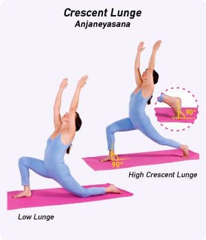
High Crescent Lunge
While standing on the mat, take a step forward to allow your right foot to be in a staggard stance *Think in a lunge position.
Bend the front knee gently while keeping the back leg straight, as this allows the heel in the back leg to be lifted off the mat; the bent front knee enables the thigh to be parallel to the floor, and the right foot is pressed flat on the mat.
Square up the hips, so they face toward the mat’s front.
Extend the arm up towards the ceiling to stretch upwards while pressing into the mat to feel the hips stretch
Hold for five breaths before slowly rising out of the lunge position and repeating on the other side.
This stretch helps release tension in the hip flexors and quads while warming up the muscles and increasing blood flow to the legs.
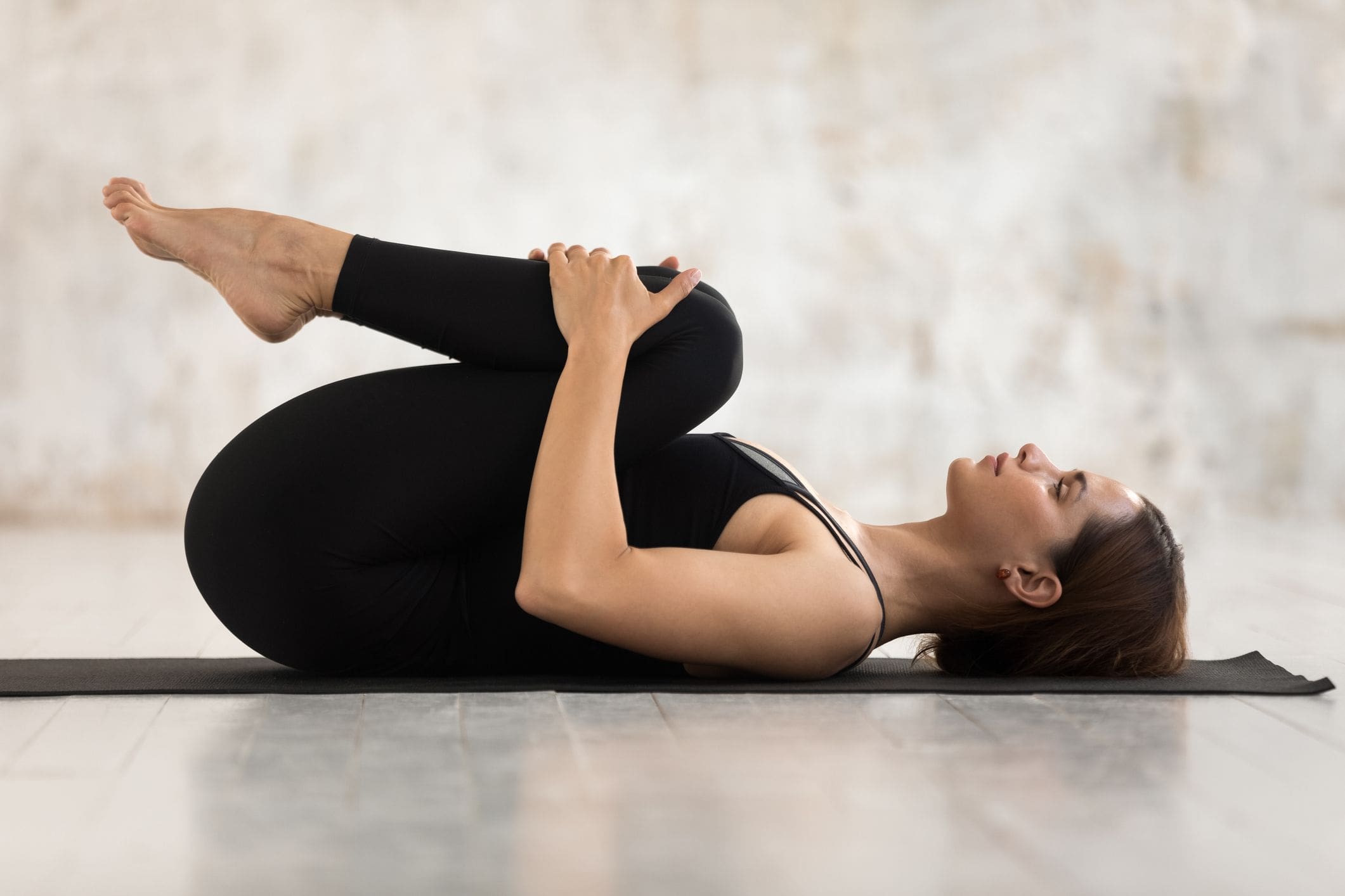
Knee-To Chest Stretch
Lie on the mat with both legs extended out and feet flexed.
Pull on the left knee to the chest while keeping the right leg straight, and the lumbar portion of the back is pressed into the mat.
Hold the position while taking deep breaths for 30 seconds to 2 minutes.
Release slowly and repeat on the right leg *You can lift both knees to your chest and rock slowly from side to side to relieve low back tension as an alternative.
This stretch is extremely helpful for tight hamstrings and allows the tense muscles on the hips and lower back to relax while increasing blood flow back to the muscles.
Piriformis Stretch
On the mat, sit with both legs extended out.
Cross the right leg over the left and place the other flat on the floor while the left foot is flexed
Place the right hand behind the body while the left elbow is on the right knee.
On inhale, press the right leg to the left while allowing the torso to twist on the right.
Take five breaths for a deeper stretch and switch sides to repeat the action with the left hand *If you have low back pain issues, the modified version allows you to use your left hand to pull the right quad in and out to the left and vice versa.
This stretch helps loosen tight muscles in the lower back, hips, and glutes. If you have sciatic nerve pain associated with piriformis syndrome, this stretch helps release muscle tension from the piriformis muscle aggravating the sciatic nerve.
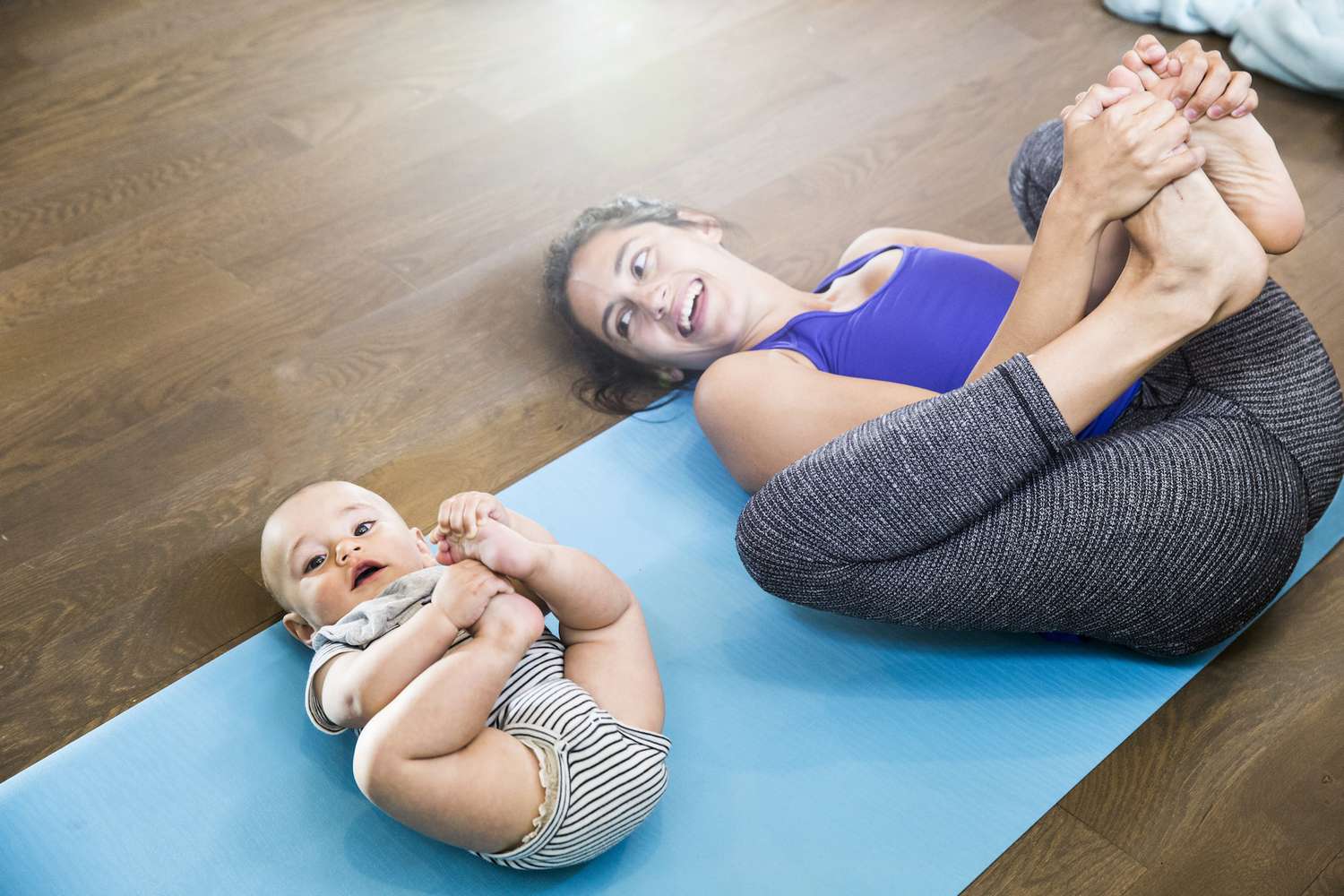
Happy Baby Pose
Lie on the mat with both knees bent and feet on the ground.
On inhale, lift the feet off the ground and grab the outer sections of the feet with your hands.
Then gently pull the feet towards the chest and allow the knees to lower to the ground, on either side of the body, while keeping the back flat on exhale.
Hold the position for at least five breaths.
This stretch helps with the inner thigh muscles or hip adductors and helps them become loose and mobile without feeling any strain or tension.
Bridge Pose
On the mat, lie on your back and sides, and extend your arms while your feet are flat on the floor with your knees bent.
Press with your heels to lift the hips and allow the feet to walk a few steps toward the body. *Keep the feet and knees hip-width apart.
Clasp hands together underneath the body and press them into the mat
Hold the position for five breaths.
This stretch helps take the pressure off the hip muscles while strengthening the glutes and abdominal muscles.
Conclusion
When it comes to releasing tight hip flexors after sitting for a long time or having hip issues affecting your low back or pelvis, Doing different stretches that target the hips can reduce the pain and release tight muscles associated with other conditions that can affect the body. The hips are important to take care of since they provide mobility and stability to the upper and lower portions of the body. They support the upper body’s weight while providing a huge range of motion to the legs. Incorporating these different stretches can reduce the pain that they have been under and help warm up the other muscles that surround the lower extremities.
References
Lee, Sang Wk, and Suhn Yeop Kim. “Effects of Hip Exercises for Chronic Low-Back Pain Patients with Lumbar Instability.” Journal of Physical Therapy Science, U.S. National Library of Medicine, Feb. 2015, https://www.ncbi.nlm.nih.gov/pmc/articles/PMC4339134/.
Moreside, Janice M, and Stuart M McGill. “Hip Joint Range of Motion Improvements Using Three Different Interventions.” Journal of Strength and Conditioning Research, U.S. National Library of Medicine, May 2012, https://pubmed.ncbi.nlm.nih.gov/22344062/.
Reiman, Michael P, and J W Matheson. “Restricted Hip Mobility: Clinical Suggestions for Self-Mobilization and Muscle Re-Education.” International Journal of Sports Physical Therapy, U.S. National Library of Medicine, Oct. 2013, https://www.ncbi.nlm.nih.gov/pmc/articles/PMC3811738/.
Reiman, Michael P, and J W Matheson. “Restricted Hip Mobility: Clinical Suggestions for Self-Mobilization and Muscle Re-Education.” International Journal of Sports Physical Therapy, U.S. National Library of Medicine, Oct. 2013, https://www.ncbi.nlm.nih.gov/pmc/articlhttps://www.ncbi.nlm.nih.gov/pmc/articles/PMC8027473/es/PMC3811738/.
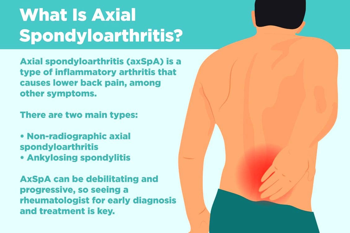
Non-radiographic axial spondyloarthritis or nr-axSpA and non-radiographic ankylosing spondylitis/AS are related. However, non-radiographic axial spondyloarthritis can present AS symptoms with active inflammation of the spine and sacroiliac/SI joints, causing back and hip pain but does not reveal joint damage on X-rays or MRIs. Injury Medical Chiropractic and Functional Medicine Clinic can explain what it means to have non-radiographic axial spondyloarthritis, how it can be managed, and what to do to prevent it from turning into ankylosing spondylitis.
Non-Radiographic Axial Spondyloarthritis
Non-radiographic axial spondyloarthritis means there are early AS symptoms but have not developed enough joint inflammation or damage to show up on an X-ray or other form of imaging. Early evidence of joint inflammation includes blurring of the joint edges and localized regions of joint erosion. It can be difficult for physicians to see these subtle changes on an x-ray.
Ankylosing Spondylitis
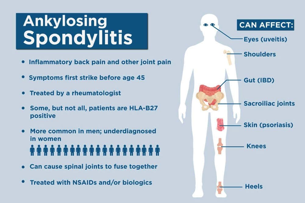
Ankylosing spondylitis, or AS, is a form of inflammatory arthritis that affects joints in the spine and elsewhere.
It is a chronic, inflammatory, autoimmune disease.
Medical research is still ongoing to determine the exact cause, but a genetic component is believed to be contributing factor.
Around 85% of individuals with ankylosing spondylitis have inherited the HLA-B27 gene, which is associated with multiple autoimmune conditions.
In the early stages, individuals will present lower back pain around the sacroiliac joints or the joints that connect the spine to the pelvis.
Later stages have more obvious X-ray findings, like the fusing of the sacroiliac joints and the lower spine that takes place over time.
Joint inflammation can progress, causing permanent joint damage and spine rigidity.
Most individuals with the condition can manage their symptoms with NSAIDs, chiropractic care, physical and massage therapy, and range of motion exercises.
Stage 1
There is no evidence of spinal inflammation on x-rays.
MRI provides more detailed images of bones and may reveal bone marrow edema or accumulation of fluid in the structures of the spinal bones and joints.
Individuals with non-radiographic axial spondyloarthritis, you are here.
Stage 2
There is visible inflammation of the spinal joints on the x-ray.
The sacroiliac joints between the spine and the pelvis are the most affected.
Stage 3
Chronic inflammation of the joints has caused bone loss and permanent joint damage, resulting in spine rigidity.
Symptoms of Non-Radiographic Axial Spondyloarthritis
There are differences between back pain associated with muscle strain and arthritis. Back pain symptoms include:
Starts to present before age 40.
It has a gradual onset and can go unnoticed for years.
Improves with movement or activity.
Eases up throughout the day.
Starts up in the evening when resting.
Other symptoms include:
Joint stiffness
Swollen fingers
Heel pain
Bilateral buttock discomfort and pain
Slowing Progression
Progression from non-radiographic axial spondyloarthritis to ankylosing spondylitis occurs in 10% – 20% of individuals over a two-year period. Progression factors include genetics, gender, degree of joint damage, and level of inflammatory markers at the time of diagnosis.
Early diagnosis and treatment can slow the progression before significant joint damage with anti-inflammatory therapy, rheumatological therapy, and targeted exercise.
Work with a specialist like an orthopedic spine specialist and rheumatologist that understands the disorder and is up to date on the most recent treatment modalities.
Individuals with non-radiographic axial spondyloarthritis should expect to have serial X-rays to gauge the progression of the disease.
Staying healthy and active is recommended to slow the progression of nr-AxSpA and AS.
Recent medical advances and lifestyle adjustments can slow the progression in most cases.
axSpA
References
Six tips for living well with ankylosing spondylitis. Available at https://www.mayoclinic.org/diseases-conditions/ankylosing-spondylitis/in-depth/6-tips-for-living-well-with-ankylosing-spondylitis/art-20478753. Accessed 11/07/2022.
Ankylosing spondylitis. Mayo Clinic. Available at https://www.mayoclinic.org/diseases-conditions/ankylosing-spondylitis/symptoms-causes/syc-20354808. Accessed 11/05/2022.
D. J. Pradeep, A. Keat, K. Gaffney, Predicting outcome in ankylosing spondylitis, Rheumatology, Volume 47, Issue 7, July 2008, Pages 942–945, https://doi.org/10.1093/rheumatology/ken195
Kucybała, Iwona, et al. “Radiologic approach to axial spondyloarthritis: where are we now and where are we heading?.” Rheumatology international vol. 38,10 (2018): 1753-1762. doi:10.1007/s00296-018-4130-1
Michelena, Xabier, López-Medina, Clementina, and Helena Marzo-Ortega. “Non-radiographic versus radiographic axSpA: what’s in a name?”.” National Center for Biotechnology Information. October 14, 2020. https://doi.org/10.1093/rheumatology/keaa422
Swift D. Ankylosing spondylitis: disease progression varies widely. Medpage Today. Accessed 11/05/2022.Available at https://www.medpagetoday.com/rheumatology/arthritis/49096
Ankylosing spondylitis is an inflammatory condition that causes pain and stiffness in the spine’s joints. Over time, it can progress to fusing the bones of the spine/vertebrae, limiting the spine’s mobility and leading to discomfort symptoms. The condition is more common in men but also affects women. There is not a known, singular cause, but individuals with a particular gene, HLA-B27, have an increased risk of developing the condition. However, the gene itself does not mean an individual has ankylosing spondylitis; genetics and other outside and environmental factors also play a role. DMARDs, or Disease-Modifying Antirheumatic Drugs, suppress inflammation. An emerging DMARD treatment uses biologics that further target and suppresses specific inflammatory compounds.
Biologics
Unlike regular medications, which are synthesized, biologics are made from and inside living sources.
They are grown in cultures or harvested from blood.
Biologics are complex and expensive.
Biologics provide some of the most powerful treatments for many conditions.
Two biologics commonly used to treat ankylosing spondylitis are:
TNF – tumor necrosis factor, TNF inhibitors are a biological medication that targets and suppresses TNF, which is involved in inflammatory processes throughout the body.
Blocking or suppressing TNF decreases inflammation and can delay the progression of ankylosing spondylitis.
It could be administered through an injection or infusion to provide the medication.
Side Effects
Side effects associated with this medication, along with other conditions, include:
IL – interleukin – IL-17 is a protein involved in inflammatory processes and conditions.
IL-17 inhibitors suppress inflammation which are newer medications that have been shown to benefit those with ankylosing spondylitis.
Doctors often administer IL-17 inhibitors through an injection.
Side Effects
Minor side effects include:
Headaches
Runny nose
Irritation at the injection site.
More serious side effects include:
Certain cancers
Severe infections
High blood pressure
Other Treatments
Treatment goals for ankylosing spondylitis include:
Slowing the disorder’s progression.
Decreasing inflammation.
Reducing pain.
Improving or maintaining joint and spinal range of motion.
Biologics are not the first-line treatment for ankylosing spondylitis.
Providers usually first treat an initial diagnosis with anti-inflammatory medications, like NSAIDs, to decrease inflammation and slow the condition’s progression.
Chiropractic care and physical therapy are recommended to maintain and/or improve posture, muscle strength, and endurance.
Recommended lifestyle and nutritional modifications.
Posture training stretches and exercises.
Optimal movement strategies to safely and confidently complete everyday tasks.
Biologic Medications
Biologics for ankylosing spondylitis may or may not be suitable. These medications are meant to help relieve the inflammation associated with the condition and slow the disorder’s progression. Your healthcare team will work with you to determine the right treatment option/s and explain the benefits, risks, and types of treatment. They will monitor the condition, track progress, and adjust the plan accordingly.
Assessing Hormone Therapy
References
Ankylosing spondylitis. (n.d.) National Institute of Arthritis and Musculoskeletal and Skin Diseases. U.S. Department of Health and Human Services. Available at: https://www.niams.nih.gov/health-topics/ankylosing-spondylitis#:~:text=Ankylosing%20spondylitis%20is%20a%20type,the%20spine%20can%20cause%20stiffness (Accessed: October 12, 2022).
Chen C, Zhang X, Xiao L, Zhang X, Ma X. Comparative Effectiveness of Biologic Therapy Regimens for Ankylosing Spondylitis: A Systematic Review and a Network Meta-Analysis. Medicine (Baltimore). 2016 Mar;95(11):e3060. doi: 10.1097/MD.0000000000003060. PMID: 26986130; PMCID: PMC4839911.
Gerriets V, Goyal A, Khaddour K. Tumor Necrosis Factor Inhibitors. [Updated 2022 July 4]. In: StatPearls [Internet]. Treasure Island (FL): StatPearls Publishing; 2022 Jan-. Available from: https://www.ncbi.nlm.nih.gov/books/NBK482425/
Lindström, U., Olofsson, T., Wedrén, S. et al. Biological treatment of ankylosing spondylitis: a nationwide study of treatment trajectories on a patient level in clinical practice. Arthritis Res Ther 21, 128 (2019). https://doi.org/10.1186/s13075-019-1908-9
Yin, Y., Wang, M., Liu, M. et al. Efficacy and safety of IL-17 inhibitors for the treatment of ankylosing spondylitis: a systematic review and meta-analysis. Arthritis Res Ther 22, 111 (2020). https://doi.org/10.1186/s13075-020-02208-w
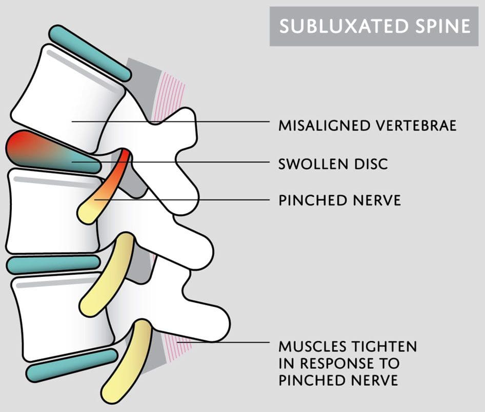
Subluxation is when a joint shifts out of alignment, which can happen to any joint in the body. Spinal subluxation indicates a misalignment of one or more portions of the spinal vertebrae. It is common in the spine from all the reaching, bending, twisting, and flexing the body goes through. Spinal subluxations, if left untreated, can cause disc degeneration, permanent nerve damage, neurological conditions, and chronic pain symptoms. A subluxation chiropractor will realign and decompress the spine combined with massage therapy to relax the muscles and restore mobility and function.
Subluxation Chiropractor
Some subluxations don’t cause any problems or pain, but that doesn’t mean they aren’t affecting the back and body. A spinal subluxation can cause long-term problems by:
Research shows that spinal subluxations can affect many facets of the body. Long-term effects may include:
Sleep problems
Low energy
Brain fog
Mood swings
Anxiety and depression
Digestive issues
Respiratory problems
Bone spurs
Spinal arthritis
Chiropractic Care
When the spine is out of alignment, it can cause issues throughout the body. Changes in one area affect the rest of the body. A subluxation chiropractor looks at the spine’s neurological and mechanical components and aims to reset everything back into its proper position. Similar to the way a massage helps the mind and body relax and de-stress, a spinal adjustment helps by:
Increasing circulation
Relieving discomfort and pain
Releasing tension
Improving mood
Reducing stress levels
Improving sleep function
Increasing energy levels
When the spine is properly aligned, the body can operate at its full potential.
Adrenal Dysfunction
References
Brian S. Budgell, Reflex effects of subluxation: the autonomic nervous system, Journal of Manipulative and Physiological Therapeutics, Volume 23, Issue 2,
2000, Pages 104-106, ISSN 0161-4754, https://doi.org/10.1016/S0161-4754(00)90076-9. (https://www.sciencedirect.com/science/article/pii/S0161475400900769)
Green, J D et al. “Anterior subluxation of the cervical spine: hyperflexion sprain.” AJNR. American journal of neuroradiology vol. 2,3 (1981): 243-50.
Meyer, S. “Thoracic spine trauma.” Seminars in roentgenology vol. 27,4 (1992): 254-61. doi:10.1016/0037-198x(92)90004-l
Neva MH, Häkkinen A, Mäkinen H, et al. High prevalence of asymptomatic cervical spine subluxation in patients with rheumatoid arthritis waiting for orthopedic surgeryAnnals of the Rheumatic Diseases 2006;65:884-888.
Nourollahi, Maryam, et al. “Awkward trunk postures and their relationship with low back pain in hospital nurses.” Work (Reading, Mass.) vol. 59,3 (2018): 317-323. doi:10.3233/WOR-182683
Vernon, Howard. “Historical overview and update on subluxation theories().” Journal of chiropractic humanities vol. 17,1 (2010): 22-32. doi:10.1016/j.echu.2010.07.001
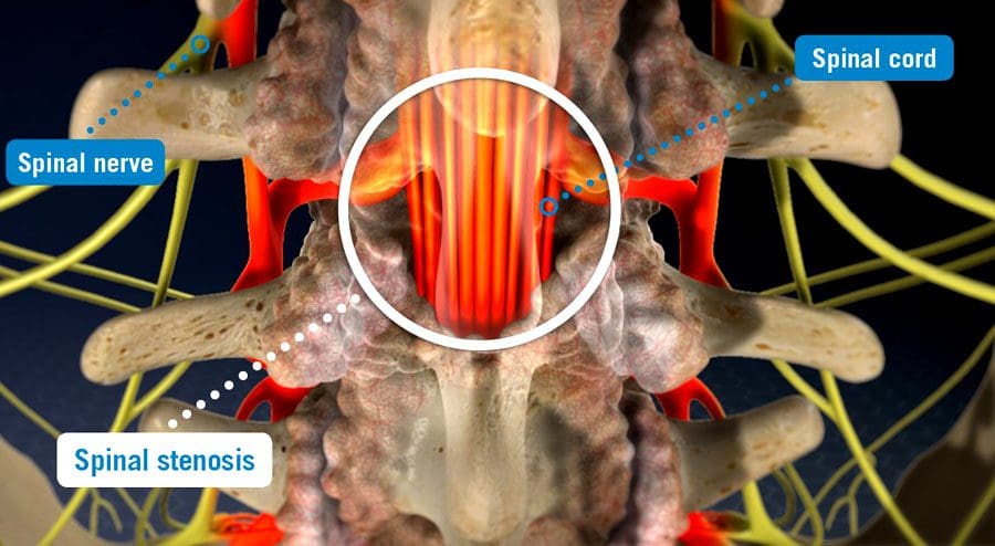
Spinal stenosis is when space somewhere along or within the spine begins to narrow, closing off the ability of normal/comfortable movement and nerve circulation. It can affect different areas, including the cervical/neck, lumbar/low back, and, less commonly, the thoracic/upper or mid-back regions causing tingling, numbness, cramping, pain, muscle weakness, or a combination in the back, leg/s, thighs, and buttocks. There can be various factors causing the stenosis; correct diagnosing is the first step, and where a spinal stenosis MRI comes in.
Spinal Stenosis MRI
Stenosis can be challenging to diagnose as it is more of a symptom/complication than a condition, often caused by herniated discs, bone spurs, a congenital condition, post-surgery, or after an infection. Magnetic resonance imaging/MRI is a common test used in diagnosis.
Diagnosis
A healthcare professional, like a chiropractor, physical therapist, spine specialist, or physician, will begin with understanding symptoms and medical history.
A physical exam will be conducted to learn more about the location, duration, positions, or activities that decrease or worsen the symptoms.
Additional tests include muscle strength, gain analysis, and balance testing to help better understand where the pain is coming from.
To confirm a diagnosis, imaging will be required to see what is going on.
An MRI uses computer-generated imaging to produce images that show bone and soft tissues, like muscles, nerves, and tendons, and if they are compressed or irritated.
A healthcare professional and MRI technician will go over the safety requirements before the imaging.
Because the machine uses powerful magnets, there can be no metal on or in the body, like implanted prostheses or devices that include:
A different imaging test may be used if an individual cannot have an MRI like a CT scan.
An MRI can range from several minutes to an hour or longer, depending on how many positions are necessary to isolate the injured area and get a clear image. The test is painless, but sometimes individuals are asked to maintain a specific position that could be uncomfortable. The technician/s will ask if there is discomfort and offer any help to make the experience as easy as possible.
Treatment
Not all cases of stenosis cause symptoms, but there are treatment options that a healthcare professional can recommend.
Conservative care is the first recommendation that includes chiropractic, decompression, traction, and physical therapy.
Treatment increases muscle strength, improves range of motion, improves posture and balance, decreases discomfort symptoms, and incorporates strategies to prevent and manage symptoms.
Prescription medications could be part of a larger treatment plan.
Surgery could become an option in more severe cases where conservative care is not working.
Spinal Stenosis
References
Database of Abstracts of Reviews of Effects (DARE): Quality-assessed Reviews [Internet]. York (UK): Centre for Reviews and Dissemination (UK); 1995-. Diagnosis of lumbar spinal stenosis: an updated systematic review of the accuracy of diagnostic tests. 2013. Available from: https://www.ncbi.nlm.nih.gov/books/NBK142906/
Ghadimi M, Sapra A. Magnetic Resonance Imaging Contraindications. [Updated 2022 May 8]. In: StatPearls [Internet]. Treasure Island (FL): StatPearls Publishing; 2022 Jan-. Available from: https://www.ncbi.nlm.nih.gov/books/NBK551669/
Gofur EM, Singh P. Anatomy, Back, Vertebral Canal Blood Supply. [Updated 2021 Jul 26]. In: StatPearls [Internet]. Treasure Island (FL): StatPearls Publishing; 2022 Jan-. Available from: https://www.ncbi.nlm.nih.gov/books/NBK541083/
Lurie, Jon, and Christy Tomkins-Lane. “Management of lumbar spinal stenosis.” BMJ (Clinical research ed.) vol. 352 h6234. 4 Jan. 2016, doi:10.1136/bmj.h6234
Stuber, Kent, et al. “Chiropractic treatment of lumbar spinal stenosis: a review of the literature.” Journal of chiropractic medicine vol. 8,2 (2009): 77-85. doi:10.1016/j.jcm.2009.02.001
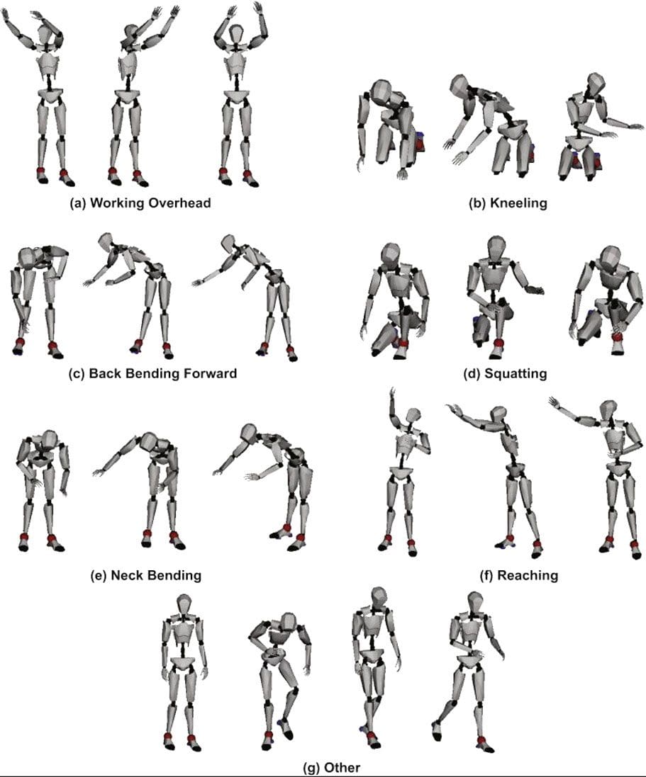
Functional fitness conditioning exercises train the muscles for everyday activities safely and efficiently. It refers to exercises that simulate daily movements like standing, bending, reaching, jumping, twisting, pushing, pulling, squatting, lunging, turning, walking, and running. These exercises improve functional body strength to train the muscles to work together and prepare for daily tasks and chores that individuals do at home, at work, or in sports activities. Strengthening the muscles the same way they are needed for everyday tasks reduces the risk of injury, allowing individuals to go through the day without worrying about straining or pulling something.
Functional Fitness Conditioning
Functional fitness conditioning exercises can be done at home, park, or gym and is an excellent way to combat restlessness and keep the body moving. Training the muscles to work the way they do daily prepares the body to move optimally and efficiently in various situations. Functional fitness exercises simultaneously utilize upper and lower body muscles to increase core stability and train the whole body. Examples include:
Combining strength training with exercises that mirror the movements of daily life increase overall strength and improves:
Balance
Endurance
Flexibility
Mobility
Various gyms or fitness centers may offer functional fitness conditioning classes or incorporate functional fitness into their exercise courses. Exercise equipment can include:
Primarily using body weight makes this strength training simple and safe for almost anybody. Functional fitness conditioning is laidback, requiring less equipment and intensity. The objective is to develop and strengthen the entire body to handle daily life while minimizing the risk of injury, making it suitable for individuals of all ages and fitness levels.
Benefits
These include benefits for overall health and physical performance.
Everyday life
Quality of life improves because of the stress relief factor.
This type of training has been reported to be more enjoyable because the training is for everyday life.
Increased muscle memory
Builds muscle and core strength and exercises the brain to increase memory.
Increased mobility
Improves balance
Coordination
Flexibility
Muscle strength
Agility
Improves balance and posture
Training and strengthening the muscles to manage/balance weight correctly reduces stress on the body.
Faster injury recovery
A strengthened body operates at optimal levels that improve repairing and healing capabilities.
It is recommended to check with your doctor before starting any new exercise program, and pregnant women should check with their doctors. A personal trainer, functional medicine, or sports chiropractor can discuss available conditioning options specific to the individual.
Functional Fitness
References
Fahlman, Mariane M et al. “Effects of resistance training on functional ability in elderly individuals.” American Journal of health promotion: AJHP vol. 25,4 (2011): 237-43. doi:10.4278/ajhp.081125-QUAN-292
Gerards, Marissa H G, et al. “Perturbation-based balance training to improve balance control and reduce falls in older adults – study protocol for a randomized controlled trial.” BMC geriatrics vol. 21,1 9. 6 Jan. 2021, doi:10.1186/s12877-020-01944-7
Pacheco, Matheus Maia, et al. “Functional vs. Strength training in adults: specific needs define the best intervention.” International Journal of sports physical therapy vol. 8,1 (2013): 34-43.
Pullyblank, Kristin, et al. “Effects of the Strong Hearts, Healthy Communities Intervention on Functional Fitness of Rural Women.” The Journal of rural health: official Journal of the American Rural Health Association and the National Rural Health Care Association vol. 36,1 (2020): 104-110. doi:10.1111/jrh.12361
IFM's Find A Practitioner tool is the largest referral network in Functional Medicine, created to help patients locate Functional Medicine practitioners anywhere in the world. IFM Certified Practitioners are listed first in the search results, given their extensive education in Functional Medicine