For wrestling athletes or those thinking about getting into the sport, can knowing about common injuries help in rehabilitation and prevention?
Wrestling Injuries
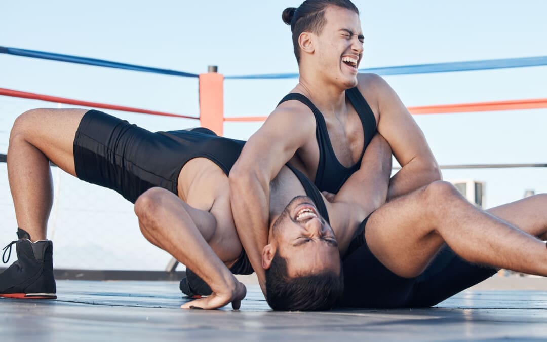
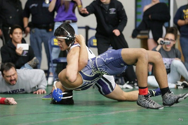
Wrestling is an intense and demanding sport. Studies have found that football and wrestling are the two high school sports with the highest risk of serious injury to athletes (Center for Injury Research and Policy, 2009). The injury rate for college wrestlers is 9 injuries per 1,000 athlete exposures. (Kroshus, E. et al., 2018) While most wrestling injuries include strains and sprains, there can also be serious traumatic and unusual injuries. Using proper safety gear and learning correct techniques can significantly reduce the risk of injuries. The majority occur during competition.
Common
The most common wrestling injuries are similar to those in other sports and include:
Muscle Soreness
Muscle soreness that is experienced 12 to 48 hours after an intense workout or competition.
Resting is often all that is needed to recover.
Bruises and Contusions
Sparring, take-downs, and hard landings can result in various bruises and contusions.
Sprains and Strains
Rest, ice, compression, and elevation are recommended to treat sprains and strains immediately.
Ankle Sprains
Ankle sprains occur when surrounding ligaments stretch and tear around the joint.
Wrist Sprains
Typically, it occurs when stretching or tearing the ligaments.
Falling or landing on the hands is a common cause.
Overtraining Syndrome
Frequently occurs in athletes who train beyond the body’s ability to recover.
Dehydration
When trying to make weight, dehydration can be a serious health problem that many wrestlers experience.
Other Injuries
Other injuries common in wrestling:
Wrist tendinitis
Finger fractures
Iliotibial band syndrome
Meniscus tears
Groin pull
Hamstring pull or tear
Pulled calf muscle
Achilles tendonitis
Achilles tendon rupture
Clavicle/Collarbone fracture
Concussion
Serious
The forcing of a joint beyond its normal range of motion is the most common cause of serious injuries. The most serious wrestling injuries affect the neck, shoulder, elbow, and knee and include:
Neck
The cervical vertebrae are often forced into vulnerable positions during various techniques and movements, which can result in a neck injury. Common types include:
Neck Strain
Whiplash
Cervical Fracture
Shoulder
A combination of leverage and twisting causes most upper body and shoulder injuries during competition. Types of shoulder injuries include:
Rotator cuff injury
Shoulder separation
Shoulder dislocation
Elbow Dislocation
Elbows are under tremendous strain when maneuvering.
Dislocations of the radial head are often related to the athlete bracing for a fall on an outstretched arm during take-downs.
Knee
Most knee injuries occur to the ligaments of the knee joint.
These include anterior and posterior cruciate ligament or ACL/PCL injuries.
Safety
Wrestling requires flexibility, strength, and proper technique to prevent injury, combined with thorough instruction and coaching and following basic safety precautions. Some tips include.
Safety Gear
Wear appropriate headgear and mouthguards during practices, meets, and tournaments.
Improving Joint Flexibility
Wrestlers with a high degree of shoulder flexibility have fewer injuries.
The flexibility of the lower back, hamstrings, elbows, and cervical spine should also be worked on.
Gain or Lose Weight Safely
Avoid dramatic weight loss and weight-gaining strategies by maintaining healthy nutrition and hydration during the season.
Avoiding Dangerous Holds and Slam Moves
Safe wrestling techniques need to be followed as these can generate severe injuries.
Regardless of how common or seemingly not serious an injury or medical condition is, it’s important to rest and recover and tell a coach and health care professional, as some injuries and conditions can become serious if left untreated. Injury Medical Chiropractic and Functional Medicine Clinic focuses on and treats injuries and chronic pain syndromes through personalized care plans that improve ability through flexibility, mobility, and agility programs to relieve pain. Our providers use an integrated approach to create personalized care plans for each patient, including Functional Medicine, Acupuncture, Electro-Acupuncture, and Sports Medicine principles. Our goal is to relieve pain naturally by restoring health and function to the body. If other treatment is needed, Dr. Jimenez has teamed up with top surgeons, clinical specialists, medical researchers, and rehabilitation providers to provide the most effective treatments.
Kroshus, E., Utter, A. C., Pierpoint, L. A., Currie, D. W., Knowles, S. B., Wasserman, E. B., Dompier, T. P., Marshall, S. W., Comstock, R. D., & Kerr, Z. Y. (2018). The First Decade of Web-Based Sports Injury Surveillance: Descriptive Epidemiology of Injuries in US High School Boys’ Wrestling (2005-2006 Through 2013-2014) and National Collegiate Athletic Association Men’s Wrestling (2004-2005 Through 2013-2014). Journal of athletic training, 53(12), 1143–1155. doi.org/10.4085/1062-6050-154-17
Can physical therapies help treat a high steppage gait from injury or medical conditions and restore normal gait patterns for individuals who have or are developing one?
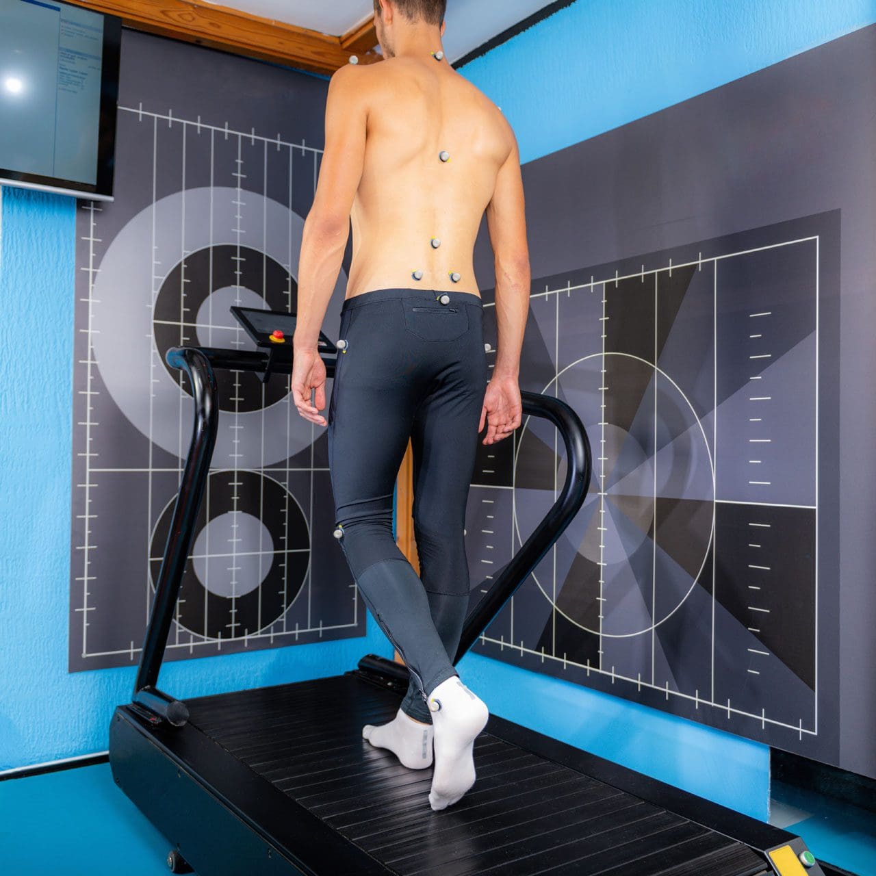
Walking or gait anthropometric analysis on a treadmill
Neuropathic Gait
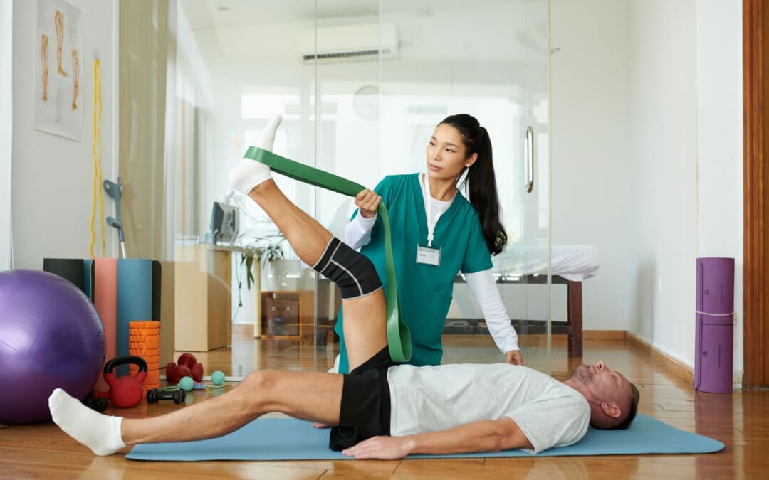
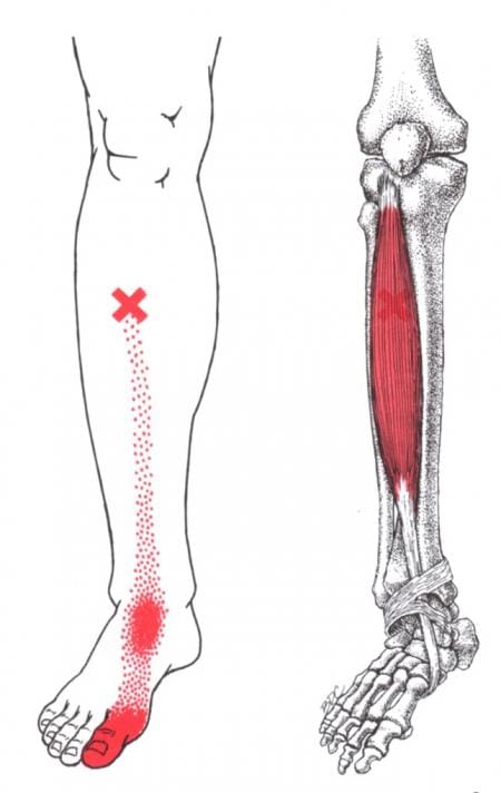
Neuropathic gait, aka equine or high steppage gait, is a type of walking abnormality that causes individuals to raise their hips to lift their legs higher than normal. It occurs when individuals have a foot drop or ankle equinus due to loss of dorsiflexion. As a result, the foot hangs with the toes pointing down, causing the toes to drag while walking. The foot may appear floppy when it drops. Foot drop is caused by weakness or paralysis of the anterior tibialis muscle in front of the shin bone. The anterior tibialis muscle contracts to help flex the foot and ankle while walking, ensuring the foot clears the floor and doesn’t drag. Individuals with anterior tibialis weakness or paralysis may have a neuropathic gait and excessively bend the hip and knee while stepping forward, lifting their leg high off the floor to clear the foot to avoid tripping. A physical therapy team can help with a high steppage gait pattern after illness or injury.
Causes
Conditions that can cause anterior tibialis weakness or paralysis and a high steppage gait pattern include:
Sciatica
Pain caused by compression or irritation of the sciatic nerve starts in the lower back and travels down the back of the leg. (McCabe, F. J., McCabe, J. P. 2016)
Peroneal Nerve Injury
Damage to the peroneal nerve branches from the sciatic nerve that help move the lower leg and foot. (Johns Hopkins Medicine. 2024)
Multiple Sclerosis
An autoimmune disease that damages nerve cells in the brain and spinal cord. (Taylor, P. N. et al., 2016)
Balance exercises will help improve overall proprioception, or the sense of the body’s position and movement.
Neuromuscular electrical stimulation, or NMES, can help improve the function of the muscle. (Hollis, S., McClure, P. 2017)
The electrical stimulation artificially contracts the muscle to restore proper function.
For anterior tibialis weakness caused by sciatica, back decompression exercises may be prescribed to relieve pressure off the sciatic nerve.
The exercises release the nerve to restore normal signal transmission up and down the nerve in the lower back.
Neuromuscular electrical stimulation may also be used to help improve muscle function.
Assistive Walking Devices
A therapist may suggest using an assistive device to help the patient walk properly. This could include a wheeled walker or a quad cane. A temporary solution to anterior tibialis weakness is to elevate the foot while walking with an elastic band. Tie a band around the leg below the knee and secure it around the ball of the foot. When swinging the leg forward, the band pulls the foot up. Using it as a temporary solution may help maintain safe mobility. Sometimes, paralysis of the anterior tibialis muscle can become permanent. In this case, individuals may benefit from a special brace called an ankle-foot orthosis. The brace helps to lift the foot and toes off the ground.
For individuals concerned about losing their balance and falling, there are ways to improve walking patterns to stay safe. A healthcare provider may recommend physical therapy to correct gait, strengthen the anterior tibialis muscle, improve balance, and educate on injury prevention. Individuals should discuss symptoms and conditions with a primary physician, healthcare provider, or specialist to guide them in the right direction and determine the best treatment.
Injury Medical Chiropractic and Functional Medicine Clinic uses an integrated approach personalized to the individual that focuses on what works for them and treats injuries and chronic pain syndromes through personalized care plans that improve ability through flexibility, mobility, and agility programs to relieve pain. If other treatment is needed, Dr. Jimenez has teamed up with top surgeons, clinical specialists, medical researchers, and rehabilitation providers to provide the most effective treatments.
Control Foot Motion and Posture
References
McCabe, F. J., & McCabe, J. P. (2016). An Unusual Presentation of Right-Sided Sciatica with Foot Drop. Case reports in orthopedics, 2016, 9024368. doi.org/10.1155/2016/9024368
Kaykisiz, E. K., & Unluer, E. E. (2017). An Unexpected Reason for Isolated Foot Drop: Acute Stroke. Pakistan journal of medical sciences, 33(5), 1288–1290. doi.org/10.12669/pjms.335.13593
Taylor, P. N., Wilkinson Hart, I. A., Khan, M. S., & Slade-Sharman, D. E. (2016). Correction of Footdrop Due to Multiple Sclerosis Using the STIMuSTEP Implanted Dropped Foot Stimulator. International journal of MS care, 18(5), 239–247. doi.org/10.7224/1537-2073.2015-038
Hollis, S., & McClure, P. (2017). Intramuscular Electrical Stimulation for Muscle Activation of the Tibialis Anterior After Surgical Repair: A Case Report. The Journal of orthopaedic and sports physical therapy, 47(12), 965–969. doi.org/10.2519/jospt.2017.7368
Can understanding night cravings help individuals who constantly eat at night plan meals that satisfy and choose nutritious snacks?
Eating At Night
Snacking after dinner and eating at night is common and not bad; however, snacking mindfully can help one truly enjoy and savor snacks. Consider some of the reasons why you might be hungry or not completely satisfied after dinner. Improving the nutritional value of nighttime snacks can make late-night hunger work toward meeting nutritional needs. Common reasons include:
Not meeting the right macronutrient balance during dinner.
Not being completely satisfied with dinner.
Dehydration.
Macronutrient Profile
Getting the right amount of carbohydrates, fat, and protein during dinner is integral to feeling satisfied. Adults need 130g of carbohydrates, 56g of protein, and 3.7L of water daily. The amount of fat required varies, but monounsaturated and polyunsaturated fats are the most healthy fats to consume, helping the body feel satisfied. Several studies show that eating protein during a meal reduces hunger and decreases cravings. (Kohanmoo, A. et al., 2020)
Unsatisfying Dinner
Another reason individuals eat at night is that they are unsatisfied with dinner. Eating satiating foods can help the mind and body feel full throughout the evening.
Satiety is the sense of satisfaction from food.
Foods high in fiber and healthy fats are known to help produce satisfaction.
When the body is full and satisfied, it produces hormones that signal to the brain there is no need to continue eating.
Try to plan healthy meals that are genuinely exciting to eat.
Create time to cook and make and eat meals you can genuinely enjoy.
Dehydration
Sometimes, when the body is dehydrated, it can have difficulty distinguishing thirst from hunger. As a result, some may eat in reaction to dehydration. This isn’t always bad, as some foods, specifically water-rich foods like melon and other fruits, can provide hydration. But sometimes, individuals don’t realize they are misreading their body’s thirst for hunger, and they reach for any food. They are still dehydrated, so they keep eating. If hunger persists after dinner, drink a glass of water and wait 20 minutes to see if that impacts hunger.
Maximize Nutrition
Snacking at night is not bad, but it is wise to plan to ensure the body gets the right balance of nutrients.
Satisfy Cravings
Many crave something sweet after dinner or later on. Eating healthy foods that satisfy cravings will help trigger hormones that tell the body it is done eating. Keep your favorite fruits and vegetables for a quick bite to get some sweetness and fiber. Vegetables like red bell peppers and carrots provide sweetness and crunchiness and can be satisfying. One small red pepper provides 100% of the daily recommended Vitamin C in 20 calories. (U.S. Department of Agriculture. Agricultural Research Service. 2018)
Foods that Promote Sleep
The foods chosen can affect sleep. Whole grains, walnuts, cherries, and kiwi increase serotonin and decrease the stress hormone cortisol. Complex carbohydrates contain melatonin, a hormone responsible for feeling sleepy. A whole-grain snack is a healthy choice before going to bed. (Nisar, M. et al., 2019) Some research shows that dark chocolate is rich in magnesium and can help promote deep sleep. However, it also contains caffeine, which can inhibit sleep. If dark chocolate is a favorite, make sure to eat it early enough in the evening.
Alternative Nighttime Routine
Some people eat out of boredom at night. To curb this, individuals in this category should change their routines. Here are a few tips to help adjust nighttime habits.
Healthy After-Dinner Activities
Go for a quick walk after dinner. 10 to 20 minutes can help, as physical activity signals the shift from dinner to other evening activities.
It also gives the body a chance to feel the fullness sensation.
Hobbies and other light meditative activities can help take the mind off eating.
Watch TV Mindfully
Many individuals eat more at night because snacking can go on and on in front of the television.
Use smart and healthy snacking strategies like portion control.
Remember to take a drink of water in between snacking.
Stay active – simple chores or activities while watching TV can help avoid overeating.
Rest and Sleep
Not getting enough sleep has been linked with increased appetite. (Hibi, M. et al., 2017)
Engage in activities to encourage rest.
Meditation can help calm down the mind and body.
Consider going to bed earlier.
Using an integrated approach, Dr. Jimenez’s Functional Medicine Team aims to restore health and function to the body through Nutrition and Wellness, Functional Medicine, Acupuncture, Electro-Acupuncture, and Sports Medicine protocols. We focus on what works for the individual through researched methods and total wellness programs.
Eating Right to Feel Better
References
Kohanmoo, A., Faghih, S., & Akhlaghi, M. (2020). Effect of short- and long-term protein consumption on appetite and appetite-regulating gastrointestinal hormones, a systematic review and meta-analysis of randomized controlled trials. Physiology & behavior, 226, 113123. doi.org/10.1016/j.physbeh.2020.113123
Nisar, M., Mohammad, R. M., Arshad, A., Hashmi, I., Yousuf, S. M., & Baig, S. (2019). Influence of Dietary Intake on Sleeping Patterns of Medical Students. Cureus, 11(2), e4106. doi.org/10.7759/cureus.4106
Hibi, M., Kubota, C., Mizuno, T., Aritake, S., Mitsui, Y., Katashima, M., & Uchida, S. (2017). Effect of shortened sleep on energy expenditure, core body temperature, and appetite: a human randomised crossover trial. Scientific reports, 7, 39640. doi.org/10.1038/srep39640
Individuals suffering from a jammed finger: Can knowing the signs and symptoms of a finger that is not broken or dislocated allow for at-home treatment and when to see a healthcare provider?
Jammed Finger Injury
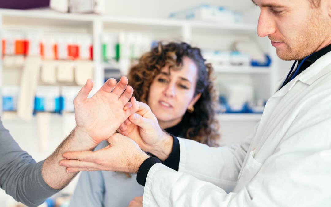
A jammed finger, also known as a sprained finger, is a common injury when the tip of a finger is forcefully pushed toward the hand, causing the joint to become compressed. This can cause pain and swelling in one or more fingers or finger joints and cause ligaments to stretch, sprain, or tear. (American Society for Surgery of the Hand. 2015) A jammed finger can often heal with icing, resting, and taping. This is often enough to allow it to heal in a week or two if no fractures or dislocations are present. (Carruthers, K. H. et al., 2016) While painful, it should be able to move. However, if the finger cannot wiggle, it may be broken or dislocated and require X-rays, as a broken finger or joint dislocation can take months to heal.
Treatment
Treatment consists of icing, testing, taping, resting, seeing a chiropractor or osteopath, and progressive regular use to regain strength and ability.
Ice
The first step is icing the injury and keeping it elevated.
Use an ice pack or a bag of frozen vegetables wrapped in a towel.
Ice the finger in 15-minute intervals.
Take the ice off and wait until the finger returns to its normal temperature before re-icing.
Do not ice a jammed finger for over three 15-minute intervals in one hour.
Try To Move The Affected Finger
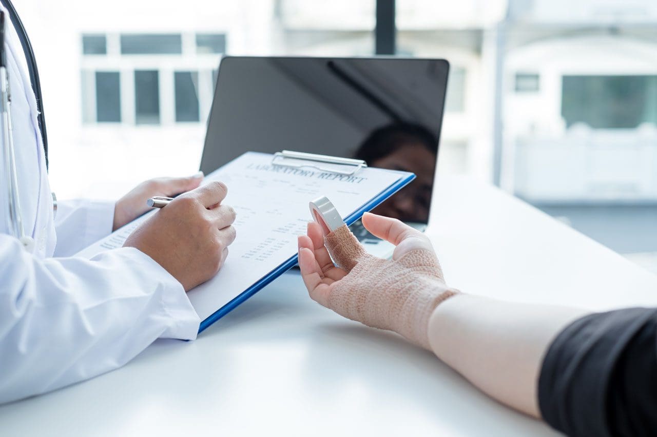
If the jammed finger does not move easily or the pain gets worse when trying to move it, you need to see a healthcare provider and have an X-ray to check for a bone fracture or dislocation. (American Society for Surgery of the Hand. 2015)
Try to move the finger slightly after swelling, and the pain subsides.
If the injury is mild, the finger should move with little discomfort for a short time.
Tape and Rest
If the jammed finger is not broken or dislocated, it can be taped to the finger next to it to keep it from moving, known as buddy taping. (Won S. H. et al., 2014)
Medical-grade tape and gauze between the fingers should be used to prevent blisters and moisture while healing.
A healthcare provider may suggest a finger splint to keep the jammed finger lined up with the other fingers.
A splint can also help prevent a jammed finger from re-injury.
Resting and Healing
A jammed finger must be kept still to heal at first, but eventually, it needs to move and flex to build strength and flexibility.
Targeted physical therapy exercises can be helpful for recovery.
A primary care provider might be able to refer a physical therapist to ensure the finger has a healthy range of motion and circulation as it heals.
A chiropractor or osteopath can also provide recommendations for helping rehabilitate the finger, hand, and arm to normal function.
Easing The Finger Back to Normal
Depending on the extent of the injury, the finger and hand can be sore and swollen for a few days or weeks.
It can take some time to start feeling normal.
Once the healing process begins, individuals will want to return to using it normally.
Avoiding using a jammed finger will cause it to lose strength, which can, over time, further weaken it and increase the risk of re-injury.
If the pain and swelling persist, see a healthcare provider to get it checked for a possible fracture, dislocation, or other complication as soon as possible, as these injuries are harder to treat if the individual waits too long. (University of Utah Health, 2021)
At Injury Medical Chiropractic and Functional Medicine Clinic, we passionately focus on treating patients’ injuries and chronic pain syndromes and improving ability through flexibility, mobility, and agility programs tailored to the individual. Our providers use an integrated approach to create personalized care plans that include Functional Medicine, Acupuncture, Electro-Acupuncture, and Sports Medicine protocols. Our goal is to relieve pain naturally by restoring health and function to the body. If the individual needs other treatment, they will be referred to a clinic or physician best suited for them. Dr. Jimenez has teamed up with the top surgeons, clinical specialists, medical researchers, and premier rehabilitation providers to provide the most effective clinical treatments.
Carruthers, K. H., Skie, M., & Jain, M. (2016). Jam Injuries of the Finger: Diagnosis and Management of Injuries to the Interphalangeal Joints Across Multiple Sports and Levels of Experience. Sports health, 8(5), 469–478. doi.org/10.1177/1941738116658643
Won, S. H., Lee, S., Chung, C. Y., Lee, K. M., Sung, K. H., Kim, T. G., Choi, Y., Lee, S. H., Kwon, D. G., Ha, J. H., Lee, S. Y., & Park, M. S. (2014). Buddy taping: is it a safe method for treatment of finger and toe injuries?. Clinics in orthopedic surgery, 6(1), 26–31. doi.org/10.4055/cios.2014.6.1.26
Can understanding the body’s hinge joints and how they operate help with mobility and flexibility problems and manage conditions for individuals with difficulty fully bending or extending their fingers, toes, elbows, ankles, or knees?
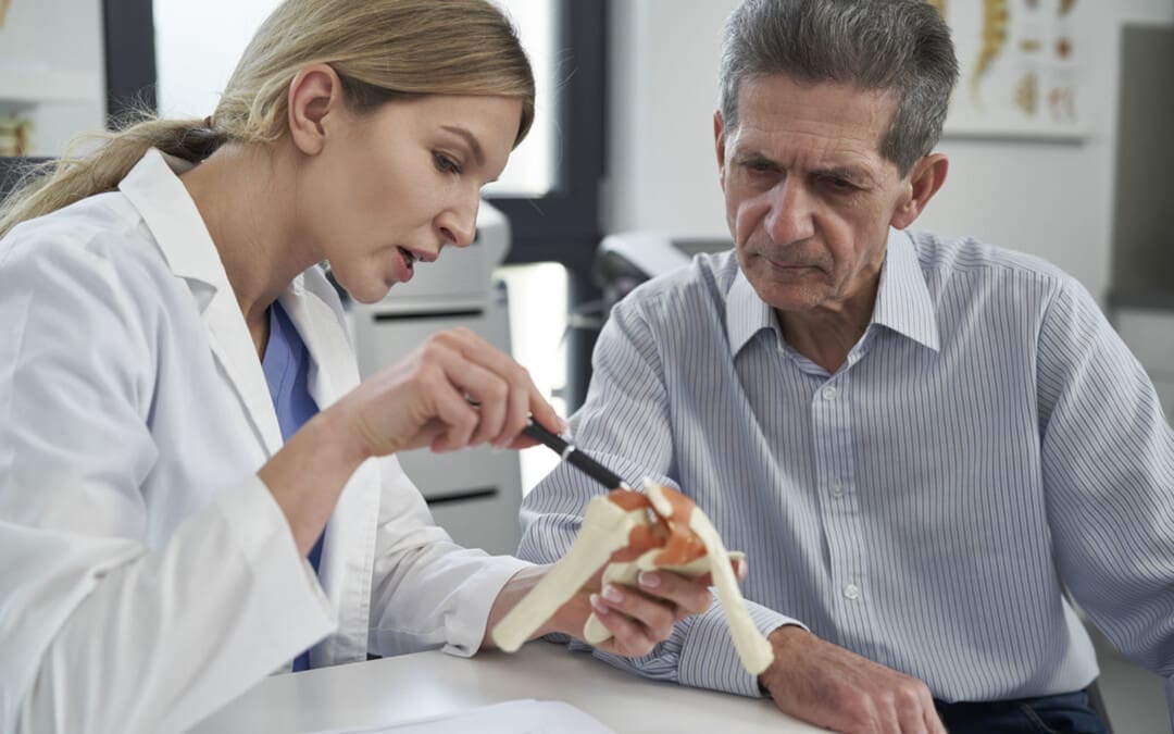
Hinge Joints
A joint forms where one bone connects to another, allowing motion. Different types of joints differ in structure and movement depending on their location. These include hinge, ball and socket, planar, pivot, saddle, and ellipsoid joints. (Boundless. General Biology, N.D.) Hinge joints are synovial joints that move through one plane of motion: flexion and extension. Hinge joints are found in the fingers, elbows, knees, ankles, and toes and control movement for various functions. Injuries, osteoarthritis, and autoimmune conditions can affect hinge joints. Rest, medication, ice, and physical therapy can help alleviate pain, improve strength and range of motion, and help manage conditions.
Anatomy
A joint is formed by the joining of two or more bones. The human body has three main classifications of joints, categorized by the degree to which they can move. These include: (Boundless. General Biology, N.D.)
Synarthroses
These are fixed, immovable joints.
Formed by two or more bones.
Amphiarthroses
Also known as cartilaginous joints.
A fibrocartilage disc separates the bones that form the joints.
These movable joints allow for a slight degree of movement.
Diarthroses
Also known as synovial joints.
These are the most common freely mobile joints that allow movement in multiple directions.
The bones that form the joints are lined with articular cartilage and enclosed in a joint capsule filled with synovial fluid that allows for smooth motion.
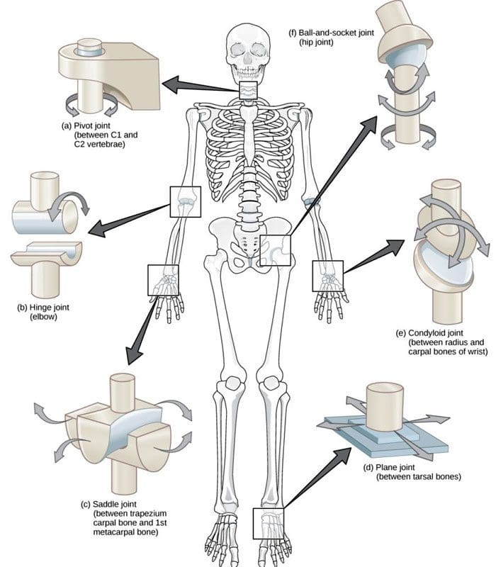
Synovial joints are classified into different types depending on differences in structure and the number of motion planes they allow. A hinge joint is a synovial joint that allows movement in one plane of motion, similar to a door hinge that moves forward and backward. Within the joint, the end of one bone is typically convex/pointed outward, with the other concave/rounded inward to allow the ends to fit smoothly. Because hinge joints only move through one plane of movement, they tend to be more stable than other synovial joints. (Boundless. General Biology, N.D.) Hinge joints include:
The finger and toe joints – allow the fingers and toes to bend and extend.
The elbow joint – allows the elbow to bend and extend.
The knee joint – allows the knee to bend and extend.
The talocrural joint of the ankle – allows the ankle to move up/dorsiflexion and down/plantarflexion.
Hinge joints allow the limbs, fingers, and toes to extend away and bend toward the body. This movement is essential for activities of daily living, such as showering, getting dressed, eating, walking, standing up, and sitting down.
Conditions
Osteoarthritis and inflammatory forms of arthritis can affect any joint (Arthritis Foundation. N.D.) Autoimmune inflammatory forms of arthritis, including rheumatoid and psoriatic arthritis, can cause the body to attack its own joints. These commonly affect the knees and fingers, resulting in swelling, stiffness, and pain. (Kamata, M., Tada, Y. 2020) Gout is an inflammatory form of arthritis that develops from elevated levels of uric acid in the blood and most commonly affects the hinge joint of the big toe. Other conditions that affect hinge joints include:
Injuries to the cartilage within the joints or ligaments that stabilize the outside of the joints.
Ligament sprains or tears can result from jammed fingers or toes, rolled ankles, twisting injuries, and direct impact on the knee.
These injuries can also affect the meniscus, the tough cartilage within the knee joint that helps cushion and absorb shock.
Rehabilitation
Conditions that affect hinge joints often cause inflammation and swelling, resulting in pain and limited mobility.
After an injury or during an inflammatory condition flare-up, limiting active movement and resting the affected joint can reduce increased stress and pain.
Applying ice can decrease inflammation and swelling.
Once the pain and swelling start to subside, physical and/or occupational therapy can help rehabilitate the affected areas.
A therapist will provide stretches and exercises to help improve the joint range of motion and strengthen the supporting muscles.
For individuals experiencing hinge joint pain from an autoimmune condition, biologic medications to decrease the body’s autoimmune activity are administered through infusions delivered every several weeks or months. (Kamata, M., Tada, Y. 2020)
Cortisone injections may also be used to decrease inflammation.
At Injury Medical Chiropractic and Functional Medicine Clinic, we passionately focus on treating patients’ injuries and chronic pain syndromes and improving ability through flexibility, mobility, and agility programs tailored to the individual. Our providers use an integrated approach to create personalized care plans that include Functional Medicine, Acupuncture, Electro-Acupuncture, and Sports Medicine protocols. Our goal is to relieve pain naturally by restoring health and function to the body. If the individual needs other treatment, they will be referred to a clinic or physician best suited for them. Dr. Jimenez has teamed up with the top surgeons, clinical specialists, medical researchers, and premier rehabilitation providers to provide the most effective clinical treatments.
Kamata, M., & Tada, Y. (2020). Efficacy and Safety of Biologics for Psoriasis and Psoriatic Arthritis and Their Impact on Comorbidities: A Literature Review. International journal of molecular sciences, 21(5), 1690. doi.org/10.3390/ijms21051690
What are the healing times of common sports injuries for athletes and individuals who engage in recreational sports activities?
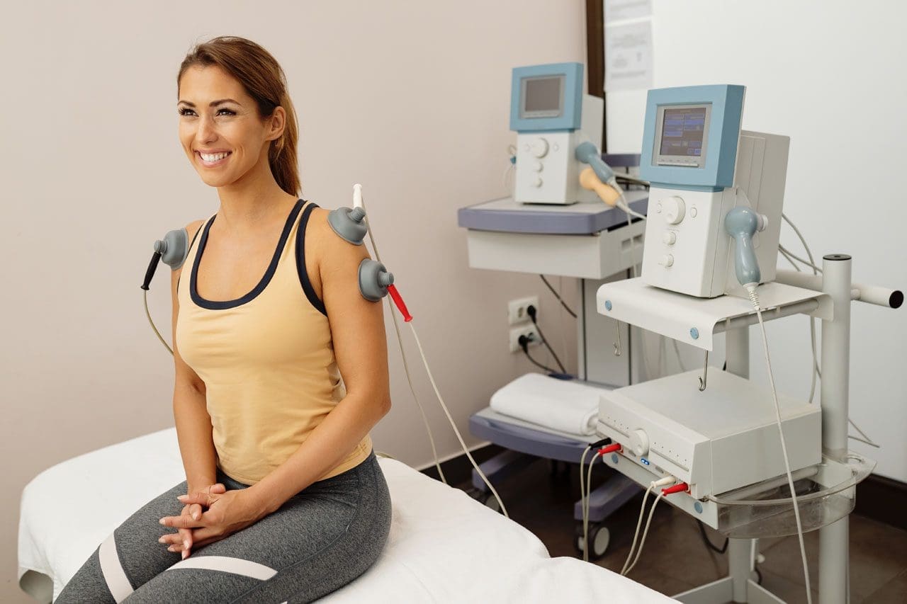
A young, happy sportswoman is getting tens-electrotherapy treatments at a medical clinic.
Healing Times for Sports Injuries
Healing time from sports injuries depends on various factors, such as the location and extent of the injury and the health of the skin, joints, tendons, muscles, and bones. It is also important to take the time to recuperate or not rush back into physical sports activities before the bones or tissues have fully healed. To prevent re-injury, ensure the doctor clears health before returning to sports or strenuous physical activity.
According to CDC research, an average of 8.6 million sports and recreation-related injuries occur annually. (Sheu, Y., Chen, L. H., and Hedegaard, H. 2016) However, most sports injuries are superficial or caused by low-grade strains or sprains; at least 20% of injuries result from bone fractures or more serious injuries. Bone fractures take longer than sprains or strains, and complete tendon or muscle ruptures can take months before one can fully return to activities. Individuals in decent physical shape with no underlying illness or impairment, here is what they can expect when recovering from the following sports injuries:
Bone Fractures
In sports, the highest rate of bone fractures occurs with football and contact sports. Most are centered around the lower extremities but can involve the neck and shoulder blades, arms, and ribs.
Simple Fractures
Depends on the individual’s age, health, type, and location.
Generally, takes at least six weeks to heal.
Compound Fractures
In this case, a bone is broken in several places.
It may require surgery to stabilize the bone.
Healing time can take up to eight months.
Fractured Clavicle/Collarbone
It may require the immobilization of the shoulder and upper arm.
It can take five to ten weeks to heal fully.
Fractured fingers or toes can heal in three to five weeks.
Fractured Ribs
Part of the treatment plan includes breathing exercises.
Painkillers may be needed short term.
Usually, it takes around six weeks to heal.
Neck Fractures
It may involve any one of the seven neck vertebrae.
A neck brace or a halo device that is screwed into the skull for stability may be used.
A sprain is the stretching or tearing of ligaments or the tough bands of fibrous tissue that connect two bones at a joint.
A strain is the overstretching or tearing of muscles or tendons.
Sprained Ankles
It can heal in five days if there are no complications.
Severe sprains involving torn or ruptured tendons can take three to six weeks to heal.
Calf Strains
Classified as grade 1 – a mild strain can heal in two weeks.
A grade 3 – severe strain may require three months or more to heal completely.
The use of calf suppression sleeves can expedite the recovery of strains and sprains in the lower leg.
Acute Neck Strain
A tackle, impact, fall, quick shifting, or whipping motion can cause a whiplash injury.
Healing time can take a couple of weeks to six weeks.
Other Injuries
ACL Tears
Involving the anterior cruciate ligament.
Usually, it requires months of recuperation and rehabilitation, depending on several factors, including the type of sports activity.
Full recovery from surgery takes six to 12 months.
Without surgery, there is no specific timeline for rehabilitation.
Achilles Tendon Ruptures
It is a serious injury.
These occur when the tendon is either partially or completely torn.
Individuals will more than likely require surgery.
Recovery time is four to six months.
Cuts and Lacerations
Depends on the depth and location of the injury.
It can take anywhere from a week to a month to heal.
If there are no accompanying injuries, stitches can be removed within two to three weeks.
If a deep cut requires stitches, more time is necessary.
Mild Contusions/Bruises
Are caused by a trauma to the skin, causing blood vessels to break.
In most cases, a contusion will take five to seven days to heal.
Shoulder Separations
When treated properly, it usually takes around two weeks of rest and recovery before the patient returns to activity.
Multidisciplinary Treatment
After the initial inflammation and swelling have subsided, a doctor will recommend a treatment plan that usually involves physical therapy, self-performed physical rehabilitation, or supervision by a physical therapist or team. Fortunately, athletes and individuals who regularly exercise tend to have a faster healing time because they are in top physical shape, and their cardiovascular system provides a stronger blood supply that speeds up the healing process. At El Paso’s Chiropractic Rehabilitation Clinic & Integrated Medicine Center, we passionately focus on treating patients’ injuries and chronic pain syndromes. We focus on improving ability through flexibility, mobility, and agility programs tailored to the individual. We use in-person and virtual health coaching and comprehensive care plans to ensure every patient’s personalized care and wellness outcomes.
Our providers use an integrated approach to create personalized care plans that include Functional Medicine, Acupuncture, Electro-Acupuncture, and Sports Medicine principles. Our goal is to relieve pain naturally by restoring health and function to the body.
If the chiropractor feels the individual needs other treatment, they will be referred to a clinic or physician best suited for them. Dr. Jimenez has teamed up with the top surgeons, clinical specialists, medical researchers, and premier rehabilitation providers to provide the top clinical treatments for our community. Providing highly noninvasive protocols is our priority, and our personalized patient-based clinical insight is what we provide.
Lumbar Spine Injuries in Sports: Chiropractic Healing
References
Sheu, Y., Chen, L. H., & Hedegaard, H. (2016). Sports- and Recreation-related Injury Episodes in the United States, 2011-2014. National health statistics reports, (99), 1–12.
For individuals experiencing pelvic pain, it could be a disorder of the pudendal nerve known as pudendal neuropathy or neuralgia that leads to chronic pain. The condition can be caused by pudendal nerve entrapment, where the nerve becomes compressed or damaged. Can knowing the symptoms help healthcare providers correctly diagnose the condition and develop an effective treatment plan?
Pudendal Neuropathy
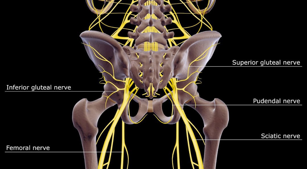
The pudendal nerve is the main nerve that serves the perineum, which is the area between the anus and the genitalia – the scrotum in men and the vulva in women. The pudendal nerve runs through the gluteus muscles/buttocks and into the perineum. It carries sensory information from the external genitalia and the skin around the anus and perineum and transmits motor/movement signals to various pelvic muscles. (Origoni, M. et al., 2014) Pudendal neuralgia, also referred to as pudendal neuropathy, is a disorder of the pudendal nerve that can lead to chronic pelvic pain.
Causes
Chronic pelvic pain from pudendal neuropathy can be caused by any of the following (Kaur J. et al., 2024)
Excessive sitting on hard surfaces, chairs, bicycle seats, etc. Bicyclists tend to develop pudendal nerve entrapment.
Trauma to the buttocks or pelvis.
Childbirth.
Diabetic neuropathy.
Bony formations that push against the pudendal nerve.
Thickening of ligaments around the pudendal nerve.
Symptoms
Pudendal nerve pain can be described as stabbing, cramping, burning, numbness, or pins and needles and can present (Kaur J. et al., 2024)
In the perineum.
In the anal region.
In men, pain in the scrotum or penis.
In women, pain in the labia or vulva.
During intercourse.
When urinating.
During a bowel movement.
When sitting and goes away after standing up.
Because the symptoms are often hard to distinguish, pudendal neuropathy can often be hard to differentiate from other types of chronic pelvic pain.
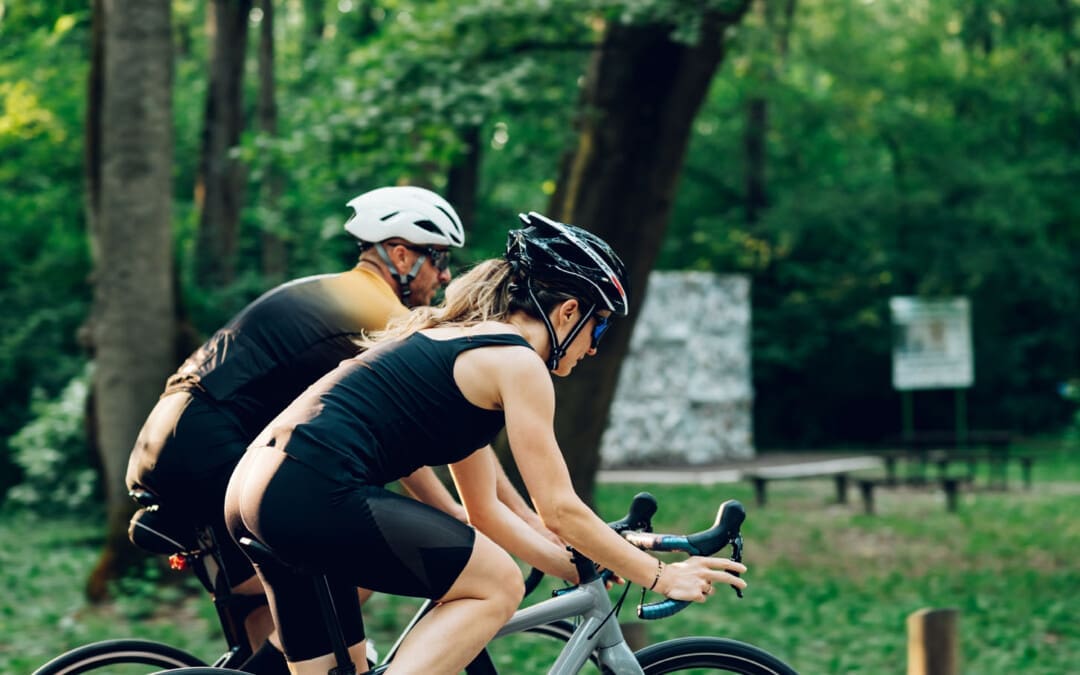
Cyclist’s Syndrome
Prolonged sitting on a bicycle seat can cause pelvic nerve compression, which can lead to chronic pelvic pain. The frequency of pudendal neuropathy (chronic pelvic pain caused by entrapment or compression of the pudendal nerve) is often referred to as Cyclist’s Syndrome. Sitting on certain bicycle seats for long periods places significant pressure on the pudendal nerve. The pressure can cause swelling around the nerve, which causes pain and, over time, can lead to nerve trauma. Nerve compression and swelling can cause pain described as burning, stinging, or pins and needles. (Durante, J. A., and Macintyre, I. G. 2010) For individuals with pudendal neuropathy caused by bicycling, symptoms can appear after prolonged biking and sometimes months or years later.
Take breaks at least 20–30 seconds after each 20 minutes of riding.
While riding, change positions frequently.
Stand up to pedal periodically.
Take time off between riding sessions and races to rest and relax the pelvic nerves. 3–10 day breaks can help in recovery. (Durante, J. A., and Macintyre, I. G. 2010)
If pelvic pain symptoms are barely starting to develop, rest and see a healthcare provider or specialist for an examination.
Seat
Use a soft, wide seat with a short nose.
Have the seat level or tilted slightly forward.
Seats with cutout holes place more pressure on the perineum.
If numbness or pain is present, try a seat without holes.
Bike Fitting
Adjust the seat height so the knee is slightly bent at the bottom of the pedal stroke.
The body’s weight should rest on the sitting bones/ischial tuberosities.
Keeping the handlebar height below the seat can reduce pressure.
The Triathlon bike’s extreme-forward position should be avoided.
A more upright posture is better.
Mountain bikes have been associated with an increased risk of erectile dysfunction than road bikes.
Shorts
Wear padded bike shorts.
Treatments
A healthcare provider may use a combination of treatments.
The neuropathy can be treated with rest if the cause is excessive sitting or cycling.
Injury Medical Chiropractic and Functional Medicine Clinic care plans and clinical services are specialized and focused on injuries and the complete recovery process. Our areas of practice include Wellness and nutrition, Chronic Pain, Personal Injury, Auto Accident Care, Work Injuries, Back Injury, Low Back Pain, Neck Pain, Migraine Headaches, Sports Injuries, severe sciatica, Scoliosis, Complex Herniated Discs, Fibromyalgia, Chronic Pain, Complex Injuries, Stress Management, and Functional Medicine Treatments. If the individual requires other treatment, they will be referred to a clinic or physician best suited for their condition, as Dr. Jimenez has teamed with the top surgeons, clinical specialists, medical researchers, therapists, trainers, and premiere rehabilitation providers.
Pregnancy and Sciatica
References
Origoni, M., Leone Roberti Maggiore, U., Salvatore, S., & Candiani, M. (2014). Neurobiological mechanisms of pelvic pain. BioMed research international, 2014, 903848. doi.org/10.1155/2014/903848
Durante, J. A., & Macintyre, I. G. (2010). Pudendal nerve entrapment in an Ironman athlete: a case report. The Journal of the Canadian Chiropractic Association, 54(4), 276–281.
Chiaramonte, R., Pavone, P., & Vecchio, M. (2021). Diagnosis, Rehabilitation and Preventive Strategies for Pudendal Neuropathy in Cyclists, A Systematic Review. Journal of functional morphology and kinesiology, 6(2), 42. doi.org/10.3390/jfmk6020042
IFM's Find A Practitioner tool is the largest referral network in Functional Medicine, created to help patients locate Functional Medicine practitioners anywhere in the world. IFM Certified Practitioners are listed first in the search results, given their extensive education in Functional Medicine

 Causes