Can individuals with Ehlers-Danlos syndrome find relief through various non-surgical treatments to reduce joint instability?
Introduction
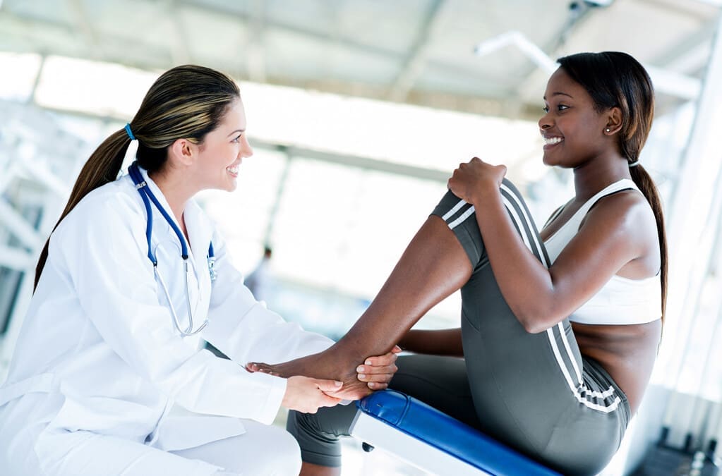
The joints and ligaments surrounding the musculoskeletal system allow the upper and lower extremities to stabilize the body and be mobile. The various muscles and soft connective tissues that surround the joints help protect them from injuries. When environmental factors or disorders start to affect the body, many people develop issues that cause overlapping risk profiles, which then affect the stability of the joints. One of the disorders that affect the joints and connective tissue is EDS or Ehlers-Danlos syndrome. This connective tissue disorder can cause the joints in the body to be hypermobile. It can cause joint instability in the upper and lower extremities, thus leaving the individual to be in constant pain. Today’s article focuses on Ehlers-Danlos syndrome and its symptoms and how there are non-surgical ways to manage this connective tissue disorder. We discuss with certified medical providers who consolidate our patients’ information to assess how Ehlers-Danlos syndrome can correlate with other musculoskeletal disorders. We also inform and guide patients on how various non-surgical treatments can help reduce pain-like symptoms and manage Ehlers-Danlos syndrome. We also encourage our patients to ask their associated medical providers many intricate and important questions about incorporating various non-surgical therapies as part of their daily routine to manage the effects of Ehlers-Danlos syndrome. Dr. Jimenez, D.C., includes this information as an academic service. Disclaimer.
What Is Ehlers-Danlos Syndrome?
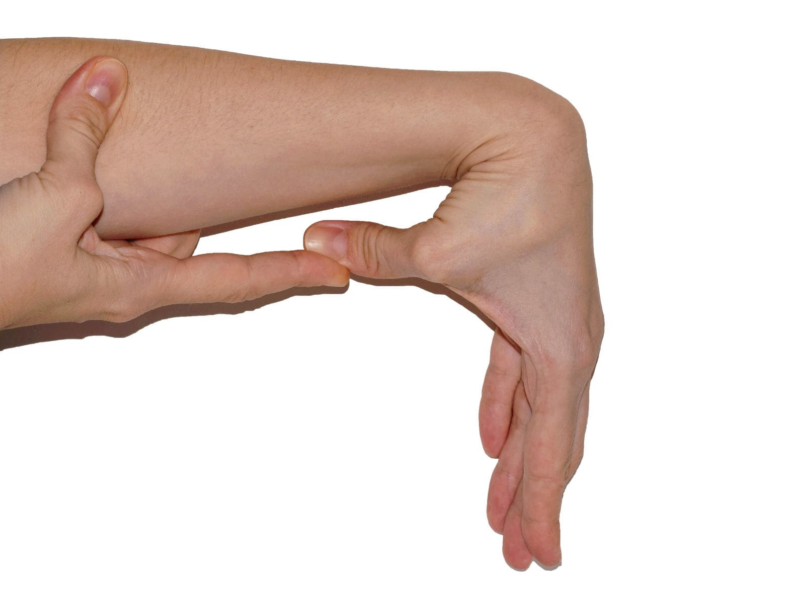
Do you often feel extremely tired throughout the day, even after a full night of sleep? Do you bruise easily and wonder where these bruises are coming from? Or have you noticed that you have an increased range in your joints? Many of these issues are often correlated with a disorder known as Ehlers-Danlos syndrome or EDS that affects their joints and connective tissue. EDS affects the connective tissues in the body. The connective tissues in the body help provide strength and elasticity to the skin, joints, as well as blood vessel walls, so when a person is dealing with EDS, it can cause a significant disruption to the musculoskeletal system. EDS is largely diagnosed clinically, and many doctors have identified that the gene coding of the collagen and proteins that interact in the body can help determine what type of EDS affects the individual. (Miklovic & Sieg, 2024)
The Symptoms
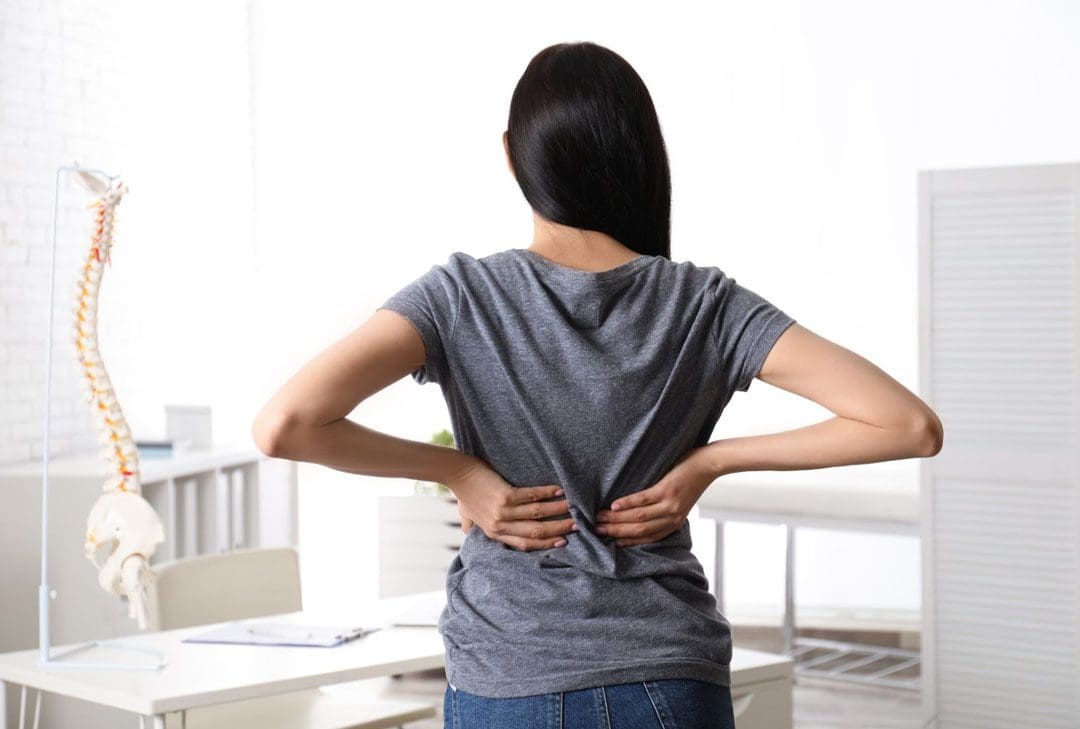
When understanding EDS, it is essential to know the complexities of this connective tissue disorder. EDS is classified into numerous types with distinct features and challenges that vary depending on the severity. One of the most common types of EDS is hypermobile Ehlers-Danlos syndrome. This type of EDS is characterized by general joint hypermobility, joint instability, and pain. Some of the symptoms that are associated with hypermobile EDS include subluxation, dislocations, and soft tissue injuries that are common and may occur spontaneously or with minimal trauma. (Hakim, 1993) This can often cause acute pain to the joints in the upper and lower extremities. With its broad range of symptoms and the personal nature of the condition itself, many often don’t realize that joint hypermobility is common in the general population and may present no complications that indicate that it is a connective tissue disorder. (Gensemer et al., 2021) Additionally, hypermobile EDS can lead to spinal deformity due to the hyperextensibility of the skin, joints, and various tissue fragility. The pathophysiology of spinal deformity associated with hypermobile EDS is primarily due to muscle hypotonia and ligament laxity. (Uehara et al., 2023) This causes many people to reduce their quality of life and daily living activities significantly. However, there are ways to manage EDS and its correlating symptoms to reduce joint instability.
Movement Medicine: Chiropractic Care-Video
Ways To Manage EDS
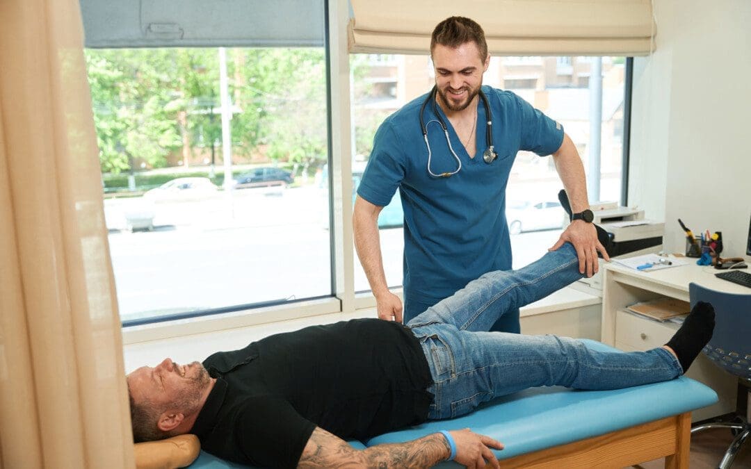
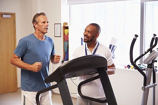
When it comes to looking for ways to manage EDS to reduce pain and joint instability, non-surgical treatments can help address the physical and emotional aspects of the condition. Non-surgical treatments for individuals with EDS commonly focus on optimizing the body’s physical function while improving muscular strength and joint stabilization. (Buryk-Iggers et al., 2022) Many individuals with EDS will try to incorporate pain management techniques and physical therapy anduse braces and assistive devices to reduce the effects of EDS and improve their quality of life.
Non-surgical Treatments For EDS
Various non-surgical treatments like MET (muscle energy technique), electrotherapy, light physical therapy, chiropractic care, and massages can help strengthen while toning the surrounding muscles around the joints, provide sufficient pain relief, and limit long-term dependence on medications. (Broida et al., 2021) Additionally, individuals dealing with EDS aim to strengthen the affected muscles, stabilize the joints, and improve proprioception. Non-surgical treatments allow the individual to have a customized treatment plan for the severity of EDS symptoms and help reduce the pain associated with the condition. Many individuals, when going through their treatment plan consecutively to manage their EDS and reduce the pain-like symptoms, will notice improvement in symptomatic discomfort. (Khokhar et al., 2023) This means that non-surgical treatments allow individuals to be more mindful of their bodies and reduce the pain-like effects of EDS, thus allowing many individuals with EDS to lead fuller, more comfortable lives without feeling pain and discomfort.
References
Broida, S. E., Sweeney, A. P., Gottschalk, M. B., & Wagner, E. R. (2021). Management of shoulder instability in hypermobility-type Ehlers-Danlos syndrome. JSES Rev Rep Tech, 1(3), 155-164. doi.org/10.1016/j.xrrt.2021.03.002
Buryk-Iggers, S., Mittal, N., Santa Mina, D., Adams, S. C., Englesakis, M., Rachinsky, M., Lopez-Hernandez, L., Hussey, L., McGillis, L., McLean, L., Laflamme, C., Rozenberg, D., & Clarke, H. (2022). Exercise and Rehabilitation in People With Ehlers-Danlos Syndrome: A Systematic Review. Arch Rehabil Res Clin Transl, 4(2), 100189. doi.org/10.1016/j.arrct.2022.100189
Gensemer, C., Burks, R., Kautz, S., Judge, D. P., Lavallee, M., & Norris, R. A. (2021). Hypermobile Ehlers-Danlos syndromes: Complex phenotypes, challenging diagnoses, and poorly understood causes. Dev Dyn, 250(3), 318-344. doi.org/10.1002/dvdy.220
Hakim, A. (1993). Hypermobile Ehlers-Danlos Syndrome. In M. P. Adam, J. Feldman, G. M. Mirzaa, R. A. Pagon, S. E. Wallace, L. J. H. Bean, K. W. Gripp, & A. Amemiya (Eds.), GeneReviews((R)). www.ncbi.nlm.nih.gov/pubmed/20301456
Khokhar, D., Powers, B., Yamani, M., & Edwards, M. A. (2023). The Benefits of Osteopathic Manipulative Treatment on a Patient With Ehlers-Danlos Syndrome. Cureus, 15(5), e38698. doi.org/10.7759/cureus.38698
Can understanding the body’s hinge joints and how they operate help with mobility and flexibility problems and manage conditions for individuals with difficulty fully bending or extending their fingers, toes, elbows, ankles, or knees?
Hinge Joints
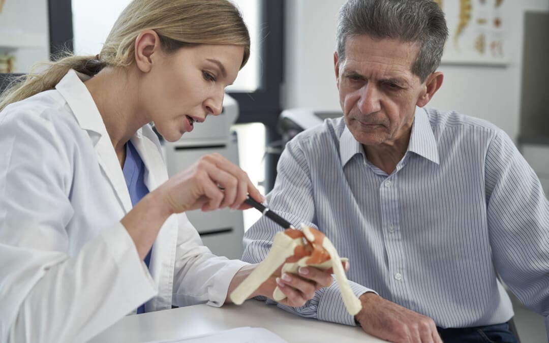
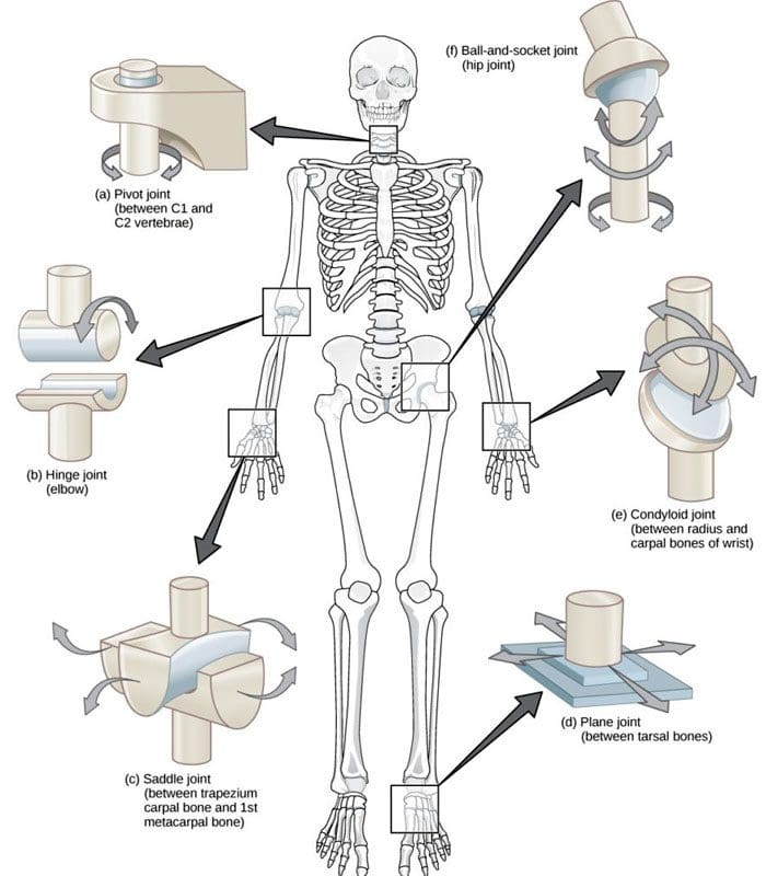
A joint forms where one bone connects to another, allowing motion. Different types of joints differ in structure and movement depending on their location. These include hinge, ball and socket, planar, pivot, saddle, and ellipsoid joints. (Boundless. General Biology, N.D.) Hinge joints are synovial joints that move through one plane of motion: flexion and extension. Hinge joints are found in the fingers, elbows, knees, ankles, and toes and control movement for various functions. Injuries, osteoarthritis, and autoimmune conditions can affect hinge joints. Rest, medication, ice, and physical therapy can help alleviate pain, improve strength and range of motion, and help manage conditions.
Anatomy
A joint is formed by the joining of two or more bones. The human body has three main classifications of joints, categorized by the degree to which they can move. These include: (Boundless. General Biology, N.D.)
Synarthroses
These are fixed, immovable joints.
Formed by two or more bones.
Amphiarthroses
Also known as cartilaginous joints.
A fibrocartilage disc separates the bones that form the joints.
These movable joints allow for a slight degree of movement.
Diarthroses
Also known as synovial joints.
These are the most common freely mobile joints that allow movement in multiple directions.
The bones that form the joints are lined with articular cartilage and enclosed in a joint capsule filled with synovial fluid that allows for smooth motion.
Synovial joints are classified into different types depending on differences in structure and the number of motion planes they allow. A hinge joint is a synovial joint that allows movement in one plane of motion, similar to a door hinge that moves forward and backward. Within the joint, the end of one bone is typically convex/pointed outward, with the other concave/rounded inward to allow the ends to fit smoothly. Because hinge joints only move through one plane of movement, they tend to be more stable than other synovial joints. (Boundless. General Biology, N.D.) Hinge joints include:
The finger and toe joints – allow the fingers and toes to bend and extend.
The elbow joint – allows the elbow to bend and extend.
The knee joint – allows the knee to bend and extend.
The talocrural joint of the ankle – allows the ankle to move up/dorsiflexion and down/plantarflexion.
Hinge joints allow the limbs, fingers, and toes to extend away and bend toward the body. This movement is essential for activities of daily living, such as showering, getting dressed, eating, walking, standing up, and sitting down.
Conditions
Osteoarthritis and inflammatory forms of arthritis can affect any joint (Arthritis Foundation. N.D.) Autoimmune inflammatory forms of arthritis, including rheumatoid and psoriatic arthritis, can cause the body to attack its own joints. These commonly affect the knees and fingers, resulting in swelling, stiffness, and pain. (Kamata, M., Tada, Y. 2020) Gout is an inflammatory form of arthritis that develops from elevated levels of uric acid in the blood and most commonly affects the hinge joint of the big toe. Other conditions that affect hinge joints include:
Injuries to the cartilage within the joints or ligaments that stabilize the outside of the joints.
Ligament sprains or tears can result from jammed fingers or toes, rolled ankles, twisting injuries, and direct impact on the knee.
These injuries can also affect the meniscus, the tough cartilage within the knee joint that helps cushion and absorb shock.
Rehabilitation
Conditions that affect hinge joints often cause inflammation and swelling, resulting in pain and limited mobility.
After an injury or during an inflammatory condition flare-up, limiting active movement and resting the affected joint can reduce increased stress and pain.
Applying ice can decrease inflammation and swelling.
Once the pain and swelling start to subside, physical and/or occupational therapy can help rehabilitate the affected areas.
A therapist will provide stretches and exercises to help improve the joint range of motion and strengthen the supporting muscles.
For individuals experiencing hinge joint pain from an autoimmune condition, biologic medications to decrease the body’s autoimmune activity are administered through infusions delivered every several weeks or months. (Kamata, M., Tada, Y. 2020)
Cortisone injections may also be used to decrease inflammation.
At Injury Medical Chiropractic and Functional Medicine Clinic, we passionately focus on treating patients’ injuries and chronic pain syndromes and improving ability through flexibility, mobility, and agility programs tailored to the individual. Our providers use an integrated approach to create personalized care plans that include Functional Medicine, Acupuncture, Electro-Acupuncture, and Sports Medicine protocols. Our goal is to relieve pain naturally by restoring health and function to the body. If the individual needs other treatment, they will be referred to a clinic or physician best suited for them. Dr. Jimenez has teamed up with the top surgeons, clinical specialists, medical researchers, and premier rehabilitation providers to provide the most effective clinical treatments.
Kamata, M., & Tada, Y. (2020). Efficacy and Safety of Biologics for Psoriasis and Psoriatic Arthritis and Their Impact on Comorbidities: A Literature Review. International journal of molecular sciences, 21(5), 1690. doi.org/10.3390/ijms21051690
Individuals that have gone through a back injury may develop a synovial spinal cyst as a way to protect the spine that could cause pain symptoms and sensations. Can knowing the signs help healthcare providers develop a thorough treatment plan to relieve pain, prevent worsening of the condition and other spinal conditions?
Spinal Synovial Cysts
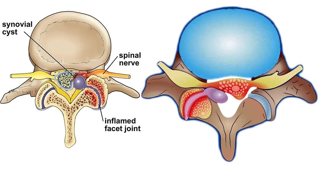
Spinal synovial cysts are benign fluid-filled sacs that develop in the spine’s joints. They form because of spinal degeneration or injury. The cysts can form anywhere in the spine, but most occur in the lumbar region/lower back. They typically develop in the facet joints or junctions that keep the vertebrae/spinal bones interlocked.
Symptoms
In most cases, synovial cysts don’t cause symptoms. However, the doctor or specialist will want to monitor for signs of degenerative disc disease, spinal stenosis, or cauda equina syndrome. When symptoms do present, they typically cause radiculopathy or nerve compression, which can cause back pain, weakness, numbness, and radiating pain caused by the irritation. The severity of symptoms depends on the size and location of the cyst. Synovial cysts can affect one side of the spine or both and can form at one spinal segment or at multiple levels.
Effects Can Include
Radiculopathy symptoms can develop if the cyst or inflammation caused by the cyst comes into contact with a spinal nerve root. This can cause sciatica, weakness, numbness, or difficulty controlling certain muscles.
Neurogenic claudication/impingement and inflammation of spinal nerves can cause cramping, pain, and/or tingling in the lower back, legs, hips, and buttocks. (Martin J. Wilby et al., 2009)
If the spinal cord is involved, it may cause myelopathy/severe spinal cord compression that can cause numbness, weakness, and balance problems. (Dong Shin Kim et al., 2014)
Symptoms related to cauda equina, including bowel and/or bladder problems, leg weakness, and saddle anesthesia/loss of sensation in the thighs, buttocks, and perineum, can present but are rare, as are synovial cysts in the middle back and neck. If thoracic and cervical synovial cysts develop, they can cause symptoms like numbness, tingling, pain, or weakness in the affected area.
Causes
Spinal synovial cysts are generally caused by degenerative changes like osteoarthritis that develop in a joint over time. With regular wear and tear, facet joint cartilage/the material in a joint that provides protection, a smooth surface, friction reduction, and shock absorption begins to waste away. As the process continues, the synovium can form a cyst.
Traumas, large and small, have inflammatory and degenerative effects on joints that can result in the formation of a cyst.
Around a third of individuals who have a spinal synovial cyst also have spondylolisthesis.
This condition is when a vertebrae slips out of place or out of alignment onto the vertebra underneath.
It is a sign of spinal instability.
Instability can occur in any spine area, but L4-5 are the most common levels.
This segment of the spine takes most of the upper body weight.
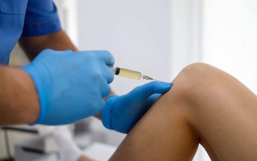
Epidural corticosteroid injections can reduce inflammation and could be an option to relieve pain.
Patients are recommended to receive no more than three injections per year.
Surgical Options
For severe or persistent cases, a doctor may recommend decompression surgery to remove the cyst and surrounding bone to relieve pressure on the nerve root. Surgical options range from minimally invasive endoscopic procedures to larger, open surgeries. The best surgical option varies based on the severity of the situation and whether associated disorders are present. Surgical options include:
Laminectomy – Removal of the bony structure that protects and covers the spinal canal/lamina.
Hemilaminectomy – A modified laminectomy where a smaller portion of the lamina is removed.
Facetectomy – The removal of part of the affected facet joint where the synovial cyst is located, usually following a laminectomy or hemilaminectomy.
Fusionof the facet joints and vertebra – Decreases vertebral mobility in the injured area.
Most individuals experience immediate pain relief following a laminectomy or hemilaminectomy.
Fusion can take six to nine months to heal completely.
If surgery is performed without fusion where the cyst originated, the pain could return, and another cyst could form within two years.
Surgery Complications include infection, bleeding, and injury to the spinal cord or nerve root.
How I Gained My Mobility Back With Chiropractic
References
Wilby, M. J., Fraser, R. D., Vernon-Roberts, B., & Moore, R. J. (2009). The prevalence and pathogenesis of synovial cysts within the ligamentum flavum in patients with lumbar spinal stenosis and radiculopathy. Spine, 34(23), 2518–2524. doi.org/10.1097/BRS.0b013e3181b22bd0
Kim, D. S., Yang, J. S., Cho, Y. J., & Kang, S. H. (2014). Acute myelopathy caused by a cervical synovial cyst. Journal of Korean Neurosurgical Society, 56(1), 55–57. doi.org/10.3340/jkns.2014.56.1.55
Epstein, N. E., & Baisden, J. (2012). The diagnosis and management of synovial cysts: Efficacy of surgery versus cyst aspiration. Surgical neurology international, 3(Suppl 3), S157–S166. doi.org/10.4103/2152-7806.98576
As the body ages, individuals want to stay active and maintain a healthy pain free lifestyle. Can regenerative cells for arthritis and cartilage damage be the future of neuromusculoskeletal medicine and joint healing?
Regenerative Cells For Arthritis and Cartilage Damage
Individuals want to continue to do the physical activities they love, which require healthy joints. Scientists are learning how to harness the abilities of regenerative cells to repair and regrow damaged and deteriorated cartilage. Current stem cell treatment of cartilage problems has not been shown to reverse the effects of arthritis and while studies show clinical improvement, further research is necessary. (Bryan M. Saltzman, et al., 2016)
Cartilage and How It Gets Damaged
Cartilage is a type of connective tissue. In the joints, there are a few types of cartilage. The most commonly referred to is the smooth lining known as articular or hyaline cartilage. This type forms a smooth layer of cushion on the end of a bone at the joint. (Rocky S. Tuan, et al., 2013)
The tissue is very strong and has the ability to compress and absorb energy.
It is very smooth allowing a joint to glide effortlessly through a limb’s range of motion.
When joint cartilage is damaged, the cushioning can wear down.
In traumatic injuries, a sudden force can cause the cartilage to break off and/or suffer damage, that exposes the underlying bone.
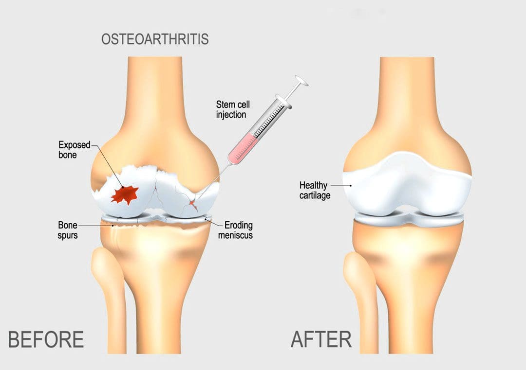
In osteoarthritis – degenerative or wear-and-tear arthritis, the smooth layer can wear down thin and unevenly.
Eventually, the cushion wears away, the joints become inflamed and swollen and movements become stiff and painful.
There are treatments for arthritis and cartilage damage, but these treatments are usually focused on relieving symptoms by smoothing down the damaged cartilage or replacing the joint surface with an artificial implant, like knee replacement or hip replacement surgeries. (Robert F. LaPrade, et al., 2016)
Regenerative Cells
Regenerative stem cells are special cells that have the ability to multiply and develop into different types of tissue. In an orthopedic surgery setting for joint problems, stem cells are obtained from adult stem cell primary sources which are bone marrow and fatty tissue. These cells have the ability to develop into cartilage cells, called chondrocytes. (Rocky S. Tuan, et al., 2013)
They also help by stimulating the body to reduce inflammation, stimulate cell repair, and improve blood circulation.
This process is caused by cellular signals and growth factors to stimulate the body to activate the healing processes.
Once stem cells have been obtained, they need to be delivered to the area of cartilage damage.
Cartilage is a complex tissue that is described as a scaffold structure that is composed of collagen, proteoglycans, water, and cells. (Rocky S. Tuan, et al., 2013)
To regenerate cartilage, the complex tissues must also be reconstructed.
There are studies on types of tissue scaffolds engineered to recreate a similar type of cartilage structure.
The stem cells can then be injected into the scaffold, in hopes of restoring a normal type of cartilage.
Non-Surgical Arthritis Treatments
Standard treatments such as cortisone shots or physical therapies work as well and provide benefits that could be utilized in combination with regenerative cells for arthritis and cartilage damage in the near future. Data takes time and therefore how this impacts the long-term health of a joint needs continued research in terms of tissue engineering and cell delivery to determine the best approach to help individuals.
Arthritis
References
LaPrade, R. F., Dragoo, J. L., Koh, J. L., Murray, I. R., Geeslin, A. G., & Chu, C. R. (2016). AAOS Research Symposium Updates and Consensus: Biologic Treatment of Orthopaedic Injuries. The Journal of the American Academy of Orthopaedic Surgeons, 24(7), e62–e78. doi.org/10.5435/JAAOS-D-16-00086
Saltzman, B. M., Kuhns, B. D., Weber, A. E., Yanke, A., & Nho, S. J. (2016). Stem Cells in Orthopedics: A Comprehensive Guide for the General Orthopedist. American journal of orthopedics (Belle Mead, N.J.), 45(5), 280–326.
Tuan, R. S., Chen, A. F., & Klatt, B. A. (2013). Cartilage regeneration. The Journal of the American Academy of Orthopaedic Surgeons, 21(5), 303–311. doi.org/10.5435/JAAOS-21-05-303
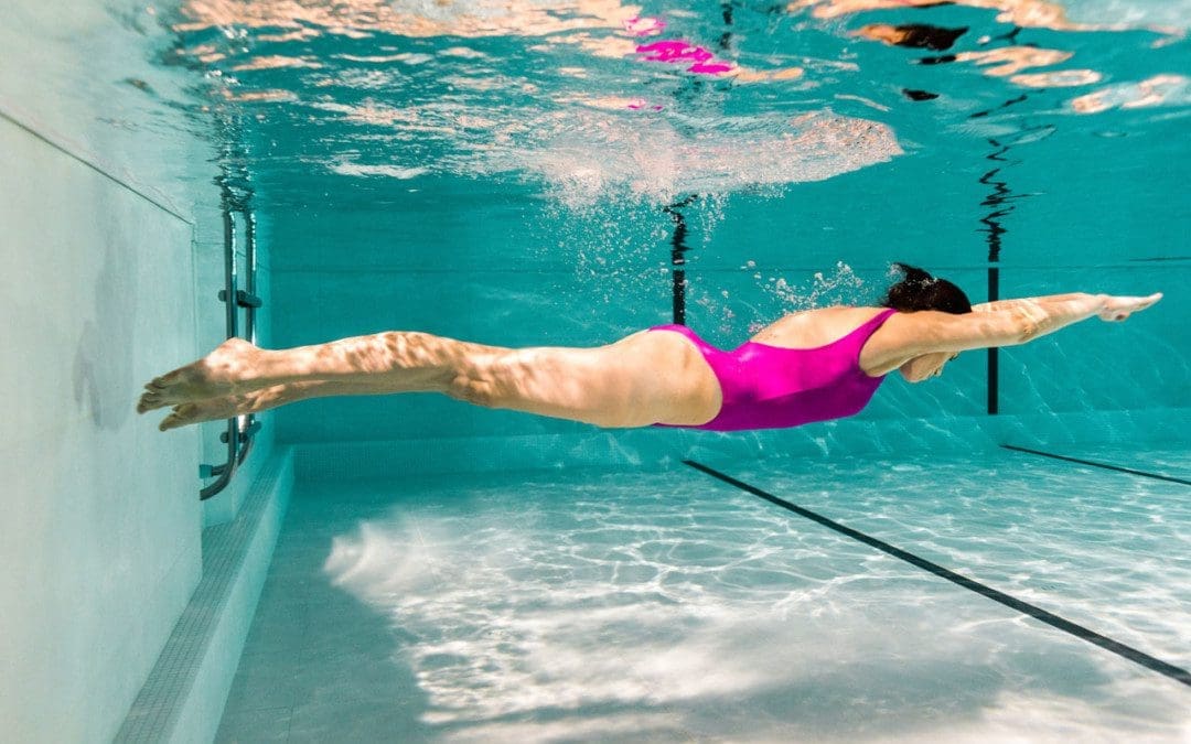
Individuals dealing with or managing chronic back pain should incorporate swimming exercises. Swimming is a low-impact aerobic conditioning exercise that’s easy on the back and healthy for the spine. When an individual struggles with back problems, they may be tempted to rest and avoid physical activity/exercise. Total rest is not recommended as it can cause the muscles that support the back to weaken or atrophy. When the muscles weaken, they cannot stabilize the spine or body correctly, which causes conditions to worsen or contribute to new injuries. Starting swimming exercises can expand the spine, relieve painful pressure or strain on the back and strengthen the muscles for spinal health.
Starting Swimming Exercises
Swimming does not impact the spine and other musculoskeletal structures because the water suspends the body.
Swimming is a full-body, low-impact exercise which is excellent for individuals of all ages and all body shapes and sizes.
Talk to a healthcare professional about any questions or concerns about how swimming may impact your body.
Swimming benefits include stress relief, a strengthened musculoskeletal system, and support in heart health.
Swimming for Back Problems
Relaxes The Nervous System
Tense muscles can cause or contribute to back problems and pain symptoms and aggravate spinal conditions.
Swimming exercises release endorphins to relax the nervous system and tense muscles.
Relieves Pressure on Joints
The water lightens the body relieving pressure on the joints and muscles.
Builds Muscle to Support the Spine
The resistance and movement strengthen the whole body with the joints and spine supported.
Swimming engages muscles not always used, specifically those needed to improve spinal stability.
Exercises for Back Relief
Checking with a physician before exercising is recommended, especially if starting a new exercise routine. When you meet with the Injury Medical Chiropractic and Functional Medicine Cline team, we can determine if starting swimming exercises would benefit you. Once cleared, here are some swimming exercises that could help bring relief:
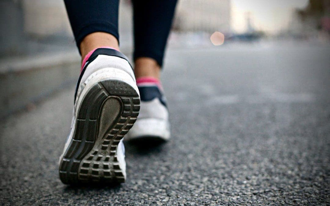
Walking
Walking around the pool means movement that the body needs to heal and build muscle without aggravating symptoms.
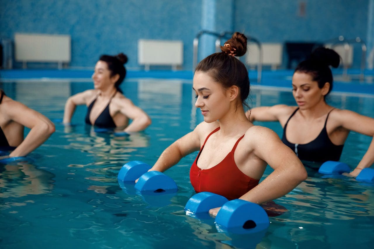
Aerobics
Water aerobics is perfect for working on cardiovascular health needed to build strength.
Increase mobility and flexibility.
Swimming Laps
Start slow when swimming laps, maybe only twice a week at first.
The different types of strokes work various muscles in the hips, chest, and back.
Treading water is a great way to get the body used to the movements.
A swim coach can provide tips on the proper technique and form.
Swim Exercise Tools and Accessories
Proper swimming equipment can make the exercise sessions much more enjoyable.
Swim Cap
Swim caps protect the hair from the water’s elements and keep hair from blocking the view.
Goggles
Goggles protect the eyes and help to see better underwater.
Look for a comfortable pair that doesn’t leak.
Sun protection and clothing
A day in the sun and water increases the risk of exposure to UV rays.
Waterproof Headphones
For listening to music or podcasts while swimming.
Kickboard
Many pools can provide kickboards that swimmers can borrow during their time there.
Lean the upper body on the board and kick, focusing on lower body movements.
Pull Buoy
Pull buoys help focus on the upper body and arm work.
It is placed between the upper thighs to help the legs float as the individual pulls with their arms.
It is recommended to take some lessons to learn how the body moves through the water. Once a basic understanding of balance and buoyancy is met, individuals can propel through the water more efficiently.
Sciatica Secrets Revealed
References
Bartels, Else Marie, et al. “Aquatic exercise for the treatment of knee and hip osteoarthritis.” The Cochrane Database of systematic reviews vol. 3,3 CD005523. 23 Mar. 2016, doi:10.1002/14651858.CD005523.pub3
Cole, A J et al. “Spine pain: aquatic rehabilitation strategies.” Journal of Back and musculoskeletal rehabilitation vol. 4,4 (1994): 273-86. doi:10.3233/BMR-1994-4407
Ferrell, M C. “The spine in swimming.” Clinics in sports medicine vol. 18,2 (1999): 389-93, viii. doi:10.1016/s0278-5919(05)70153-8
Su, Yanlin, et al. “Swimming as Treatment for Osteoporosis: A Systematic Review and Meta-analysis.” BioMed research international vol. 2020 6210201. 15 May. 2020, doi:10.1155/2020/6210201
Wirth, Klaus, et al. “Strength Training in Swimming.” International Journal of environmental research and public health vol. 19,9 5369. 28 Apr. 2022, doi:10.3390/ijerph19095369
Many people don’t often realize that stability and balance are two of the most reliable abilities to keep the body from falling, and it is often taken for granted from the earlier stages, where infants and toddlers are learning to stand upright, to adulthood where we are walking, running or performing any physical activities. Our bodies are complex machines comprised of upper and lower portions that provide balance and stability. The lower half of our bodies helps stabilize and balance the upper half weight and allows us to move around. This is known as gait. However, when the body begins to age naturally or chronic issues begin to affect the muscles and cause an imbalance in the lower half, it can lead to many disorders associated with these imbalances. Today’s articles examine what gait is, how gait disturbances are associated with the body, and how the MET technique improves gait. We provide information about our patients to certified medical providers that offer available therapy techniques like MET (muscle energy techniques) for individuals dealing with chronic conditions associated with gait disturbances that could affect a person’s ability to walk. We encourage each patient appropriately by referring them to our associated medical providers based on their diagnosis results. We accept that education is a spectacular way when asking our providers the most crucial questions at the patient’s acknowledgment. Dr. Alex Jimenez, D.C., assesses this information as an educational service. Disclaimer
What Is Gait?
Have you been dealing with issues when walking for a short or long distance? Do your feet or ankles seem to feel tired or ache when stepping? Or have you been dealing with mobility issues in your hips? Many of these issues are associated with gait and can cause balance disturbances in the body. So what is gait? In the book by Leon Chaitow, N.D, D.O., and Judith Walker DeLany, L.M.T, titled “Clinical Applications of Neuromuscular Techniques,” gait is defined as how you walk and how each lower body section contributes to how you walk. This includes:
Feet
Ankles
Knees
Hips
Spine
The book also mentions how a person progresses from one location to another using muscular action and gravity to make them walk. Two functional units are in a casual relationship contributing to gait: the passenger and locomotor units. The passenger unit consists of the upper extremities, like the head, neck, arms, trunk, and pelvis, to be the center of gravity when moving forward. At the same time, the locomotor unit comprises the pelvis and lower extremities, like the legs, knees, feet, and ankles, to support the weight of the upper extremities and perform structural stability and mobility to make the body move forward.
Gait Disturbances Associated With The Body
So what happens when traumatic factors or natural aging begins to affect the body and causes gait disturbances. Research studies reveal that since gait depends on the interplay of the nervous, musculoskeletal, and cardiorespiratory system which can be influenced by age and other factors that can lead to issues in the lower extremities causing falls and injuries. Many factors can lead to gait disturbances that can affect how a person walks and how it can affect the joints and muscles, which can lead to pain-like symptoms. Additional studies mentioned that gait disorder affects elderly adults, increasing their fall risk and leading to mobility issues in their hips. Muscle shortening and joint health are other issues that can cause gait disturbances in the lower extremities. When the muscles in the lower extremities are tight and weak, it can cause them to be short and be accompanied by joint dysfunction. The health of the joints in the lower extremities depends on the balancing strength of the opposing flexor muscles. When the flexor muscles lose part or all of their function, it can cause the joint to be hyperextended. To that point, it causes abnormal joint stress, corresponding to lower back pain associated with gait disorders affecting a person’s ability to walk and keep their body balanced.
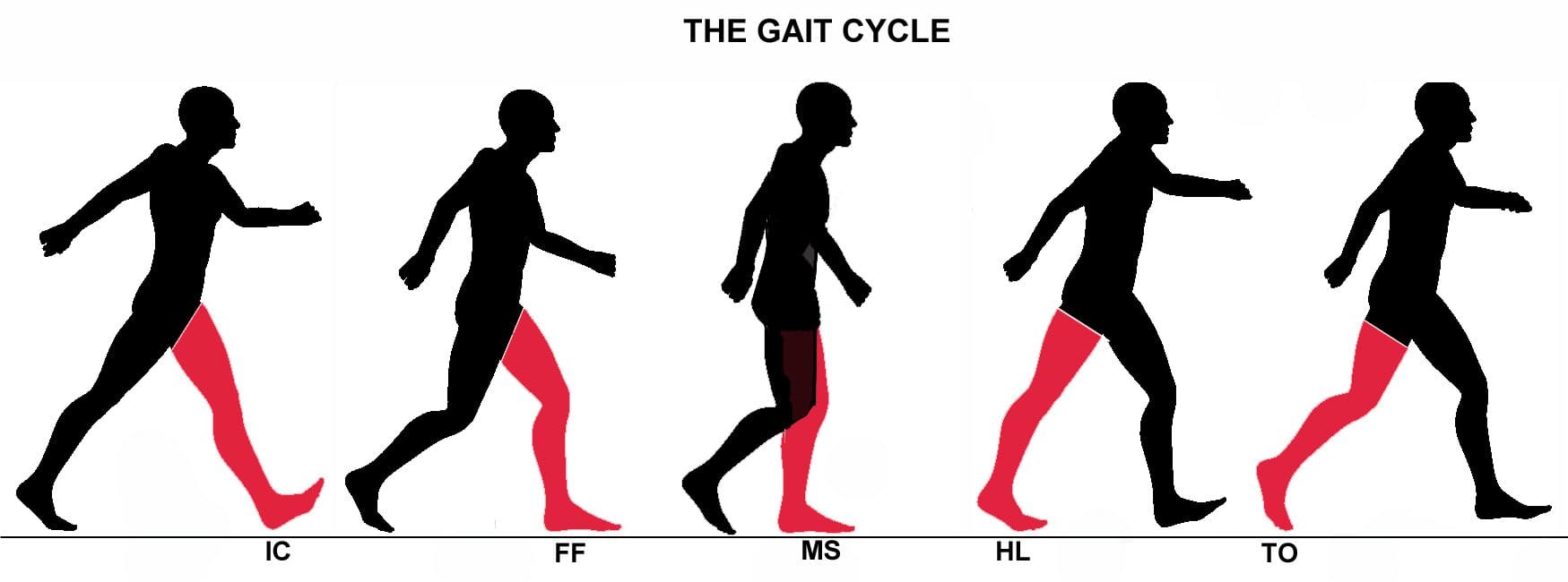
An Overview Of Gait Analysis-Video
Have you been dealing with mobility issues in your joints? Do you find yourself becoming unstable every time you walk? Or do your leg muscles feel tight? If you have been dealing with these issues, it could be due to a gait problem. Many people have different ways of walking; if there are issues, they can be pointed out in an examination. When there is an issue with gait, it can indicate pain and other revealing problems that can affect the entire body. The video above explains the gait cycle and gait analysis of a person’s walk. Gait analysis is often used in a normal examination to evaluate how a person walks, their body mechanics, and muscle activity to provide insight into the issue. A person’s gait can offer many important clues that doctors and pain specialists can see and identify the problem by developing a treatment plan to improve a person’s gait and reduce pain-like symptoms.
How The MET Technique Improves Gait
So many treatment plans can effectively improve balance and gait disorders in the body. Many pain specialists like chiropractors use manual spinal manipulation to re-align the spine to loosen stiff joints that may have contributed to imbalances in the lower extremities. MET (muscle energy technique) and physical therapy can help stretch the tight muscles and strengthen the muscle groups affected. MET and other approaches to improve gait allow many individuals to regain their stamina and adopt new strategies for their posture and movement. These therapy treatments will enable a person to feel more confident and more aware of how they walk while providing muscle strength to the affected muscles to prevent fatigue and decrease the chances of injuries in the future, as studies reveal.
Conclusion
Walking is determined by a person’s gait and how they move in different scenarios. Our bodies are comprised of upper and lower portions that correspond with gait and allow us stability and balance when we are in motion. When various issues like traumatic factors or just normal aging affect the body, the joints and muscles can cause problems with a person’s gait, leading to balance issues and fall injuries. Incorporating treatment plans to improve gait can help prevent future chances of injuries and help stretch and strengthen the affected muscles while loosening up stiff joints. This allows a person to regain their balance and improve stability in their bodies.
References
Baker, Jessica M. “Gait Disorders.” The American Journal of Medicine, U.S. National Library of Medicine, 27 Dec. 2017, pubmed.ncbi.nlm.nih.gov/29288631/.
Chaitow, Leon, and Judith Walker DeLany. Clinical Applications of Neuromuscular Techniques. Churchill Livingstone, 2003.
Pirker, Walter, and Regina Katzenschlager. “Gait Disorders in Adults and the Elderly : A Clinical Guide.” Wiener Klinische Wochenschrift, U.S. National Library of Medicine, Feb. 2017, www.ncbi.nlm.nih.gov/pmc/articles/PMC5318488/.
Van Abbema, Renske, et al. “What Type, or Combination of Exercise Can Improve Preferred Gait Speed in Older Adults? A Meta-Analysis.” BMC Geriatrics, U.S. National Library of Medicine, 1 July 2015, www.ncbi.nlm.nih.gov/pmc/articles/PMC4488060/.
Prolonged standing can cause the pelvis to push backward, increasing the curve of the lower back/lumbar region. This increased pressure on the soft tissues surrounding the spine causes the lower back muscles to tighten and/or spasm, resulting in discomfort in the joints and nerves. Weakened core muscles and unhealthy posture/postural syndrome are the most common causes, but injury, aging, congenital malformations, or a disease/condition can also contribute to the symptoms. Injury Medical Chiropractic and Functional Medicine Clinic has a top team of professional therapists to evaluate the problem, diagnose the cause/s accurately, and develop a customized treatment and rehabilitation plan.
Prolonged Standing Back Discomfort
Back Structure
The lower back is one of the most used areas of the spine, moving around and bending during a normal day. When the body stands, the spine naturally curves both in and outwards.
The inward curve, called lordosis, curves towards the front of the body at the lower back and neck regions.
The outward curve, called kyphosis, curves towards the back of the body at the chest.
When bending over while standing, the five lumbar vertebrae of the lower back change position and shift from lordosis to kyphosis when bent completely.
When standing up from bending, the lumbar vertebrae change position again and return to the lordosis position.
Causes
The facet joints allow movement between each spine level. The standing spinal curvature can increase contact between the facet joints. As the body ages, the facet joints and discs begin to wear out, which can cause the discs and facet joints to become inflamed. Prolonged standing during normal daily activity combined with inflammation in these joints can aggravate the inflammation and cause symptoms. Regular routines and habits may contribute to low back discomfort during prolonged standing. These include:
Sleeping on a sinking or unsupportive mattress.
Practicing unhealthy postures that cause imbalances with proper weight distribution.
Not wearing proper footwear and/or supportive orthotics forces the lower spine into increased curvature and can compress the facet joints.
Not getting enough physical activity that strengthens the core.
Chiropractors are experts on the musculoskeletal system. They will:
Listen to the patient about symptoms, medical history, and occupation.
A physical examination of muscle tone, strength, and range of motion.
Therapeutic massage, electric muscle stimulation, and ultrasound therapy can help reduce muscle inflammation and increase circulation to injured soft tissues.
Chiropractic adjustments will reset joints, removing pressure from the surrounding muscles and nerves.
Targeted therapeutic strength training is recommended for core and leg muscles to improve hip flexibility.
Non-surgical decompression or traction, either with a machine or suspension, can reverse the pressure in spinal discs.
Standing Lower Back Relief Exercises
References
Hasegawa, Tetsuya, et al. “Association of low back load with low back pain during static standing.” PloS one vol. 13,12 e0208877. 18 Dec. 2018, doi:10.1371/journal.pone.0208877
Jo, Hoon, et al. “Negative Impacts of Prolonged Standing at Work on Musculoskeletal Symptoms and Physical Fatigue: The Fifth Korean Working Conditions Survey.” Yonsei medical journal vol. 62,6 (2021): 510-519. doi:10.3349/ymj.2021.62.6.510
Ognibene GT, Torres W, von Eyben R, Horst KC. Impact of a sit-stand workstation on chronic low back pain: randomized trial results. J Occup Environ Med. 2016;58(3):287-293. Abstract. www.ncbi.nlm.nih.gov/pubmed/26735316. Accessed March 2, 2017.
Parry, Sharon P et al. “Workplace interventions for increasing standing or walking for decreasing musculoskeletal symptoms in sedentary workers.” The Cochrane database of systematic reviews vol. 2019,11 CD012487. November 17, 2019, doi:10.1002/14651858.CD012487.pub2
Rodríguez-Romero, Beatriz, et al. “Thirty Minutes Identified as the Threshold for Development of Pain in Low Back and Feet Regions, and Predictors of Pain Intensity During 1-h Laboratory-Based Standing in Office Workers.” International journal of environmental research and public health vol. 19,4 2221. February 16, 2022, doi:10.3390/ijerph19042221
Smith, Michelle D et al. “The Influence of Using a Footstool during a Prolonged Standing Task on Low Back Pain in Office Workers.” International journal of environmental research and public health vol. 16,8 1405. April 18. 2019, doi:10.3390/ijerph16081405
IFM's Find A Practitioner tool is the largest referral network in Functional Medicine, created to help patients locate Functional Medicine practitioners anywhere in the world. IFM Certified Practitioners are listed first in the search results, given their extensive education in Functional Medicine