Can individuals dealing with psoriatic arthritis find non-surgical treatments to reduce joint pain and improve skin health?
Introduction
The body is a complex machine with a communal relationship with the joints, organs, bones, muscles, and tissues, each of which has a specific function to ensure the host is alive. The brain is the main control system that gives each body’s systems, organs, muscles and joints a job. When environmental factors or pathogens enter the body, the immune system is the first one to defend the body and eliminate the pathogens to initiate the natural healing process. However, when the immune system is attacking the body constantly, over time, it can lead to autoimmunity and cause overlapping risk profiles in the body. This, in turn, can lead the development of chronic conditions and cause individuals pain and discomfort. In today’s article, we look at an autoimmune disorder known as psoriatic arthritis, its causes and symptoms, and how treatment can help reduce its pain like effects. We discuss with certified medical providers who inform our patients how psoriatic arthritis can affect the joints. While asking informed questions to our associated medical providers, we advise patients to incorporate various non-surgical treatments to reduce psoriatic arthritis symptoms and help manage joint pain that affects a person’s quality of life. Dr. Alex Jimenez, D.C., encompasses this information as an academic service. Disclaimer.
What Is Psoriatic Arthritis?
Do you experience swelling in your joints that causes pain and discomfort when moving? Do you feel your skin itchier and warmer than usual? Or have you noticed that everyday tasks are more difficult now than before? Many of these pain-like scenarios are associated with joint pain associated with an autoimmune or musculoskeletal condition. Since autoimmunity is developed when environmental factors cause the immune system to attack healthy cells, it can envoke pain and discomfort to the body. When a person is dealing with an autoimmune condition, it can impact not only the individual but also the rest of the body’s system. For instance, if a person is dealing with an autoimmune condition, they could be dealing with skin issues that can cause them to develop scaly, itchy, red skin patches known as psoriasis. If they have joint pain combined with psoriasis, it can develop into psoriatic arthritis. (Skornicki et al., 2021) Now psoriatic arthritis is a heterogeneous autoimmune condition with musculoskeletal involvement that can manifest various symptoms including inflammation and arthritis. (Hackett et al., 2022) This in turn causes many individuals to think they are dealing with rheumatoid factors.
Causes & Symptoms
As a multifactorial pathology, psoriatic arthritis causes care often influenced by a complex interplay of immunological, environmental, and genetic factors that cause its development. (Zalesak et al., 2024) When the immune system starts to attack the healthy cells in the body, the inflammatory cytokines can become haywire and attack the “problem,” causing the development of autoimmunity. Within this integration, some of the causes can include predisposing genetic backgrounds with the presence of environmental factors that can activate the innate immune system precipitate the onset of psoriatic arthritis. (Azuaga et al., 2023) Some of the symptoms can many individuals with psoriatic arthritis can include:
Tendon tenderness
Joint pain
Stiffness
Fatigue
Joint swelling
Skin rash
Musculoskeletal pain
However, there are ways to manage psoriatic arthritis, reduce joint pain from affecting a person, and help them manage the symptoms.
Arthritis Explained-Video
Treatments For Psoriatic Arthritis
When it comes to treating psoriatic arthritis, healthcare professionals need to develop therapeutic options to educate the individual and develop a treatment plan that can include conventional therapies and non-pharmacological therapies that are affordable and customized with the individual’s condition. (Ogdie et al., 2020) Since psoriatic arthritis is a complex inflammatory musculoskeletal condition and skin disease, individuals must try to find ways to reduce the inflammatory effects of causing more joint issues. Non-surgical treatments like chiropractic care, physical therapy, and diet changes can help mitigate the burden of psoriatic arthritis by reducing the complications and the socio-economic impact. (McGonagle et al., 2022)
Another way individuals can reduce the inflammatory effects of psoriatic arthritis is by consuming anti-inflammatory foods to dampen the inflammatory cytokines attacking the joints and going to physical therapy sessions like water aerobics to help stabilize the joints and help people be more mindful about their bodies. Since psoriatic arthritis can be managed through non-surgical therapy, low-weight bearing exercise regimes and incorporating anti-inflammatory foods to reduce inflammation can help people minimize psoriatic arthritis symptoms from impacting their health and wellness journey.
References
Azuaga, A. B., Ramirez, J., & Canete, J. D. (2023). Psoriatic Arthritis: Pathogenesis and Targeted Therapies. Int J Mol Sci, 24(5). https://doi.org/10.3390/ijms24054901
Hackett, S., Ogdie, A., & Coates, L. C. (2022). Psoriatic arthritis: prospects for the future. Ther Adv Musculoskelet Dis, 14, 1759720X221086710. https://doi.org/10.1177/1759720X221086710
McGonagle, D. G., Zabotti, A., Watad, A., Bridgewood, C., De Marco, G., Kerschbaumer, A., & Aletaha, D. (2022). Intercepting psoriatic arthritis in patients with psoriasis: buy one get one free? Ann Rheum Dis, 81(1), 7-10. https://doi.org/10.1136/annrheumdis-2021-221255
Ogdie, A., Coates, L. C., & Gladman, D. D. (2020). Treatment guidelines in psoriatic arthritis. Rheumatology (Oxford), 59(Suppl 1), i37-i46. https://doi.org/10.1093/rheumatology/kez383
Skornicki, M., Prince, P., Suruki, R., Lee, E., & Louder, A. (2021). Clinical Burden of Concomitant Joint Disease in Psoriasis: A US-Linked Claims and Electronic Health Records Database Analysis. Adv Ther, 38(5), 2458-2471. https://doi.org/10.1007/s12325-021-01698-7
Zalesak, M., Danisovic, L., & Harsanyi, S. (2024). Psoriasis and Psoriatic Arthritis-Associated Genes, Cytokines, and Human Leukocyte Antigens. Medicina (Kaunas, Lithuania), 60(5). https://doi.org/10.3390/medicina60050815
Can individuals with rheumatoid arthritis incorporate various exercises to reduce joint pain and inflammation in their hands and feet?
Introduction
The joints in the human help provide function, mobility, and flexibility to the upper and lower extremities. The joints are part of the musculoskeletal system and have an outstanding relationship with the muscles, ligaments, and soft tissues that give the body structure and support that lets the individual move around and protects the important organs to function normally. However, when a person is dealing with injuries or illnesses that affect the body’s musculoskeletal function, it can cause pain to the individual. One of the symptoms that often correlate in the joints is chronic inflammation, leading to the development of an autoimmune disease known as rheumatoid arthritis. Today’s article looks at how rheumatoid arthritis affects the joints in the musculoskeletal system and how various exercises can help manage and reduce the symptoms associated with rheumatoid arthritis. We discuss with certified associated medical providers who consolidate our patients’ information to assess rheumatoid arthritis and its associated pain symptoms affecting the joints. We also inform and guide patients while asking their associated medical provider intricate questions to integrate various exercises into their personalized treatment plan to manage the pain correlated with rheumatoid arthritis. Dr. Jimenez, D.C., includes this information as an academic service. Disclaimer.
How RA Affects The Joints
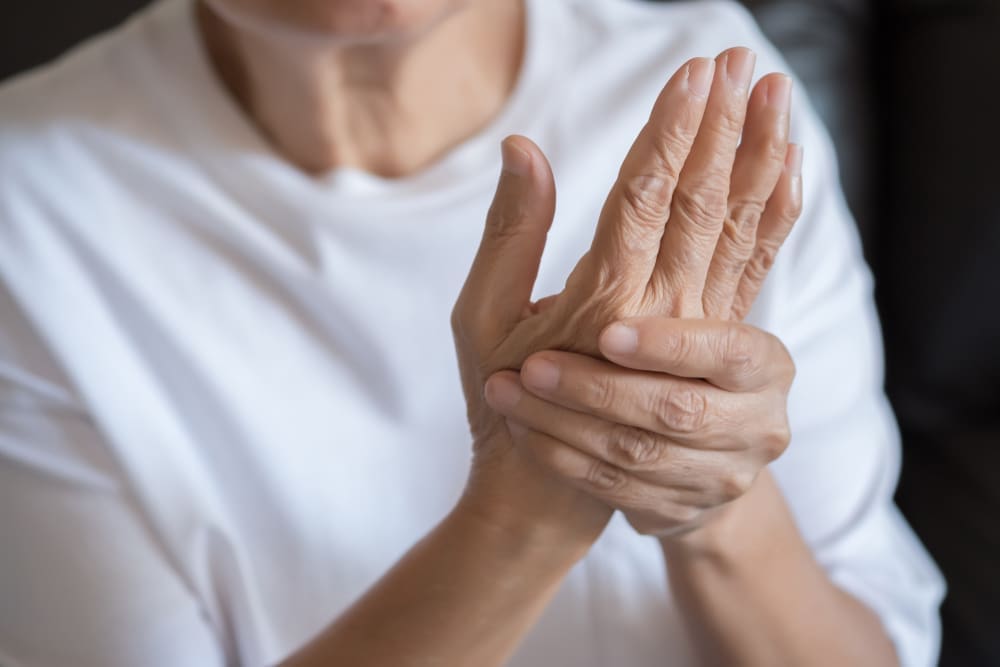
Do you feel pain and tenderness in your joints affecting your daily routine? Do you experience stiffness first thing in the morning, and it goes away throughout the day? Or do you feel fatigued throughout the day, even after a good night’s sleep? Many individuals with these symptoms could be dealing with early development of rheumatoid arthritis in their joints. Now, rheumatoid arthritis is a chronic inflammatory autoimmune disorder that affects the body’s joints but is more prominent on the hands, wrists, and feet. The symptoms of rheumatoid arthritis can develop early or slowly depending on the environmental factors contributing to the development. Since rheumatoid arthritis is categorized as a systemic autoimmune disease, genetic and environmental risk factors that can contribute to rheumatoid arthritis development can trigger overlapping risk profiles on the joints. (Jang et al., 2022) When a person is dealing with the symptoms of rheumatoid arthritis, one of the key pain symptoms that can affect the joints drastically is inflammation. Inflammation is associated with rheumatoid arthritis; it is reflected by joint pain, leading to swelling and subsequent destruction of the cartilage and bone. (Scherer et al., 2020) This causes many individuals to be in constant pain and prevents them from doing any activities.
Additionally, when a few joints are being affected by rheumatoid arthritis in the early stages, some of the symptoms include:
Fatigue
Flu-like symptoms
Swollen & tender joints
Stiffness
However, when rheumatoid arthritis reaches the later stages in the joints, the autoantigens that are specific to rheumatoid arthritis can lead to a self-perpetuating chronic inflammatory state on the joints, thus causing an expansion on the periarticular bone at the cartilage-bone junction, leading to bone erosion and cartilage degradation. (Lin et al., 2020) Luckily, there are therapeutic options to reduce the pain and inflammatory effects of rheumatoid arthritis and help manage the symptoms that are affecting the joints.
Arthritis Explained- Video
How Various Exercises Can Help With RA
When it comes to reducing the inflammatory effects of rheumatoid arthritis, many individuals can seek out therapeutic options to restore mobility, function, and flexibility. Many individuals can incorporate various physical activities to relieve stress on the inflamed tissues while slowing the progression of rheumatoid arthritis. (Radu & Bungau, 2021) When people with rheumatoid arthritis incorporate various physical activities, they can include a healthy diet and nutrition to suppress pro-inflammatory effects associated with rheumatoid arthritis, help provide symptomatic improvement, and restore bodily function to the joints. (Gioia et al., 2020)
When people with rheumatoid arthritis start exercising as part of their personalized treatment, it can have beneficial properties as they can help with the following:
Reduce joint pain & stiffness
Improve muscle strength around the joints
Enhance physical function
Boost mental health
Reduces inflammation
Increase energy levels
The main priority of incorporating exercises to reduce rheumatoid arthritis is choosing gentle exercises on the person’s joints while providing enough movement to keep the body flexible and strong. Below are some exercises to reduce rheumatoid arthritis.
Range of Motion Exercises
Range of motion exercises can help maintain normal joint function by improving flexibility and reducing stiffness for individuals with rheumatoid arthritis. Some examples include:
Finger Bends: Gently bend your fingers into a fist and straighten them. Repeat several times.
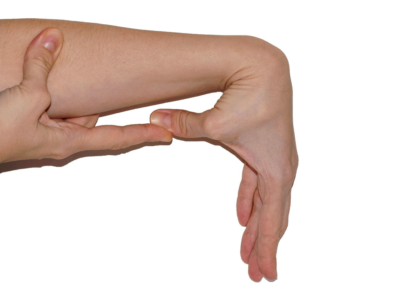
Wrist Stretch: Extend your arm with the palm facing down. Gently use your other hand to press the extended hand down and back for a stretch.
Shoulder Rolls: Roll the shoulders in a forward circular motion, then reverse the direction.
Strength Training Exercises
Strength training can help build the surrounding muscles around the joints. This allows many individuals with rheumatoid arthritis to provide better support and reduce stress on the joints. Some examples include:
Resistance Bands: Use resistance bands to perform bicep curls, leg extensions, and chest presses.
Light Weights: Incorporate light dumbbells to perform exercises like shoulder presses, tricep extensions, and squats.
Bodyweight Exercises: Engage in wall push-ups, seated leg lifts, and modified planks.
Water-Based Exercises
Water-based exercises provide resistance without impact on the joints, making it ideal for those with rheumatoid arthritis. The water helps cushion the joints by easing the stiffness, building strength, and helping relax sore muscles. Some examples of water-based exercises include:
Water Aerobics: Join a water aerobics class that offers structured routines in a supportive environment.
Aqua Jogging: Use a buoyancy belt to jog in the pool’s deep end.
Swimming: Perform laps or engage in gentle exercises like the backstroke or breaststroke.
Tips For Exercising With RA
It is important to remember that when exercising with rheumatoid arthritis, it is important to always start with a gentle warm-up and always end with a cool down to prepare the muscles and joints when beginning to exercise. Another thing to remember is to stay consistent and modify when needed. This allows many individuals to listen to their bodies and modify exercises to avoid pain and discomfort. Incorporating exercises is highly effective in reducing rheumatoid arthritis activity as it can help enhance the body’s immune function and help manage the inflammatory response associated with rheumatoid arthritis. (Li & Wang, 2022)
References
Gioia, C., Lucchino, B., Tarsitano, M. G., Iannuccelli, C., & Di Franco, M. (2020). Dietary Habits and Nutrition in Rheumatoid Arthritis: Can Diet Influence Disease Development and Clinical Manifestations? Nutrients, 12(5). https://doi.org/10.3390/nu12051456
Jang, S., Kwon, E. J., & Lee, J. J. (2022). Rheumatoid Arthritis: Pathogenic Roles of Diverse Immune Cells. Int J Mol Sci, 23(2). https://doi.org/10.3390/ijms23020905
Li, Z., & Wang, X. Q. (2022). Clinical effect and biological mechanism of exercise for rheumatoid arthritis: A mini review. Front Immunol, 13, 1089621. https://doi.org/10.3389/fimmu.2022.1089621
Lin, Y. J., Anzaghe, M., & Schulke, S. (2020). Update on the Pathomechanism, Diagnosis, and Treatment Options for Rheumatoid Arthritis. Cells, 9(4). https://doi.org/10.3390/cells9040880
Can physical therapies help relieve muscle contractures in individuals who have endured prolonged bed rest, inactivity, or lack of use of certain muscle groups?
Muscle Contracture
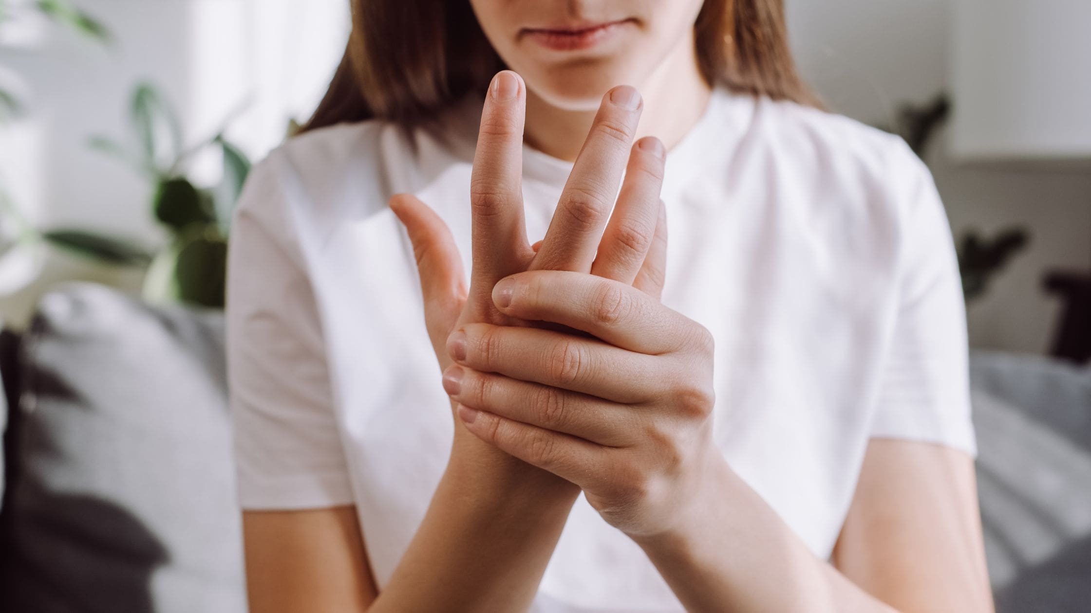
A muscle contracture, or contracture deformity, is caused when a muscle loses elasticity. This causes permanent shortening and tightening of muscle fibers, which reduces flexibility and makes movement difficult. Muscles that cannot move and stretch cause the surrounding joints to lose mobility and develop pain symptoms. When trying to stretch the contracted muscle, the individual will feel the muscle become very rigid, which can increase pain. (Lieber, R. L., and Fridén, J. 2019) Delaying treatment can potentially cause irreversible and chronic symptoms.
Commonly Affected Muscles
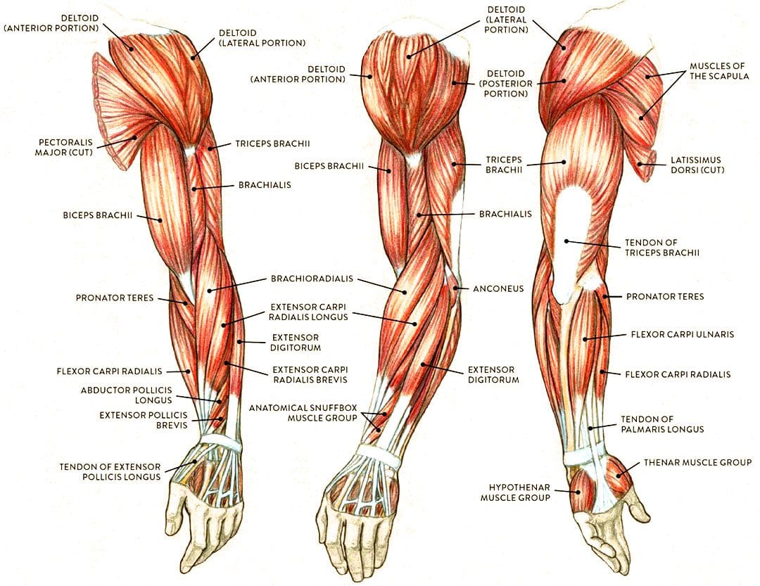
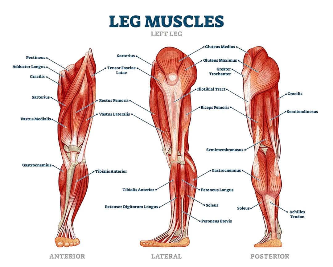
Flexor muscles bend the joints and are those most affected by contractures. The stiffening and tightening prevent the body parts from moving out and away. The most common include:
Wrist and Finger Flexors
Muscle groups that bend the wrist and fingers.
Biceps
The primary elbow flexor that bends the arm.
Gastrocnemius and Soleus
The calf muscles which allow the ankle to point the foot/plantarflexion.
Hamstrings
A group of three muscles behind the thigh that bend the knee.
Causes
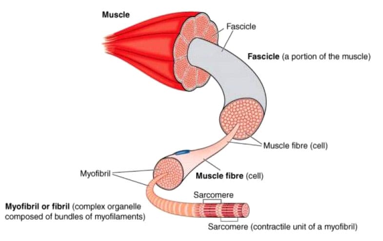
The permanent shortening of muscle fibers and changes in muscle structure cause muscle contractures or stiffer-than-normal tissue that is difficult to stretch. Sarcomeres are structural units of muscles that cause fibers to contract.
With contractures, the sarcomeres overly lengthen when the muscle fibers tighten. This increase in sarcomere length prevents the muscle from contracting normally, resulting in weakness. Muscle fibers are encased in an extracellular matrix, a mesh composed of collagen and other proteins that help transmit force and provide muscle contraction. Muscle contractures cause the amount of collagen within the extracellular matrix to increase, causing a stiffening of fibers that restricts movement. (Lieber, R. L., and Fridén, J. 2019)
Muscle contractures also form from decreased satellite cells. Satellite cells are specialized stem cells that can rebuild muscle and are necessary for muscle regeneration and repair. Without the proper amount of satellite cells, other cells like fibroblasts significantly increase in the muscle tissue, causing the fibers to become stiff and fibrotic or more fibrous. These changes to the sarcomeres, collagen within the extracellular matrix, and decreased satellite cells all result from conditions in which neurological input to the brain and spinal cord muscles becomes reduced. This is caused by lack of use, injury, or neurological and neuromuscular conditions. (Lieber, R. L., and Fridén, J. 2019)
Cerebral Palsy
Contractures often occur from upper motor neuron lesions, which prevent signals from the brain and spinal cord from reaching the motor neurons that control muscle contraction. When these signals are weakened or blocked, muscles become stiff and weak from lack of stimulation. (Lieber, R. L., and Fridén, J. 2019)
Cerebral palsy is a group of disorders affecting mobility caused by an upper motor neuron lesion that is present at birth and is the most common motor disability in children. It causes:
Cognitive impairment
Decreased muscle strength
Problems with movement, coordination, and functional motions.
Because cerebral palsy prevents the muscles of the legs from being sufficiently stimulated, contractures commonly develop in the hips, knees, and ankles. Individuals can have a 75% decrease in satellite cells to repair muscle tissue and prevent muscle fibrosis or stiffening. Specific genes linked to collagen production are also altered, causing irregular changes to the extracellular matrix of muscles. (Lieber, R. L., and Fridén, J. 2019)
Muscular Dystrophy
Muscular dystrophy is a group of inherited neuromuscular disorders characterized by muscle weakness and wasting. Deficient nerve supply to muscles causes them to become stiff and tight, inhibiting the functional range of motion needed to move joints and activate muscles to move. Clinical research suggests that individuals with muscular dystrophy have decreased levels of satellite cells to repair, increasing the risk of developing muscle contracture. (Lieber, R. L., and Fridén, J. 2019)
Disuse-induced Muscle Wasting or Disuse Atrophy
When muscles are not used for some time because of hospitalization, prolonged bed rest, or immobilization from wearing braces, splints, or casts after injuries, the blood circulation and electrical signaling from nerves to muscles decreases. This results in weakness, increased muscle tightness and stiffness, and muscle wasting/atrophy. Over time, stiff and tight muscles can progress to contractures that become extremely difficult to stretch.
Trauma or Injury
Muscle or tendon injuries can cause contractures as scar tissue develops, joining muscle fibers and joints together. This can significantly restrict movement. Large burns can also cause skin, muscles, and joint contractures. The range of motion can become significantly limited, and the changes can become irreversible if not aggressively treated.
Other Causes
Other forms of upper motor neuron lesions that can cause contractures because of weak or blocked electrical input to muscles as a result of brain or spinal cord damage include:
Neuromuscular disorders like spinal muscular atrophy – SMA.
Conditions that cause inflammation and joint stiffening, like juvenile rheumatoid arthritis.
A history of diabetes also increases the risk of developing contractures affecting finger flexors, like Dupuytren’s contractures and stenosing tenosynovitis
or trigger finger. (Lieber, R. L., and Fridén, J. 2019)
Symptoms
Symptoms include:
Extremely stiff and tight muscles resistant to stretching.
Pain from the inability to stretch.
Loss of range of motion.
Impaired joint mobility.
Severe contractures can interfere with the functional range of motion needed to move joints to complete normal tasks and movements, such as standing up from a chair and walking.
Treatment
Physical Therapy
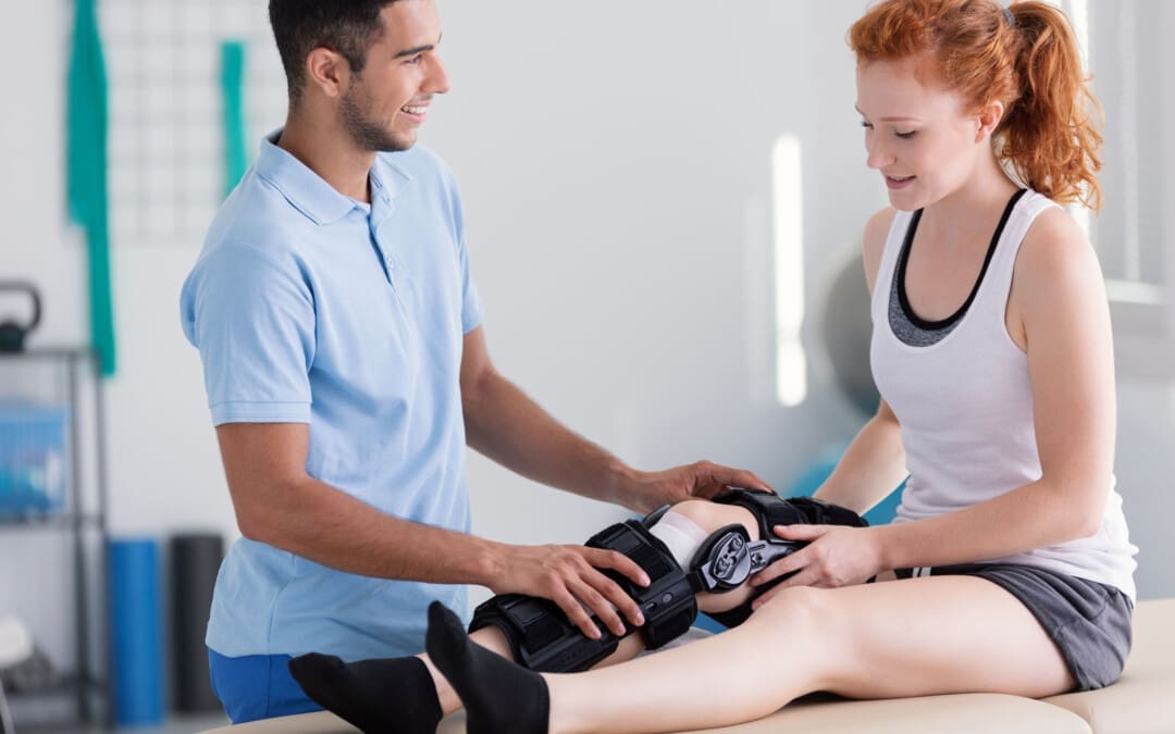
Physical therapies can help reduce the severity through stretching and soft tissue mobilization to decrease tightness. (Lieber, R. L., and Fridén, J. 2019)
Specialized braces or splints can be custom-made to fit different body parts.
These provide a prolonged low-intensity stretch over a period of time to increase muscle length.
Once the muscle has stretched, a new brace or splint may be needed to adjust to the increased range of motion. (Lieber, R. L., and Fridén, J. 2019)
Surgery
In severe cases where muscle contractures limit the functional range of motion needed for activities of daily living or ADLs, surgical release of the contracted tissue may be recommended. This surgery can improve functional movements like walking, getting in and out of bed, and standing up from chairs. The tight muscles can be surgically cut, and the tendons can be lengthened to allow more mobility. (Lieber, R. L., and Fridén, J. 2019)
The causes of muscle contracture are not always avoidable, but various treatment options are available to help loosen up tight muscles and preserve or restore the range of motion. It’s important to move daily and stretch common areas like the fingers, arms, and legs to reduce the risk of muscle tightness and prevent contractures from developing. It is imperative to seek medical treatment for severe contractures resulting from neuromuscular disorders, including physical and occupational therapy, to prevent contractures from worsening and regaining as much functional range as possible.
Injury Medical Chiropractic and Functional Medicine Clinic uses an integrated approach personalized to the individual that focuses on what works for them and treats injuries and chronic pain syndromes through personalized care plans that improve ability through flexibility, mobility, and agility programs to relieve pain. Our providers use an integrated approach to create personalized care plans for each patient, including Functional Medicine, Acupuncture, Electro-Acupuncture, and Sports Medicine principles. Our goal is to relieve pain naturally by restoring health and function to the body. If other treatment is needed, Dr. Jimenez has teamed up with top surgeons, clinical specialists, medical researchers, and rehabilitation providers to provide the most effective treatments.
Chiropractic Treatment for Cerebral Palsy
References
Lieber, R. L., & Fridén, J. (2019). Muscle contracture and passive mechanics in cerebral palsy. Journal of applied physiology (Bethesda, Md. : 1985), 126(5), 1492–1501. https://doi.org/10.1152/japplphysiol.00278.2018
Can individuals with Ehlers-Danlos syndrome find relief through various non-surgical treatments to reduce joint instability?
Introduction
The joints and ligaments surrounding the musculoskeletal system allow the upper and lower extremities to stabilize the body and be mobile. The various muscles and soft connective tissues that surround the joints help protect them from injuries. When environmental factors or disorders start to affect the body, many people develop issues that cause overlapping risk profiles, which then affect the stability of the joints. One of the disorders that affect the joints and connective tissue is EDS or Ehlers-Danlos syndrome. This connective tissue disorder can cause the joints in the body to be hypermobile. It can cause joint instability in the upper and lower extremities, thus leaving the individual to be in constant pain. Today’s article focuses on Ehlers-Danlos syndrome and its symptoms and how there are non-surgical ways to manage this connective tissue disorder. We discuss with certified medical providers who consolidate our patients’ information to assess how Ehlers-Danlos syndrome can correlate with other musculoskeletal disorders. We also inform and guide patients on how various non-surgical treatments can help reduce pain-like symptoms and manage Ehlers-Danlos syndrome. We also encourage our patients to ask their associated medical providers many intricate and important questions about incorporating various non-surgical therapies as part of their daily routine to manage the effects of Ehlers-Danlos syndrome. Dr. Jimenez, D.C., includes this information as an academic service. Disclaimer.
What Is Ehlers-Danlos Syndrome?
Do you often feel extremely tired throughout the day, even after a full night of sleep? Do you bruise easily and wonder where these bruises are coming from? Or have you noticed that you have an increased range in your joints? Many of these issues are often correlated with a disorder known as Ehlers-Danlos syndrome or EDS that affects their joints and connective tissue. EDS affects the connective tissues in the body. The connective tissues in the body help provide strength and elasticity to the skin, joints, as well as blood vessel walls, so when a person is dealing with EDS, it can cause a significant disruption to the musculoskeletal system. EDS is largely diagnosed clinically, and many doctors have identified that the gene coding of the collagen and proteins that interact in the body can help determine what type of EDS affects the individual. (Miklovic & Sieg, 2024)
The Symptoms
When understanding EDS, it is essential to know the complexities of this connective tissue disorder. EDS is classified into numerous types with distinct features and challenges that vary depending on the severity. One of the most common types of EDS is hypermobile Ehlers-Danlos syndrome. This type of EDS is characterized by general joint hypermobility, joint instability, and pain. Some of the symptoms that are associated with hypermobile EDS include subluxation, dislocations, and soft tissue injuries that are common and may occur spontaneously or with minimal trauma. (Hakim, 1993) This can often cause acute pain to the joints in the upper and lower extremities. With its broad range of symptoms and the personal nature of the condition itself, many often don’t realize that joint hypermobility is common in the general population and may present no complications that indicate that it is a connective tissue disorder. (Gensemer et al., 2021) Additionally, hypermobile EDS can lead to spinal deformity due to the hyperextensibility of the skin, joints, and various tissue fragility. The pathophysiology of spinal deformity associated with hypermobile EDS is primarily due to muscle hypotonia and ligament laxity. (Uehara et al., 2023) This causes many people to reduce their quality of life and daily living activities significantly. However, there are ways to manage EDS and its correlating symptoms to reduce joint instability.
Movement Medicine: Chiropractic Care-Video
Ways To Manage EDS
When it comes to looking for ways to manage EDS to reduce pain and joint instability, non-surgical treatments can help address the physical and emotional aspects of the condition. Non-surgical treatments for individuals with EDS commonly focus on optimizing the body’s physical function while improving muscular strength and joint stabilization. (Buryk-Iggers et al., 2022) Many individuals with EDS will try to incorporate pain management techniques and physical therapy anduse braces and assistive devices to reduce the effects of EDS and improve their quality of life.
Non-surgical Treatments For EDS
Various non-surgical treatments like MET (muscle energy technique), electrotherapy, light physical therapy, chiropractic care, and massages can help strengthen while toning the surrounding muscles around the joints, provide sufficient pain relief, and limit long-term dependence on medications. (Broida et al., 2021) Additionally, individuals dealing with EDS aim to strengthen the affected muscles, stabilize the joints, and improve proprioception. Non-surgical treatments allow the individual to have a customized treatment plan for the severity of EDS symptoms and help reduce the pain associated with the condition. Many individuals, when going through their treatment plan consecutively to manage their EDS and reduce the pain-like symptoms, will notice improvement in symptomatic discomfort. (Khokhar et al., 2023) This means that non-surgical treatments allow individuals to be more mindful of their bodies and reduce the pain-like effects of EDS, thus allowing many individuals with EDS to lead fuller, more comfortable lives without feeling pain and discomfort.
References
Broida, S. E., Sweeney, A. P., Gottschalk, M. B., & Wagner, E. R. (2021). Management of shoulder instability in hypermobility-type Ehlers-Danlos syndrome. JSES Rev Rep Tech, 1(3), 155-164. https://doi.org/10.1016/j.xrrt.2021.03.002
Buryk-Iggers, S., Mittal, N., Santa Mina, D., Adams, S. C., Englesakis, M., Rachinsky, M., Lopez-Hernandez, L., Hussey, L., McGillis, L., McLean, L., Laflamme, C., Rozenberg, D., & Clarke, H. (2022). Exercise and Rehabilitation in People With Ehlers-Danlos Syndrome: A Systematic Review. Arch Rehabil Res Clin Transl, 4(2), 100189. https://doi.org/10.1016/j.arrct.2022.100189
Gensemer, C., Burks, R., Kautz, S., Judge, D. P., Lavallee, M., & Norris, R. A. (2021). Hypermobile Ehlers-Danlos syndromes: Complex phenotypes, challenging diagnoses, and poorly understood causes. Dev Dyn, 250(3), 318-344. https://doi.org/10.1002/dvdy.220
Hakim, A. (1993). Hypermobile Ehlers-Danlos Syndrome. In M. P. Adam, J. Feldman, G. M. Mirzaa, R. A. Pagon, S. E. Wallace, L. J. H. Bean, K. W. Gripp, & A. Amemiya (Eds.), GeneReviews((R)). https://www.ncbi.nlm.nih.gov/pubmed/20301456
Khokhar, D., Powers, B., Yamani, M., & Edwards, M. A. (2023). The Benefits of Osteopathic Manipulative Treatment on a Patient With Ehlers-Danlos Syndrome. Cureus, 15(5), e38698. https://doi.org/10.7759/cureus.38698
Can understanding the body’s hinge joints and how they operate help with mobility and flexibility problems and manage conditions for individuals with difficulty fully bending or extending their fingers, toes, elbows, ankles, or knees?
Hinge Joints
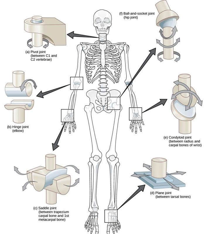
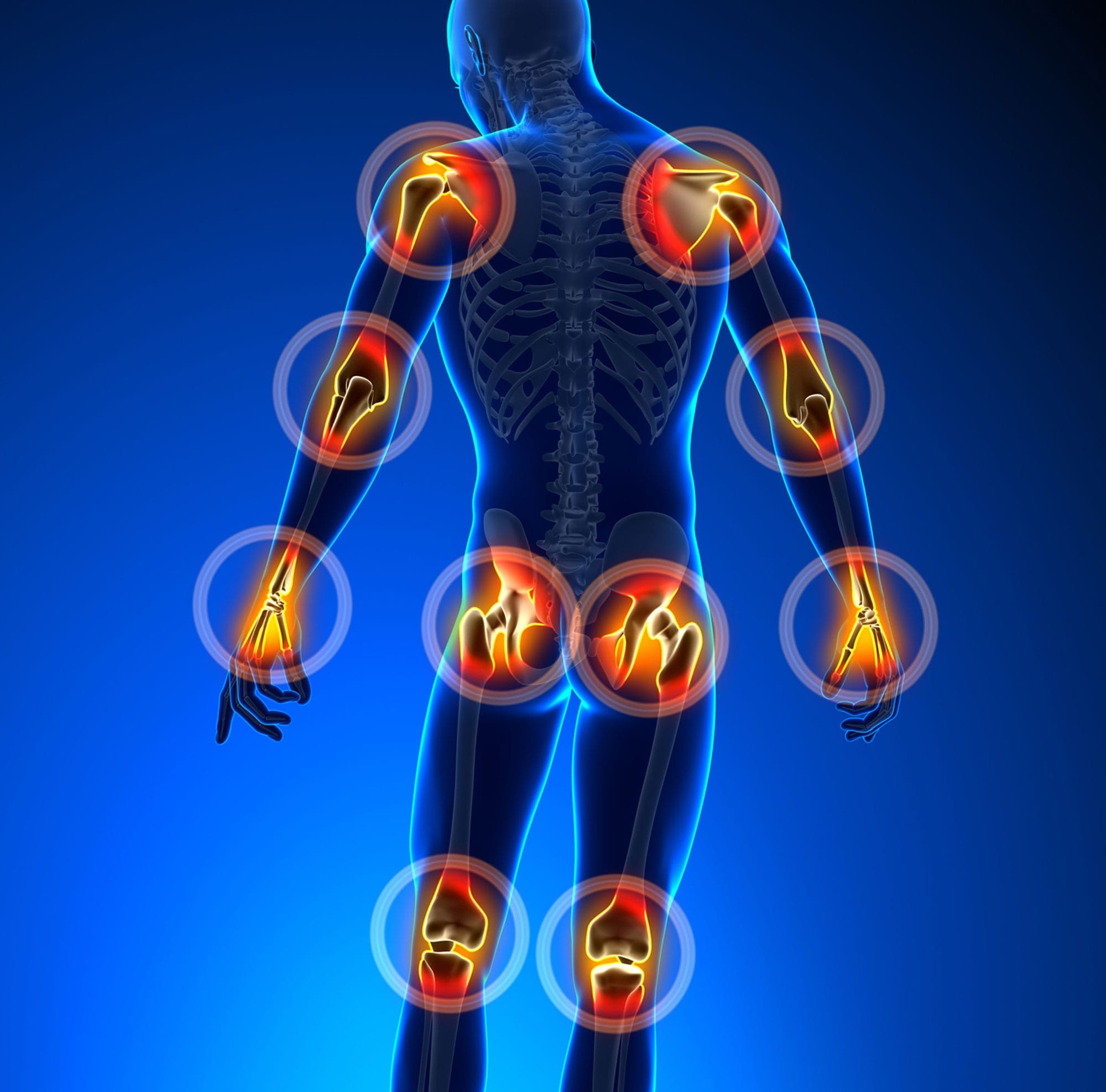
A joint forms where one bone connects to another, allowing motion. Different types of joints differ in structure and movement depending on their location. These include hinge, ball and socket, planar, pivot, saddle, and ellipsoid joints. (Boundless. General Biology, N.D.) Hinge joints are synovial joints that move through one plane of motion: flexion and extension. Hinge joints are found in the fingers, elbows, knees, ankles, and toes and control movement for various functions. Injuries, osteoarthritis, and autoimmune conditions can affect hinge joints. Rest, medication, ice, and physical therapy can help alleviate pain, improve strength and range of motion, and help manage conditions.
Anatomy
A joint is formed by the joining of two or more bones. The human body has three main classifications of joints, categorized by the degree to which they can move. These include: (Boundless. General Biology, N.D.)
Synarthroses
These are fixed, immovable joints.
Formed by two or more bones.
Amphiarthroses
Also known as cartilaginous joints.
A fibrocartilage disc separates the bones that form the joints.
These movable joints allow for a slight degree of movement.
Diarthroses
Also known as synovial joints.
These are the most common freely mobile joints that allow movement in multiple directions.
The bones that form the joints are lined with articular cartilage and enclosed in a joint capsule filled with synovial fluid that allows for smooth motion.
Synovial joints are classified into different types depending on differences in structure and the number of motion planes they allow. A hinge joint is a synovial joint that allows movement in one plane of motion, similar to a door hinge that moves forward and backward. Within the joint, the end of one bone is typically convex/pointed outward, with the other concave/rounded inward to allow the ends to fit smoothly. Because hinge joints only move through one plane of movement, they tend to be more stable than other synovial joints. (Boundless. General Biology, N.D.) Hinge joints include:
The finger and toe joints – allow the fingers and toes to bend and extend.
The elbow joint – allows the elbow to bend and extend.
The knee joint – allows the knee to bend and extend.
The talocrural joint of the ankle – allows the ankle to move up/dorsiflexion and down/plantarflexion.
Hinge joints allow the limbs, fingers, and toes to extend away and bend toward the body. This movement is essential for activities of daily living, such as showering, getting dressed, eating, walking, standing up, and sitting down.
Conditions
Osteoarthritis and inflammatory forms of arthritis can affect any joint (Arthritis Foundation. N.D.) Autoimmune inflammatory forms of arthritis, including rheumatoid and psoriatic arthritis, can cause the body to attack its own joints. These commonly affect the knees and fingers, resulting in swelling, stiffness, and pain. (Kamata, M., Tada, Y. 2020) Gout is an inflammatory form of arthritis that develops from elevated levels of uric acid in the blood and most commonly affects the hinge joint of the big toe. Other conditions that affect hinge joints include:
Injuries to the cartilage within the joints or ligaments that stabilize the outside of the joints.
Ligament sprains or tears can result from jammed fingers or toes, rolled ankles, twisting injuries, and direct impact on the knee.
These injuries can also affect the meniscus, the tough cartilage within the knee joint that helps cushion and absorb shock.
Rehabilitation
Conditions that affect hinge joints often cause inflammation and swelling, resulting in pain and limited mobility.
After an injury or during an inflammatory condition flare-up, limiting active movement and resting the affected joint can reduce increased stress and pain.
Applying ice can decrease inflammation and swelling.
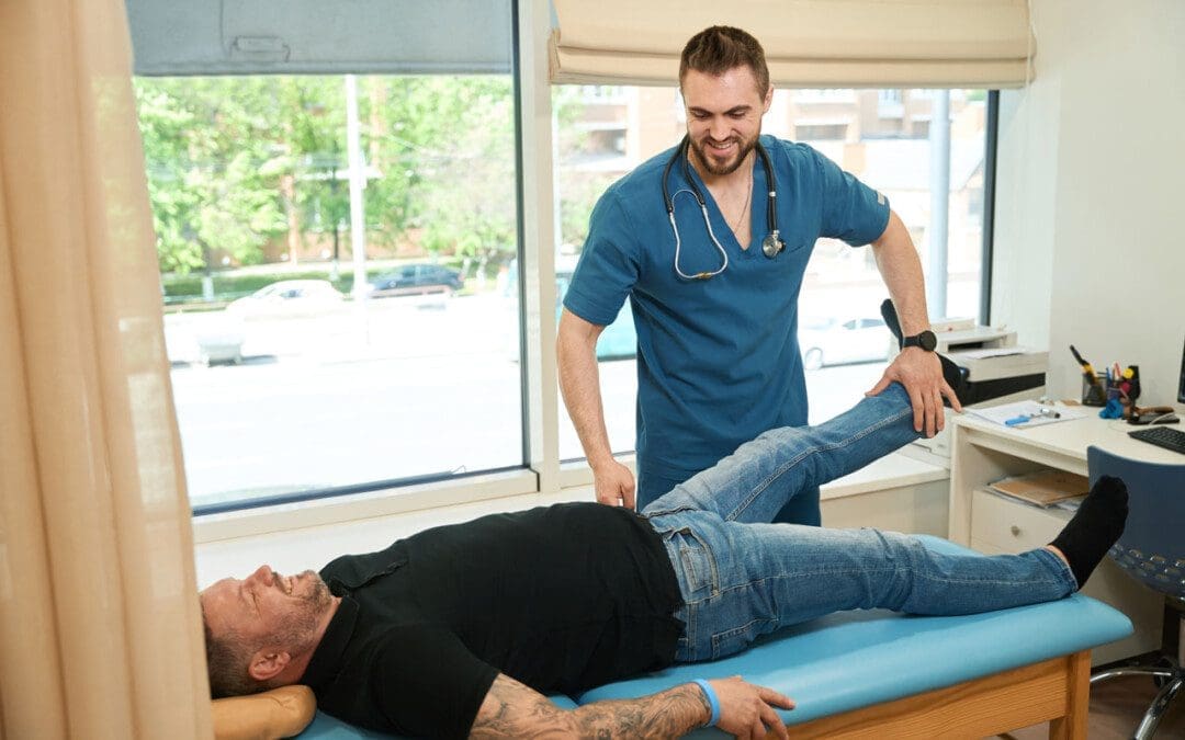
Once the pain and swelling start to subside, physical and/or occupational therapy can help rehabilitate the affected areas.
A therapist will provide stretches and exercises to help improve the joint range of motion and strengthen the supporting muscles.
For individuals experiencing hinge joint pain from an autoimmune condition, biologic medications to decrease the body’s autoimmune activity are administered through infusions delivered every several weeks or months. (Kamata, M., Tada, Y. 2020)
Cortisone injections may also be used to decrease inflammation.
At Injury Medical Chiropractic and Functional Medicine Clinic, we passionately focus on treating patients’ injuries and chronic pain syndromes and improving ability through flexibility, mobility, and agility programs tailored to the individual. Our providers use an integrated approach to create personalized care plans that include Functional Medicine, Acupuncture, Electro-Acupuncture, and Sports Medicine protocols. Our goal is to relieve pain naturally by restoring health and function to the body. If the individual needs other treatment, they will be referred to a clinic or physician best suited for them. Dr. Jimenez has teamed up with the top surgeons, clinical specialists, medical researchers, and premier rehabilitation providers to provide the most effective clinical treatments.
Chiropractic Solutions
References
Boundless. General Biology. (N.D.). 38.12: Joints and Skeletal Movement – Types of Synovial Joints. In. LibreTexts Biology. https://bio.libretexts.org/Bookshelves/Introductory_and_General_Biology/Book%3A_General_Biology_%28Boundless%29/38%3A_The_Musculoskeletal_System/38.12%3A_Joints_and_Skeletal_Movement_-_Types_of_Synovial_Joints
Kamata, M., & Tada, Y. (2020). Efficacy and Safety of Biologics for Psoriasis and Psoriatic Arthritis and Their Impact on Comorbidities: A Literature Review. International journal of molecular sciences, 21(5), 1690. https://doi.org/10.3390/ijms21051690
Individuals that have gone through a back injury may develop a synovial spinal cyst as a way to protect the spine that could cause pain symptoms and sensations. Can knowing the signs help healthcare providers develop a thorough treatment plan to relieve pain, prevent worsening of the condition and other spinal conditions?
Spinal Synovial Cysts
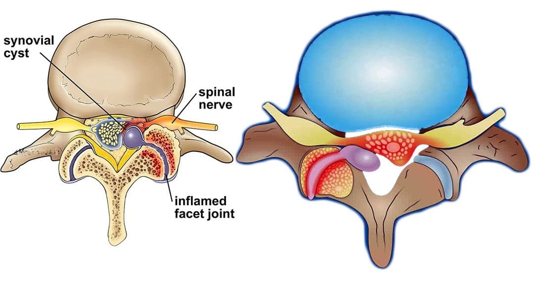
Spinal synovial cysts are benign fluid-filled sacs that develop in the spine’s joints. They form because of spinal degeneration or injury. The cysts can form anywhere in the spine, but most occur in the lumbar region/lower back. They typically develop in the facet joints or junctions that keep the vertebrae/spinal bones interlocked.
Symptoms
In most cases, synovial cysts don’t cause symptoms. However, the doctor or specialist will want to monitor for signs of degenerative disc disease, spinal stenosis, or cauda equina syndrome. When symptoms do present, they typically cause radiculopathy or nerve compression, which can cause back pain, weakness, numbness, and radiating pain caused by the irritation. The severity of symptoms depends on the size and location of the cyst. Synovial cysts can affect one side of the spine or both and can form at one spinal segment or at multiple levels.
Effects Can Include
Radiculopathy symptoms can develop if the cyst or inflammation caused by the cyst comes into contact with a spinal nerve root. This can cause sciatica, weakness, numbness, or difficulty controlling certain muscles.
Neurogenic claudication/impingement and inflammation of spinal nerves can cause cramping, pain, and/or tingling in the lower back, legs, hips, and buttocks. (Martin J. Wilby et al., 2009)
If the spinal cord is involved, it may cause myelopathy/severe spinal cord compression that can cause numbness, weakness, and balance problems. (Dong Shin Kim et al., 2014)
Symptoms related to cauda equina, including bowel and/or bladder problems, leg weakness, and saddle anesthesia/loss of sensation in the thighs, buttocks, and perineum, can present but are rare, as are synovial cysts in the middle back and neck. If thoracic and cervical synovial cysts develop, they can cause symptoms like numbness, tingling, pain, or weakness in the affected area.
Causes
Spinal synovial cysts are generally caused by degenerative changes like osteoarthritis that develop in a joint over time. With regular wear and tear, facet joint cartilage/the material in a joint that provides protection, a smooth surface, friction reduction, and shock absorption begins to waste away. As the process continues, the synovium can form a cyst.
Traumas, large and small, have inflammatory and degenerative effects on joints that can result in the formation of a cyst.
Around a third of individuals who have a spinal synovial cyst also have spondylolisthesis.
This condition is when a vertebrae slips out of place or out of alignment onto the vertebra underneath.
It is a sign of spinal instability.
Instability can occur in any spine area, but L4-5 are the most common levels.
This segment of the spine takes most of the upper body weight.
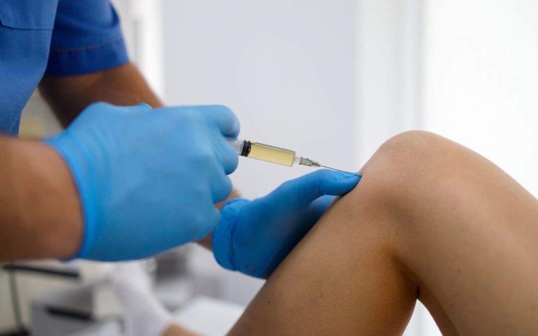
Epidural corticosteroid injections can reduce inflammation and could be an option to relieve pain.
Patients are recommended to receive no more than three injections per year.
Surgical Options
For severe or persistent cases, a doctor may recommend decompression surgery to remove the cyst and surrounding bone to relieve pressure on the nerve root. Surgical options range from minimally invasive endoscopic procedures to larger, open surgeries. The best surgical option varies based on the severity of the situation and whether associated disorders are present. Surgical options include:
Laminectomy – Removal of the bony structure that protects and covers the spinal canal/lamina.
Hemilaminectomy – A modified laminectomy where a smaller portion of the lamina is removed.
Facetectomy – The removal of part of the affected facet joint where the synovial cyst is located, usually following a laminectomy or hemilaminectomy.
Fusionof the facet joints and vertebra – Decreases vertebral mobility in the injured area.
Most individuals experience immediate pain relief following a laminectomy or hemilaminectomy.
Fusion can take six to nine months to heal completely.
If surgery is performed without fusion where the cyst originated, the pain could return, and another cyst could form within two years.
Surgery Complications include infection, bleeding, and injury to the spinal cord or nerve root.
How I Gained My Mobility Back With Chiropractic
References
Wilby, M. J., Fraser, R. D., Vernon-Roberts, B., & Moore, R. J. (2009). The prevalence and pathogenesis of synovial cysts within the ligamentum flavum in patients with lumbar spinal stenosis and radiculopathy. Spine, 34(23), 2518–2524. https://doi.org/10.1097/BRS.0b013e3181b22bd0
Kim, D. S., Yang, J. S., Cho, Y. J., & Kang, S. H. (2014). Acute myelopathy caused by a cervical synovial cyst. Journal of Korean Neurosurgical Society, 56(1), 55–57. https://doi.org/10.3340/jkns.2014.56.1.55
Epstein, N. E., & Baisden, J. (2012). The diagnosis and management of synovial cysts: Efficacy of surgery versus cyst aspiration. Surgical neurology international, 3(Suppl 3), S157–S166. https://doi.org/10.4103/2152-7806.98576
As the body ages, individuals want to stay active and maintain a healthy pain free lifestyle. Can regenerative cells for arthritis and cartilage damage be the future of neuromusculoskeletal medicine and joint healing?
Regenerative Cells For Arthritis and Cartilage Damage
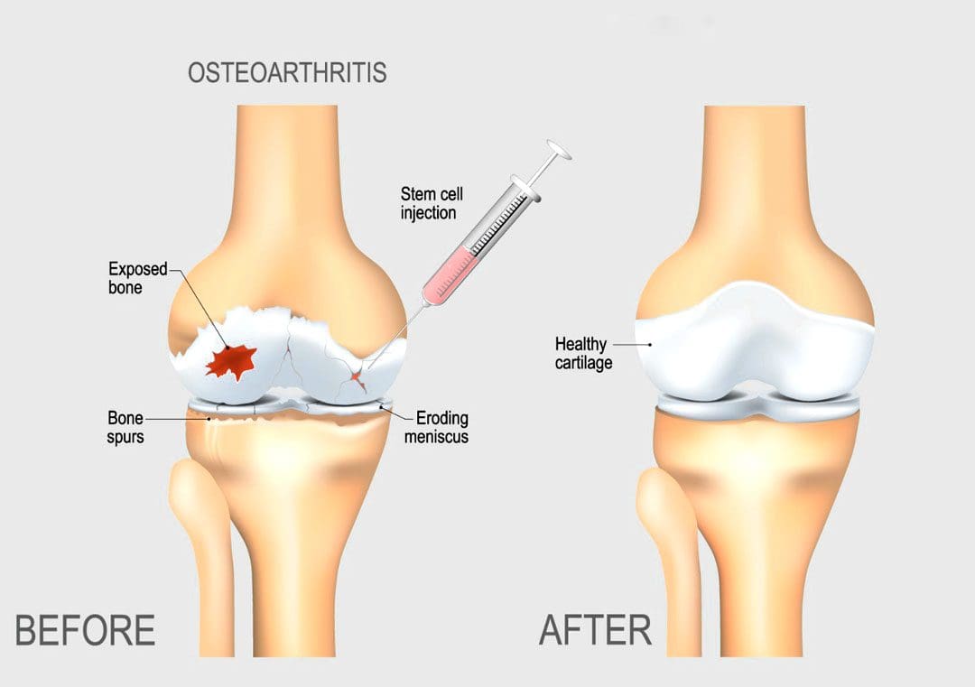
Individuals want to continue to do the physical activities they love, which require healthy joints. Scientists are learning how to harness the abilities of regenerative cells to repair and regrow damaged and deteriorated cartilage. Current stem cell treatment of cartilage problems has not been shown to reverse the effects of arthritis and while studies show clinical improvement, further research is necessary. (Bryan M. Saltzman, et al., 2016)
Cartilage and How It Gets Damaged
Cartilage is a type of connective tissue. In the joints, there are a few types of cartilage. The most commonly referred to is the smooth lining known as articular or hyaline cartilage. This type forms a smooth layer of cushion on the end of a bone at the joint. (Rocky S. Tuan, et al., 2013)
The tissue is very strong and has the ability to compress and absorb energy.
It is very smooth allowing a joint to glide effortlessly through a limb’s range of motion.
When joint cartilage is damaged, the cushioning can wear down.
In traumatic injuries, a sudden force can cause the cartilage to break off and/or suffer damage, that exposes the underlying bone.
In osteoarthritis – degenerative or wear-and-tear arthritis, the smooth layer can wear down thin and unevenly.
Eventually, the cushion wears away, the joints become inflamed and swollen and movements become stiff and painful.
There are treatments for arthritis and cartilage damage, but these treatments are usually focused on relieving symptoms by smoothing down the damaged cartilage or replacing the joint surface with an artificial implant, like knee replacement or hip replacement surgeries. (Robert F. LaPrade, et al., 2016)
Regenerative Cells
Regenerative stem cells are special cells that have the ability to multiply and develop into different types of tissue. In an orthopedic surgery setting for joint problems, stem cells are obtained from adult stem cell primary sources which are bone marrow and fatty tissue. These cells have the ability to develop into cartilage cells, called chondrocytes. (Rocky S. Tuan, et al., 2013)
They also help by stimulating the body to reduce inflammation, stimulate cell repair, and improve blood circulation.
This process is caused by cellular signals and growth factors to stimulate the body to activate the healing processes.
Once stem cells have been obtained, they need to be delivered to the area of cartilage damage.
Cartilage is a complex tissue that is described as a scaffold structure that is composed of collagen, proteoglycans, water, and cells. (Rocky S. Tuan, et al., 2013)
To regenerate cartilage, the complex tissues must also be reconstructed.
There are studies on types of tissue scaffolds engineered to recreate a similar type of cartilage structure.
The stem cells can then be injected into the scaffold, in hopes of restoring a normal type of cartilage.
Non-Surgical Arthritis Treatments
Standard treatments such as cortisone shots or physical therapies work as well and provide benefits that could be utilized in combination with regenerative cells for arthritis and cartilage damage in the near future. Data takes time and therefore how this impacts the long-term health of a joint needs continued research in terms of tissue engineering and cell delivery to determine the best approach to help individuals.
Arthritis
References
LaPrade, R. F., Dragoo, J. L., Koh, J. L., Murray, I. R., Geeslin, A. G., & Chu, C. R. (2016). AAOS Research Symposium Updates and Consensus: Biologic Treatment of Orthopaedic Injuries. The Journal of the American Academy of Orthopaedic Surgeons, 24(7), e62–e78. https://doi.org/10.5435/JAAOS-D-16-00086
Saltzman, B. M., Kuhns, B. D., Weber, A. E., Yanke, A., & Nho, S. J. (2016). Stem Cells in Orthopedics: A Comprehensive Guide for the General Orthopedist. American journal of orthopedics (Belle Mead, N.J.), 45(5), 280–326.
Tuan, R. S., Chen, A. F., & Klatt, B. A. (2013). Cartilage regeneration. The Journal of the American Academy of Orthopaedic Surgeons, 21(5), 303–311. https://doi.org/10.5435/JAAOS-21-05-303
IFM's Find A Practitioner tool is the largest referral network in Functional Medicine, created to help patients locate Functional Medicine practitioners anywhere in the world. IFM Certified Practitioners are listed first in the search results, given their extensive education in Functional Medicine


 With contractures, the sarcomeres overly lengthen when the muscle fibers tighten. This increase in sarcomere length prevents the muscle from contracting normally, resulting in weakness. Muscle fibers are encased in an extracellular matrix, a mesh composed of collagen and other proteins that help transmit force and provide muscle contraction. Muscle contractures cause the amount of collagen within the extracellular matrix to increase, causing a stiffening of fibers that restricts movement. (