ClickCease
+1-915-850-0900 spinedoctors@gmail.com
Select Page
The Knee

The Knee

The Knee | MRI may be requested for:

  • Ligament injuries
  • Meniscal tears and degeneration
  • Rheumatoid arthritis
  • Osteochondral fractures
  • Tendon disruptions

Bones & Cartilage Of The Knee

The knee joint is the largest, most complicated, and most vulnerable joint in the body, as it does not have a stable bony configuration. It consists of the tibiofemoral and patellofemoral articulations, which include the femur, tibia, and patella. The knee is a synovial joint that is enclosed by a ligament capsule. The capsule contains synovial fluid that keeps the joint lubricated (Figure 82). The knee provides flexible movement, but must also bear large weight and pressure loads. During walking, the knees support 1.5 times your body weight. When climbing stairs, they support 3-4 times your body weight. When squatting, your knees support 8 times your body weight.

the knee

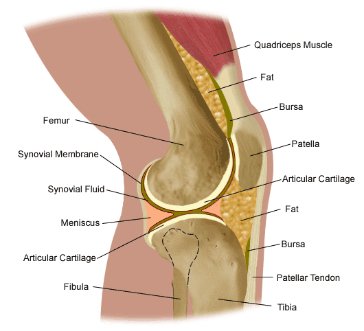
Figure 82. Anatomy of the knee.

The tibiofemoral articulation is a modified hinge joint that allows bending and straightening, but also allows for slight rotation. This articulation consists of the lateral and medial condyles of the femur resting on the lateral and medial aspects of the tibial plateau. The femoral condyles make up the distal portion of the femur, which is expanded in order to assist with weight distribution at the knee joint. The medial femoral condyle is typically larger and rounder. The condyles are united anteriorly to provide the articular surface for the patella, but they are separated posteriorly by the intercondylar notch. This notch, or fossa, is the attachment site for the cruciate ligaments, the ligaments of Humphrey and Wrisberg, and the frenulum of the patellar fat pad. A large part of the posterior distal femur is called the popliteal surface. This area is covered by fat, which separates it from the popliteal artery. The medial and lateral edges of the popliteal surface are attachment sites for muscles. Superior to the femoral condyles are the epicondyles, which are the attachment sites for muscles, tendons, and capsular ligaments. The medial epicondyle is the attachment site for the medial (or tibial) collateral ligament (Figure 83). The lateral femoral epicondyle is the attachment site for the lateral (or fibular) collateral ligament, as well as the tendon of the popliteus muscle, fibers of the iliotibial tract, and the lateral capsular ligament. Superior and posterior to the epicondyles is the most distal extent of the linea aspera, the bony ridge of the femur.

The tibia is the distal portion of the tibiofemoral articulation at the knee. The tibia is the second longest bone in the body, ranked just behind the femur. Its proximal end is flattened and expanded to provide a larger surface for the body weight that is transmitted through the femur. Like the femur, the proximal tibia has medial and lateral condyles. The medial condyle is larger, and somewhat flattened where it contacts the medial meniscus. The lateral condyle has a circular look to its femoral articular surface. The lateral tibial condyle articulates with the head of the fibula posteriorly, which is as close as the fibula comes to any involvement in the knee joint. Both the medial and lateral condyles rise in the center of the superior aspect of the tibia to form the intercondylar eminence. Posterior to this eminence are the attachments sites for the posterior horns of the medial and lateral menisci, which will be discussed with the ligaments of the knee. The medial and lateral tibial condyles, and the area of the intercondylar eminence are often grouped together and referred to as the tibial plateau (Figure 84). This is a critical weight-bearing area, and greatly affects the stability of the knee joint. The tibial tuberosity (or tubercle) is located on the anterior surface of the proximal tibial shaft. It has a smooth upper portion, and a roughened lower portion, which is the insertion site for the patellar tendon. The lateral side of the tibial tuberosity has a ridge for the attachment of fibers from the iliotibial tract. This is the strongest direct attachment site for the iliotibial tract. The IT tract, or band, helps in limiting lateral movement of the knee.

the knee

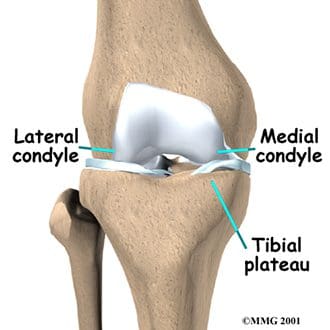
Figure 84. Tibial plateau.

the knee

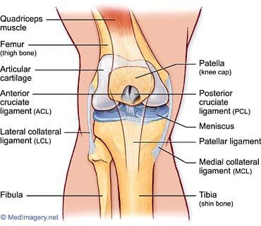
Figure 83. Tibiofemoral anatomy.


The patella is the third bone involved in the knee joint, specifically in the patellofemoral articulation. Patella means �little plate� in Latin, which describes the look and function of this sesamoid bone. The patella develops in the tendon of the quadriceps femoris muscle (Figure 85). It moves when the leg moves, and protects the knee joint by relieving friction between the bones and muscles when the knee is bent or straightened. The patellofemoral joint is a saddle-type synovial joint, allowing the patella to glide along the bottom front surface of the femur between the femoral condyles in the patellofemoral groove. Ossification of the patella is typically completed in females by age 10, and in males between the ages of 13-16. If the patella has more than one ossification center, and the additional center does not fuse, it is termed a bipartite patella (Figure 86).

the knee

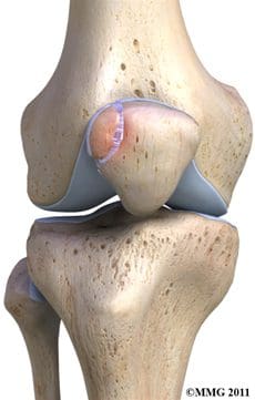
Figure 86. Bipartite patella.

the knee

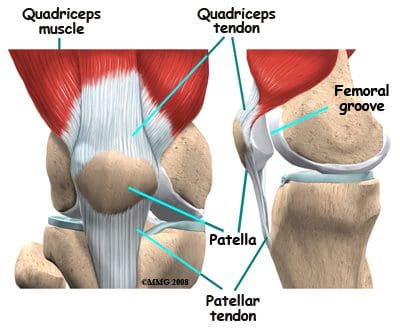
Figure 85. Patella location.


Articular, or hyaline, cartilage covers the ends of the bones involved in any joint. In the knee joint, this includes the distal end of the femur, the proximal end of the tibia, and the posterior aspect of the patella (Figure 87). In larger joints, this cartilage is approximately �� thick. Articular cartilage is white, shiny, rubbery, and slippery, enabling surfaces to slide against one another without damage. Articular cartilage is very flexible, due in part to its high water content, which also makes it highly visible on MRI. In contrast to the bones that it covers, articular cartilage has almost no blood vessels, so it is not good at repairing itself. Bones, on the other hand, have numerous blood vessels, and are good at self-repair.

the knee

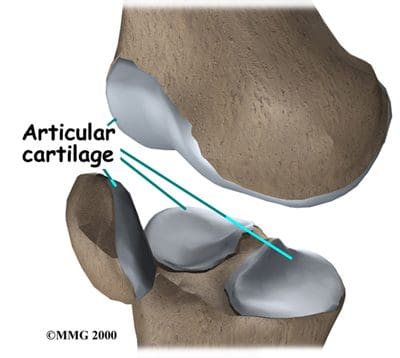
Figure 87. Articular cartilage.

Another type of cartilage is found between the femur and tibia- the fibrous cartilage that makes up the medial and lateral menisci. The menisci, also referred to as �articular disks�, wrap around the round ends of the femur to fill the space between the femur and tibia (Figure 88). Since the menisci are more fibrous in composition, they have tensile strength and can resist pressure. They can help spread the force from our body weight over a larger area. By helping with weight distribution, the menisci protect the articular cartilage on the ends of the bones from excessive forces. The menisci are fashioned to be thicker on their outsides, creating a shallow socket on the tibial surface. They act like a wedge on the rounded distal portion of the femur, improving the overall stability of the knee joint by preventing any �rolling� of the femur. Despite how strong they sound, the menisci can crack or tear when the knee is forcefully rotated or bent. The medial meniscus is fused with the medial collateral ligament, so it is less mobile than the lateral meniscus. It is often injured when the anterior or posterior cruciate ligaments are injured. The inner 2/3 of the medial meniscus receives a limited blood supply, so the entire meniscus is usually slow to heal. The lateral meniscus suffers from fewer injuries than the medial meniscus. Meniscal tears are one of the most common causes of knee pain, with suspected meniscal tears the most common indication for an MRI of the knee joint.

the knee

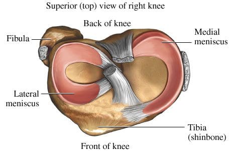
Figure 88. Superior view of menisci of right knee.

Symptoms that might indicate a problem with the bones of the knee joint include locking of the joint, the knee giving way, crackling or grinding felt in the joint, and pain and swelling. Locking of the joint can be indicative of a �loose body� (bone, cartilage, or foreign object) in the joint space, which can often be removed through arthroscopy (Figure 89). A knee that gives way can indicate that the patella is out of the patellofemoral groove, which leaves the knee unstable. Crackling and grinding at the joint can result from degenerative arthritis or osteoarthritis, as well as from a dislocating patella. An increase in pain with activity can occur due to a stress fracture or bone fracture. One of the pathologic conditions that can affect the bones of the knee joint is osteochondritis dissecans, which can affect the distal femur, and was discussed previously with the femur anatomy. Various types of arthritis manifest in the bones of the knee joint, including osteoarthritis, infectious arthritis, and rheumatoid arthritis. Chondromalacia patella, also known as patellofemoral syndrome or �runner�s knee� results from an irritation of the undersurface of the patella (Figure 91). If the patella is not tracking correctly in the patellofemoral groove, the articular cartilage may rub against the knee joint (Figure 90). The cartilage degenerates, and becomes irritated and painful. This condition is most common amongst young, healthy athletes, especially females and runners that are flat-footed. Treatment is typically rest and physical therapy to stretch and strengthen the quads and hamstrings. If surgery is required, it may be to perform a �lateral release�, as the abnormal tracking of the patella can cause a tightening of the lateral tissues of the knee. The lateral release procedure cuts the tight tissues, so the patella can return to its normal position and tracking. Osgood-Schlatter disease involves the anteriorly located tibial tuberosity, and the patellar tendon that inserts on that tuberosity (Figures 92, 93). This condition affects children during their growth spurts, and is typically found more in boys. During growth spurts, contractions of the quad muscle put additional stress on the patellar tendon at its attachment site on the tibial tuberosity. This can result in multiple subacute avulsion fractures and inflammation of the tendon. Excess bone growth occurs on the tuberosity, and a lump on the tuberosity can be seen and felt. This lump can become irritated and swollen, causing knee and leg pain. This condition is typically worsened with running, jumping, and climbing stairs. Osgood-Schlatter usually resolves with rest, ice, compression and elevation, as well as maturity of the youngster�s skeleton.

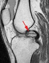
Figure 89. Intraarticular loose body.


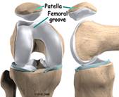
Figure 90. Patellofemoral groove.

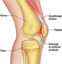
Figure 91. Patellofemoral syndrome or �runner�s knee�.


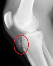
Figure 92. Xray displaying Osgood-Schlatter disease.


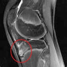
Figure 93. MRI displaying Osgood- Schlatter disease.


Ligaments Of The Knee

Ligaments are the tough bands of tissue that connect bones. They are considered to be �viscoelastic�, meaning they can gradually lengthen under tension, but return to their original shape when the tension is removed. However, if they are stretched for a prolonged period of time, or past a certain point, the ligaments cannot retain their original shape, and may eventually tear or snap. This is one of the reasons that a dislocated joint should be re-located as quickly as possible. If the ligaments lengthen, they leave the joint weakened and prone to future dislocations. Controlled stretching exercises to lengthen ligaments, and make the joints more supple, are part of the daily routines of athletes, gymnasts, dancers, etc. Damaged ligaments can lead to unstable joints, wearing of the cartilage, and eventually osteoarthritis. The numerous ligaments of the knee joint are the most important structures in controlling stability of the knee. Many of these ligaments were mentioned in the femur anatomy section, as they have attachments on the distal femur. The more important ligaments will be reviewed here in greater detail, in regards to their functions in the knee joint. The main intracapsular ligaments are the anterior and posterior cruciates (Figures 94, 95). Intracapsular ligaments are not very common in synovial joints. They provide stability, but permit a larger range of motion as compared to capsular or extracapsular ligaments. The anterior cruciate ligament (ACL) stretches from the lateral femoral condyle to the anterior intercondylar area of the tibia, preventing the tibia from being pushed too far anterior relative to the femur. It is the more commonly injured of the cruciate ligaments, and can be torn during twisting and bending of the knee. Women are at higher risk for ACL ruptures due to the facts that the maximum diameter of the intercondylar fossa is in its posterior aspect (the ACL attaches anteriorly), and the overall width of the intercondylar fossa is smaller in females. The posterior cruciate ligament (PCL) stretches from the medial femoral condyle to the posterior intercondylar area of the tibia, preventing posterior displacement of the tibia relative to the femur. It is the stronger of the two cruciate ligaments, and is injured less frequently; however, it can be injured from direct force or trauma. The menisci are also considered to be intracapsular structures, with connections to ligaments inside and outside the joint capsule. Two of their intracapsular ligaments are the anterior and posterior transverse meniscomeniscal ligaments. They attach the medial and lateral menisci to each other at their anterior and posterior aspects. Posterior transverse meniscal ligaments are very rare- only 1-4% of knees will have them. Two additional intermeniscal ligaments are the medial and lateral oblique meniscomeniscal ligaments (Figure 96). Their names describe their anterior horn attachment sites; they attach on the posterior horn of the opposite meniscus (i.e. medial oblique meniscomeniscal attaches to the anterior horn of the medial meniscus and posterior horn of the lateral meniscus). The oblique meniscomeniscal ligaments both traverse the intercondylar notch, and pass between the anterior and posterior cruciate ligaments (Figure 97).

the knee

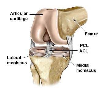
Figure 94. Cruciate ligaments and menisci.

the knee

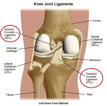
Figure 95. Posterior view of cruciate ligaments of left knee.

the knee

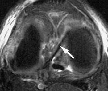
Figure 96. Axial fatsat T2 FSE image with arrow indicating
oblique meniscal ligament coursing from anterior horn of
medial meniscus to posterior horn of lateral meniscus.

the knee

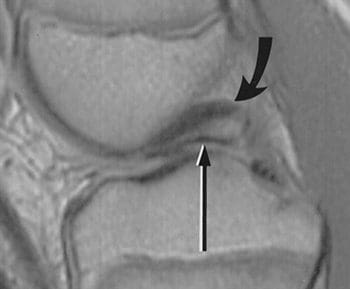
Figure 97. Sagittal dual-echo T2 through the intercondylar notch at the level of the posterior cruciate ligament (curved arrow); thin linear structure of low signal intensity inferior to PCL represents the oblique meniscomeniscal ligament (straight arrow); sometimes misinterpreted as displaced meniscal fragment.


The medial (or tibial) collateral ligament is considered a capsular ligament, as it is part of the articular capsule surrounding the synovial knee joint. It acts as mechanical reinforcement for the joint, protecting the knee from valgus force, or being bent open medially due to stress on the lateral side of the knee. The medial collateral ligament (MCL) is one of the most commonly injured of all knee ligaments, occurring in all sports, in all ages, and often times with medial meniscal tears (Figures 98-101). It has both superficial and deep components. Fibers from the superficial portion of the MCL attach to the medial epicondyle of the femur and the medial tibial condyle. Fibers from the deep medial collateral ligament attach to the medial meniscus. Proximal to the attachment point, this ligament is referred to as the meniscofemoral ligament, as it attaches the medial meniscus to the medial aspect of the femur. Distal to the meniscal attachment, the ligament is referred to as the meniscotibial (or coronary) ligament, as it attaches the medial meniscus to the medial aspect of the tibia. The meniscofemoral and meniscotibial are also referred to as the meniscocapsular or medial capsular ligaments, as they play an important role in anchoring peripheral parts of the medial meniscus in the medial side of the knee. The meniscotibial ligament is typically injured more often than the meniscofemoral ligament. The meniscotibial ligament attaches to the tibia several millimeters inferior to the articular cartilage. Its job is to stabilize and maintain the meniscus in its proper position on the tibial plateau. Disruption of the meniscotibial ligament can result in a floating meniscus or meniscal avulsion, while the meniscofemoral ligament may not be affected. The deep medial collateral ligament is short, and tightens quickly with rotation motions. It is often damaged, along with the ACL, when the mechanism of injury involves tibial rotation. Diagnosis and surgical repair of the deep medial collateral ligament can be challenging.

the knee

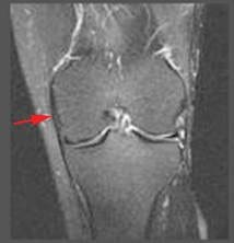
Figure 98. Normal MCL is linear,
has low signal intensity.

the knee

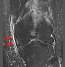
Figure 99. Grade 1 sprain shows adjacent edema, no change in signal intensity of MCL.

the knee

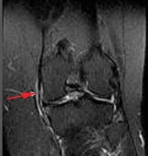
Figure 100. Grade 2 sprain or partial tear shows increased edema,
abnormal signal intensity,
thickening or thinning of ligament.

the knee

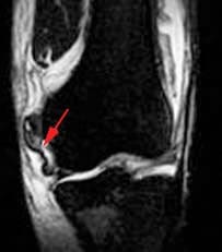
Figure 101. Grade 3 involves complete disruption of ligaments or attachments.


In addition to fibers of the medial collateral ligament, the deep portion of the capsular compartment of the medial knee is the location of the medial knee�s posterior support. The posterior oblique ligament is attached proximally to the medially located adductor tubercle of the femur, and distally to the tibia and the posterior aspect of the knee joint capsule. If the posterior oblique is injured, it is usually torn from its femoral origin. The posterior oblique ligament provides static resistance to valgus loads as the knee moves into full extension, as well as dynamic stabilization to valgus forces (stress from lateral side) as the knee moves into flexion. It acts as an important restraint to posterior tibial translation in cases of posterior cruciate ligament injury. The posterior oblique ligament has three �arms�. Its superior capsular �arm� becomes continuous with the posterior knee capsule, and the proximal portion of the oblique popliteal ligament. The oblique popliteal ligament is also an important posterior stabilizing structure for the knee joint Figure 102). It extends from the posteromedial aspect of the tibia, running obliquely and laterally upward to insert near the lateral epicondyle of the femur.

the knee

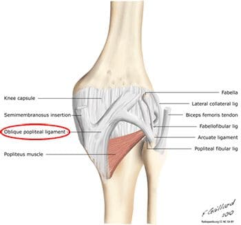
Figure 102. Oblique popliteal ligament in posterior view of knee.

the knee

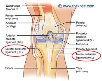
Figure 103. Medial (tibial) and lateral (fibular) collateral ligaments.


The lateral (or fibular) collateral ligament is considered an extracapsular ligament. It helps to provide joint stability and protects the lateral side of the knee from varus forces, or inside bending forces that are directed at the medial side of the knee. Injuries to the lateral collateral ligament are less common than injuries to the medial collateral, as the opposite leg can guard against medial forces that can lead to lateral collateral injuries. Injuries can occur in sports such as soccer and rugby, where the knee is extended and unprotected during running. The lateral, or fibular, collateral ligament stretches obliquely downward and backward, from the lateral epicondyle of the femur to the head of the fibula (Figure 103). It is not fused with the capsular ligament or with the lateral meniscus, so it has increased flexibility and decreased incidence of injury when compared to the medial collateral ligament. Similar to the medial meniscus, the lateral meniscus has a meniscotibial, or coronary, ligament. It connects the inferior edges of the lateral meniscus to the periphery of the tibial plateau. The lateral meniscus also has a meniscofemoral ligament that extends from the posterior horn of the lateral meniscus to the lateral aspect of the medial femoral condyle. It is given two distinct names, based on its location in relation to the posterior cruciate ligament (PCL). The ligament of Humphrey passes in front of the posterior cruciate ligament. It is less than 1/3 the diameter of the posterior cruciate ligament, but may be confused for the posterior cruciate during arthroscopy. The ligament of Wrisberg passes behind the posterior cruciate ligament, and is about � of the posterior cruciate�s diameter (Figure 104). Its femoral origin often merges with the posterior cruciate ligament. Both ligaments are present in only about 6% of knees. Approximately 70% of people have one or the other of these ligaments, with the majority possessing the more posterior ligament of Wrisberg (Figure 105). MRI is the preferred imaging modality for medial collateral or lateral collateral ligament injuries, as it can detect any associated internal knee derangements, cruciate-collateral ligament injuries, or cartilage deficiencies.

the knee

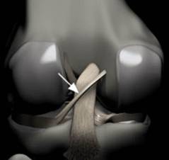
Figure 104. Rendering of posterior knee, arrow indicates Ligament of Wrisberg; courses obliquely from lateral aspect of medial femoral condyle to posterior horn of lateral meniscus,
remains posterior to PCL.

the knee

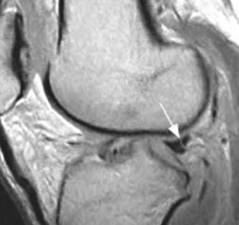
Figure 105. Arrow indicates �Wrisberg pseudo-tear�; intermediate signal
intensity line at junction of
Ligament of Wrisberg and normal posterior horn of lateral meniscus; often mistaken for a meniscal tear.

The patellar ligament is the connection between the patella and the tibia, extending from the apex (inferior aspect) of the patella to the tibial tuberosity. Technically, it is connecting two bones, so it is a ligament. However, it is most often referred to as the patellar tendon, because the superficial fibers that cover the front of the patella and extend to the tibia are continuous with the central portion of the common tendon of the quadriceps femoris muscle. The posterior surface of the patellar ligament is separated from the synovial membrane of the knee joint by a large infrapatellar pad of fat. Injuries to the patellar ligament can occur from overuse, such as sports that involve jumping and quick directional changes, as well as running-related sports. This is the ligament that is injured in jumper�s knee (or patellar tendonitis), which begins with inflammation, and can lead to degeneration or rupture of the patellar ligament and the tissue around it (Figure 106). Patients with patellar ligament injuries typically complain of pain in the area below the kneecap, which will increase with walking, running, squatting, etc. They can often be treated in the same manner as other soft tissue injuries- with rest, ice, compression and elevation. The patellar ligament attachment at the tibial tuberosity is the site of Osgood-Schlatter disease, which was discussed previously.

the knee

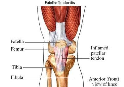
Figure 106. Patellar tendonitis (jumper�s knee).

Along the sides of the patella and the patellar ligament are the medial and lateral patellar retinacula (Figure 107). They are fibrous tissue stabilizers for the patella that form from the medial and lateral portions of the quad tendons as they pass down to insert on either side of the tibial tuberosity. The lateral retinaculum is the thicker of the two, but both have superficial and deep layers. Within the deep layers are various ligaments (whose names indicate the structures they connect) that help support the patella in its position, relative to the femur below it. The deep layer of the lateral patellar retinaculum is the location where the lateral patellofemoral ligament meets the iliopatellar band, which is a tract of fibers from the iliotibial (IT) band that connects to the patella. The deep layer of the medial patellar retinaculum has three focal capsular thickenings, referred to as the medial patellofemoral, medial patellomeniscal, and medial patellotibial ligaments. The medial patellofemoral ligament is strong enough to influence patellar tracking, and acts as a major medial restraint. Imbalances in the forces that control patellar tracking during flexion and extension of the knee can lead to patellofemoral pain syndrome (runner�s knee), one of the most common causes of knee pain. This can result from overuse, trauma, muscle dysfunction, patellar hypermobility, and poor quadriceps flexibility. Typical symptoms include pain behind or around the patella that is increased with running, and activities that involve knee flexion. MRI is typically not necessary for this diagnosis. Physical therapy has been found to be effective for the treatment of patellofemoral pain syndrome.

the knee

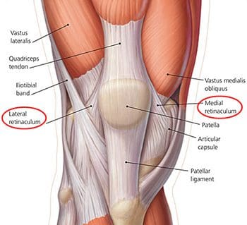
Figure 107. Lateral and medial retinaculum.

Muscles & Tendons Of The Knee

The flexor and extensor muscles of the knee have been discussed previously, as the majority of them are the anterior and posterior muscles of the thigh. We will review the thigh muscles involved in knee movement, and add two muscles of the lower leg that also affect the knee. The quadriceps femoris muscles of the anterior thigh are the main knee extensors (Figure 108). As these muscles contract, the knee joint straightens. The tendons of the vastus medialis, vastus intermedius, vastus lateralis, and rectus femoris join at the superior aspect (base) of the patella to form the patellar tendon. This tendon continues over the patella and attaches it to the tibial tuberosity (since it is connecting bone to bone, it is sometimes called the patellar ligament). The quadriceps, along with the gluteal muscles, are responsible for the thrusting forces necessary for walking, running, and jumping. The quads also help control movement of the patella, as they are attached to it by the quadriceps tendons (Figure 109). The patella increases the force exerted by the quadriceps muscles as the knee is straightened.

the knee

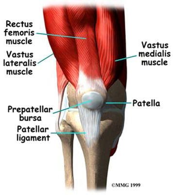
Figure 108. Anterior thigh muscles – knee extensors.

the knee

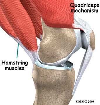
Figure 109. Quadriceps controlling the patella.


The posterior thigh muscles, also known as the hamstrings, are the main knee flexors, with assistance from the sartorius, gracilis, gastrocnemius, and popliteus muscles. The knee bends when the hamstrings contract. The hamstring muscles give the knee joint the strength needed for propulsion in running and jumping. They also help to stabilize the knee by protecting the collateral and cruciate ligaments, especially when the knee twists. The three hamstring muscles have varying attachment sites around the knee joint (Figure 110). The biceps femoris attaches to the head of the fibula and the superolateral aspect of the tibia. The semitendinosus attaches on the anterior aspect of the tibia, medial to the tibial tuberosity, crossing over the medial collateral ligament. The tendon of the semitendinosus muscle is sometimes used for cruciate ligament reconstruction. The semimembranosus attaches at the posteriomedial aspect of the medial tibial condyle. The sartorius muscle is also a knee flexor, although it is an anterior thigh muscle. It inserts on the anterior medical aspect of the tibia. The gracilis muscle of the medial thigh is one of the hip adductors, but also plays a part in knee flexion. Like the semitendinosus tendon, the tendon of the gracilis is sometimes used for cruciate ligament reconstructions. The gracilis attaches to the medial aspect of the proximal tibia.

the knee

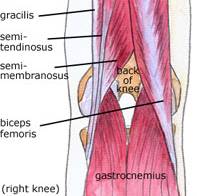
Figure 110. Posterior knee
muscles – knee flexors.

Additional flexors of the knee joint include some of the posterior muscles of the lower leg. The large superficial gastrocnemius muscle has a medial and a lateral head, which originate from the medial and lateral femoral condyles, respectively. It runs the length of the posterior lower leg, attaching to the calcaneus by the Achilles tendon. The gastrocnemius gives us the ability to flex our knee while our foot is flexed, as it connects to both joints. It is involved in standing, walking, running, and jumping. The popliteus is a deep posterior lower leg muscle that helps with knee flexion, and also rotates the tibia medially, which aids in knee stability. The popliteus originates from the outer margin of the lateral meniscus of the knee joint. It extends posteriorly and inserts on the medial aspect of the tibia, inferior to the medial tibial epicondyle.

The important tendons of the knee include the quadriceps, patellar, and hamstring tendons, and the iliotibial band (Figure 111). Tendons attach muscles to bones. These major knee tendons have all been discussed with either the bones or the muscles that they attach. The quadriceps tendon was mentioned with the quadriceps muscle as the muscle�s attachment to the patella. The quad tendon continues over the patella, then attaches the apex of the patella to the tibial tuberosity. It is then called the patellar tendon (or ligament). Hamstring tendons were discussed with the hamstring muscles, the posterior muscles that are flexors of the knee. Hamstring tendons are sometimes used for cruciate ligament reconstructions. Tendonitis, which is the inflammation of a tendon, is a common knee injury amongst athletes in a variety of sports. The iliotibial band (or IT tract) functions like a tendon, as it attaches the knee to the tensor fasciae latte muscle. The band is actually a fibrous reinforcement of the fascia lata, or deep tissue of the thigh. It runs from the ilium to the tibia. Proximally, it acts as a hip abductor, while distally it acts as lateral stabilization for the knee, and aids with medial rotation of the tibia. The IT band is in constant use during walking and running, which can lead to irritation at the point where it passes over the lateral femoral epicondyle. A �tight� IT band can cause inflammation and/or irritation at the femoral epicondyle, or at the point of insertion on the lateral tibial condyle. This condition is called IT band friction syndrome. It is common amongst runners, hikers, and cycling enthusiasts.

the knee

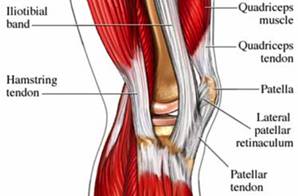
Figure 111. Tendons of the knee.

Nerves Of The Knee

The main nerves to the knee that come from the sacral plexus of nerves are the tibial nerve and the common peroneal nerve (Figure 112). Both are branches of the sciatic nerve, and begin posteriorly, slightly above the actual knee joint. Both of these nerves, or their branches, continue through the lower leg and foot, providing sensation and muscle control. The tibial and common peroneal nerves are also both involved in cutaneous innervation, which is the supply of nerves to the skin of the knee. The tibial nerve remains posterior and more medial, branching at the medial ankle to innervate the foot. The common peroneal nerve begins posterolaterally, moving anteriorly near the neck of the fibula. It then branches into the superficial and deep peroneal nerves, which continue their anterior descent to the foot. The tibial and common peroneal nerves are the most commonly injured nerves when a knee is dislocated. Nerves can grow back, but they do so at a rate of approximately � inch per month.

the knee

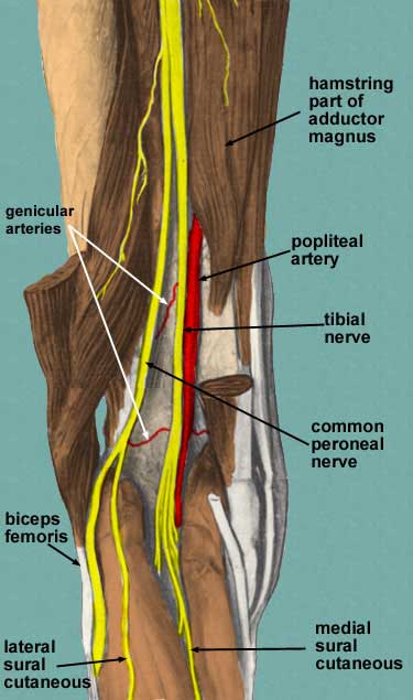
Figure 112. Sacral plexus nerves of knee.

Nerves from the lumbar plexus that affect the knee include the lateral femoral cutaneous, and the saphenous, which is a branch of the femoral nerve (Figure 113). The saphenous nerve travels more medially and gives off infrapatellar branches around the knee joint. Below the knee, the saphenous nerve sends branches to the skin of the anterior and medial lower leg. The lateral femoral cutaneous nerve sends an anterior branch to the skin of the anterior and lateral thigh, down to the area of the knee. Terminal filaments of this nerve communicate with the infrapatellar branch of the saphenous nerve, forming the peripatellar plexus.

the knee

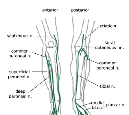
Figure 113. Lumbar plexus nerves of knee.

Arteries & Veins Of The Knee

The popliteal artery, a branch of the superficial femoral artery, is the main arterial supply to the knee joint. It runs along the posterior aspect of the distal femur, behind the knee joint. At the supracondylar ridge, the popliteal artery gives off the blood supply to the knee, which consists of various genicular arteries (Figure 114). Inferior to the knee joint, the popliteal branches into the anterior and posterior tibial arteries, which supply the lower leg. The popliteal artery is a common site for both atherosclerosis and aneurysms, and is listed as the most common site for peripheral arterial aneurysms. Approximately 50% of these aneurysms are bilateral. Although they rarely rupture, popliteal aneurysms may serve as a focus for abrupt thrombotic occlusion of the involved popliteal artery, which can affect the foot on the same side. A thrombus within an aneurysm can also lead to a distal embolism. The genicular arteries are sources of continued blood flow to the knee and lower limb, in case of an obstructed popliteal artery. The descending genicular, also called the highest or supreme genicular, branches from the femoral artery, just superior to the popliteal branch. It supplies the adductor magnus and hamstring muscles, then joins with the network of genicular arteries around the knee joint. The middle genicular pierces the oblique popliteal ligament, and supplies the ligaments and synovial membrane inside the knee articulation (including the ACL and PCL). The sural artery joins the anastomoses of the genicular arteries, and also supplies muscles of the lower leg, including the large gastrocnemius muscle. The anastomotic pattern around the knee joint is supplied by the popliteal artery posteriorly, the descending genicular artery medially, and the descending branch of the lateral circumflex femoral artery laterally. The genicular arteries involved in the anastomosis are labeled as the medial and lateral superior geniculars, and the medial and lateral inferior geniculars.

the knee

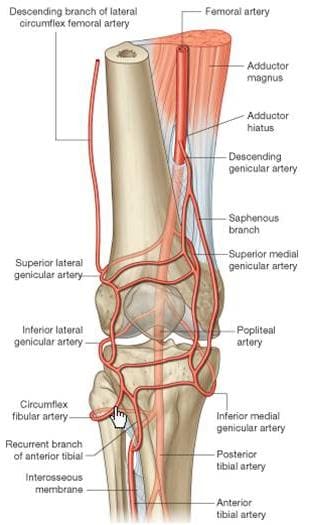
Figure 114. Arteries of knee.

The major deep veins around the knee joint are the popliteal vein, and the anterior and posterior tibial veins (Figure 115). The popliteal vein begins at the junction of the tibial veins in the posterior aspect of the lower leg, just inferior to the knee joint. It ascends posteriorly, continuing as the femoral vein about halfway up the thigh. As deep veins typically follow the arteries, the genicular veins accompany the genicular arteries around the knee joint, then drain into the popliteal vein. The important superficial veins around the knee joint are the small and great saphenous veins. Superficial veins typically do not follow arteries, but rather travel with cutaneous nerves. The small saphenous ascends the lower leg posteriorly, angling from lateral to medial. It merges with the popliteal vein at a position slightly superior to the knee joint. The great saphenous vein, the longest vein in the body, has a medial and anterior course in the lower leg. It moves to a posterior position, but stays medial along the knee joint, moving alongside the medial epicondyle of the femur. The great saphenous then moves anteriorly again through the thigh.

the knee

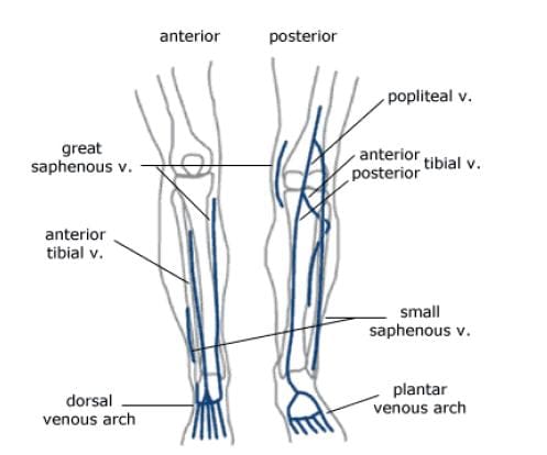
Figure 115. Veins of knee.

Varicose and �spider� veins are often seen in the leg in the posterior aspect of the knee joint. As mentioned previously, in the femoral vein discussion, veins have valves to ensure the �one-way� uphill flow of blood back to the heart (Figure 116). Communicating vessels, also called perforating veins, exist between the deep and superficial veins to help compensate for valves that may be incompetent, and are allowing blood reflux. If venous walls are weakened or dilated, the cusps of the valves can no longer close properly, and the valves can become incompetent. This leads to an increase in the weight of the column of blood for the veins that are �downstream� from the bad valve. Blood can pool in these veins, causing them to become varicose, where the veins swell, become tortuous, and even bulge through the skin surface. Reticular veins, which are smaller varicose veins that do not bulge through the skin, as well as very small �spider� veins are both typically less severe conditions, but both still involve the backwards flow of blood. Removal of severe varicose veins will actually help blood flow, as the blood will no longer be stagnant in the pooled areas.

the knee

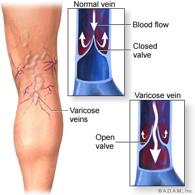
Figure 116. Varicose veins around knee.

Bursae Of The Knee

The synovial knee joint is home to a large number of bursae (Figure 117). These are fluid sacs and synovial pockets that surround and sometimes communicate with the joint cavity. They facilitate friction-free movement between the bones and moving structures (tendon, muscle). Fluid or debris can collect in the bursa, or fluid can extend into the bursa from the adjacent joint in situations such as excessive friction, infection or direct trauma. This type of pathological enlargement of the bursa is referred to as bursitis, which can mimic several peripheral joint and muscle abnormalities. Radiologists must be able to accurately identify bursal pathology, especially amongst the numerous knee bursae (14 reported in some literature). We will identify a few of the more common bursa, beginning with the suprapatellar bursa. This bursa lies between a quadriceps tendon and the femur, superior to the patella (Figure 118). Fluid is commonly found here when patients have a joint effusion. Bursitis of the prepatellar bursa is also known as �housemaid�s knee�. It occurs from repetitive trauma from kneeling, as seen with housemaids, wrestlers, and carpet-layers. This bursa is found between the patella and the skin (Figure 119). Inflammation of the superficial infrapatellar bursa may be called �Clergyman�s knee�, another bursitis that can occur from excessive kneeling. This bursa is located between the distal third of the patellar tendon and the overlying skin (Figure 120).

the knee

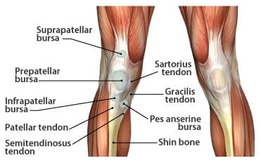
Figure 117. Bursae in the knee.

the knee

Figure 118. T2 gradient
displaying suprapatellar
bursa.

the knee

Figure 119. T2
fatsat displaying
prepatellar bursa.

the knee

Figure 120. T2 fatsat
displaying infrapatellar
bursa.


The synovial sac of the knee joint sometimes forms a posterior bulge, known as a Baker�s cyst or popliteal cyst (Figure 121). It typically forms between the tendons of the medial head of the gastrocnemius muscle and the semimembranosus muscle, posterior to the medial femoral condyle. Baker�s cysts are not true cysts, as they typically maintain open communication with the synovial sac. However, they can pinch off, and they can rupture. They are usually asymptomatic, but can be indicative of another problem of the knee, such as arthritis or a meniscal tear. Aspiration of the synovial fluid can be performed if the cyst becomes problematic. Treatment is usually necessary if a Baker�s cyst ruptures, as it can cause acute pain behind the knee, and swelling of the calf muscles. A ruptured cyst can also mimic a DVT or thrombophlebitis. Ultrasound and MRI can both be used for confirmation of a Baker�s cyst (Figure 122).

the knee

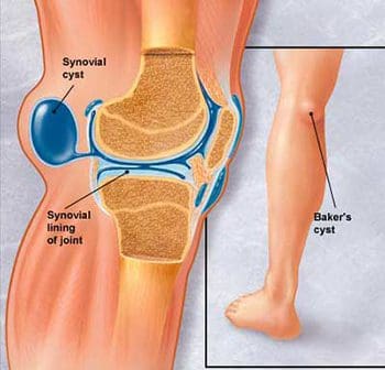
Figure 121. Lateral view of Baker�s cyst.

the knee

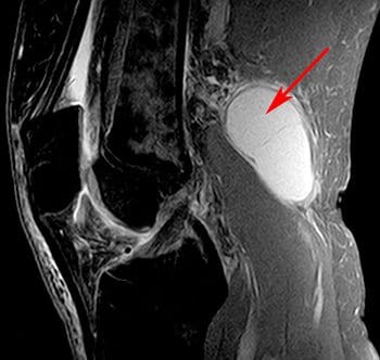
Figure 122. Sagittal image of Baker�s cyst on MRI.

Scan Setups

The following are HMSA suggestions for knee imaging. Knee protocols should be designed to yield diagnostic images of the menisci, bones, articular cartilage, and all ligamentous structures of the knee. While many radiologists may require additional imaging of the ACL, protocols that are designed for optimal imaging of the cartilage and menisci should also produce adequate images of the ACL. Always check with your radiologist for his/her imaging preferences.

Axial Scans

When positioning axial slices for the knee, sagittal and coronal images can be used to insure inclusion of all pertinent anatomy. The slices should extend superiorly to include the entire patella, and inferiorly to include the tibial tuberosity and patellar tendon insertion. A presat can be placed over the unaffected lower extremity to reduce the possibility of wrap-around artifact, as seen in the coronal image in Figure 139.

the knee

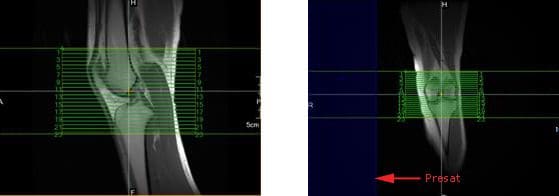
Figure 139. Axial slice setup using sagittal and coronal images.

Coronal Scans

Coronal slices of the knee should include the anatomy from the posterior femoral condyles to the anterior portion of the patella. Visualize a line connecting the lateral and medial condyles of the femur. Typically, the coronal slices are angled so that they are parallel to that line, as seen in the axial image in Figure 140.

the knee

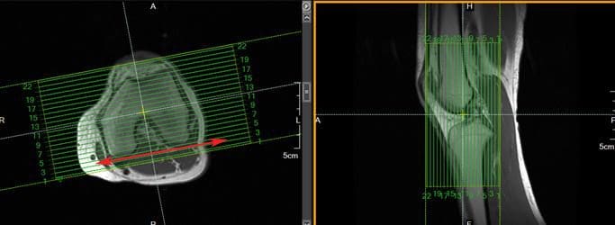
Figure 140. Coronal slice setup using axial and sagittal images.

Sagittal Scans

Sagittal slices should include the anatomy from the medial condyle to the lateral condyle. The slice group may be angled per your radiologist�s preference, but should remain perpendicular to the coronal slices. Typically, the slice group is angled so that it is parallel to the medial border of the femoral condyle, as seen in the axial image in Figure 141.

the knee

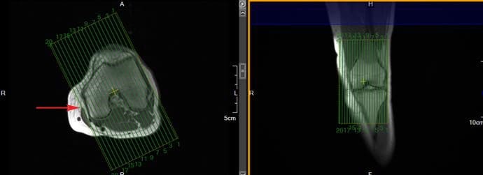
Figure 141. Sagittal slice setup using axial and coronal images.

In addition to routine oblique sagittal images, some radiologists prefer an additional sagittal scan of the ACL with thin slices and high spatial resolution. Axial and coronal images can be used for slice setup. Referenced literature recommends that the angle of the slice group should not exceed 10� from a line drawn perpendicular to the bicondylar line (line that connects the posterior femoral condyles), as seen in Figure 142.

the knee

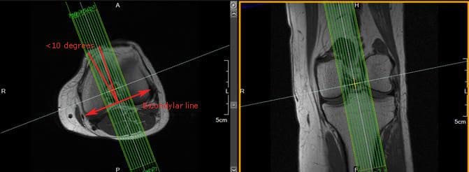
Figure 142. Sagittal ACL slice setup using axial and coronal images.


blank
References:

Kapit, Wynn, and Lawrence M. Elson. The Anatomy Coloring Book. New York: HarperCollins, 1993.

Hip Anatomy, Function, and Common Problems. (Last updated 28July2010). Retrieved from http://healthpages.org/anatomy-function/hip-structure-function-common-problems/

Cluett, J. M.D. (Updated 22May2012). Labral Tear of the Hip Joint. Retrieved from http://orthopedics.about.com/od/hipinjuries/qt/labrum.htm

Hughes, M. D.C. (15July2010). Diseases of the Femur Bone. Retrieved from http://www.livestrong.com/article/175599-diseases-of-the-femur-bone/

A Patient�s Guide to Perthes Disease of the hip. (n.d.). Retrieved from http://www.orthopediatrics.com/docs/Guides/perthes.html

Hip Injuries and Disorders. (Last reviewed 10February2012). Retrieved from http://nlm.nih.gov/medlineplus/hipinjuriesanddisorders.html

Ligament of head of femur. (Updated 20December2011). Retrieved from http://en.wikipedia.org/wiki/Ligament_of_head_of_femur

Ewing�s sarcoma. (Last modified 06January2012). Retrieved from http://en.wikipedia.org/wiki/Ewing%27s_sarcoma

Hip Anatomy. (n.d.). Retrieved from http://www.activemotionphysio.ca/Injuries-Conditions/Hip

Iliotibial Band Friction Syndrome. (n.d.). Retrieved from http://www.physiotherapy-treatment.com/iliotibial-band-friction-syndrome.html

Snapping hip syndrome. (Last modified 09November2011). Retrieved from http://en.wikipedia.org/wiki/Snapping_hip_syndrome

Sekul, E. (Updated 03February2012). Meralgia Paresthetica. Retrieved from http://emedicine.medscape.com/article/1141848-overview

Yeomans, S. D.C. (Updated 07July2010). Sciatic Nerve and Sciatica. Retrieved from http://www.spine-health.com/conditions/sciatica/sciatic-nerve-and-sciatica

Mayo Clinic staff. (26July2011). Meralgia paresthetica. Retrieved from http://www.mayoclinic.com/health/meralgia-paresthetica/DS00914

Deep Vein Thrombosis (DVT)-Blood Clots in the Legs. (n.d.). Retrieved from http://catalog/nucleusinc.com/displaymonograph.php?MID=148

Petersilge, C. M.D. (03May2000). Chronic Adult Hip Pain: MR Arthrography of the Hip. Retrieved from http://radiographics.rsna.org/content/20/suppl_1/S43.full

Acetabular branch of medial circumflex femoral artery. (Last modified 17November2011). Retrieved from http://en.wikipedia.org/wiki/Acetabular_branch_of_medial_circumflex_femoral_artery

Cluett, J. M.D. (Updated 26March2011). Hip Bursitis. Retrieved from http://orthopedics.about.com/cs/hipsurgery/a/hipbursitis.htm

Steinbach, L. M.D., Palmer, W. M.D., Schweitzer, M. M.D. (10June2002). Special Focus Session MR Arthrography. Retrieved from http://radiographics.rsna.org/content/22/5/1223.full

Schueler, S. M.D., Beckett, J.M.D., Gettings, S.M.D. (Last updated 05August2010). Ischial Bursitis/Overview. Retrieved from http://www.freemd.com/ischial-bursitis/overview.htm

Hwang, B., Fredericson, M., Chung, C., Beaulieu, C., Gold, G. (29October2004). MRI Findings of Femoral Diaphyseal Stress Injuries in Athletes. Retrieved from http://www.ajronline.org/content/185/1/166.full.pdf

The Femur (Thigh Bone). (n.d.). Retrieved from http://education.yahoo.com/reference/gray/subjects/subject/59

Norman, W. PhD, DSc. (n.d.). Joints of the Lower Limb. Retrieved from http://home.comcast.net/~wnor/lljoints.htm

Femur. (Last modified 24September2012). Retrieved from http://en.wikipedia.org/wiki/Femur

Wheeless, C. III, M.D. (Last updated 25April2012). Ligaments of Humphrey and Wrisberg. Retrieved from http://wheelessonline.com/ortho/ligaments_of_humphrey_and_wrisberg

Muscle Strains in the Thigh. (Last reviewed August2007). Retrieved from http://orthoinfo.aaos.org/topic.cfm?topic=A00366

Shiel, W. Jr., M.D. (Last reviewed 23July2012). Hamstring Injuries. Retrieved from http://www.medicinenet.com/hamstring_injury/article.htm

Hamstring Muscle Injuries. (Last reviewed July 2009). Retrieved from http://orthoinfo.aaos.org/topic.cfm?topic=a00408

Knee. (Last modified 19September2012). Retrieved from http://en.wikipedia.org/wiki/Knee

DeBerardino, T. M.D. (Updated 30March2012). Quadriceps Injury. Retrieved from http://emedicine.medscape.com/article/91473-overview

Kan, J.H. (n.d.). Osteochondral Abnormalities: Pitfalls, Injuries, and Osteochondritis Dissecans. Retrieved from http://www.arrs.org/shopARRS/products/s11p_sample.pdf

Nerves of the Lower Limb. (Last updated 30March2006). Retrieved from http://download.videohelp.com/vitualis/med/lowrnn.htm

The Adductor Canal. (Last updated 30March2006). Retrieved from http://download.videohelp.com/vitualis/med/addcanal.htm

Nabili, S. M.D. (n.d.). Varicose Veins & Spider Veins. Retrieved from http://www.medicinenet.com/varicose_veins/article.htm

Basic Venous Anatomy. (n.d.). Retrieved from http://vascular-web.com/asp/samples/sample104.asp

Femoral nerve. (Last modified 23September2012). Retrieved from http://en.wikipedia.org/wiki/Femoral_nerve

Peron, S. RDCS. (Last modified 16October2010). Anatomy � Lower Extremity Veins. Retrieved from http://www.vascularultrasound.net/vascular-anatomy/veins/lower-extremity-veins

Medical Multimedia Group, L.L.C. (n.d.). Knee Anatomy. Retrieved from http://www.eorthopod.com/content/knee-anatomy

Knee Joint Anatomy, Function and Problems. (Last updated 06July2010). Retrieved from http://healthpages.org/anatomy-function/knee-joint-structure-function-problems/

Coronary ligament of the knee. (Last modified 09May2010). Retrieved from http://en.wikipedia.org/wiki/Coronary_ligament_of_the_knee

Walker, B. (n.d.). Patellar Tendonitis Treatment � Jumper�s Knee. Retrieved from http://www.thestretchinghandbook.com/archives/patellar-tendonitis.php

Osgood-Schlatter disease. (Last reviewed 12November2010). Retrieved from http://www.ncbi.nlm.nih.gov/pubmedhealth/PMH0002238/

Grelsamer, R. M.D. (n.d.). The Anatomy of the Patella and the Extensor Mechanism. Retrieved from http://kneehippain.com/patient_pain_anatomy.php

Oblique popliteal ligament. (Last modified 24March2012). Retrieved from http://en.wikipedia.org/wiki/Oblique_popliteal_ligament

Shiel, W. Jr., M.D. (Last reviewed 27July2012). Chondromalacia Patella (Patellofemoral Syndrome). Retrieved from http://www.medicinenet.com/patellofemoral_syndrome/article.htm

Knee. (Last modified 19September2012). Retrieved from http://en.wikipedia.org/wiki/Knee

Mosher, T. M.D. (Last updated 11April2011). MRI of Knee Extensor Mechanism Injuries Overview of the Knee Extensor Mechanism. Retrieved from http://emedicine.medscape.com/article/401001-overview

Carroll, J. M.D. (December 2007). Oblique Menisco-meniscal Ligament. Retrieved from http://radsource.us/clinic/0712

DeBerardino, T. M.D. (Last updated 30March2012). Medial Collateral Knee Ligament Injury. Retrieved from http://emedicine.medscape.com/article/89890-overview#a0106

Farr, G. (Last updated 31December2007). Joints and Ligaments of the Lower Limb. Retrieved from http://becomehealthynow.com/article/bodyskeleton/951/

Knee anatomy overview. (02March2008). Retrieved from http://www.kneeguru.co.uk/KNEEnotes/node/741

Dixit, S. M.D., Difiori, J. M.D., Burton, M. M.D., Mines, B. M.D. (15January2007). Management of Patellofemoral Pain Syndrome. Retrieved from http://www.aafp.org/afp/2007/0115/p194.html

Knee Muscles. (Last updated 05September2012). Retrieved from http://www.knee-pain-explained.com/kneemuscles.html

Popliteus muscle. (Last updated 20February2012). Retrieved from http://en.wikipedia.org/wiki/Popliteus_muscle

Kneedoc. (10February2011). Nerves. Retrieved from http://thekneedoc.co.uk/neurovascular/nerves

Wheeless, C. III, M.D. (Last updated 15December2011). Popliteal Artery. Retrieved from http://wheelessonline.com/ortho/popliteal_artery

The Popliteal Artery. (n.d.) Retrieved from http://education.yahoo.com/reference/gray/subjects/subject/159

Knee bursae. (Last updated 09May2012). Retrieved from http://en.wikipedia.org/wiki/Bursae_of_the_knee_joint

Hirji, Z., Hunjun, J., Choudur, H. (02May2011). Imaging of the Bursae. Retrieved from http://www.ncbi.nlm.nih.gov/pmc/articles/PMC3177464/

Kimaya Wellness Limited. (n.d.). Organ>Popliteal Artery. Retrieved from http://kimayahealthcare.com/OrganDetail.aspx?OrganID=103&AboutID=1

Total Vein Care. (Last updated 24February2012). Varicose Vein Anatomy and Function for Patients. Retrieved from http://www.veincare.com/education/

Tibia. (Last updated 01April2012). Retrieved from http://en.wikipedia.org/wiki/Tibia

Norkus,S., Floyd, R. (Published 2001). The Anatomy and Mechanisms of Syndesmotic Ankle Sprains. Retrieved from http://www.ncbi.nlm.nih.gov/pmc/articles/PMC155405/

Soleus muscle. (Last updated 10April2012). Retrieved from http://en.wikipedia.org/wiki/Soleus_muscle

Achilles Tendinitis. (Last reviewed June2010). Retrieved from http://orthoinfo.aaos.org/topic.cfm?topic=A00147

Wheeless, C. III,M.D. (Last updated 11April2012). Sural Nerve. Retrieved from http://wheelessonline.com/ortho/sural_nerve

Medical Multimedia Group, L.L.C. (Last updated 26July2006). Ankle Syndesmosis Injuries. Retrieved from http://www.orthogate.org/patient-education/ankle/ankle-syndesmosis-injuries.html

Cluett, J. M.D. (Last updated 16September2008). Exertional Compartment Syndrome. Retrieved from http://orthopedics.about.com/od/overuseinjuries/a/compartment.htm

Leg Veins (Thigh, Lower Leg) Anatomy, Pictures and Names. (Last updated 21November2010). Retrieved from http://www.healthype.com/leg-veins-thigh-lower-leg-anatomy-pictures-and-names.html

Cluett, J.M.D. (Last updated 6October2009). Stress Fracture. Retrieved from http://orthopedics.about.com/cs/otherfractures/a/stressfracture.htm

Ostlere, S. (1December2004). Imaging the ankle and foot. Retrieved from http://imaging.birjournals.org/content/15/4/242.full

Inverarity, L. D.O. (Last updated 23January2008). Ligaments of the Ankle Joint. Retrieved from http://physicaltherapy.about.com/od/humananatomy/p/ankleligaments.htm

Golano, P., Vega, J., DeLeeuw, P., Malagelada, F.,Manzanares, M., Gotzens, V., van Dijk, C. (Published online 23March2010). Anatomy of the ankle ligaments:a pictorial essay. Retrieved from http://www.ncbi.nlm.nih.gov/pmc/articles/PMC2855022/

Numkarunarunrote, N., Malik, A., Aguiar, R.,Trudell, D., Resnick, D. (11October2006). Retinacula of the Foot and Ankle: MRI with Anatomic Correlation in Cadavers. Retrieved from http://www.ajronline.org/content/188/4/W348.full

Medical Multimedia Group, L.L.C. (n.d.). A Patient�s Guide to Ankle Anatomy. Retrieved from http://www.eorthopod.com/content/ankle-anatomy

The Anterior Tibial Artery. (n.d.). Retrieved from http://education.yahoo.com/reference/gray/subjects/subject/160

Foot and Ankle Anatomy. (Last updated 28July2011). Retrieved from http://northcoastfootcare.com/pages/Foot-and-Ankle-Anatomy.html

Donnelly, L., Betts, J., Fricke, B. (1July2009). Skimboarder�s Toe: Findings on High-Field MRI. Retrieved from http://www.ajronline.org/content/184/5/1481.full

Foot. (Last updated 28August2012). Retrieved from http://en.wikipedia.org/wiki/Foot

Wiley, C. (n.d.). Major Ligaments in the Foot. Retrieved from http://www.ehow.com/list_6601926_major-ligaments-foot.html

Turf Toe: Symptoms, Causes, and Treatments. (Last reviewed 9August2012). Retrieved from http://www.webmd.com/fitness-exercise/turf-toe-symptoms-causes-and-treatments

Cluett, J. M.D. (Last updated 02April2012). Turf Toe. Retrieved from http://orthopedics.about.com/od/toeproblems/p/turftoe.htm

Neurology and the Feet. (n.d.) Retrieved from http://footdoc.ca/www.FootDoc.ca/Website%20Nerves%20Of%20The%20Feet.htm

The Veins of the Lower Extremity, Abdomen, and Pelvis. (n.d.). Retrieved from http://education.yahoo.com/reference/gray/subjects/subject/173

Corley, G., Broderick, B., Nestor, S., Breen, P., Grace, P., Quondamatteo, F., O�Laighin, G. (n.d.). The Anatomy and Physiology of the Venous Foot Pump. Retrieved from http://www.eee.nuigalway.ie/documents/go_anatomy_of_the_plantar_venous_plexus_manuscript.pdf

Morton�s neuroma. (Last modified 8August2012). Retrieved from http://en.wikipedia.org/wiki/Morton%27s_metatarsalgia

References For Anatomy Pics:

Figures 1, 5, 6, 24- http://www.orthopediatrics.com/docs/Guides/perthes.html

Figures 2, 3, 11, 12, 14, 15, 16, 18, 23, 25- http://www.activemotionphysio.ca/Injuries-Conditions/Hip/Hip-Anatomy/a~299/article.html

Figure 4- http://hipkneeclinic.com/images/uploaded/hipanatomy_xray.jpg

Figures 7, 8, 9- http://hipfai.com/

Figure 10- http://en.wikipedia.org/wiki/File:Ewing%27s_sarcoma_MRI_nci-vol-1832-300.jpg

Figure 13- http://www.chiropractic-help.com/Patello-Femoral-Pain-Syndrome.html

Figure 17- http://www.thestretchinghandbook.com/archives/ezine_images/adductor.jpg

Figure 19- http://media.summitmedicalgroup.com/media/db/relayhealth-images/hipanat.jpg

Figures 20-22- http://www.ajronline.org/content/182/1/137.full.pdf+html

Figure 43, 44- http://radiographics.rsna.org/content/20/suppl_1/S43.full

Figure 45- http://www.exploringnature.org/db/detail.php?dbID=24&detID=2768

Figures 46-48- http://www.ajronline.org/content/185/1/166.full.pdf

Figure 49- http://arrs.org/shopARRS/products/s11p_sample.pdf

Figure 50- http://www.thestretchinghandbook.com/archives/medial-collateral-ligament.php

Figures 51, 52- http://www.radsource.us/clinic/0712

Figures 53, 54- http://www.osteo-path.co.uk/BodyMap/Thighs.html

Figure 55- http://www.ncbi.nlm.nih.gov/pmc/articles/PMC1963576/

Figure 56- http://legacy.owensboro.kctcs.edu/gcaplan/anat/Notes/API%20Notes%20M%20%20Peripheral%20Nerves.htm

Figure 57- http://www.keywordpictures.com/keyword/lateral%20cutaneous%20nerve%20of%20thigh/

Figure 58- http://home.comcast.net/~wnor/postthigh.htm

Figure 59- http://becomehealthynow.com/glossary/CONG437.htm

Figure 60- http://fitsweb.uchc.edu/student/selectives/Luzietti/Vascular_pvd.htm

Figure 61- http://www.fashion-res.com/peripheral-vascular-disease-with-stenting-in-the/

Figure 62- http://www.wpclipart.com/medical/anatomy/blood/femoral_artery_and_branches_in_leg.png.html

Figure 63- http://www.globalteleradiologyservices.com/Deep_Vein_Thrombosis_Overview.htm

Figure 64- http://www.vascularultrasound.net/vascular-anatomy/veins/lower-extremity-veins

Figure 82- http://www.jeffersonhospital.org/diseases-conditions/knee-ligament-injury.aspx?disease=658f267f-75ab-4bde-8781-f2730fafa958

Figure 83- http://javierjuan.ifunnyblog.com/anatomybackofknee/

Figure 84- http://www.kneeandshouldersurgery.com/knee-disorders/tibial-osteotomy.html

Figure 85- http://www.disease-picture.com/chondromalacia-patella-physical-therapy/

Figure 86- http://www.eorthopod.com/content/bipartite-patella

Figure 87- http://www.orthogate.org/patient-education/knee/articular-cartilage-problems-of-the-knee.html

Figure 88- http://www.webmd.com/pain-management/knee-pain/menisci-of-the-knee-joint

Figure 89- http://sumerdoc.blogspot.com/2008_07_01_archive.html

Figure 90- http://www.concordortho.com/patient-education/topic-detail-popup.aspx?topicID=55befba2d440dc8e25b85747107b5be0

Figure 91- http://trialx.com/curebyte/2011/08/16/pictures-for-chondromalacia-patella/

Figure 92- http://radiopaedia.org/images/1059

Figure 93- http://radiologycases.blogspot.com/2011/01/osgood-schlatter-disease.html

Figure 94- http://www.physioquestions.com/2010/09/07/knee-injury-acl-part-i/

Figure 95- http://www.jeffersonhospital.org/diseases-conditions/knee-ligament-injury.aspx?disease=4e3fcaf5-0145-43ea-820f-a175e586e3c8

Figures 96, 97- http://radiology.rsna.org/content/213/1/213.full

Figures 98-101- http://appliedradiology.com/Issues/2008/12/Articles/Imaging-the-knee–Ligaments.aspx

Figure 102- http://radiopaedia.org/images/408156

Figure 103- http://aftabphysio.blogspot.com/2010/08/joints-of-lower-limb.html

Figures 104, 105- http://www.radsource.us/clinic/0310

Figure 106- http://nwrunninglab.com/patellar-tendonitis.html

Figure 107- http://www.aafp.org/afp/2007/0115/p194.html

Figure 108- http://www.reboundsportspt.com/blog/tag/knee-pain

Figure 109- http://www.norwellphysicaltherapy.com/Injuries-Conditions/Knee/Knee-Issues/Quadriceps-Tendonitis-of-the-Knee/a~1803/article.html

Figure 110- http://kneeguru.co.uk/KNEEnotes/node/479

Figure 111- http://www.magicalrobot.org/BeingHuman/2010/03/fascia-bones-and-muscles

Figure 112- http://home.comcast.net/~wnor/postthigh.htm

Figures 113, 115, 157-159- http://ipodiatry.blogspot.com/2010/02/anatomy-of-foot-and-ankle_26.html

Figure 114- http://medchrome.com/basic-science/anatomy/the-knee-joint/

Figure 116- http://www.sharecare.com/question/what-are-varicose-veins

Figure 117- http://mendmyknee.com/knee-and-patella-injuries/anatomy-of-the-knee.php

Figures 118-120- http://www.ncbi.nlm.nih.gov/pmc/articles/PMC3177464/

Figure 121- http://www.riversideonline.com/health_reference/Disease-Conditions/DS00448.cfm

Figure 122- http://arthritis.ygoy.com/2011/01/01/what-is-an-arthritis-knee-cyst/

Figure 143- http://usi.edu/science/biology/mkhopper/hopper/BIOL2401/LABUNIT2/LabEx11week6/tibiaFibulaAnswer.htm

Figure 144- http://web.donga.ac.kr/ksyoo/department/education/grossanatomy/doc/html/fibula1.html

Figure 145- http://becomehealthynow.com/popups/ligaments_tib_fib_bh.htm

Figure 146- http://www.parkwayphysiotherapy.ca/article.php?aid=121

Figure 147- http://aidmyankle.com/high-ankle-sprains.php

Figure 148- http://legsonfire.wordpress.com/what-is-compartment-syndrome/

Figures 149, 152- http://www.stepbystepfootcare.ca/anatomy.html

Figures 150, 151- http://www.gla.ac.uk/ibls/US/fab/tutorial/anatomy/jiet.html

Figure 153- http://www.athletictapeinfo.com/?s=tennis+leg

Figure 154- http://radsource.us/clinic/0608

Figure 155- http://www.eorthopod.com/content/achilles-tendon-problems

Figure 156- http://achillesblog.com/assumptiondenied/not-a-rupture/

Figure 181- http://www.orthopaedicclinic.com.sg/ankle/a-patients-guide-to-ankle-anatomy/

Figure 182- http://www.activemotionphysio.ca/article.php?aid=47

Figure 183- http://www.ajronline.org/content/193/3/687.full

Figures 184, 186- http://www.eorthopod.com/content/ankle-anatomy

Figure 185- http://www.crossfitsouthbay.com/physical-therapy/learn-yourself-a-quick-anatomy-reference/ankle/

Figures 187, 227- http://www.activemotionphysio.ca/Injuries-Conditions/Foot/Foot-Anatomy/a~251/article.html

Figure 188- http://inmotiontherapy.com/article.php?aid=124

Figures 189, 190- http://home.comcast.net/~wnor/ankle.htm

Figure 191- http://skillbuilders.patientsites.com/Injuries-Conditions/Ankle/Ankle-Anatomy/a~47/article.html

Figure 192- http://metrosportsmed.patientsites.com/Injuries-Conditions/Foot/Foot-Anatomy/a~251/article.html

Figure 193- http://musc.edu/intrad/AtlasofVascularAnatomy/images/CHAP22FIG30.jpg

Figure 194- http://musc.edu/intrad/AtlasofVascularAnatomy/images/CHAP22FIG31B.jpg

Figure 195- http://veinclinics.com/physicians/appearance-of-vein-disease/

Figure 196- http://mdigradiology.com/services/interventional-services/varicose-veins.php

Figure 216- http://kidport.com/RefLib/Science/HumanBody/SkeletalSystem/Foot.htm

Figure 217- http://www.joint-pain-expert.net/foot-anatomy.html

Figure 218- http://www.thetoedoctor.com/turf-toe-symptoms-and-treatment/

Figures 219, 220- http://radsource.us/clinic/0303

Figure 221- http://www.ajronline.org/content/184/5/1481.full

Figure 222- http://www.answers.com/topic/arches

Figure 223- http://www.mayoclinic.com/health/medical/IM00939

Figure 224- http://radsource.us/clinic/0904

Figure 225- http://www.ortho-worldwide.com/anfobi.html

Figure 226- http://www.coringroup.com/lars_ligaments/patientscaregivers/your_anatomy/foot_and_ankle_anatomy/

Figure 228- http://www.stepbystepfootcare.ca/anatomy.html

Figure 229- http://iupucbio2.iupui.edu/anatomy/images/Chapt11/FG11_18aL.jpg

Figure 230- http://www.ajronline.org/content/184/5/1481.full.pdf

Figure 231- http://metrosportsmed.patientsites.com/Injuries-Conditions/Foot/Foot-Anatomy/a~251/article.html

Figure 232- http://www.painfreefeet.com/nerve-entrapments-of-the-leg-and-foot.html

Figures 233, 234- http://emedicine.medscape.com/article/401417-overview

Figure 235- http://web.squ.edu.om/med-Lib/MED_CD/E_CDs/anesthesia/site/content/v03/030676r00.HTM

Figure 236- http://www.nysora.com/peripheral_nerve_blocks/classic_block_tecniques/3035-ankle_block.html

Figure 237- http://ultrasoundvillage.net/imagelibrary/cases/?id=122&media=464&testyourself=0

Figure 238- http://www.joint-pain-expert.net/foot-anatomy.html

Figure 239- http://jap.physiology.org/content/109/4/1045.full

Figure 240- http://microsurgeon.org/secondtoe

Figure 241- http://elu.sgul.ac.uk/rehash/guest/scorm/406/package/content/common_iliac_veins.htm

Close Accordion
Ankle Injuries: Chiropractic Care Rehab

Ankle Injuries: Chiropractic Care Rehab

An ankle can be injured as glamorously as falling off a $400 platform stiletto heel, stepping off a Parisian curb, or as mundanely as tripping over a toy truck, or falling over a rock on your way to the mailbox. No matter the cause, ankle injuries are painful and problematic, and cause recurring problems if left untreated.

The poor ankle sure has it rough. It supports a person’s entire body weight, twists and turns many times a day, and maintains proper balance. This heavy responsibility takes its toll. Emergency rooms treat approximately one million patients ever year for ankle injuries.

Ankles are technically “the joint where the foot joins the leg.”� In reality, there are more moving parts involved than that simplified definition allows. Multiple bones and two separate joints actually converge in the ankle area, which increases the chance of an ankle injury.

Ankle Injuries:

ankle injuries

Sprains:

When you roll your ankle outward, the movement damages the ligaments on the outside of the ankle. This is a common sports injury and, unfortunately, once you have sprained your ankle it’s more likely to recur. Up to half of the people who suffer from a sprained ankle will sprain it again.

Strains:

There are two tendons in the ankle that are commonly strained, usually over stretching from overuse or trauma.

Fractures:

This injury happens when one or more of the three bones in the ankle is injured. While less common than a sprain or strain, a fractured ankle may also involve damaged ligaments and require surgery.

Many instances of ankle injuries are avoidable. Be sure to wear proper shoes when exercising or participating in sports, avoid uneven walking surfaces, and keep stairways and floors in your home clear of clutter

And, ladies, avoid the really high heels. We know, we know, they are just so cute! 🙂

Even with ankle-protecting precautions, you still may end up on your rump in the grass nursing your swollen ankle. What should you do if you injure your ankle? There are several forms of treatment for an ankle injury depending on its severity.

Rest and ice: For mild injuries, stay off your ankle and use ice packs to reduce the swelling. Rest allows the injured area to heal faster.

Visit a doctor: If you experience severe pain, swelling, and are unable to put weight on your ankle, see a doctor, as some ankle injuries grow worse without treatment. Injuries may require a brace, cast, or even surgery.

See a chiropractor: Patients frequently see strongly positive results in ankle injuries from a series of chiropractic treatments. Chiropractors understand the way the ankle is built, and use chiropractic adjustments to reduce pain and inflammation and promote faster healing.

Exercise rehab: Once you are healed, it’s vital to build up the ankle’s strength to avoid re-injury. Your chiropractor can lay out an exercise routine that you can employ into your regular workouts that will improve your balance and increase mobility. Performing these moves helps dramatically decrease dealing with this again down the road.

Ankle injuries are common and, whether or not you maintain an active lifestyle, you may end up suffering from one. By visiting a chiropractor on the front end, you can better plan a course of treatment that will heal your ankle quickly, reduce the pain effectively, and minimize the chance of a recurrence.

Golf & Chiropractic Care

Whiplash Injuries Explained

Whiplash Injuries Explained

Whiplash Injuries Explained: Whiplash Associated Disorders

  • Approximately 15 to 40% of those injured in automobile accidents will struggle with chronic pain for the rest of their life. Journal of the American Academy of Orthopedic Surgeons, 2007
  • Whiplash injuries not only increase your chances of chronic neck and shoulder pain, they also increase the probability of other seemingly unrelated health problems. Journal of Clinical Epidemiology, 2001
  • Chronic Pain does bad things to people. According to standardized assessment tests, 100% of those struggling with chronic pain caused by whiplash injuries have abnormal psychological profiles. The only way to resolve these abnormal psychological profiles is to relieve / remove the chronic back pain, neck pain and headaches. Counseling / Psychiatry has not been shown to improve the pain nor the psychological profiles of people suffering from the effects of their automobile accident. Pain, 1997
  • The longest-running study ever done on whiplash patients looked at the overall health of whiplash patients almost twenty years after their automobile accident. Nearly two decades after their accident, 55% of those patients still deal with chronic pain. Accident Analysis and Prevention, 2002
  • Unless you have a fracture or specific ligament tear, Cervical Collars are no longer recommended for treating patients with whiplash injuries. When cervical collars are used as a whiplash injury treatment, there is a 90% probability that you will still have chronic neck pain in six months. Spine, 2000
  • One in one hundred people around the world (1% of the population, or just over 70 million people) suffer from ongoing chronic neck pain due to an automobile-induced whiplash injury. Injury, 2005
  • One in fifty people injured in Whiplash-like accident deal with chronic pain severe enough to need diagnostic testing, medications, and doctor visits, on an ongoing basis —– nearly eight years after the accident occured. Pain, 1994

“Statistically, every American can expect to be in a motor vehicle collision once every ten years. Motor vehicle collisions have been the number one cause of death of our children for decades. Since 9/11 (September 11, 2001), about 3,000 Americans have died as a consequence of terrorism; about 360,000 Americans have died in motor vehicle crashes. Since the start of the American Revolution in 1775, about a million Americans have died in our wars. Since Henry Ford introduced the mass-produced motorcar in 1913, more than 2.5 million Americans have met their deaths on the road. And millions of Americans who did not die from motor vehicle collisions were injured.” Orthopedist and one of the world’s foremost experts on whiplash, Dr. Dan Murphy. There are 3,000,000 new cases of whiplash in the US every year.

Whiplash Injuries Explained

The word �whiplash� is a layperson�s term —- and although it is typically associated with Car Crashes, crashes are certainly not the only way to get a whiplash injury. Whiplash Associated Disorders (WAD) are typically referred to in the medico-legal literature as �Acceleration / Deceleration� injuries, or “Hyperflexion / Hyperextension” injuries. And, as many of you have come to understand the hard way, they can be incredibly violent � even in seemingly minor accidents that had surprisingly little vehicular damage. With over three million new cases of Acceleration / Deceleration injuries occurring each year, and over 50% of those progressing to at least some degree of unresolved or �chronic� symptoms, it is clear that Whiplash Associated Disorders are taking a massive toll on our country financially, physically, and emotionally.

When people think of �whiplash� they tend to think of motor vehicle accidents (MVA�s). Although MVA is probably the single most common cause of the symptoms most frequently associated with and experienced by those suffering with Whiplash Associated Disorders (neck pain, upper back pain, shoulder pain, fuzzy thinking, numbness, tingling and / or weakness of the hands, dizziness, etc), whiplash can occur in about a thousand and one different ways. And while there are certain symptoms that we see over and over and over in our clinic (neck pain and headaches, for instance), whiplash can seemingly cause about a thousand and one different symptoms as well. Some of the most common causes of WAD that I see in my office include sports injuries, work injuries (think logging here), spousal abuse, fights, horse accidents (falls), and almost anything else that has the capacity to �snap� your head suddenly and violently.

Although the most common problems associated with Whiplash Associated Disorders are related to the neck (neck pain, numb hands, headaches), scientific research shows that Acceleration / Deceleration injuries routinely cause all sorts of other injuries as well. For instance, I commonly see people whose low back pain started with an MVA. I even see people whose FIBROMYALGIA was brought on by the emotional and physical stress of an MVA! One of the most shocking conclusions concerning Whiplash Associated Disorders, was written by a pair of the most well known whiplash researchers on the planet � medical researchers, not chiropractic researchers. Drs. Gargan & Bannister stated in a study that was done in the 1990?s, that whiplash-like injuries frequently result in a whole host of, �bizarre and seemingly unrelated symptoms�. Although there are plenty of malingerers, fakers, scam artists, money-grubbers, and drug seekers out there; far too many people are lumped into these categories simply because their problems do not show up on traditional medical tests such as MRI / CT.

Even though there are literally scores of scientific studies concluding that Whiplash Associated Disorders are difficult (often to the point of being impossible) to image on x-rays, CT’s, or MRI�s, these are still the chief method the medical community is using to determine whether or not you were injured, and just how serious this injury might be. The problem is, if the vast majority of soft-tissue injuries (injuries to LIGAMENTS, TENDONS, MUSCLES, FASCIA, etc) do not image well with advanced imaging techniques, and imaging is the medical community�s chief method of diagnosis; unless you have a herniated disc, you will invariably be treated like nothing is really wrong with you � like you are a scam artist trying to extort a huge settlement from an insurance company. Stop and think for a moment about how problematic that fascia, arguably the single most pain-sensitive tissue in your entire body, will not show up on any tests —- including MRI.

When you are taken the the ER, you will have some tests run and the doctor will look at you and say, �Thank God Mrs. Smith. Nothing is broken! Now, go home and rest, and call your family doctor tomorrow. In the mean time, wear this collar, and take these Anti-Inflammatory Medications, pain pills, and muscle relaxers. Oh, and don�t forget to use a heat pack as well.� Is this good advice? Sure it is � if you own a medical clinic! Follow this advice and you are certain to become a lifetime ARTHRITIC! The truth is, when it comes to the evaluation and treatment of injuries to fascia and other elastic, collagen-based connective tissues, all of our hi-tech equipment with its bells and whistles is simply not helping diagnose or help most injured people. You are reading a page on whiplash —- my guess is that you completely understand this concept because you have been there, and done that! The Old Model of tissue injury evaluation and treatment went out the door about 25 years ago. It just seems like no one has remembered to tell treating physicians about the NEW MODEL.

Brain Based Injury

Your short drive to work was no different than any other day —- until you began slowing down for the school bus stopping in front of you. Just as you’re coming to a complete stop, BAM; your world explodes as someone plows into your car from behind, knocking you into the bus. Turns out the kid driving the full-sized crew cab pickup truck that hit you was texting, and never even hit his brakes. You’re having a hard time remembering exactly what happened. You remember a flash of light and your head being slammed backwards over the top of your headrest. You vaguely recall that your head rocketed forward as you hit the bus — almost hitting the windshield. You step out of your 1997 Toyota Camry to take stock of the situation. There is no blood or guts. In fact, you don’t even have a bruise to show for your trouble. But by the time the State Troopers arrive to work the accident, you not only have a neck pain unlike anything you have ever felt before, you have a banging headache as well. You’re having trouble putting the pieces in order for them. They ask if you need an ambulance, but you do not want to go to the Emergency Room. But a few weeks later, you’re still having trouble with your memory. Work is not going well because on top of the pain and exhaustion (yeah, since the accident you can’t sleep either), everything seems fuzzy, foggy, and hazy. Who would have thought that whiplash could cause these sorts of symptoms —– particularly without any overt / obvious injuries?

Whiplash Injuries are particularly dangerous because they are a common cause of MTBI (Mild Traumatic Brain Injury). MTBI results from the brain bouncing off the inside of the skull during the hyperextension / hyperflexion of the neck. As you can imagine, this damages / destroys nerve cells. Depending on which part of the brain is injured, a person might have problems in some of the following areas…

  • Walking / Moving
  • Balance
  • Coordination
  • Strength / Endurance
  • Ability to Communicate
  • Ability to Understand
  • Ability to Think
  • Memory
  • Strange or Unexplainable Pain Patterns or Symptoms (these are some of the “bizarre and seemingly unrelated symptoms” talked about by whiplash researchers Gargan and Bannister.)
  • Altered Psychological Profiles

Because these symptoms are often subtle, not very specific, and do not show up on standard medical tests such as x-rays or MRI’s, it�s common for patients with MTBI not to complain about them — at least initially. For many people it can be embarrassing “complaining” to the chiropractor or doctor about these vague and difficult-to-describe symptoms that have no external findings to relate them to (bruising, abrasions, broken bones, etc). Believe it or not, many patients are relieved to find out that there is a physiological reason that they feel the way they do, and that it is not “all in their head”. The good news is that with the correct kind of care, most of the patients who are struggling with these injuries will recover within a year’s time. But unfortunately, not all do. It is for this group of people that the term MTBI or “Post Concussive Syndrome” is used.

Factors That Worsen Whiplash Injury

The �old� model of whiplash said that WAD was simply caused by stretched or torn tissue, which was solely the result of the head flying around upon impact. That model simply did not explain the injuries being reported in low-speed collisions (15 mph and under). The most current whiplash models shows that a wave is �shot� through the spine upon impact —- quite similar to the wave you create to move the garden hose a couple of feet to the left. This wave, which occurs in a fraction of a second, can tear both connective tissue and nerve tissue microscopically. It also momentarily induces a tremendous amount of pressure in the smallest blood vessels (capillaries) which is known as �blood hammer�. Blood Hammer, FASCIAL TEARING, and subsequent Neurological Damage, helps to explain some of these “bizarre and seemingly unrelated symptoms” that are almost epidemic in those who have suffered whiplash injuries due to MVA’s.

What Can Make Whiplash Injury Worse?

FACTORS THAT POTENTIALLY INCREASE WHIPLASH SEVERITY

  • Unaware of approaching impact
  • Being Female (less muscle mass)
  • Incorrectly positioned headrest (too low)
  • Wet, Icy, or Slick roads (or gravel)
  • Automatic Transmission
  • Your vehicle is small and light or struck by a larger vehicle
  • Elderly or arthritic spine (or history of previous whiplash injury)
  • Head turned at impact
  • Angled or side-impact accidents (rear-enders are particularly bad)

FACTORS THAT POTENTIALLY DECREASE WHIPLASH SEVERITY

  • Aware of approaching impact
  • Being Male (more muscle mass)
  • Headrest positioned at mid-ear
  • Dry Pavement
  • Manual Transmission
  • Your vehicle is large, heavy, or struck by a much smaller vehicle
  • Younger or more flexible and healthy spine (no previous injury)
  • Head facing forward at impact
  • Straight impacts

Relationship: Severity Of Injury & Amount Of Vehicle Damage

statistics

“Different parts of the human body have different inertial masses. The mechanism of injury from a rear-end motor vehicle collision, is, as a rule, an inertial injury. This means the injury does not occur as a consequence of direct contact of vehicle parts to the patient�s body; rather, injury occurs as a consequence of different inertial masses moving independently from one another.” Dr. Daniel Murphy, Board Certified Orthopedist and Leading Expert in Whiplash Diagnosis and Treatment

In 1687, famed astronomer / mathematician / physicist / philosopher / and theologian, Sir Issac Newton, wrote his still-renowned Philosophiae Naturalis Principia Mathmatica (now referred to as Principia or simply “Principles”), that is still considered to be the greatest scientific textbook in human history.

In Principia, Newton laid out his three Laws of Motion. These laws are able to explain whiplash and the subsequent injury that follows better than anything else I have seen thus far. For understanding whiplash injuries and their relationship to vehicle damage, Newton’s first law is the most important —- The Law of Inertia. Channel your 8th grade science class and stay with me here as we take a brief science / physics review. Newton’s First Law: Objects at rest remain at rest unless they are acted on by an outside force. Likewise, objects in motion stay in motion unless they are acted on by an outside force. And remember this; Like Dr. Murphy described above, whiplash injuries occur because different parts of your body can and will have different inertias — sometimes very different inertias.

Let’s say that you are sitting at a stoplight and minding your own business. You’re humming along to Manfred Mann’s Blinded by the Light, when all of a sudden —- BAM! You are slammed from behind and launched across the intersection like you were shot from a cannon! You are not sure what happened, but you feel like you just got knocked into next week. PHYSICS LESSON: When your vehicle was struck from behind, it shot forward. Much of this had to do with the fact that you were driving a 1992 Toyota Corolla, and the kid that hit you (he was texting of course) was headed to the sale barn for his dad, driving a F-350 Supercab, and pulling a stock trailer loaded with eight steers. When he hit you, there was a huge instantaneous change in momentum. In a fraction of a second, your Corolla was accelerated from zero to over 50 mph. Let’s look at this event in frame-by-frame fashion.

As the Corolla shot forward, so did your torso that was sitting in the seat. Follow me, because here is the precise point where whiplash occurs. As your body was accelerated forward, your head (at least in the initial milliseconds) did not move. The head is much smaller (and lighter) than your torso, and attached by a thin column of muscles, tissues, and tiny vertebrate we call the neck or Cervical Spine. Because of the weight difference between the head and the body, as well as the fact that the connector between them (the neck) is stretchy and relatively thin; the head has a completely different inertia than the body. This was magnified by the fact that the seat back kept your torso from moving very far backwards, but did nothing to stop your neck — and unfortunately, your head restraint was not adjusted to the proper height. In other words, your body was essentially driven out from under your head; then a fraction of a second later, your head not only caught up with your body, it actually accelerated to a greater velocity than your body, and overshot it as your head slammed forward.

Let’s review: As the vehicle, the seat, and your body rocketed forward with the explosive energy and momentum shift from the impact, your head remained stationary for a split second. Your body was essentially driven out from under your head, making it appear that your head slammed backwards. As your head’s momentum began catch up to that of your body, the tissues in your neck began to stretch and deform. Unfortunately, when the force of the accident is greater than the forces holding your tissues together, these tissues begin to tear —- at least on a microscopic basis (remember, most of the time this tearing and SCAR TISSUE will not show up on an MRI). The result was a whiplash injury —- an inertial injury to the SPINAL LIGAMENTS, SPINAL DISCS, FASCIA, TENDONS, and other soft tissues of the neck and upper back. In fact, there are studies showing that even though they are too small to be effectively imaged with current MRI technology, there are often (usually) microscopic fractures of the FACET JOINTS present with intense whiplash injuries. Frequently, there is also sub-clinical brain injury as well.

Interestingly enough, one of the things that make muscles contract with greater intensity is to maximally stretch them (think of the windup and cocked arm of a baseball pitcher here). When the neck is stretched to such a great degree, it’s muscles contract to an equally intense degree. When coupled with the acceleration and subsequent deceleration of the vehicle, this causes the neck to slam forward causing still more tissue tearing in the neck and upper back. And the most important thing to grasp is that your neck and head never hit anything throughout the entire process. The injury to the neck itself (which happened in a matter of milliseconds) occurred because of a huge momentary shift in momentum, energy, and inertia between your body and your head —- just like what you see in Shaken Baby Syndrome.

Although you are slightly dazed, you get out of your Corolla and begin to appraise the situation. You look at your limbs. They look intact. You can move. You are breathing. There’s no blood. Nothing looks bruised or feels broken. In fact, you do not have as much as a scratch on you. You do not want to go to the Emergency Room, but the State Trooper working the accident talks you in to it. You have several spinal x-rays and a CT of your neck. Everything is negative. The ER doctor comes in, pokes you, prods you a couple times, and has you move a bit. He then delivers a short monologue — one he has delivered hundreds of times previously, “Wow Mr. Jones. Sounds like you were born under a lucky star. Thank God nothing is broken. Neurologically you check out fine. You’ll be sore, but just go see your family doctor tomorrow. You’ll get some PAIN PILLS, NSAIDS, CORTICOSTEROIDS, and MUSCLE RELAXERS. Don’t worry. You’ll be just fine.”

But that’s just it. You saw your doctor, and as the weeks go by, you’re not fine. Far from it. You are in pain, and it’s getting worse. But you have nothing to show for it. Like I said, there were no broken bones and no bruises. Heck, there was not even a cut or scratch. There is nothing that would alert anyone (let alone a doctor who is not up on the most current research) that you are in pain —- and that it’s getting worse. And on top of that, the damage to the rear end of your Corolla looked surprisingly light compared to how hard you were hit and the way that you feel (for Pete’s sake, the car is actually drivable). The other fellow’s insurance company paid you $2,000 for your Toyota, which was over double the Kelly Blue Book value. They took care of the ambulance ride and Emergency Room visit, and even offered you $1,500 for pain and suffering. You hired an attorney, but he acts like he does not really believe how much you hurt either. What’s going on here?

Almost half a century ago (1964), the prestigious medical journal, American Journal of Orthopedics revealed a still well-concealed fact — that there is no relationship (none, nada, zilch, zero) between the damage done to the vehicle and the amount of injury to the vehicle’s occupants. Since that time, the medical and scientific communities have proved this fact over and over and over again via research. It is a fact that I have heard verified over and over and over again by the Law Enforcement Officers and Paramedics that I adjust on a regular basis. Although most of the time, Insurance Companies and the Attorneys that represent them would have you believe just the opposite (there was not enough vehicle damage to have an injury), it’s just not true. Decades worth of scientific studies tell us that the severity of the vehicle damage cannot predict….

  • If patients will suffer whiplash injuries.
  • How severe those injuries might be.
  • How long it will take to effectively treat / heal the injury — or whether they will ever really heal at all.
  • Whether or not the injured party will end up with Chronic Pain and / or Arthritis as a direct result of the accident.

Dozens upon dozens of studies on Motor Vehicle Accidents have shown that vehicles that do not crumple upon impact will be accelerated with a far greater force and momentum. The faster that your vehicle is accelerated upon impact, the greater the inertial stresses to the neck and upper back. This is why today’s vehicles are made with “crumple zones”. You are much better off if the force of impact is absorbed by vehicular deformation, than by deformation of your body, particularly the soft tissues and discs of your neck. The larger the inertial stresses to the neck and upper back, the greater the damage to the soft tissues of the cervical spine / neck.

So, it stands to reason that harder impacts and greater amounts of vehicle damage lead to greater amounts of bodily injury. Not only is this not true, but most of the medical research on whiplash injuries today is being done on the effects of low speed impacts (those under 15 mph). Here are a few of the Scientific / Medical / Legal profession’s journals saying that there is no relationship between the amount of vehicular damage and the amount of injury to the vehicle’s occupants.

  • The Spine, 1982
  • Orthopedic Clinics of North America, 1988
  • Society of Automotive Engineers, 1990
  • Injury, 1993
  • Trial Talk, 1993
  • Injury, 1994
  • American Journal of Pain Management, 1994
  • Society of Automotive Engineers, 1995
  • Society of Automotive Engineers, 1997
  • Archives of Physical Medicine and Rehabilitation, 1998
  • Journal Of Whiplash & Related Disorders, 2002
  • Spine, 2004
  • Journal of Neurology, Neurosurgery, and Psychiatry, 2005
  • Spine, 2005
  • Whiplash Injuries, 2006

One of the problems, however, with whiplash injuries is that they frequently end up causing DEGENERATIVE ARTHRITIS. This has to do with the fact that these inertial injuries damage tissues in ways that cannot be imaged using even the most advanced technologies. Because most doctors are not up on current whiplash research, and feel you are looking for a big settlement, they frequently treat you like a malingerer (faker). However, these injuries cause the microscopic fibrosis that causes abnormal joint motion over time. This leads to arthritis so frequently, that I can often predict with a great deal of accuracy when a person’s injury occurred — just by looking at a current x-ray of their neck.

Arthritis After An Automobile Accident

  • X-rays taken an average of seven years after a whiplash injury revealed that arthritis in the neck’s spinal discs in almost 40% of the patients. The study’s uninjured group showed only a 6% rate of arthritis. What did the authors conclude? �Thus, it appeared that the injury had started the slow process of disc degeneration.� The Cervical Spine Research Society, 1989
  • Whiplash patients who already had degenerative arthritis of their cervical spine (neck), showed evidence of degenerative arthritis at previously non-arthritic discs and vertebrates in 55% of cases. The Cervical Spine Research Society, 1989
  • Compared to the necks of uninjured patients, a single incidence of whiplash increases the occurance of neck arthritis by 10 years. The Journal of Orthopedic Medicine, 1997
  • Pre-exisiting arthritis of the neck / Cervical Spine, greatly worsens the effects of a whiplash injury. Numerous studies show how this slows recovery times and increases the probability of ending up with Chronic Pain and even more arthritis than you started with. British Journal of Bone and Joint Surgery, 1983; The American Academy of Orthopedic Surgeons, 1987; Orthopedic Clinics of North America, 1988; Spine, 1994; British Journal of Bone and Joint Surgery, 1996
  • A great example of Inertia Injuries involves the sport of soccer. Soccer players who regularly “head” soccer balls, speed up degenerative arthritis of the neck by as much as twenty years. European Spine Journal, 2004 This is not new information, however. I wrote a newspaper column on the subject clear back in 1993. We saw that professional soccer players had double the amount of neck arthritis as their non-soccer playing peer group.

Whiplash Disorders: Difficult To Diagnose Despite Advanced Imaging

WAD is difficult to properly diagnose or evaluate using standard medical tests. X-rays do not ever show soft connective tissues, and dozens of studies show that MRIs, contrary to popular belief, do a poor job of imaging injured soft tissues — ESPECIALLY FASCIA. This is why you might feel like you are �dying�, but all of the tests are negative. People go through this experience over and over. They are then sent home from the E.R. or doctor�s office with pain killers, muscle-relaxers, and anti-inflammation drugs which can actually cause injured tissue to heal approximately 1/3 weaker and less elastic than it otherwise would, and told that in time it will heal. Just like a broken arm that is cocked off at a funny angle but never set or put in a cast; it will heal�.. It just won�t heal the right way or with the proper amount of joint function / motion.

So just how should a problem like this be addressed? The key to a functional recovery is controlled motion. CHIROPRACTIC ADJUSTMENTS, specific stretches, and strengthening exercises are the number one way to accomplish this! Because FASCIAL ADHESIONS are usually part of the whiplash equation, you will probably need to undergo some form of Tissue Remodeling as well. Restoring movement, function, and strength (both to individual joints or vertebrate, and to the spine or limb as a whole) is the only proven method that is effective in truly reducing the symptoms of whiplash. Contrary to popular belief, using drugs to simply cover symptoms, is never a good option.

If the only treatment you receive for your whiplash injury is palliative (meaning covering symptoms with drugs, without addressing the underlying cause of those symptoms), then any relief achieved is temporary, and the end product of this process will likely be dysfunction, degeneration, and chronic pain!

Doctor/s Cannot Find Anything Wrong: What To Do

whiplash injuries explained

I would seriously consider getting a new doctor. As you have already read, whiplash is frequently a “clinical” diagnosis. This simply means that it is not going to show up well on standard imaging tests such as x-rays, CT, and even MRI. If your doctor is not up on the most current whiplash research, you lose — in more ways than one. Let me show you the results of one study that wanted to determine if the effects of whiplash were real (“organic”) or in the patient’s head (“psychometric”). By the way, this study comes from a 1997 issue of one of the planet’s most prestigious medical journals, The Journal of Orthopedic Medicine. They compared a large control group to a large whiplash group, ten years after the accident. Not only does this give us a long-term look at the effects of whiplash, it also removes the potential effects of litigation on the research as any legal issues would have been long settled.

NON-WHIPLASH INJURED GROUP

  • Neck Pain
  • Headaches
  • Numbness, Tingling, Pain, Paresthesia in Arms / Hands
  • Combined Back and Neck Pain
  • Neck Degeneration as Seen on X-rays

WHIPLASH INJURED GROUP

  • Eight Times more Neck Pain
  • Eleven Times more Headaches
  • Sixteen Times more Numbness, Tingling, Pain, Paresthesia in Arms / Hands
  • Thirty Two Times more Combined Back and Neck Pain
  • Neck Degeneration was Ten Years Advanced when Compared to the Control Group

Hyperflexion/Hyperextension Of The Cervical Spine

whiplash injuries explained

Hyperflexion

whiplash injuries explained

Hyperextension

whiplash injuries explained

With Hyperflexion, the spine goes forward, which drives the Nucleus of the disc to the back. This is why Herniated Discs are a frequent result of Whiplash Injuries. In Hyperextension, the spine is slammed backward. Although this rarely if ever results in frontal Disc Herniations, it jams the facets (the two little joints to the rear and on either side of the disc). This can lead to a degenerative condition called Facet Syndrome.

Notice in this Flexion / Extension X-ray that there is Spinal Degeneration occurring at the level of the C5-C6 Spinal Disc. This means that either this X-ray is being taken years (maybe decades) after an injury, or that this person had pre-existing degeneration (bone spurs, thin discs, and calcium deposits) prior to this latest injury. Either way, the individual being X-rayed had a Flexion / Extension injury of some sort probably 20 years ago or so. How can we predict this. Although there is a certain degree of “guesswork” that goes into knowing this, we know that DEGENERATIVE ARTHRITIS occurs due to loss of joint motion over time, and that whiplash tends to strike worst at C5-C6.

Soft Tissue Injuries?: How Long Do They Take To Heal?

That the spine and its supporting Connective Tissues can take up to two years to heal is not really new information. It can be found at least as far back as a 1986 issue of the Canadian Family Physician. More recent studies showing these longer healing times include a 1994 issue of the journal Pain, a 1994 issue of the journal Spine, and a 2005 issue of the medical journal Injury. In fact, the 1994 issue of Spine said that appropriately treated whiplash patients took an average time of over seven months to heal. This means that for every person who took 4-6 weeks to heal from their injuries, someone else is taking well over a year.

For people injured in Automobile Accidents, falls, Horse Accidents, Motorcycle Crashes, or any number of other ways that people end up with “Whiplash Injuries”, this is a commonly-asked question.� But it’s also a commonly asked question for those whose soft tissue injury was not traumatic, but was due to chronic, repeated, sub-maximal loading.� It’s more than understandable.� No matter how the injury occurred or what it is, everyone wants to know how long it is going to take to get better.� Just bear in mind that healing takes time.� And although you will often hear “6-8 weeks” bantered around, this is only partially true.� If you will notice the chart below, you can see that after about 3-4 weeks, the only thing going on is “Maturation and Remodeling”.� Do not be fooled!� This phase is not only critical, but far too often ignored by those who have a financial interest in your injury.

Tissue Repair & Healing Phases

STAGE I (Inflammatory Phase): This phase lasts from 12-72 hours, and is characterized by a release of inflammatory chemicals by injured cells. When cells are injured and die, they rupture and release their contents into the extracellular fluid (WHAT IS INFLAMMATION). These �Inflammatory Chemicals� that are released from ruptured cells are a necessary and vital component of the healing process. However, in excessive amounts, they can cause a great deal of pain. They also promote excessive microscopic scarring. Be aware that if you visit your doctor for a soft tissue injury, you will be given anti-inflammatory medications. These have serious side-effects (heart, liver, kidneys, etc). However, the real kick in the teeth is the fact that this class of drug has been scientifically proven to cause injured connective tissues to heal significantly weaker and with less elasticity than they otherwise would. Nowhere is this more true tha with Corticosteroids. Do a quick search of the Medico-Scientific Literature on Corticosteroids and soft tissue injuries. You will see over and over again that they are detrimental to the healing process and should play no part in the treatment of these injuries (HERE is an example from the field of Sports Injuries).

STAGE II (Passive Congestion): In this phase that begins by the 2nd to 4th day, we begin to see swelling (sometimes we do not see it, because it is not on the body�s surface). Remember; �inflammation� is not synonymous with swelling. Inflammatory Chemicals released by dying cells attract the fluid that causes swelling. This is why using cold therapy (ice) to control both inflammation and swelling is such an important part of the healing process � particularly in its earliest stages. However, the best method for moving out this “Congestive Swelling” is via controlled motion if possible. Oh, and your doctor may tell you to use heat during these initial two phases of soft tissue healing; don’t do it. Use ICE to control the inflammation!

STAGE III (Regeneration & Repair Phase): The Repair Phase is where new collagen fibers are made by fibroblasts. The body then uses these collagen fibers as a sort of soft tissue �patch�. Just like with your old blue jeans, a patch is not ideal. But once those old Levis tear or rip, what else are you going to do? In the body, this collagen patch (scar tissue) tends to be different than the tissue around it in a number of ways. Scar Tissue is weaker, less elastic, MUCH MORE PAIN SENSITIVE, has SEVERELY DIMINISHED PROPRIOCEPTIVE ABILITIES, etc). Be aware that the Repair Phase of tissue healing only lasts about 6 weeks, with the majority being completed in half that time. WARNING: This 3rd stage of healing is where many of the so-called �experts� want you to believe the process of Tissue Healing & Repair ends because this phase ends within a month of injury. But that’s not where the story ends. Dr. Dan Murphy uses dozens of studies to, “document that the best management of soft tissue injuries during this phase of healing is early, persistent, controlled mobilization. In contrast, immobilization is harmful, leading to increased risk of slowed healing and chronicity”.

STAGE IV (Maturation / Remodeling Phase): Not only is it the longest, but the Remodeling Phase is by far the most critical of the four stages of Connective Tissue healing. Yet it is the phase that most often gets overlooked. It is also where people most often get duped (sometimes inadvertently, but more often than not, purposefully) by doctors, insurance companies, and attorneys. Many of you reading this know exactly what I am talking about. The most current research shows that in case of serious Connective Tissue Injury, the Remodeling Phase can last up to two years; making the old �6-8 weeks� sound ridiculous (gulp)! The Remodeling Phase is characterized by a �realignment� (REMODELING) of the individual fibers that make up the injured tissue (the collagen �patch� that we call Scar Tissue). What is interesting is that each study that comes out on this topic, seems to be saying that this phase of healing lasts longer than what the study that came out before it said. This is a good thing. However, bear in mind that if you have not improved within 90 days after injury, standard forms of treatment become much less likely to help you. Phase IV can also be risky because although a person’s pain may have dissipated, the injury itself has not completely healed and is vulnerable to re-injury.

As Controlled Loading / Tensile Loading is applied to the healing tissues via CHIROPRACTIC ADJUSTMENTS, Scar Tissue Remodeling, STRETCHING and strengthening exercises, Proprioceptive Re-education, Massage Therapy, TRIGGER POINT THERAPY, PNF, etc; the individual tissue fibers move from a more random, tangled, and twisted wad of unorganized collagen fibrils; to a tissue that is much more organized, parallel, and orderly as far as its microscopic configuration is concerned. Again, this takes time! Although our Scar Tissue Remodeling Therapy can frequently bring immediate relief (just look at our VIDEO TESTIMONIALS), it is obvious from the medical literature that there is a healing processes that cannot be bypassed. Because numerous Scientific Studies have proved Cold Laser Therapy to be effective in regenerating Collagen (SEE HERE), we highly recommend it for our more seriously injured patients as well.

Everyone has heard the old cliche that is still used by doctors, “You�d have been better off to break the bone than to tear the ligaments”. Knowing what we know about the healing of the Collagen-Based, Elastic Connective Tissues; this statement makes a lot of sense! Soft tissues heal much slower than other tissues (including bones). Do not let anyone try and convince you otherwise! This is why following the complete stretching and strengthening protocol that goes hand-in-hand with our �Tissue Remodeling� treatment, is the one and only way that it will work properly over the long haul. By the way, we have dealt extensively with the fact that whiplash injuries heal best with forms of therapy that employ controlled motion such as does chiropractic. Now I want to explore what the scientific literature says about using medications for whiplash injuries explained.

Whiplash Injuries Explained: Relationship Of Inflammation To Pain & Scar Tissue

In 2007, the renowned pain researcher Dr. Sota Omoigui, published an article in the medical journal Medical Hypothesis called, “The Biochemical Origin of Pain: The Origin of All Pain is Inflammation and the Inflammatory Response”. In it, he showed the relationship between pain, inflammation, and fibrosis (Scar Tissue). Most people tend to think of Inflammation as a “local” phenomenon. You know; sprain an ankle, and it swells — sometimes a whole bunch. But it is critical to remember that the terms “swelling” and “inflammation” are in no ways synonymous. When cells of soft tissues are seriously injured (like in Whiplash Injuries), they die. These dead then rupture their contents into the surrounding extra-cellular fluid. In response to this, the Immune System makes a group of chemicals that we collectively refer to as “Inflammation”, which in small amounts, are normal and good. Their local presence is indicated by five well known signs and symptoms. The classical names for the various signs of Local Inflammation come from Latin and include:

  • Dolar (Pain)
  • Calor (Heat)
  • Rubor (Redness)
  • Tumor (Swelling) Chemicals we collectively call “Inflammation” are not synonymous with swelling, but they attract swelling.
  • Functio Laesa (Loss of Function)

Although these chemicals can remain in a local area (I stub my toe, the toe gets red and inflamed), they can invade the blood stream and have a systemic (whole body) effect as well. But inflammation does not end there. These immune system chemicals that we refer to collectively as “inflammation” (prostaglandins, leukotrienes, thromboxanes, cytokines, chemokines, certain enzymes, kinnins, histamines, eicosanoids, substance P, and dozens of others) are being touted by the medical community as the primary cause of a whole host of physical ailments, when there are too many of them in the body. Some of the other problems that Inflammation is known to cause includes;

  • Disc Injuries, Slipped Disc, Disc Herniation, and Disc Rupture
  • Heart Disease and virtually all forms of Cardiovascular Problems
  • Skin conditions including Eczema and Psoriasis
  • Arthritis & Fibromyalgia
  • Asthma
  • ADD, ADHD, Depression, and various forms of Dementia
  • Neurological Conditions
  • Female Issues
  • Cancer
  • Inflammatory Bowel Disease / Leaky Gut Syndrome
  • Diabetes, Insulin Resistance, Hypoglycemia, and other Blood Sugar Regulation Problems
  • Obesity

Inflammation causes pain, ill health, and eventually, death. But this list is not the thrust of this section. To understand is the way that inflammation is related to Scar Tissue, Adhesion, and Fibrosis.

Born in 1904, Dr. James Cyriax, a Cambridge-educated M.D. widely known as the “The Einstein of Physical Medicine” wrote his Magnum Opus, Orthopaedic Medicine, Diagnosis of Soft Tissue Lesions, in 1982 shortly before he passed away. Cyriax is still considered one of the brilliant pioneers of soft tissue research. One of Dr. Cyriax’ groundbreaking discoveries is that Scar Tissue / Fibrosis can and will generate an Inflammatory Response long after the Fourth Stage of Healing (Maturation & Remodeling) is over. Pay attention to what Cyriax wrote over three decades ago.

�Fibrous tissue appears capable of maintaining an inflammation, originally traumatic, as the result of a habit continuing long after the cause has ceased to operate…… It seems that the inflammatory reaction at the injured fibers continues, not merely during the period of healing, but for an indefinite period of time afterwards, maintained by the normal stresses to which such tissues are subject.�

Why would what Cyriax refers to as “normal mechanical stresses” cause an “indefinite period” of inflammation? This one is easy. Scar Tissue and Fibrosis are so dramatically different from normal tissue. One of the most obvious ways that this can be seen is by looking at any good Pathology Textbook. Scar Tissue and Fibrosis is far weaker and much less elastic than normal Connective Tissue. What does this mean? Only that it is easily re-injured. This starts the whole vicious cycle over again. Injury —-> Inflammation —> Pain —> Fibrosis & Scar Tissue Formation —> Re-injury —> Repeat indefinitely. Just remember that the end result of this cycle is degeneration of the affected bones and spinal discs!

whiplash injuries explained

HEALTHY CONNECTIVE TISSUE

whiplash injuries explained

SCAR TISSUE & FIBROSIS

Notice how the Connective Tissue on the left is uniformly wavy. This is due to the collagen fibrils that provide stretchiness and elasticity. Now notice how the cells of the Scar Tissue and Fibrosis run and swirl in many different ways. This decreases both elasticity and strength of the Scar Tissue.

Scar Tissue & Fibrosis: Different From Normal Tissue, 3 Ways

SCAR TISSUE IS WEAKER

Repaired soft tissues are weaker than the body’s undamaged soft tissues. The diameter of the collagen fibers of scar tissue are smaller than those of normal tissue. Also, as you can see from the pictures above, the structure has been physically changed. This weakness leads to a viscous cycle of instability, re-injury, and degeneration.

SCAR TISSUE IS LESS ELASTIC

Repaired soft tissues are always less elastic and “stiffer” than the body’s undamaged soft tissues. This has to do with the fact that the individual collagen fibers will never identically align themselves quite like the original uninjured soft tissue. This is all easy to see because range of motion testing on injured individuals will always show areas of decreased ranges of motion.

SCAR TISSUE IS MORE PAIN-SENSITIVE

Repaired soft tissues have a strong tendency to be more pain-sensitive than their uninjured counterparts. In fact, for reasons that are not completely understood, Scar Tissue has the neurological capability of going into something called “super-sensitivity”, and can end up 1,000 times more sensitive to pain than normal tissue.

Relationship: Inflammation, Pain, & Fibrosis/Scar Tissue

Dr. Soto Omoigui had this to say about the relationship between pain, inflammation, and fibrosis, “The origin of all pain is inflammation and the inflammatory response…. Irrespective of the type of pain, whether it is acute or chronic pain, peripheral or central pain, nociceptive or neuropathic pain, sharp, dull, aching, burning, stabbing, numbing or tingling, the underlying origin is inflammation and the inflammatory response.” Fellow pain researcher Doctor Manjo stated in the “Chronic Inflammation” chapter of his 2004 pathology textbook that (slightly paraphrased for patients), “After a day or two of acute inflammation, the connective tissue�in which the inflammatory reaction is unfolding�begins to react, producing more fibroblasts, more capillaries, more cells�more tissue, but it cannot be mistaken for normal connective tissue. Fibrosis means an excess of fibrous connective tissue. It implies an excess of collagen fibers. When fibrosis develops in the course of inflammation it may contribute to the healing process. By contrast, an excessive or inappropriate stimulus can produce severe fibrosis and impair function. Why does fibrosis develop? In most cases the beginning clearly involves chronic inflammation. Fibrosis is largely secondary to inflammation.”

It is not difficult to connect the dots! Chronic Inflammation of a whiplash injury leads to Scar Tissue Formation, and Scar Tissue Formation leads to even more pain. And like I mentioned earlier, the whole mess leads to Spinal Degeneration. How can you break free? Dr. Cyriax goes on to say in his book that immobilization of injured soft tissues is a bad thing, and mobilization of injured soft tissues is not only good, but necessary for proper healing to take place. But under the umbrella of America’s PHARMACEUTICAL DRUG CULTURE, functional restoration frequently takes a back seat to different kinds of medicines. Don’t get me wrong; if you need something for the pain after a whiplash injury, there is no dishonor in doing something on a short-term basis. However, this is never the solution. It is masking symptoms to get you through a rough place. As long as you understand this, OK. However, there is one class of drugs that should play no part in the healing of your Whiplash Injury…

Inflammation Medications For Whiplash & Soft Tissue Injuries

  • The most prestigious medical school on the planet, John’s Hopkins proved that 1,000 200 mg capsules of Tylenol consumed over the course of a person’s lifetime doubles that person’s chances of dialysis. Furthermore, 5,000 pills increase kidney failure by nearly nine times. New England Journal of Medicine, 1994
  • Regular use of Tylenol and other similar medications is a top cause of liver disease / liver failure. New England Journal of Medicine, 1997
  • NSAID’s (Non-Steroidal Anti-Inflammatory Drugs) used by arthritis sufferers causes 16,500 Americans to die of bleeding ulcers each year. Fatal GI bleeds are the 15th most common cause of death in America. New England Journal of Medicine, 1999
  • Gastrointestinal (GI) toxicity caused by NSAID use is one of the most commonly seen and serious drug side effects in modern cultures. Spine, 2003 & Surgical Neurology, 2006
    Regular use of Tylenol doubles one’s chances of developing high blood pressure. Hypertension, 2005
    All NSAIDs (Non-Steroidal Anti-Inflammatory Drugs) increase chances of Myocardial Infarction (heart attack) by about 40%. This risk starts the first day the drug is consumed. European Heart Journal, 2006
  • Celebrex increases your chances of intestinal bleeding by four times (nearly 400%). Vioxx increases your chances of bleeding ulcers and other GI Bleeds by over three times (nearly 330%). Medications taken for pain increase your chances of GI Bleeds by nearly 140%. Drug Safety, 2009
  • Vioxx was removed from the market in 2004 because it increased one’s chances of a heart attack by 230% (exponentially more if you already had a congestive heart). Celebrex increased the risk of heart attack by 44%. Pain Medications, on average, increase your chances of a heart attack by nearly half 50%. While Vioxx was pulled from the market, the others are considered to be “acceptably safe” and they were allowed to stay on the market. Drug Safety, 2009
  • Those who took the greatest amounts of NSAID pain medications increased their chances of all types of dementia —– Alzheimer�s included. The increase was a whopping 2/3 (66%). Neurology, 2009

So, what is a person supposed to do? Despite decades of research saying that NSAID’s are not “therapeutic” (actually helps you get better), but are instead, “palliative” (makes you feel better without any therapeutic benefits), the medical community continues to hand these and other dangerous drugs out almost like candy. Just remember that any pain relief achieved without addressing the underlying components of the Whiplash Injury, are temporary. And that’s not all. When joints and tissues heal in RESTRICTED FASHION, they always end up with copious amounts of decay, degeneration, and deterioration. And the final kick in the teeth for those of you who have been on this MEDICAL MERRY-GO-ROUND is that much of this research is at least two decades old. As I have said for a very long time, much of the medical community is caught in a time warp. They are treating whiplash injuries using outdated models, often times very outdated models.

Chiropractic Benefits: Whiplash, Neck/Back Pain

  • Over 70 years ago, the best available research said that soft tissue injuries require early and regular joint motion in order to heal properly. American Journal of Anatomy, 1940
  • Over 50 years ago, research pointed out that the most effective treatment for whiplash injury does not involve medication, but instead needs mobilization, manipulation and traction to heal. The best results for patients with whiplash injuries require early and regular joint mobilization. Furthermore, it must be done by someone expertly trained in rehabilitation of injured joints. Journal of the American Medical Association, 1958
  • For injured soft tissues to heal properly requires joint movement / motion. Joint immobilization should be avoided. Textbook of Orthopedic Medicine, 1982 & Continuous Passive Motion, 1993
  • Chiropractic spinal adjustments fix over 4/5 of disabled patients suffering from chronic low back and sciatica. This is true despite the failure of other approaches. Canadian Family Physician, 1985
  • Chiropractic spinal adjustments have been proven superior in the treatment of chronic and acute low back pain, when compared to hospital outpatient treatment. These benefits of chiropractic adjustments were still seen 3 years post-treatment. British Medical Journal, 1991
  • Chiropractic spinal adjustments have been shown to be more effective than physical therapy mobilizations and manipulations. Lancet, 1991
  • 93% of those struggling with chronic pain due to whiplash injury —- who have already failed medical care and physical therapy —- improve significantly under chiropractic care. Injury, 1996
  • When it comes to chronic neck pain, manual manipulation of the neck has been shown to be significantly better than pain meds and exercise. Annals of Internal Medicine, 2002
  • Chiropractic spinal adjustments have been clinically proven to be over five times more effective than NSAID’s (Non-steroidal Anti-Inflammatory Drugs) for chronic neck and low back pain. In this study, the chiropractic group suffered from no adverse reactions, but the the NSAID group had more patients reporting adverse drug reactions than were actually helped. Half the NSAIDS used in the study are now off the market. Spine, 2003
  • For chronic neck and back pain, chiropractic spinal adjustments proved significantly better than both acupuncture and pain medicines. Furthermore, chiropractic adjustments were the only treatment studied that showed therapeutic benefit one year post-treatment. Journal of Manipulative and Physiological Therapeutics, 2005
  • In patients with chronic pain from DEGENERATIVE ARTHRITIS, 59% can eliminate their pain meds by taking omega-3 fatty acids found in fish oil (EPA & DHA). Surgical Neurology, 2006
  • In the recent medical publication called, �A Review of the Evidence for the American Pain Society and the American College of Physicians Clinical Practice Guideline�, only spinal manipulation was touted as effective for the treatment of both acute and chronic low back pain. Annals of Internal Medicine, 2007
  • A joint research effort from the University of California, San Francisco, and Harvard Medical School, showed that �Chiropractic care is more effective than other modalities for treating low back and neck pain�. Do Chiropractic Services for the Treatment of Low Back and Neck Pain Improve the Value of Health Benefits Plans? An Evidence-Based Assessment of Incremental Impact on Population Health and Total Health Care Spending, 2009

Long Term Prognosis: Whiplash

Despite the fact that you can see from the current scientific literature how successful chiropractic care is at helping people with severe, debilitating, whiplash injuries; not everyone injured in an MVA will recover. Unfortunately, many will never recover —- even after several decades. It seems that whiplash caused by Motor Vehicle Accidents is the portal whereby numerous people enter into the realm of Chronic Pain and dysfunction. The truth is that there is a great deal of scientific research done of this particular topic. And furthermore, as you can see from the small comments in red made by the authors of each individual study, litigation seems to have little or no effect on clinical outcomes.

  • The Journal of Bone and Joint Surgery published research in 1964 showing that of 145 patients involved in a study of whiplash injuries; as many as 83% of the injured patients continued to suffer from pain two years after the accident. The study’s authors said this, “If the symptoms resulting from an extension-acceleration injury of the neck are purely the result of litigation neurosis, it is difficult to explain why [at least] 45%of the patients should still have symptoms two years or more after settlement of their court action.”
  • A 1989 issue of Neuro-Orthopedics published a study was carried out on patients suffering with whiplash for well over a decade. Despite the length of time involved, nearly two thirds still struggled with moderate to severe pain symptoms due to their accident. The study’s authors said this, “If symptoms were largely due to impending litigation it might be expected that symptoms would improve after settlement of the claim. Our results would seem to discount this theory, with the long-term outcome seeming to be determined before the settlement of compensation.”
  • A 7-year study on whiplash-injured patients published in a 2000 issue of the Journal of Clinical Epidemiology showed that 40% of those suffering an accident-induced whiplash injury continued to suffer from neck and shoulder pain seven years post-accident.
  • A 2005 research project published in the medical journal Injury, showed that over 20% of those injured in a whiplash injury struggled with Chronic Pain nearly 8 years post-injury. Furthermore, almost half of those in the study suffered from “Nuisance Pain” during the same time frame.
  • An 11 year study published in a 1990 issue of the British Journal of Bone and Joint Surgery showed 40% of the whiplash patients struggling with Chronic Pain over a decade after the fact. 40% of the remainder of the study’s people dealt with “Nuisance Pain” during the same period. The study’s authors said this, “The fact that symptoms do not resolve even after a mean 10 years supports the conclusion that litigation does not prolong symptoms.”
  • A fifteen and a half year study published in a 1996 issue of the British Journal of Bone and Joint Surgery reported that well over 40% of whiplash-injured patients struggled with Chronic Pain from the accident over a decade and a half after the fact. Almost 30% of the rest dealt with “Nuisance Pain” over the course of the study. The study’s authors said this, “Symptoms did not improve after settlement of litigation, which is consistent with previous published studies”.
  • The European Spine Journal published a nearly two decade long study on whiplash-injured patients in 2002. Well over half (55%) of those studied had pain seventeen years post-accident. One quarter of these dealt with daily neck pain, and almost one quarter had radiating arm pain on a daily basis. The study’s authors said this, “It is not likely that the patients exposed to motor vehicle accidents would over-report or simulate their neck complaint at follow-up 17 years after the accident, as all compensation claims will have been settled.”
  • In one of the longest studies done to date on whiplash injured patients, a 2006 issue of the British Journal of Bone and Joint Surgery looked at whiplash-injured patients three decades after their initial injury. 15% of these patients struggled with daily pain severe enough to require treatment. Four out of ten of the remainder dealt with “Nuisance Pain” over the same time frame.

Attorney’s, Insurance, Fees & Medical Pay

After 20 years of practice, I can almost say that I have seen it all. Almost. One thing that I have not seen is an improvement in the way that the financial responsibility for Motor Vehicle Accidents (MVA) is handled by insurance companies. This is a big part of the reason that I do not accept automobile insurance (yours or the other party�s) for the treatment of injuries sustained in MVA�s. Attorneys tend to get involved, and I have found that in most cases, attorneys don’t really work for you, they work for themselves.

WHERE DOES THIS ALL LEAD?

Although, I do not treat huge numbers of MVA cases acutely (they tend to go wherever their attorney sends them usually whoever can run up the highest bills), I treat scores of MVA victims once they have reached the chronic stage. After their attorney reaches a settlement for their injured client, any treatment they were receiving typically ends. As you can tell from both our Patient Testimonial Page, as well as our Blog Post called the WEEKLY TREATMENT DIARY, the treatment frequently ends without ever effectively dealing with the underlying scar tissue and Fibrotic Adhesions that leave so many people in Chronic Pain, long after they have settled their injury claim.

These folks enter the miserable world of CHRONIC NECK / BACK PAIN and HEADACHES, and then wonder what the heck they are going to do because their $3,000 settlement check is long gone. The patient is then left with a choice. They can climb back on the Medical Merry-Go-Round and continue to spin in circles. Tests, blood work, MRI�s, CT scans, drugs, drugs, and more drugs; and therapy � more of the same (expensive) stuff you went through before you settled your case, with more of the same crappy results. Or they can do something different.

Prevent Whiplash Injuries & Lessen The Effects

whiplash injuries explained

There are several ways to go about preventing or at the very least, lessening the potential effects of a whiplash-like accident / injury. one of the most effective would be driving a vehicle that is highly rated in crash tests. What is the safest vehicle on the road today? Without a doubt, the Volvo and Saab brands have out-performed every other auto maker in the market today as far as safety is concerned. However, there are a number of things you can do to protect yourself besides trading your Chevy in for a Volvo.

  • DRIVE A SAFE VEHICLE: Make sure that the vehicle you drive is highly rated by the organizations that rank automobile safety. This information can be found HERE.
  • DRIVE SAFELY AND DEFENSIVELY: This is common sense. Because I rode a motorcycle for many years, I learned how to drive defensively. I always thought that by paying attention and trying to think one step ahead of everything going on around me, crashes with other vehicles could be avoided. That was until I hit a drunk who ran a stop sign (I was in a full-sized Chevy Silverado). Things happen quickly, that you have no control over. However, driving your automobile in an unsafe manner definitely puts you at a higher risk for suffering a Whiplash Injury.
  • WEAR YOUR SEAT BELTS: The simple truth of the matter is that seat belts will probably not lessen the “Whiplash” component of an Automobile Accident. In fact, by holding your body in place while your head flies around, they can potentially worsen a neck injury to the soft tissues. However, seat belts will help to keep you alive.
  • MAKE SURE YOUR HEAD RESTRAINT IS ADJUSTED PROPERLY: This is by far the most important thing you can do diminish your chances of Whiplash Injury should you end up in an MVA. The truth is, most of us refer to these things that stick out of the top of our seats as “Head Rests” instead of “Head Restraints”, and actually have them adjusted improperly (all the way down). The purpose of these devices is not to “rest” your head because you are tired, it is to “restrain” your head from flying backwards during a rear-ender accident. The top of the Head Restraint should be level with the top of your head, and the gap between the two should not be more than about two inches. For the record; if you recline your seat more than 20 degrees, all bets are off. A serious rear-ender will cause you to ramp up in your seat rendering the Head Restraint useless.

2018 Destroy Chronic Pain / Doctor Russell Schierling

Soccer Injuries: Avoid/Treat With Chiropractic Care

Soccer Injuries: Avoid/Treat With Chiropractic Care

Soccer is one of the most popular team sports in the United States, and offers an excellent form of exercise to children and adults alike. Unfortunately, the nature of the sport, the repeated movement and the chance of collision, add up to quite a few opportunities for injury.

Lower and upper extremity injuries, overuse injuries, and head, neck, and face injuries are commonplace. According to Stanford Children’s Health, “88,000 children 8-14 were treated in an emergency room for soccer-related injuries.”

Soccer players who take certain precautionary measures decrease their chances of injury. Let�s look at three ways you can avoid injury as a soccer player:

#1: Soccer: Use Proper Equipment

Donning proper fitting cleats, uniforms, and shin guards decrease the risk of being hurt in the first place. Make adjustments often, especially if the player is growing rapidly or fluctuates in weight.

#2: Get Checked Out By A Chiropractor Pre-Season

Soccer players who allow their fitness less to lapse increase the chance of injury. Visit a Doctor of Chiropractic to ensure there are no underlying issues with participating in strenuous activity. A chiropractor is also able to make sure the spine is aligned and muscles and joints are strong and functioning properly.

#3: Pay Attention To The Surroundings

A field that is not kept up well offers a greater chance of turning an ankle or falling. It’s vital to check out the playing area beforehand and note any uneven areas that could cause a player to trip.

In addition, consider the weather. Muddy, slick fields create extra issues, and particularly hot temperatures make players run the risk of dehydration or heat stroke. Prepare for weather issues in advance of the game.

If, even though you take all of these precautions, you still end up injured, there are several options for treatment. The injury is hopefully mild and heals on its own after a few days of rest. More serious injuries require a doctor visit, and one of these three treatments.

First, ice and elevate it: Keep weight off the injured area as much as possible, and elevate it with pillows. Use an ice bag wrapped in a towel to keep down swelling and inflammation. If the injury is painful, over the counter medication helps reduce discomfort.

Then, take a break: The last decision you want to make is to begin playing too soon and re-injure yourself. With more serious injuries, sitting out of a few games, or even an entire season, is a choice that promotes healing and health. Talk to your chiropractor about the timeframe the injury needs to be able to recover correctly, and follow his or her advice.

Finally, keep your chiropractic adjustments: Chiropractors are trained in treating the neuromusculoskeletal system as a whole. Many of the injuries suffered from soccer show an improvement after a few chiropractic visits.

soccer

Spinal and joint alignment, muscle healing, and tendon relaxation are all techniques chiropractors employ to promote and hasten healing. Additionally, chiropractors give insight on valuable ways to use nutrition and exercise to keep the body functioning at optimum capacity, to avoid re-injury.

Enjoying physical activity is essential to maintain a routine that provides a healthy, active lifestyle, and joining a soccer team is a great choice for children as well as adults. Knowing the advance precautions to put in place to avoid injury will help keep you strong and safe.

If, however, you or your child end up hurt, these forms of treatment will lessen healing time and get you back in the game at full speed. So give us a call to schedule your next appointment before you get back out on the pitch.

Chiropractic Treatment For Concussions

This article is copyrighted by Blogging Chiros LLC for its Doctor of Chiropractic members and may not be copied or duplicated in any manner including printed or electronic media, regardless of whether for a fee or gratis without the prior written permission of Blogging Chiros, LLC.

Baseball Injuries: Chiropractic Works Wonders

Baseball Injuries: Chiropractic Works Wonders

Baseball Injuries: The crack of a ball against your bat, good! The crack of a back or shoulder, bad!

Baseball, the nationwide pass time, heats up in summer. From little league on up, individuals enjoy swinging the bat and running the bases. Unfortunately, the movements baseball requires can wreak havoc on a person’s body, leaving them with strained backs, hurt shoulders, and pulled muscles. According to Livestrong, there are over 600,000 injuries from playing baseball per year, and 5-14 year olds suffer from 117,000 of them.

The aspects of the game � running, sliding, twisting, and jumping � cause the body to maneuver into awkward positions. If you or a loved one has slid into first and felt a pop, or twisted to catch a fly ball and felt a snap, chiropractic care offers several ways to help put you on the road to a complete recovery.

Baseball Injuries: Manage Pain

Baseball injuries frequently involve large muscles, resulting in a high degree of pain. Chiropractic treatment offers relief from severe pain of many injuries involving the spine, muscles, and joints.

Using spinal adjustments, a chiropractor is able to help the body align itself properly, and sooth the injured area. Once the body functions normally, pain is diminished. Sometimes this can be accomplished in one visit, while other injuries require a few sessions before pain diminishes.

Increase Mobility

A strained neck, pulled back, or overextended knee may cause the individual serious issues in being able to move. Limping around slowly is nobody’s idea of fun!

Chiropractic adjustments are proven to help reduce inflammation and improve mobility in many injury cases. If that last baseball game has you unable to put weight on your knee, your neck won’t twist without pain, or your shoulder won’t lift your arm, it may be time to visit a chiropractor for evaluation.

baseball injuriesPromote Healing

The premise of chiropractic care is to treat the entire body as a whole, not just the injured part. As the body becomes better aligned, it functions at a higher capacity, and begins to heal itself.

Most injuries resulting from playing baseball, such as tendonitis, strained muscles, torn rotator cuffs, and the like, heal faster with chiropractic care. Increased blood flow to the injured area, and less pressure on the body part (usually due to misalignment of the spine) allow the injured area to regenerate, and a greater chance to begin healing faster than without chiropractic care.

Avoid Medication

Even over-the-counter pain and anti-inflammatory drugs cause side effects in certain individuals. Allergies, stomach issues, and other reasons result in many injured people steering clear of medication to relieve pain and inflammation.

Chiropractic care provides these individuals a drug-free, less invasive way to help manage the pain and promote healing. More people every year are choosing to visit a chiropractor for pain relief instead of popping pain medicine.

Before jumping into a baseball game, or even if you or your children play regularly, remember that prevention is worth its weight in gold. Take care of your body by stretching beforehand, being aware of your physical limits, maintaining proper posture, and staying hydrated during the game. With a little extra effort, the chance of a baseball injury can be dramatically decreased.

Summer fun often includes activities such as baseball, whether in a league or in your back yard with your family. Avoid suffering from an injury as best you can, and, if you or your child end up with an achy knee, twisted elbow, or smarting shoulder, call you chiropractor to schedule a thorough evaluation as quickly as possible.

Prevention, Recognition & Management Of Youth Sports Injuries

This article is copyrighted by Blogging Chiros LLC for its Doctor of Chiropractic members and may not be copied or duplicated in any manner including printed or electronic media, regardless of whether for a fee or gratis without the prior written permission of Blogging Chiros, LLC.

How To Choose Running Shoes For IT Band Syndrome

How To Choose Running Shoes For IT Band Syndrome

El Paso, TX. Chiropractor, Dr. Jimenez takes a look at top running shoes that are great for knee pain and Iliotibial (IT) Band Syndrome.

Running Shoes: Knee pain is one of the common problems with most active people. It could get worse for those who love running, especially the athletes. A majority of them suffer from knee pains each year. This pain hinders you from enjoying your daily sports activities and might even become worse with time if not treated correctly. There are causes and cures for such pains that this article is going to look at, but the main focus is on the best shoes for knee pain, also referred to as Iliotibial (IT) Band Syndrome.

This can happen due to various causes like overtraining, running many hills, and wrong running form, among others. These injuries are very frustrating as they can take up to months to go away. This is the reason different companies have designed shoes that will offer you support for any knee problem.

What Goes Wrong

The iliotibial band (ITB) is usually a structure whose job is to provide leg stability whenever you take a step. It works with the hip muscles in a thigh’s outward movement and also helps counter the movements within the knee joint. This band starts in the hip and ends just under the knee joint.

Repeated use of the ITB leads to stress, causing knee pain. You will also notice clicking sensations from the joint as ITB snaps across it. This pain is always experienced when the heel comes into contact with the ground; running slowly or downhill tends to make the symptoms worse.

ITBS will usually start as tightness while running but continues to a point where the pain is severe and unbearable. Although ITB continues to tighten when overstressed or injured from training, this is not the main problem. What causes the injury is how the ITB functions and the weakness around it.

The ITB is generally a weak structure and any weakness around it will lead to injury. Most runners have weak core muscles due to the fact that they don’t do strength training or have never been in any sports with side-to-side movement.

running shoes itb-syndrome3
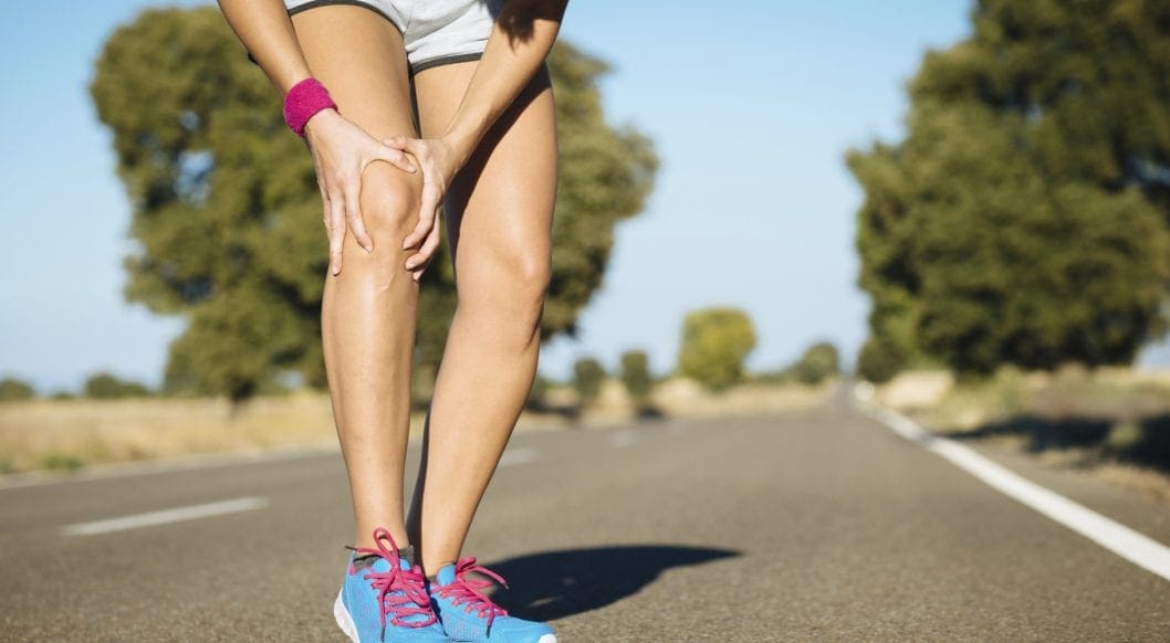
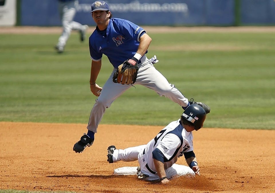
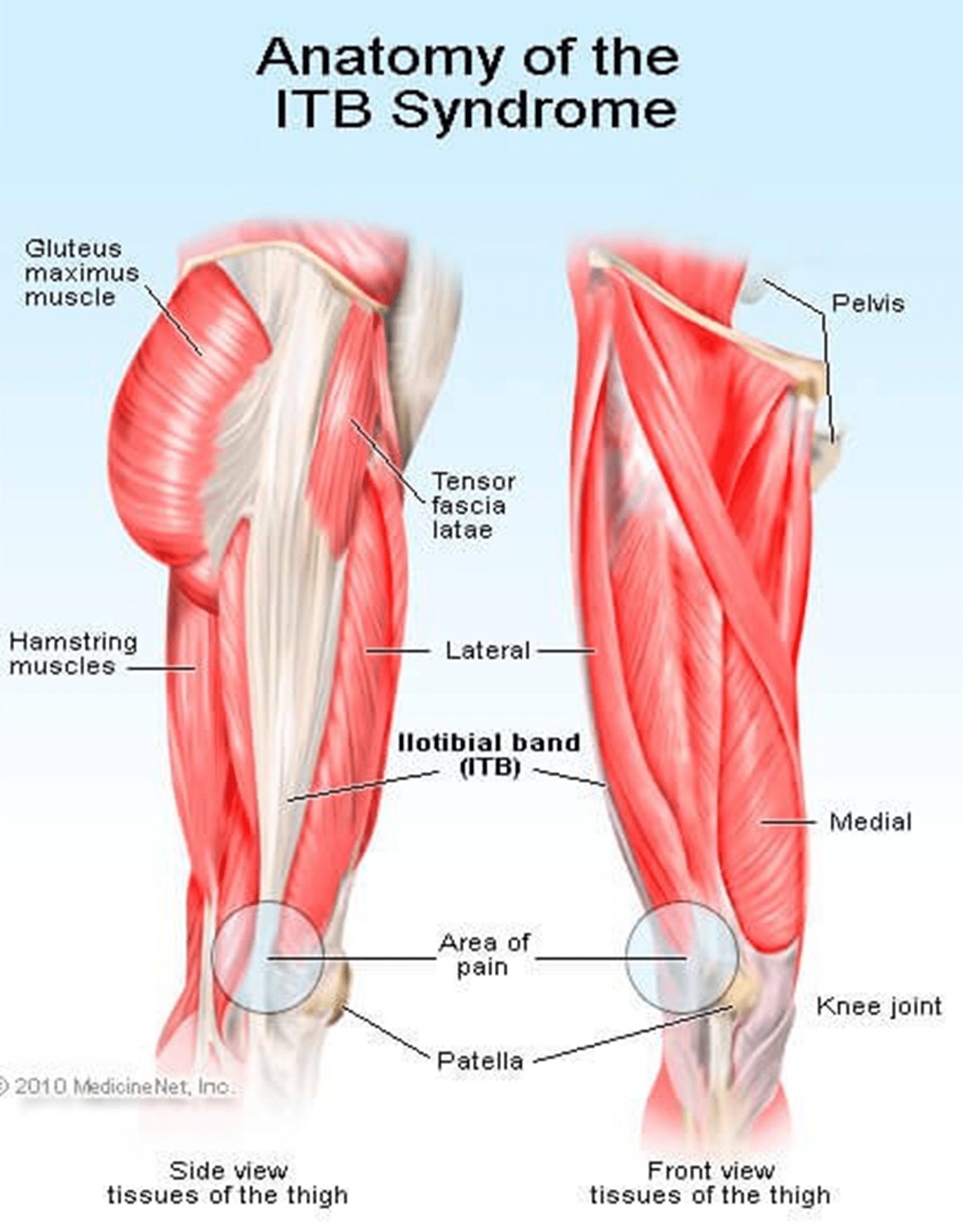
Signs Of IT Band Syndrome

Knee-Hurts-768x511If you are a runner, you will be able to distinguish ITBS by:

  • A swelling
  • A cracking feeling when stretching the knee
  • A feeling of burning, stinging and aching on the outer side of the knee that might migrate to the thigh. You will notice these discomforts especially, on your second half of the run.
  • Bending the knee at 45 degrees causes severe external knee pain

Criteria You Should Follow When Selecting The Best Running Shoes for ITBS

?There are various things that you should always consider when buying running shoes. Since most runners experience knee pain, it is wise to look for shoes that will help alleviate this pain without slowing them down. Below are some of the features to look out for in running shoes:

Stability/ Support

Since it is common to have knee pains due to lack of motion control and lack of stability, it is good to choose shoes that will offer you the support you need while running. If your running shoes don’t have any stability, you will end up stressing out your knee, which will result in pain and discomfort while running.

Fit

running shoes running2If you want to do away with pain, you might consider looking for a fit pair of shoes as they will reduce any pain, causing issues in the long run. Pay attention to small specifics like shoes that offer enough heel space, sufficient toe box room, and enough space for wide feet. Your toes should be able to move freely without being constricted.

If your foot cannot move freely and the toes are restricted from spreading, it could lead to painful issues in your feet, legs, and knees.

Motion control footwear is not the whole solution; you need to ensure your feet can still function naturally as they are supposed to.

Comfort

No one wants to wear uncomfortable shoes! Each of these selected best shoes come with upper and underfoot comforts to ensure you get to enjoy your run.

Most of these shoes are made with DNA technology, Gel cushioning, and REVlite midsole for ultimate comfort.

Durability

Your running shoes should run their course without falling apart as this will cause you pain in the long-run. If they promise to offer you support, they should do just that and not start peeling off and tearing when you are on the run.

The ??below 5 shoes have passed the durability test to ensure they give you maximum performance.

Breathability

Although this has nothing to do with knees, it is paramount that your running shoes have enough breathing space to avoid accumulating excess moisture, which might bring discomfort and other feet related problems.

There is no magical cure for knee pain and you should always know the root cause. This way, you will be able to come up with the best solution of minimizing or even eliminating the pain entirely. Although there are various causes of knee pain, this article is focusing on ITB syndrome which happens to be one of the causes.

Reviews Of The Top 5 Shoes

These shoes have been selected with the runner’s welfare in mind. They will help deal with the ITBS, which is a problem for most of them. Since one way of dealing with this condition is getting good running shoes, here is a review of such products.

Asics Gel Kayano 23

running shoes 41eQ53NInwL._SL250_This upgraded version is lightweight to help with any knee problems. It offers you comfort through cushioning that help absorb shock as you run as well as other features like grip, fit, and durability. The shoe has an added outer sole to ensure it lasts you as long as possible.

PROS

  • ?Gel cushioning will act as a shock absorber for more comfort
  • ?Has superb breathability feature
  • ?Is ideal for overpronation and knee pain
  • ?The outsole’s traction will offer the intended support on various surfaces

CONS

  • ?It is a bit pricey

New Balance 890v5

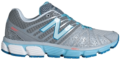
It tops the list of 5 best running shoes. Also, it has remained the first choice for most runners with knee pain issues. This pair offers all the above functionalities too, making it your best choice.

PROS

  • ?It comes with one of a kind breathability and fit due to its great FantomFit design
  • ?Its smooth upper construction will ensure no irritation occurs
  • ?The REVlite midsole will give you much needed cushioning

CONS

  • ?It has a narrow toe box and might not fit a person with a wide foot

?Puma Faas 600 V3

running shoes 41alXU8oEPL._SL250_
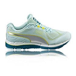
Puma models have never disappointed, and this one is no exception. Puma Faas 600 is the solution to your knee pain. It is also an affordable option for the short-handed.

PROS

  • ?Great breathability
  • ?Comes at a reasonable price
  • ?It’s lacing system and fit offers you a secure and comfortable run
  • ?It is designed to fit perfectly

CONS

  • ?There have been reported concerns about the outsole’s durability

New Balance 1080v7

running shoes 51PjsjoRrgL._SL250_
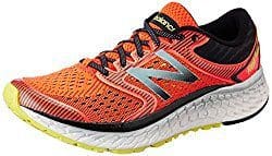
This is another great choice on the list. It is one of the New Balance Fresh Foam Series. Its midsole offers you the required support coupled with comfort to eliminate knee pains.

PROS

  • ?Very durable
  • ?Enough breathability for long runs
  • ?Good amount of cushioning and support from the Fresh Foam midsole
  • ?It fits like a sock giving you a confident use

CONS

  • ?The upper design is not seamless
  • ?Can be stiff

Saucony Hurricane 16

running shoes 41onfkrTORL._SL250_
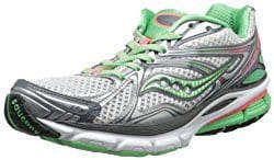
This is the 16th edition of the Saucony Hurricane, which offers a combination of steadiness and protection. Those with knee pain have agreed with the stability offered by this shoe. It is also cushioned to help you go for long runs without any pain or injury. It is perfect for heavy runners and those who are out of shape due to inactivity.

PROS

  • ?Superb stability
  • ?Lightweight rubber offers protection and cushioning
  • ?Great ground contact
  • ?Reflective parts allow you to have a safe run
  • ?Comes with Sauc-Fit Technology that enhances its comfortability

CONS

  • ?It is a bit narrow
  • ?Limited colors to choose from
  • ?Might be heavy for fast runners

If you are a long-distance runner, it is good to know that your shoes cushioning will wear out quite easily and you might be tempted to continue using them since they look good on the outside. This is a big mistake. The following will help you prevent any more ITBS recurrences:

  • Replace running shoes frequently to avoid wearing those with worn out inner cushioning
  • Always give your shoes time to rest so that the cushioning can get restored; it would be wise to have two pairs of running shoes.

Although shoes can offer you relief from ITBS, it is better to look out for other ways of helping you cope with or eliminate the pain entirely. Also, know what triggers the problem and avoid it at all costs.

These shoes have been tried and tested and found to offer support and help in managing the iliotibial band syndrome. Asics takes the lead on these best shoes. It comes with gel cushioning that will offer you the best shock absorption and maximum comfort as seen above. Its sole is also made to help you tackle any terrain and you can be assured that your knees will thank you later. The only drawback is the price, which is on the upper-side. However, always remember that cheap is expensive.

If you are an active person or an athlete suffering from ITBS, go ahead and get yourself a pair of these shoes as per your preference and choice.

in running

Zoey Miller

Zoey Miller

Hey there, I’m Zoey, founder and the main editor of The Babble Out. I know nobody’s life is smooth as they wish, and it�s the same with mine. I had some terrible news a few years ago and running was the way I got through these issues. This has given me enough motivation to create this blog, so that I can give you a helping hand for as many daily problems as I can. If you are curious why “babble out” is the? name of the blog, then check the “About” page and find out more about me.


read more button

Ergonomics: Office And Workplace

Ergonomics: Office And Workplace

Ergonomics in the work place. Back pain is one of the most frequent work-related injuries and is often brought on by ordinary work activities like sitting in an office chair or heavy lifting.� the study of the workplace as it relates to the worker – helps prevent back pain and back injury and help maintain a healthy back.

The objective of an ergonomics program in business is to adapt the workplace to a specific worker, determined by the job description, required tasks, and physical make up of the employee performing these tasks.

  • Non-accidental injury, where pain occurs as a result of normal activities and needs of the task. This might occur from sitting in an office chair or standing for too long in one position.
  • Accidental injury results when an unexpected event triggers injury during the task. A load that changes as it is being lifted, and fall and a slip or hitting one’s head on a cabinet door or slips are typical examples. These injuries can jolt other joints, back, and the neck with consequent muscle strain or tearing of soft tissue at the back.

Occupations which are physically demanding and require repetitive lifting (such as in nursing or heavy industry) are at greatest risk for both non-accidental and accidental spine injury. For instance, a number of health workers have problems because patients are of weight and different stature with needs. Often, the patients need help changing position, rising from a chair and walking. Similarly, the physical effort needed to release a trapped individual or save a life is unpredictable. The same problems occur in the building industry where consistencies of tasks are a challenge.

Office Chair Back Injuries

ergonomics work injury office chair el paso txIndividuals who sit most of the day, like those working in a computer while sitting in an office chair, are also at high risk for non-accidental spine injury. Office ergonomics, or computer ergonomics, can help minimize the risk such as the dangers associated with prolonged sitting in an office chair, and carpal tunnel syndrome, such as lower back pain, neck strain, and leg pain.

Office Chair: Ergonomics To Reduce Back Pain?

This guide summarize the use of ergonomic concepts, mechanical apparatus and decent body mechanics (biomechanics) that can contribute to reducing back injuries in the work environment for several jobs. Significantly, staying strong, physically fit and flexible improves the probability of preventing back injuries.

There are certain basic ergonomic tips which may help an employee avoid back pain or back injury:

  • Develop a job description based on the forces within a particular work environment, the time spent performing the task and the biomechanics (which define human moves and seated posture in an office chair) used in the task.
  • Use body posture as a tool which may be changed to fit the job demands with minimal stress on the muscles, ligaments, bones and joints.
  • Learn and use proper body mechanics to restrict extra mechanical stress in completing the job.
  • Maintain fitness and flexibility and create a reserve of strength.

Identifying Poor Posture And Risks

ergonomics work injury office el paso txMany potentially harmful situations that lead to back injury can be identified and avoided by following four basic rules of thumb:

Prolonged static posture is your enemy. The healthy body can only tolerate staying in one position for around 20 minutes. That is sitting at a movie theatre, in a desk in an office chair, or on an airplane becomes uncomfortable after a short time. Standing in one area, such as standing on a floor at an assembly line tends to cause back pain. Holding the same position gradually reduces elasticity in the soft tissues (muscles, ligaments, and tendons in the back). Stress builds up and causes discomfort and/or leg discomfort back.

The remedy is simple. Whether you are sitting in an office chair or standing in a line, change positions frequently. Just move. Stand or sit, stretch, have a short walk. After returning to the standing or sitting posture, use an alternate posture for only a couple minutes and some.

Frequent or repetitive stretching to the end range of motion or embarrassing, angled positions can liquefy the joints. Unlike jobs that need seating in an office chair, jobs that require motion can cause discomfort. Such tasks involve lifting overhead lifting from the floor, moving loads, or utilizing force or twisting while managing material and which signal back injuries might be on the way.

Heavy loads offer greater risk. It is important to have the proper tools or get help if the job requires moving objects.

Fatigue�from sitting in an office chair, from work or from insomnia can make people move more awkwardly. If one is overtired or feels fatigued, it is advisable to avoid lifting heavy objects alone or quickly.

ergonomics work injury office chair el paso txThese ergonomic rules of thumb will help the worker and their backs. Otherwise the worker is at risk of sustaining or aggravating a back injury.

Mastodon