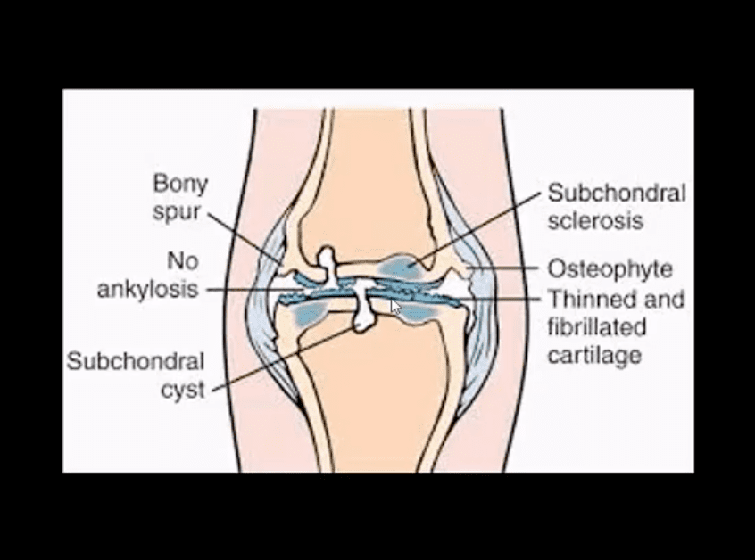
Pathology: da disease of the articular cartilage. Continuing mechanical stimulation follows by an initial increase in water and cartilage thickness. Gradual loss of proteoglycans and ground substance. Fissuring/splitting. Chondrocytes are damaged and release enzymes into the joint. Cystic progression and further cartilage loss. Subchondral bone is denuded and exposed to mechanical stresses. It becomes hypervascular forming osteophytes. Subchondral cysts and bone thickening/sclerosis develop.
Imaging plays a crucial role in Dx/grading and management
Clinically: pain on walking/rest, crepitus, swelling d/t synovitis, locking/catching d/t osseocartilaginous fragments and gradual functional loss. Knee OA typically presents as mono and oligoarthritis. DDx: morning pain/stiffness is >30-min DDx from inflammatory arthritis
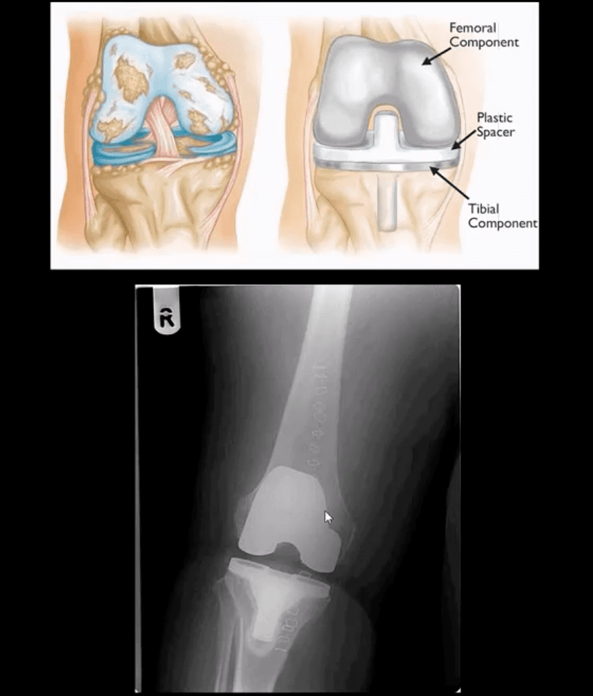
Treatment: in mild to moderate cases-conservative care. Severe OA-total knee arthroplasty
Grade 4: severe JSN, large osteophytes, marked subchondral sclerosis and definite bony deformity
Typical report language will state:
Minor, mild, moderate or severe aka advanced arthrosis
Technique
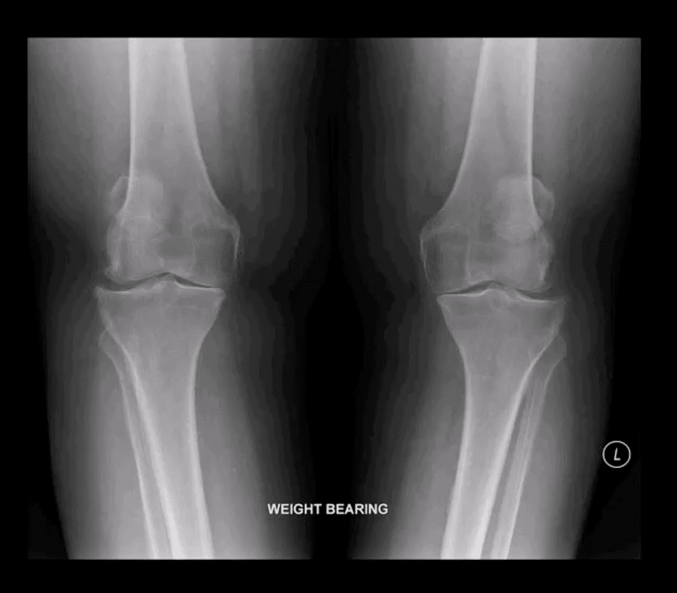
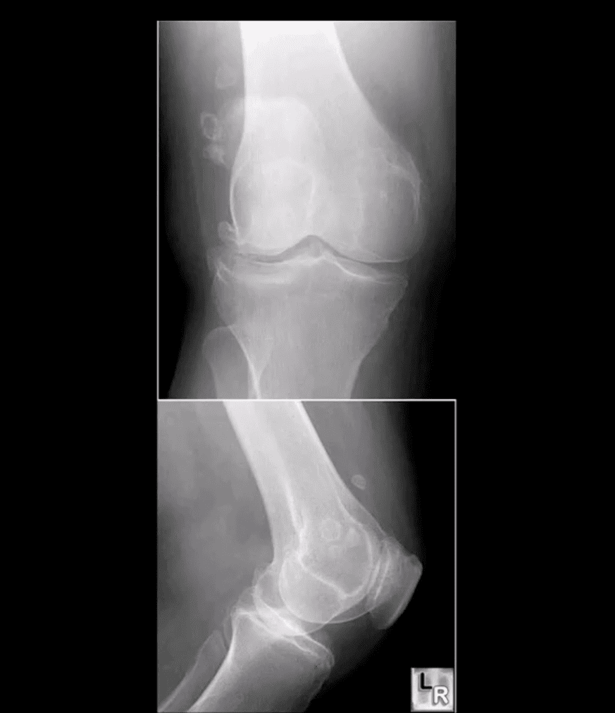
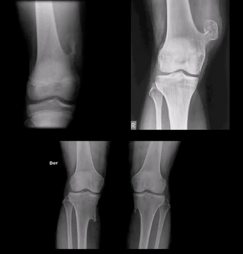
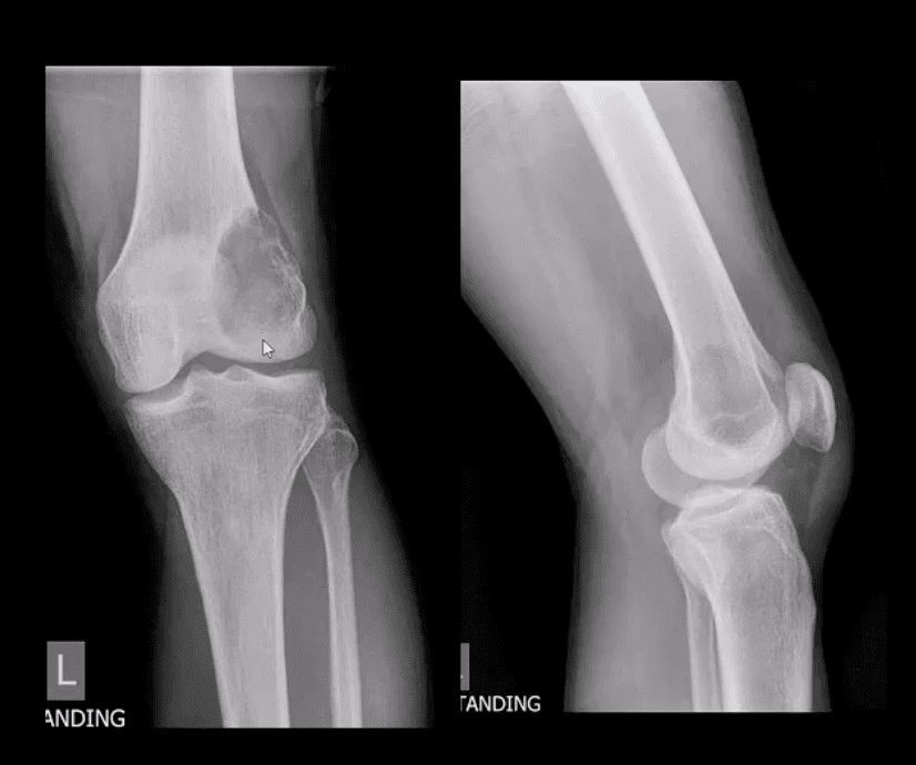
Radiography: AP weight-bearing knees: note severe JSN of the medial compartment more severely with lateral knee compartment. Osteophytes and marked genu varum deformity and bone deformation
Typically medial femorotibial compartment is affected early and more severely
The patellofemoral compartment is also affected and best visualized on the lateral and Sunrise views
Impressions: severe tri-compartmental knee arthrosis
Recommendations: referral to the orthopedic surgeon
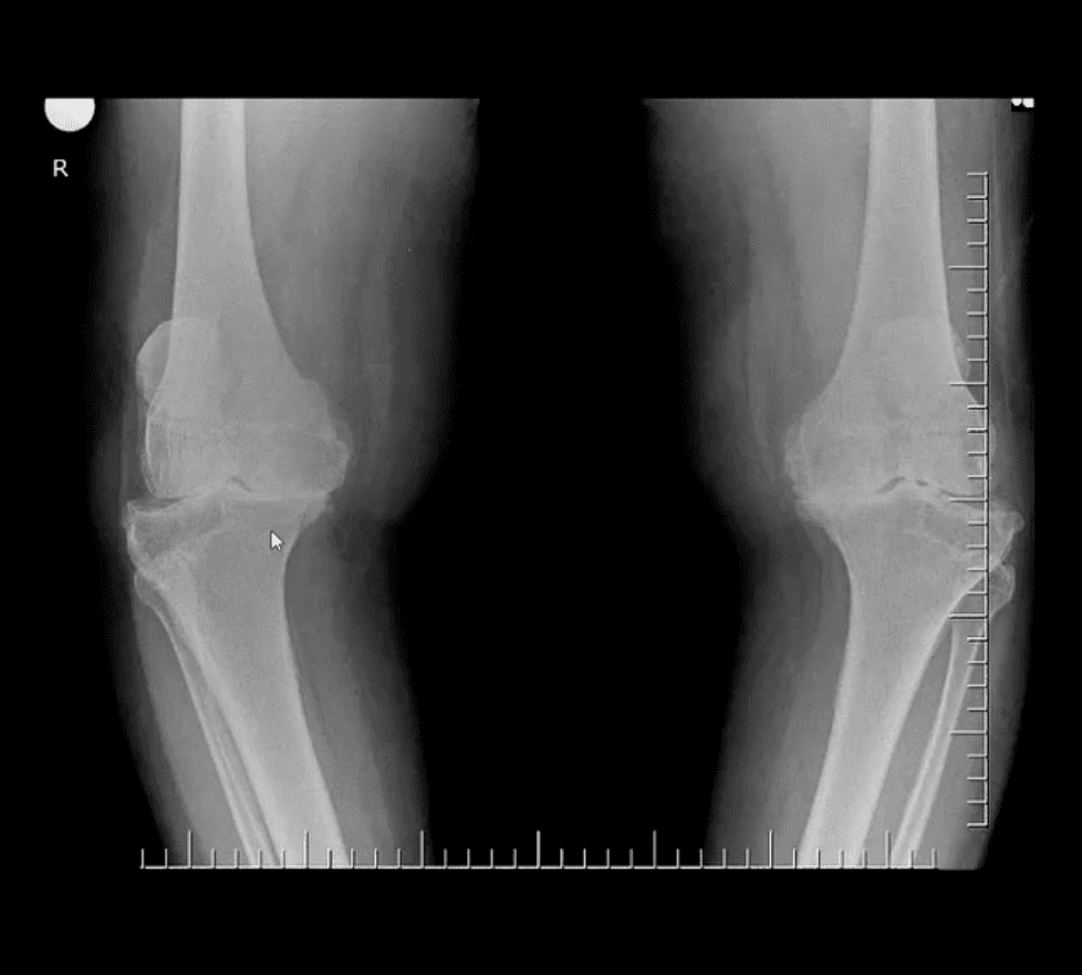
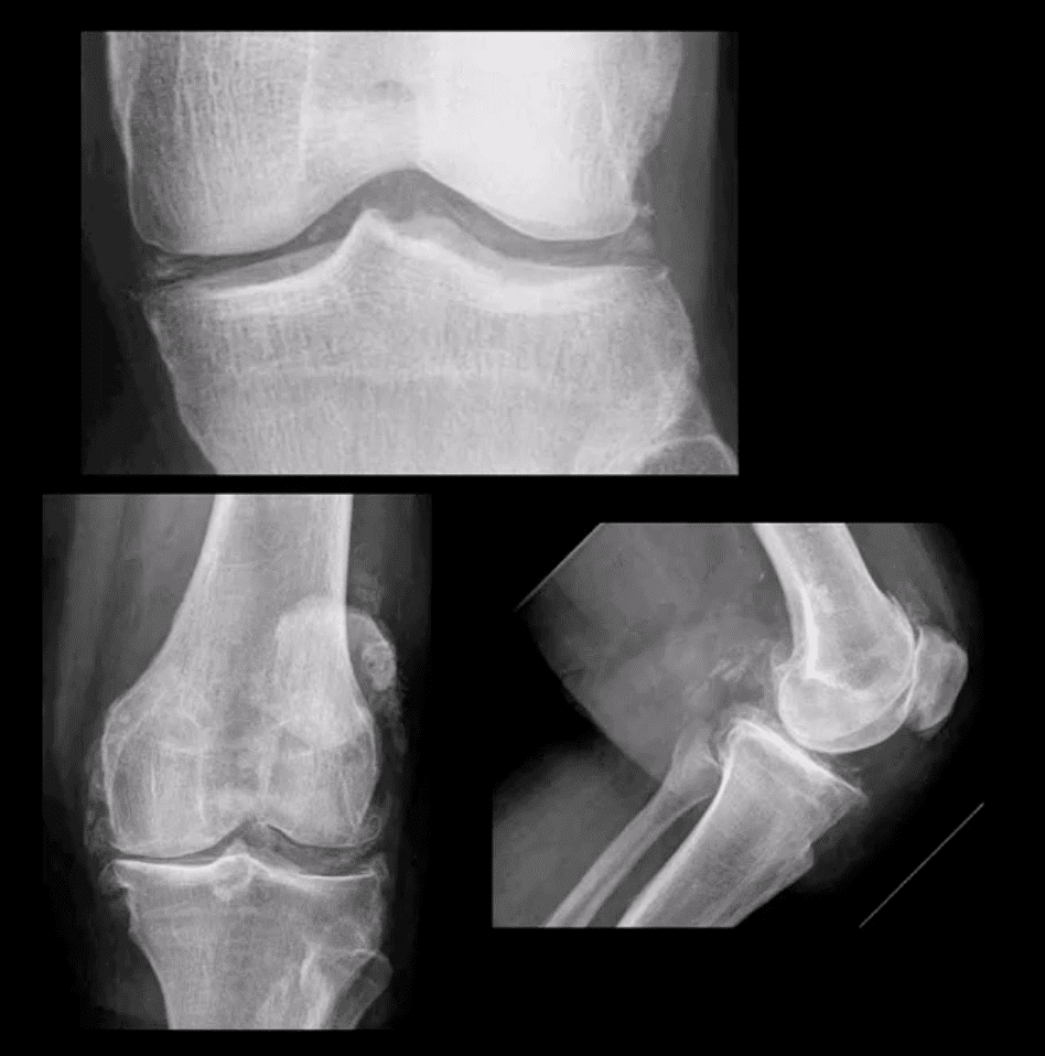
Moderate JSN
B/L AP weight-bearing view (above top image): Moderate JSN primarily of the medial femorotibial compartment. Osteophytosis, subchondral sclerosis and mild bone deformation (genu varum)
May present as asymptomatic chondrocalcinosis, CPPD arthropathy resembling DJD with pan predominance of large subchondral cysts. Often found as isolated PFJ DJD
Pseudogout with an acute attack of knee pain resembling gouty arthritis
Radiography is the 1st step and often reveals the Dx
Arthrocentesis with polarized microscopy may be helpful to DDx between CPPD and Gouty arthritis
Rheumatoid Arthritis
RA: an autoimmune systemic inflammatory disease that targets soft tissues of joints synovium, tendons/ligaments, bursae and extra-articular sites (e.g., eyes, lungs, cardiovascular system)
RA is the m/c inflammatory arthritis, 3% of women and 1% of men. Age: 30-50 F>M 3:1, but may develop at any age. True RA is uncommon in children and should not be confused with Juvenile Idiopathic Arthritis
RA most often affects small joints of the hands and feet as symmetrical arthritis (2nd 3rd MCP, 3rd PIPs, wrists & MTPs, sparing DIPs of fingers and toes)
Radiographically: RA presents with joint effusion leading to hyperemia and marginal erosions and periarticular osteoporosis. In the knee, the lateral compartment is affected more frequently leading to valgus deformity. Uniform aka concentric/symmetrical JSN affects all compartments and remains a key Dx clue
An absence of subchondral sclerosis and osteophytes. Popliteal cyst�(Baker’s cyst) may represent synovial pannus and inflammatory synovitis extending into the popliteal region that may rapture and extend into posterior leg compartment
N.B. Following initial RA joint destruction, it is not unusual to note superimposed 2nd OA
Radiography is the 1st step but early joint involvement may be undetectable by x-rays and can be helped by US and/or MRI.
Final Dx is based on Hx, clinical exam, labs, and radiology
Clinical pearls: patients with RA may present with a single knee being affected
Most patients are likely to have bilateral symmetrical hands/feet RA.
Cervical spine, particularly C1-2 is affected in 75-90% of cases throughout the course of the disease
N.B. Sudden exacerbation of joint pain in RA should not underestimate septic arthritis because patients with pre-existing RA are at higher risk of infectious arthritis. Joint aspiration may help with Dx.
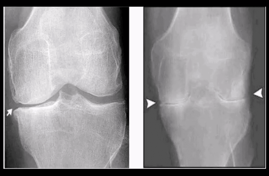
Radiographic DDx
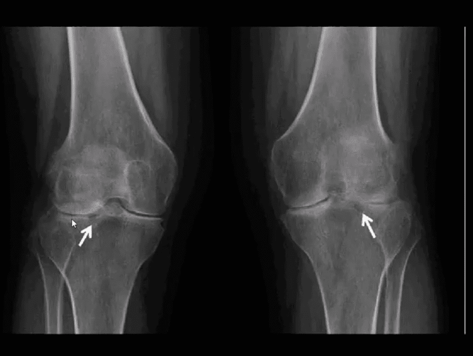
RA (above left) vs. OA (above right)
RA: concentric (uniform) joint space loss, lack of osteophytes and juxta-articular osteopenia.
Clinical Pearls: patients with RA may present radiographically with subchondral sclerosis d/t superimposed DJD. The latter feature should not be interpreted as OA but instead considered as secondary OA
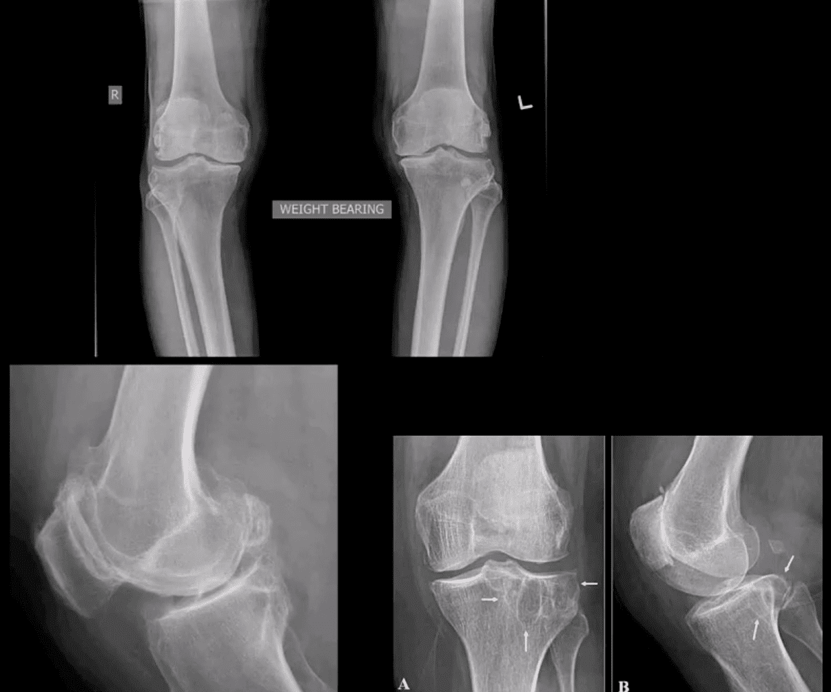
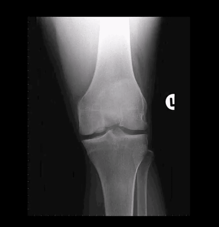
AP Knee Radiograph
Note marked uniform JSN, juxta-articular osteopenia and subchondral cystic changes
Clinical Pearls: subcortical cysts in RA will characteristically lack sclerotic rim noted in OA-associated subcortical cysts.
MRI Sensitivity
MRI is very sensitive and may aid during early Dx of RA.
T2 fat-sat or STIR and T1 + C gad contrast fat-suppressed sequences may be included
MRI Dx of RA: synovial inflammation/effusion, synovial hyperplasia, and pannus formation decreased cartilage thickness, subchondral cysts, and bone erosions
MRI is very sensitive to reveal juxt-articular bone marrow edema, a precursor to erosions
Intra-articular fibrinoid fragments known as “Rice bodies” are characteristic MR sign of RA
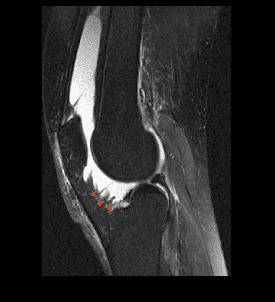
Note: T2 fat-sat sagittal MRI revealing large inflammatory joint effusion and pannus synovial proliferation (above arrowheads). No evidence of radiographic or MRI bone erosions present. Dx: RA
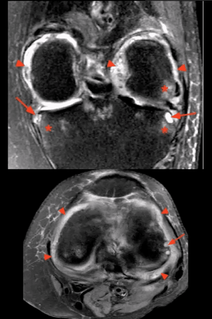
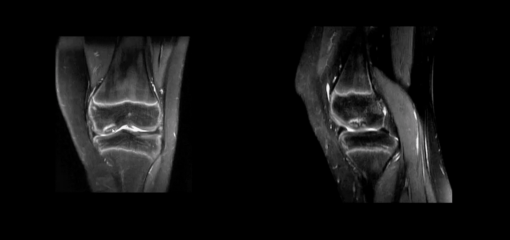
STIR MR Slices
Note: STIR MR slices in the axial (above bottom image) and coronal planes (above top image) demonstrate extensive synovitis/effusion (above arrowheads) and multiple erosions in the medial and lateral tibial plateau (above arrows)
Additionally, scattered patchy areas of bone marrow edema are noted (above asterisks) such marrow edema changes are indicative and predictive of future osseous erosions.
Additional features: note thinning and destruction of joint cartilage
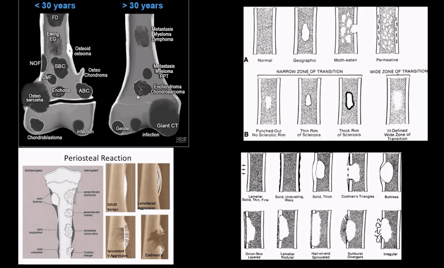
Bone neoplasms and tumor-like conditions affecting the knee can be benign or malignant. Age at Dx is crucial for DDx
In patients <40: Benign bone neoplasms: Osteochondroma, Enchondroma are relatively frequent
Fibrous cortical defect (FCD) & Non-ossifying fibroma (NOF) are particularly frequent in children
Giant cell tumor (GCT) is the m/c benign neoplasm of the knee in patients between 20-40 years of age
Malignant bone neoplasms in <40: m/c Osteosarcoma and 2nd m/c Ewing sarcoma
In patients >40: malignant neoplasms: m/c are secondaries d/t bone metastasis. Primary bone malignancy:�the m/c
Multiple Myeloma (MM). Less frequently:�a 2nd�peak of Osteosarcoma (post-radiation or Paget�s), Fibrosarcoma or Malignant�Fibrous�Histiocytoma�(MFH) of bone.
Clinically: knee pain, pathological fracture
Some tumor-like conditions like FCD/Non-ossifying fibroma are asymptomatic and may regress spontaneously. Occasionally NOF may present with pathologic fracture. N.B. any knee/bone pain in a child/adolescents should be�treated with clinical suspicion and adequately investigated.
Imaging: 1st step: radiography
MRI with T1+C is crucial for lesion characterization/regional extent, staging and pre-operative planning. CT may�help with pathologic Fxs detection. If malignant bone neoplasms considered, CXR/CT, PET-CT to investigate�metastatic spread and staging are important
Imaging Approach Bone Neoplasms
Approach to imaging Dx of bone neoplasms includes age, bone location (epiphysis vs. metaphysis vs. diaphysis), zone of transition surrounding the lesion, periosteal response, type of matrix, permeating or moth-eaten destruction vs. sclerotic, ground-glass, osteoid, cartilaginous matrix, soft tissue invasion, etc.
Key x-radiography features to DDx benign vs. malignant bone neoplasm:
Zone of transition: lesion is geographic with a narrow zone of transition vs. ill-defined wide zone of transition suggesting aggressive bone resorption
What type of bone destruction occurred: soap-bubbly appearance vs. osteolytic vs. osteosclerotic changes
Is there a round-glass matrix? Is there a well-defined rim of the sclerotic border with septations potentially suggesting slow growth and encapsulation like most benign processes.
Periosteal proliferation: solid vs. aggressive spiculated/sunburst/hair-on-end with local soft tissue invasion and Codman triangle (study next slide)
FCD & NOF
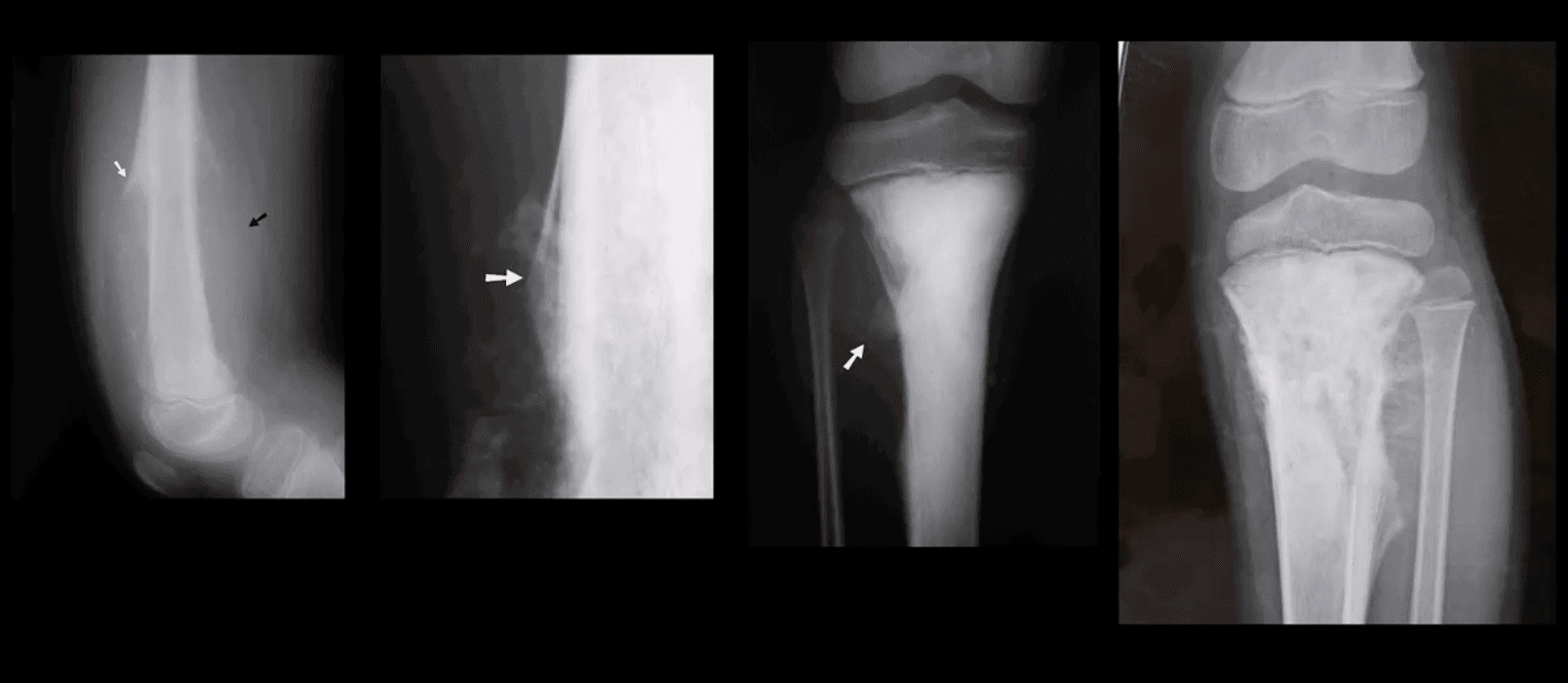
FCD & NOF or more appropriately Fibroxanthoma of the bone are benign bone processes that m/c seen in children. DDx based on the size with FCD presenting as <3-cm and NOF >3cm lesion composed of a fibrous heterogeneous matrix. FCD are asymptomatic and may regress in many cases. Some may progress to NOF. Location: identified in the knee region as an eccentric cortical based lesion.
FCD must be DDx from an avulsive irregularity d/t repeated stress along Linea aspera by extensors muscles
Dx: radiography
Management: leave-me-alone lesion. Occasionally NOF may progress and lead to pathologic fracture requiring orthopedic consult
Osteochondroma
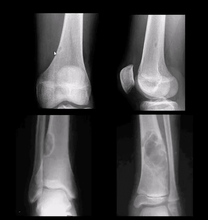
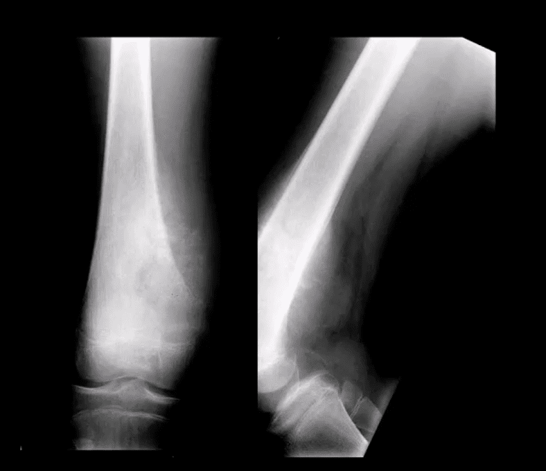
Osteochondroma: m/c benign bone neoplasm. Knee is the m/c location. Contains all bone elements with a cartilaginous cap. Presented as pedunculated or sessile bone exostosis pointing away from the joint.
1% malignant degeneration to chondrosarcoma if solitary lesion and 10-15% in cases of HME
Other complications: fracture (top left image) pseudoaneurysm of the Popliteal artery, adventitious bursa formation
Hereditary Multiple Exostosis (HME)– autosomal dominant process. Presents with multiple osteochondromas (sessile-type dominates). May lead to limb deformities (Madelung deformity, coxa valga) reactive ST pressure, malignant degeneration
Dx: radiography, MRI helps to Dx malignant degeneration to chondrosarcoma by changes in size and activity of cartilaginous cap (>2-cm in adults may manifest malignant degeneration). MRI will also help with Dx of regional complications
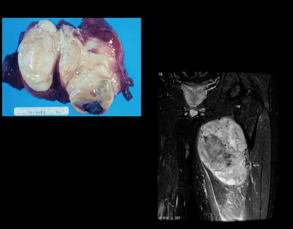
HME & Knee Pain
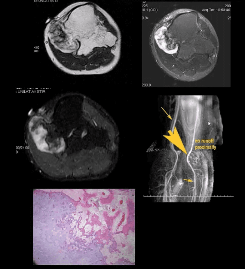
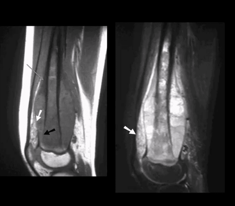
37-y.o male with HME and knee pain. Axial T1, T2 and STIR MRI slices at the popliteal region. Large cartilaginous cap and possible compression of the popliteal artery by osteochondroma. MRA was performed to evaluate popliteal A. pseudoaneurysm (large arrow). Pathology specimen obtained from the cartilaginous cap showed increased cellularity suggestive of malignant degeneration. Operative care was planned
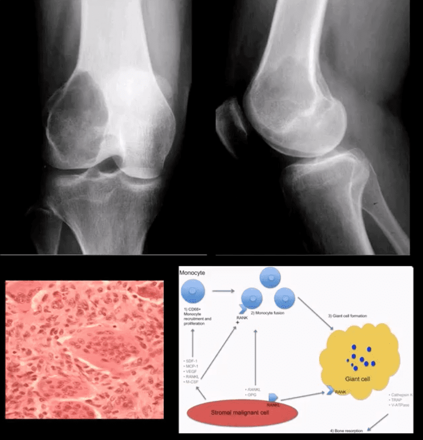
Giant Cell Tumor (GCT) aka Osteoclastoma
GCT- is a relatively common primary benign bone neoplasm. Age 25-40. M>F slightly.
GCT is the M/C benign sacral tumor. In 50% of cases, GCT occurs about the knee.
GCT is histologically benign, but lung Mets may develop esp. if in distal radius and hands, often termed Malignant GCT
<1% unresponsive/recurring GCTs may undergo malignant transformation to high-grade bone sarcoma
Pathology: histologically composed of osteoclasts-multinucleated giant cells with stromal cells derived from precursors monocyte-macrophage type. Produces cytokines and osteolytic enzymes. GCT may contain blood and associated with secondary Aneurysmal Bone Cyst (ABC)
Clinically: knee pain unresponsive to conservative care. Pathologic Fx may occur
Imaging: always begins with radiography followed by MRI and surgical biopsy that are crucial to Dx.
Rx: operative with curettage and cementing, a surgical appliance may be used if pathological fx present and cortical breach. In more severe cases other options available
Radiologic-Pathologic Dx
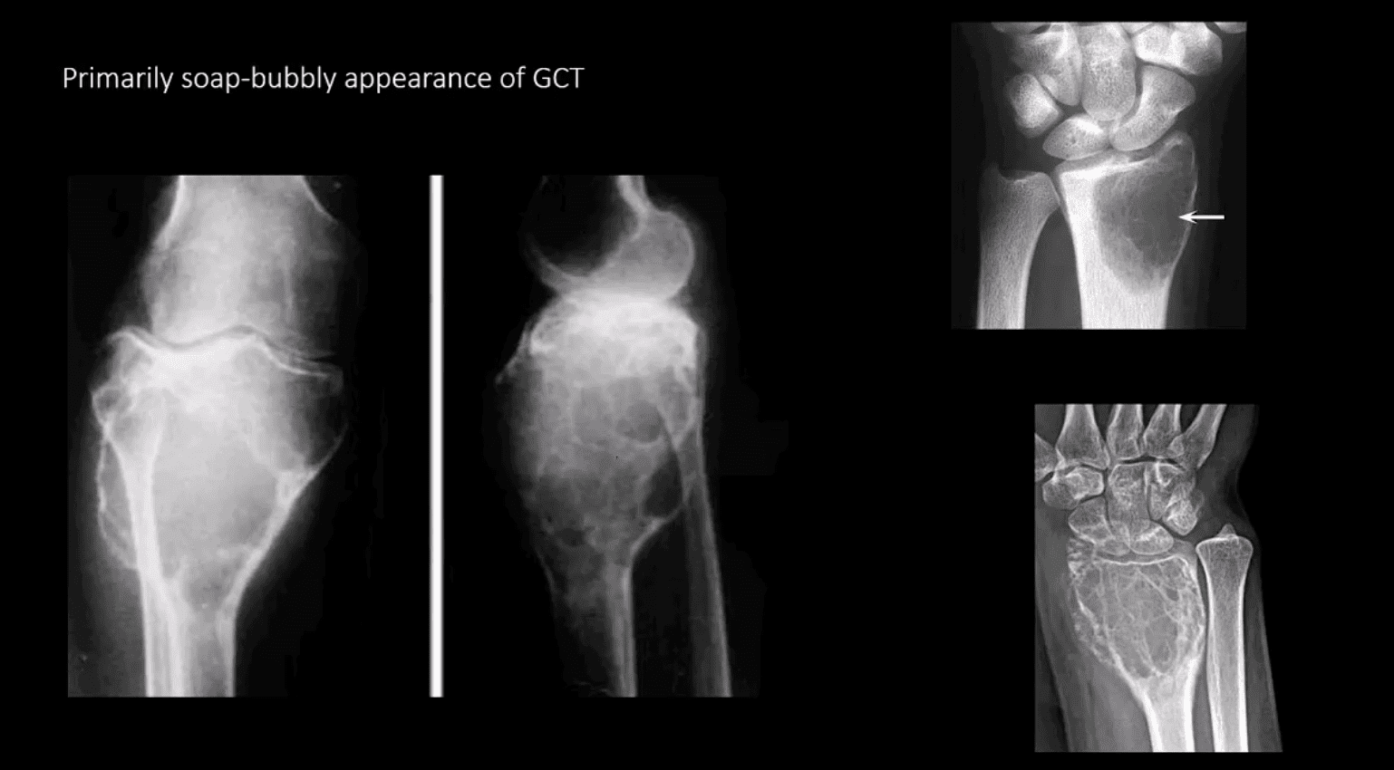
Radiologic-pathologic Dx: osteolytic and soap-bubbly lesion typically involving metaphysis and into epiphysis (classic key feature) with subarticular extension. Zone of transition is generally narrow but occasionally in aggressive lesions wide zone of transition may be seen.
MRI: low T1, highT2/STIR, characteristic fluid-fluid levels noted that are present in GCT and ABC. Histology is crucial to Dx.
DDx: ABC, Brown cell tumor of HPT (osteoclastoma), Telangiectatic Osteosarcoma
Radiological rule: if the physeal growth plate is present Dx of GCT is taken off the list in favor of chondroblastoma and vice versa.
Primarily Soap-Bubbly Appearance of GCT
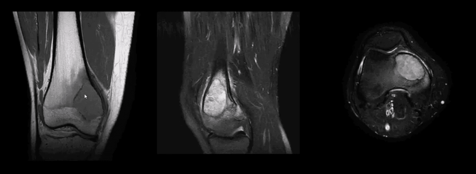
Coronal, Fat-Sat Sagittal & Axial MRI Slices of GCT
T1 coronal, T2 fat-sat sagittal and T2 axial MRI slices of GCT. Typically: low T1, highT2/STIR and fluid-fluid levels
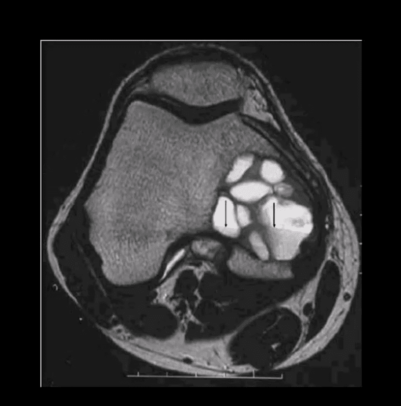
Characteristic MRI Appearance of GCT
Fluid-fluid levels d/t different composition of blood degradation products
Important DDx: ABC
Malignant Neoplasms About the Knee
In children and very young adults, m/c primary malignant neoplasm is central aka intramedullary (osteogenic) osteosarcoma (OSA). Second peak of OS: >70 y.o d/t Paget�s (1%) and/or post radiation OSA.
The knee is the m/c location of OSA (distal femur, prox. Tibia)
A 2nd m/c malignant pediatric primary is Ewing sarcoma.
In adults >40 y.o. the m/c primary is Multiple Myeloma (MM) or Solitary Plasmacytoma
Overall m/c bone neoplasms in adults d/t bone Mets from lung, breast, prostate, renal cell, thyroid (discussed)
Dx: clinical and radiological with surgical biopsy
Imaging is crucial to Dx. 1st step x-radiography. MRI+ gad C is vital
CT scanning occasionally helps to evaluate pathological fracture
Central (Intramedullary) Osteosarcoma (OSA)
m/c age: 10-20. m/c location: knee, males>females. Increased risk in some
congenital syndromes and mutation of the retinoblastoma gene: Rothmund-Thompson AR syndrome.
Early Dx is important d/t 10-20% present with Lung Mets at Dx. Prognosis depends on stages. Early stages with local bone invasion and no
mets 76% of survival.
Rx: limb salvage procedures preferred with 8-12 weeks of chemo, amputation if encased neurovascular tissue, path Fx, etc.
Imaging: radiography and MRI.
Clinically: bone pain, Inc. Alkaline Phosphatase
Chest CT if lung Mets considered
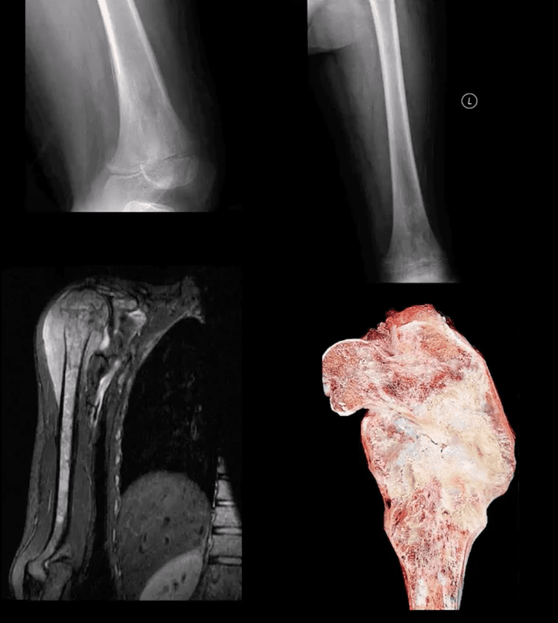
Classic Rad Features of OSA
Osteoid forming a sclerotic mass with aggressive hair-on-end/speculated/sun-burst periosteal reaction, Codman’s triangle and soft tissue invasion. Order MRI for staging and extent. Chest CT is crucial for Lung Mets dx.
MRI is Crucial for Dx/Staging
Note sagittal T1 (left) and STIR (right) MR slices: large mass extending from distal femoral metaphysis to remaining shaft. A low signal on T1 and high on STIR d/t marrow invasion with edema, hemorrhaging and tumor invasion. Local ST invasion seen (white arrows). Periosteal lifting and Codman�s triangle (green arrow) are additional signs of aggressive neoplasm.
Note an interesting feature that the epiphysis is spared d/t physeal plate serving temporarily as an additional barrier to the tumor spread.
Ewing Sarcoma
Ewing sarcoma: age: 2-20, uncommon in black patients. 2nd m/c highly malignant bone neoplasm in children that typically arises from medullary cavity (Round cell tumors). Key symptom: bone pain that may mimic infection (ESR/CRP/WBC) Considered PNET Key Rad Dx: aggressive moth-eaten/permeative lucent lesions in the shaft of long bones with sizeable soft tissue invasion/typical onion skin periostitis. May produce saucerisation May affect flat bones. May appear as sclerotic in 33%. Early lung Mets (25-30%) bone-to-bone Mets Poor prognosis if delayed Dx. Imaging steps: 1st step x-rad, MRI is v. important followed by a biopsy. CXR/CT PET-CT Rx: combined rad-chemo, operative.
Note aggressive expansile osteolytic lesion in the distal femur metaphysis into epiphysis. No periosteal reaction present. Following further work up with abdominal and chest CT scanning, Dx of Renal cell carcinoma was established
Distal Mets into lower extremity are more common with lung, renal cell, thyroid and breast CA.
Renal cell and Thyroid will typically present with aggressive osteolytic expansile mass aka �blowout Mets.�
In general, imaging approach should consist of Radiographic knee series, followed by MRI if x-rays are unrewarding
Tc99 Bone scintigraphy is the modality of choice to evaluate metastatic bone disease
Soft Tissue Neoplasms About the Knee
Malignant fibrous histiocytoma (MFH) reclassified as Pleomorphic Undifferentiated Sarcoma (PUS) is the m/c S.T. sarcoma. MFH is aggressive biologically with poor prognosis M>F (1.2:1) 30-80 with a peak in a 6th decade. 25-40% of all adults sarcomas m/c extremities. Retroperitoneum next (worst prognosis d/t late Dx and large growth w/o symptoms) Clinically: painful, hard mass typically about the knee or thigh. Histology: poorly differentiated/undifferentiated malignant fibroblasts, myofibroblasts, and other mesenchymal cells Imaging: MRI is the modality of choice with T1, T2, T1+C. Typically appears as an aggressive heterogeneous mass intermediate to low signal on T1 and high signal on T2 with areas of necrosis and enhancement on T1+C. May appear misleadingly encapsulated w/o true capsule Management: operative with radiation and chemotherapy. Tumor depth is crucial for prognosis. 80% 5-year survival if <5cm deep in ST and 50% if >5-cm deep in ST.
Synovial Sarcoma
Synovial sarcoma: common malignant ST neoplasm esp. in younger patients or older children/adolescents. M/C found in knee area Clinically: can present slowly as a palpable mass in the extremity often ignored d/t slow growth Imaging is the key: radiography may reveal ST. density/mass. Some synovial sarcomas may show calcification and mistaken for Myositis Ossificanse or heterotopic bone formation MRI with T1, T2 and T1+C are Dx modality of choice. Other modalities: US, CT are non-specific DDx: MFH Management: operative, chemo-radiation Prognosis: variable depending on size, invasion, metastasis
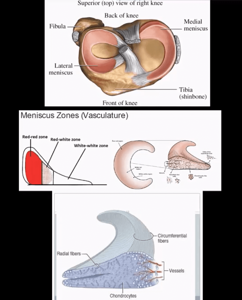
Types, location, and stability of tears are v. important during MRI Dx
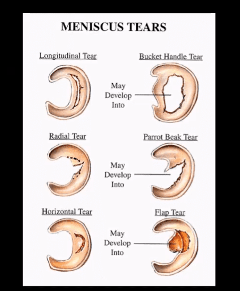
Vertical/longitudinal tears especially occur in acute ACL tears. Some longitudinal tears found at the periphery or “red zone” may heal
Bucket handle tear: longitudinal tear in the inner edge that is deep and vertical extending through the long axis and may displace into a notch
Oblique/flap/parrot-beak are complex tears
Radial tear at 90-degree to plateau
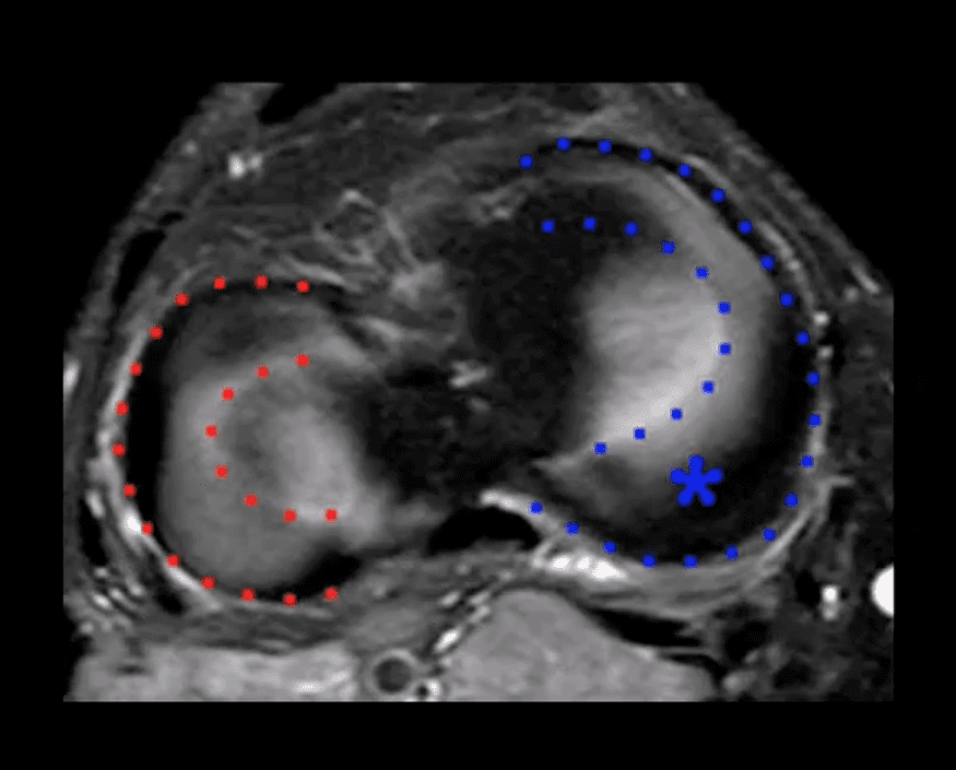
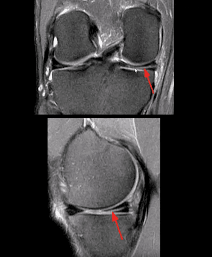
Axial T2
Axial T2 WI fat-sat and coronal STIR slices of the posterior horn of the medial meniscus.
Note a radial tear of the posterior horn of the medial meniscus near the meniscal root. This is potentially an unstable lesion requiring operative care
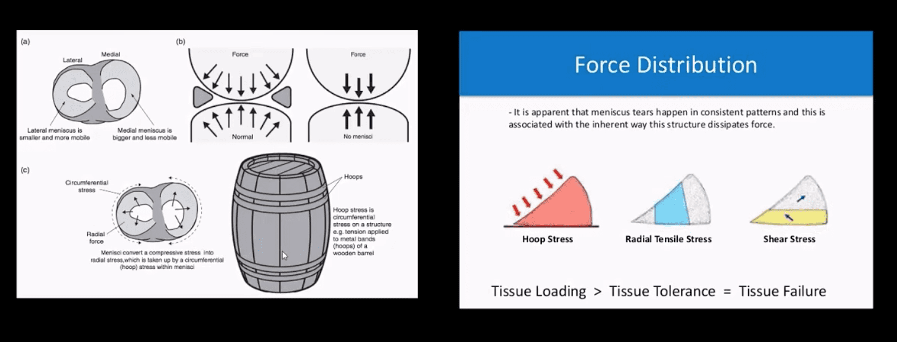
The meniscus, in this case, is unable to provide a “hoop-stress mechanism.”
MRI Slices Coronal & Sagittal
Fat-sat coronal and sagittal proton density MRI slices revealing horizontal (cleavage) tear that is more typical in the aged meniscus
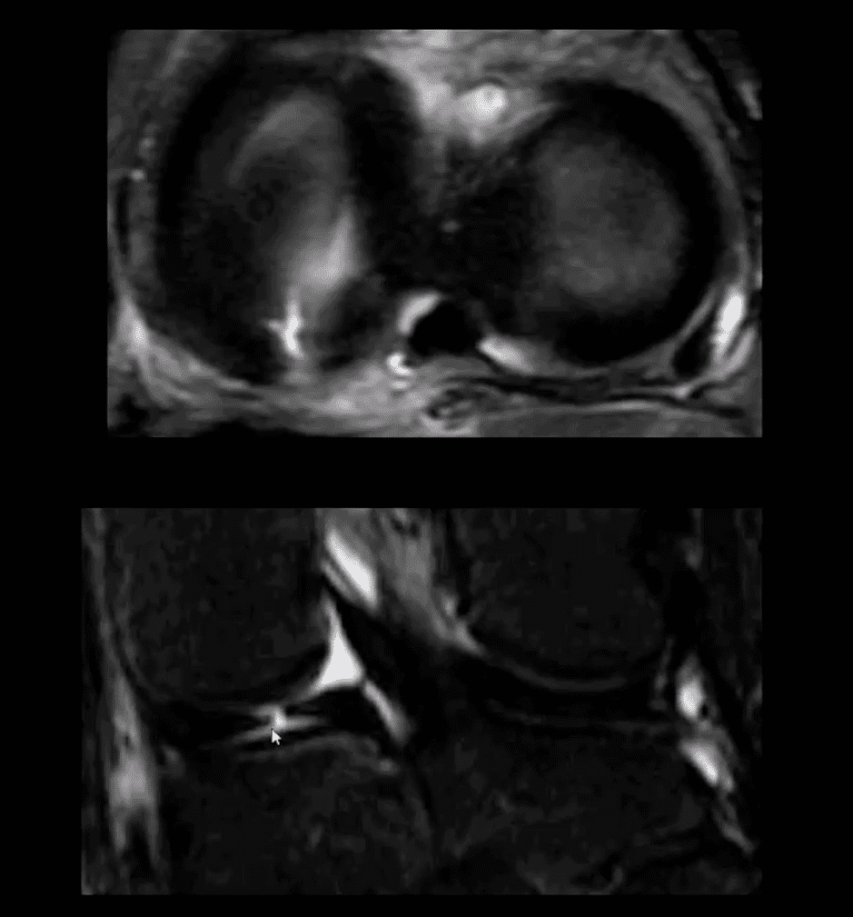
In some cases, when this tear does not contain a radial component, it may partially heal obviating the need for operative care
T2 w GRE Sagittal MRI Slice
Complex tear with a horizontal oblique and radial component.
This type of tear is very unstable and in most cases may need operative care
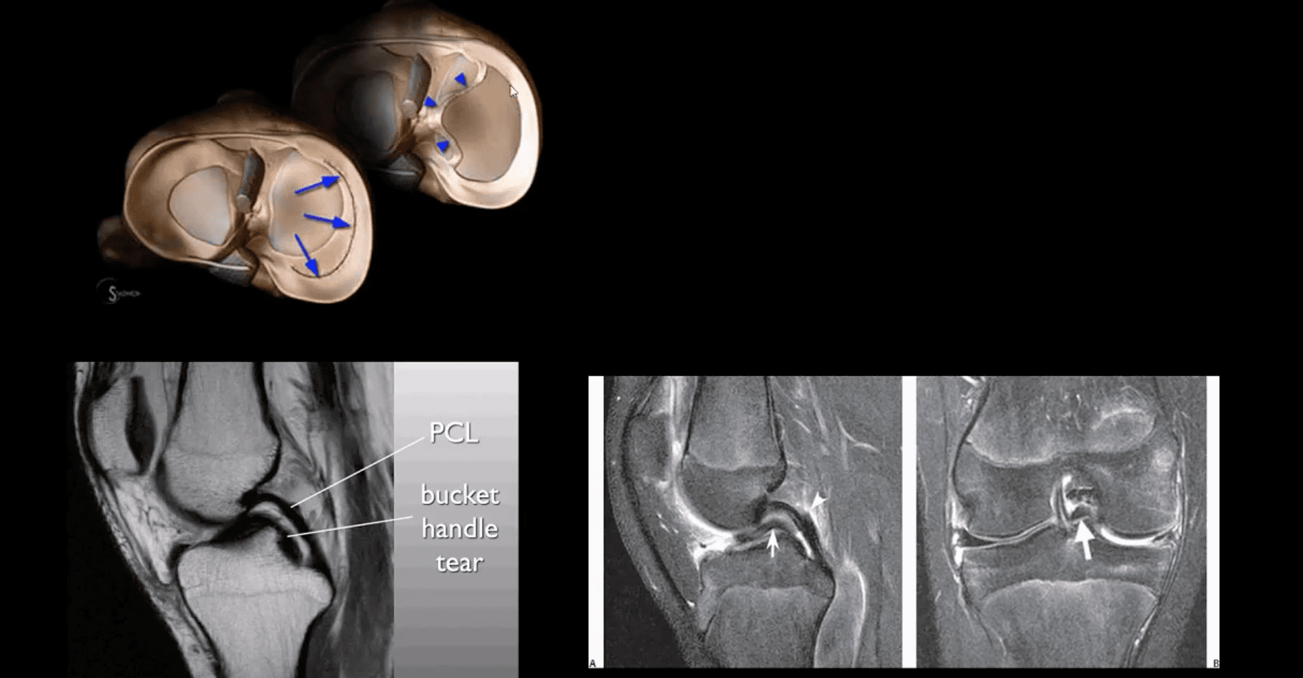
Bucket Handle Tear
Bucket handle tear are m/c in the medial meniscus esp. with acute ACL and MCL tear
MRI signs; double PCL sign on sagittal slices
Absent “bow-tie” sign and others
Most cases require operative care
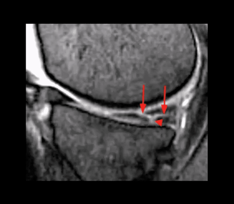
DDx From Meniscal Degeneration
Occasionally meniscal tears need to be DDx from meniscal degeneration which may also appear bright (high signal) on fluid-sensitive MRI
The simplest rule is that if there is a true meniscal tear aka Grade 3 lesion, it always reaches/extends to the tibial plateau surface
The Role of MSK Ultrasound (US) in Knee Examination
MSK US of the knee permits high resolution and dynamic imaging of primarily superficial anatomy (tendons, bursae, capsular ligaments)
MSK US cannot adequately evaluate cruciate ligaments and the menisci in their entirety
Thus MR imaging remains modality of choice
Potential Pathologies Successfully Evaluated by MSK US
Patellar tendionosis/patellar tendon rupture
Quadriceps tendon tear
Prepatellar bursitis
Infrapatellar bursitis
Pes Anserine bursitis
Popliteal cyst (Baker cyst)
Inflammation/joint effusion with synovial thickening and hyperemia can be imaged with US (e.g., RA) especially with the addition of color power Doppler
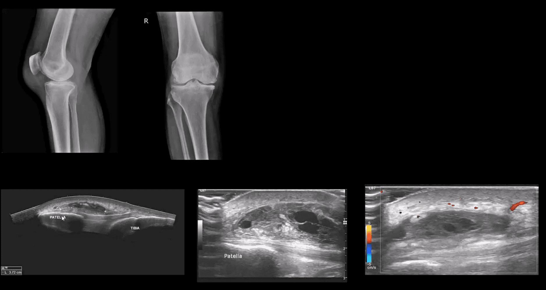
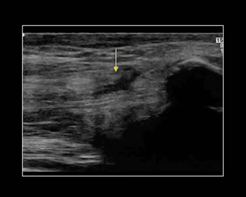
Patient Presented With Atraumatic Knee Pain & Swelling
Radiography revealed sizeable soft tissue density within the superficial pre-patella region along with mild-to-moderate OA
MSK US demonstrated large septated heterogeneous fluid collection with mild positive Doppler activity on the periphery indicating inflammation d/t Dx of Superficial pre-patella bursitis
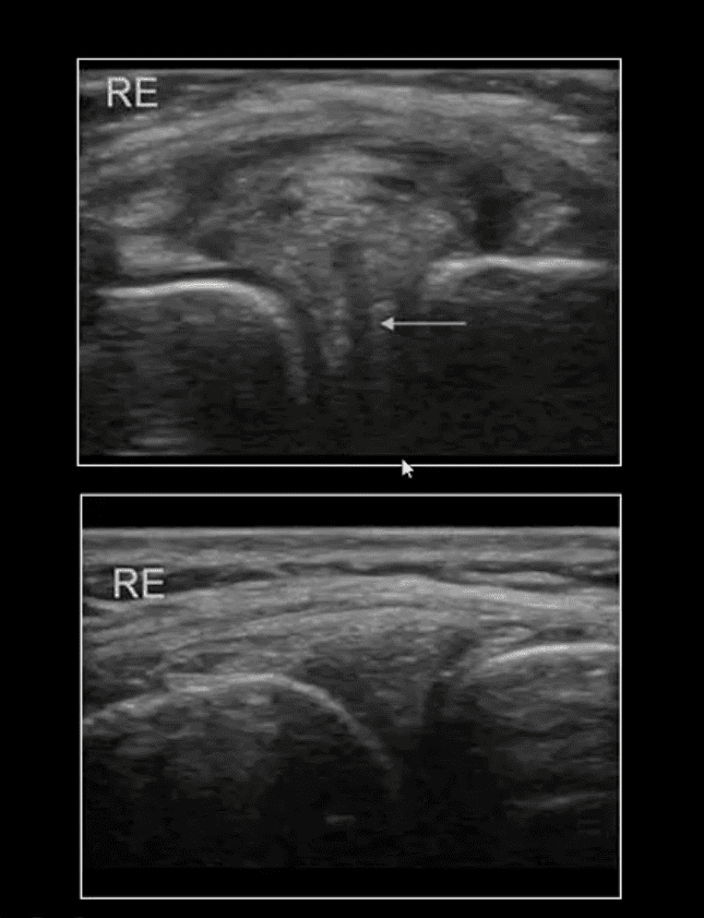
Long Axis US Images
Note normal lateral meniscus and fibers of LCL (above bottom image) compared to
Horizontal degenerative cleavage tear along with protrusion of lateral meniscus and LCL bulging (above top image)
Major limitation: unable to visualize the entire meniscus and the ACL/PCL
MRI referral is suggested
Rupture of Distal Tendon of Quadriceps
Note rupture of distal tendon of the Quadriceps muscle presented as fiber separation and fluid (hypo to anechoic) fluid collection within the substance of the tendon
Advantages of MSK US over MRI to evaluate superficial structures:
Dynamic imaging
Availability
Cost-effective
Patient’s preparation
Disadvantages: limited depth of structures, inability to evaluated bone and cartilage, etc.
Osteochondral Knee Injuries (OI)
osteochondral knee injuries can occur in children 10-15 y.o presented as Osteochondritis Dissecance (OCD) and in mature skeleton m/c following hyperextension and rotation trauma, particularly in ACL tear.
OCD-typically develops from repeated forces in immature bone and affects m/c postero-lateral portion of the medial femoral condyle.
OI in mature bone occurs m/c during ACL tears mainly affecting so-called terminal sulcus of the lateral femoral condyle at the junction of the weight-bearing portion opposed to tibial plateau and the part articulating with the patella
Osteochondral injuries may potentially damage the articular cartilage causing secondary OA. Thus need to be evaluated surgically
Imaging plays an important role and should begin with radiography often followed by MR imaging and orthopedic referral.
OCD Knee
95% associated with some trauma. Other etiology: ischemic bone necrosis especially in adults
Other common location for osteochondral injuries: elbow (capitellum), talus
1st step: radiography may detect osteochondral fragment potentially attached or detached
Location: a posterior-lateral aspect of the medial femoral condyle. Tunnel (intercondylar notch) view is crucial
MRI: modality of choice >90% specificity and sensitivity. Crucial for further management. T1-low signal demarcating line with T2 high signal demarcating line that signifies detachment and unlikely healing. Refer to orthopedic surgeon
Management: stable lesion esp. in younger children>off weight-bearing-heals in 50-75%
Unstable lesion and older child or impending physeal closure>operative fixation.
Result from valgus or varus stress with or w/o axial loading
Associated with periarticular soft tissues injury
High-stress injury m/c due to jumps falls and axial loading, often with the splitting of the tibial plateau. Men>women. Patients are in their 30s
Low impact or no trauma in patients with osteoporosis d/t insufficiency fractures
Impaction injury is more common with depression of tibial plateau. Women>men. Patients are in their 70s
Lateral Tibial Plateau Fractures More Common
Functional anatomy plays a significant role
60% of weight bearing is by the medial plateau
The medial plateau is more concave
Lateral plateau is slightly higher and more convex. Valgus stress impacts lateral plateau.
Tibial plateau fractures considered intra-articular and prone to delayed healing, non-union, meniscal injury (m/c lateral) ACL tear, secondary OA. Other complications: compartment syndrome, vascular injury.
Management: operative in many cases especially if >3-mm step-off at the plateau
If medial plateau or bicondylar Fxs present, ORIF will be required.
Imaging Plays A Crucial Role
Begins with x-radiography. X-radiography may not reveal the complexity and extent of this injury.
CT scanning w/o contrast will further delineate fracture complexity and pre-operative planning
MR imaging may be considered to evaluate for internal derangement: meniscal, ACL injuries.
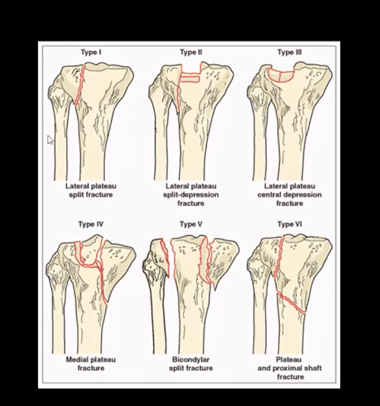
Shatzke classification may help to evaluate the complexity of this injury
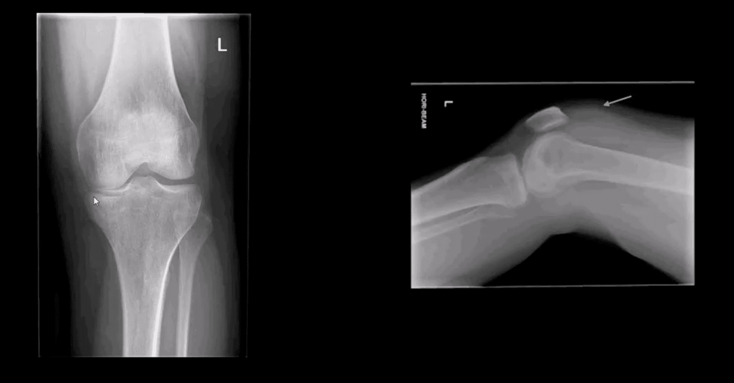
Key Diagnostic Sign
AP and lateral horizontal beam (cross table) left knee radiograph. Note subtle depression of the lateral plateau manifested by the lateral plateau appearing at the same level or lower as the medial. A critical diagnostic sign is the presence of fat-blood-interphase or FBI sign on cross-table lateral (above arrow) indicating intra-articular knee fracture
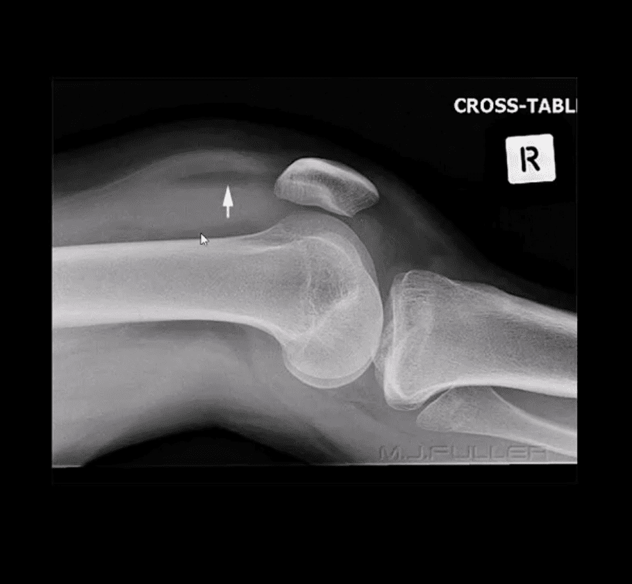
Lipohemarthorosis aka FBI Sign
Can be detected by radiography, CT or MR imaging
FBI sign is a reliable secondary radiographic sign of intra-articular knee fractures, regardless of how small they are
Mechanism: fracture results with acute hemarthrosis
Hemarthrosis will also occur w/o Fx. However, Fx will result with a fatty marrow being released into the joint cavity. Fat is a less dense medium (lighter) and will appear on the top of the hemorrhage if the patient is held in the supine position for 5-10-minutes before the cross-table radiograph is taken
FBI sign confirms the intra-articular Fx.
ACL/PCL, meniscal tears will not result in FBI sign
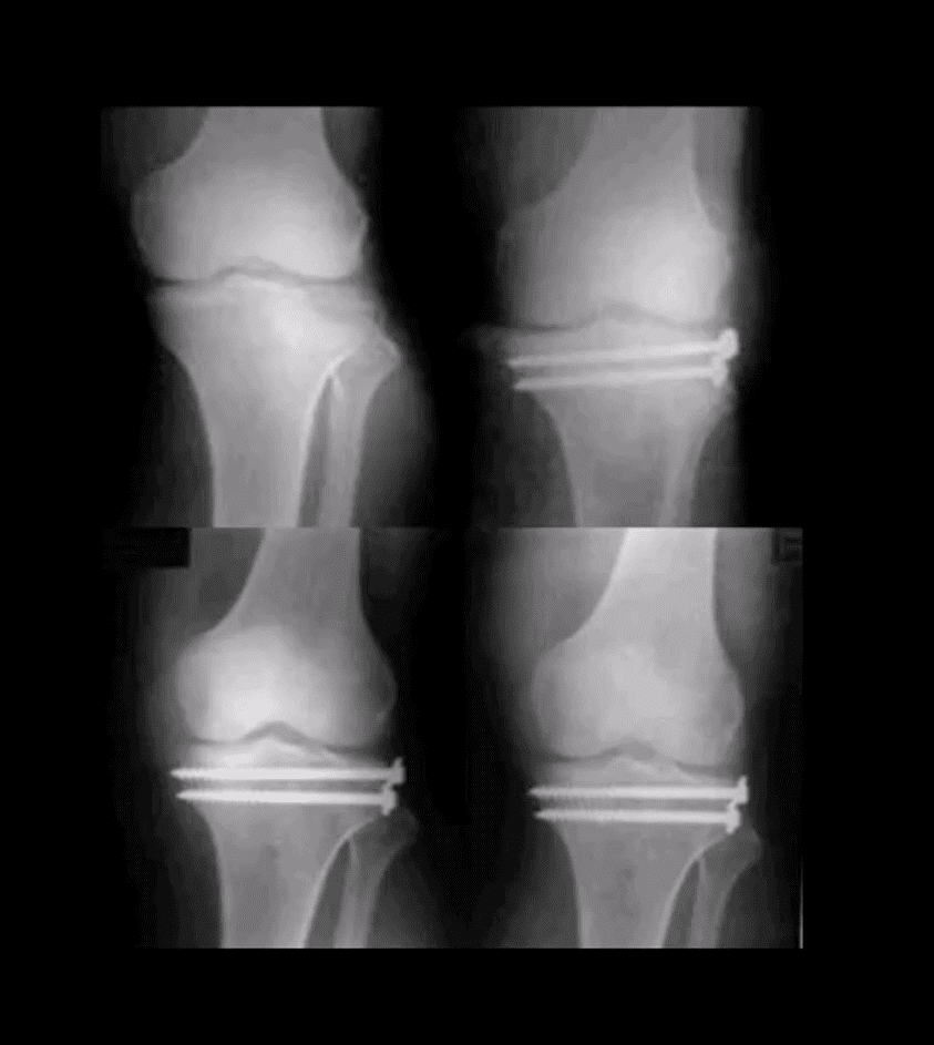
Lateral Tibial Plateau Fx
Lateral tibial plateau Fx that was managed operatively
Most common complication: premature secondary OA
More complex injuries may result in more extensive operative care
Knee Internal Derangement
Acute or chronic injuries of meniscal fibrocartilages and ligamentous restraints
Tears of the ACL and posterior horn of the medial meniscus are the most common
Acute ACL tears, however, often result with a lateral meniscus tear
Acute ACL tear may occur as a combined injury of the ACL, MCL, and medial meniscus
Functional anatomy: ACL prevents anterior displacement of the tibia and secondary varus stress
MCL functions together with ACL in resisting external rotation of the tibia especially when the foot is planted (closed chain position)
MCL is firmly attached to the medial meniscus, explaining the classic triad of ACL, MCL and medial meniscal tear (O’Donahue terrible triad)
Cruciate ligaments (ACL/PCL) are intra-articular but extra-synovial. Less likely to be torn in closed pack position (full extension). When all articular facets of tibia and femur are in full contact, the ACL/PCL are at least tension and stable
When the knee is flexed 20-30-degrees or more ACL is taut and remains unstable
ACL is a significant mechanoreceptor that feeds the info to CNS about the joint position. Thus the majority of previous ACL tears will lead to some degree of knee instability
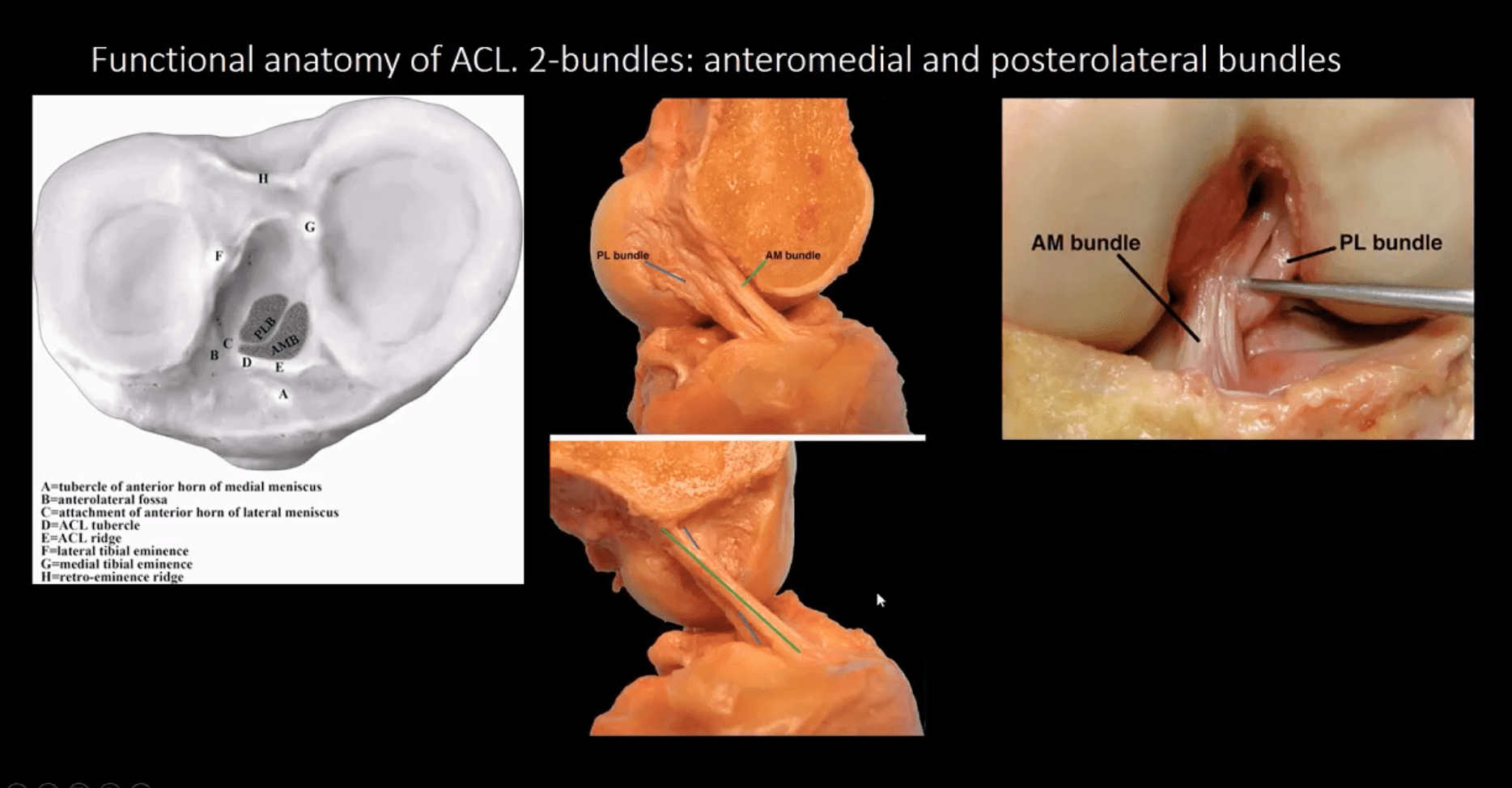
Functional Anatomy of ACL
Diagnosis of ACL Tear
Diagnosis of ACL tear requires MR imaging
Concerns exist of not only ligamentous injuries but injuries to the articular cartilage and menisci.
Most vendors will perform at least: one T1 WI in coronal or sagittal planes. Sagittal and coronal Proton-density slices to evaluate cartilaginous structures. Fast spin-echo sagittal, axial and coronal T2 fat-saturated or sagittal and coronal STIR images are crucial to demonstrate edema within the substance of knee ligaments
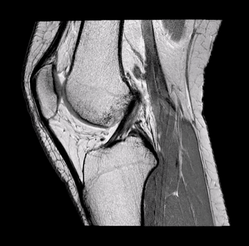
ACL is aligned along the Blumensaat line or oblique line corresponding the intercondylar roof of Femoral condyles. Lack of such alignment by the ACL is significant for ACL tear
Imaging Dx of Internal Derangement
MRI shows 78-100% sensitivity and 78-100% specificity
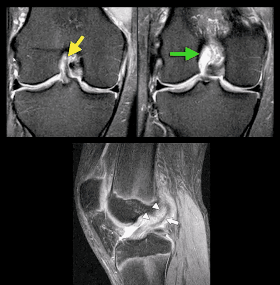
Primary signs of ACL tear: non-visualization of ACL (above green arrow), loss of its axis along the Blumensaat line (above triangle heads), wavy appearance and substance tear (above white arrow) or edema and cloud-like indistinctness (above yellow arrow)
Reliable Secondary Signs of ACL Tear
May be observed on the radiographs and MRI
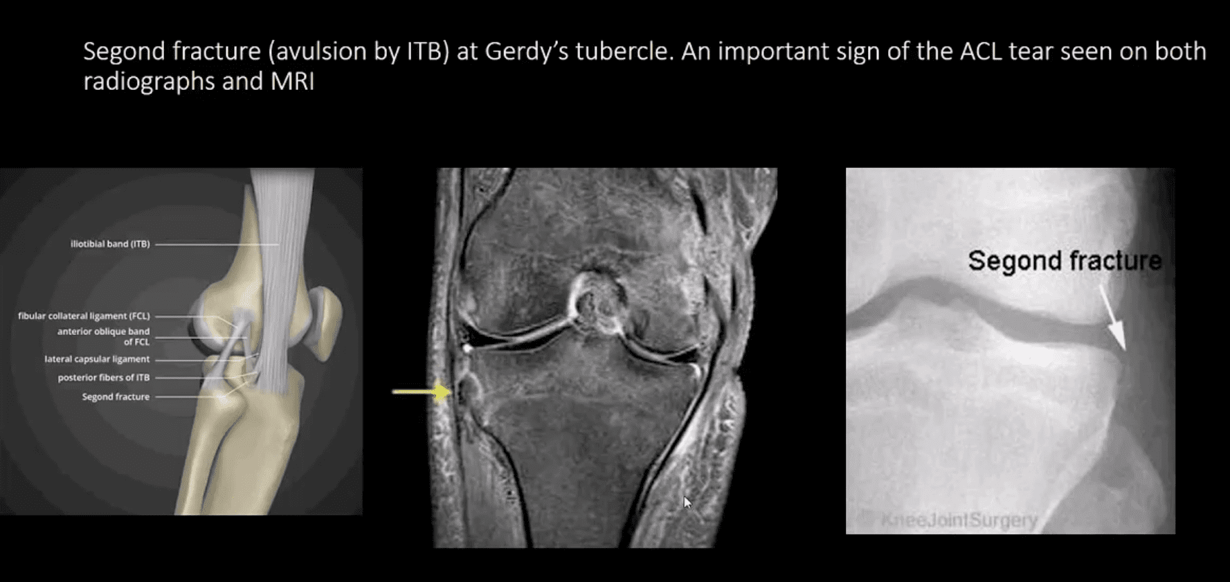
Segond avulsion fracture (80% specificity for ACL tear) (next slide)
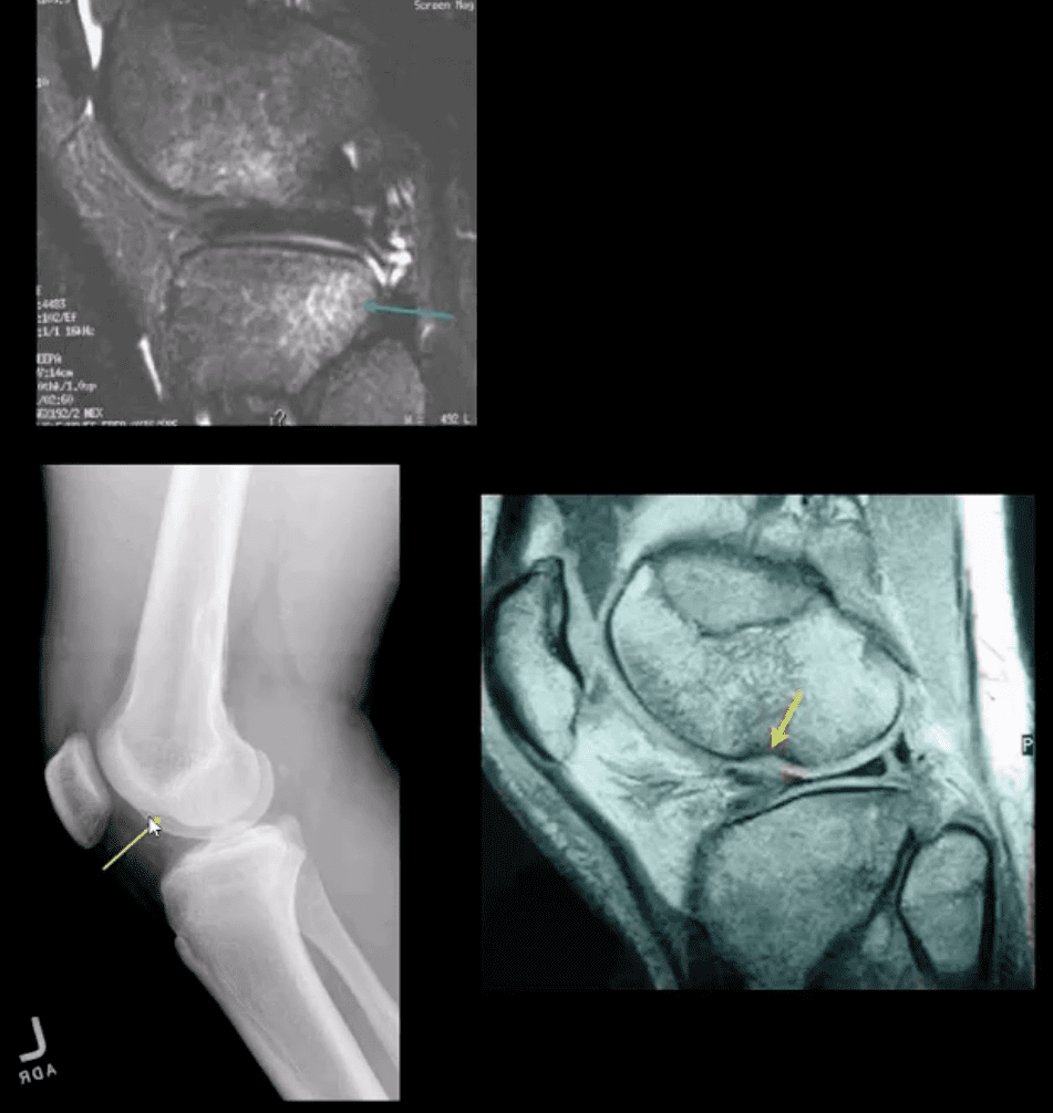
Deep femoral notch sign indicating osteochondral fracture (above bottom images) and
Pivot -shift bone marrow edema in the posterolateral tibial condyle d/t external rotation and often valgus impact by the lateral femoral condyles (above top image)
Segond Fracture (Avulsion by ITB)
Segond fracture at Gerdy’s tubercle. A vital sign of the ACL tear seen on both radiographs and MRI
Management of ACL Tears
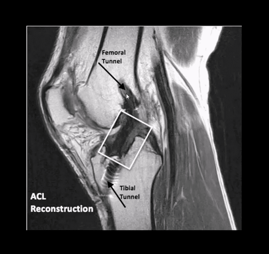
In acute cases, usually operative using cadaveric or autograft (patella ligament or hamstring) ACL reconstruction
Complications: graft tear, instability and premature DJD, joint stiffness d/t lack of postoperative rehab or gaft shortening. More rare, infection, a formation of intraosseous synovial cysts, etc.
aka Spondylodiscitis and vertebral osteomyelitis overall are relatively infrequent and may present with bimodal distribution: children and adults >50’s
Occasionally considered as two separate entities due to variations in the blood supply of pediatric vs. adult spines
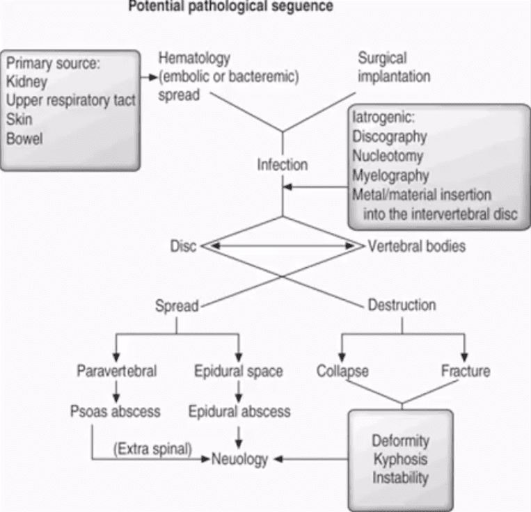
Risk factors/causes: distant site of infection in the body (25-35%), e.g., oropharynx, urogenital infections, bacterial endocarditis, indwelling catheters, florid skin infections furunculosis/abscess, etc.
Iatrogenic:�operative (e.g., discectomy) interventional or diagnostic/therapeutic procedures
Penetrating trauma
Immunocompromised patients
Diabetics
Malnourished patients or patients with low protein
IV drug users
Chronic disease patients, cancer patients etc.
Potential Pathological Sequence
Clinical Presentation
Back pain with or w/o high fever and other “septic” signs. Fever may only present in 50% of children
Exacerbation of pre-existing back pain in post-surgical cases
Neurological complications in advanced cases of vertebral destruction and epidural abscess
Meningitis, septicemia etc.
Labs: Blood tests are unspecific, may or may not indicate elevated ESR/CRP, WBC
Diagnostic imaging is important but
If clinical suspicion is strong, prompt I.V. antibiotics are needed to prevent serious complications
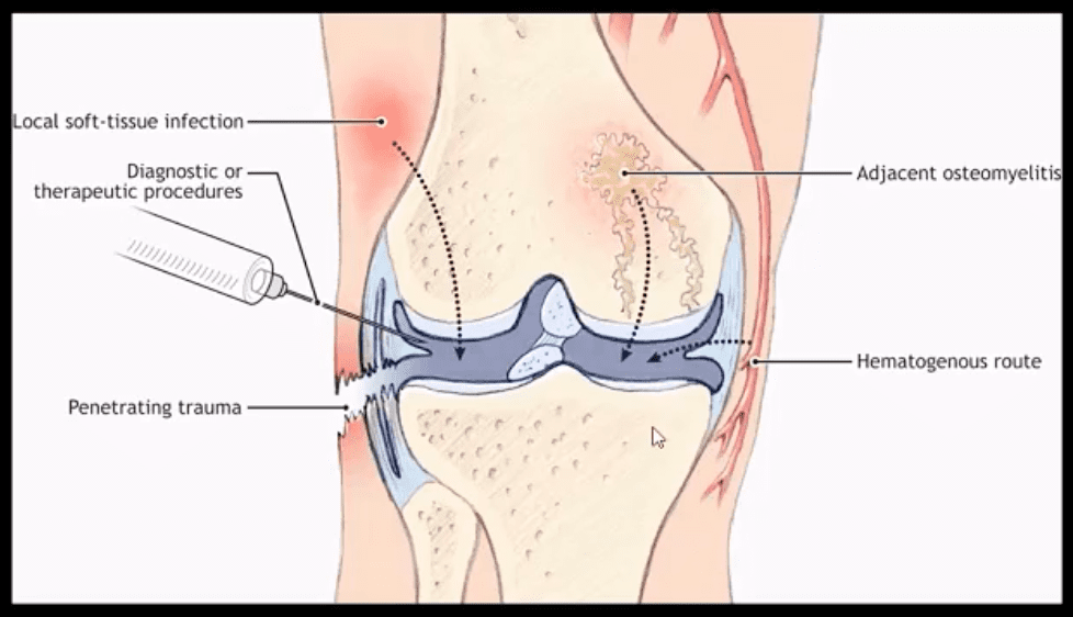
Routes of Infection
Infection routes to the spine are similar to bone in general
3-distinct routes:
1) Hematogenous spread as bacteremia (most common)
2) Adjacent site of infection (e.g., soft tissue abscess)
3)Direct inoculation (e.g., iatrogenic or traumatic)
M/C organism Staph. Aureus
Mycobacterium TB (tuberculous spinal osteomyelitis) aka Pott’s disease can be presented in cases of re-activated or disseminated pulmonary TB
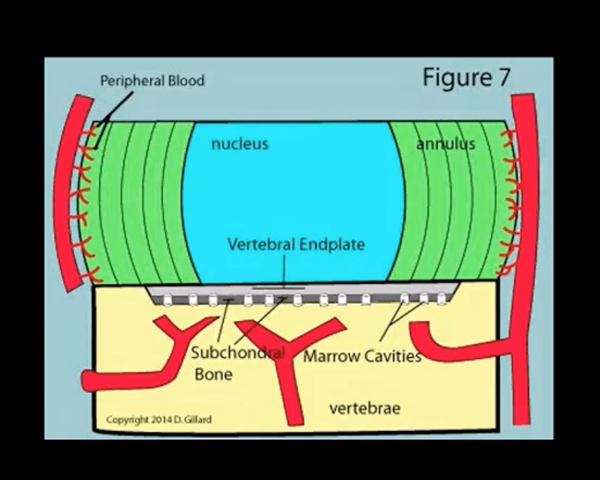
Mechanisms of Spinal Infection
May vary depending on the patients’ age
In children, the IVD receives direct blood supply and can be infected directly spreading to adjacent bone and causing spondylodiscitis
In Adults
The disc is avascular
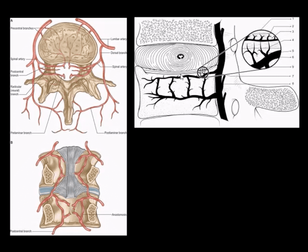
Pathogens invade adjacent vertebral end-plates via end-arterial supply of the vertebral body that may facilitate infection due to slow, turbulent flow
Organisms may then quickly gain access to disc substance rich in nutrients (discitis) often w/o significant initially visible destruction to the bone
Thus, one of the earliest rad. findings of spinal infection or sudden reduction of disc height
Later end-plate irregularity/sclerosis may develop, subsequently affecting the entire adjacent vertebral bodies
Diagnostic Imaging
Initially, in most cases of MSK complaints, radiography is the 1st imaging step
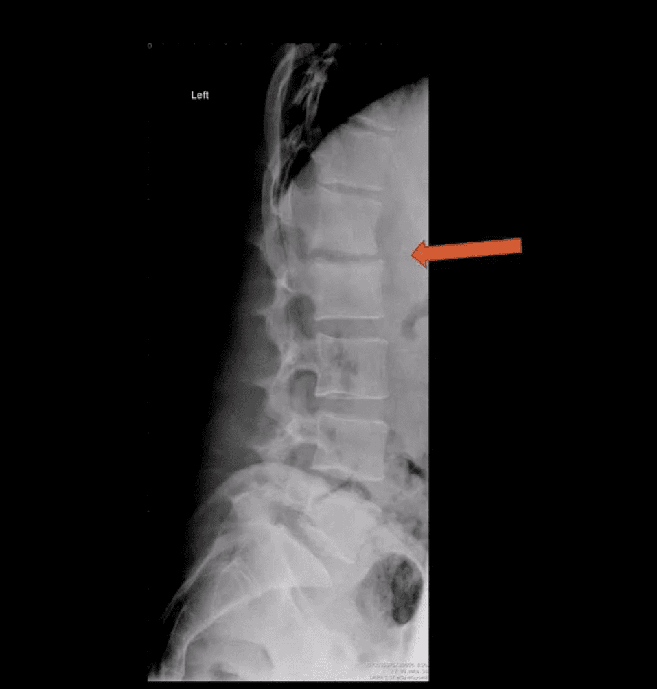
Initially, X-radiography is often unrewarding and may appear unremarkable for 7-10 days or presents with some subtle soft tissue changes (e.g., obscuration of Psoas shadows etc.)
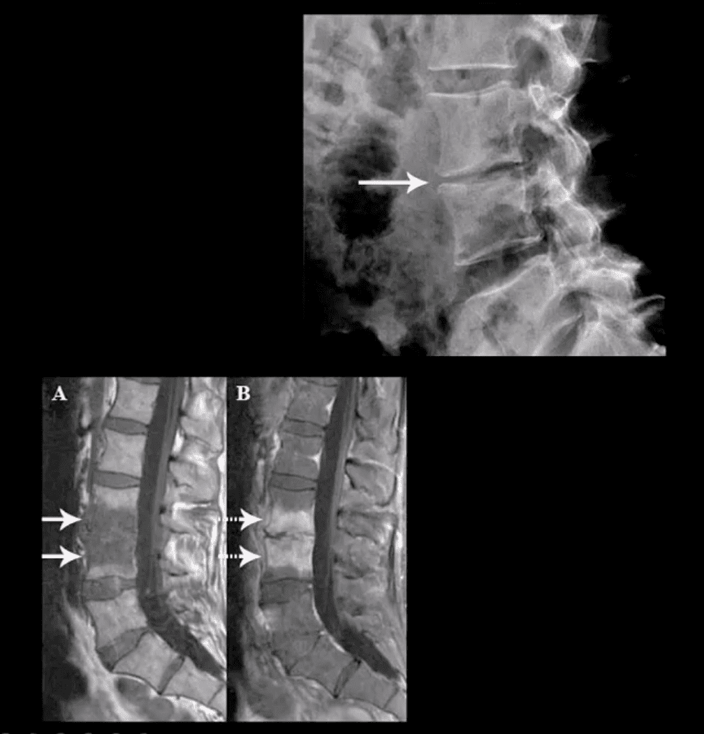
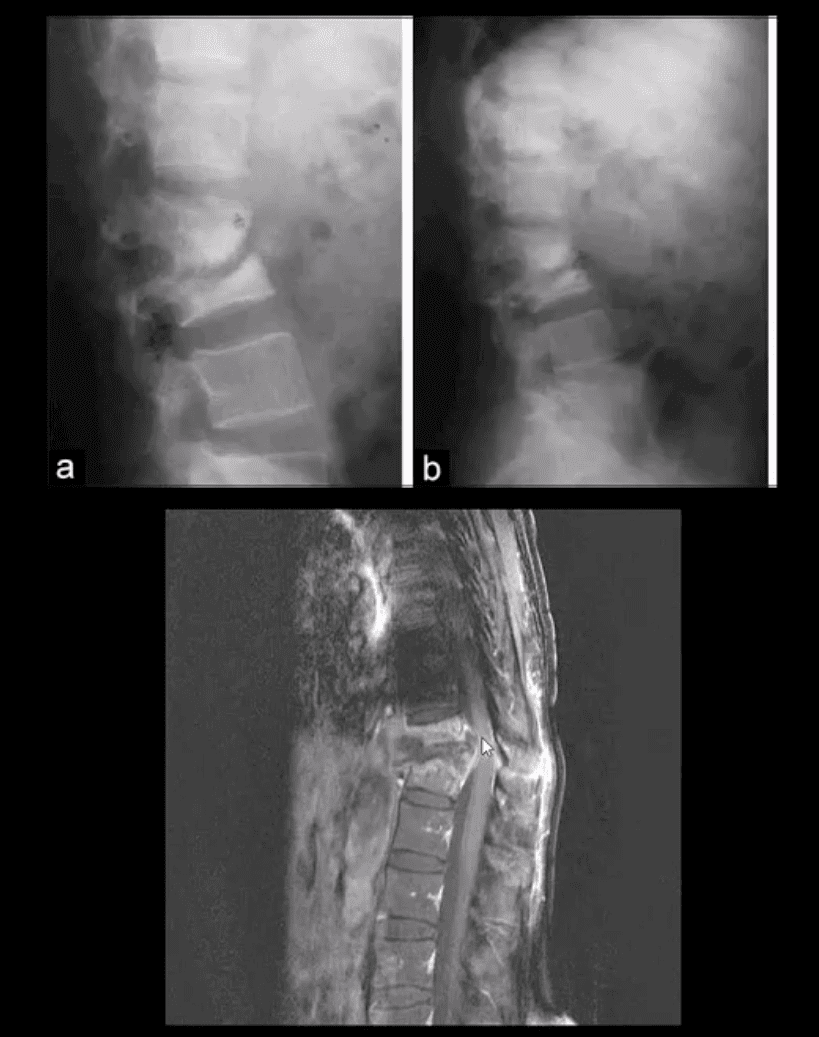
Some of the earliest x-ray signs of pyogenic spondylodiscitis: sudden reduction of disc height (above arrow) during initial 7-10 days
Subsequently (10-20 days) some end-plate irregularity and adjacent sclerosis may be noted
In more advanced cases, subsequent vertebral destruction and collapse may occur
N.B. Reliable feature to DDx between spinal infection and metastasis is the preservation of disc height in the latter
Note:�sudden disc narrowing with no appreciable spondylosis (above the first image) is suspicious for infection (discitis)
MRI +C is required to evaluate suspected infection
N.B. 50-60% of pyogenic spondylodiscitis occur in the lumbar region
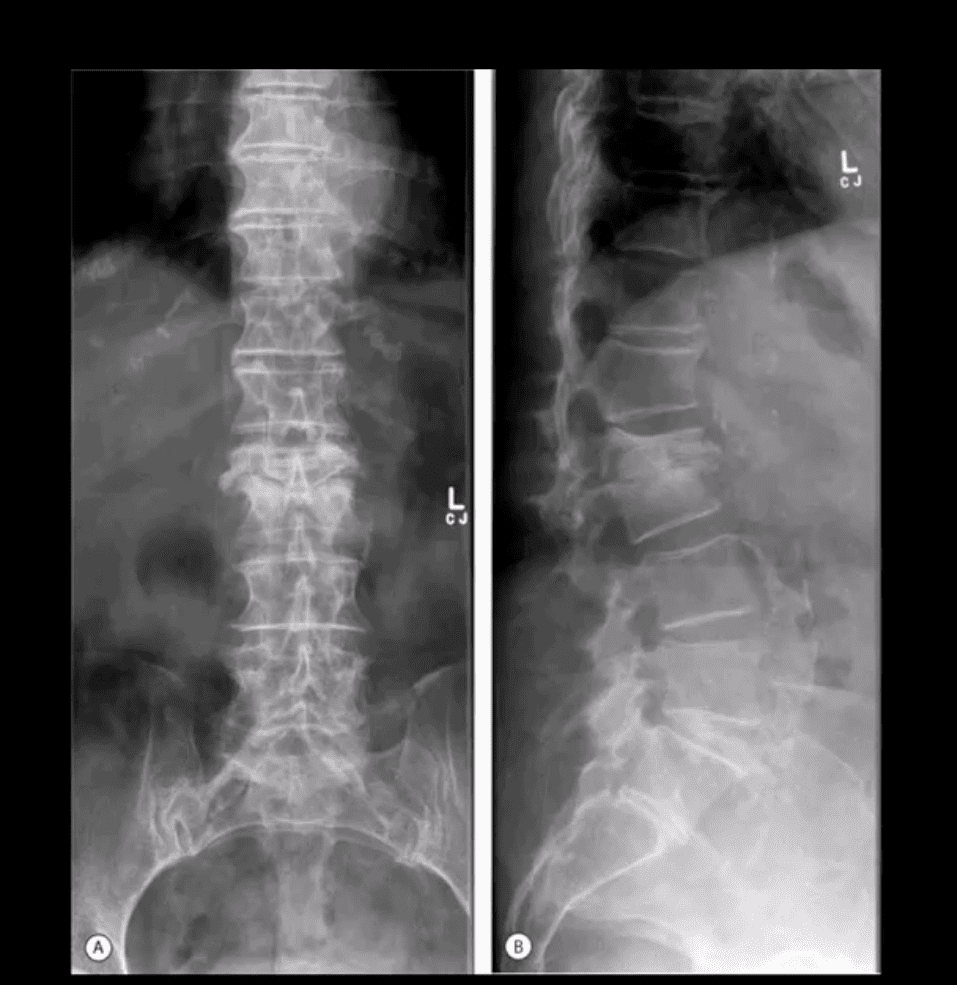
AP & Lateral Lumbar Radiographs
Note severe disc narrowing and adjacent vertebral body destruction at L1-L2 in a 68 -y.o.-female with a known Hx of type 2 DM
Additional imaging modalities should be used to support the Dx
Final Dx: Pyogenic Spondylodiscitis
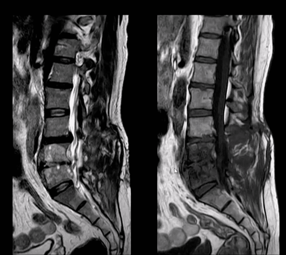
Sagittal T1 & T2 MRI
Weighted MRI slices of a patient who had laminectomy at L4
MR imaging with gad contrast is the modality of choice for Dx of spinal infection
Early septic changes affecting the disc and adjacent vertebral end-plates are readily demonstrated as a low signal on T1 and high T2/STIR d/t edema and inflammation
T1 FS +C gad images show avid enhancement of the lesion due to granulation tissue around the phlegmon. Peripheral enhancement is also characteristic of an abscess.
Epidural extension/abscess can also be successfully detected my MRI
N.B. 50% of epidural abscess cases present with neurological signs
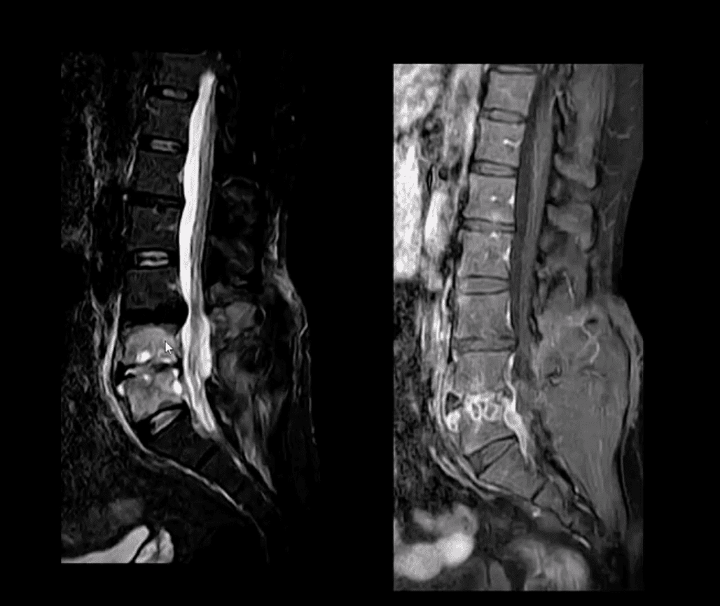
STIR & T1 FS +C Gad Sagittal MRI
Marked septic collection and edema affecting L4-5 disc and vertebral body with some epidural extension and paraspinal soft tissue edema. Avid contrast enhancement is noted surrounding low signal foci within the bone and disc tissue, some gad. Enhancement is noted in posterior paraspinal muscles and dural spaces
Management: Dx of spondylodiscitis requires prompt I.V antibiotics. If instability and neurological complications develop referral to a Neurosurgeon is required
MRI Unavailable or Contraindicated
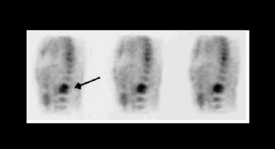
Bone scintigraphy is very sensitive but non-specific for spinal infection but overall is of great value d/t higher sensitivity than x-rays and relatively low cost.
An area of increased flow with radiopharmaceutical uptake is characteristic but not specific sign of spondylodiscitis
If neurological signs are present and MRI is contraindicated than CT myelography may be used
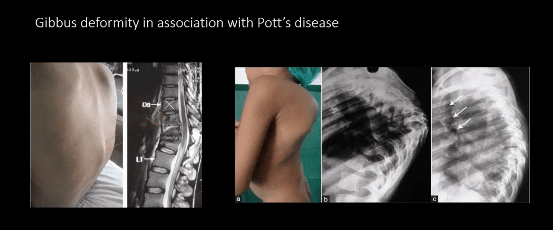
TB Osteomyelitis aka Pott’s Disease
TB osteomyelitis is increasing d/t HIV and other immunocompromised states. Extrapulmonary TB m/c affects the spine and especially the thoracic spine (60%)
Radiographic Pathology:�TB bacillus infects the vertebral body and often spreads subligamentously. “Cold” paraspinal abscess collection may develop and spreads along fascial planes, e.g., Psoas abscess. Disc spaces are preserved until v. late and skip areas are noted helping to DDx TB from pyogenic infection. Severe vertebral destruction aka Gibbus deformity may develop (>60-degree sometimes) and may become permanent. Neurologic and many regional complications may develop
Imagingapproach:�CXR with spinal x-rays 1st step that may be unrewarding but may potentially reveal VB destruction w/o disc narrowing. CT scanning is more superior than x-rays. MRI with gad C is a modality of choice
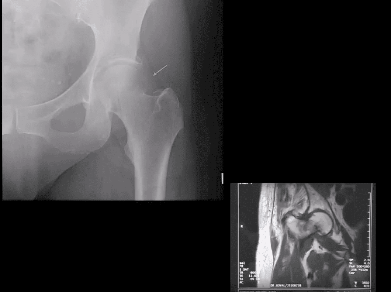
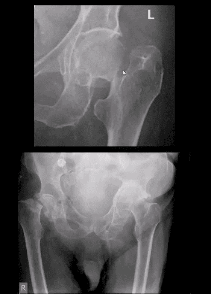
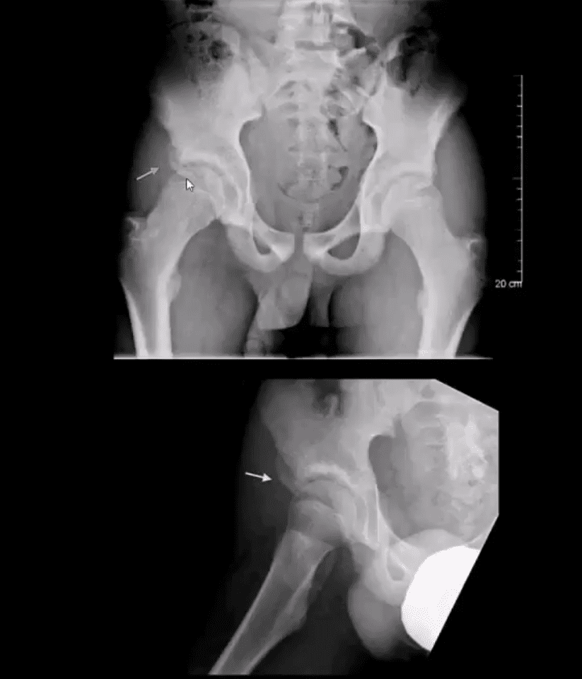
X-radiography pitfalls: some undisplaced Garden 1 & 2 Fxs may be missed d/t pre-existing DJD and osteophytes along the femoral head-neck junction that may overly the Fx line
Fx line is incomplete and too small/subtle especially if the study is read by non-radiologists
Incomplete Fxs if left untreated will not heal and likely to progress to complete Fxs
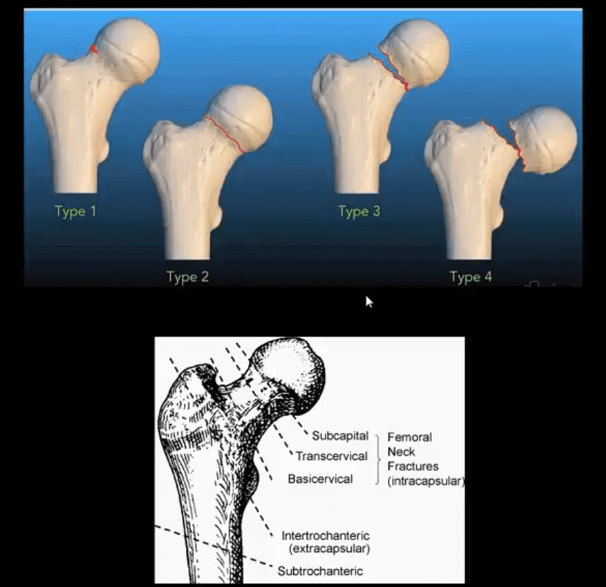
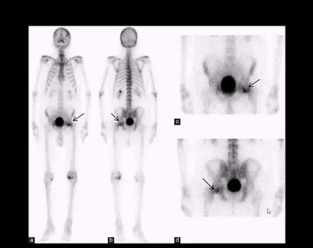
AP hip spot view: note valgus deformity of the head (above yellow arrow) with a small/subtle line of sclerosis in the sub-capital region representing Garden 1 Fx. MRI may help with Dx of subtle radiographic Fxs. If MRI contraindicated, Tc 99 radionuclide bone scan may help demonstrate high uptake of the radiopharmaceutical in Fx (below image)
Above – Tc99 Radionuclide Bone Scan Reveals Left Subcapital Femoral Neck Fx
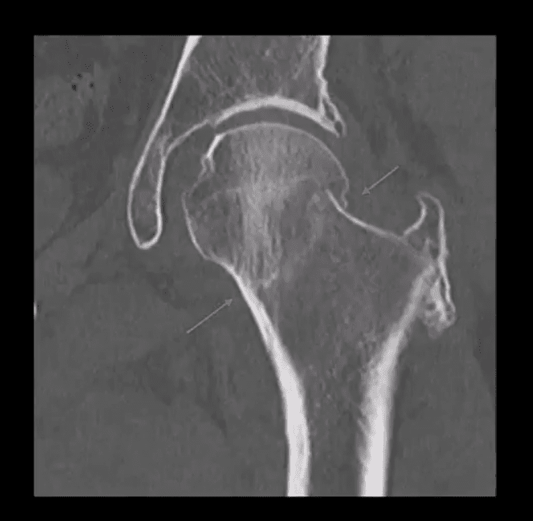
Garden 2 complete undisplaced (above green arrows) Fx
AP hip: Garden 3 complete partially displaced Fx (above the first image)
AP pelvis: complete displaced Garden 4 Fx (above the second image)
Clinical pearls: in some cases of Garden 4 Fx, DDx may be difficult to differentiate from OSP vs. pathologic fx d/t to bone Mets of Multiple myeloma (MM)
Management: depends on patients age and activity level
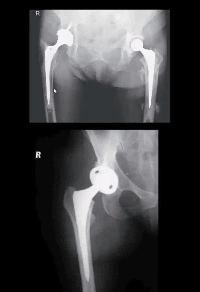
Garden 3 & 4� require total hip arthroplasty in patients <85-y.o.
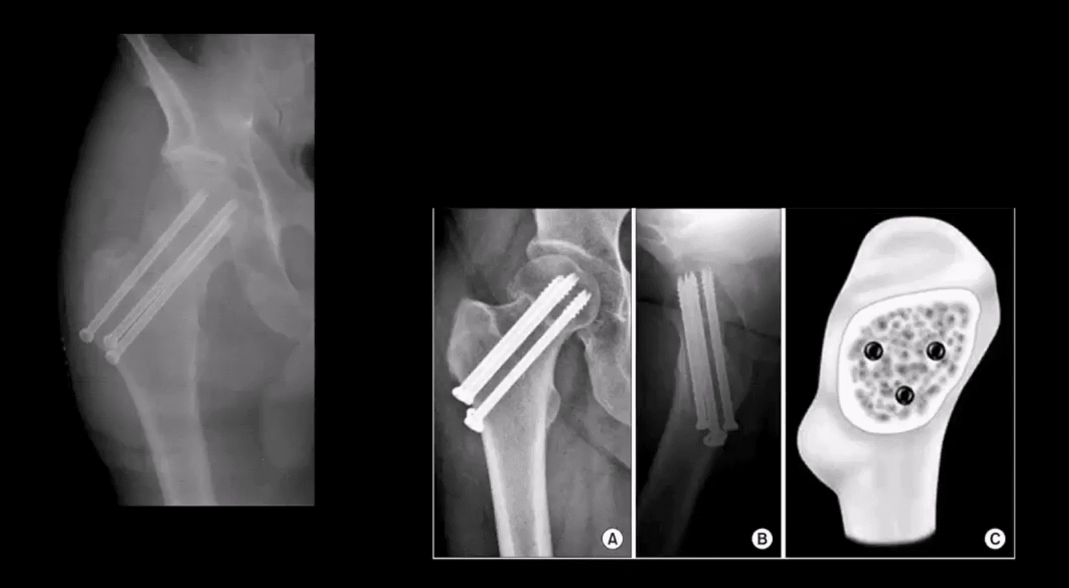
Garden 1 & 2 may be treated with closed reduction of fx and open capsule and 3-cannulated fixating screws
Pre-existing DJD may require total arthroplasty
Occasionally observation may be performed on patients who are not active and significant risks of surgery and depends on surgical centers
m/c Rx of Garden 1 & 2 undisplaced Fx with 3-screws. Screws proximity depends on the bone quality and Fx type
THA aka hip replacement: cemented THA with bone cement (above the first image) vs. non-cemented (biologic) that is used mostly in younger patients
2-types: metal on metal vs. metal on polyethylene
The femoral angle of the prosthesis should have slight valgus but never >140 degrees
The non-cemented component uses porous metal allowing the bone to integrate sometimes coating in bone cement from osteoconduction
THA has good outcome and prognosis
Occasionally cement failure, fractures, and infections may complicate this procedure
Unstable Fx: a result of high energy trauma with >50% d/t MVA
20% closed Fx and 50% of open Fx result in mortality
Mortality is associated with vascular and internal organs injuries
Vascular injury: 20% arterial 80% venous
Chronic morbidity/disability and prolonged pain
Unstable Fx are rarely seen in the outpatient setting and typically and present to the ED
Stable pelvic Fx are usually caused by muscles/tendons avulsions and more often seen in pediatric cases
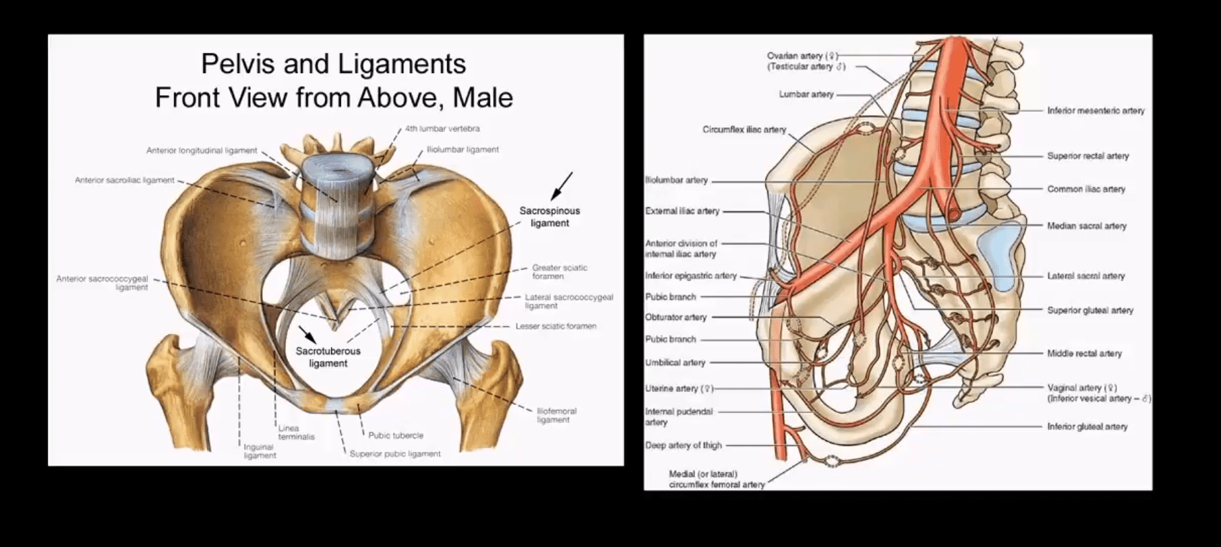
Understanding Pelvic Anatomy Is The Key To Successful Imaging Dx
The bony pelvis is a continuous ring of bone held by strong ligaments
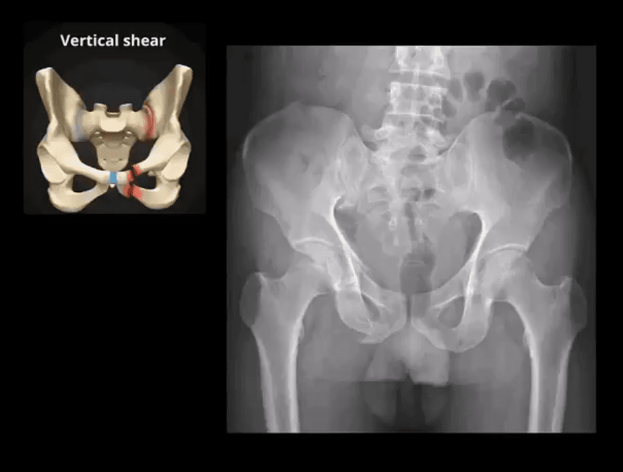
During significant impact, pelvic fractures may occur in more than one location because forces applied to one region of the ring will also correspond to injury on the other, usually the opposite side of the ring (above image)
Thus the majority of unstable pelvic Fx will typically demonstrate more than one break
Pelvic is seen as a ring of� bone connected by some of the strongest ligaments in the body
The pelvic ring comprises 2-semirings: anterior to the acetabulum and posterior to the acetabulum
The bony pelvis is in close proximity to major vessels carrying a greater chance of vascular injury
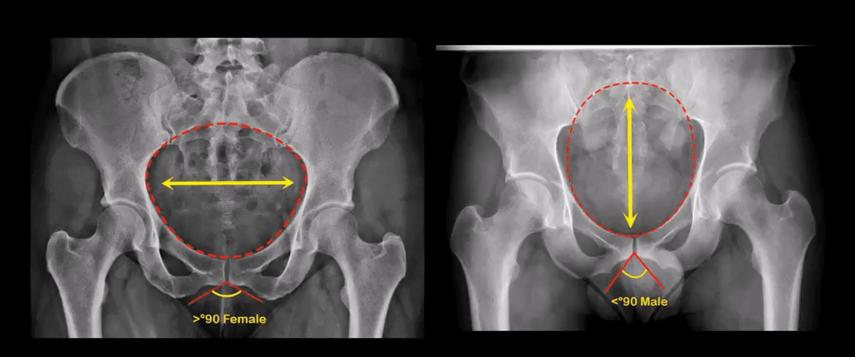
Anatomical Differences of The Female and Male Pelvis
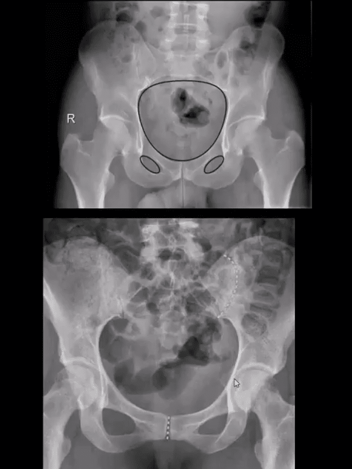
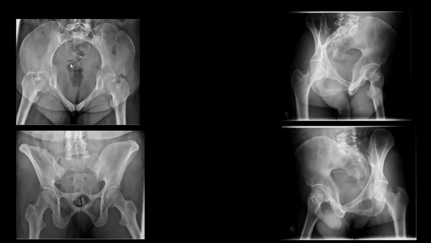
Post-Traumatic Pelvic Views May Vary and Include:
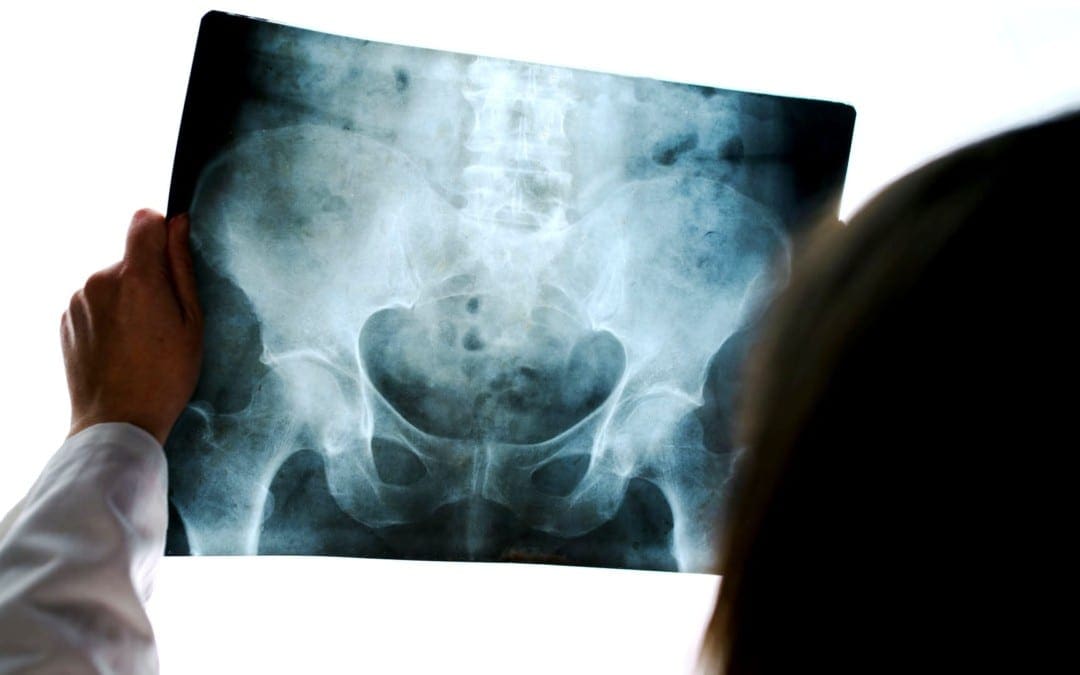
Standard AP Pelvis (above images)
Judet views evaluating the acetabulo-pelvic region
Inlet/Outlet views helping with the symphysis and SIJ regions
Rad survey of the pelvis should include evaluation of the continuity of pelvic rings:
Inlet/outlet, obturator rings (above the first image)
Symphysis pubis and SIJ for diastasis and post-trauma separation (above the second image)
Lumbosacral spine and hips should also be carefully examined
Pelvic inlet (above top left) and Outlet (above bottom left)
Judet views: left and right posterior oblique views
Additional Survey:
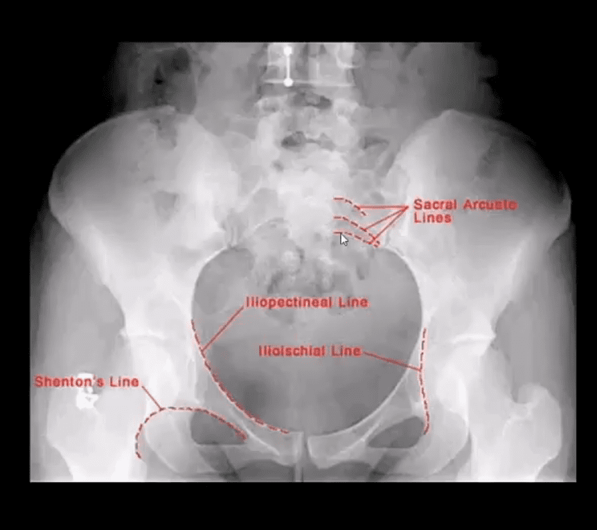
Iliopectineal, ilioischial, Shenton and Sacral arcuate lines will help detection of sacral, acetabular and hip fracture/dislocations
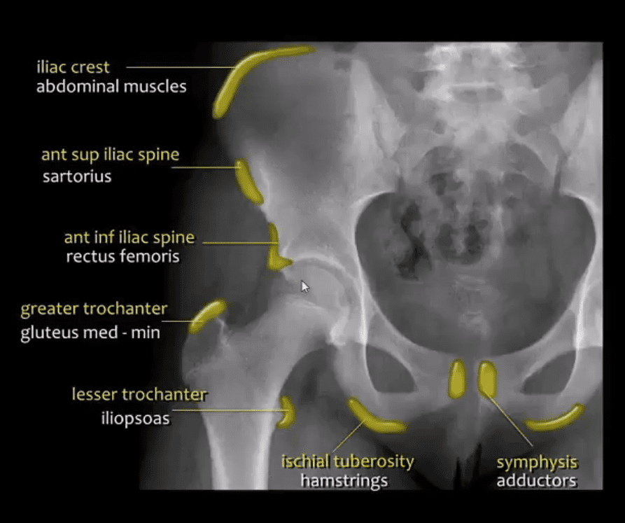
Stable Pelvic Fractures aka Avulsion Injury
Appreciating anatomical sites of pelvic origin/insertion of different muscles will help Dx of pelvic avulsion Fx
Avulsion Fx of the AllS (origin of the direct head of Rectus femoris M)
Pelvic avulsions occur by sudden eccentric contraction especially during kicking or jumping
Imaging: x-radiography will suffice
Clinically: sudden snap or pop followed by local pain. Pt can weight bear
Care: non-operative with rest for 4-weeks. Non-union is rare. No major complications
DDx: key rad DDx feature is not to mistake an avulsion from an aggressive pediatric bone tumor-like osteosarcoma that may show some exuberant new bone formation d/t healing and bone callus
Commonly Encountered Unstable Pelvic Fractures
Malgaigne Fx: d/t vertical shear injury to the ipsilateral pelvis
Rad Dx: ipsilateral superior and inferior pubic rami Fx (anterior ring) with ipsilateral SIJ separation/Fx of the sacrum and adjacent ilium (posterior ring). Symphysis pubis diastasis can be seen. An additional clue is an avulsion of L4 and/or L5 TP that often signifies serious pelvic injury
Clinically: marked leg shortening, shock, inability to weight bear.
Damage to Superior Gluteal Artery can occur
Imaging: x-radiography followed by CT scanning w/o and with IV contrast esp. if visceral injury present
Care: surgical in most cases d/t significant instability. ORIF. Hemostasis, Pelvic stabilization
Prognosis: depends on the complexity, rate of visceral complications and stability. 10% Superior glut artery bleed requiring rapid hemostasis
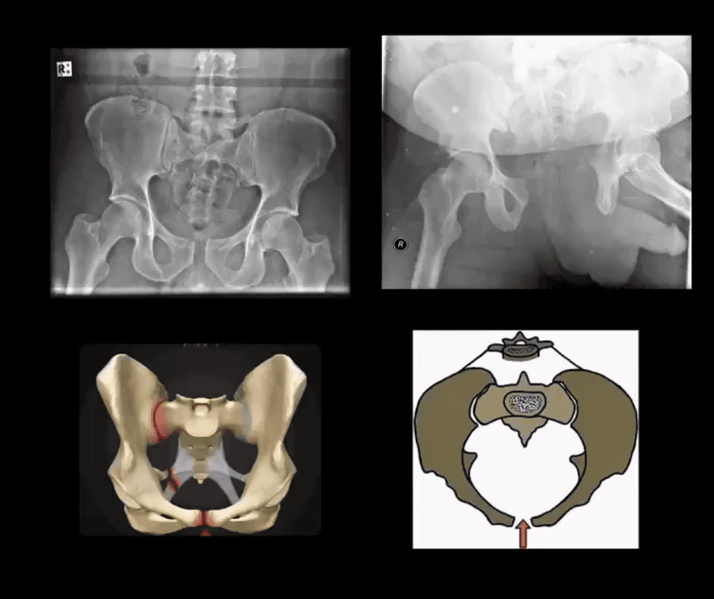
Open Book Pelvis (major instability)
Mechanism: AP compression of different force magnitude (picture depiction)
Rad Dx: diastasis of symphysis pubis with diastasis of SIJ with and w/o adjacent Fx of the ala
Imaging steps: x-radiographic, CT scanning with and w/o contrast for vascular injury, cystography for acute urinary bladder rupture
Immediate and delayed complications may occur: vascular injury, urethral/bladder injury
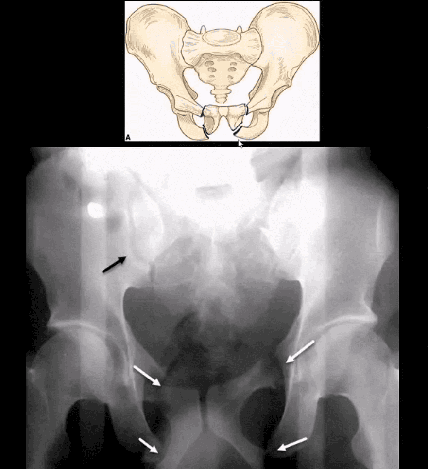
Straddle Injury: Unstable Fx
Mechanism: direct impact/collision
High risk of urinary bladder/urethral injury
Imaging: bilateral superior and inferior pubic rami Fx with or w/o diastasis and Fx of SIJ
CT with and w/o contrast for vascular injury
Cystourethrogram additionally evaluates a urogenital injury
2) Osteoporotic patients with low impact, trivial or no trauma (i.e., insufficiency Fx)
X-radiography is crucial to early Dx and prevention of complications which include:
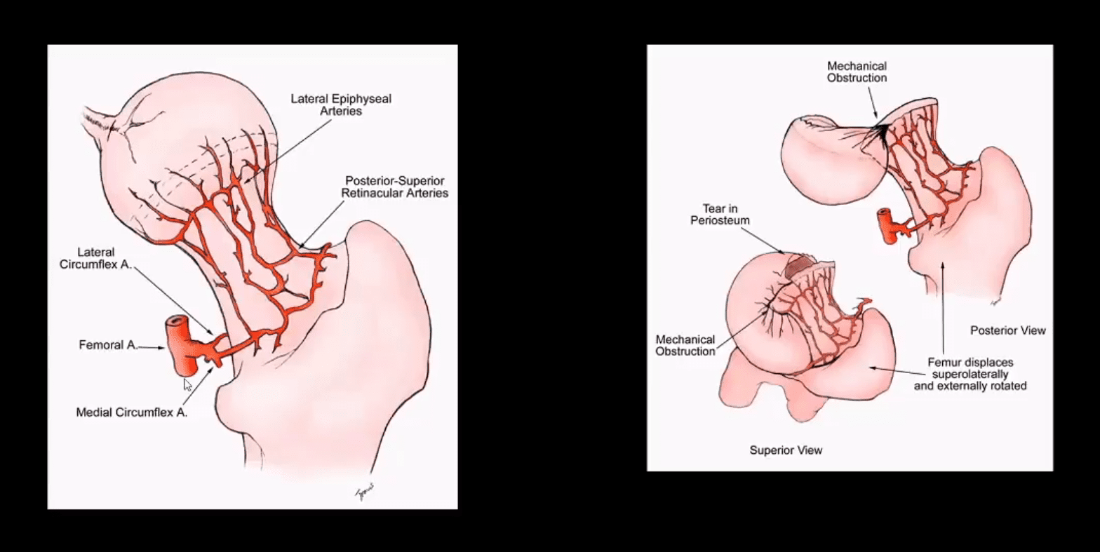
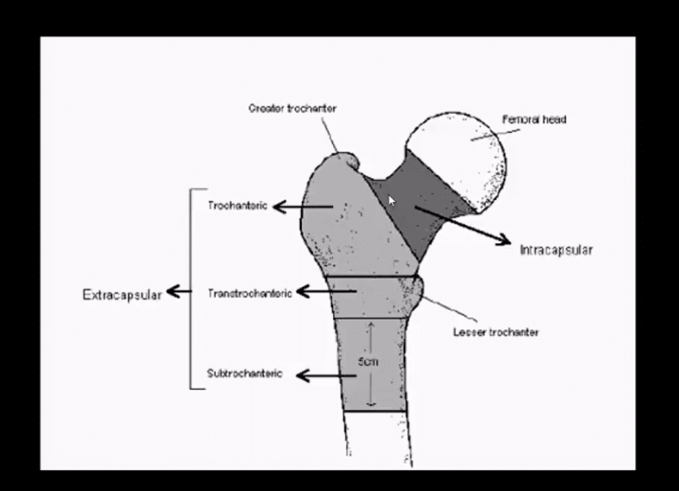
Dx: intra-capsular vs. extra-capsular Fx
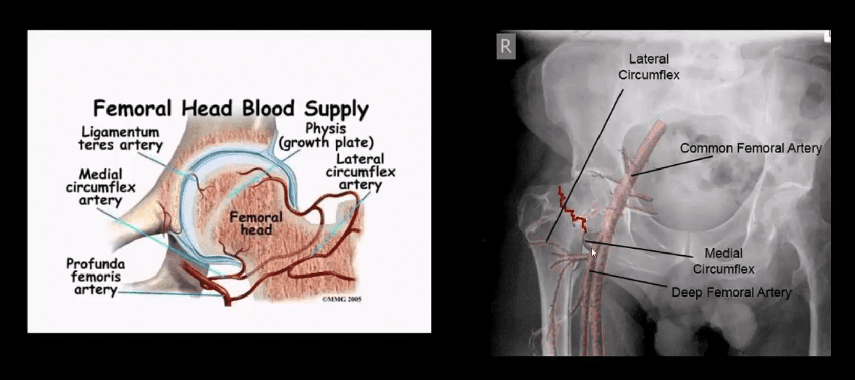
Ischemic osteonecrosis aka avascular necrosis (AVN) of the femoral head and rapid disabling DJD
Epidemiology: USA has some of the highest rates of OSP hip Fx worldwide. Highest healthcare cost Fx to treat overall
Women>men, Caucasians>African-Americans
25-30% mortality within the 1st year. Mortality depends on co-morbidities and stat of activity prior Fx
Pathophys: the femoral neck is intra-capsular and transmits arterial flow to the head. The neck is uncovered by the periosteum and unable to develop a good callus. The neck transmits maximum tensile forces through the proximal femur and prone to Fx and non-union
IFM's Find A Practitioner tool is the largest referral network in Functional Medicine, created to help patients locate Functional Medicine practitioners anywhere in the world. IFM Certified Practitioners are listed first in the search results, given their extensive education in Functional Medicine