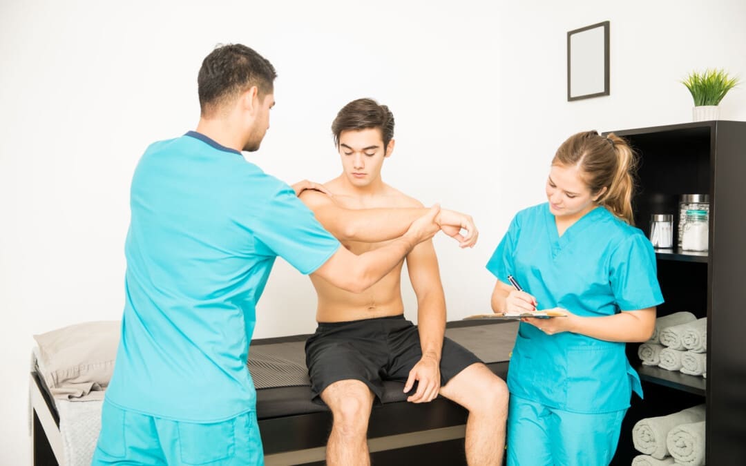
Can understanding the mechanics of the throwing motion help to understand why it may cause shoulder pain, the symptoms of a shoulder problem, the diagnosis, and the treatment options available?
Throwing a Ball and Shoulder Pain
The throwing motion is a complex shoulder movement that requires the mechanics of muscles, tendons, joints, ligaments, and bones. They all must move in a synchronized and stable pattern to move the shoulder joint. When these mechanics are interrupted or altered, inflammation can result in pain symptoms. (Wardell M., Creighton D., & Kovalcik C., 2022)
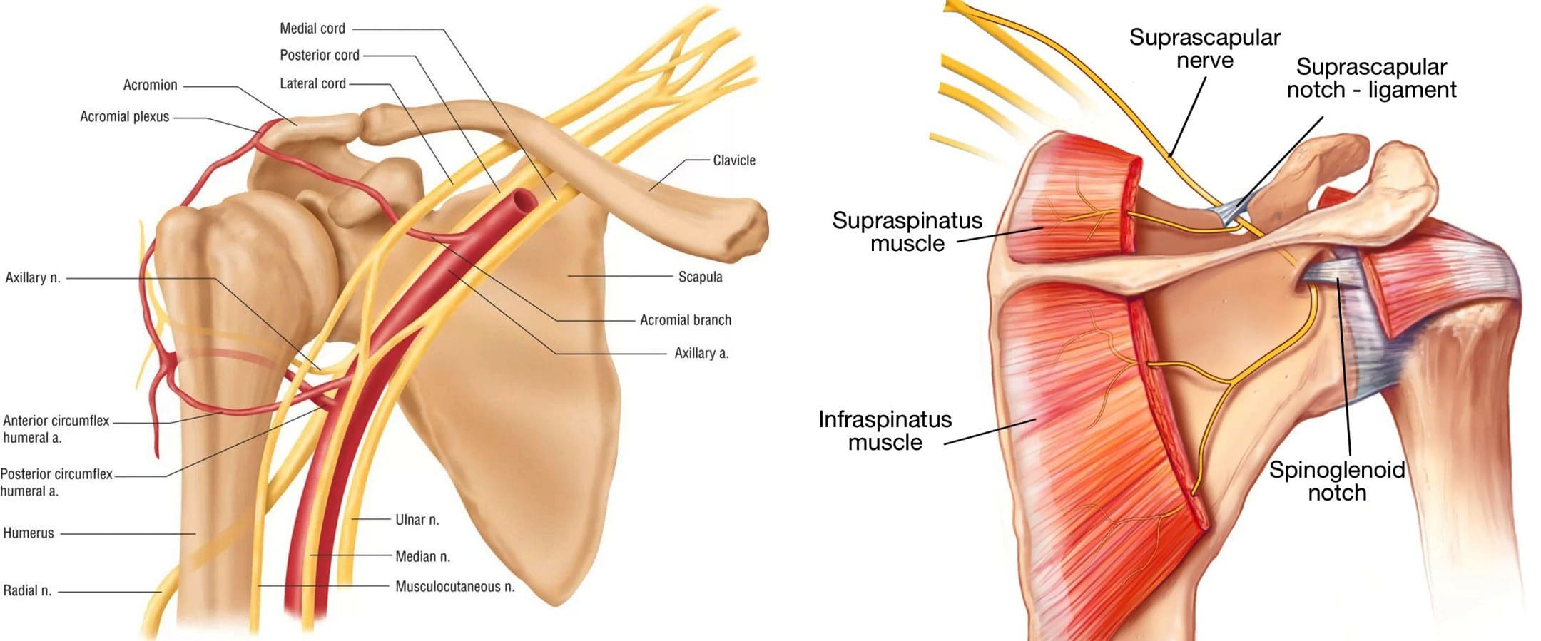
The labrum stabilizes the ball in the socket of the shoulder.
The shoulder blade rotation coordinates with the arm to ensure mobility. (Itoigawa Y. et al., 2023)
The throwing motion generates high torque and acceleration forces acting on the shoulder joint and the surrounding muscles, ligaments, and tendons.
Causes of Pain
Pain when throwing can come from the:
Shoulder blade
Shoulder joint – cartilage and labrum
Rotator cuff muscles and tendons
Nerves that control the muscles’ function
The shoulder blade is attached to the upper back by ligaments, muscles, and tendons. The various muscles and tendons that control the movement of the shoulder blade impact movements. Abnormalities of any area can lead to shoulder dysfunction and pain when throwing. (Wardell M., Creighton D., & Kovalcik C., 2022) The most common is the tightness of the posterior shoulder capsule, causing a loss of normal internal rotation of the shoulder. If this is causing pain, individuals may notice that they can’t reach up as high on the side with the painful shoulder when reaching behind their back.
Symptoms
Whether an athlete or playing catch in the backyard, shoulder function abnormalities can cause significant pain. Some symptoms include.
Aching Pain
Often deep in the shoulder or extending down the upper arm.
Dead Arm
Lack of strength in the throwing motion.
Pain at Night
Pain can awaken you from sleep.
Diagnosis
Finding a healthcare provider familiar with sports injuries can be helpful. They can best determine if a structural abnormality needs to be addressed. (American Academy of Orthopaedic Surgeons, 2021)
Treatment
Most can improve with nonsurgical treatments. The earliest phase of treatment is resting the joint and reducing inflammation. Treatments can include:
Ice
Anti-inflammatory medications
Cortisone injection
Once the inflammation has subsided, the source of the discomfort can be addressed.
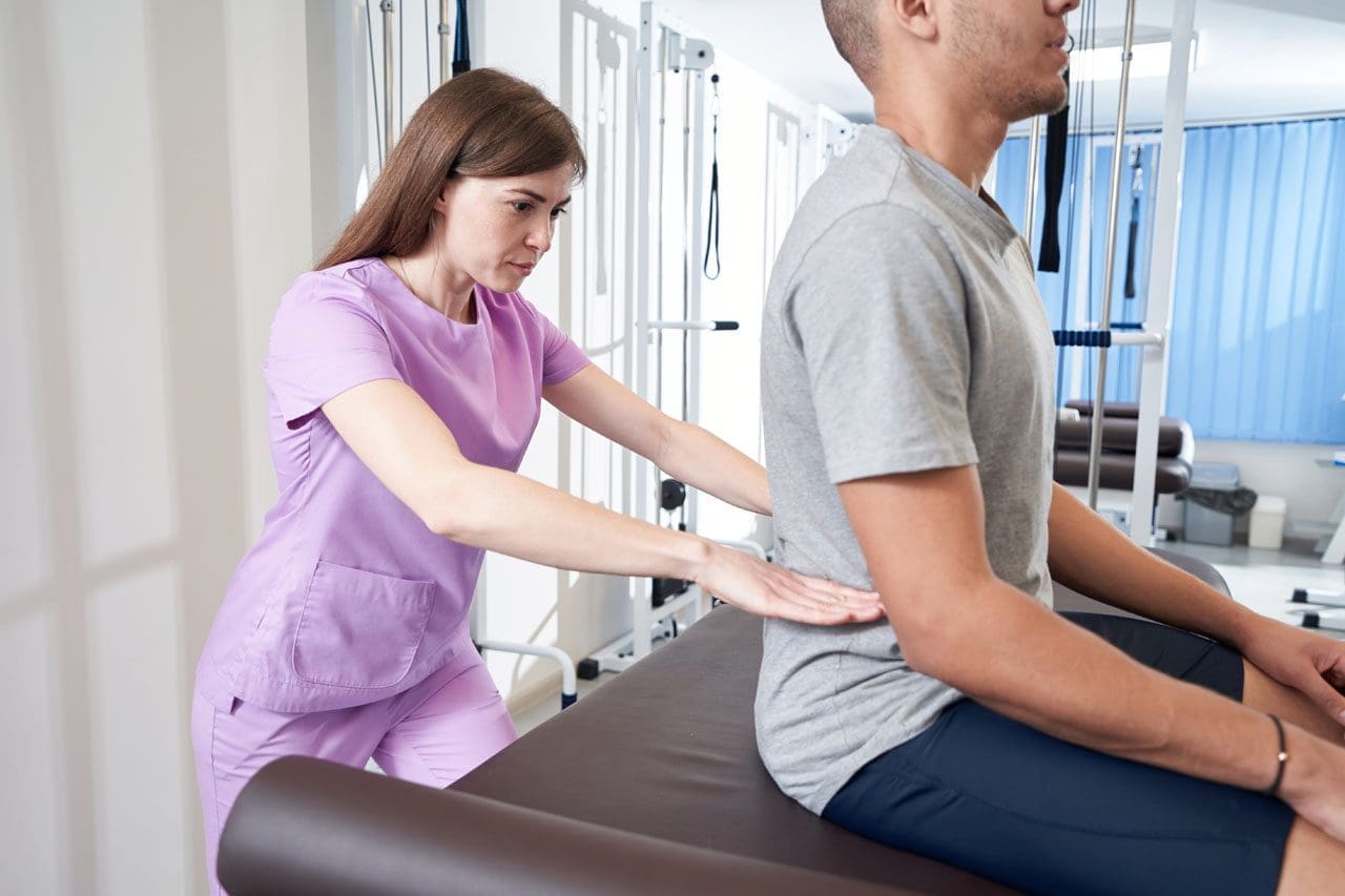
Physical Therapy
Therapy can include:
A structured shoulder stretching and strengthening program will help.
The physical therapist will focus on scapular mobility when managing shoulder joint problems.
Stretching to improve internal rotation or any other lost motion can help allow a more normal throwing motion.
Strength exercises are often aimed at the rotator cuff, as these muscles initiate proper shoulder movements and stabilize the shoulder joint.
Maintaining flexibility and strength of the periscapular muscles (muscles that attach to the scapula bone) is important to ensure that the scapular movements are coordinated with the throwing motion.
If improvements are not made within three months of therapy, or individuals can’t return to competitive sports within six months. In that case, the individual may need to return to their healthcare provider or see an orthopedic specialist who may recommend surgery. (American Academy of Orthopaedic Surgeons, 2024)
Injury Medical Chiropractic and Functional Medicine Clinic
As a Family Practice Nurse Practitioner, Dr. Jimenez combines advanced medical expertise with chiropractic care to address various conditions.
Wellness & Nutrition: Personalized plans to optimize health and prevent disease.
Chronic Pain Management: Non-invasive solutions for fibromyalgia, sciatica, and low back pain.
Personal Injury & Auto Accident Care: Tailored rehabilitation for whiplash, soft tissue injuries, and more.
Sports Injuries & Orthopedic Care: Treatment for sprains, strains, and complex injuries.
Functional Medicine: Root-cause analysis for chronic disorders, incorporating nutrition, lifestyle, and environmental factors.
Neuromusculoskeletal Health: Care for neck pain, migraines, herniated discs, and scoliosis.
Our clinic integrates Functional Medicine, Acupuncture, Electro-Acupuncture, and Sports Medicine to create customized care plans that promote natural healing, mobility, and long-term wellness. By focusing on flexibility, agility, and strength, we empower patients to thrive, regardless of age or health challenges.
At El Paso’s Chiropractic Rehabilitation Clinic & Integrated Medicine Center, we passionately focus on treating patients after frustrating injuries and chronic pain syndromes. We focus on improving your ability through flexibility, mobility, and agility programs tailored for all age groups and disabilities. We use in-person and virtual health coaching and comprehensive care plans to ensure every patient’s personalized care and wellness outcomes.
Lumbar Spine Injuries in Sports: Chiropractic Healing
References
Wardell, M., Creighton, D., & Kovalcik, C. (2022). Glenohumeral Instability and Arm Pain in Overhead Throwing Athletes: A Correlational Study. International journal of sports physical therapy, 17(7), 1351–1357. https://doi.org/10.26603/001c.39800
Itoigawa, Y., Koga, A., Morikawa, D., Kubota, A., Uehara, H., Maruyama, Y., Takazawa, Y., & Ishijima, M. (2023). Posterior shoulder stiffness was associated with shoulder pain during throwing in college baseball players: assessment of shear wave elastography. European journal of orthopaedic surgery & traumatology: orthopedie traumatologie, 33(4), 1237–1244. https://doi.org/10.1007/s00590-022-03286-z
American Academy of Orthopaedic Surgeons. (2021). Shoulder Injuries in the Throwing Athlete. https://orthoinfo.aaos.org/en/diseases–conditions/shoulder-injuries-in-the-throwing-athlete/
American Academy of Orthopaedic Surgeons. (2024). Shoulder Impingement/Rotator Cuff Tendinitis. https://orthoinfo.aaos.org/en/diseases–conditions/shoulder-impingementrotator-cuff-tendinitis
Should individuals experiencing nerve pain or various sensations get a nerve conduction velocity study to examine nerve health and function?
Nerve Conduction Velocity
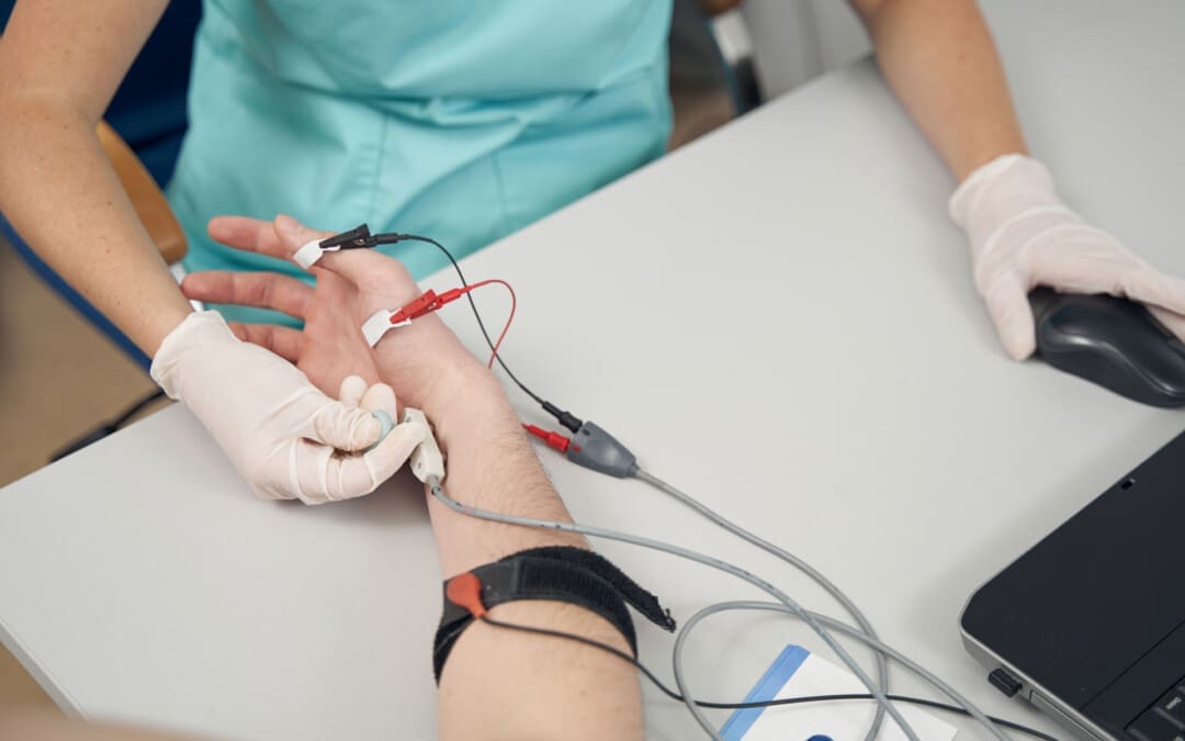
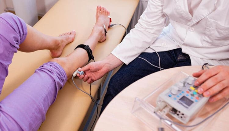
A nerve conduction velocity (NCV) is a noninvasive test that measures the speed and strength of nerve stimulation using electrical probes placed on the skin. It’s used to diagnose nerve damage or disease, often alongside an EMG (electromyogram) to differentiate between nerve and muscle problems. It can also evaluate sensory issues, pain, and weakness of the extremities.
This test involves safe electrical shocks that can be slightly uncomfortable but not painful.
Nerve conduction velocity (NCV) measures the speed at which electrical impulses travel along a nerve fiber, which measures how quickly electrical signals travel through a nerve.
This information indicates nerve health and function.
Electromyography (EMG) is a nerve test that involves placing tiny needles into the muscles.
A slower NCV can indicate nerve injury or dysfunction.
Test Uses
Generally, the test is ordered to assess peripheral nerve diseases, those that connect from the muscles, organs, and skin to the spinal cord or brain. It can help identify the type and location of nerve damage.
Peripheral nerve conditions typically cause pain, sensory loss, tingling, or burning.
Mild weakness and diminished reflexes can be detected during a neurological examination.
Conditions
Nerve conduction studies are performed to help diagnose conditions.
Nerve damage (neuropathy), such as from diabetes, chemotherapy, or autoimmune disorders
Charcot-Marie-Tooth disease
Nerve compression
Many different conditions, including trauma, inflammation, and tumors, can compress one or more nerves.
Radiculopathy
Often described as a pinched nerve, radiculopathy can affect an arm or a leg, causing pain and weakness.
Peripheral Neuropathy
This nerve damage begins in the most distal nerves, those farthest from the center of the body, such as the toes and fingers. It is often due to chronic alcohol misuse, uncontrolled diabetes, nutritional deficits, and inflammatory diseases. (Ferdousi M. et al., 2020)
Carpal Tunnel Syndrome
Commonly caused by inflammatory diseases or overuse of the wrists, such as from assembly line work, carpal tunnel syndrome causes numbness, pain, and weakness of the fingers and hands. (Tada K. et al., 2022)
Ulnar neuropathy
This common condition causes arm pain and sensory changes, usually due to repetitive movements or a prolonged position that causes pressure on the ulnar nerve.
Guillain-Barré syndrome (GBS)
This inflammatory condition causes demyelination, or loss of the insulating covering around nerves, which results in leg weakness.
It begins in the motor nerves, which send signals to muscles in the legs. (Shibuya K. et al., 2022)
The inflammation travels to nerves of the upper body, often affecting the muscles that control breathing.
Respiratory support is necessary until the condition improves.
Chronic Demyelinating Polyneuropathy (CIDP)
This condition is a chronic, recurrent form of GBS that usually affects the legs and causes episodes of weakness.
ICU neuropathy
Metabolic changes, severe illness, and not moving enough can cause nerves to develop a pattern of weakness and sensory loss.
Myasthenia gravis (MG)
This autoimmune condition affects the junction between the nerves and the muscles.
Myasthenia gravis causes drooping eyelids and weakness of the arms and shoulders.
Amyotrophic lateral sclerosis (ALS)
ALS is a serious, degenerative disease affecting the spinal cord’s motor neurons.
Amyotrophic lateral sclerosis progresses rapidly, resulting in substantial weakness of muscles throughout the body.
How it’s Done
Surface electrodes are placed on the skin over nerves, and a small electrical current is applied to stimulate the nerve.
The time it takes for the electrical signal to travel between the electrodes is measured, and this time is used to calculate the NCV.
Values
Normal NCV values are generally between 50 and 70 meters per second. However, these values can vary depending on the nerve and the individual.
NCV Factors
Various factors can influence NCV.
Age
Sex
Medical conditions like diabetes
Interpretation
A slower NCV can indicate nerve damage or demyelination (loss of the myelin sheath, which insulates nerve fibers), while an EMG can help determine if the problem is with the nerve or the muscle.
Results
The results of NCV testing can be used to determine the type, severity, and location of nerve damage. The results will be ready in report form about a week after the test.
The test measures velocity (how fast a nerve transmits signals) and amplitude (how many nerve fibers were activated). (Tavee J. 2019)
The measurements are transmitted to a computer and shown as waves and numerical values.
The values are compared to a standard measurement based on the tested nerve.
The distance between the electrodes.
The person’s age.
Compared to the standard, the NCV results can identify certain patterns of nerve damage. (Tada K. et al., 2022) Outcomes include: (Tavee J. 2019)
If one or more nerves are affected.
If motor nerves (control movement), sensory nerves (transmit sensory signals), or both are affected.
Whether a nerve is blocked or damaged.
The severity of the damage.
The type of nerve damage
Axonal (damage to the nerve itself)
Demyelination (damage to the protective fatty layer around the nerve)
The results can help point to certain diagnoses.
Preparation Before the Test
Individuals will not need to change their diet before having an NCV. However, patients will be asked to avoid lotions or creams on their skin before the test. Individuals who are also having an EMG at the time of their NCV might be asked to stop taking medications or supplements that increase the risk of bleeding and bruising. If a healthcare provider says not to stop taking the medicines for health reasons, the patient might be warned that they could have some bruising after the EMG test.
NCV may advise against getting the test for those with electrical device implants.
Make sure your healthcare providers are aware of your whole medical history.
Injury Medical Chiropractic & Functional Medicine Clinic
Injury Medical Chiropractic and Functional Medicine Clinic works with primary healthcare providers and specialists to develop an optimal health and wellness solution. We focus on what works for you to relieve pain, restore function, and prevent injury. Regarding musculoskeletal pain, specialists like chiropractors, acupuncturists, and massage therapists can help mitigate the pain through spinal adjustments that help the body realign itself. They can also work with other medical professionals to integrate a treatment plan to resolve musculoskeletal issues.
Peripheral Neuropathy and Chiropractic Care
References
Ferdousi, M., Kalteniece, A., Azmi, S., Petropoulos, I. N., Worthington, A., D’Onofrio, L., Dhage, S., Ponirakis, G., Alam, U., Marshall, A., Faber, C. G., Lauria, G., Soran, H., & Malik, R. A. (2020). Corneal confocal microscopy compared with quantitative sensory testing and nerve conduction for diagnosing and stratifying the severity of diabetic peripheral neuropathy. BMJ open diabetes research & care, 8(2), e001801. https://doi.org/10.1136/bmjdrc-2020-001801
Tada, K., Murai, A., Nakamura, Y., Nakade, Y., & Tsuchiya, H. (2022). In Carpal Tunnel Syndrome, Sensory Nerve Conduction Velocities Are Worst in the Middle Finger Than in the Index Finger. Frontiers in Neurology, 13, 851108. https://doi.org/10.3389/fneur.2022.851108
Shibuya, K., Tsuneyama, A., Misawa, S., Suzuki, Y. I., Suichi, T., Kojima, Y., Nakamura, K., Kano, H., Ohtani, R., Aotsuka, Y., Morooka, M., Prado, M., & Kuwabara, S. (2022). Different patterns of sensory nerve involvement in chronic inflammatory demyelinating polyneuropathy subtypes. Muscle & Nerve, 66(2), 131–135. https://doi.org/10.1002/mus.27530
Tavee J. (2019). Nerve conduction studies: Basic concepts. Handbook of Clinical Neurology, 160, 217–224. https://doi.org/10.1016/B978-0-444-64032-1.00014-X
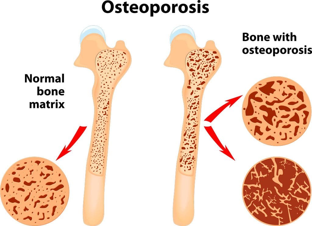
What is a bone density test, how is it performed, and what do the results mean?
Bone Density Test
A bone density test examines bone mass, which indicates overall bone strength. Assessing bone density or mass is necessary for diagnosing osteopenia or osteoporosis, conditions that increase the risk of broken bones. The scan is performed through dual-energy X-ray absorptiometry (DEXA), which examines the thickness of the bones. Results from DEXA scans are compared to standardized values to determine whether bone density is lower than normal and whether osteopenia or osteoporosis is present.
Examination
The procedure examines bone density, or bone mass. The bones’ density, or mass, is an overall indicator of bone strength. The greater the bone density, the thicker and stronger the bones are. The test is used to diagnose osteoporosis, a condition characterized by brittle bones at risk of breaking due to significantly low bone density. A bone density test can also diagnose osteopenia, a condition characterized by lower than normal bone mass that can lead to osteoporosis. (National Institute of Arthritis and Musculoskeletal and Skin Diseases, 2025) It is recommended that all women aged 65 and older and all men aged 70 and older have a bone density scan to screen for bone loss to help prevent fractures. (Kling J. M., Clarke B. L., & Sandhu N. P. 2014)
Bone density scans can establish a baseline level of bone density and track changes over time.
For individuals with osteoporosis or osteopenia, a bone density scan can help track how well their bones respond to treatment.
During a DEXA scan, the patient will lie on their back on a table with their legs elevated on a padded platform.
An X-ray scanner will pass over the spine and hips while another scans beneath.
While the scan takes place, the patient will be asked to hold very still to obtain an accurate image.
The scan will obtain bone density readings from the spine and hip, the two most commonly fractured bones, and generally takes less than 30 minutes.
Results
A DEXA scan measures bone density in grams per centimeter squared (g/cm²). This number indicates how densely bone cells are packed together in a specific area of bone. This bone density reading is then compared to a standardized value to determine if bone density is within a normal range or lower than average.
Between minus 1.0 and minus 2.5: Low bone density (osteopenia)
Equal to minus 2.5 or below: Osteoporosis
Bone density values are reported as a Z score for women who have not undergone menopause and men under 50 years old.
Z scores are compared to bone density levels of individuals of the same age and sex.
A Z score of minus 2.0 or lower indicates low bone density, which can be caused by factors other than aging, such as medication side effects, nutritional deficiencies, or thyroid problems.
Arthritis Diagnosis
Because a DEXA scan only measures the thickness of bones, it doesn’t work to diagnose arthritis. An X-ray of the affected joint is currently the most accurate way to diagnose arthritis. The Kellgren-Lawrence classification system categorizes the extent of arthritis based on the severity of joint damage seen on an X-ray. According to this system, arthritis can be classified as: (Kohn M. D., Sassoon A. A., & Fernando N. D. 2016)
Grade 1 (minor)
Minimal or no joint space narrowing, with possible bone spur formation.
Grade 2 (mild)
Possible joint space narrowing, with definite bone spur formation.
Grade 3 (moderate)
Definite joint space narrowing, moderate bone spur formation, mild sclerosis (abnormal thickening of bone), and possible deformation of bone ends.
Grade 4 (severe)
Severe joint space narrowing, large bone spur formation, marked sclerosis, and definite deformation of bone ends.
Injury Medical Chiropractic & Functional Medicine Clinic
Exercise can be incredibly beneficial for improving bone density, joint mobility, and the strength of surrounding muscles, which support and protect joints and bones. Talk to a healthcare provider to learn what interventions and available treatment options would be the most effective. Injury Medical Chiropractic and Functional Medicine Clinic works with primary healthcare providers and specialists to develop an optimal health and wellness solution. We focus on what works for you to relieve pain, restore function, and prevent injury. Regarding musculoskeletal pain, specialists like chiropractors, acupuncturists, and massage therapists can help mitigate the pain through spinal adjustments that help the body realign itself. They can also work with other medical professionals to integrate a treatment plan to resolve musculoskeletal issues.
Osteoporosis
References
National Institute of Arthritis and Musculoskeletal and Skin Diseases. (2025). Bone mineral density tests: what the numbers mean. Retrieved from https://www.niams.nih.gov/health-topics/bone-mineral-density-tests-what-numbers-mean
Kling, J. M., Clarke, B. L., & Sandhu, N. P. (2014). Osteoporosis prevention, screening, and treatment: a review. Journal of women’s health (2002), 23(7), 563–572. https://doi.org/10.1089/jwh.2013.4611
Kohn, M. D., Sassoon, A. A., & Fernando, N. D. (2016). Classifications in Brief: Kellgren-Lawrence Classification of Osteoarthritis. Clinical orthopaedics and related research, 474(8), 1886–1893. https://doi.org/10.1007/s11999-016-4732-4
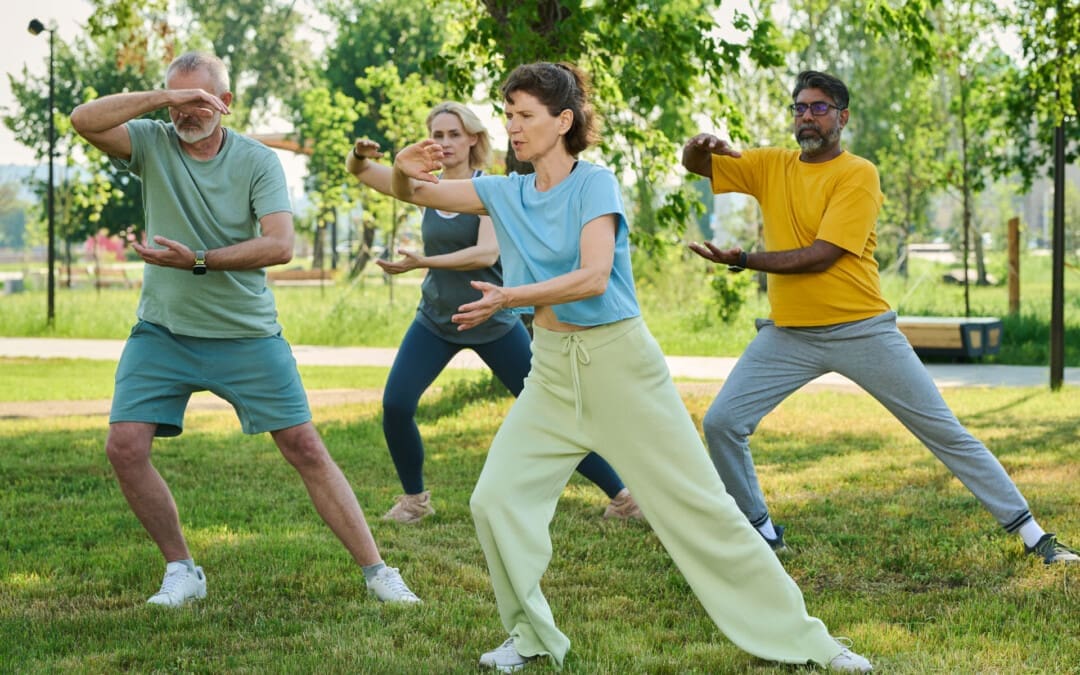
For individuals dealing with digestive issues and conditions, can incorporating Tai Chi help improve gut health?
Tai Chi For Gut Health
Tai Chi is an exercise that has been referred to as moving meditation. The practice is rooted in Chinese medicine, which fuses martial arts and meditation. The art of Tai Chi is used to improve physical health. It can also positively impact gut health by improving digestive function, reducing inflammation, and influencing the gut microbiota composition. The slow, controlled movements and deep breathing can stimulate abdominal and pelvic organs, promoting digestion. Additionally, it has been shown to reduce inflammation in the gut and increase the diversity of gut bacteria, including beneficial butyrate-producing bacteria. (Kang D., Wang X., & Wang J., 2023)
Types
The idea is to slow down your mind and body by repeating rhythmic choreography and breathwork for about 30 to 60 minutes, culminating in finding a sense of inner peace and tranquility.
Primary Forms/Styles
These include Chen, Yang, Wu, Sun, and Wu/Hao. Each follows the same origins and principles with variations. (Tai Chi for Health Institute, 2007)
Chen Style
Considered the oldest and original style, the Chen style is characterized by explosive power, low stances, and a combination of fast and slow movements, including jumping, kicking, and striking.
Chen also utilizes a movement called “silk reeling,” a spiral-esque, flowing movement that starts at the feet and moves into the hands.
Yang Style
Yang is often considered the most popular form of Tai Chi and is practiced worldwide.
Yang Tai Chi focuses more on improving flexibility through grand, sweeping movements executed slowly and gracefully.
Wu Style
Wu Tai Chi emphasizes small, compact movements and a medium stance. Its focus is on extending the body by leaning forward and backward.
Sun Style
Sun Tai Chi combines elements of Tai Chi, Xing Yi, and Ba Gua, resulting in a unique style with fluid, circular movements.
Hao Style
This style is characterized by small-frame movements focusing on accurate position and internal strength.
Tai Chi can indirectly benefit gut health by reducing stress levels.
Its emphasis on slow movements and deep breathing can help reduce stress.
Combining meditative practices with physical movement can help calm the mind, improve focus, and even trigger the release of endorphins.
Improved Digestion
Gentle, flowing movements, particularly those involving the diaphragm, can massage and stimulate the abdominal and pelvic organs, aiding the digestive process.
Reduced Inflammation
Tai Chi can help reduce gut inflammation, a common issue in conditions like inflammatory bowel disease (IBD).
Gut Microbiota Changes
Tai Chi has been found to positively influence the gut microbiota composition, increasing the diversity and abundance of beneficial bacteria.
Improved Gut Barrier Function
Tai Chi may help improve the integrity of the gut barrier, which is essential for preventing harmful substances from entering the bloodstream and causing inflammation.
Increased Butyrate Production
Tai Chi can promote the growth of butyrate-producing bacteria, which are important for intestinal health and can reduce inflammation.
Overall Health Benefits
Increases Cognitive Function
In addition to improving your mental well-being, Tai Chi has also been found to boost cognitive abilities.
A meta-analysis stated that physical exercise, in general, improves cognitive function, and researchers specifically recommended Tai Chi for elderly individuals since it’s a gentler and more accessible form of physical exercise that also combines mental exercises through repeated choreography. (Yin Wu, et al., 2013)
Increases Flexibility and Agility
Similar to yoga, Tai Chi often involves body extensions that can improve flexibility and agility.
This is useful in daily activities and makes you more agile and capable in other sports.
Improves Balance and Coordination
In addition to improving flexibility and agility, the intricate movements can help balance and coordination.
This skill is useful in daily life.
It can help with fine motor skills and even prevent trips, stumbles, falls, and other sports.
Enhances Strength and Stamina
As with any form of physical exercise, Tai Chi can build upon existing strength and stamina.
With ongoing practice, individuals become leaner, their muscles are more defined, and they can exercise longer.
Injury Medical Chiropractic & Functional Medicine Clinic
Talk to a healthcare provider to learn what interventions would help the most. Injury Medical Chiropractic and Functional Medicine Clinic works with primary healthcare providers and specialists to develop an optimal health and wellness solution. We focus on what works for you to relieve pain, restore function, and prevent injury. Regarding musculoskeletal pain, specialists like chiropractors, acupuncturists, and massage therapists can help mitigate the pain through spinal adjustments that help the body realign itself. They can also work with other medical professionals to integrate a treatment plan to resolve musculoskeletal issues.
Body Maintenance
References
Kang, D., Wang, X., & Wang, J. (2023). Intervention study of tai chi training on the intestinal flora of college student basketball players. Medicine, 102(36), e35044. https://doi.org/10.1097/MD.0000000000035044
Wu Y, W. Y., Burgess EO, Wu J. (2013). The effects of Tai Chi exercise on cognitive function in older adults: A meta-analysis. Journal of Sport and Health Science, 2(4), 193-203. https://doi.org/https://doi.org/10.1016/j.jshs.2013.09.001
Tai Chi for Health Institute. (2018). History of Tai Chi. https://taichiforhealthinstitute.org/history-of-tai-chi/#:~:text=Based%20on%20Qigong%20and%20martial%20art%20techniques,It%20contains%20explosive%20power%20and%20low%20stances.
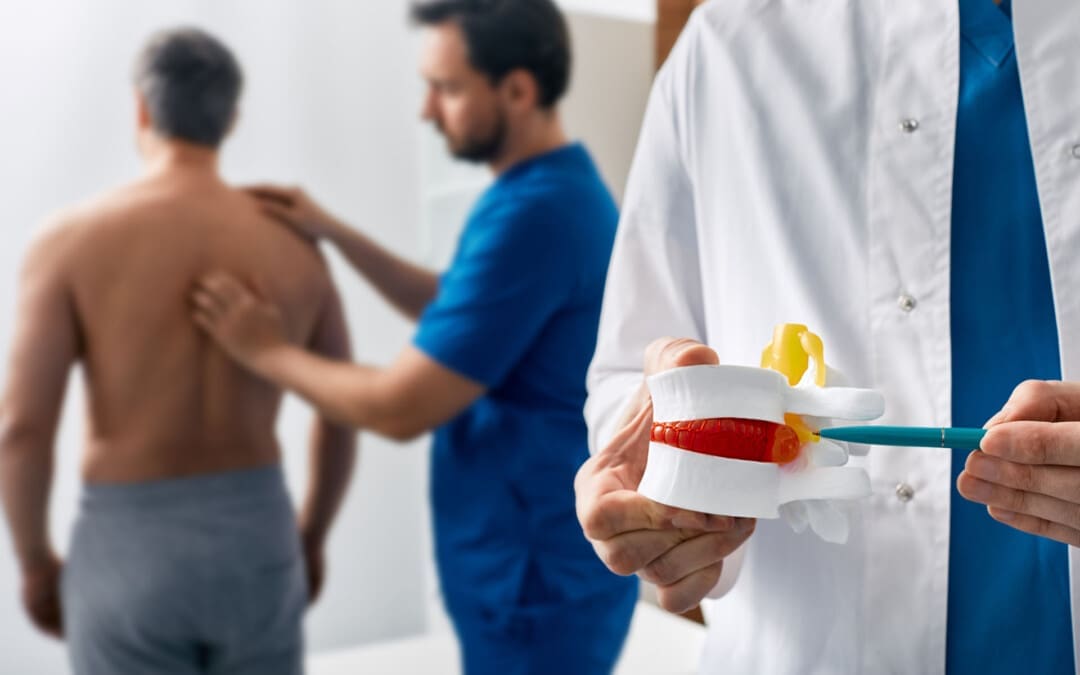
Can postural assessments help identify problems with how a person stands or sits, which can lead to various musculoskeletal issues?
Postural Assessment
Proper posture is an exercise in which the muscles support the skeleton in a comfortable, stable, and efficient alignment. Healthy posture is present when the body is still and when moving. However, numerous factors can affect and hinder posture. These include daily wear and tear, injury, illness, or a condition. A posture assessment is a process that identifies posture issues and their root causes, often using visual and palpation techniques, and can help determine appropriate treatment or exercises. (Science Direct, 2007)
Visual Assessment
Observing the body’s alignment and symmetry from different angles (anterior, posterior, and lateral views).
Consider their daily activities, work environment, and any previous injuries. (Du, S. H. et al., 2023)
Wall Test
The patient stands against a wall with their feet shoulder-width apart and heels about 6 inches from the baseboard.
If they have good posture, their ears will be vertically aligned with their shoulders, and their head will be no more than three finger widths from the wall. (Physiopedia, 2025)
Professionals Who Can Perform a Posture Assessment
Physiotherapists
Professionals trained in assessing and treating musculoskeletal problems.
Chiropractors
Professionals who focus on the spine and nervous system.
Fitness Professionals
Personal trainers or other fitness professionals can use posture assessments to help clients improve their posture and movement.
Ergonomists
Professionals who specialize in designing workspaces and environments to promote good posture and reduce strain.
Injury Medical Chiropractic & Functional Medicine Clinic
Talk to a healthcare provider to learn what interventions would help the most. Injury Medical Chiropractic and Functional Medicine Clinic works with primary healthcare providers and specialists to develop an optimal health and wellness solution. We focus on what works for you to relieve pain, restore function, and prevent injury. Regarding musculoskeletal pain, specialists like chiropractors, acupuncturists, and massage therapists can help mitigate the pain through spinal adjustments that help the body realign itself. They can also work with other medical professionals to integrate a treatment plan to resolve musculoskeletal issues.
Singla, D., & Veqar, Z. (2014). Methods of postural assessment used for sports persons. Journal of clinical and diagnostic research: JCDR, 8(4), LE01–LE4. https://doi.org/10.7860/JCDR/2014/6836.4266
Du, S. H., Zhang, Y. H., Yang, Q. H., Wang, Y. C., Fang, Y., & Wang, X. Q. (2023). Spinal posture assessment and low back pain. EFORT open reviews, 8(9), 708–718. https://doi.org/10.1530/EOR-23-0025
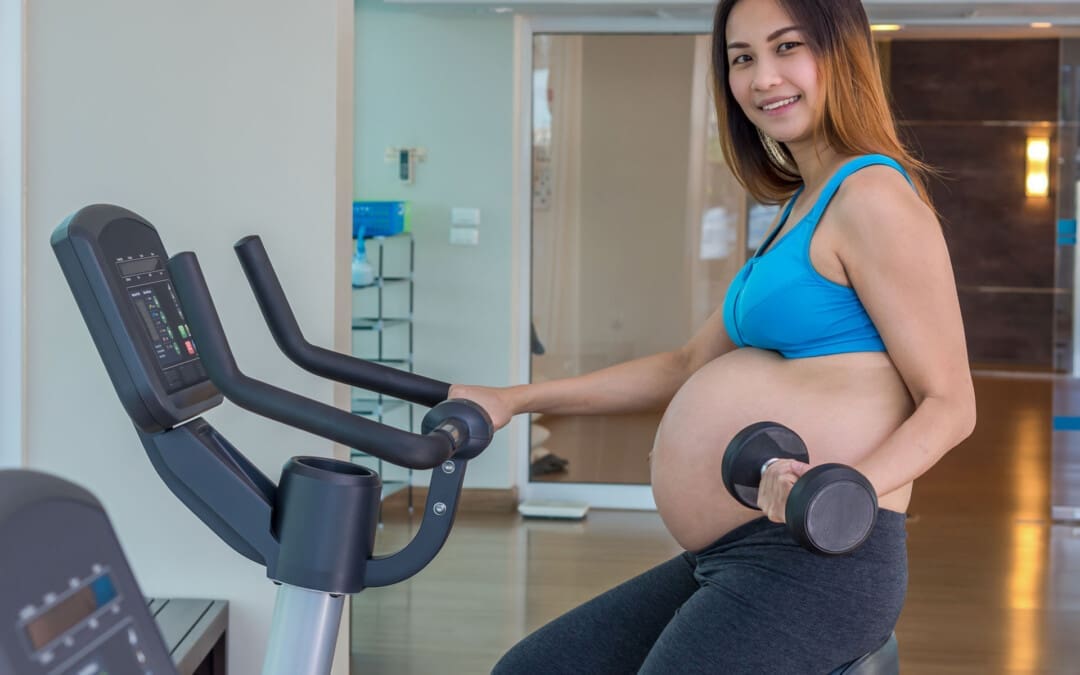
The American College of Obstetricians and Gynecologists, or ACOG, advises pregnant individuals to exercise regularly during pregnancy. (The American College of Obstetricians and Gynecologists, 2024) The guidelines indicate that individuals who regularly engage in vigorous-intensity aerobic exercise before pregnancy should continue these activities during their pregnancy. (Syed H., Slayman T., & DuChene Thoma K. 2021) According to ACOG, observational studies of pregnant individuals who exercise show benefits such as:
Indoor cycling is ideal because individuals won’t have to deal with balance challenges or generate a heavy impact on their joints. There are many indoor cycling workouts to try, whether spin or on-demand classes. Indoor cycling is safer during pregnancy than outdoor cycling, which is not recommended because of the risk of falls from traffic and weather conditions. Although indoor cycling is generally considered safe during pregnancy, individuals should get clearance from their OB/GYN if they have any underlying medical conditions that might limit physical activity options.
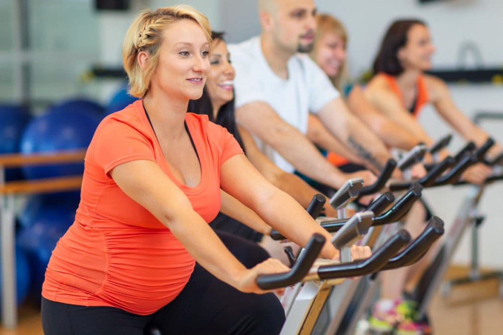
Cycling Classes
Taking cycling classes during pregnancy is safe if a healthcare provider has no concerns. It’s important to take precautions.
It’s recommended to continue with any exercise you were doing before pregnancy rather than start a new routine.
Remember that this is an exercise for two, so the heart rate will elevate quickly and become overheated more easily.
Take it easier on the bike, and don’t push too hard.
Consult With the Instructor
It’s recommended to seek out an instructor with some prenatal exercise training. Individuals may benefit from sticking with the same instructor whenever possible to get to know them and familiarize themselves with their modifications and needs. Whether or not you’re showing, tell the instructor that you’re pregnant before the class starts. This way, they can monitor progress and will not push too hard. The instructor can also give important pointers on modifying the ride to suit your needs.
Modify Bike Set-Up
Individuals may need to adjust the saddle position and raise the handlebars to stay comfortable as their bodies change. Sitting more upright is recommended to relieve strain on the lower back, and increasing the handlebars and bringing them closer instead of leaning forward is another goal. Another goal is to keep the weight more evenly distributed between the hands and body. Also, avoid movable indoor bikes that mimic outdoor riding. They can lean sideways, which might cause a fall.
Dial Down Intensity
With indoor cycling, it’s best to exercise moderately during pregnancy. Consider wearing a heart rate monitor to ensure a safe intensity. It’s also helpful to pay attention to the Rating of Perceived Exertion scale/RPE. Even if the heart rate isn’t too high, slow down or stop exercising immediately if you’re gasping for breath or feeling lightheaded. ACOG guidelines indicate that 13-14 “somewhat hard” on the Borg RPE scale is a safe and acceptable level of exertion. The guidelines also state that RPE is a better gauge of exertion than heart rate and that the talk test (holding a conversation while exercising) can indicate safe workout intensity.
Stay Cool and Properly Hydrated
Wear comfortable, breathable clothing to help you stay cool and a bra with plenty of support. Drink lots of water throughout the workout, actually more than usual. Overheating and dehydration are common during pregnancy and can be dangerous for both parents and babies. Carrying an extra 20 to 30 pounds and having 40% more blood pumping through the body toward the end of pregnancy makes you likely to sweat more and can easily lead to dehydration. Using a fan for home gyms is highly recommended.
Avoid Standing and Stay In a Seated Position
During the early months, you may be able to ride in a standing position without any problems. But as the belly grows, it changes the body’s center of gravity, putting more pressure on the joints and making it difficult to ride standing. Joints are looser or more flexible during pregnancy, which makes standing while cycling more difficult and risky. It is still a healthy workout if you stay seated the whole time—and, most importantly, avoid overdoing it or injuring yourself.
Body Signs
Listen to the body while exercising. If you get winded, dizzy, or unwell while riding, take a break or reduce your effort by a few notches. If a 45-to-60-minute class is too intense, feel free to depart early; just let the instructor know you’re OK. Energy will likely ebb and flow during pregnancy, so pay attention to the body’s signals and take care of them accordingly. Stop exercising if you experience any of the following (Syed H., Slayman T., & DuChene Thoma K. 2021)
Abdominal pain
Dyspnea: shortness of breath before exertion
Headache
Dizziness
Calf pain or swelling
Muscle weakness affects balance
Chest pain
Amniotic fluid leakage
Regular painful contractions
Vaginal bleeding
Call your doctor if you experience sharp pain, contractions, a surge of fluid, a sudden severe headache, prolonged swelling, or decreased baby movement.
Injury Medical Chiropractic & Functional Medicine Clinic
It’s important to exercise wisely during the nine months to accommodate body changes, the extra weight, the increasingly relaxed ligaments, and the shift in the center of gravity. The stationary bike provides a personalized, low-impact workout. You get to control the intensity and the duration of the ride. Monitor your heart rate and/or RPE to avoid overdoing it. Injury Medical Chiropractic and Functional Medicine Clinic works with primary healthcare providers and specialists to develop an optimal health and wellness solution. We focus on what works for you to relieve pain, restore function, and prevent injury. Regarding musculoskeletal pain, specialists like chiropractors, acupuncturists, and massage therapists can help mitigate the pain through spinal adjustments that help the body realign itself. They can also work with other medical professionals to integrate a treatment plan to resolve musculoskeletal issues.
Chiropractic Lower Back Pain Pregnancy Treatment
References
Hinman, S. K., Smith, K. B., Quillen, D. M., & Smith, M. S. (2015). Exercise in Pregnancy: A Clinical Review. Sports Health, 7(6), 527–531. https://doi.org/10.1177/1941738115599358
The American College of Obstetricians and Gynecologists. (2024). Exercise during pregnancy. https://www.acog.org/womens-health/faqs/exercise-during-pregnancy?utm_source=redirect&utm_medium=web&utm_campaign=int
Syed, H., Slayman, T., & DuChene Thoma, K. (2021). ACOG Committee Opinion No. 804: Physical Activity and Exercise During Pregnancy and the Postpartum Period. Obstetrics and gynecology, 137(2), 375–376. https://doi.org/10.1097/AOG.0000000000004266
Individuals dealing with pain in the buttocks and in the back of the thigh, along with numbness and tingling down to the bottom of the foot, may be experiencing hamstring syndrome, a condition caused by pressure on the sciatic nerve. What is the recommended treatment?
Hamstring-Syndrome Relief
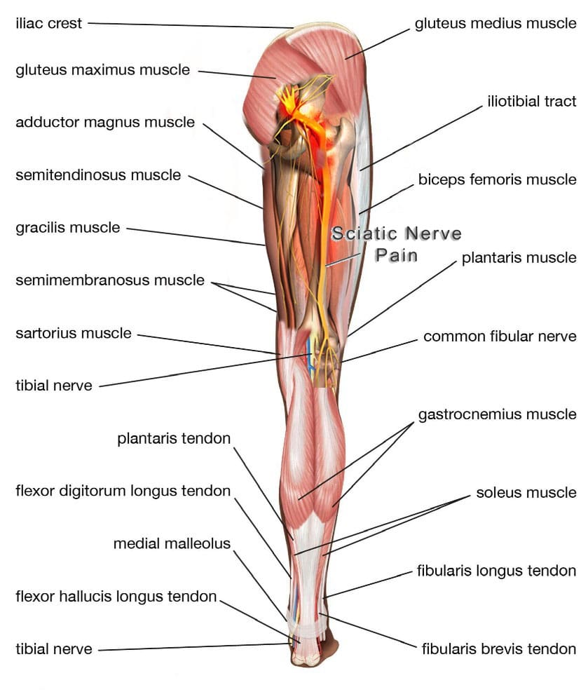
The hamstrings are three muscles in the back of the thigh, extending from the pelvis or upper thigh across the back of the knee to the leg. This muscle group is important for bending the knee, straightening the hip, and stabilizing the knee. The sciatic nerve is a large nerve that runs from the lower back down the legs. It usually passes near or through these muscles, and the pelvis then runs under these muscles in the thigh. Hamstring syndrome refers to pain in the buttock and back of the thigh, often radiating down the leg, caused by compression or irritation of the sciatic nerve at the hamstring-insertion point on the ischial tuberosity, typically due to tight or scarred tissue. (Sakari Orava, 1997)
Pain Location
The pain is primarily felt in the buttock and back of the thigh, sometimes extending down the leg. It’s characterized by pressure on the sciatic nerve, which runs through the buttock and into the back of the thigh, where it supplies the hamstring muscles. (Kaiser Permanente, 2024)
In some cases, injections with cortisone and numbing medicine may be used to reduce nerve inflammation and pain. (Lower Limb Surgery, 2024)
Surgery
In severe cases, surgery may be necessary to release the compressing bands and free the sciatic nerve. (Lower Limb Surgery, 2024)
Injury Medical Chiropractic & Functional Medicine Clinic
Talk to a healthcare provider about what interventions would help the most. Injury Medical Chiropractic and Functional Medicine Clinic works with primary healthcare providers and specialists to develop an optimal health and wellness solution. We focus on what works for you to relieve pain, restore function, and prevent injury. Regarding musculoskeletal pain, specialists like chiropractors, acupuncturists, and massage therapists can help mitigate the pain through spinal adjustments that help the body realign itself. They can also work with other medical professionals to integrate a treatment plan to resolve musculoskeletal issues.
Kaiser Permanente. (2024). Hamstring Syndrome: Care Instructions. https://healthy.kaiserpermanente.org/health-wellness/health-encyclopedia/he.hamstring-syndrome-care-instructions.abr3618
Puranen, J., & Orava, S. (1988). The hamstring syndrome. A new diagnosis of gluteal sciatic pain. The American Journal of Sports Medicine, 16(5), 517–521. https://doi.org/10.1177/036354658801600515
Zion Physical Therapy. (2023). Hamstring Tendinitis Vs. Hamstring Syndrome. https://www.zionpt.com/post/hamstring-tendinitis-vs-hamstring-syndrome
IFM's Find A Practitioner tool is the largest referral network in Functional Medicine, created to help patients locate Functional Medicine practitioners anywhere in the world. IFM Certified Practitioners are listed first in the search results, given their extensive education in Functional Medicine