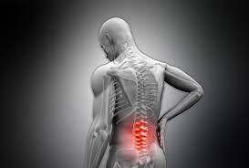
Question: I’ve had back pain for awhile and I was able to function through it for a time. But now it’s taking its toll on me, as well as my family. I never believed in chiropractors, but now I’ll try anything. What should I ask my physician about chiropractic?
El Paso, TX. Chiropractor, Dr. Alexander Jimenez has seen this scenario all too often and is happy to inform anyone about chiropractic care and it’s benefits.
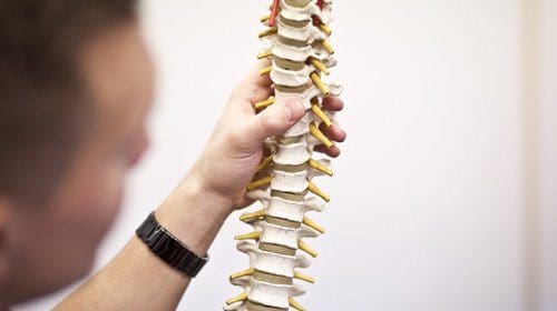
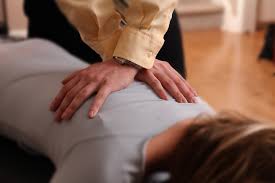
About two out of every three people will likely experience significant low back pain at some point. A doctor might suggest any range of potential treatments and therapies. But one that almost is never considered is a referral for spinal manipulation.
It seems doctors have been mistaken for initial treatment of lower back pain.�It may be time for doctors to rethink their chiropractic biases.
Spinal Manipulation:
Along with other less traditional therapies like heat, acupuncture and meditation, chiropractic appears to be as powerful as any other medical treatments doctors prescribe and as safe, if not safer.
Physicians are traditionally wary of spinal manipulation (applying pressure on bones and joints), in part because the practitioners are often not doctors. Patients with back pain don’t seem as skeptical. A survey from 2002 through 2008 found that over 30 percent sought chiropractic care, significantly more than those who sought massage, acupuncture or homeopathy.
Researchers have been looking at the evidence supporting spinal manipulation for a while. Nearly 35 years ago, a systematic review assessed the available research found that there may be some short-term benefits from the process. Two reviews from 2003 agreed for the most part, finding that spinal manipulation worked better than a “sham procedure”, or placebo, but no better or worse than other options.
Recently, in The Journal of the American Medical Association, researchers looked for new studies since 2011, in addition to those that had been performed before.
Evidence from 15 randomized controlled trials, which included more than 1,700 patients, revealed that spinal manipulation minimized their back pain of about 10 points on a 100-point scale. The evidence from 12 randomized controlled trials that overlapped of almost 1,400 patients showed that spinal manipulation also resulted in improved mobility and function.
In February, in Annals of Internal Medicine, another systematic review of non-pharmacologic therapies generally agreed with the recent trials. Based on this review, and other evidence, the American College of Physicians released new clinical practice guidelines for the noninvasive treatment of back pain. They recommended that patients should try spinal manipulation, heat, massage and acupuncture as first-line therapies.
What If The Spinal Adjustment Causes Other Injuries:
Because of the fear of those injuries, physicians are hesitant to refer patients to chiropractors or physical therapists for care. However, in the studies outlined above, there were no serious adverse events reported. It is possible to find stories of injury to the spine from improper adjustments, but these are rare, and almost never involve the lower spine.
What About The Cost:
Some doctors are concerned about the cost of chiropractic, especially since many insurance carriers don’t cover chiropractic. Seeing a chiropractor costs more than taking many non-narcotic pain medications. However, more invasive interventions cost a lot of money. In addition, studies have proven that, in general, users of complementary and alternative medicine spend less for back pain compared to users of only traditional medicine.
Medication and surgery may also result in injuries and prescription pain medicines, like opioids, may result in huge costs, particularly when they’re misused.
Some physicians are uncomfortable that they do not have a clear picture of how spinal manipulation actually works. The reality is that doctors don’t understand why a number of other therapies work either.
Back pain is still the issue and no matter what you do, chiropractic is definitely an option to consider.
Chiropractic: Alternative Approach To Treating Chronic Pain
Pain is defined as a physical discomfort caused by illness or injury. Most cases of pain are temporary, or they disappear once the source of the symptom is treated or healed, however, what happens when the pain becomes persistent?
How can you diagnose chronic pain?
Diagnosing chronic pain can be a long procedure. Because there are lots of possible causes behind the painful symptoms, the health care provider may need to conduct a variety of examinations and tests to attempt to find the source of your own pain. Below are a few of the methods the physician may try to diagnose chronic pain.
Medical History
Your doctor will take a comprehensive health history, going over any injuries, illnesses, and medical problems that run in your family. It is essential to be thorough so that the doctor can take a look at the full assortment of causes because chronic pain can generally be a result after injury or illness.
You’ll also have to be very particular about your pain: description, frequency, intensity, duration, activities that make it worse, even whether it is better at a certain time of day, etc.. It may be a good idea to initiate a pain journal where you record details of your pain. You’ll be better able to share information with the healthcare professional.
Physical, Neurological, and Mental Health Exams
For the physical examination, the physician will see your range of movement (that is how well and how much you can move specific joints), posture, and general physical condition. He or she will make note of any movements that increase or cause you pain.
The neurological examination will test your nerves, so this test is particularly critical for chronic pain. The doctor will test your reflexes, muscle power, and how well it is possible to feel. The physician might test if you can feel a touch in your skin. That may indicate nerve damage if you can’t. The healthcare provider will see whether your pain is currently spreading through the examination, �or whether you came in complaining of back pain.
Because chronic pain frequently has an emotional or psychological element, you might have to have a mental health examination. This is to check for symptoms such as stress or depression, that could develop alongside pain. The health exam may also give your doctor a complete picture of your overall health and wellness.
Diagnostic Tests
To see if there is an injury or identifiable illness causing your chronic pain, the healthcare professional will need to conduct diagnostic tests. For the imaging evaluations (x-rays, MRIs), you may have to go to an imaging center to have these done; the results will probably be sent back to your physician, who will interpret them for you.
Some possible diagnostic tests include:
Blood evaluation: Your doctor may be able to tell if you have specific forms of arthritis or a disease based on a blood test.�A blood test also allows the doctor to check your liver and kidney functions.
Bone scan: To help your doctor detect spinal problems such as osteoarthritis, sacroiliac joint dysfunction, fractures, or illnesses (which can all lead to chronic pain), you could have a bone scan. You will have a small amount of radioactive substance. That can travel through your blood flow and be absorbed by your bones. An area where there is abnormal action, like an inflammation, will absorb substances. A scanner can detect the quantity of radiation from all your muscles and also show the “hot spots” (the areas with more radioactive material) to help your doctor figure out where the issue is.
CT scan: A CT scan, which stands for computerized axial tomography, reveals the bones, but in addition, it reveals the soft tissues and nerves.
EMG: An electromyography (EMG) will check if your muscles are responding well to nerve stimuli.
MRI: An MRI, which stands for magnetic resonance imaging, shows the bones, but in addition, it shows the soft tissues and nerves. MRIs do not expose you to radiation by using magnets to get the picture.
Myelogram: To see whether you have a spinal canal or spinal cord disorder, maybe nerve compression causing weakness and pain, you might need a myelogram. In this evaluation, you’ll have a special dye injected into the fluid that surrounds your spinal cord and nerves. Then you’ll have a CT scan or an x-ray. The image will offer a detailed anatomic picture of your spine of the bones, which will assist your doctor.
NCV: A nerve conduction velocity (NCV) test will help the doctor evaluate your nerves and ascertain if there is any damage. This test is done together using the EMG test.
Nerve block: When the doctor suspects that a particular nerve is damaged and that is what is causing your chronic, they may do a nerve block. This is a special type of injection that may help identify if the nerve is the source of pain.
X-ray: This gives your doctor a clear picture of your bones.
The scope of our information is limited to chiropractic and spinal injuries and conditions. To discuss options on the subject matter, please feel free to ask Dr. Jimenez or contact us at 915-850-0900 .�
By Dr. Alex Jimenez
Additional Topics: Wellness
Overall health and wellness are essential towards maintaining the proper mental and physical balance in the body. From eating a balanced nutrition as well as exercising and participating in physical activities, to sleeping a healthy amount of time on a regular basis, following the best health and wellness tips can ultimately help maintain overall well-being. Eating plenty of fruits and vegetables can go a long way towards helping people become healthy.
Question: I work at a desk all day and have started to feel pain in my neck, back, shoulders and arms. Can chiropractors help with sitting posture and general posture?
El Paso, TX. Chiropractor, Dr. Alexander Jimenez provides insight into proper sitting posture and general posture through chiropractic.
Have people asked you about using a posture brace to help correct their sitting habits? Chiropractic patients with moderate posture problems, meaning that they may be chronic but are not tied to any spinal column or other medical condition, may benefit from a posture back brace to improve this condition. There are a number of great ones available, and you can see more information about some of the top ones here.
A patient such as this will almost certainly enjoy the quick referral as well as the relatively fast results, and chiropractors get an easy opportunity to help people, which explains why many enter the profession in the first place.
But if the patient does have an underlying chiropractic ailment, a back brace will probably do little if any good. These issues are usually easily diagnosed using a quick examination, and afterwards, treatment is relatively straightforward as well.
Diagnosing Conditions
All good exams begin with thorough health history, because in most cases, family history is among the most powerful indicators about current issues.
Next, observe the individual’s gait, as how someone walks shows a number of issues. For instance, those who stand or walk with their legs abnormally spread might have fallen arches. Because the bottom of the foot isn’t in appropriate contact with the floor, the whole body has been thrown off kilter, resulting in poor posture. While it isn’t really a chiropractic condition, fallen arches are, as simple as, a referral to an orthopedist.
Moreover, if the patient is experiencing pain, the location can be an indicator as to where the muscles may be weak and there is too much strain on the spine or a different region of the body. Muscle weakness is among the leading causes of poor posture. So, follow-up tests that measure muscle strength and range of movement are usually a good idea.
If the gait examination signals issues in a particular area of the body, the professional should concentrate further diagnostic efforts in that region.
Ultimately, one of the most simple and effective evaluations is just holding a yardstick or comparable object against the patient’s back, so the two of you can definitely see any abnormalities. This comprehensive approach generally describes some of the very frequent chiropractic posture issues, including:
Tissue Damage:
If the neck, lower back, mid back, or any other area is weak and/or inflexible, the individual will probably not see very much posture improvement until these conditions are adjusted and the muscles have been strengthened.
Kyphosis:
Hunchback is a degenerative spine condition that’s very prevalent in women over 60. While more advanced cases may be life threatening and may require spinal fusion surgery, most men and women respond well to therapeutic adjustments and other treatments.
Scoliosis:
This condition is much like genetically-induced kyphosis, since there’s no cure but there are several therapies available. In extreme cases, surgery may be necessary.
Un-Level Pelvis/Pelvic Tilt:
A pelvic tilt, a lower sacral base, and a femur head discrepancy can indicate a lower extremity source, but not�whether it is an anatomical or functional short leg. A clinical postural exam with lower extremity screening is the only way to make this determination.
Forward Head Posture:
The anterior positioning of the cervical spine. This posture is sometimes called Scholar’s Neck, Wearsie Neck, Hunch & or Reading Neck.It is a posture problem that is due to several factors including sleeping with the head raised too high, prolonged use of computers and cellphones, lack of developed back muscle strength and deficiency of nutrients like calcium. Potential negative effects include tingling and numbness in the arms, and a burning pain between the shoulder blades.
Treatment
As stated previously, back braces frequently mend postural issues, like slouching. As for structural problems, like scoliosis, more aggressive treatments are needed.
Conventional treatment consists of:
Heat
Massage
Stretching
Strength exercises
Supportive braces
There are a number of biofeedback tools that accurately evaluate patient progress.
As a chiropractor, your patients count on you to get much better. That almost always means accurately assessing the problem, which also means a quick and capable referral or an aggressive and well thought out therapy regimen.
Millions of people live with some form of back or neck pain every day. While most of these are only temporary and resolve on their own, many others will continue to experience painful symptoms long after the source of their pain has been treated or healed. Unfortunately, the exact cause of chronic pain is often misunderstood.
What causes chronic pain?
First of all, what is chronic pain? It is technically defined as pain that lasts for 12 weeks or longer, even after the pain is no longer acute (short-term, acute pain) or the injury has healed. Naturally there are numerous causes of chronic pain that can influence any level of the spine, cervical (neck), mid back (thoracic), low back (lumbar), sacral (sacrum) or a combination of levels (eg, lumbosacral).
Common Causes of Chronic Pain
Back pain, among one of the most common forms of chronic pain, is a great example to use for this particular cause. Let’s say that over time and through normal wear and tear, you’ve worn out the cartilage that surrounds the joints in your spine (the facet joints). The joints subsequently become swollen, and you develop osteoarthritis in the spine. This inflammation makes it quite painful for you to move, and it is a source of pain. Your damaged facet joints and cartilage may have developed into chronic pain. That’s only one example. Other common back pain causes include:
Trauma or injury:�In a car accident, injury and trauma can occur and it might still cause pain once it’s healed.
Bad posture: Years of slumping over can cause parts of your spine to wear out faster resulting in chronic pain.
Obesity: It’s well-documented that excessive weight puts strain and pressure on the spine. It can wear quicker or simply not work out as well, possibly leading to chronic pain.
Aging: Various areas of the spinal anatomy, as well as other joints, may wear out over time (a process called degeneration). There is no guarantee that spine degeneration will be debilitating, however: it depends on how it is affected by the degeneration process, to begin with.
The medical community is not certain severe (short-term) pain occasionally develops to chronic, (long-term) pain after the original pain source was treated. It is possible to learn more about chronic pain, in addition to other kinds of chronic pain, such as fibromyalgia or rheumatoid arthritis.
Chronic Pain Due to Nerve Damage
Nerves can be damaged or injured in a variety of ways. For instance, a nerve root may be pinched by a herniated disc, causing pain. After healing the herniated disc, the pain can persist because of �the initial nerve damage. Nerves may be injured by arachnoiditis (inflammation of a tissue that protects the nerve roots), arthritis (again, the inflammation may compress the nerve), diabetes, cancer, Lyme disease, an infection, and more. Nerves may have trouble relaying essential neurological communications between the brain and the rest of the body, if they’ve been damaged. One theory about pain is that the nerves don’t stop sending pain signals to the mind, even after the source of pain exists.
Unknown Causes of Chronic Pain
As you can tell from the preceding two sections, there’s a good deal of doubt when it comes to chronic pain. Through study, the community is starting to better understand pain and its causes, but they can’t pinpoint a cause for chronic pain. That does not mean that they can’t help you find pain relief and approaches to take control of your life again. It’s possible to achieve this with the proper diagnosis and treatments, although it’ll be a process.
Accurate Diagnosis
Obtaining an accurate diagnosis of the cause of your neck or back pain is essential for the result of your treatment plan. Depending upon the severity and cause of your chronic pain, you may have to seek the help of other healthcare professionals, such as pain medicine specialists, spine surgeons, neurosurgeons, physical therapists and/or chiropractors. Over time your pain may need to be reevaluated, and might vary in the way it is managed.
Your chronic neck or back pain treatment generally includes a combination of therapies to optimize outcomes. Some of the treatments your doctor prescribes and recommends may include:
Physical Therapy: Stretching and strengthening muscles is critical in treating chronic back or neck pain. Your physical treatment may include ice, heat, transcutaneous electrical nerve stimulation (TENS), ultrasound, and myofascial release.
Chiropractic Care: Based on your diagnosis, chiropractic care may be recommended by your physician. Studies have shown that two weeks of manipulation by a chiropractor may bring about significant improvement in some patients with chronic low back pain and referred leg pain, or sciatica.
Medications: There are many prescription drugs to treat inflammation, pain, muscle spasms, and pain. Furthermore, there are medications to treat conditions that often accompany chronic pain, such as medication to assist you alleviate depression, sleep, and decrease anxiety.
Procedures: Your doctor may recommend spinal cord stimulator spinal injections, drug pump, or spinal column operation. You can acquire a second opinion if you are not certain regarding the treatment program recommended.
Coping skills: Never underestimate the ability of the mind. Your mood and mental prognosis can impact your degree of pain. Assess with a professional to find out relaxation and coping skills.
Complementary Treatments: Doctors recommend acupuncture and other forms of traditional Chinese medicine. Speak to your doctor.
Fitness and Chronic Pain
While there’s some things you can’t stop from affecting your chronic pain, like the cellular changes which reduce muscle mass and/or bone density, it is possible to take steps to improve fitness which may benefit your symptoms.
It’s known that patients with chronic neck or back pain may not feel like going to the gym, being active or even following a house exercise program. However, being active can help improve chronic pain because during exercise, the body releases endorphins, your body’s natural painkiller.
Furthermore, by teaming up with your physical therapist, it is possible to start to construct abdominal muscles, essential for the core strength which helps support your spine of your body. Several studies show that aerobic exercise is successful in maintaining the integrity of the intervertebral discs in the lower spine, which can lessen back pain.
Were you aware that by losing even 5 to 10 pounds you can reduce back pain? It is true. Carrying around extra weight translates to added stress. In many cases, a difference can be made by early and aggressive therapy of chronic back or neck pain. But keep in mind that knowledge is power: Be certain that you understand your options.
The scope of our information is limited to chiropractic and spinal injuries and conditions. To discuss options on the subject matter, please feel free to ask Dr. Jimenez or contact us at 915-850-0900 .�
By Dr. Alex Jimenez
Additional Topics: Wellness
Overall health and wellness are essential towards maintaining the proper mental and physical balance in the body. From eating a balanced nutrition as well as exercising and participating in physical activities, to sleeping a healthy amount of time on a regular basis, following the best health and wellness tips can ultimately help maintain overall well-being. Eating plenty of fruits and vegetables can go a long way towards helping people become healthy.
Question: My parents are elderly, and both hurt their backs. What type of doctor or physician should I look for?
Chiropractic care is safe and effective for elderly adults with back pain, according to new research. To better understand the risk and benefits of chiropractic care in elderly individuals, El Paso, TX. Chiropractor, Dr. Alexander Jimenez, answers questions regarding chiropractic care for elderly individuals with back pain.??
Q: Is Chiropractic Care Safe For Elderly People?
Dr. Jimenez: Chiropractic care is one of the safest interventions for the care of back pain. The dangers of a serious problem after a chiropractic adjustment would be like the danger of having a serious problem after an immunization–exceptionally small. However, minor side effects lasting up to approximately 3 days are very common, most commonly muscle stiffness and soreness. These side effects are similar to those you get when you begin a workout program. People have a smaller chance of severe side effects from manipulation and manual treatment than they do from medication taken for the very same ailments. You should always discuss any side effects from treatment with your chiropractor right away.
Q: What Are The Benefits Of Chiropractic Care For The Treatment Of Low Back Pain In Older Adults?
Dr. Jimenez: Chiropractic care has been used for over 100 years to assist patients with back pain. One of the best benefits of trying chiropractic care first is that you can avoid the risk of side effects from taking medications for pain. Opioid drug addiction is a serious crisis in America. Trying non-drug therapies, such as chiropractic care first may help to reduce the high rates of opioid dependency in the USA.
Actually, the most recent treatment principle for low back pain (from the American College of Physicians), specifically states that people should attempt non-drug options first. The health dangers of pain medications are even greater for older adults compared to younger people, since many older people have other illnesses that affect their wellbeing, and their own bodies process drugs more slowly. Also, many drugs, particularly opioids, increase older people’s risk of falls, which is a very considerable risk in this age group.
Q: What Questions Should I Ask My Chiropractor Before Receiving Treatment?
Dr. Jimenez:�When first talking with a chiropractor, inquire which sort of treatments he or she may use for back pain. Chiropractic care incorporates many different non-drug approaches in addition to spinal manipulation, including other kinds of manual therapy in addition to exercise. �Ask the chiropractor to explain or illustrate the type of spinal manipulation he or she uses (many chiropractors have photographs or can demonstrate what they do), so that you feel comfortable with it. Should you feel uncomfortable with the therapy, feel free to ask whether there are alternate kinds of manipulation he or she may use as there are many different ways to perform spinal manipulation for optimum patient comfort, and also achieve the same targets.
Q: What Should I Tell A Chiropractor About My Medical History, Medicines, & Spinal Condition?
Dr. Jimenez: First of all, since chiropractors are trained to choose a thorough patient history, including questions regarding medication, past injuries or surgeries, and any recent changes in your health, most likely you won’t have to volunteer this information. You’ll be asked about your health history on a patient intake form. Then the doctor will ask you more questions in person. Make sure to talk about all health information about current and previous conditions, drugs, history of accidents and surgeries or alternative therapy, and current changes in your health, even if you think they are not important, they may be.
The chiropractor will also inquire what makes your pain worse or better, and if you have had changes in strength, recent falls, or balance issues, changes in your bowel or bladder function along with other relevant health factors like eating and sleep habits. The chiropractor should then perform a thorough physical examination, including your neurological system, to be certain that spinal manipulation is ideal for you. He or she’ll also see what kinds of adjustments will make your pain better or worse. The chiropractor will use all of this information to develop a treatment plan that’s specific for you to help you fulfill your goals for returning to a normal lifestyle.
Backpack pain is an all too common condition of school-age children. While back pain is a known and widely-studied issue in adults, its prevalence in school-aged children has received relatively little scientific attention. Elementary, middle, and high school students must often carry backpacks that weigh enough to trigger chronic back pain, bad posture, and even decreased lung volume. I have written about this issue earlier, but lately, several studies reveal the truths behind childhood back pain and ways to mitigate it.
Are Backpacks Too Heavy For Kids?
Recent research supports that children carrying backpack loads of over ten percent of their body weight have a greater chance of creating back pain and related difficulties. An global study found that an alarmingly large percentage of school-age kids in Australia, France, Italy, and the United States often carried backpacks weighing more than the ten percent threshold.
In a second study involving a sample of 1540 metropolitan school-aged children, more than a third of the children surveyed reported backpack pain. Along with carrying heavy backpacks, female students and those diagnosed with scoliosis had a larger association with back pain pain. Children with access to lockers reported less pain.
The number of straps on the back had little effect on the respondent’s replies. Children also reported restricted physical activity due to back pain, and some took drugs to alleviate the pain.
Girls who transported bags in addition to wearing a backpack reported considerably greater back pain. Adolescents with back pain spent more time watching television than their peers. More than 80 percent of the surveyed thought that carrying a heavy backpack due to their back pain.
Backpack Pain Solutions
The research revealed several things that might help reduce back pain in school-aged children. The best way to prevent back pain is to refrain from carrying heavy loads.
Kids ought to make the most of locker breaks and only carry items necessary for a couple of courses at one time. When lifting a back pack, children should crouch down and bend their knees rather than curve the spine.
Backpack Safety
Appropriate Backpack Carrying Techniques
While not conclusive, research also supports that carrying the weight otherwise, e.g., by hand rather than by back pack, may help stop or reduce back pain. The American Occupational Therapy Association and the American Chiropractic Association provide these additional safe backpack etiquette tips:
Children should avoid carrying over 10 percent of the bodyweight in their backpack. For instance, an 8th-grader weighing 120 pounds should take no more than 12 lbs.
Place the heaviest objects at the back of the pack.
Make sure the items fit as snugly as possible to minimize back pain due to shifting weight.
Adjust the shoulder straps so they fit snugly over your kid’s shoulders and the back pack doesn’t drag your child backward. The bottom of the pack ought to be less than four inches under your child’s waist.
Children should avoid carrying backpacks slung over one shoulder, as it could cause spinal pain and general discomfort.
Encourage your child to carry only necessary items in their own backpack. Extra items can be carried in hand.
Look for backpacks with useful features like multiple compartments for even weight distribution, cushioned straps to protect the neck and shoulders, and waist belt.
If your child’s school permits, think about a roller pack, which rolls on the floor like luggage.
If problems persist, talk to your child’s teacher or principal about implementing paperback textbooks, lighter materials, or electronic versions.
Most of us will experience it at some point — but how does it influence on athletic performance? Chiropractic injury specialist, Dr. Alexander Jimenez investigates.
Research postulates that 80 percent of the populace will undergo an acute onset of back pain at least once in their lifetimes. This adds a considerable financial burden not just on the medical system (physician consultations, prescribed drugs, physiotherapy) but also the financing of the workforce in lost employee hours and loss in productivity.
The types of lower back pain that an individual may experience include (but are not limited to):
1. Lumbar spine disc herniation with/ without sciatica
8. Inflammatory arthritis such as rheumatoid and anklyosing spondylitis
9. Facet joint sprains
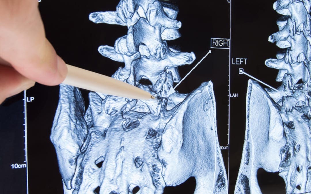
10. Bone injuries such as stress fractures, pars defects and spondylolisthesis.
The focus for this paper will be on the previous group — that the bone injuries. This may be simply postural (slow onset repetitive trauma) or related to sports; for instance, gymnastics.
The two demographic groups that tend to endure the most extension-related low back pain are:
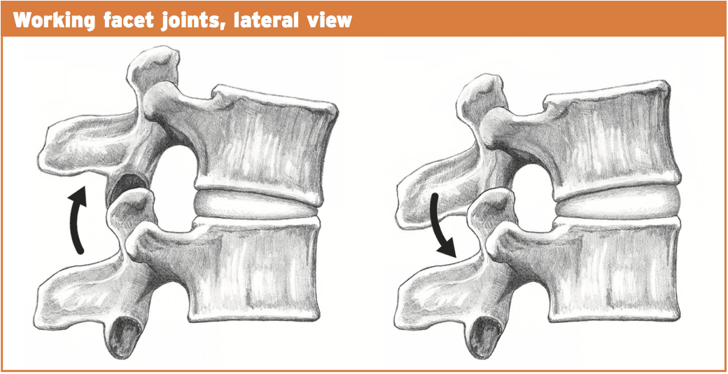
1. People who endure all day, for instance, retailers, army, security guards etc.. Prolonged position will obviously force the pelvis to start to migrate to an anterior tilt management. This may begin to place compressive pressure on the facet joints of the spinal column as they also change towards an expansion position since they accompany the pelvic tilt.
2. Extension sports such as gymnastics, tennis, swimming, diving, football codes, volleyball, basketball, track and field, cricket fast bowlers. This is more pronounced in sports that involve extension/rotation.
Pathomechanics
With normal extension of the lumbar spine (or backward bending), the facet joints begin to approximate each other and compress.�The articular processes of this facet above will abut the articular process of the facet below. This is a normal biomechanical movement. However, if the extension ranges are excessive, the procedures will impinge quite aggressively and damage to the cartilage surfaces within the facet joint can result. Sports such as gymnastics, functioning in tennis, and handling in American Soccer may all involve uncontrolled and excessive extension.
It would be unlikely that a bone stress response or even a stress fracture could be brought on by an isolated expansion injury. It would be more likely that a sudden forced extension injury may damage an already pre-existing bone strain reaction.
Similarly, if an individual stands daily and the pelvis migrates into lateral tilt, then the aspects will be placed under low load compression but for extensive intervals.
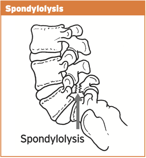
With ongoing uncontrolled loading, stress is then transferred from the facet joint to the bone below (pars interarticularis). This originally will manifest as a pressure reaction on the bone. This bone strain may advance to a stress fracture throughout the pars if uncorrected. This fracture is also referred to as a “pars flaw”, or spondylolysis.
It was initially considered that stress fractures of the pars was a congenital defect that introduced itself at the teenage years. However, it is now agreed that it is probably obtained through years of overuse into extension positions, especially in young sportspeople involved with expansion sports. What’s more, one-sided pars defects often occur more commonly in sport which also included a rotational component such as tennis serving or fast bowling in cricket.
The stress fracture can then advance to impact the opposite side, causing a bilateral strain fracture, with anxiety subsequently being transferred to the disk in between both levels.
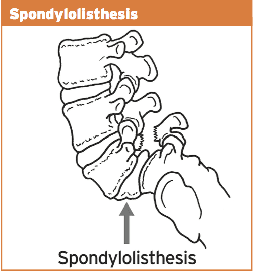
Spondylolisthesis features bilateral pars defects which could possibly be a result of repetitive stress into the bilateral pars in extension athletics, but more likely it is an independent pathology that manifests in the early growing stages (9-14) as this pathology is often viewed in this age category. If they become symptomatic in later years because of involvement in expansion sports, it is exceedingly likely that the defects were there by a young age but presented asymptomatically. As a result of rapid growth spurts in teenage years and the high-volume training experienced by teenaged athletes, it is possible that these dormant spondylolisthesis then pose as ‘acute onset’ back pain in teenage years.
In summary, the progression of this bone stress reactions tends to follow the following continuum:
1. Facet joint irritation
2. Pars interarticularis stress response
3. Stress fracture to the pars
4. Pars defect (or spondylolysis)
5. Spondylolisthesis due to activity or more likely congenital and found later in teenage years due to participation in�extension sports.
The landmark publication related to spondylolysis and spondylolisthesis was presented by Wiltse et al (1976) and they classified these injuries as follows:
1. Type I: dysplastic � congenital abnormalities of L5 or the upper sacrum allow anterior displacement of L5 on the sacrum.
2. Type II: isthmic � a lesion in the pars interarticularis occurs. This is subclassified as
a. lytic, representing a fatigue fracture of the pars,
b. elongated but intact pars, and c. acute fracture.
3. Type III: degenerative � secondary to long-standing intersegmental instability with associated remodeling of the articular processes.
4. Type IV: traumatic � acute fractures in vertebral arch other than the pars.
5. Type V: pathological � due to generalized or focal bone disease affecting the vertebral arch.
The vast majority of spondylolysis and sponylolisthesis accidents are Type II — the isthmic variety.
For the purposes of this paper, we will refer to the above stages as the posterior arch bone stress injuries (PABSI).
Epidemiology
It is a lot more widespread at the L5 level (85-90 percent). It’s a high asymptomatic prevalence in the general population and is often found unintentionally on x ray imaging. Nonetheless, in athletes, particularly young athletes, it is a common reason for persistent low back pain. From the young athlete, the problem is often referred to as ‘active spondylolysis’.
Active spondylolysis is normal in virtually every gamenevertheless, sports such as gymnastics and diving and cricket pose a much greater danger due to the extension and turning character of the sport. The progression from an active spondylolysis into a non-union type spondylolisthesis has been associated with a greater prevalence of spinal disk degeneration.
Early detection through screening and imaging, therefore, will highlight those early at the bone stress phase and if caught early enough and managed, the progression to the larger and more complicated pathologies are avoided as a result of therapeutic capacity of the pars interarticularis in the early stages.
It is more common to find teens and young adults afflicted by PABSI. This will highlight the rapid growth of the spine through growth spurts that is also characterized by a delay in the motor control of the muscle system during this period. Furthermore, it’s thought that the neural arch actually gets stronger in the fourth decade hence possibly explaining the low incidence of bone stress reactions in mid ages.
The incidence of spondylolysis has been reported to be around 4-6% in the Caucasian population (Friedrikson et al 1984). The rates seem to be lower in females and also in African-American males. It has also been suggested that a link exists between pars defects and spina bifida occulta.
The incidence of spondylolysis seems to be higher in the young athletic population than in the general population. Studies in gymnasts, tennis, weightlifting, divers and wrestlers all show disproportionately high incidence of spondylolysis compared with the general population of age-matched subjects.
Tennis
The tennis serve generates excessive extension and rotation force. In addition, the forehand shot may also produce elevated levels of spinning/ extension. The more traditional forehand shot demanded a great deal of weight shift through the legs to the torso and arms. However, a more favorite forehand shot is to currently face the ball and also generate the force of this shot utilizing hip rotation and lumbar spine extension. This action does increase ball speed but also puts more extension and compressive loads on the spine potentially resulting in a greater degree of stress on the bone components.
Golf
The most likely skill component involved in golf that may cause a PABSI are the tee shot with a 1 wood when forcing for distance. The follow-through of this shot entails a significant quantity of spine rotation with maybe a level of spine expansion.
Cricket
Fast bowlers in cricket are the most susceptible to PABSI. This will occur on the opposite side to the bowling arm. As the front foot engages on plant stage, the pelvis abruptly stops moving but the spine and chest continue to proceed. With the wind-up of this bowling action (rotation), when coupled with expansion this can place large forces on the anterior arch of the thoracic. More than 50% of fast bowlers will create a pars stress fracture. Young players (up to 25) are most vulnerable. Cricket governments have implemented training and competition guidelines to avoid such injuries by restricting the number of meals in training/games.
Field Events
The more common field events to cause a PABSI would be high leap followed by javelin. Both these sports create enormous ranges of backbone extension and under significant load.
Contact Sports
Sports like NFL, rugby and AFL all require skill components that need backbone expansion under load.
Gymnastics/Dancers
It goes without saying that gymnastics and dancing involves a substantial amount of repetitive spine expansion, particularly backflips and arabesques. It has been suggested that nearly all Olympic degree gymnasts could have suffered from a pars defect. Many organizing bodies now put limits on the number of hours young gymnasts can instruct to prevent the repetitive loading on the spine.
Diving
Spine extension injuries occur mostly off the spring board and on water entrance.
Diagnosis Of PABSI In Athletes
Clinical investigation
These can pose as preventable injuries. Research shows that the incidence was emphasized from the general population that have nil indicators of back pain. But, individuals will typically complain of back ache that is deep and generally unilateral (one side). This may radiate into the buttock area. The most offending movements tend to be described as expansion moves or backward bending movements. This may be a slow progression of pain or might be initiated by one acute episode of back pain in a competitive extension motion.
On clinical examination:
1. Pain may be elicited with a one-leg extension/rotation test (standing on the leg on the affected side) � stork test.
2. Tenderness over the site of the fracture.
3. Postural faults such as excessive anterior tilt and/or pelvic asymmetry.
The one-legged hyperextension test (stork test) was suggested to be pathognomonic for busy spondylolysis. A negative evaluation was stated to effectively exclude the diagnosis of a bone stress-type injury, thus creating radiological investigations unnecessary.
But, Masci et al (2006) examined the connection between the one-legged hyperextension test and gold standard bone scintigraphy and MRI. They discovered that the one-legged hyperextension test was neither sensitive nor specific for active spondylolysis. Moreover, its negative predictive value was so poor. Thus, a negative test can’t exclude energetic spondylolysis as a possible cause.
Masci et al (2006) go on to indicate that the bad relationship between imaging and the one-legged test may be because of a number of factors. The extension test would be expected to move a significant extension force on to the lower back spine. In addition to putting substantial strain on the pars interarticularis, it might also stress different regions of the spinal column like facet joints as well as posterior lumbar disks, and this may subsequently induce pain in the existence of other pathology such as facet joint arthropathy and spinal disc disease. This will explain the poor specificity of the test. Conversely, the inadequate sensitivity of the test may be related to the subjective reporting of pain by issues performing the maneuvre, which may vary based on individual pain tolerance. Additionally, this evaluation can preferentially load the fifth cervical vertebra, and so bone stress located in the upper lumbar spine may not test positive.
Grade 1 spondylolisthesis are normally asymptomatic; nonetheless, grade 2+ lesions often present with leg pain, either with or without leg pain. On examination, a palpable slip could be evident.
Imaging
Clinical assessment of active spondylolysis and the more severe pars defects and spondylolisthesis can be notoriously non-specific; this is, not all patients suffering PABSI will present with favorable abstract features or positive signs on analyzing. Thus, radiological visualization is important for diagnosis. The imaging methods available in the diagnosis of bone stress injury are:
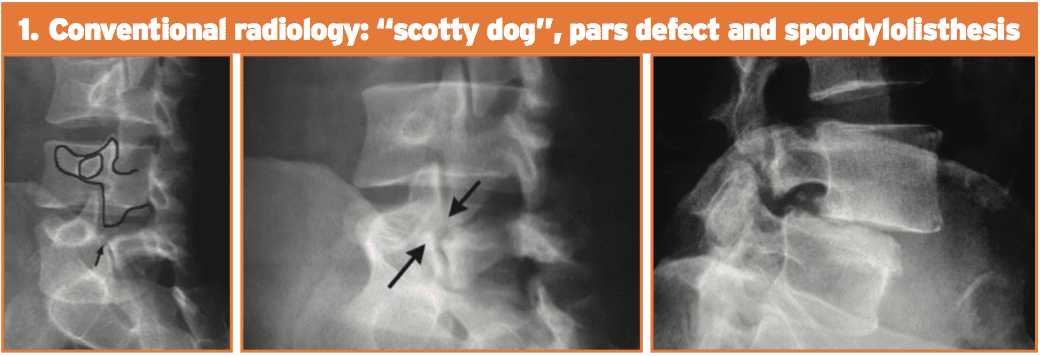
1. Conventional radiology. This test is not very sensitive but is highly unique. Its limits are partially because of the cognitive orientation of the pars defect. The oblique 45-degree films may show the timeless ‘Scotty Dog’ appearance. Spondylolisthesis can be looked at simply on a lateral movie x-ray.
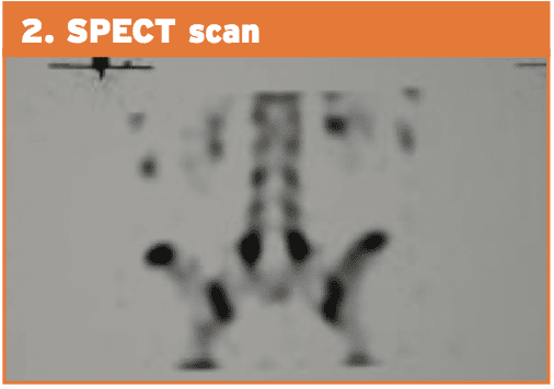
2. Planar bone scintigraphy (PBS) and single photon emission computed tomography (SPECT). SPECT enhances sensitivity in addition to specificity of PBS than straightforward radiographic study. Comparative research between PBS and conventional radiology have shown that scintigraphy is more sensitive. Patients with positive SPECT scan must then undergo a reverse gantry CT scan to assess whether the lesion is active or old.
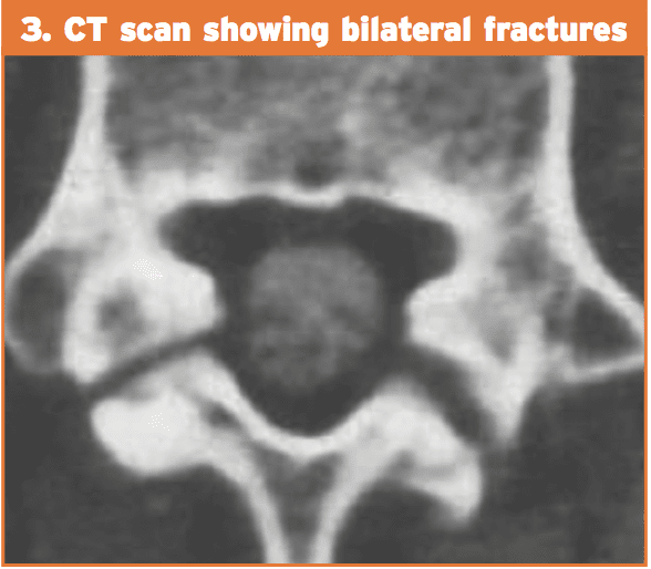
3. Computed tomography (CT). The CT scan is considered to be more sensitive than conventional radiology and with higher specificity than SPECT. Regardless of the type of cross-sectional image utilized, the CT scan provides information on the state of the flaw (intense fracture, unconsolidated flaw with geodes and sclerosis, pars in procedure for consolidation or repair). The “inverse gantry” perspective can evaluate this condition better. Repeat CT scan can be used to track progress and recovery of the pars defect.
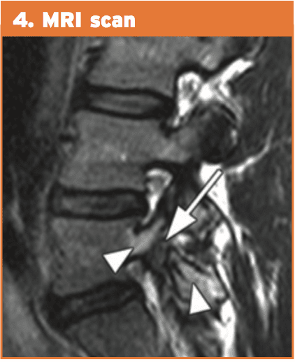
4. Magnetic resonance imaging (MRI). This technique shows pronounced changes in the signal in the amount of the pars. This is recognized as “stress response” and can be classified into five different degrees of action. MRI can be helpful for evaluating elements that stabilize isthmic lesions, for example intervertebral disc, common anterior ligament, and related lesions. The MRI isn’t as specific or sensitive as SPECT and CT combination.
Therefore, the current gold standards of investigation for athletes with low back pain are:
1. bone scintigraphy with single photon emission computed tomography (SPECT); if positive then
MRI has many advantages over bone scintigraphy, for instance, noninvasive nature of the imaging along with the absence of ionizing radiation. MRI changes in active spondylolysis include bone marrow edema, visualized as increased signal in the pars interarticularis on edema-sensitive sequences, and fracture, visualized as reduced signal in the pars interarticularis on T1 and T2 weighted sequences.
However, there is greater difficulty in detecting the changes of busy spondylolysis from MRI. Detecting pathology from MRI relies on the interpretation of distinct contrasts of signals compared with normal tissue. Unlike stress fractures in different parts of the body, the little region of the pars interarticularis may make detection of those changes harder.
However, unlike MRI, computed tomography has the capability to differentiate between acute and chronic fractures, and this differentiation might be an important determinant of fracture healing. Accordingly, in areas using pars interarticularis fractures discovered by MRI, it might nonetheless be necessary to execute thin computed tomography slices to determine whether or not a fracture is severe or chronic — an important factor in fracture resolution.
IFM's Find A Practitioner tool is the largest referral network in Functional Medicine, created to help patients locate Functional Medicine practitioners anywhere in the world. IFM Certified Practitioners are listed first in the search results, given their extensive education in Functional Medicine








 Are Backpacks Too Heavy For Kids?