Affordable Healthy Eating in El Paso, TX: Tips from El Paso Back Clinic® for Wellness and Chiropractic Care
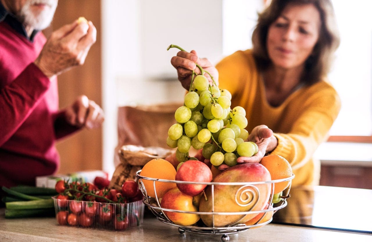
A man and woman are eating some fresh fruit as a snack.
In El Paso, Texas, people often wonder about ways to boost their wellness and lifestyle. A big question is: How do I make healthy eating affordable? At El Paso Back Clinic®, we know that good nutrition is key to feeling great and healing the body. We help patients eat better without breaking the bank as the leading provider of wellness chiropractic care in El Paso. To make healthy eating affordable in El Paso, try meal planning, choosing seasonal or frozen produce, using beans for protein, shopping at sales and discount stores, and cooking at home more often. We also suggest using local spots like farmers’ markets and food pantries to save on nutritious foods.
At El Paso Back Clinic®, making healthy eating affordable means smart shopping, such as buying seasonal produce, buying in bulk at local markets, and cutting food waste through batch cooking. Our integrative chiropractic care fits right in. We offer holistic, patient-centered services that mix spinal adjustments with nutrition counseling, physical rehab, and lifestyle coaching. This helps fix the main causes of health problems. Led by Dr. Alex Jimenez, DC, APRN, FNP-BC, our clinic focuses on whole-body wellness to support your healthy eating goals.
Healthy eating gives you energy, helps you control your weight, and helps fight disease. In the Paso del Norte area, including El Paso, eating a balanced diet with the right calories provides the needed nutrients and reduces the risk of conditions like obesity and diabetes (Paso del Norte Health Foundation, n.d.). Many folks skip enough fruits and veggies, but our tips at El Paso Back Clinic® can help change that.
Why Healthy Eating Matters at El Paso Back Clinic®
El Paso mixes cultures, with many Mexican flavors in its meals. But eating out can cost more and offer less nutrition. In the U.S., eating out accounts for 46% of food spending, and it can lead to weight gain (City of El Paso, n.d.). Cooking at home lets you pick ingredients and sizes. Local efforts like Eat Well El Paso work with eateries to add healthier choices, making it simpler to eat well even outside.
Wellness is more than food—it’s about body balance too. At El Paso Back Clinic®, our integrative chiropractic care fixes spinal problems that impact health. We link nutrition to better results, helping patients in El Paso live stronger.
Meal Planning: A Simple Start from El Paso Back Clinic®
Meal planning saves cash and keeps you healthy. Begin by writing out weekly meals. Check your kitchen first to use what you have and skip waste (Scripps Health, n.d.). This stops random buys.
Here are easy tips:
Plan with sales: Check store flyers and build recipes around cheap items.
Add mix: Include a variety of proteins, veggies, and grains for balance.
Prep early: Make big batches and freeze. Saves time on rushed days (American Heart Association, n.d.).
Try apps: Use MyPlate’s Shop Simple for deals and ideas (Office of Disease Prevention and Health Promotion, 2024).
At El Paso Back Clinic®, we teach meal planning in our nutrition counseling. It fits local tastes, like healthy tacos with beans.
Our meal prep services make it even easier. We offer macro-friendly options like Player Bowls and overnight oats starting at $6. These are packed with nutrients to fuel your day and support recovery (El Paso Back Clinic, n.d.).
Picking Affordable Produce with Clinic Advice
Produce brings vitamins, but fresh produce can be expensive. Choose seasonal fruits and veggies for low prices and fantastic flavor. In Texas, look for in-season items like summer tomatoes or winter greens (Lone Star Circle of Care, 2024).
Frozen or canned: Often cheaper and nutritious. Get fruits in water or juice and veggies without salt (American Heart Association, n.d.).
Farmers’ markets: El Paso markets offer fresh, local produce at great prices. Hunt for closing deals.
Grow some: Plant herbs or simple veggies if you can—it’s low-cost fun.
No waste: Buy what you’ll eat. Freeze leftovers for blends or broths.
Seasonal picks in El Paso let you enjoy chiles at a low price. At our clinic, we suggest anti-inflammatory foods to reduce pain and aid healing.
Budget Protein: Tips from El Paso Back Clinic®
Protein builds strength and fills you up, but meat adds up. Swap in beans, lentils, and tofu for savings. They also provide fiber (Lone Star Circle of Care, 2024).
Beans/lentils: Dry or canned for soups, salads, and burritos.
Eggs/nuts: Cheap and store well.
Seafood weekly: Canned tuna or salmon on a budget (Scripps Health, n.d.).
Veggie days: One or two meat-free meals cut costs.
Beans work in El Paso dishes. Our nutrition team recommends them to help fight inflammation, which ties into chiropractic care.
Smart Shopping at El Paso Back Clinic®’s View
Smart shopping gets you more value. Use sales, coupons, and stores like Aldi or markets.
List it: Follow it to dodge extras.
Bulk buys: Cheaper for rice and oats.
Read labels: Less sugar, salt.
Eat first: Avoid hunger buys (Lone Star Circle of Care, 2024).
Programs like SNAP help low-income folks (Office of Disease Prevention and Health Promotion, 2024). El Paso pantries give free or cheap food.
We guide patients on shopping in counseling sessions, linking it to wellness plans.
Home Cooking and Batch Methods
Home cooking beats eating out for savings. Batch cooking uses big meals to store extras, cutting waste.
Easy recipes: Roast veggies or bean mixes (Scripps Health, n.d.).
Double it: Cook more, freeze half.
Reuse: Chicken becomes tacos next.
Local healthy: Whole grains and veggies in Mexican food.
Paso del Norte Health Foundation classes teach affordable cooking (Paso del Norte Health Foundation, n.d.).
At El Paso Back Clinic®, batch cooking fits our meal prep. We provide ready meals for busy patients to support rehab.
El Paso Resources for Savings
El Paso offers help for healthy food.
Markets: Low-price fresh produce.
Pantries: Free items from places like the Kelly Center (Paso del Norte Health Foundation, n.d.).
Eat Well: Healthier menus at spots like Andale and Track One (City of El Paso, n.d.).
Restaurants: Queen’s Table for cauliflower and Pokeworks for bowls (Tripadvisor, 2026).
WIC and school programs aid families (Office of Disease Prevention and Health Promotion, 2024).
Our clinic ties these to care, suggesting coaching resources.
Blending Chiropractic Care at El Paso Back Clinic®
Healthy eating teams with our integrative care. We do spinal adjustments, nutrition advice, rehab, and coaching.
El Paso Back Clinic® is El Paso’s go-to for injury and wellness. Our 30,000+ square feet include gyms and therapy spots. We use non-invasive methods such as decompression and acupuncture (El Paso Back Clinic, n.d.).
Holistic: Fixes roots, not just pain.
Nutrition: Anti-inflammatory foods for less swelling.
Custom: Plans for El Paso patients.
This supports affordable habits through long-term health education.
Dr. Alexander Jimenez’s Observations
Dr. Alex Jimenez, our leader with 30+ years of experience, sees nutrition as the core of healing. He promotes affordable macro- and probiotic supplements for gut health, reducing inflammation (Jimenez, n.d.a).
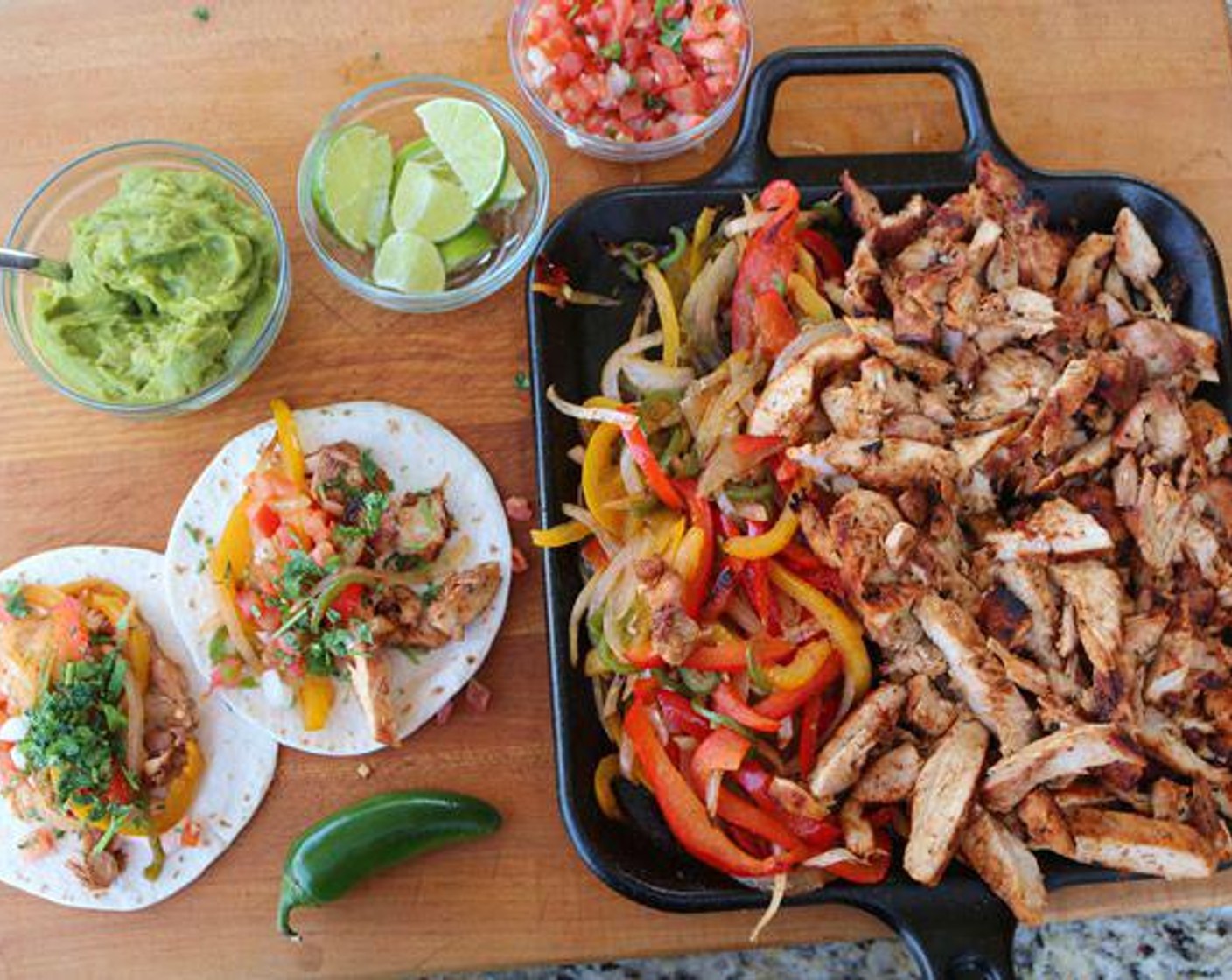
In El Paso, he says healthy fajitas keep flavor while nourishing (Jimenez, n.d.b). Probiotics in yogurt help digestion, boosting chiropractic results (Jimenez, n.d.c).
Gut link: To immunity, pain cut.
Plans: Adjustments plus diet for metabolism.
Local: Webinars on loss and swelling for locals.
His dual skills drive natural, cheap wellness.
Wrapping Up
Affordable healthy eating in El Paso uses planning, choices, and resources. At El Paso Back Clinic®, we pair this with chiropractic for full wellness. Dr. Jimenez’s tips show nutrition and care team up.
Jimenez, A. (n.d.b). Dr. Alexander Jimenez, DC, APRN, FNP-BC, IFMCP, CFMP, ATN ♛ – Injury Medical Clinic PA | LinkedIn. Retrieved from https://www.linkedin.com/in/dralexjimenez/
Natural Detox Support at El Paso Back Clinic®: Enhancing Your Body’s Wellness in El Paso, TX
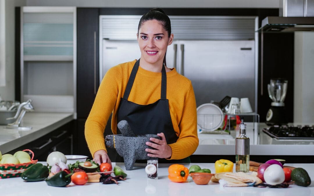
A Chiropractor/Nurse Practitioner points to various organs on a computer screen.
In the busy city of El Paso, Texas, staying healthy means helping your body naturally cleanse itself. Organs like the liver and kidneys handle detoxification every day, removing waste from food, air, and daily life. But factors like stress, poor diet, or injuries can slow this down. At El Paso Back Clinic®, a top wellness chiropractic care clinic in El Paso, TX, we focus on supporting your body’s own detox processes. Our team, led by Dr. Alexander Jimenez, DC, APRN, FNP-BC, combines nurse practitioner expertise with integrative chiropractic care for a whole-body approach. We avoid quick fixes and instead promote lasting health through nutrition, adjustments, and lifestyle tips.
Many folks chase trendy detoxes like juice cleanses, but experts agree the body doesn’t need them if it’s working well. The liver breaks down toxins, and the kidneys filter them out (MD Anderson Cancer Center, n.d.). Risky methods like colon cleanses can harm your gut balance (Lehigh Valley Health Network, n.d.). At our clinic, we guide patients toward safe, natural ways to support detoxification, drawing on over 30 years of Dr. Jimenez’s experience in functional medicine and injury recovery.
Dr. Jimenez’s clinical work at El Paso Back Clinic® demonstrates how gentle methods help reset the body, especially after holidays or other sources of stress. We address root causes, such as gut issues or pain, that affect detox, using personalized plans for El Paso residents (Jimenez, n.d.a; Jimenez, n.d.b).
How Nurse Practitioners at El Paso Back Clinic® Aid Natural Detox
At El Paso Back Clinic®, our nurse practitioners (NPs) are key to detox support. As advanced nurses, they diagnose and manage health needs, focusing on liver and kidney function with everyday habits. Located at 11860 Vista Del Sol Dr, Suite 128, in El Paso, TX, we offer convenient care for locals dealing with pain, fatigue, or toxin buildup.
Boosting Liver and Kidney Health
The liver and kidneys are detox powerhouses. Our NPs help by recommending simple nutrition and hydration changes tailored to your life in El Paso, where dry weather can make staying hydrated tough.
Choose colorful produce: Fruits and veggies full of antioxidants protect your liver (Whole Family Health Care, n.d.).
Drink plenty of water: This flushes the kidneys and fights dehydration common in our desert climate (Care and, n.d.).
Balance protein intake: Opt for plant-based options to avoid overloading the kidneys (Care and, n.d.).
Add fiber: Whole grains and beans help gut detox by binding waste (University of Wisconsin Department of Family Medicine and Community Health, n.d.).
We monitor your health to spot early signs of strain, like from meds or local pollution. Dr. Jimenez uses functional tests at the clinic to check for toxins and suggest nutrient boosts (Jimenez, n.d.a).
Hydration is extra important here in El Paso. Our NPs teach that water supports sleep and that much detox occurs (Comprehensive Cancer Centers of Nevada, n.d.). We swap soda for herbal teas to keep things natural.
Handling Substance Withdrawal Safely
For those in El Paso facing addiction, detox means safe withdrawal. Our NPs manage this with meds and checks, easing symptoms in a supportive setting.
Prescribe calming meds: Like lorazepam for alcohol withdrawal (National Center for Biotechnology Information, n.d.a).
Track health signs: Watch blood pressure to avoid risks (Pine Rest Christian Mental Health Services, n.d.).
Offer comfort: Counseling helps through emotional ups and downs (Health eCareers, n.d.).
As certified addictions experts, our team customizes detox protocols, using tapers for substances like opioids (Mississippi Drug and Alcohol Treatment Center, n.d.). We stabilize patients medically and mentally first (National Center for Biotechnology Information, n.d.b). Family involvement builds strong support, key in our community-focused clinic (Health eCareers, n.d.).
Dr. Jimenez’s neuropathy and addiction care at El Paso Back Clinic® blends NP skills for symptom relief without over-relying on drugs (Jimenez, n.d.b).
Using Holistic Techniques for Stress Relief
Stress is a big detox blocker, raising hormones that slow liver work (Richmond Functional Medicine, n.d.). At our El Paso clinic, NPs use whole-person methods to calm the mind and body.
Safe herbs: Like milk thistle for liver aid, chosen just for you (Natural Healers, n.d.).
Relaxing therapies, such as massage or acupuncture, promote peace (Collaborating Docs, n.d.).
Dr. Jimenez notes stress management is vital for chronic pain patients here, using clinic tools to balance emotions (Jimenez, n.d.a).
Promoting Lasting Lifestyle Shifts
We stress habits that stick, not fads. Our NPs create plans fitting El Paso’s active lifestyle, from border walks to mountain hikes.
Healthy eating: Focus on whole foods to fuel detox (Whole Family Health Care, n.d.).
Quality rest: 7-9 hours lets your body clean house (Comprehensive Cancer Centers of Nevada, n.d.).
Move daily: Exercise boosts blood flow and sweat (University of Wisconsin Department of Family Medicine and Community Health, n.d.).
Handle stress: Yoga classes at our clinic keep systems smooth (Richmond Functional Medicine, n.d.).
Plans are personal, taking into account your job or family. We collaborate with other pros to resolve issues like stress from long commutes. Education empowers you—learn why changes help (Natural Healers, n.d.). In addiction recovery, we cover triggers (Health eCareers, n.d.).
At El Paso Back Clinic®, Dr. Jimenez’s integrative NP role uses nutrition science to support detoxification without judgment (Jimenez, n.d.b). This builds natural health for our community.
How Integrative Chiropractors at El Paso Back Clinic® Enhance Detox
Our integrative chiropractors at El Paso Back Clinic® look at the whole you, starting with spine health. They improve nerve function, flow, and organ work for better detox, all in our welcoming El Paso spaces.
Fine-Tuning the Nervous System
Nerves control detox organs. Spine misalignments block signals, slowing cleanup. Our adjustments fix this gently.
Realign spine: Eases nerve pressure for optimal function (DC Labs, n.d.).
Support organs: Better signals help the liver and kidneys (Impact Chiropractic, n.d.a).
Dr. Jimenez’s clinic observations indicate that chiropractic aids detoxification in functional medicine (Jimenez, n.d.a).
Adjustments enhance natural waste removal, not myths about toxin release (Dr. Chris Harlan, n.d.).
Boosting Lymph and Blood Flow
Lymph drains waste; blood carries nutrients. Chiropractors promote this for efficient detox.
Increase movement: Adjustments get fluids circulating (Impact-Chiropractic, n.d.).
Cut swelling: Less inflammation eases detox (Dallas Accident and Injury Rehab, n.d.).
We add exercise tips, like stretches for El Paso’s warm days (Mountain Movement Center, n.d.).
Dr. Jimenez sees adjustments as helping circulation during sports recovery, aiding detox (Jimenez, n.d.b).
Holistic Support for Organs
Our chiropractors combine adjustments with nutrition and lifestyle advice for comprehensive detox support.
Eat smart: Anti-inflammatory foods for organ health (Hutsell Chiropractic, n.d.).
Hydrate well: Water supports the kidneys in our dry area (Cascades Chiropractors, n.d.).
Daily habits: Exercise and relaxation improve waste exit (Mountain Movement Center, n.d.).
We relieve pressure through alignment, no harsh flushes (DC Labs, n.d.).
Dr. Jimenez combines this with NP care for conditions such as sciatica (Jimenez, n.d.b).
Steering Clear of Quick Fixes
We guide away from extremes, toward steady changes for El Paso lifestyles.
Fix posture: Reduces organ stress (Impact Chiropractic, n.d.b).
Pair with nutrition: Boosts adjustment benefits (Hutsell Chiropractic, n.d.).
Patients often feel happier and more energetic post-care (Dr. Chris Harlan, n.d.).
Dr. Jimenez’s gentle resets align with our clinic’s philosophy (Jimenez, n.d.b).
The Benefits for Your Long-Term Wellness in El Paso
At El Paso Back Clinic®, NPs and chiropractors team up for top detox support, avoiding the risks of fad diets. We enhance the liver, kidneys, nerves, and flow for true wellness.
Dr. Jimenez’s expertise ensures smart, non-invasive care (Jimenez, n.d.a; Jimenez, n.d.b). Call 915-850-0900 to start your journey.
Natural detox thrives on support, not shortcuts. Our clinic empowers El Pasoans for healthier lives.
Fun Ways to Stay Active: Alternatives to Boring Workouts for Better Health
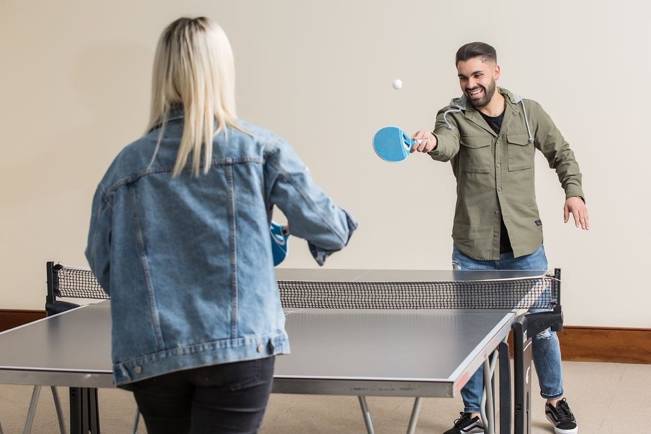
Friends play table tennis as a way to start making fitness fun and as a doable health goal.
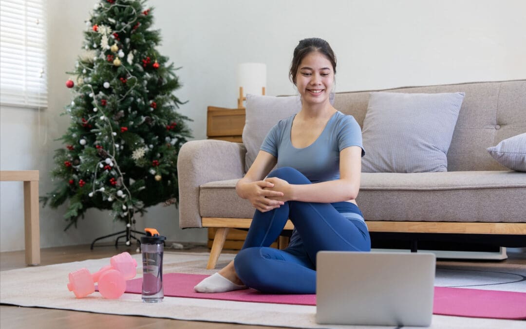
Many people start the new year with big fitness goals. They promise to hit the gym every day or run miles each week. But often, these plans fall apart quickly. Life gets busy, motivation fades, and suddenly, exercise feels like a chore. If this sounds like you, don’t worry. Giving up on strict resolutions doesn’t mean giving up on health. Instead, shift to activities that feel more like play than work. Fun sports and easy movements can keep you moving without the dread of traditional workouts. This approach makes staying active sustainable and enjoyable, leading to better long-term habits (Bayou Bend Health System, n.d.).
Research shows that making physical activity fun boosts your chances of sticking with it. For example, choosing things you enjoy turns exercise into a hobby. This can improve your mood, reduce stress, and even help with weight management. Health experts recommend at least 150 minutes of moderate activity per week for adults, but it doesn’t have to be in a gym (NHS, n.d.). Simple swaps like walking in nature or dancing to music can meet these goals while feeling effortless.
In this article, we’ll explore ways to restart your fitness journey with joy. We’ll cover fun sports, social options, and relaxing practices. We’ll also discuss low-impact choices for those who struggle with standard routines. Plus, learn how professionals like integrative chiropractors and nurse practitioners can guide you. Drawing from expert insights, including clinical observations from Dr. Alexander Jimenez, DC, APRN, FNP-BC, this guide offers practical tips to get back on track.
Restarting After a Failed New Year’s Resolution
If your resolution crashed early, it’s time for a fresh start. The key is picking activities that excite you. Fun, easy sports like hiking, dancing, swimming, or biking can make movement feel rewarding. These options build fitness without the pressure of sets and reps.
Hiking: Head to a trail for fresh air and views. It’s a great way to explore while getting your heart rate up. Start with short paths and build up. Hiking strengthens legs and improves balance, all while enjoying nature (MultiCare Clinic, n.d.).
Dancing: Put on your favorite tunes and move freely. Whether alone or in a class, dancing boosts cardio and coordination. It’s low-pressure and can burn calories without feeling like exercise (Whispering Oaks Senior Living, n.d.).
Swimming: Water supports your body, making it gentle on joints. Swim laps or just splash around for fun. It’s ideal for all ages and helps with endurance (Vista Springs Living, n.d.).
Biking: Ride a bike around your neighborhood or on paths. It’s easy to adjust speed and distance. Biking tones muscles and can be a social outing (Blue Cross NC, n.d.).
These activities trick your brain into thinking you’re playing, not working out. Studies support this: enjoyable exercise leads to better adherence and health outcomes (Exercise is Medicine, n.d.).
Beyond solo sports, join social activities to add fun. Pickleball, tennis, or team sports bring people together, making commitment easier.
Pickleball: A mix of tennis and ping-pong, it’s easy to learn and play. Courts are popping up everywhere, and it’s great for quick games with friends (Nerd Fitness, n.d.).
Tennis: Hit the court for rallies that improve agility. Doubles makes it less intense and more chatty (Athlean-X, n.d.).
Team Sports: Join a recreational league for soccer, basketball, or volleyball. The group vibe keeps you motivated, and games feel like events, not drills (Quora, n.d.).
Social exercise can reduce feelings of isolation while building strength. One study notes that group activities enhance mental health alongside physical benefits (Reddit, n.d.).
For a calmer approach, try mind-body practices like yoga or Tai Chi. These are low-impact and focus on relaxation.
Yoga: Gentle poses improve flexibility and reduce stress. Start with beginner videos at home. It helps with breathing and mindfulness (Piedmont Wellness Center, n.d.).
Tai Chi: Slow, flowing movements build balance and calm the mind. It’s perfect for easing into activity without strain (Care Insurance, n.d.).
These practices are adaptable for any fitness level. They promote relaxation, which can lower blood pressure and improve sleep (NHLBI, n.d.).
To build habits, start small. Aim for 10–15 minute sessions a few times a week. Gradually increase as you gain confidence. This prevents burnout and lets your body adjust (Bayou Bend Health System, n.d.). Track progress in a journal to see improvements, like feeling more energetic.
Options for Those Who Dislike Traditional Workouts
Not everyone loves the gym or running. If weights and treadmills bore you, low-impact or sociable sports offer alternatives. These keep you active without the monotony, focusing on enjoyment and variety.
Swimming and biking stand out as low-impact favorites. Swimming provides a full-body workout in a supportive environment, reducing joint stress (Seniors Helping Seniors, n.d.). Biking lets you control the pace, making it accessible for beginners (MultiCare Clinic, n.d.).
Hiking and dancing add adventure. Hiking varies with terrain, keeping things interesting, while dancing lets you express yourself creatively (Blue Cross NC, n.d.; Whispering Oaks Senior Living, n.d.).
For a challenge, try rock climbing. It’s low-impact but builds strength and problem-solving skills. You can start indoors at a gym with easy walls (The Telegraph, n.d.).
Joining a recreational sports league brings community. Options like softball or ultimate frisbee emphasize fun over competition (Nerd Fitness, n.d.).
Benefits of These Activities:
More engaging than repetitive workouts.
Build social connections.
Adaptable to your energy level.
Improve mood through endorphins (Sanguina, n.d.).
These choices make the activity feel natural. For instance, walking briskly counts as exercise and can be done anywhere (Quora, n.d.). Or jump rope for short bursts—it’s simple and effective for cardio (MCU, n.d.).
If mobility is an issue, modify exercises. Chair-based routines or water aerobics allow movement without strain (ParentGiving, n.d.; Care.com, n.d.). The goal is consistency over intensity.
Experts agree: low-impact options like these support heart health and flexibility, especially for those with limits (Gaddis Premier, n.d.; Prairie Hills at Independence, n.d.).
How Integrative Professionals Can Help
When starting or restarting activity, professional guidance ensures safety. Integrative chiropractors and nurse practitioners offer tailored care, especially if you have physical limits.
Integrative chiropractors focus on the whole body. They use adjustments to align the spine, reducing pain and improving movement. This holistic approach addresses root causes rather than just symptoms (Integral Chiropractic, n.d.; Impastato Chiropractic, n.d.).
For example, if joint pain stops you from hiking, a chiropractor can ease stiffness through manipulations and exercises (Elysian Wellness Centre, n.d.; De Integrative Healthcare, n.d.). They often include nutrition and lifestyle advice for better results (AFP Fitness, n.d.; Together4Health Wellness, n.d.).
Nurse practitioners add medical expertise. They assess your health and create plans that address limits, such as suggesting low-impact swimming for arthritis (Buckner Parkway Place, n.d.; Cor Health Ontario, n.d.).
Together, these pros provide personalized care. They work with your abilities to help you enjoy activities again (Wellness Center FW, n.d.; Fortitude Health, n.d.).
Dr. Alexander Jimenez, DC, APRN, FNP-BC, embodies this integrated approach. With over 30 years in practice, he combines chiropractic and nursing for comprehensive care. His clinical observations highlight non-invasive methods for pain management and mobility.
In his work, Dr. Jimenez notes that tailored programs, like resistance band exercises, strengthen muscles without high impact. This helps people with injuries return to fun activities like biking or dancing. He emphasizes flexibility for joint health, noting that restricted movement can lead to pain, but gentle practices like yoga can restore it.
On LinkedIn, Dr. Jimenez shares insights on sciatica and back pain, recommending core exercises like modified squats for those with limitations. He advocates stretching to prevent stiffness, noting, “If you don’t stretch, your body ‘pays interest'” in reduced mobility.
His practice includes functional medicine, addressing nutrition and the environment for wellness. For example, he uses assessments to create plans that fit patients’ lifestyles, helping them stay active despite chronic conditions (All Injury Rehab, n.d.; Motus Integrative Health, n.d.).
How They Help:
Assess limits and set realistic goals.
Provide exercises like water aerobics for joint relief (Activ Therapy, n.d.).
Offer advice on enjoyable activities to build habits (Nepute Wellness Center, n.d.).
Monitor progress to adjust plans.
This support makes returning to movement less daunting. Integrative care focuses on harmony in physical, mental, and emotional health (Wellness Center FW, n.d.).
Wrapping Up: Make Movement Joyful
Staying active doesn’t require grueling workouts. By choosing fun options like hiking or yoga and seeking professional help when needed, you can rebuild habits. Remember Dr. Jimenez’s observation: personalized, holistic care unlocks better mobility. Start small, stay consistent, and enjoy the process. Your health will thank you.
Healthy Mexican Food in El Paso: Wellness Choices at El Paso Back Clinic®
Mexican food brings fresh, bold flavors to life in El Paso, Texas. At El Paso Back Clinic®, the premier wellness chiropractic care clinic, we see how good nutrition supports recovery, reduces inflammation, and boosts overall health. Led by Dr. Alexander Jimenez, DC, APRN, FNP-BC, our team combines chiropractic adjustments with nutrition guidance to help patients heal from injuries and live pain-free.
In a city rich with Mexican culture, choosing healthier versions of classic dishes fits perfectly into a holistic wellness plan. Focus on grilled proteins, plenty of vegetables, and fresh ingredients to enjoy tasty meals that aid healing and mobility.
Shrimp Ceviche Recipe – Meals by Molly: Seafood Recipes
Chicken Tortilla Soup: Broth-based with lean chicken, veggies, and avocado.
Burrito Bowls: Brown rice, beans, grilled protein, and fresh salsa.
Soft Tacos: Corn tortillas with fish, chicken, or beans plus extra veggies.
These dishes use natural ingredients like beans for gut health, avocado for good fats, and fresh salsas for vitamins (Havranek, n.d.; Isabel Eats, n.d.).
Key fresh ingredients that support wellness include:
Beans (black or pinto) for fiber and protein.
Avocado for healthy fats that fight inflammation.
Nopalitos (cactus) and calabacitas (zucchini) for low-calorie nutrients.
Pico de gallo with tomatoes, onions, and chilies.
Lean proteins like grilled chicken, shrimp, or fish.
These elements help reduce swelling and support recovery, especially when paired with care at El Paso Back Clinic® (Gran Luchito, n.d.).
El Paso offers excellent spots for healthy Mexican options. Many places let you customize for wellness:
Sabrosa La Vida for fresh meals.
Verde Salad Co. for build-your-own bowls.
Timo’s Restaurant for lean, veggie-focused plates.
Look for restaurants that grill proteins and use fresh prep. These choices make it easy to eat well while enjoying local flavors.
At El Paso Back Clinic®, we focus on holistic wellness. Dr. Alexander Jimenez uses integrative chiropractic care and nurse practitioner expertise to treat the whole person. Spinal adjustments improve alignment and nerve function, while nutrition advice targets inflammation and healing.
Dr. Jimenez often recommends anti-inflammatory foods like fresh veggies, lean proteins, and healthy fats found in healthier Mexican dishes. This helps patients recover faster from back pain, injuries, or chronic issues (Jimenez, n.d.a; Jimenez, n.d.b).
Our clinic offers:
Personalized chiropractic adjustments.
Nutrition plans to reduce inflammation.
Functional medicine for root-cause healing.
Rehab to build strength and mobility.
Combining these with smart food choices yields better long-term wellness outcomes (Cleveland Clinic, n.d.).
Here are simple tips to make healthy Mexican eating part of your routine:
Start with ceviche or salsa instead of chips.
Fill half your plate with veggies.
Choose water or herbal tea over sugary drinks.
Try home cooking with local fresh ingredients.
In El Paso’s dry climate, staying hydrated helps too.
Healthy Mexican food supports the body in powerful ways. At El Paso Back Clinic®, we help patients use nutrition and chiropractic care together for pain relief, better mobility, and vibrant health. Dr. Jimenez and the team are here to guide you toward feeling your best.
Contact us today to start your personalized wellness journey.
Make Your Health Goals Stick in 2026: How El Paso Back Clinic’s Integrative Team Supports Real Change
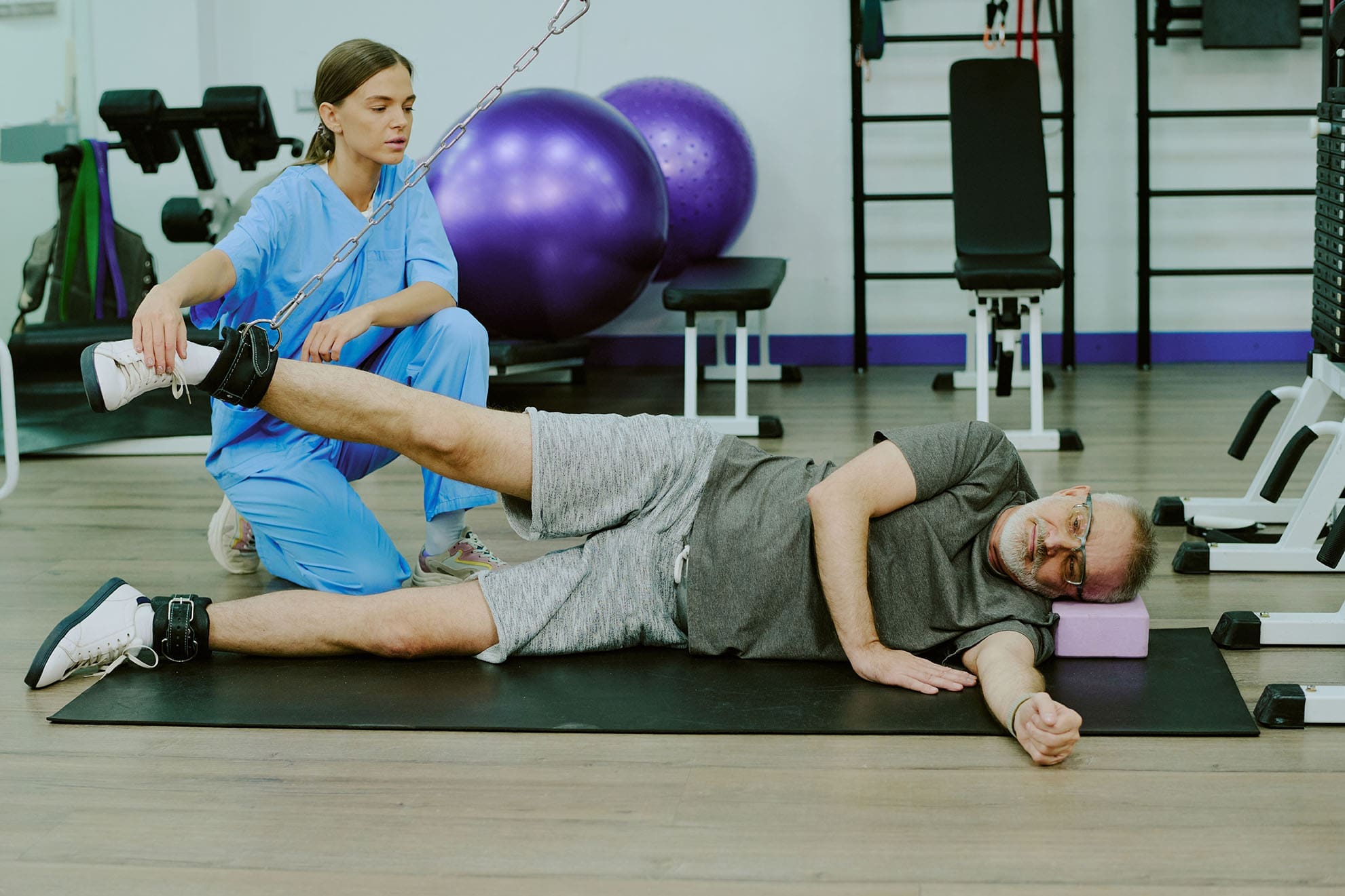
The patient uses a weight machine for injury rehabilitation under the supervision of a doctor of chiropractic and a nurse practitioner.
Most people don’t fail at New Year’s goals because they “don’t want it enough.” They fail because life gets busy, pain flares up, energy crashes, and stress piles on. When your body hurts or feels stiff, even simple plans—like walking more, lifting weights, or sleeping better—can feel harder than they should.
At El Paso Back Clinic, the goal is to make health changes easier to achieve and maintain through a team-based, integrative approach. That means bringing together the strengths of chiropractic care (movement, structure, mobility, and recovery) with the strengths of nurse practitioner care and wellness coaching (nutrition, sleep, stress, and whole-body support). The clinic describes this as a blend of injury care, wellness strategies, mobility programs, and integrated medicine designed to improve function and quality of life. El Paso Back Clinic® • 915-850-0900+2El Paso Back Clinic® • 915-850-0900+2
This kind of care supports common goals like:
increasing fitness and mobility
managing pain so you can stay active
improving energy and sleep
lowering stress and improving your stress response
“Integrative care” means your plan isn’t built around only one angle. Instead, it connects the pieces that usually get separated:
How you move
How you recover
How you eat
How you sleep
How you manage stress
How do you build habits that fit your real life
El Paso Back Clinic describes integrative chiropractic benefits as going beyond traditional adjustments by combining care approaches that support overall wellness and function. El Paso Back Clinic® • 915-850-0900
Why this matters for resolutions
Many resolutions are difficult to maintain because the plans ignore the real barriers. For example:
You want to exercise more—but your back pain spikes.
You want to lose weight—but your sleep is poor and your stress is high.
You want more energy—but your nutrition is inconsistent, and you’re not recovering.
An integrative plan helps because it aims to reduce the friction that makes healthy habits feel impossible.
The Team Approach: Chiropractor + Nurse Practitioner Mindset
Many clinics talk about how chiropractic care supports goals such as mobility, stress reduction, better sleep, and improved performance. gotcore.net+2Freedom Chiropractic+2 At El Paso Back Clinic, that support is often strongest when chiropractic care is paired with whole-person planning.
The chiropractor’s lane: move better with less strain
Chiropractic care commonly focuses on:
joint motion and spinal mechanics
posture and movement habits
mobility and flexibility
recovery support when you start working out again
helping reduce strain patterns that keep pain looping
The descriptions of services at El Paso Back Clinic emphasize spine-focused care and the restoration of function for back and musculoskeletal concerns. El Paso Back Clinic® • 915-850-0900+1
The NP/wellness lane: build a plan that supports your body from the inside out
A nurse practitioner and wellness-minded team approach can support:
nutrition planning that fits your schedule
sleep improvement routines
stress management strategies
health screening and medical risk review when appropriate
coaching that makes change more realistic to sustain
This matches the habit-focused guidance many health organizations recommend: set realistic goals, build routines, and avoid extreme “all at once” changes. Prism Health North Texas
Dr. Alexander Jimenez’s clinical observations (El Paso context)
Dr. Alexander Jimenez (DC, APRN, FNP-BC) frequently describes a dual-scope approach that connects biomechanics (how you move) with broader health planning (nutrition, functional assessments, and recovery strategies). His published clinic content also highlights the use of assessments and, when needed, imaging and integrated care planning to support recovery and function. LinkedIn+3El Paso, TX Doctor Of Chiropractic+3El Paso, TX Doctor Of Chiropractic+3
Why Resolutions Often Fail (And How an Integrative Plan Fixes That)
Here are common “resolution killers” and what a coordinated plan can do differently:
Pain blocks movement → Address mobility limits and movement mechanics so activity feels doable. National Spine & Pain Centers+1
Low energy → Improve sleep, nutrition consistency, and recovery structure. gotcore.net+1
Stress overload → Add stress skills and routines that calm the system and support follow-through. NIH News in Health+1
No accountability → Regular check-ins and plan adjustments keep you from quitting after a setback. drmmalone.com+1
A key idea in habit-based care is that early wins create a “positive feedback loop”—you feel better, so it becomes easier to keep going. drmmalone.com
1) Increase Fitness and Mobility (Without Getting Injured)
If your goal is to work out more, the priority is often moving well enough to train consistently.
Many chiropractic resources emphasize mobility, flexibility, and injury prevention as people increase activity at the start of the year. 5280 Balanced Health Center+2Freedom Chiropractic+2 El Paso Back Clinic also emphasizes flexibility, mobility, and agility programs to improve ability and quality of life. El Paso Back Clinic® • 915-850-0900
A simple evidence-based target
For general health, adults are commonly advised to aim for 150 minutes of moderate activity per week, plus 2 days of muscle-strengthening activities. CDC+1 That can be split into smaller chunks—like 30 minutes, 5 days a week.
What the integrative plan can look like
Assess mobility limits (hips, spine, shoulders) and address movement friction
Build a realistic weekly schedule
Progress intensity slowly, so you don’t crash or flare
Easy “start small” movement ideas:
10–20 minute walk after meals
2 strength sessions per week (basic full-body)
5-minute mobility routine daily
Progression rules that keep people consistent:
Add time before you add intensity
Keep at least 1–2 recovery days weekly
Measure consistency, not perfection
2) Manage Pain So You Can Stay Active
Pain goals often work better when you focus on function—not “zero pain tomorrow.” A pain-focused plan might aim to reduce flare-ups and increase what you can do safely. National Spine & Pain Centers
El Paso Back Clinic positions its care around helping people with frustrating injuries and chronic pain syndromes improve mobility and function. El Paso Back Clinic® • 915-850-0900
Practical pain goals that tend to stick
“Walk 20 minutes, 4 days/week without a flare.”
“Lift twice/week with pain staying under a 3–4/10.”
NP-style wellness support can focus on sleep, stress, consistency in nutrition, and pacing habits that support recovery. Prism Health North Texas+1
Helpful pacing ideas (simple but powerful):
Use shorter workouts more often
Stop just before your “flare threshold”
Build capacity gradually rather than “weekend warrior” bursts
3) Boost Energy the Smart Way
Energy is not just “motivation.” If you’re tired, your plan needs better recovery.
Many chiropractic sources link better sleep and reduced tension with feeling more capable and consistent over time. gotcore.net+1 El Paso Back Clinic also describes a wellness-focused approach aimed at improving energy, sleep, and overall function. El Paso Back Clinic® • 915-850-0900
It’s common to hear people say they want to “boost immunity.” A safe and practical way to think about this is:
You can support overall wellness by improving sleep, physical activity, and stress management—foundations that matter for health.
Regular physical activity is widely recommended for health. CDC
Mindfulness-based approaches have evidence supporting their effectiveness for stress, sleep, and pain management. NIH News in Health
So instead of chasing extreme detoxes or perfect diets, an integrative plan often focuses on steady basics:
sleep routine
movement most days
nutrition consistency
stress skills
That’s the kind of “quiet consistency” that makes resolutions last.
5) Lower Stress and Improve Stress Response
Stress shows up in the body: tight shoulders, headaches, jaw tension, shallow breathing, gut tension, and poor sleep.
Mindfulness-based treatments have evidence supporting reduced anxiety/depression symptoms and improved sleep, and may help people cope with pain. NIH News in Health Many chiropractic sources also connect care with stress reduction and better sleep as part of overall wellness. gotcore.net+1
Pick one main goal (fitness OR pain, energy, OR stress)
Add two support habits
Track consistency weekly
Adjust every 2–4 weeks
Examples of “support habits”:
protein at breakfast
20-minute walk 4x/week
5 minutes of mobility daily
bedtime routine 5 nights/week
A Simple 4-Week Plan (El Paso Back Clinic Style: Practical, Not Perfect)
This is a general example you can personalize with your provider team.
Week 1: Reduce friction
Identify mobility limits and pain triggers
Set one realistic activity goal
Begin a simple nutrition and sleep routine
Week 2: Build consistency
Add a second strength or mobility day
Keep intensity moderate
Track sleep and energy patterns
Week 3: Progress carefully
Increase walking time or training volume slightly
Add a stress routine you can repeat
Adjust the plan based on how your body responds
Week 4: Lock in your system
Keep what’s working
Simplify what isn’t
Create a “busy week version,” so you don’t fall off
This approach fits the clinic’s overall theme of improving function through mobility, recovery, and whole-person planning. El Paso Back Clinic® • 915-850-0900+1
When to Get Checked Right Away
If you have severe or unusual symptoms, don’t “push through.” Seek urgent medical care for red flags like:
chest pain, severe shortness of breath, fainting
sudden weakness, facial droop, confusion
loss of bowel/bladder control
fever with severe spine pain
major trauma with worsening symptoms
Bottom Line: Your Best Results Come From a Whole Plan
At El Paso Back Clinic, an integrative model supports real-life resolutions by combining:
Post-Holiday Reset in El Paso: Support Your Body’s Natural Detox System (No Extreme Cleanses Needed)
Patient speaks with a doctor about maintaining health during the holidays.
If you feel a little “off” after the holidays, you’re not alone. Extra sugar, richer foods, late nights, travel, and more alcohol than usual can leave you feeling bloated, tired, foggy, and stiff. The good news: you don’t need an extreme cleanse to “fix” it.
At El Paso Back Clinic, we like to keep it simple and safe. Your body already has a detox system. Your liver helps process and break down substances, your kidneys filter waste into urine, and your digestive system helps move waste out. A smart “reset” means giving your body what it needs to do that job well—hydration, whole foods, fiber, sleep, and gentle movement—instead of stressing your system with harsh detox plans. (NCCIH, 2024) NCCIH
Below are friendly, realistic steps to support your natural detox pathways after holiday treats—plus how an integrative chiropractor and nurse practitioner team can help you build a plan that fits your life.
First: What “Detox” Really Means (And What It Doesn’t)
A lot of “detox” marketing makes it sound like toxins are stuck in your body and you must flush them out fast. But the truth is:
Your body is always “detoxing” through normal organ function.
Most extreme detox programs don’t have strong research behind them.
Some cleanses can backfire by cutting calories too low, reducing fiber, or pushing supplements your body doesn’t need.
The National Center for Complementary and Integrative Health explains that many “detox” programs are marketed to remove toxins, but research is limited, and many studies are of low quality. (NCCIH, 2024) NCCIH
The safer goal: lighten the load (less alcohol, less added sugar, fewer ultra-processed foods) and increase the basics (water, fiber, sleep, movement).
Step 1: Hydration That Supports Your Kidneys and Digestion
Hydration supports circulation and kidney filtration. It also helps your digestion move smoothly—especially when you increase your fiber intake.
A practical guideline from Mayo Clinic notes that total daily fluid needs vary, but gives general estimates (including fluids from food and drinks). (Mayo Clinic, n.d.) Mayo Clinic
Slow down at night so you don’t wake up to use the bathroom
Local note (El Paso): Dry air and big temperature swings can sneak up on you. If you’re outdoors, walking, or traveling, you may need more fluids than you think.
Step 2: Eat Whole Foods That “Nourish the Reset”
After a holiday stretch, your body usually does best with simple, colorful, balanced meals.
A “reset plate” you can repeat all week
½ plate: vegetables (raw, roasted, steamed, soups)
¼ plate: protein (fish, chicken, turkey, eggs, tofu, beans)
¼ plate: high-fiber carbs (oats, quinoa, brown rice, potatoes, fruit)
Many post-holiday reset guides emphasize returning to whole foods and cutting back on processed foods as a core step in recovery. (UPMC, 2015; Baptist Health, 2018) El Paso Back Clinic® • 915-850-0900+1
Step 3: Keep an Eye on Alcohol and Added Sugar
Two common holiday stressors on your system are alcohol and added sugar.
Alcohol: why “less is better” for a reset
The CDC notes your liver can only process small amounts of alcohol, and the rest can harm your liver and other organs as it moves through the body. (CDC, 2025) CDC The NIAAA also explains that alcohol affects many body systems—not only the liver. (NIAAA, 2025) NIAAA
Try this for 3–7 days:
Pick alcohol-free days
If you drink, slow down and alternate with water
Eat before drinking (not after)
Added sugar: a simple limit to remember
The FDA explains the Daily Value for added sugars is 50 grams per day (based on a 2,000-calorie diet), and the Dietary Guidelines recommend keeping added sugars under 10% of total calories. (FDA, 2024) U.S. Food and Drug Administration The CDC provides similar guidance and explains how added sugars can accumulate quickly. (CDC, 2024) CDC
Easy swaps that still feel satisfying
Replace soda with sparkling water + citrus
Replace candy with fruit + nuts
Replace pastries with Greek yogurt + berries
Replace sugary coffee drinks with lightly sweetened or unsweetened options
Step 4: Sleep Is One of Your Strongest “Reset Tools”
Sleep is not lazy. It’s repair time.
The CDC notes adults generally need at least 7 hours of sleep per night. (CDC, 2024) CDC When sleep drops, people often notice more cravings, a worse mood, and lower pain tolerance—so the reset gets harder.
A simple sleep reset checklist
Keep the same wake time most days
Get daylight in your eyes in the morning (even 5–10 minutes helps)
Stop heavy meals 2–3 hours before bed
Reduce screen time 30–60 minutes before sleep
Keep your room cool and dark
Dr. Jimenez also discusses how irregular sleep and late-night light exposure can disrupt your rhythm and contribute to brain fog and fatigue patterns. (Jimenez, n.d.) El Paso, TX Doctor Of Chiropractic
Step 5: Light Exercise Supports Circulation and Helps You Feel “Unstuck”
You don’t need a hard workout to support your body after the holidays. You need consistent, gentle movement.
El Paso Back Clinic often emphasizes circulation and movement as a supportive strategy for overall function, including how exercise helps blood and lymph flow and how integrative therapies can support the body’s natural processes. (Jimenez, 2025) El Paso Back Clinic® • 915-850-0900
Pick one simple movement option daily
10–30 minute walk
Gentle yoga flow
Light stretching + deep breathing
Easy cycling
Mobility work (hips, spine, shoulders)
“After-meal” movement (small but powerful)
5–10 minute walk after meals
Gentle spinal twists (seated or lying)
Calf raises or marching in place while cooking
Step 6: Stress, Digestion, and the Vagus Nerve Connection
After the holidays, stress can show up in the body as:
tight shoulders/neck
shallow breathing
bloating or “nervous stomach”
headaches
trouble sleeping
Dr. Jimenez explains that vagal tone is a key factor in maintaining calm and balance, influencing the stress response and digestion. (Jimenez, 2025) El Paso, TX Doctor Of Chiropractic
Two “reset” tools that take 2 minutes
Box breathing: inhale 4 seconds, hold 4, exhale 4, hold 4 (repeat 4 times)
These are small steps, but they can help your body shift from “fight or flight” to “rest and digest.”
How El Paso Back Clinic Can Help You Reset (The Integrative Way)
A post-holiday reset is easier when pain, stiffness, or stress is not getting in your way. El Paso Back Clinic describes a multidisciplinary approach that includes chiropractic care and functional medicine-style wellness support. El Paso Back Clinic® • 915-850-0900+1
Integrative chiropractic care may support your reset by helping you:
move better (so walking and exercise feel doable)
reduce tension patterns that build up during travel and long sitting
improve posture and mobility habits that affect breathing and comfort
Dr. Jimenez also writes about how travel and routine changes can increase postural strain, stiffness, and fatigue—and how chiropractic and integrative care can help people restore balance after those disruptions. (Jimenez, 2025) El Paso, TX Doctor Of Chiropractic
Nurse practitioner + functional medicine support can help you:
check for health issues that make fatigue worse (when appropriate)
create realistic nutrition and sleep plans (not extreme rules)
El Paso Back Clinic lists ways to connect, including calling 915-850-0900 and using online appointment options. El Paso Back Clinic® • 915-850-0900+1
A Simple 7-Day Post-Holiday Reset Plan (Doable, Not Perfect)
Days 1–2: Hydrate + simplify
Water on waking + water with meals
One big veggie-based meal per day (salad, soup, stir-fry)
10–20 minute walk
Lights out a little earlier
Days 3–5: Add fiber + cut added sugar
Add beans, oats, chia, berries, greens
Skip sugary drinks
Keep alcohol low or pause it
Add 5–10 minutes of stretching daily
Days 6–7: Lock in your “normal”
Choose 2–3 simple meals you can repeat next week
Pick your exercise rhythm (walks, yoga, mobility)
Keep the same sleep/wake schedule
Plan your next grocery run so your kitchen supports your goals
When to Get Checked Instead of “Resetting” at Home
Call a clinician if you have:
severe belly pain, ongoing vomiting, blood in stool
chest pain, fainting, or shortness of breath
yellowing of skin/eyes, dark urine, extreme fatigue
symptoms of dehydration that don’t improve
concerns about alcohol dependence or withdrawal
Also, if you have kidney disease, liver disease, heart conditions, or diabetes on medication, or you’re pregnant, avoid detox supplements and extreme plans. NCCIH recommends caution with many detox/cleanse products, citing limited evidence and potential safety concerns. (NCCIH, 2024) NCCIH
Takeaway: The Best “Detox” Is Basic Care Done Consistently
After holiday treats, your body usually doesn’t need punishment—it needs support:
And if pain, stress, or stubborn symptoms are blocking your progress, a team that blends chiropractic care + nurse practitioner support can help you reset in a smart, structured way. El Paso Back Clinic® • 915-850-0900+1
Beat Holiday Stress with Fun Movement and Smart Body Care
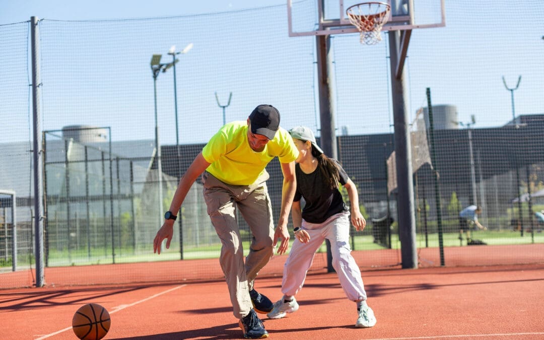
A man and a woman play table tennis to ease holiday stress.
The holiday season brings joy, family time, and tasty food, but it can also be stressful. Busy schedules, shopping, travel, and extra tasks can make anyone feel overwhelmed. One great way to feel better is through simple movement and exercise. Physical activity releases endorphins, chemicals in your brain that improve mood and reduce stress (Mayo Clinic, 2023). Even short sessions of fun activities can clear your mind and boost energy.
Many experts agree that almost any form of movement helps manage stress. It acts like a natural reset for your body and brain (Kitsap Physical Therapy, n.d.). Adding some holiday cheer to your routine makes it easier to stick with. This guide shares easy, enjoyable ways to stay active and calm during the holidays.
Why Movement Helps Reduce Holiday Stress
Exercise does more than keep you fit. It pumps up endorphins, boosting a happier feeling, and distracts you from worries. Activities like walking or dancing provide “meditation in motion,” helping you forget daily irritations (Mayo Clinic, 2023). Regular movement also improves sleep, builds confidence, and helps your body better handle stress.
During the holidays, people often move less due to cold weather or busy plans. This can make stress worse. But even one quick workout can lift your mood for hours (Gorman, 2022). Fun, low-pressure activities work best to avoid adding more pressure.
Releases feel-good chemicals to fight anxiety
Clears the mind and improves focus
Boosts energy and helps you sleep better
Builds strength to handle physical holiday demands, like carrying bags
Fun Sports-Inspired Activities to Boost Endorphins
Try activities that feel like play. Sports-inspired moves get your heart pumping and bring smiles.
Jumping rope: A quick cardio blast that raises your heart rate fast. Do it for 10-15 minutes while listening to holiday music (Avec Apartments, n.d.).
Dance breaks: Turn on your favorite songs and dance freely. Join a family dance party or try simple steps. Dancing combines rhythm and fun for great stress relief (NMC Health, n.d.; Triathlete Magazine, n.d.).
Pickup games: Play basketball, tennis, volleyball, or soccer with friends or family. These team sports combine exercise with social time, which further lowers stress (King Chiropractic, n.d.).
Shadowboxing: Punch the air like a boxer. This low-impact move releases tension without needing equipment. It’s perfect for a hotel room or living room (FightCamp, n.d.; Triathlete Magazine, n.d.).
These activities are easy to start and don’t require much space or gear.
Quick and Easy Bodyweight Exercises for Fast Relief
No gym? No problem. These simple moves use only your body and take little time.
Here are some top picks:
High knees: Run in place, lifting knees high. Do it for 1 minute to get your blood flowing (Echelon Fit, n.d.).
Planks: Hold a straight body position on your forearms and toes. Start with 30 seconds of core strength work (Echelon Fit, n.d.).
Bodyweight squats: Lower as if sitting in a chair, then stand up. Great for legs and glutes (Hydrow, n.d.).
Push-ups: Modify on knees if needed. Strengthen your upper body quickly (Hydrow, n.d.).
Jumping jacks: Classic move to warm up and boost mood (Echelon Fit, n.d.).
Try a 20-minute circuit: 30 seconds of each, with short rests in between. Repeat a few times (FightCamp, n.d.). Add holiday twists, like “present pick-up” squats—bend down as if grabbing gifts (Performance Health Academy, n.d.).
Mindful Practices for Calm and Flexibility
For gentler options, try mindful movements that focus on breath and flow.
Yoga flows: Simple poses like downward dog or warrior help stretch and center your mind. A 15-20 minute session reduces tension (Avec Apartments, n.d.; King Chiropractic, n.d.).
Tai Chi: Slow, flowing moves called “meditation in motion.” It improves balance and eases stress without strain (Mind Body Spine, n.d.; FightCamp, n.d.).
These practices calm the nervous system and pair well with busier days.
Outdoor Options: Walks and Hikes for Mind Clearing
Fresh air makes everything better. A brisk walk or hike builds endurance and clears thoughts.
Go for a festive neighborhood walk to see lights. Make it fun with a scavenger hunt for decorations (NMC Health, n.d.).
Hike in nature for extra calm. Being outdoors boosts positive feelings, such as gratitude (Triathlete Magazine, n.d.).
Add active games, such as playing in the yard or stair climbing, between tasks (Muscle MX, n.d.).
Aim for 30 minutes most days. No special gear needed—just good shoes (Club Getaway, n.d.).
Make It Festive: Holiday-Themed Active Fun
Keep things light by tying movement to celebrations.
Dance to holiday tunes or play charades that get everyone moving.
Try “Santa bag throws” or “candy cane curls” with simple weights or air motions (Performance Health Academy, n.d.).
Family games like obstacle courses or mini-golf indoors keep energy high and stress low (NMC Health, n.d.).
These ideas turn exercise into shared joy.
How Integrative Chiropractic Care Fits In
Physical tension from stress often shows up as tight muscles or misalignment. Integrative chiropractic care helps by using gentle adjustments to ease tension and support the nervous system. This improves your body’s stress response and promotes better flexibility (Chiropractic Works Collinsville, n.d.).
Chiropractors may suggest stretches or movements to help maintain alignment. This holistic approach complements exercise for full-body relief. Dr. Alexander Jimenez, a chiropractor and nurse practitioner with over 30 years of experience, notes that spinal health drives overall wellness. His integrative methods combine adjustments with posture exercises and stress management for better mobility and calm (Jimenez, n.d.; Jimenez, 2025a). He often sees that staying active and making adjustments help prevent holiday-related tension and support recovery (Jimenez, 2025b).
Pairing chiropractic visits with daily movement creates a balanced way to enjoy the season.
Tips to Get Started and Stay Consistent
Starting small is key during busy times.
Pick activities you enjoy to make it fun.
Schedule short sessions, like 10-20 minutes.
Involve family or friends for accountability.
Listen to your body—keep it light to avoid extra stress.
Combine with deep breathing for extra calm.
Consistency brings the best results. Even small efforts add up to less stress and more energy (American Fitness Professionals & Associates, n.d.).
By adding these fun movements and mindful care, you can handle holiday demands with ease. Focus on feeling good, not perfect. Your body and mind will thank you.
IFM's Find A Practitioner tool is the largest referral network in Functional Medicine, created to help patients locate Functional Medicine practitioners anywhere in the world. IFM Certified Practitioners are listed first in the search results, given their extensive education in Functional Medicine


:max_bytes(150000):strip_icc()/281448-grilled-chicken-fajitas-mfs-4-6c901272e8e34755bf0c5a535c8cb9fa.jpg)