Individuals dealing with health problems, UTIs, and skin issues can become chronic, what are the effects and benefits of drinking cranberry juice?
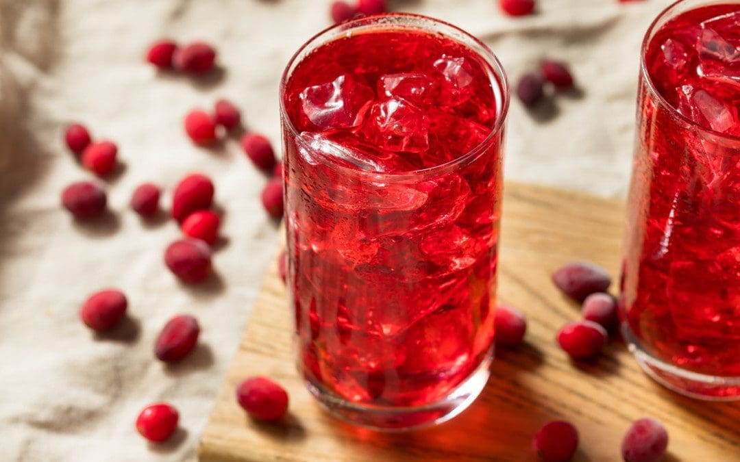
Cranberry Juice
Cranberries are a healthy source of nutrients and antioxidants. Cranberry juice is a recommended source of vitamin C, with the added benefits of promoting digestive, heart, immune, and skin health. Most individuals can safely drink cranberry juice to their diet with no issues, but women who are pregnant or individuals that take blood thinners, or medications should discuss adding cranberry intake with a doctor or specialist first.
One cup of unsweetened cranberry juice provides 23.5 milligrams or 26% of the daily value for vitamin C. (USDA 2018)
To avoid excess consumption of added sugars and maximize the benefits, it is recommended to drink unsweetened cranberry juice.
Digestive Health
Cranberries contain antioxidant compounds/polyphenolsthat have been shown to help with digestive health.
A study found that drinking cranberry juice was associated with increased beneficial gut bacteria and decreased constipation.
Research funded by a cranberry juice company found participants who consumed cranberry juice twice daily had lower levels of several risk factors for heart disease, stroke, and diabetes than those who received a placebo. (USDA 2016)
A systematic review and meta-analysis found that cranberry supplementation may improve body weight and blood pressure levels.
Cranberries may also help improve high-density lipoprotein (HDL) cholesterol—considered “good” cholesterol—in younger adults.
Cranberry juice contains vitamin C, which is important for immune system function.
Research suggests that inadequate vitamin C consumption can lead to decreased immunity and an increased risk of infections. (Carr A, Maggini S, 2017)
Skin Health
Thanks to its high antioxidant content, cranberry juice may help protect your skin against damage caused by free radicals that contributes to premature aging.
The vitamin C in cranberry juice is also needed for collagen production.
Collagen is a type of protein that provides strength, elasticity, and structural support to the skin, helping to keep it firm and smooth.(Pullar JM, et al., 2017)
Infection Prevention
A study found that cranberry components known as proanthocyanidins, can promote oral health.
Cranberries activate antibacterial processes that can prevent bacteria from binding together, reducing periodontitis/gum disease and the formation of dental plaque. (Chen H, et al., 2022)
Urinary Tract Infection Prevention
Cranberries have gone through many studies for home treatment of UTIs.
It is believed the chemical compounds/proanthocyanidins can help prevent certain bacteria from sticking to the lining of the urinary tract, thus reducing the risk of UTIs. (Das S. 2020)
A study found cranberry products in the form of juice or tablets may lower the risk of UTIs in at-risk groups by approximately 30%.
At-risk groups include those with recurrent UTIs, pregnant women, older adults, and individuals with chronic indwelling catheters (devices used for short-term bladder drainage) and neurogenic bladder (conditions in which people lack bladder control due to problems in the brain, spine, or spinal cord). (Xia J Yue, et al., 2021)
Daily Amount
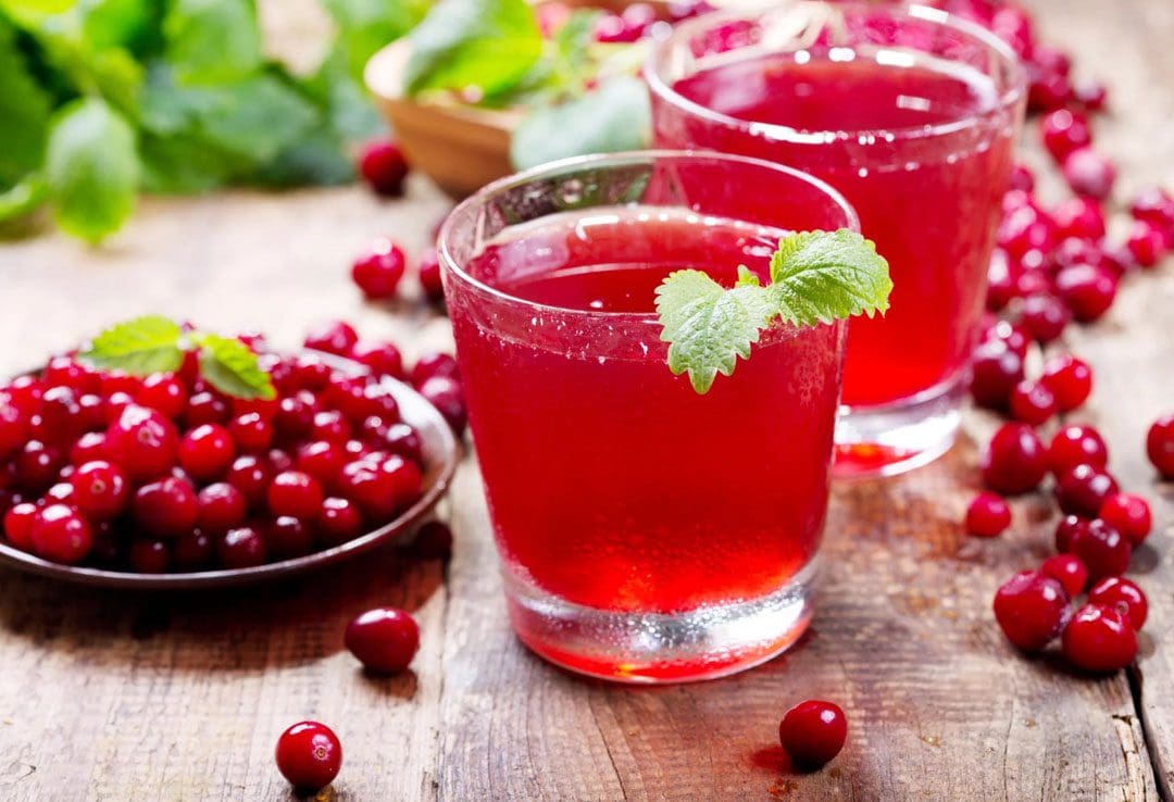
There is no official recommendation on the optimal amount of juice an individual should consume for health benefits. Most studies examining the benefits have used amounts ranging from 8 to 16 ounces, or around 1 to 2 cups per day. (The Cranberry Institute) However, cranberry juice with large amounts of added sugar can contribute to increased calories, leading to weight gain and other health concerns. Therefore, it is important to read the product label and look for pure, 100% cranberry juice.
If the pure juice is too tart, dilute it with some ice or water.
Avoid cranberry cocktails that are often mixed with other juices, like grape or apple juice, and contain added sugars that can decrease the benefits.
Chicas MC, Talcott S, Talcott S, Sirven M. Effect of cranberry juice supplementation on the gut microbiome and inflammatory markers: a randomized, double-blind, placebo-controlled study in overweight individuals. Curr Dev Nutr. 2022;6(Suppl 1):272. doi:10.1093/cdn/nzac053.013
Chen H, Wang W, Yu S, Wang H, Tian Z, Zhu S. Procyanidins and their therapeutic potential against oral diseases. Molecules. 2022;27(9):2932. doi:10.3390/molecules27092932
Pourmasoumi M, Hadi A, Najafgholizadeh A, Joukar F, Mansour-Ghanaei F. The effects of cranberry on cardiovascular metabolic risk factors: A systematic review and meta-analysis. Clinical Nutrition. 2020;39(3):774-788. doi:10.1016/j.clnu.2019.04.003
Pullar JM, Carr AC, Vissers MCM. The roles of vitamin C in skin health. Nutrients. 2017;9(8):866. doi:10.3390/nu9080866
Xia J Yue, Yang C, Xu D Feng, Xia H, Yang L Gang, Sun G ju. Consumption of cranberry as adjuvant therapy for urinary tract infections in susceptible populations: A systematic review and meta-analysis with trial sequential analysis. PLoS One. 2021;16(9):e0256992. doi:10.1371/journal.pone.0256992
The digestive system breaks down the foods eaten so the body can absorb the nutrients. During digestion, the unnecessary parts of these foods are turned into waste/stool, which is evacuated during a bowel movement. When the digestive system stops functioning properly due to factors such as diet change, eating unhealthy foods, lack of physical activity/exercise, medications, and certain health conditions, can cause constipation. Constipation occurs when the body cannot have a regular bowel movement. The distention, gas, bloating and not being able to have a bowel movement cause irritability and stress, which can worsen constipation. Incorporating recommended nutrition can help restore regular bowel movements and gut function.
Recommended Nutrition For Constipation
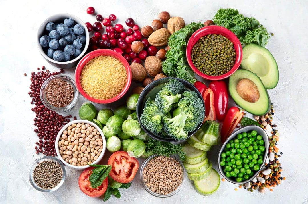
Symptoms like abdominal pain, bloating, and difficult bowel movements are common. Diet and proper hydration have a significant role in digestive health, especially in relieving and preventing constipation. High-fiber foods, prebiotics, and adequate hydration from foods and beverages are essential for healthy bowel movements.
Fiber is found in whole grains, starches, fruits, and vegetables.
Soluble and insoluble fiber are important for digestive health.
Focusing on incorporating high-fiber fruits, vegetables, and whole grains.
Foods rich in prebiotics like fermented foods are recommended when constipated.
The recommended nutrition for constipation, according to a dietitian includes.
Avocados
Avocados can be paired with just about anything and are full of nutrients and fiber.
One avocado contains around 13.5 grams of fiber.
One avocado will provide almost half daily fiber needs.
Other high-fiber fruits: pomegranates, guava, raspberries, blackberries, and passionfruit.
Figs
Figs can be eaten fresh and dried.
Figs are considered a laxative and have been shown to treat and reduce constipation.
They contain antioxidants, polyphenols, polyunsaturated fatty acids, and vitamins.
Other fruits similar to a fig: dried apricots, prunes, and plums.
Plums
Plums, prunes dried plums are packed with fiber and prebiotics that have a natural laxative effect.
The added H2O makes the stools softer and easier to pass.
Natural fruit juices, like pear, apple, or prune are often prescribed for constipation.
Other fruits that aid in bowel movements: peaches, pears, and apples.
Kefir
Fermented foods like kefir are rich in beneficial bacteria that work to maintain digestive system health.
It can be consumed on its own or used in smoothies, cooking, and baking recipes.
Other fermented foods: kombucha, yogurt, sauerkraut, kimchi, miso, and tempeh.
Oat Bran
Oat bran is oatmeal that has not had the bran removed.
The bran contains beneficial nutrients including fiber, antioxidants, vitamins, and minerals.
Oat bran contains soluble and insoluble fiber, as well as beta-glucan/non-starchy polysaccharides.
All improve the composition of gut bacteria and promote healthy bowel movements.
Other beneficial grains: oatmeal, wheat bran, rye, and barley.
Incorporating Gut-Beneficial Foods
How to incorporate recommended nutrition gut-beneficial foods into a regular menu:
Smoothie
Use kefir or yogurt as a base then balance it out with fiber-rich fruits like mango, blueberries, and kiwi.
Snacks
Diversify snacks with a plate of fiber and prebiotics.
Nuts, cheese, crackers, fruit, and a yogurt or avocado dip.
Oatmeal
Try oat bran to increase fiber.
Sprinkle a serving of flaxseeds, chia seeds, or hemp seeds for added fiber and healthy fats.
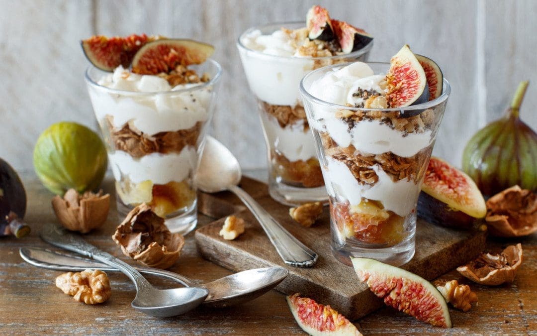
Parfait
Yogurt parfaits can maximize nutrients, flavor, and textures in a bowl.
Layer up on a favorite yogurt with granola, nuts, fruit, and seeds.
Grain Bowl
Fiber found in whole grains and seeds like barley, farro, and quinoa, helps promote healthy digestion.
Make a bowl with a grain base, then top with a protein, fresh or grilled veggies, avocado, and dressing.
Talk with a registered nutritionist or other healthcare provider to discuss recommended nutrition plan options.
Balancing Body and Metabolism
References
Arce, Daisy A et al. “Evaluation of constipation.” American family physician vol. 65,11 (2002): 2283-90.
Bharucha, Adil E. “Constipation.” Best practice & research. Clinical gastroenterology vol. 21,4 (2007): 709-31. doi:10.1016/j.bpg.2007.07.001
Gray, James R. “What is chronic constipation? Definition and diagnosis.” Canadian Journal of Gastroenterology = Journal Canadien de Gastroenterology vol. 25 Suppl B, Suppl B (2011): 7B-10B.
Jani, Bhairvi, and Elizabeth Marsicano. “Constipation: Evaluation and Management.” Missouri medicine vol. 115,3 (2018): 236-240.
Naseer, Maliha, et al. “Therapeutic Effects of Prebiotics on Constipation: A Schematic Review.” Current clinical pharmacology vol. 15,3 (2020): 207-215. doi:10.2174/1574884715666200212125035
National Institute of Diabetes and Digestive and Kidney Diseases. Symptoms and Causes of Constipation.
National Institute of Diabetes and Digestive and Kidney Disease. Your Digestive System and How It Works.
Sinclair, Marybetts. “The use of abdominal massage to treat chronic constipation.” Journal of bodywork and movement therapies vol. 15,4 (2011): 436-45. doi:10.1016/j.jbmt.2010.07.007
With the summer heatwave blasting through, some individuals can experience digestive health problems. The relationship between the temperature outside and the temperature in the body translates to the digestive system. As the heat rises, it can make the digestive system slow down and become weakened feeling bloated, nauseated, and tired. The body’s balance may feel off because the body lowers its internal temperature to protect itself. Individuals must be careful not to overload themselves with the wrong foods. One way to avoid problems and maintain digestion working smoothly is to eat lighter, eat smaller portions for each meal, and eat easily digestible foods. Doing this will allow the body will feel cooler, and maintain alertness and energy throughout the hot day.
The objective is not to skip meals but to eat regular meals, just smaller and easily digestible ones. Foods low in fiber tend to be easy to digest and can help the body feel better.
White Rice
White rice is low in fat and fiber, making it easy on the stomach and easy to digest.
It is not associated with any gastrointestinal issues and is considered a safe starch because it is an easy source of carbohydrates that provides instant energy.
To digest rice even more easily, eat it by itself or pair it with foods low in fat.
Certain foods that are high in fat, like vegetable oils, can take longer to digest and could cause discomfort.
A 1/2 cup of cooked white rice:
210 calories
4g protein
0g fat
49g carbohydrates
1g fiber
Bananas
Ripe bananas are an easily digestible fruit that only contains a moderate amount of fiber.
They are associated with improvements in both constipation and diarrhea,
Individuals with a variety of digestive issues may experience relief when incorporating bananas into their diets.
Cooking bananas makes them even easier to digest as it makes certain nutrients easier to absorb.
Make sure the bananas are ripe enough.
Unripe bananas can be more difficult to digest.
1 medium raw/ripe banana:
105 calories
1.3g protein
0.4g fat
27g carbohydrates
3g fiber
Applesauce
Although made from apples, applesauce is low in fiber and a great source of vitamin C.
Cooked, canned, or processed fruits tend to be lower in fiber and easier to digest.
Applesauce is recommended to calm a variety of stomach-related ailments like constipation, diarrhea, and gastroparesis.
A 4-ounce serving of applesauce:
90 calories
0g protein
0g fat
22g carbohydrates
2g fiber
White Bread
Plain white bread is low in fiber and easier to digest than bread made with whole-grain wheat bread.
It is often fortified with nutrients including folic acid, B vitamins, vitamin D3, and more.
Try plain toast for breakfast
Use low-fat fillings for an easily digestible sandwich for lunch or dinner.
2 slices of plain white bread:
150 calories
4g protein
28g carbohydrates
2g fat
1g fiber
Chicken and Turkey
Lean proteins low in fat like chicken breast and turkey are easy to digest.
Individuals experiencing digestive problems are recommended to consume lean protein over fattier red meats.
A 3-ounce serving of skinless, boneless chicken breast:
128 calories
26g protein
2.7g fat
0g carbohydrates
0g fiber
Sweet Potatoes
Cooked potatoes of all varieties are examples of easily digestible foods.
Sweet potatoes are gentle on the digestive tract because they are mostly insoluble fiber, which speeds up digestion and increases regularity.
To make potatoes easier to digest, remove the skins and mash the inside.
Removing the skins decreases the fiber content, and mashing them makes digestion easier.
1 medium sweet potato that is cooked and peeled:
135 calories
3g protein
0.2g fat
31g carbohydrates
5g fiber
Other recommendations that could help stimulate digestion include drinking more water, getting more sleep, reducing stress levels, and exercising.
The Healing Diet
References
Howard, Sally, and Geetanjali Krishna. “How hot weather kills: the rising public health dangers of extreme heat.” BMJ (Clinical research ed.) vol. 378 o1741. 14 Jul. 2022, doi:10.1136/bmj.o1741
Kong, Fanbin, et al. “Physical changes in white and brown rice during simulated gastric digestion.” Journal of food science vol. 76,6 (2011): E450-7. doi:10.1111/j.1750-3841.2011.02271.x
Nguyen, Hoang Chinh et al. “Bioactive Compounds, Antioxidants, and Health Benefits of Sweet Potato Leaves.” Molecules (Basel, Switzerland) vol. 26,7 1820. 24 Mar. 2021, doi:10.3390/molecules26071820
Remes-Troche, José María. “Too hot” or “too cold”: effects of meal temperature on gastric function.” Digestive diseases and sciences vol. 58,9 (2013): 2439-40. doi:10.1007/s10620-013-2789-4
Salfi, Salvatore F, and Karyn Holt. “The role of probiotics in diarrheal management.” Holistic nursing practice vol. 26,3 (2012): 142-9. doi:10.1097/HNP.0b013e31824ef5a3
Singh, Balwinder, et al. “Bioactive compounds in banana and their associated health benefits – A review.” Food Chemistry vol. 206 (2016): 1-11. doi:10.1016/j.foodchem.2016.03.033
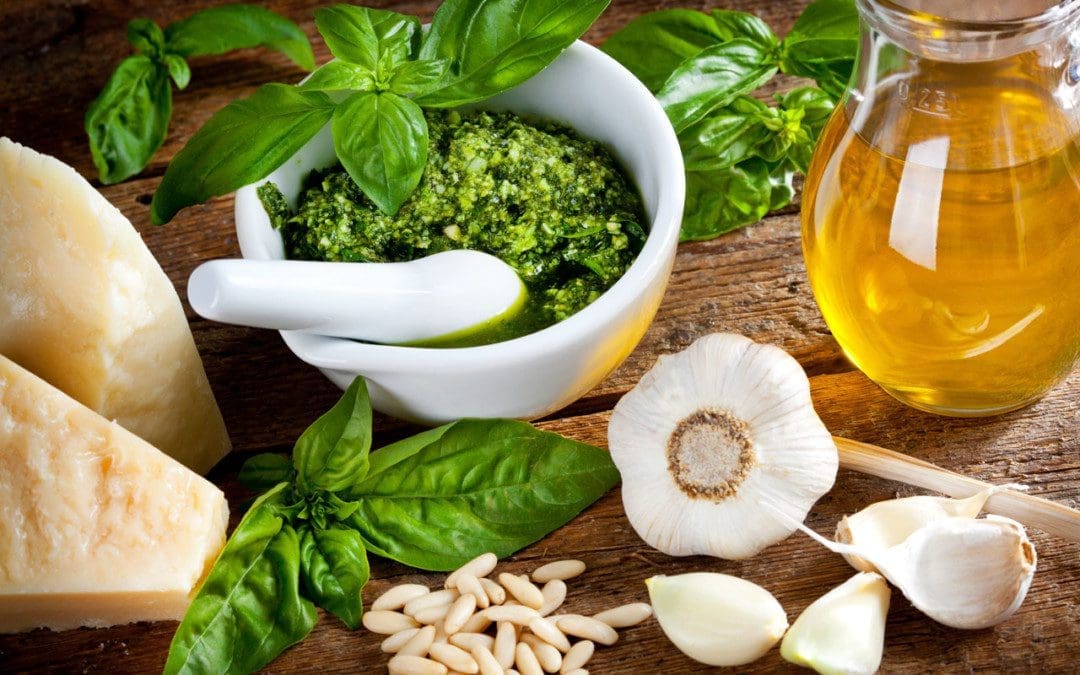
Pesto is a sauce that is made with garlic, pine nuts, basil, cheese, and olive oil, which creates a strong, rich flavor. It is used as a marinade, dip, salad dressing, sandwich spread, and a topping for dishes like pasta and pizza. It can be homemade or bought premade, including vegan varieties. It is made with nutritious ingredients and can be consumed as part of a balanced diet. The sauces can vary in nutrition, but in general, it is a rich source of healthy fats and is also part of the Mediterranean Diet.
Pesto
Carbohydrates
The sauce is not a significant source of complex carbohydrates, dietary fiber, or sugar.
A spoonful contains under 1 gram of carbohydrates.
It is often paired with foods rich in carbohydrates, like sandwiches, pizza, and pasta.
Fats
Nearly 60% of the calories in pesto come from fats, provided by the olive oil, cheese, and pine nuts.
There are 9.47 grams of fats per spoonful, which includes:
5.63 grams of monounsaturated fatty acids.
1.53 grams of saturated fatty acids.
1.68 grams of polyunsaturated fatty acids.
It also contains 2.56mg of cholesterol.
According to U.S. Dietary Guidelines for Americans, 20% to 35% of daily calories should come from fat.
Protein
The sauce is not a protein-rich food with only 1.38 grams of protein per tablespoon.
It is often used as a condiment, it can add flavor to other foods higher in protein.
Vitamins and Minerals
Pesto contains:
33.1mg of calcium.
36.8mg of phosphorus.
31.8mg of potassium.
9.76mg of magnesium.
Health Benefits
Some of the potential health benefits of pesto.
Antioxidant Properties
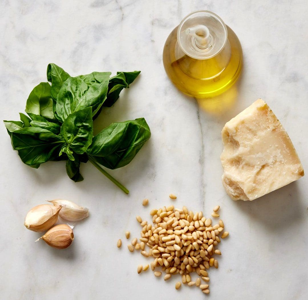
Garlic, pine nuts, olive oil, and basil are rich sources of antioxidants.
Antioxidants have an essential role in defending the body against free radical damage, which can lead to disease.
Diets high in antioxidants reduce the risk of various diseases, like heart disease and cancer.
Consuming antioxidant-rich foods like pesto on a regular basis can increase antioxidant levels.
Cardiovascular Benefits
Olive oil health benefits include the reduced risk of heart disease.
Substituting other high-fat foods like margarine, butter, and mayonnaise with olive oil can help lower the risk of heart disease and stroke.
Help Lower Cholesterol
Four different types of fats – saturated, trans, monounsaturated, and polyunsaturated fats.
Saturated fats can raise LDL/unhealthy cholesterol levels.
Foods rich in monounsaturated and polyunsaturated fats like pesto can help lower LDL cholesterol levels in the blood and support HDL/healthy cholesterol levels.
Weight Management
Pesto is commonly consumed on the Mediterranean diet and can be part of a nutritious lifestyle that supports a healthy weight.
Research has shown that following a Mediterranean diet can lead to and maintain long-term changes in weight management.
Store-bought pesto can contain a large amount of sodium.
Individuals following a low-sodium diet or taking heart medications should check with their doctor before consuming these products.
To control the amount of sodium, consider making a homemade recipe that uses less salt and aligns with individual dietary preferences.
Basil is the main ingredient but some varieties are made with other herbs.
Pesto traditionally contains parmesan cheese/milk products and pine nuts/tree nuts which are common food allergens.
The sauce is a vegetarian-friendly sauce, but vegans can look for cheese and dairy-free versions.
It is possible that an allergic reaction can occur in individuals allergic to dairy products and nuts.
Individuals allergic to nuts can choose nut-free varieties.
From Consultation to Transformation
References
Agnoli C, Sieri S, Ricceri F, et al. Adherence to a Mediterranean diet and long-term changes in weight and waist circumference in the EPIC-Italy cohort. Nutr Diabetes. 2018;8(1):22. doi:10.1038/s41387-018-0023-3
Bolling, Bradley W et al. “Tree nut phytochemicals: composition, antioxidant capacity, bioactivity, impact factors. A systematic review of almonds, Brazils, cashews, hazelnuts, macadamias, pecans, pine nuts, pistachios, and walnuts.” Nutrition research reviews vol. 24,2 (2011): 244-75. doi:10.1017/S095442241100014X
Bower, Allyson, et al. “The Health Benefits of Selected Culinary Herbs and Spices Found in the Traditional Mediterranean Diet.” Critical Reviews in food science and nutrition vol. 56,16 (2016): 2728-46. doi:10.1080/10408398.2013.805713
Guasch-Ferré M, Liu G, Li Y, et al. Olive oil consumption and cardiovascular risk in U.S. adults. J Am Coll Cardiol. 2020;75(15):1729-1739. doi:10.1016/j.jacc.2020.02.036
Liu, Qing, et al. “Antibacterial and Antifungal Activities of Spices.” International journal of molecular sciences vol. 18,6 1283. 16 Jun. 2017, doi:10.3390/ijms18061283
Marcelino, Gabriela et al. “Effects of Olive Oil and Its Minor Components on Cardiovascular Diseases, Inflammation, and Gut Microbiota.” Nutrients vol. 11,8 1826. 7 Aug. 2019, doi:10.3390/nu11081826
Nicastro, Holly L, et al. “Garlic and onions: their cancer prevention properties.” Cancer prevention research (Philadelphia, Pa.) vol. 8,3 (2015): 181-9. doi:10.1158/1940-6207.CAPR-14-0172
Sestili, Piero, et al. “The potential effects of Ocimum basilicum on health: a review of pharmacological and toxicological studies.” Expert opinion on drug metabolism & toxicology vol. 14,7 (2018): 679-692. doi:10.1080/17425255.2018.1484450
Sun, Liangzi, et al. “Tryptophan targeted pulsed electric field treatment for enhanced immune activity in pine nut peptides.” Journal of food biochemistry vol. 44,6 (2020): e13224. doi:10.1111/jfbc.13224
Before the body can benefit from consumed nutrients, the gastrointestinal tract has to digest and absorb the foods. Before eating, the body needs to feel hungry. However, hunger is not the same as appetite. Hunger is a physical reaction caused by hormonal and chemical changes in the body when fuel is needed. Appetite is more of a desire to eat and can be a learned response. It is one reason why individuals can eat when they are not hungry. The body comprises different hormones that regulate hunger, digestion, and appetite.
Hunger Digestion Regulating Hormones
Hunger Hormones
Hunger is the feeling when the body needs food. When the body has enough, hunger should subside. That’s because various hormones regulate hunger.
Leptin
Leptin is a hormone secreted by adipose tissue/fat into the bloodstream.
The more fat in the body, the higher the blood levels of leptin.
Leptin level also increases with food intake and is higher in females than males, but overall, it lowers with age.
Increased leptin levels trigger the hypothalamus to reduce hunger.
Ghrelin
Ghrelin is a hormone produced by the stomach and small intestine when the stomach is empty.
Like leptin, it also works with the hypothalamus.
However, instead of suppressing hunger, it increases hunger.
Insulin
The pancreas produces this hormone.
It is mostly known for regulating blood sugar levels.
It also suppresses hunger.
Adiponectin
Adiponectin is a hormone secreted by fat cells.
As body fat levels go down, this hormone goes up.
If fat levels go up, adiponectin levels go down.
Cholecystokinin
Cholecystokinin is a hormone produced in the small intestine during and after a meal.
It triggers the release of bile and digestive enzymes into the small intestine.
These suppress hunger and make the body feel full.
Peptide YY
This hormone suppresses appetite for about 12 hours after eating.
Made by both the large and small intestines after eating.
Glucocorticoids
Adrenal glands make these hormones, and their primary function is to regulate inflammation and other processes, but they also impact hunger.
A cortisol deficiency reduces appetite, but excessive amounts of glucocorticoids increase hunger.
Digestion Hormones
Digestion is coordinated and regulated by hormones.
Gastrin
Gastrin is a hormone the stomach and the small intestine release when eating.
Gastrin stimulates the release of hydrochloric acid and pepsinogen in the stomach to speed up digestion.
Gastrin stimulates glucagon, which works with insulin to regulate blood sugar.
Secretin
Secretin is a hormone made by the small intestine.
It is secreted into the bloodstream when the acidic chyme from the stomach enters the small intestine.
Secretin stimulates the pancreas to release bicarbonate digestive liquids into the small intestine.
The bicarbonate neutralizes the acidity.
Secretin acts on the stomach to trigger the production of pepsinogen to help break down proteins.
Cholecystokinin – CCK
The small intestine makes and releases CCK into the bloodstream.
Essential fat digestion stimulates the gallbladder to release bile into the small intestine.
It also triggers the pancreas to release various digestive enzymes so they can break down fats, carbohydrates, and proteins.
It stimulates the pancreas to release insulin and slows down stomach digestive activity.
Peptide YY and Enterogastrone
Released by the small intestine, two more hormones slow digestion down and decrease the production of digestive secretions.
Chiropractic Care and Metabolism
References
Chandra, Rashmi, and Rodger A Liddle. “Cholecystokinin.” Current Opinion in Endocrinology, diabetes, and Obesity vol. 14,1 (2007): 63-7. doi:10.1097/MED.0b013e3280122850
Davis, Jon. “Hunger, ghrelin and the gut.” Brain Research vol. 1693, Pt B (2018): 154-158. doi:10.1016/j.brainres.2018.01.024
Gupta K, Raja A. Physiology, Gastric Inhibitory Peptide. [Updated 2022 Sep 26]. In: StatPearls [Internet]. Treasure Island (FL): StatPearls Publishing; 2023 Jan-. Available from: https://www.ncbi.nlm.nih.gov/books/NBK546653/
Konturek, S J et al. “Brain-gut axis and its role in the control of food intake.” Journal of Physiology and Pharmacology: an official journal of the Polish Physiological Society vol. 55,1 Pt 2 (2004): 137-54.
Prosapio JG, Sankar P, Jialal I. Physiology, Gastrin. [Updated 2023 Apr 6]. In: StatPearls [Internet]. Treasure Island (FL): StatPearls Publishing; 2023 Jan-. Available from: https://www.ncbi.nlm.nih.gov/books/NBK534822/
Rix I, Nexøe-Larsen C, Bergmann NC, et al. Glucagon Physiology. [Updated 2019 Jul 16]. In: Feingold KR, Anawalt B, Blackman MR, et al., editors. Endotext [Internet]. South Dartmouth (MA): MDText.com, Inc.; 2000-. Available from: https://www.ncbi.nlm.nih.gov/books/NBK279127/
Suzuki, Keisuke, et al. “The role of gut hormones and the hypothalamus in appetite regulation.” Endocrine Journal vol. 57,5 (2010): 359-72. doi:10.1507/endocrine.k10e-077
Tack, Jan, et al. “The gastrointestinal tract in hunger and satiety signaling.” United European gastroenterology journal vol. 9,6 (2021): 727-734. doi:10.1002/ueg2.12097
Zanchi, Davide, et al. “The impact of gut hormones on the neural circuit of appetite and satiety: A systematic review.” Neuroscience and biobehavioral reviews vol. 80 (2017): 457-475. doi:10.1016/j.neubiorev.2017.06.013
Olympic athletes are so fit they make everything look easy. It is one of the most amazing examples of athletic prowess to behold. Attempting these feats of athleticism could lead to injury without proper training and conditioning. However, you can draw inspiration from these incredible skills and commitment to reach your fitness goals. Individuals can emulate the Olympic athlete’s discipline to get the most out of physical activity and workouts.
Olympic Athlete Discipline
This is not about the literal workout routines these athletes engage in but the mental state and discipline that keeps them motivated, especially when things get tough. This can help individuals get the most out of every workout. When momentum or motivation starts to dwindle, look to Olympic athletes and then apply that discipline to recharge motivation and achieve the goal.
Learn to Train Daily
Many can succumb to weekend warrior syndrome. After sitting on the couch after a long day of work all week, individuals try to make up for the inactivity by overexerting themselves with hours of exercise on the weekend. This is a perfect setup for injury.
Instead, train, work out, exercise, and move around daily. Professional athletes know they won’t get the top results in one shot.
They approach it in incremental steps for a solid foundation and understanding of the final and optimal result.
Daily physical activity and exercise maintain body conditioning, strength, and overall fitness.
And makes the body more efficient at burning fat.
Maintain Goal Focus
Olympic athletes have specific goals in their training. There has to be when competing against the best athletes in the world and winning.
Individual health and fitness goals may be smaller, but they are just as important and should be treated as such for motivation to exercise each day.
Keep goals simple, specific, and reachable/doable when setting them.
SMART goals are Specific, Measurable, Achievable, Realistic, and Time-based.
Use daily reminders of what the goal is and strategies to achieve it.
Utilize Specific Training
Olympic athletes must have incredible endurance, strength, and skill sets to complete their event.
They must incorporate specific training drills, exercises, stretches, nutrition, etc., to improve and advance.
Whatever the goal, make sure the training is appropriate.
If trying to build muscle, focus on heavy strength training and consuming quality calories.
If the goal is to lose weight, break down the goal into achievable steps to get there, like.
What type of workouts are needed?
Consulting a nutritionist for recommendations.
Fuel the Body for Top Performance
Individuals can be concerned about food and whether it contains too much fat or calories.
Olympic athletes worry about getting the right nutrients and calories to fuel their bodies for competition.
Use a similar approach and ask what is the best thing I should eat right now for the workout and…
What foods will maintain optimal body health?
Listen to The Body and Rest
Olympic athletes have to learn to maintain the balance of maintaining top performance and not getting burnt out.
This is when they know to listen to their bodies, extend recovery days, or take a mini-vacation.
Overtraining can lead to injury, frustration, discouragement, and loss of motivation.
Whatever the activity, make sure proper form is followed.
For example, distance runners hold their heads high, have relaxed faces and upper bodies, have a natural arm swing, and do not overstride.
Apply the same form principles to stretches and exercises.
Stronger Body = Better Life
References
Casa, Douglas J et al. “Fluid Needs for Training, Competition, and Recovery in Track-and-Field Athletes.” International Journal of sports nutrition and exercise metabolism vol. 29,2 (2019): 175-180. doi:10.1123/ijsnem.2018-0374
Bailey RR. Goal Setting and Action Planning for Health Behavior Change. Am J Lifestyle Med. 2019;13(6):615-618. doi:10.1177/1559827617729634
Hackett, Daniel, et al. “Olympic weightlifting training improves vertical jump height in sportspeople: a systematic review with meta-analysis.” British Journal of sports medicine vol. 50,14 (2016): 865-72. doi:10.1136/bjsports-2015-094951
Huebner, Marianne, et al. “The Masters’ athlete in Olympic weightlifting: Training, lifestyle, health challenges, and gender differences.” PloS one vol. 15,12 e0243652. 4 Dec. 2020, doi:10.1371/journal.pone.0243652
Swift DL, Johannsen NM, Lavie CJ, Earnest CP, Church TS. The Role of Exercise and Physical Activity in Weight Loss and Maintenance. Prog Cardiovasc Dis. 2014;56(4):441-447. doi:10.1016/j.pcad.2013.09.012
Regular exercise and physical activity help with cardiovascular health, improved mood, better management of chronic conditions, and can help digestion. For individuals with any GI distress or inflammatory bowel disease that has caused digestive enzyme deficiencies, exercise, and physical movement have been found to provide digestive aid. Here we look at activities to help digestion.
Exercises To Help Digestion
When exercising the body, the cardiac output/volume of blood the heart pumps every minute increases as the demand for oxygenated blood throughout the body increases, particularly in the working muscles. During exercise, the same increase in blood circulation happens within the digestive system’s muscle groups. The blood flow to digestive organs causes peristalsis, which is involuntary constriction and relaxation of the muscles in the digestive tract. This process helps move food efficiently through the gastrointestinal tract. Exercise supports the growth of beneficial gut bacteria to maintain a healthy digestive system.
Exercise helps relieves stress which means lower amounts of cortisol.
Research has found that elevated cortisol levels are associated with compromised digestive function.
Tai chi has been shown to improve immune function and inflammation of the gut and is a helpful tool for maintaining homeostasis/gut balance.
This is a form of moderate-intensity exercise and meditative practice.
The emphasis is on slow controlled movements and deep breathing.
This makes it recommended for individuals looking to improve digestion and those with gastrointestinal conditions.
Pilates
Pilates is the practice that targets abdominal muscles and utilizes diaphragmatic breathing to help individuals perform a series of movements to strengthen and elongate the body’s muscles.
Individuals who perform this exercise often report relief from bloating and gas.
Whether new to exercise or returning, a plan can help you get there. Meeting with a fitness trainer or sports chiropractor is a great place to begin if you have limited knowledge about what works best for your body and schedule.
A certified trainer can help guide you toward an achievable program that focuses on gut health.
Individuals with a GI disorder should talk with their doctors before starting a new exercise plan.
This does not mean you can’t do intense exercises like running; you’ll want to work with a doctor to set up a program that doesn’t cause flare-ups.
Aim for roughly three hours of moderate-intensity weekly exercise to support a healthy digestive system.
Sit less and move more.
Do at least two or more muscle-strengthening activities of moderate intensity every week.
Cherpak, Christine E. “Mindful Eating: A Review Of How The Stress-Digestion-Mindfulness Triad May Modulate And Improve Gastrointestinal And Digestive Function.” Integrative medicine (Encinitas, Calif.) vol. 18,4 (2019): 48-53.
Drouin, Jacqueline S et al. “Comparisons between Manual Lymph Drainage, Abdominal Massage, and Electrical Stimulation on Functional Constipation Outcomes: A Randomized, Controlled Trial.” International Journal of environmental research and public health vol. 17,11 3924. June 1. 2020, doi:10.3390/ijerph17113924
Hamasaki, Hidetaka. “Exercise and gut microbiota: clinical implications for the feasibility of Tai Chi.” Journal of integrative medicine vol. 15,4 (2017): 270-281. doi:10.1016/S2095-4964(17)60342-X
Joyner, Michael J, and Darren P Casey. “Regulation of increased blood flow (hyperemia) to muscles during exercise: a hierarchy of competing physiological needs.” Physiological Reviews vol. 95,2 (2015): 549-601. doi:10.1152/physrev.00035.2013
LeBouef T, Yaker Z, Whited L. Physiology, Autonomic Nervous System. [Updated 2023 May 1]. In: StatPearls [Internet]. Treasure Island (FL): StatPearls Publishing; 2023 Jan-. Available from: https://www.ncbi.nlm.nih.gov/books/NBK538516/
Singhal, Rashi, and Yatrik M Shah. “Oxygen battle in the gut: Hypoxia and hypoxia-inducible factors in metabolic and inflammatory responses in the intestine.” The Journal of biological chemistry vol. 295,30 (2020): 10493-10505. doi:10.1074/jbc.REV120.011188
van Wijck, Kim, et al. “Physiology and pathophysiology of splanchnic hypoperfusion and intestinal injury during exercise: strategies for evaluation and prevention.” American Journal of Physiology. Gastrointestinal and liver physiology vol. 303,2 (2012): G155-68. doi:10.1152/ajpgi.00066.2012
IFM's Find A Practitioner tool is the largest referral network in Functional Medicine, created to help patients locate Functional Medicine practitioners anywhere in the world. IFM Certified Practitioners are listed first in the search results, given their extensive education in Functional Medicine