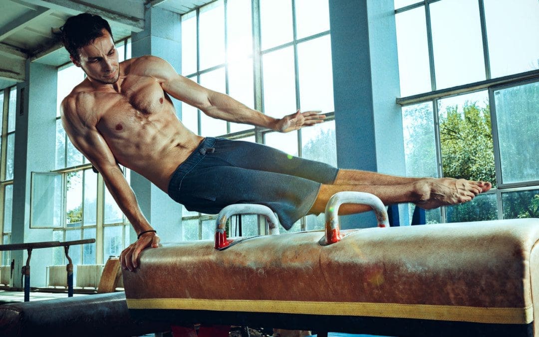
Athletes train and practice constantly to prepare their mind and bodies for the big games, matches, etc. When the game is on, it is normal/natural to feel anxious and nervous, especially at the beginning, but then the athlete settles in and relaxes, letting their training take over. However, for some athletes, the anxiousness and nervousness doesn’t go away but intensifies, the heart starts racing, and the individual can’t stop thinking about choking, failing, and losing. This is known as sports performance anxiety, or competitive anxiety, and is common.
Competitive Anxiety
Research shows that 30 to 60 percent of athletes experience the disorder. Doctors divide the signs and symptoms into mental and physical categories.
Physical Symptoms
Rapid Heartbeat
The stress can cause overproduction of adrenaline and cortisol, making the heart beat rapidly.
Muscle Tension
The muscles can tighten up, become painful, and cause tension and pain in the head.
Trembling
The hands could shake while holding the ball, bat, racket, or foot twitching could present.
Hyperventilation
Individuals report a sensation of choking or being unable to catch their breath.
Digestion Issues
The stress can cause foods to be quickly digested, causing cramping and/or the sudden urge to use the bathroom.
Mental Symptoms
Fear of Failing
The athlete imagines themselves losing all the time.
Worrying about letting the coach and team down or the audience or other athletes criticizing and laughing at your performance.
Unable to Focus
The athlete may have concentration issues and become absorbed in how others react to their performance.
Overthinking
The athlete can temporarily forget how to perform specific actions that are typically automatic.
Self-confidence issues
The athlete can start doubting their abilities.
Stress and Anxiety
The Yerkes-Dodson law explains how stress, anxiety, and arousal levels affect performance and how stress levels must be maintained within a range to perform well.
Low Arousal
It could be the athlete is not as into the sport as when they began, so they do not put forth the total effort.
High Arousal
This means the sport could be causing so much stress that the athlete panics or freezes up.
Competitive anxiety sets in.
Optimal Arousal
This means the athlete is fully engaged in pushing themselves to the fullest.
This can be applied to any performing task like play rehearsals to a tennis match.
Some recommended steps can be taken to handle and prevent sports competitive anxiety when trying to overcome those overwhelming feelings of nervousness and tension.
Positive self-talk
Self-talk is having a positive conversation with yourself.
Athletes who practiced positive self-talk reported:
Improved self-confidence
Reduced physical anxiety symptoms
Improved sports performance
Listen to Music
When anxious before a meet, game, match, etc., consider listening to some favorite or relaxing music.
Meditation
Meditation has been found to reduce all types of anxiety, including sports.
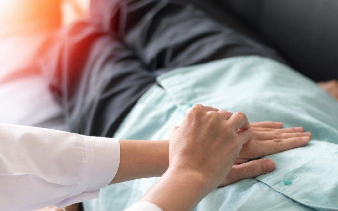
Chiropractic
Chiropractic treatment specializes in the musculoskeletal system and can realign the body and release any muscle tension and restriction through hands-on manipulation techniques and mechanical decompression. Treatment involves manipulating the muscles, ligaments, tendons, fascia, and soft tissues to relieve pain through therapeutic muscle therapies that include:
One or a combination of therapies can alleviate symptoms related to muscle spasms, delayed onset muscle soreness, fascia restrictions, soft tissue injuries, and pain and dysfunction throughout the body, restoring function, movement, and strength.
Using The DRX9000 For Spinal Decompression
References
Elliott, Dave, et al. “The effects of relaxing music for anxiety control on competitive sport anxiety.” European journal of sports science vol. 14 Suppl 1 (2014): S296-301. doi:10.1080/17461391.2012.693952
Ford, Jessica L et al. “Sport-related anxiety: current insights.” Open access journal of sports medicine vol. 8 205-212. 27 Oct. 2017, doi:10.2147/OAJSM.S125845
Rice, Simon M et al. “Determinants of anxiety in elite athletes: a systematic review and meta-analysis.” British journal of sports medicine vol. 53,11 (2019): 722-730. doi:10.1136/bjsports-2019-100620
Rowland, David L, and Jacques J D M van Lankveld. “Anxiety and Performance in Sex, Sport, and Stage: Identifying Common Ground.” Frontiers in psychology vol. 10 1615. 16 Jul. 2019, doi:10.3389/fpsyg.2019.01615
Walter N, et al. (2019). Effects of self-talk training on competitive anxiety, self-efficacy, volitional skills, and performance: An intervention study with junior sub-elite athletes. mdpi.com/2075-4663/7/6/148
Motor vehicle crashes and accidents cause significant trauma in a few seconds changing an individual’s life completely. Severe injuries include traumatic brain injury, spinal cord damage, fractures, and amputations. Many individuals experience post-traumatic stress disorder – PTSD after a vehicle collision; even a minor accident can cause emotional trauma symptoms. PTSD commonly presents with other symptoms that range from depression to heart disease, and the most frequent symptom is physical pain. Chiropractic decompression, physical therapy, and therapeutic massage can help alleviate physical pain.
PTSD Physical Pain
Physical trauma can cause immediate physical effects and injury, as well as physical symptoms that present later on.
Trying not to talk or think about the crash or accident with friends, family, places, or anything associated with the trauma.
Avoiding activities.
Emotional numbness.
Detachment.
All can generate physical muscle tension and chronic stress, leading to headaches, migraines, back pain, stomach pain, and body aches. Long-term physical pain symptoms can turn chronic pain and medication dependency into a vicious cycle.
Chiropractic Therapy
Chiropractic care diagnoses and treats disorders of the musculoskeletal system. Chiropractic treatment is recommended to help alleviate the physical symptoms of PTSD. Trauma causes individuals to store intense emotions in their bodies. Chiropractic manipulation and decompression release the tension in the muscles caused by the trauma and the emotional stress. Adjustments restore the body’s alignment and open the nervous system circulation, allowing signals to flow freely, leading to a healthier mind-body connection.
Non-Surgical Spinal Decompression Therapy
References
Beck, J Gayle, and Scott F Coffey. “Assessment and treatment of PTSD after a motor vehicle collision: Empirical findings and clinical observations.” Professional psychology, research, and practice vol. 38,6 (2007): 629-639. doi:10.1037/0735-7028.38.6.629
Elder, Charles et al. “Comparative Effectiveness of Usual Care With or Without Chiropractic Care in Patients with Recurrent Musculoskeletal Back and Neck Pain.” Journal of general internal medicine vol. 33,9 (2018): 1469-1477. doi:10.1007/s11606-018-4539-y
Hu, JunMei, et al. “Chronic widespread pain after motor vehicle collision typically occurs through immediate development and nonrecovery: results of an emergency department-based cohort study.” Pain vol. 157,2 (2016): 438-444. doi:10.1097/j.pain.0000000000000388
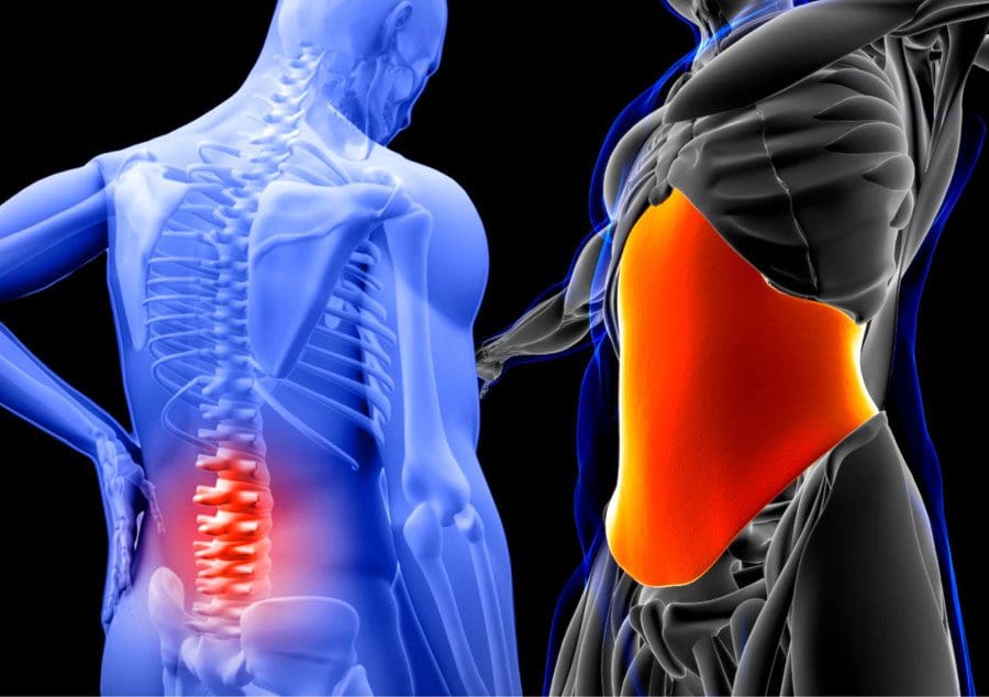
The lower back core muscles initiate and control movement and posture. Total body movements start at the pelvis and are carried out by the low back and abdominal muscles. These core muscles provide control and stability when moving. When the body is not moving, standing and sitting posture habits (healthy and unhealthy) develop based on how an individual controls/holds their pelvis posture. The lower back muscles are in constant demand for performing repetitive motions or forceful actions. The muscles become chronically tight and sore, joints and nerves get compressed, and bones and connective tissue stay in a stressed state, causing intense pain and damage to the body’s structure. These problems can result from muscle memory, the nervous system’s operating procedure to make movements automatic.
Muscle Memory
The nervous system controls the movement and contraction in the muscles and constantly reinforces and learns new movement patterns to become more efficient through muscle memory. However, this learning process allows for the development of healthy muscle habits and unhealthy muscular habits. An example is sitting using a slouching posture. The nervous system will store that posture data and subconsciously contract the abdominal and pectoral muscles, so the slouched posture takes over. Because of the continual usage and compromised postures, various muscles start to tighten up. Individuals are typically not aware of the gradual tightening until stiffness, soreness, and pain sets in. Tight muscles pull the skeleton in awkward ways that cause body misalignments, causing:
Individuals develop unique muscle patterns and tension levels throughout the body due to repetitive daily activities, stress responses, injuries sustained, and physical activities. Muscle memory issues can contribute to chronic back pain and sciatica. Unhealthy muscle memory causes the muscles not to return to their natural state but to the awkward position and makes that the natural state. Healthy muscle memory leads to instant reflexes that make movements smooth and effortless.
Chiropractic will relieve back and sciatica pain by inducing deep muscle relaxation that releases endorphins. The soreness and tension will be massaged, and mechanical decompression if necessary. Massage and stretching will help retrain muscle and movement memory, along with exercises to reinforce the retraining.
Spinal Decompression Therapy
References
Campbell, James N, and Richard A Meyer. “Mechanisms of neuropathic pain.” Neuron vol. 52,1 (2006): 77-92. doi:10.1016/j.neuron.2006.09.021
Wilder, David G et al. “Effect of spinal manipulation on sensorimotor functions in back pain patients: study protocol for a randomized controlled trial.” Trials vol. 12 161. 28 Jun. 2011, doi:10.1186/1745-6215-12-161
After traveling, body/musculoskeletal aches and pains can present from continual standing in line, sitting, staying in the same position, carrying a heavy bag or pulling a suitcase, and sleeping in an unfamiliar bed with different pillows, on a plane or car can cause body imbalances, jerking the spine out of alignment, straining the neck, shoulders, and back causing headaches, soreness, stiffness, back pain, and compression. Chiropractic massage and decompression will relieve travel pains, loosen stiff and sore joints, soothe aching muscles, realign the spine, and restore body health and comfort.
Musculoskeletal Health
The musculoskeletal system is comprised of:
Muscles
Tendons
Ligaments
Bones
Joints
Tissues that move the body and help maintain structure and form.
The health of the musculoskeletal system is defined as the absence of injury, disease, or illness within the system. Keeping this system healthy is crucial for the health of the other body systems.
Common Muskuloskeletal Conditions
Musculoskeletal injuries – work-related, personal, automobile, sports, or physical activity
Musculoskeletal health is enhanced by increased circulation that increases oxygen flow that relaxes the mind and body. Overworked sore, tired muscles after traveling can keep the body tense which could lead to chronic stress symptoms. Chiropractic massage therapy effectively reduces stress and anxiety by manipulating the body tissues like the muscles, tendons, connective tissues, and ligaments, increasing blood circulation and improving flexibility.
Jet-Lag
The body is weak after traveling into different time zones, which can cause jet lag, stress, and relaxation problems.
Jet lag can cause sleep problems, leading to extreme fatigue, headaches, and nausea.
Chiropractic can alleviate jet lag effects quickly by working/massaging/stretching out the muscles, increasing blood flow, flushing out toxins, and calming the body.
Travel Stress
The stress starts right before the trip when preparing, packing, and setting up the house while away.
The body tenses up when traveling to maintain focus, stay alert, and be ready for surprises.
When on vacation, having fun and doing all kinds of activities can make individuals forget to relax.
Waking up early and going to bed late can contribute to travel stress.
Chiropractic therapy will relieve all the strain and return the body to a relaxed state.
Relieve Back Pressure
Sitting for hours can increase pressure on the spine.
The muscles become stiff from the immobility accumulating pressure on the body and the mind, increasing the risk for other pains.
Chiropractic therapy will help lower heart rate and blood pressure relieving discomfort and stiffness.
Relax and Sleep
The internal body clock can get thrown off when traveling, resulting in little to no sleep and irritability.
Driving or flying will drain the body and mind, which can cause fatigue and anxiety.
Chiropractic will restore body and mind functions resulting in sleep and relaxation.
Spinal Decompression Texas
References
Crofford, Leslie J. “Chronic Pain: Where the Body Meets the Brain.” Transactions of the American Clinical and Climatological Association vol. 126 (2015): 167-83.
Nichols B, Nova P, Jacobs, K. Ergonomic Strategies for Using a Suitcase. The American Occupational Therapy Association. https://www.aota.org/About-Occupational-Therapy/Patients-Clients/Adults/Ergonomic-Strategies-Suitcase.aspx. August 2018. Accessed May 2019.
Sadler, Sean G et al. “Restriction in lateral bending range of motion, lumbar lordosis, and hamstring flexibility predicts the development of low back pain: a systematic review of prospective cohort studies.” BMC musculoskeletal disorders vol. 18,1 179. 5 May. 2017, doi:10.1186/s12891-017-1534-0
Waterhouse, J et al. “The stress of travel.” Journal of sports sciences vol. 22,10 (2004): 946-65; discussion 965-6. doi:10.1080/02640410400000264
The digestive system in the body helps with the process of digesting food that the host consumes. The food being digested goes through a bio-transformation where it turns into nutrients and is stored in the intestines, liver, and gallbladder, where it turns into bile to be excreted out of the system to ensure a healthy functional gut system and body. But when disruptive factors like poor eating habits or gut issues start to affect the body and gallbladder, this causes many problems that can make an individual miserable. This affects their quality of life since they are dealing with painful issues in their bodies that overlap the primary source risk profiles. Today’s article looks at the gallbladder, how it functions with the body and parasympathetic nervous system, and how referred shoulder pain and gallbladder dysfunction are connected. We refer patients to certified providers specializing in gastroenterology and chiropractic treatments that help those with issues that affect their shoulders and gallbladder. We also guide our patients by referring to our associated medical providers based on their examination when it’s appropriate. We find that education is the solution to asking our providers insightful questions. Dr. Alex Jimenez DC provides this information as an educational service only. Disclaimer
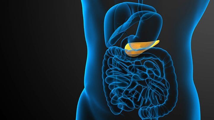
What Is The Gallbladder?
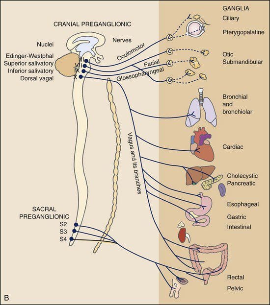
The digestive system comprises the mouth, the internal organs from the GI tract, the liver, the gallbladder, and the anus, where food is consumed, digested, and excreted out of the body to keep it healthy. The gallbladder is a small organ that store and releases bile at the appropriate time into the intestines to be mixed with the digested foods to be excreted out of the body. This pear-shaped organ inflates and deflates like a balloon when it stores and releases bile while having a casual relationship with the nerves and hormones that help regulate the gallbladder functioning properly. Studies reveal that the ganglia become the target of causing the hormone cholecystokinin and the parasympathetic nerve to up or downregulate the neurotransmission to the gallbladder. This causes the gallbladder to be functional in the body.
What Are Its Functions In The Parasympathetic Nervous System?
So what are the functions that the gallbladder provides to the body? For starters, the parasympathetic nervous system allows the body to rest and digest the consumed food to be turned into nutrients. The parasympathetic nervous system also provides gallbladder stimulation as studies reveal that the gallbladder receives innervation from the parasympathetic nervous system connected to the vagus nerve that transmits information to the spine and the brain. Keeping and releasing bile from this pear-shaped organ helps regulate the gastrointestinal tract. This causal relationship between the gallbladder and the parasympathetic nerve is essential because the body needs to know when to store and release bile from the gallbladder, or it might trigger some issues that can do more harm to the body and even affect the gallbladder itself.
Do You Have Shoulder Pain?- Video
Have you been experiencing gut issues causing a sharp or dull ache in your back or sides? How about questionable shoulder pain that seems to come out of nowhere? Or are your experiencing inflammation in your digestive system? Many of these symptoms are signs of visceral-somatic pain affecting the gallbladder. Visceral-somatic pain is defined when there is damage to the organ, and it starts to affect the muscles in a different location in the body. The video above gives an excellent example of visceral-somatic pain in the gallbladder and the shoulder. Now many people wonder how shoulder pain is the mediator of the gallbladder? Well, inflammation in the liver and gallbladder causes the nerve roots to be hypersensitive and compressed. This leads to overlapping profiles, triggering pain in the shoulder muscles and associated with upper mid-back pain.
Referred Shoulder Pain & Gallbladder Dysfunction
Now say the individual is experiencing shoulder pain; however, when they rotate their shoulder, there is no pain? Where is the source of shoulder pain localized, and what is causing the issue? And why is it correlating to the gallbladder? This is known as referred pain, where the source of pain is poorly localized when it is located elsewhere. Studies reveal that gallbladder dysfunctions like cholecystitis might be associated with acute thoracolumbar shoulder pain. So what does this mean? It means that any referred pain that is the causation of shoulder pain gives the impression that something is wrong with the gallbladder. This would provide much-needed information when individuals are being examined by their physicians.
Conclusion
The body needs the digestive system to help process food the host consumes and excretes for a healthy functioning system. The gallbladder stores and releases bile to the digested food. This ensures that the nutrients and bile are transported and passed out of the body. When disruptive factors cause gut issues and affect the gallbladder, it can correlate to different problems impacting the body. An example would be gallbladder issues associated with shoulder pain. This is referred to as pain, which is from an affected organ and associated with the muscle in a different location. This can make the individual feel miserable and wonder what is going on with their shoulders when it might be something associated with their gallbladder. Available treatments can provide better knowledge to determine the problem and how to alleviate the issues.
References
Carter, Chris T. “Acute Thoracolumbar Pain Due to Cholecystitis: A Case Study.” Chiropractic & Manual Therapies, BioMed Central, 18 Dec. 2015, https://www.ncbi.nlm.nih.gov/pmc/articles/PMC4683782/.
Jones, Mark W, et al. “Anatomy, Abdomen and Pelvis, Gallbladder.” In: StatPearls [Internet]. Treasure Island (FL), StatPearls Publishing, 8 Nov. 2021, https://www.ncbi.nlm.nih.gov/books/NBK459288/.
Mawe, Gary M., et al. “Nerves and Hormones Interact to Control Gallbladder Function.” Physiology, 1 Apr. 1998, https://journals.physiology.org/doi/full/10.1152/physiologyonline.1998.13.2.84.
Medical Professional, Cleveland Clinic. “Gallbladder: What Is It, Function, Location & Anatomy.” Cleveland Clinic, 28 July 2021, https://my.clevelandclinic.org/health/body/21690-gallbladder.
Everyone around the world has dealt with pain that makes them feel uncomfortable and has them place their hands on the location where the pain is originating in their bodies. Many factors can become issues in the body, like a poor, unhealthy lifestyle that causes problems in the gut system and develop painful symptoms that affect the intestines. Stressful events that cause headaches that affect the neck and upper back muscles or gut issues that cause discomfort in the abdominal and back region. All these issues are known as referred pain, where a person feels pain in one part of their body, but it is caused by a different source of pain in a different location. An example would be an individual with back pain, but the pain is originating in their abdominal organs. Today’s article looks at various issues that mask low back pain in the body, how organ issues mimic low back pain, and how to alleviate these issues affecting the body. We refer patients to certified providers specializing in gastroenterology and chiropractic treatments that help those with issues that affect their back and gut system. We also guide our patients by referring to our associated medical providers based on their examination when it’s appropriate. We find that education is the solution to asking our providers insightful questions. Dr. Alex Jimenez DC provides this information as an educational service only. Disclaimer
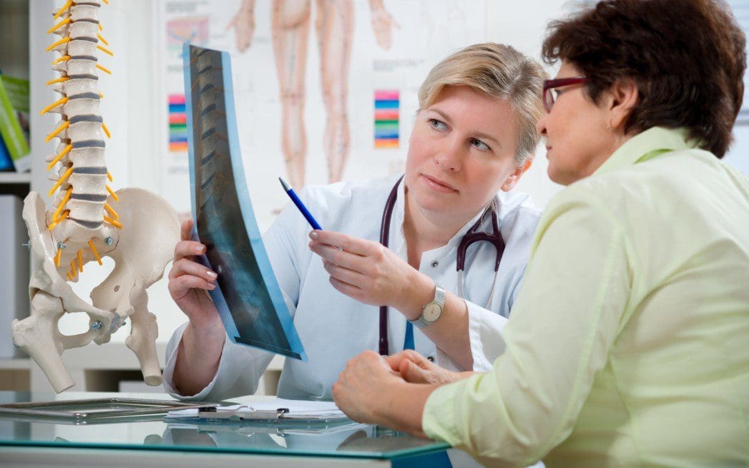
Different Issues Masking Low Back Pain
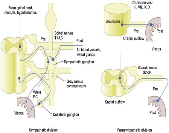
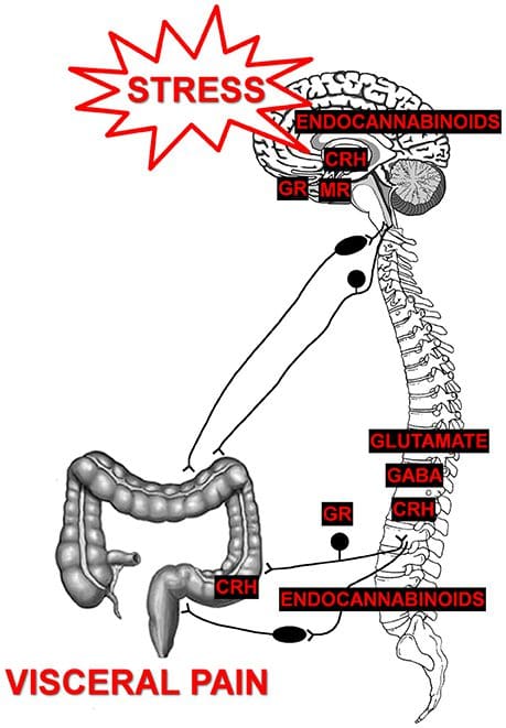
Have you experienced discomfort in your abdominal region causing pain in your lower back? How about pelvic issues that are causing bowel discomfort? Or constipation issues that are compressing the nerves in your lower back? These signs and symptoms correlate to visceral-somatic pain, defined as poorly localized pain characterized by irritated internal organs that cause muscle hypersensitivity from the same nerve. So what does this mean for a person experiencing back issues affecting their quality of life? Well, this might be an indication of the individual that might be suffering from gastrointestinal problems that are correlating to low back pain. Studies reveal any disturbances causing musculoskeletal or gastrointestinal complaints that could induce referred pain through the sympathetic nervous system. An example will be if the body suffers from infections from the kidneys that are associated with low back pain.
So how would the kidneys be associated with low back pain? What is the correlation? For example, a person is constantly eating foods with either a high salt content or a high protein in their system. These high food contents begin to form kidney stones in one or both organs, thus causing a sharp pain that triggers low back pain. As the kidney stones pass through the urinary tract, it administrates radiating pain to the body’s lower abdominal and pelvic region. Another example of issues that can mask low back is constipation in the abdominal area associated with pelvic dysfunction. How does this correlate to the lower back? Think of your abdominal organs overlapped by risk profiles associated with gut disorders. Signs like hypothyroidism, SIBO, celiac disease, or IBS can increase the risk associated with pelvic floor dysfunction, which causes bloating and constipation to the abdominal organs. These issues are co-morbidities to IBS as studies reveal that the pelvic floor and abdominal muscles are co-activated to increase spine stability and intra-abdominal pressure. Now it may seem not a bad thing to the body unless the individual is constantly standing for an extended period or is obese, thus becoming a mediator for the host to suffer from low back pain while being associated with pelvic dysfunction.
Organ Issues Mimicking Low Back Pain- Video
Have you been feeling muscle tenderness in the lower extremities of your body? How about gut issues that are associated with low back pain? Or are you feeling bowel dysfunction in your pelvic region? All these issues correlate to viscerosomatic pain, where the infected organ is causing issues to the muscle in a different location. The video above explains how various organ issues can mimic spinal and back pain in the body. One of the examples that the video explains is how kidney infections are associated with back pain. Studies reveal that visceral pain originating from the upper urinary tract coincidently correlates with the characteristics of referred pain and changes in the somatic tissues of the body.
Alleviating Issues Affecting The Body
Say an individual is suffering from low back pain issues; as they get their mandatory examination, they explain to their physician about their low back pain and what is happening. Once the suffering individual is situated, the physician begins to look over the body where the pain is located, either by physical examination or through the intake form they are looking over. So what does this implicates in the body? Well, studies have revealed that systemic pathologies of the visceral organs can mimic or mask musculoskeletal pain. An example would be someone who is experiencing gastrointestinal issues in their gut, and it’s triggering muscle spasms in the back. This causes the nerve roots to be hypersensitive to the visceral organs and increases the risk associated with low back pain.
Conclusion
Dealing with pain is no joke, primarily when the pain is located in a different body region. Sometimes the pain can be an organ issue that mimics muscle pain in the back. This is known as viscero-somatic pain, defined where infected organs are either mimicking or triggering muscle issues in different body locations. This causation is usually due to various factors like unhealthy lifestyle habits affecting the visceral organs and affecting the muscles that correspond to the organs, like IBS issues affecting the lower back. Available treatments are there to figure out what problems affect the body and provide a better understanding to alleviate them.
References
Basso, Francesca Lo, et al. “Manual Treatment for Kidney Mobility and Symptoms in Women with Nonspecific Low Back Pain and Urinary Infections.” De Gruyter, De Gruyter, 1 May 2021, https://www.degruyter.com/document/doi/10.1515/jom-2020-0288/html.
Bussey, Melanie Dawn, et al. “Is Pelvic Floor Dysfunction Associated with Development of Transient Low Back Pain during Prolonged Standing? A Protocol.” Clinical Medicine Insights. Women’s Health, SAGE Publications, 27 May 2019, https://www.ncbi.nlm.nih.gov/pmc/articles/PMC6537301/.
J;, Stowell T;Cioffredi W;Greiner A;Cleland. “Abdominal Differential Diagnosis in a Patient Referred to a Physical Therapy Clinic for Low Back Pain.” The Journal of Orthopaedic and Sports Physical Therapy, U.S. National Library of Medicine, Nov. 2005, https://pubmed.ncbi.nlm.nih.gov/16355918/.
Lacy, Brian E, et al. “Management of Chronic Abdominal Distension and Bloating.” Clinical Gastroenterology and Hepatology : the Official Clinical Practice Journal of the American Gastroenterological Association, 1 Apr. 2020, https://www.cghjournal.org/article/S1542-3565(20)30433-X/fulltext.
PJ;, Pedersen KV;Drewes AM;Frimodt-Møller PC;Osther. “Visceral Pain Originating from the Upper Urinary Tract.” Urological Research, U.S. National Library of Medicine, 16 May 2010, https://pubmed.ncbi.nlm.nih.gov/20473661/.
The body is a set of complex systems, including bones, organs, nerves, muscles, and tissue. Breathing disorders are increasing, including chronic bronchitis, asthma, emphysema, and other conditions. Viscerosomatic reflexes include poor breathing quality brought on by allergies, breathing disorders like COPD that can cause intense coughing, sneezing, hunching, arching of the back, and heaving that causes back pain and referred pain.
The brain sends electronic impulses to the different areas of the body through the spine/nervous system. If the nerves get shifted, stretched, compressed, or knocked out of position, the brain could start sending messages of pain and discomfort, which can also cause other body systems to malfunction. If the body is constantly transmitting pain signals, it can disrupt sleep, dietary habits, and overall well-being. Misalignment can disrupt the information delivered by the nervous system, leading to inflammation, irritation, and imbalances in the body.
Regular chiropractic maintains the nervous system to operate the way it was designed. Proper alignment of the spine and body will improve the nervous system’s health and function, encouraging the brain to release endorphins achieving pain relief, and leading to optimal health. When the nervous system performs optimally, the other systems will follow, including better breathing quality.
Poor Breathing
Breathing difficulties are widespread with various causes that include:
Allergies
Environmental pollutants
Viral and bacterial infections that cause inflammation
An overactive immune response can all contribute to poor breathing quality.
Individuals might not notice that their breathing quality is poor but instead notice they are:
Frequent exhaustion
Having to stop constantly in the middle of activities.
Experience brain fog.
Memory issues/forgetfulness.
Physical performance – endurance, flexibility, and muscle is deteriorating.
The breathing quality impacts how well the body’s systems can carry out their essential functions and be prepared for unexpected events. The body adjusts oxygen intake capacity in line with the energy required to perform physical activity. All bodily systems, including the cardiovascular, immune, and muscular systems, depend on the respiratory system to generate energy.
Immune protection against viruses, bacteria, fungi, and other diseases.
Chiropractic
A crucial part of the respiratory system’s function is transporting nutrients and oxygen throughout the body. Chiropractic treatment releases tension by moving muscle fascia and the spine that may have become stuck, compressed, or shifted out of position, causing poor posture and injury. Chiropractic eliminates toxins and cellular waste from tight, knotted areas by breaking up stagnant tissues.
Circulation Improvement
Chiropractic increases circulation, allowing fresh blood, lymphatic fluid, nutrients, and oxygen to enter the deprived tissues. These regions include:
Muscles in the shoulder, neck, back
Bones and joints across the spine
Body tissues
Ligaments
Tendons
Chiropractic treatment can be manual/mechanical traction/decompression, combined with therapeutic tissue massage, exercise, and diet recommendations.
Decompression De La Espalda
References
McCarty, Justin C, and Berrylin J Ferguson. “Identifying asthma triggers.” Otolaryngologic clinics of North America vol. 47,1 (2014): 109-18. doi:10.1016/j.otc.2013.08.012
Purnomo, Ariana Tulus, et al. “Non-Contact Monitoring and Classification of Breathing Pattern for the Supervision of People Infected by COVID-19.” Sensors (Basel, Switzerland) vol. 21,9 3172. 3 May. 2021, doi:10.3390/s21093172
Schend, Jason, et al. “An Osteopathic Modular Approach to Asthma: A Narrative Review.” The Journal of the American Osteopathic Association vol. 120,11 (2020): 774-782. doi:10.7556/jaoa.2020.121
IFM's Find A Practitioner tool is the largest referral network in Functional Medicine, created to help patients locate Functional Medicine practitioners anywhere in the world. IFM Certified Practitioners are listed first in the search results, given their extensive education in Functional Medicine