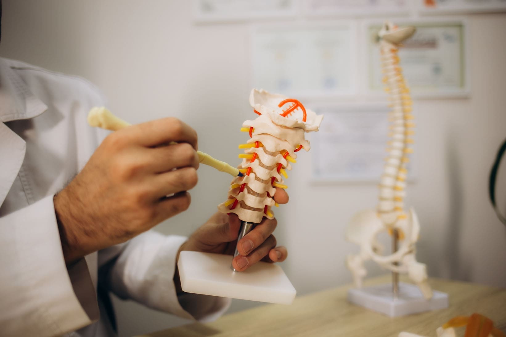
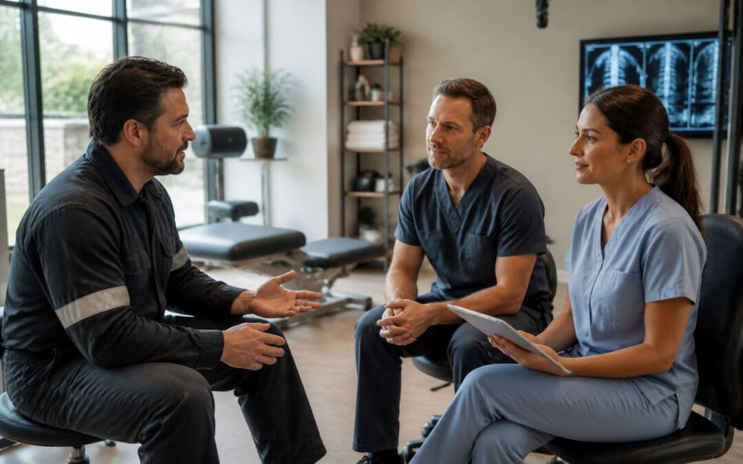
Back Clinic Treatments. There are various treatments for all types of injuries and conditions here at Injury Medical & Chiropractic Clinic. The main goal is to correct any misalignments in the spine through manual manipulation and placing misaligned vertebrae back in their proper place. Patients will be given a series of treatments, which are based on the diagnosis. This can include spinal manipulation, as well as other supportive treatments. And as chiropractic treatment has developed, so have its methods and techniques.
Why do chiropractors use one method/technique over another?
A common method of spinal adjustment is the toggle drop method. With this method, a chiropractor crosses their hands and pressed down firmly on an area of the spine. They will then adjust the area with a quick and precise thrust. This method has been used for years and is often used to help increase a patient’s mobility.
Another popular method takes place on a special drop table. The table has different sections, which can be moved up or down based on the body’s position. Patients lie face down on their back or side while the chiropractor applies quick thrusts throughout the spinal area as the table section drops. Many prefer this table adjustment, as this method is lighter and does not include twisting motions used in other methods.
Chiropractors also use specialized tools to assist in their adjustments, i.e., the activator. A chiropractor uses this spring-loaded tool to perform the adjustment/s instead of their hands. Many consider the activator method to be the most gentle of all.
Whichever adjustment method a chiropractor uses, they all offer great benefits to the spine and overall health and wellness. If there is a certain method that is preferred, talk to a chiropractor about it. If they do not perform a certain technique, they may recommend a colleague that does.
Annular Tear and Disc Height Loss After Loop 375 Lateral Impact: Mechanism-to-MRI Linkage
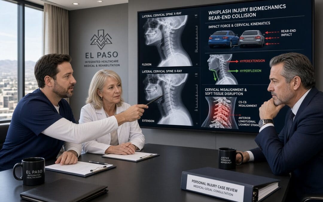
A high-speed side impact on El Paso’s Loop 375 can transmit focused lateral shear, axial compression, and rotational forces through the lumbar or cervical spine in a fraction of a second. When those force vectors exceed the tensile strength of the annulus fibrosus—particularly its relatively weaker posterolateral fibers—an acute annular tear can form, allowing nuclear material to migrate and producing measurable disc height loss visible on MRI. At Injury Medical Clinic PA in El Paso, the multidisciplinary team of Dr. Alex Jimenez, DC, APRN, FNP-BC, CFMP, IFMCP (Texas License #1191402, Prescriptive Authority #59628, NPI 1205907805) and Medical Director Dr. Maria Guadalupe Cardenas, MD (Board Certified Internal Medicine, Texas License #J2933, NPI 1164426748), systematically links these biomechanical mechanisms to objective imaging so that clinical and proximate causation are clear for patients and attorneys.
How a Loop 375 Lateral Impact Generates Disc Injury
Loop 375 (Joe Battle Boulevard and related segments) is a high-volume corridor where lane changes, side-swipes, and T-bone or angle collisions are common. In a pure lateral impact, the striking vehicle imparts a force vector roughly perpendicular to the struck vehicle’s longitudinal axis. Occupants experience rapid lateral torso acceleration while the pelvis and lower extremities lag or rotate, creating shear across the intervertebral discs and facet joints. At the same time, the sudden change in velocity produces axial compression and, often, a twisting moment as the body rebounds against the seatbelt or door.
The annulus fibrosus consists of concentric lamellae whose fiber orientation alternates for strength; however, the posterolateral region contains a higher proportion of vertically oriented fibers and is a known site of relative weakness (StatPearls, 2023). When lateral shear and rotation are superimposed on compression, these fibers can separate, producing a radial or transverse annular tear. Nuclear material may then extrude or lose hydration and volume, resulting in a measurable reduction in disc height at the affected level. Because the forces are asymmetric, the tear and height loss are often unilateral or more pronounced on the side of impact—an important morphologic clue when compared with the more uniform, multilevel pattern of ordinary age-related degeneration.
MRI Linkage: Reading the Images Through the Force Vectors
On T2-weighted MRI, an acute annular tear typically appears as a high-intensity zone (HIZ)—a focal bright signal within the normally dark annulus caused by increased water content and inflammatory fluid (StatPearls, 2023; Teraguchi et al., 2022). Supporting signs of acute traumatic change, as opposed to a chronic degenerative baseline, include:
A single or dominant level of abnormality that matches the predicted biomechanics of the impact (for example, L4-L5 or L5-S1 after a low lumbar lateral load, or mid-cervical levels after a higher side-impact).
Asymmetric height loss or annular signal that is greater on the side of the force vector.
Associated soft-tissue findings such as ligamentous edema, facet fluid, or paraspinal muscle signal change at the same or adjacent levels.
Higher T2 hyperintensity and involvement of the outer third of the annulus are more consistent with traumatic disruption than the lower-intensity, inner-two-thirds fissures typical of slow degenerative breakdown (Khurana et al., 2025).
Degenerative disc disease is extremely common and increases with age; imaging studies of asymptomatic volunteers show annular fissures in roughly 19–29% of adults depending on the decade (Brinjikji et al., as cited in clinical radiology reviews). The clinic therefore never equates the mere presence of an HIZ or reduced disc height with trauma. Instead, the team correlates the precise morphology, laterality, and associated acute soft-tissue changes with the documented mechanism, the patient’s immediate post-collision symptoms, and serial clinical examinations. When pre-existing degeneration is present, the records explicitly separate the baseline findings from the new traumatic aggravation—language that prevents defense arguments that “the MRI only shows wear and tear.”
Distinguishing Acute Traumatic Change from Degenerative Baseline
Clear, plain-language documentation is essential. An example of phrasing used in clinic notes and narrative reports reads: “The left posterolateral L4-L5 annular high-intensity zone and 2 mm asymmetric disc-height reduction are morphologically and temporally consistent with the lateral shear and rotational forces of the 14 July Loop 375 side-impact collision. These findings differ from the mild, symmetric multilevel disc desiccation and small inner annular fissures that represent age-related baseline change.” Such statements rest on the mechanism-to-MRI linkage rather than on the radiologist’s impression alone. Digital imaging, functional testing (range-of-motion, neurologic, and strength measures), and Colossus-compliant electronic records further corroborate that the structural changes produce the patient’s objective functional deficits.
Future-Care Modeling with ESWT and Non-Surgical Decompression
Once causation is established, the clinic models evidence-based future care to address both the annular tear and height loss. Non-surgical spinal decompression creates controlled negative intradiscal pressure that can increase disc height and reduce pain; one retrospective cohort demonstrated a statistically significant correlation between restored disc height and decreased low-back pain scores (Apfel et al., 2010). Additional studies confirm improvements in radicular pain and disc morphology after motorized decompression protocols (Choi et al., 2022; Egyptian Rheumatology and Rehabilitation, 2019).
Extracorporeal shock-wave therapy (ESWT) supplies a complementary biologic stimulus. In laboratory models, low-energy ESWT combined with low-tension traction has been shown to restore disc height, improve rehydration, and reduce matrix-degrading enzymes (MMP-3, MMP-13, ADAMTS-4), thereby reshaping the microenvironment for repair (Spine Journal, 2021). Clinical series report pain reduction and facilitation of herniation resorption when ESWT is integrated into staged conservative care (Orthopedic Reviews, 2026; Rehabilitation Medicine, 2024). Because both modalities are non-invasive and drug-free, they align with non-maleficence: they lower the risk of progression to surgery or long-term opioid dependence while still delivering measurable structural and symptomatic benefit.
In the future-care cost spreadsheet these interventions appear as a defined course of decompression sessions (typically 20–30 over 6–8 weeks) plus a series of ESWT treatments, accompanied by supportive chiropractic adjustments, neurofunctional acupuncture or electroacupuncture, and functional-medicine laboratory monitoring directed by Dr. Cardenas. The combination targets root-cause healing—restoring disc height and annular integrity where possible, reducing inflammatory cytokines, and improving segmental stability—rather than merely masking pain (beneficence). Patients receive clear education about expected timelines, self-care strategies, and how the plan coordinates with any outside specialists they already see, preserving autonomy and informed decision-making.
Documentation That Withstands Scrutiny
Every element—mechanism narrative, MRI description distinguishing acute from baseline change, functional deficits, and itemized future-care costs—is recorded in a manner suitable for letters of protection and deposition. The dual-licensed perspective of Dr. Jimenez (structural alignment plus advanced practice medical diagnostics), together with Dr. Cardenas’s internal-medicine oversight, supplies both the biomechanical and medical foundation attorneys require. The result is a transparent, mechanism-linked record that supports the patient’s recovery while meeting El Paso personal-injury litigation evidentiary standards.
Patients and counsel seeking this level of integrated causation analysis and future-care planning may contact Injury Medical Clinic PA for a coordinated evaluation.
References
Apfel, C. C., Cakmakkaya, O. S., Martin, W., Richmond, C., Macario, A., George, E., Schaefer, M., & Pergolizzi, J. V. (2010). Restoration of disk height through non-surgical spinal decompression is associated with decreased discogenic low back pain: A retrospective cohort study. BMC Musculoskeletal Disorders, 11, Article 155. https://doi.org/10.1186/1471-2474-11-155
Choi, E., Gil, H. Y., Ju, J., Han, W. K., Nahm, F. S., & Lee, P. B. (2022). Effect of nonsurgical spinal decompression on intensity of pain and herniated disc volume in subacute lumbar herniated disc. International Journal of Clinical Practice, 2022, Article 6343837. https://doi.org/10.1155/2022/6343837
Rehabilitation Medicine. (2024). Effect of extracorporeal shock wave therapy on different types of lumbar disc herniation. Rehabilitation Medicine, 34(1), 84–89.
Spine Journal. (2021). Low energy extracorporeal shock wave therapy combined with low tension traction can better reshape the microenvironment in degenerated intervertebral disc regeneration and repair. The Spine Journal, 21(1), 160–177. https://doi.org/10.1016/j.spinee.2020.08.012
El-Sayed, A. M., et al. (2019). Spinal decompression therapy as an alternative modality for management of low back pain and radicular pain caused by lumbar disc herniation or protrusion. Egyptian Rheumatology and Rehabilitation, 46, 161–168. https://doi.org/10.4103/err.err_34_18
Brinjikji, W., Luetmer, P. H., Comstock, B., Bresnahan, B. W., Chen, L. E., Deyo, R. A., Halabi, S., Turner, J. A., Avins, A. L., James, K., Wald, J. T., Kallmes, D. F., & Jarvik, J. G. (2015). Systematic literature review of imaging features of spinal degeneration in asymptomatic populations. American Journal of Neuroradiology, 36(4), 811–816. https://doi.org/10.3174/ajnr.A4173
Uncover the importance of causation with chiropractic care in proving the connection between trauma and injuries in personal injury cases.
Abstract
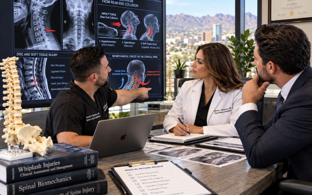
Causation is the essential legal and medical bridge connecting an incident—such as a motor vehicle collision, slip-and-fall, or workplace trauma—to the injuries that justify recovery in personal injury claims. Without clear proof that the event caused or materially contributed to the harm, even severe pain and disability may not support full compensation. In my practice at Injury Medical Clinic PA in El Paso, Texas, I have observed for more than three decades that musculoskeletal injuries from trauma rarely exist in isolation. They disrupt joint mechanics, muscle balance, nerve signaling, and overall function, often leading to secondary problems such as chronic pain, reduced mobility, sleep disruption, and compensatory strain in distant body regions. Chiropractic care addresses these issues directly by restoring joint motion, reducing abnormal afferent input to the nervous system, decreasing local inflammation, and facilitating the body’s natural healing processes. When integrated with medical oversight from Dr. Maria Guadalupe Cardenas, MD, board-certified in Internal Medicine, functional medicine principles, targeted rehabilitation, and non-surgical regenerative options, this approach provides both clinical recovery and the objective documentation needed to establish medical causation. This educational post explains what causation means in personal injury, what chiropractic care is, how the two work together, why chiropractic care reduces musculoskeletal injuries and their comorbidities, and how an integrative team model supports durable, non-surgical recovery. Drawing on current evidence from leading researchers and my clinical observations, the goal is to help readers understand the journey from injury event to functional restoration.
Understanding Causation in Personal Injury Cases
Causation is the legal and medical requirement that links a defendant’s negligent or wrongful act to the plaintiff’s injury. Courts and insurance evaluators require evidence that the incident was both the actual cause and the proximate cause of the harm. Actual cause, often called the “but-for” test, asks whether the injury would have occurred but for the incident. Proximate cause, or legal cause, asks whether the harm was a reasonably foreseeable result of the conduct and whether the connection is close enough to justify liability. In practice, medical causation is established to a reasonable degree of medical probability—more likely than not—that the trauma caused or substantially contributed to the diagnosed condition.
The Bradford Hill criteria, originally proposed in 1965 by epidemiologist Sir Austin Bradford Hill, provide a structured way to evaluate whether an observed association is likely causal. These include strength of association, consistency across studies, specificity, temporality (the exposure must precede the outcome), biological gradient or dose-response relationship, biological plausibility, coherence with existing knowledge, experimental evidence, and analogy. In musculoskeletal personal injury, temporality is especially important: symptoms that begin or clearly worsen after a documented collision, with a mechanism consistent with known tissue tolerances, support causation. Medical records that capture the mechanism of injury, the timing of symptom onset, objective findings such as restricted range of motion, positive orthopedic tests, and imaging that correlates with clinical presentation become central evidence. Expert testimony from treating providers who have examined the patient, reviewed the history, and followed the response to treatment carries significant weight because causation opinions are inherent in clinical care.
In personal injury, delayed symptom onset does not automatically defeat causation. Soft-tissue injuries such as strains, sprains, and disc injuries can produce progressive inflammation and pain that peaks hours or days later as chemical mediators accumulate and protective muscle guarding develops. Thorough documentation of the incident details, the patient’s position and restraint use, the direction of impact, and the progressive clinical findings helps distinguish trauma-related injury from pre-existing conditions. When alternative explanations are less likely, and treatment produces measurable improvement consistent with the injury’s expected natural history, the causal link is strengthened.
What Is Chiropractic Care
Chiropractic care is a licensed healthcare profession focused on diagnosing, treating, and preventing disorders of the neuromusculoskeletal system, particularly those involving the spine and extremities. Doctors of Chiropractic are trained to evaluate joint dysfunction, soft-tissue injury, and related functional impairments, then deliver manual therapies—most commonly spinal and extremity adjustments or mobilizations—along with rehabilitative exercise, soft-tissue techniques, and patient education. The core clinical goal is to restore normal joint motion, reduce abnormal mechanical stress, modulate pain signaling, and support the body’s capacity for recovery without reliance on surgery or long-term pharmaceuticals when those interventions can be avoided or delayed.
Modern, evidence-based chiropractic practice is grounded in biomechanics and neurophysiology rather than older theories of “vertebral subluxation” as a cause of systemic disease. High-velocity, low-amplitude thrusts and graded mobilizations stimulate mechanoreceptors in joint capsules, ligaments, and muscle spindles. This afferent barrage can produce short-term changes in segmental spinal excitability, alter descending pain-modulatory pathways, and reduce local muscle hypertonicity. Clinical practice guidelines from multiple organizations support spinal manipulative therapy as part of multimodal care for acute, subacute, and chronic low back pain, neck pain, and certain headache presentations. Systematic reviews and randomized trials have shown reductions in pain intensity and improved function, particularly when chiropractic care is combined with exercise and education.
In the personal injury setting, chiropractic care is often the patient’s first and most continuous contact after trauma. Early evaluation identifies red flags that require imaging or medical referral, establishes objective baseline measures, and begins conservative management while the inflammatory phase of healing is still active. Because many musculoskeletal injuries from collisions or falls are graded sprains and strains rather than fractures or complete ruptures, non-surgical restoration of motion and load-sharing is both clinically appropriate and consistent with the principle of beginning with the least invasive effective option.
How Chiropractic Care Works Hand in Hand with Causation in Personal Injury
Chiropractic care and the proof of causation reinforce each other in several practical ways. First, the initial examination captures the patient’s mechanism of injury in their own words and correlates it with clinical findings. A restrained driver who reports neck pain after a rear-end impact, who demonstrates restricted cervical rotation, and who exhibits positive orthopedic tests for soft-tissue strain supplies the temporality and biological plausibility elements of causation analysis. Subsequent visits document the recovery trajectory: decreasing pain scores, increasing measurable range of motion, improved strength, and reduced functional limitations. These serial observations create a clear timeline that is difficult to dismiss as unrelated to the incident.
Second, chiropractic providers routinely record objective data—goniometric measurements, muscle testing, orthopedic special tests, and postural observations—that can be compared over time. When these findings improve in parallel with the expected phases of tissue healing (inflammatory, proliferative, and remodeling), the records support the conclusion that the trauma produced a treatable injury whose resolution is consistent with the care provided. Third, chiropractic documentation often includes the absence of prior similar symptoms or the clear aggravation of pre-existing conditions. In my clinical experience, patients who had stable, low-level back or neck complaints before a collision frequently report a distinct increase in intensity, new radiation of pain, or new functional loss after the event. Documenting that distinction is essential when defense argues the current symptoms are merely the natural progression of degenerative changes.
Finally, the treating chiropractor’s ongoing relationship with the patient supplies continuity that a one-time independent medical examination cannot match. When the same provider observes both the acute presentation and the response to care, the opinion that the incident caused or materially contributed to the injury rests on direct clinical observation rather than retrospective chart review alone. Courts and juries give substantial weight to this contemporaneous evidence, especially when it is consistent across chiropractic and medical notes within the same clinic.
Physiological Underpinnings of Chiropractic Care for Musculoskeletal Injuries
Spinal and extremity joints that lose normal motion after trauma generate continuous abnormal input from mechanoreceptors and nociceptors. This input can facilitate segmental spinal cord neurons, expand pain receptive fields, and maintain protective muscle co-contraction that further restricts motion. High-velocity, low-amplitude manipulation produces a brief, high-frequency burst of discharge from muscle spindle primary afferents and other dynamically sensitive receptors. Animal and human studies show that this afferent volley can reduce the excitability of dorsal horn neurons involved in pain transmission and modulate descending inhibitory pathways originating in the brainstem and higher centers.
Beyond neural effects, restoring joint motion improves local circulation and reduces concentrations of inflammatory mediators in peri-articular tissues. When synovial fluid redistributes and capsular tension normalizes, the chemical environment that sustains peripheral sensitization improves. Soft-tissue techniques used with joint mobilization address myofascial trigger points and adhesions that develop from muscle guarding. Together, these changes reduce the mechanical and chemical drivers of pain, allowing patients to resume more normal movement patterns that further promote healing.
For disc-related injuries, controlled distraction or flexion-distraction techniques can reduce intradiscal pressure and may improve nutrient diffusion into the avascular disc. Combined with core stabilization exercise, this strategy aims to restore load-sharing among the spine’s passive (ligaments and discs), active (muscles), and neural control subsystems. Clinical observations in my practice confirm that patients who regain segmental motion early and then progress through graded loading recover function more reliably than those who remain immobilized or who rely solely on passive modalities.
How Chiropractic Care Reduces Musculoskeletal Injuries and Their Comorbidities
Musculoskeletal injuries from trauma commonly produce a cascade of secondary problems. Restricted motion leads to muscle atrophy and altered recruitment patterns. Persistent nociceptive input can contribute to central sensitization, sleep fragmentation, and reduced physical activity. Reduced activity then worsens cardiovascular fitness, glycemic control, and mood. Chiropractic care interrupts this cascade at several points.
Randomized trials in active-duty military personnel have shown that adding chiropractic care to usual medical management produces moderate improvements in low-back-pain intensity and disability at six weeks, along with higher patient satisfaction and lower self-reported pain-medication use. Observational studies of older Medicare beneficiaries with chronic low back pain and multiple comorbidities found lower overall costs and lower rates of subsequent surgery among those who received chiropractic manipulative treatment. Systematic reviews support spinal manipulative therapy as part of multimodal care for neck pain and cervicogenic headache, with reductions in both pain and associated disability.
In personal injury populations, the comorbidities I most frequently observe are compensatory shoulder or hip strain from altered gait or posture, tension-type or cervicogenic headache secondary to upper cervical dysfunction, and sleep disruption that prolongs recovery. By restoring cervical and lumbar motion, reducing muscle hypertonicity, and enabling earlier return to graded activity, chiropractic care decreases the likelihood that these secondary problems become entrenched. Patients who recover segmental mobility and core endurance report better tolerance for work and daily activities, which in turn supports mental health and reduces the risk of prolonged opioid exposure.
Integrative Chiropractic Care in Non-Surgical Treatments
Non-surgical management of traumatic musculoskeletal injury is most effective when mechanical, neurological, and systemic factors are addressed together. Integrative chiropractic care combines joint mobilization or manipulation with rehabilitative exercise, soft-tissue therapy, nutritional support for tissue repair, and, when indicated, regenerative options such as platelet-rich plasma under appropriate medical direction. The reasoning is straightforward: restoring motion reduces abnormal loading and afferent drive, while progressive loading stimulates collagen remodeling and motor-pattern retraining. Functional medicine evaluation can identify co-factors—such as vitamin D insufficiency, sleep apnea, or inflammatory dietary patterns—that slow healing even when local mechanics are improved.
Spinal decompression is used when imaging and clinical findings indicate disc-related nerve compression; controlled traction reduces intradiscal pressure and can create a window for nutrient exchange and symptom relief, allowing patients to participate more fully in active rehabilitation. Soft-tissue techniques and instrument-assisted mobilization address adhesions that limit excursion. Acupuncture or electroacupuncture is sometimes added for additional pain modulation via endogenous opioid and non-opioid pathways. The plan adjusts throughout based on the healing phase and the patient’s response, with medical oversight ensuring red-flag conditions are identified and any necessary imaging, lab evaluation, or referral occurs promptly.
The Multidisciplinary Team at Injury Medical Clinic PA and the Role of Dr. Maria Guadalupe Cardenas
At Injury Medical Clinic PA (Mission Plaza Injury Medical Clinic) in El Paso, Texas, a coordinated team delivers care under dual clinical leadership. I provide chiropractic evaluation and treatment together with nurse-practitioner-level medical assessment. Dr. Maria Guadalupe Cardenas, MD, Board Certified in Internal Medicine, NPI #1164426749, Texas MD License #J2933, serves as Medical Director and Collaborative Physician. With more than 40 years of experience as an internist, Dr. Cardenas provides physician-level oversight for complex cases, reviews systemic medical issues that may affect recovery, and collaborates on treatment plans that require medical decision-making beyond the scope of chiropractic or advanced-practice nursing alone.
This structure is common in integrative injury clinics. The chiropractor focuses on joint mechanics, soft-tissue function, and progressive rehabilitation. The internist evaluates comorbidities such as hypertension, diabetes, or inflammatory conditions that can influence healing, coordinates any necessary imaging or specialty referral, and ensures that documentation meets both clinical and medicolegal standards. Physical therapists, massage therapists, and additional nurse practitioners contribute specific modalities and exercise progression. The result is a single, coherent record that captures the mechanism of injury, objective findings, response to care, and medical correlation—precisely the elements needed to support causation analysis.
Patients benefit because decisions consider both mechanical and systemic perspectives. When a patient with pre-existing diabetes sustains a disc injury, for example, glycemic control is optimized alongside spinal decompression and core training so that microvascular factors do not impede soft-tissue healing. When sleep disruption or mood changes appear, we address them rather than ignore them. The collaborative model therefore supports both clinical recovery and the clear, consistent documentation that personal injury claims require.
Clinical Observations from Dr. Alexander Jimenez
In more than three decades of practice in El Paso, I have repeatedly observed that the patients who recover most completely are those whose care begins with a precise description of the mechanism of injury and whose treatment plans are adjusted according to measured progress rather than a fixed schedule. A mid-20s heavy-labor worker who sustained neck and low-back injuries in a collision recovered approximately 80 percent of function over nineteen weeks, and then experienced a second collision, requiring careful re-evaluation of both the new trauma and the residual deficits from the first event. The integrated record—chiropractic measurements of motion and strength together with medical assessment of systemic factors—allowed the team to distinguish the effects of each incident and to modify the plan accordingly.
I have also noted that delayed symptom onset is common after low-speed collisions when the head is rotated or when the patient is looking over the shoulder. Soft-tissue strain and facet irritation can cause progressive inflammation that peaks 1 to 3 days later. Early documentation of the exact body position and the progressive clinical findings has proven essential when causation is later questioned. Patients who receive early restoration of joint motion, soft-tissue care, and graded activity consistently report better sleep, fewer compensatory pains, and faster return to work than those whose care is limited to passive modalities or prolonged rest.
These observations align with the broader literature: multimodal care that includes spinal manipulative therapy produces meaningful reductions in pain and disability, and contemporaneous records from the treating team provide the strongest evidence of both the injury and its causal relationship to the incident.
Why Specific Techniques and Protocols Are Used
Spinal and extremity adjustments are used because restricted joint motion after trauma generates ongoing abnormal afferent input that facilitates pain and maintains muscle guarding. By restoring motion, the technique reduces that input and allows more normal movement patterns to re-emerge. The reasoning is both biomechanical—normalization of load distribution—and neurophysiological—modulation of segmental and descending inhibitory pathways.
Spinal decompression is selected when clinical and imaging findings indicate disc-related compression because controlled distraction can lower intradiscal pressure and may improve fluid exchange. It is paired with stabilization exercise so the temporary pressure reduction is followed by improved muscular support.
Soft-tissue techniques and therapeutic exercise are included because muscle guarding and trigger points develop secondary to joint dysfunction and nociceptive drive. Addressing them directly reduces local ischemia and referred pain, while progressive loading stimulates collagen alignment during remodeling. The protocol includes medical oversight and functional-medicine evaluation because systemic factors—inflammation, nutrient status, sleep, and comorbidities—modulate tissue repair. Optimizing these factors increases the probability that local mechanical improvements translate into durable functional gains.
The protocol is individualized throughout according to injury grade, healing phase, and measured response. Conservative care continues when progress is documented; imaging or specialty referral occurs when red flags appear, or expected improvement does not occur. This reasoned, stepwise approach supports both clinical recovery and the clear causal narrative required in personal injury documentation.
Conclusion
Causation in personal injury is established by demonstrating that an incident more likely than not caused or substantially contributed to the diagnosed musculoskeletal injuries and their functional consequences. Chiropractic care supports this process by providing early, continuous, objective documentation of the mechanism, findings, and treatment response while restoring joint motion, modulating pain signaling, and facilitating tissue healing. When delivered within an integrative model that includes medical direction from an experienced internist such as Dr. Maria Guadalupe Cardenas, functional-medicine principles, and progressive rehabilitation, the approach reduces both the primary injury and the secondary comorbidities that commonly prolong disability. The physiological rationale is grounded in current neurophysiological and biomechanical research, and clinical results in practice align with published trials showing improved pain, function, and reduced reliance on more invasive interventions. Readers seeking recovery after trauma are encouraged to pursue care that addresses both the mechanical and systemic dimensions of injury and that produces the clear, contemporaneous records needed to support both healing and the proof of causation.
Goertz, C. M., Long, C. R., Vining, R. D., Pohlman, K. A., Walter, J., & Colter, I. (2018). Effect of usual medical care plus chiropractic care vs usual medical care alone on pain and disability among US service members with low back pain: A comparative effectiveness clinical trial. JAMA Network Open, 1(1), e180105. https://jamanetwork.com/journals/jamanetworkopen/fullarticle/2680417
Weeks, W. B., Leininger, B., Whedon, J. M., Lurie, J. D., Tosteson, T. D., Swenson, R., O’Malley, A. J., & Goertz, C. M. (2016). The association between use of chiropractic care and costs of care among older Medicare patients with chronic low back pain and multiple comorbidities. Journal of Manipulative and Physiological Therapeutics, 39(2), 63–75. https://www.jmptonline.org/article/S0161-4754(16)00007-5/abstract
Gevers-Montoro, C., Provencher, B., Descarreaux, M., Ortega de Mues, A., & Piché, M. (2021). Neurophysiological mechanisms of chiropractic spinal manipulation for spine pain. European Journal of Pain, 25(7), 1429–1448. https://doi.org/10.1002/ejp.1773
Côté, P., Hartvigsen, J., Axén, I., Leboeuf-Yde, C., Corso, M., Shearer, H., Wong, J., Marchand, A. A., Cassidy, J. D., French, S., Kawchuk, G. N., Mior, S., Poulsen, E., Srbely, J., Ammendolia, C., Blanchette, M. A., Busse, J. W., Bussières, A., Cancelliere, C., … Yu, H. (2021). The global summit on the efficacy and effectiveness of spinal manipulative therapy for the prevention and treatment of non-musculoskeletal disorders: A systematic review of the literature. Chiropractic & Manual Therapies, 29, 8. https://doi.org/10.1186/s12998-021-00362-9
SEO Tags
causation personal injury, chiropractic care personal injury, medical causation Bradford Hill, integrative chiropractic El Paso, non-surgical musculoskeletal injury treatment, Dr Alex Jimenez DC APRN FNP-BC, Dr Maria Guadalupe Cardenas MD, Injury Medical Clinic PA El Paso, spinal manipulation neurophysiology, whiplash recovery chiropractic, low back pain evidence-based care, personal injury documentation, multidisciplinary injury clinic Texas
Segmental Disc Compression Patterns Generated by Loop 375 Lateral Impacts
Abstract
Loop 375 lateral impacts in El Paso generate rapid, side-directed force vectors that compress lumbar segments, tear the annulus, and accelerate disc height loss visible on structural MRI. These patterns create clear mechanism-to-injury linkages that support clinical and proximate causation for lumbar claims. At Injury Medical Clinic PA, the dual-licensed team of Dr. Alex Jimenez, DC, APRN, FNP-BC, and collaborating medical director Dr. Maria Guadalupe Cardenas, MD, documents these objective findings with Colossus-compliant notes, functional testing, and imaging timelines. Non-invasive care—chiropractic decompression and ESWT—restores disc hydration and height, reduces surgical risk, and keeps patients in control of their recovery decisions. The resulting impairment ratings are packaged inside the medical-legal causation documentation framework at https://dralexjimenez.com.
Highway-side impacts on El Paso’s Loop 375 do more than crumple doors. They deliver concentrated lateral force vectors that simultaneously load lumbar discs in compression, shear, and side-bending. The result is a recognizable pattern of segmental disc compression: annular fissures, progressive height loss, and altered load-sharing that shows up clearly on structural MRI. For personal-injury attorneys and patients, this pattern clearly links crash mechanics to documented objective injury.
How Lateral Force Vectors Create Segmental Compression
In a typical Loop 375 side-impact or side-swipe, the struck vehicle’s occupant experiences a rapid lateral acceleration. The lumbar spine, already bearing axial load, is forced into lateral bending while the upper body lags. Finite-element and cadaver studies show that axial compression plus lateral bending produces peak stresses along the posterolateral annulus—the same region where clinical annular tears most often appear.
The outer annulus, whose fibers run nearly vertical in the posterolateral quadrant, is mechanically weakest in this direction. Once a tear begins, nuclear material can migrate, and the disc loses height. Sequential MRI demonstrates the progression: an initially bright T2 fissure evolves into a broader annular disruption accompanied by measurable reduction in disc height at the involved segment (commonly L4–L5 or L5–S1).
MRI Correlation That Withstands Scrutiny
Structural MRI is the cornerstone of objective documentation. High-resolution T2 sequences reveal hyperintense annular fissures; sagittal and axial measurements quantify disc-height loss relative to adjacent normal levels. When these imaging findings are correlated with the known crash kinematics—ΔV, principal direction of force, and seat-belt usage—the clinical causation statement becomes precise: the lateral impact produced the observed segmental compression pattern and annular disruption.
Digital diagnostic imaging, functional range-of-motion and strength testing, and detailed electronic notes are packaged so they align with Colossus valuation factors. These records also support Letters of Protection and provide a clear timeline that patients and counsel can share.
Beneficence: Restoring Disc Hydration and Height
The clinical goal is root-cause recovery, not symptom suppression. Non-surgical spinal decompression creates negative intradiscal pressure that draws nuclear material centrally and allows water and nutrients to re-enter the disc. When low-energy extracorporeal shockwave therapy (ESWT) is combined with controlled traction, animal and clinical data show improved disc height, rehydration, and favorable remodeling of the annular matrix.
At the clinic, Dr. Jimenez (DC, APRN, FNP-BC, CFMP, IFMCP; Texas License #1191402, Prescriptive Authority #59628, NPI 1205907805) integrates these mechanical therapies with targeted regenerative options under the medical direction of Dr. Cardenas, MD (Board Certified Internal Medicine, Texas License #J2933, NPI 1164426748). The combination addresses both the structural compression and any concurrent metabolic or inflammatory factors that slow healing.
Non-Maleficence: Delaying or Avoiding Surgery
Invasive procedures carry risks of infection, hardware, and adjacent-segment disease. By restoring disc height and reducing annular stress through decompression and ESWT, many patients experience durable pain relief and improved mobility without immediate surgical intervention. This approach minimizes iatrogenic harm while still producing the objective measurements insurers and defense counsel expect.
Autonomy: Patient-Owned Imaging and Decision Making
Patients retain full access to their imaging timeline and functional data. They review the mechanism-to-injury linkage with both the clinical team and their attorney, then choose the care pathway that fits their goals. Coordination with existing medical providers remains seamless; the clinic’s dual-licensure model keeps chiropractic, acupuncture/electroacupuncture, and advanced medical diagnostics within one coordinated plan.
Future-Care Projections That Drive Case Value
Documented residual disc-height loss and annular disruption support projections for ongoing decompression sessions, periodic ESWT, and monitoring MRI. These future-care items are stated in plain language and tied directly to the original lateral-impact mechanism, so they withstand Colossus review and support appropriate impairment ratings.
The resulting impairment ratings are packaged inside the medical-legal causation documentation framework at https://dralexjimenez.com.
Detailed Biomechanics of Loop 375 Lateral Impacts
Loop 375’s high-speed corridors and frequent lane-change geometry produce a distinctive set of force vectors. When a vehicle is struck broadside or sideswiped, the occupant’s torso is accelerated laterally while the pelvis remains partially restrained by the seat. The resulting moment arm loads the lumbar spine in combined compression, lateral bending, and shear.
Peak annular stresses concentrate at the posterolateral annulus of L4–L5 and L5–S1. In vitro studies using complex loading protocols that include lateral bending demonstrate endplate junction failures and isolated outer-annulus disruptions—exactly the lesions seen clinically after side impacts.
These mechanics explain why patients present with segmental rather than diffuse disc changes. The MRI shows focal height loss and annular high-intensity zones at the levels that experienced the greatest lateral moment. Correlating the police report’s principal direction of force with these segmental findings supplies the proximate-causation statement required for both medical and legal purposes.
Structural MRI Protocols and Quantification
Standard lumbar MRI protocols are augmented with thin-slice T2 sequences and precise mid-sagittal height measurements. Disc height is compared with the adjacent normal disc and with published normative data. We grade annular tears by radial extent and T2 signal intensity. When serial imaging is available, the timeline shows progressive height loss beginning after the documented impact rather than from age-related change alone.
These measurements are entered into the electronic record in a Colossus-ready format: diagnosis codes, objective findings, and functional limitations appear in structured fields. The same images are released to counsel so the patient retains ownership of the visual evidence.
Clinical Examination and Functional Correlates
The physical examination maps MRI findings to clinical signs: restricted lateral flexion, positive straight-leg or femoral-nerve tension, and segmental motion restriction. Instrumented strength and range-of-motion testing quantifies the deficit. These functional data corroborate imaging and show that segmental compression produces real activity limitations—information that directly influences impairment ratings and future care needs.
Multidisciplinary Treatment: Decompression and ESWT
Chiropractic decompression tables apply computer-controlled distraction forces that cycle between hold and rest phases, generating negative intradiscal pressures sufficient to retract nuclear material and promote rehydration. ESWT delivers acoustic energy that, when combined with traction, down-regulates matrix-degrading enzymes and stimulates annular repair.
Sessions are scheduled according to the severity of height loss and annular disruption. We track progress with clinical scores and, when indicated, follow-up MRI. The dual-licensed model allows Dr. Jimenez to integrate these mechanical therapies with medical oversight from Dr. Cardenas, ensuring concurrent management of any metabolic factors.
Beneficence is realized when disc height and hydration improve and patients regain mobility and sleep. Non-maleficence is served by prioritizing non-invasive options that delay or eliminate the need for fusion or discectomy. Autonomy is preserved because every patient receives a clear explanation of the mechanism, imaging findings, and expected outcomes of each treatment choice; the patient retains possession of the imaging timeline and shares it with counsel at the patient’s direction.
Documentation That Survives Defense Scrutiny
Every encounter generates Colossus-compliant electronic notes that link the original crash kinematics to current objective findings. Digital imaging is archived with measurement annotations. Functional testing results are dated and signed. The complete package supports both the Letter of Protection and the impairment rating that is later incorporated into the medical-legal causation report hosted at https://dralexjimenez.com.
Impairment Ratings and Future-Care Projections
Residual disc-height loss and annular disruption are translated into AMA Guides impairment percentages when appropriate. Future-care projections specify the number and frequency of decompression and ESWT sessions needed to maintain restored height and prevent further annular progression. These projections are written in plain language and tied explicitly to the Loop 375 lateral-impact mechanism.
Coordination with Legal Counsel and Existing Providers
The clinic supplies counsel with a concise mechanism-to-injury summary, annotated imaging, and the functional data set. When a patient already has a primary-care or specialty physician, the dual-licensure team coordinates with that relationship rather than replacing it, preserving continuity while adding the structural and regenerative expertise needed for the injury claim.
Multidisciplinary Call to Action and Reference List
Patients injured in Loop 375 lateral impacts and their attorneys can obtain a coordinated evaluation that links crash mechanics to MRI-documented segmental disc compression, restores disc height non-surgically through decompression and ESWT, and packages the entire record for legal use.
Call to Action
Schedule a comprehensive evaluation at Injury Medical Clinic PA in El Paso. Dr. Alex Jimenez, DC, APRN, FNP-BC, and Dr. Maria Guadalupe Cardenas, MD, will map the force vectors of your collision to your current MRI findings, design a drug-free plan to restore disc hydration and height, and prepare the objective documentation your counsel needs. Contact the clinic or visit https://dralexjimenez.com to begin.
References
Che, Y.-J., Guo, J.-B., Wang, Y.-F., Zhu, L.-M., Zhou, C.-X., & Liu, Y.-L. (2021). Low energy extracorporeal shock wave therapy combined with low tension traction can better reshape the microenvironment in degenerated intervertebral disc regeneration and repair. The Spine Journal, 21(1), 91–102. https://doi.org/10.1016/j.spinee.2020.08.004
When Leg Pain Is Too Irritated for Rehab: What an Epidural Injection Is Actually Trying to Accomplish
Abstract
When radiating leg pain from lumbar nerve-root inflammation becomes so intense that walking, sitting, sleeping, or starting rehabilitation feels impossible, an image-guided epidural injection may be considered. This post explains radicular pain, why the procedure aims to reduce inflammation rather than repair a disc, how fluoroscopic guidance places medication in the epidural space, what patients typically experience, and how temporary symptom relief can open a practical window for decompression, movement, and active recovery. It also explains when an injection isn’t the right first step.
You finish a long Amazon fulfillment shift, a night of data-center rack work, a full day of programming, or a long drive, and the walk to the car lights the leg up so badly that even the idea of starting rehabilitation the next day feels out of reach. Sitting for more than a few minutes becomes a negotiation. Sleep is broken by position changes. Walking the length of a parking lot or a warehouse aisle turns into a calculated risk. For many adults with significant radiating leg pain, the barrier is not motivation. The barrier is that the nerve is too irritated to tolerate the very movements that would otherwise help.
What Radicular Pain Actually Is
Radicular pain travels along the path of a spinal nerve root. In the lumbar spine, it often follows the sciatic nerve distribution or one of its roots, causing burning, shooting, or electric sensations that can reach the buttock, thigh, calf, or foot. Numbness, tingling, or a feeling of weakness in the leg can accompany it.
A herniated disc, foraminal narrowing, or other structural change can mechanically crowd the nerve root. At the same time, biochemical factors amplify the problem. Degenerated or herniated disc material releases inflammatory mediators such as phospholipase A2, cytokines, and prostaglandins. These substances increase local edema, sensitize the nerve, and can cause ectopic firing even with only moderate mechanical pressure (McLain et al., 2005). The result is a nerve root that is both compressed and chemically inflamed. Pain and limited function often exceed what imaging alone would predict.
Why an Epidural Is Not Trying to “Repair the Disc”
An epidural steroid injection places medication into the epidural space—the area outside the dural sac that surrounds the spinal cord and nerve roots.
It does not enter the disc.
It does not shrink, reseal, or surgically remove a herniation.
It does not enlarge a stenotic canal or reverse degenerative change.
The intended effect is to locally reduce inflammation around the irritated nerve root. Corticosteroids decrease inflammatory mediator production, reduce capillary permeability and edema, and can dampen ectopic nerve firing (Patel et al., 2024). When included, local anesthetic may provide more immediate, temporary relief and help confirm that the targeted root is relevant. The goal is symptom reduction, not structural repair.
Guidelines that synthesize higher-quality fluoroscopically guided trials describe meaningful short- to intermediate-term benefit for lumbar radicular pain associated with disc herniation when the injection is properly placed (Manchikanti et al., 2021). Duration varies widely; some patients get weeks of useful relief, while others experience several months. The injection is best understood as a bridge, not a destination.
Fluoroscopic Guidance, the Epidural Space, and Medication Placement
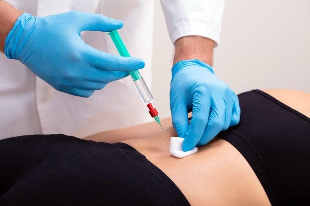
Modern lumbar epidural procedures are performed under fluoroscopic (live X-ray) guidance. The patient is positioned on a table, the skin is cleaned, and local anesthetic is used at the insertion site. A thin needle is advanced toward the epidural space. Approaches include interlaminar (through the ligamentum flavum between the laminae) and transforaminal (more selective placement near a specific exiting nerve root). The physician injects contrast dye under continuous imaging to confirm the medication spreads in the epidural space and does not enter a blood vessel or the subarachnoid space.
Once the physician verifies correct placement, they inject the corticosteroid (often with local anesthetic). The entire procedure typically lasts 10–20 minutes. Patients remain awake or lightly sedated so they can report unusual sensations. After a short observation period, most people go home the same day.
What Patients Commonly Experience
During the injection, some people feel pressure or a brief reproduction of their usual leg symptoms as the medication spreads. Immediate relief from the local anesthetic can last a few hours. The steroid effect usually begins over the following 2–7 days. A temporary increase in soreness at the injection site or a short flare of familiar leg pain can occur in the first day or two as the medication disperses; this is usually mild and self-limited.
Relief is not guaranteed and varies from person to person. When it occurs, patients often notice that they can sit longer, walk farther, sleep with fewer position changes, or tolerate the early stages of guided movement. The practical value is that these improvements create a usable window.
The Window for Movement, Decompression, and Rehabilitation
When chosen, the central purpose of the injection is to lower nerve-root irritation enough that active care becomes possible. Once leg pain no longer dominates every transition, spinal decompression therapy can be introduced or progressed more comfortably. Chiropractic adjustments and mobilization that respect the irritated root can be applied with clearer feedback. Targeted exercises for hip mobility, core control, and walking tolerance can start without an immediate severe flare.
For Amazon associates, this may mean returning to a modified floor shift with better standing and walking tolerance.
For data center technicians, it may mean safer ladder and rack work.
For programmers and drivers, it may mean longer sitting blocks without the same electric build-up.
For any adult, it may mean restoring sleep and basic daily mobility.
At Injury Medical Clinic PA, we never view the injection as a stand-alone fix. Dr. Alex Jimenez, DC, APRN, FNP-BC, CFMP, IFMCP, works under the collaborative medical direction of Dr. Maria Guadalupe Cardenas, MD, so the procedure, when indicated, is coordinated with structural care, rehabilitation, and any needed functional medicine support. The aim is beneficence: using the least invasive, effective option that serves the patient’s overall recovery and safety.
When an Injection Is Not the Appropriate First Step
Several situations call for a different sequence or urgent evaluation rather than an elective epidural:
Progressive motor weakness, saddle anesthesia, or new bowel or bladder changes (possible cauda equina) require immediate medical assessment, not a scheduled injection.
Active infection, uncontrolled coagulopathy, or allergy to the planned medication are contraindications.
Pure axial low-back pain without a clear radicular pattern is not a classic indication; other sources (facet, discogenic, myofascial) should be considered first.
Mild or improving symptoms that still allow basic walking, sleep, and a trial of conservative care often respond to decompression, targeted exercise, and activity modification without injection.
If the clinical pattern doesn’t correlate with imaging, or if a properly placed prior injection fails, the clinic should re-examine the diagnosis rather than repeat the same procedure.
Non-maleficence guides these boundaries. The clinic prioritizes options that avoid unnecessary procedural risk, prolonged high-dose medication, or premature surgery. Autonomy remains central: patients receive a clear explanation of expected benefits, realistic duration, risks, and alternatives so they can decide in coordination with their existing medical team and any group insurance requirements.
A Practical Next Step
If radiating leg pain is currently blocking rehabilitation, sleep, or basic work and home function, an evaluation can determine whether an image-guided epidural is appropriate or whether a different sequence is safer. Many patients with excellent group insurance coverage can coordinate the integrated evaluation, procedure (when indicated), and subsequent rehabilitation plan efficiently.
To schedule a visit at Injury Medical Clinic PA in El Paso, call 915-850-0900 or use the online scheduler. The multidisciplinary team—led by Dr. Alex Jimenez, DC, APRN, FNP-BC, and Dr. Maria Guadalupe Cardenas, MD—will review your history, examination, and imaging to develop a plan that supports lasting movement and function.
Why Getting Out of the Car Can Hurt More Than the Drive Itself
Abstract
Getting out of the car can hurt more than the drive because your hips stay flexed, your lumbar spine stiffens, trunk rotation is limited, and the first stand creates a sudden load change. This post explains seat position and road vibration without blaming one bad posture. It covers why the first steps may sting, how a chiropractic exam assesses sit-to-stand, and how hip mobility, trunk work, and spinal decompression can make exiting easier.
The parking brake clicks. The engine is off. For forty minutes the drive felt tolerable. Then you step onto the pavement in El Paso, and your lower back seizes up. Network engineers between sites, field technicians hauling kits, Amazon drivers on stop after stop, and hybrid employees finishing a commute describe the same surprise. The seat was fine. The exit was not.
Why the Drive Can Feel Fine
Sitting is acceptable, and one slumped inch is not the whole story. Reviews of posture and low back pain have not found a single causal pose that explains clinical pain (Swain et al., 2020). When researchers measure sitting time, many adults report an immediate rise in back discomfort, even though that does not prove sitting causes a lasting episode (De Carvalho et al., 2020).
A car seat adds its pattern. Hips stay bent. The lumbar spine often rests toward flexion. The trunk barely rotates because the belt, the console, and traffic keep you facing forward. A field technician may twist once for a meter. An Amazon driver may twist at every stop for a tote. A hybrid worker may not twist until the garage. Less variety means that tissues remain in a single length for an extended period.
De Carvalho and Callaghan (2011) measured lumbar posture and passive stiffness during two hours of simulated driving. Beach and colleagues (2005) found that passive flexion stiffness can rise after prolonged sitting, especially in men after about an hour. A spine that has been quiet in flexion may not love a sudden stand.
What Changes While You Are Still Buckled
Prolonged hip flexion. Hip flexors, including the iliopsoas, stay shortened while you drive. After a long sit, the hip may not extend easily. The pelvis can tip, and the lumbar joints take motion; the hip did not yield
Lumbar stiffness. Discs slowly lose some fluid under sustained load. Ligaments and capsules creep into the seated shape. Stiffness means motion is not shared evenly. One level may catch.
Reduced trunk rotation. A belted seat removes most of that. When you rotate to unbuckle, reach for a bag, or step out sideways, that turn is a new demand.
Seat position. A seat that is too far back forces you to round your body to reach the wheel. A seat too low sharpens the hip angle and hardens the stance. None of these is the sole cause. Together they change how load is shared between disc, facet, hip, and muscle.
Vibration. Road vibration is separate from posture. A systematic review linked whole-body vibration with higher odds of low back pain and sciatica, with some concern about publication bias (Burström et al., 2015). Vibration does not diagnose a disc. It is one more input during a long shift.
The First Steps Are a Different Task
Sitting and standing are not the same job. Intradiscal pressure changes with posture and load. Unsupported sitting can produce higher intradiscal pressure than easy standing, though the number depends on how you sit (Roman-Liu et al., 2023).
Sit-to-stand requires hip extension, knee extension, a forward weight shift, and then trunk rise. People with low back pain often coordinate the lumbar spine and hip differently during this task (Shum et al., 2005). If the hip is stiff, the low back may extend early.
That is why the first steps can hurt more than the drive:
The spine must change shape under body weight.
Hip extension is requested before the hip is ready.
Rotation returns all at once as you pivot out.
A bag, scanner, or tote is often added at the stand.
Cold air or a hot cab can make stiff tissue feel sharper.
Amazon drivers repeat this exit dozens of times. Network engineers may do it fewer times, but they still have to cover highway miles and then work in a ladder or crawl space. Hybrid employees may do it once, then sit again at a desk. The pattern is the same. The dose is not.
This Is Not One Bad Posture
Blaming the slouch oversimplifies a mixed evidence base (Swain et al., 2020). Two coworkers can share a truck, and only one limps to the tailgate. A useful exam asks what the exit demands, not whether your shoulders looked perfect at a red light.
Red flags need a same-day medical look: new bowel or bladder changes, saddle numbness, progressive leg weakness, fever, unexplained weight loss, or pain after a crash.
How a Back-Focused Exam Looks at the Exit
At El Paso Back Clinic, the question is mechanical. Can you stand without a catch?
Does one hip stop early?
Does lumbar motion hinge at one segment?
Does leg pain start after the stand, or did it ride the whole drive?
Dr. Alex Jimenez, DC, APRN, FNP-BC, CFMP, IFMCP, brings chiropractic assessment and board-certified family nurse practitioner training to that visit. He holds Texas APRN license #1191402 and prescriptive authority #59628 (NPI 1205907805). Dr. Maria Guadalupe Cardenas, MD, is board-certified in internal medicine and has more than 40 years of experience (Texas license #J2933, NPI 1164426748) and provides medical direction at Injury Medical Clinic PA.
A typical function check may include:
Sit-to-stand from seat height, then with arms crossed
Hip extension and rotation, side to side
Lumbar flexion, extension, and rotation
A short walk right after the sit
A nerve screen if pain travels below the knee
Questions on routes, ladder days, commute length, and group benefits
The goal is not a posture label. The goal is to see which link fails when the load changes.
Making the Transition Easier
Chiropractic care aims to restore segmental motion so the first steps are shared instead of hinged. Soft-tissue work can ease hip flexors and glute guards from the drive.
Scoot forward so both feet plant before you stand.
Lean the trunk forward as one unit, then rise. Do not twist and stand at once.
Take three slow steps before you lift a bag or tote.
On a longer stop, gently extend each hip for a few breaths.
If the low back prefers extension, skip a deep toe-touch after the drive.
The trunk has to accept load while the hips extend. Hip hinges with a light load, short carries, slow pivots, and step-ups help rebuild the exit you already do for work.
For disc-related lumbago or leg symptoms after the drive, targeted spinal decompression can reduce load long enough to retrain the stand. Add electroacupuncture when pain blocks sleep or the next shift. MLS laser or shockwave therapy is considered when the complaint is more tendon than disc. An image-guided epidural, when medically directed, is a later window for radicular pain, not the first habit.
These options are chosen for their benefit and for restraint. Non-invasive care can lower the chance that a stiff exit becomes a long medication course or an early surgical talk. You remain the decision-maker.
A Simple Exit Routine
Use this technique on a commute, a field loop, or a delivery block.
Set the seat so you are not reaching, with your hips near knee height if the cab allows.
Every 20 to 30 minutes, shift the pelvis. Change is the point.
Feet out, then stand, then reach.
Carry the bag on the side that does not twist a painful hip.
If the third step still grabs after two weeks, have the transition checked.
Many tech, logistics, and hybrid employers in El Paso offer group plans that include chiropractic and coordinated medical visits. Work injuries and crash-related exits are documented differently from a commute ache, and that distinction protects you.
Call if the catch spreads down a leg, if stairs feel weak, or if you plan the day around which door hurts less. You need a spine and hips that can change shape when the door opens.
Call 915-850-0900 or visit 11860 Vista Del Sol, Suite 128. Ask how group insurance applies to a sit-to-stand evaluation.
The One-Sided Carry: What a Laptop Bag, Tool Case, or Work Tote Can Do to Your Back and Hips
Abstract: A laptop bag, tool case, or work tote may feel harmless because each trip is short. But carrying the same load on the same side can change how your trunk, shoulder, hips, and legs share the work. This article explains asymmetrical loading, what clinicians look for, and practical ways to redistribute load and build capacity.
The Bag Is Not “Putting Your Spine Out”
Picture a network engineer leaving a parking lot with a laptop bag over the right shoulder. The bag is not extremely heavy, and the walk is only ten minutes. Yet by Thursday, the right shoulder feels elevated, the left low back feels tight, and one hip seems tired on stairs.
That pattern does not prove the bag has “misaligned” the spine. A better explanation is compensation.
When weight hangs on one side, the body has to keep the center of mass controlled while walking. The trunk may lean or stiffen, one shoulder may elevate, the pelvis may shift, and the hips may produce different forces from side to side. Classic biomechanical research found that asymmetric load carriage changed trunk muscle demands and hip and knee moments during walking (DeVita et al., 1991). Human bodies tolerate asymmetry every day. Concern grows when repetition exceeds a person’s strength, endurance, recovery, or tissue tolerance.
Why Repetition Matters More Than One Carry
Load-carriage research shows that load size and placement can alter posture, walking mechanics, and balance. A systematic review of backpack studies found changes such as more trunk flexion, altered hip and ankle motion, shorter strides, and higher cadence while carrying loads (Liew et al., 2016). Another systematic review found that load carriage can reduce postural stability, with load magnitude and placement influencing the effect (Martin et al., 2023).
These studies do not mean every worker carrying a bag will develop pain. They support a practical idea: the body adapts to repeated demands.
Think about the total “dose”:
How heavy is the bag?
How far do you carry it?
How many times per shift?
Do you always use the same side?
Are you climbing stairs or walking uneven ground?
Are you already fatigued from sitting, lifting, driving, or poor sleep?
Do you have enough trunk, hip, and shoulder endurance for the task?
A moderate load repeated many times can become more meaningful than one unusually heavy carry.
What Your Trunk and Hips Do With an Uneven Load
Carrying involves a coordinated system that includes the rib cage, spinal muscles, pelvis, hips, legs, and feet.
Trunk compensation
If a bag pulls downward on the right, your body may lean, brace, or rotate to keep its center of mass controlled. Some people stiffen through the torso; others allow more side-to-side motion. Either strategy can increase fatigue.
Shoulder elevation
A slipping strap can encourage shoulder hiking. Over time, neck and shoulder muscles may stay active longer than necessary, contributing to fatigue, tightness, headaches, or discomfort between the shoulder blades.
Hip loading
The hips help control the pelvis every time one foot leaves the ground. With an uneven load, one side may need to work differently to stabilize the body. Asymmetric carrying has been shown to change hip moments during walking, although the response depends on load, placement, speed, and the individual (DeVita et al., 1991).
Walking changes
People often shorten their stride, adjust cadence, change pelvic movement, or spend more time stabilizing when loads increase. These are normal adaptations, not proof of damage. Problems are more likely when the strategy becomes uncomfortable, inefficient, or difficult to recover from.
Signs the Carrying Pattern Deserves Attention
Occasional muscle fatigue is not automatically a medical problem. However, repeated symptoms that follow a predictable carrying pattern deserve a closer look.
Useful clues include:
pain that builds during or after carrying;
one shoulder consistently feeling higher or more tense;
aching on one side of the low back or outer hip;
symptoms that improve when the bag is removed or sides are changed;
reduced walking tolerance;
numbness, tingling, or weakness;
pain that changes how you climb stairs, bend, or lift.
Severe pain after trauma, progressive weakness, loss of bowel or bladder control, saddle-region numbness, fever, unexplained weight loss, or other major neurological or systemic symptoms require timely medical evaluation.
What a Chiropractic Assessment Should Actually Examine
At El Paso Back Clinic, the goal should not be to look at a shoulder bag and declare that the spine is “out.” A useful assessment asks what tissues and movements are struggling and why.
A clinician may examine spinal and hip range of motion, tenderness, neurological findings, gait, single-leg balance, trunk endurance, hip strength, shoulder mechanics, and how the patient carries. The examination may also consider work demands, prior injuries, training level, footwear, and recovery.
Watching the patient walk with the actual laptop bag or tool case can be especially informative.
Does the trunk lean?
Does the shoulder hike?
Does pain appear after several minutes?
Can symptoms improve by changing strap position, load distribution, or carrying side?
That information turns treatment from a generic back-pain plan into a task-specific rehabilitation strategy.
Treatment Should Improve Capacity, Not Create Dependence
For mechanical back or hip symptoms, chiropractic care may be one part of a broader plan to improve movement and reduce pain. Current guidance for chronic primary low back pain supports individualized nonsurgical care that can include education, structured exercise, and selected physical therapies such as spinal manipulation (World Health Organization, 2023).
The practical goal is to help the patient tolerate real life again.
A plan may include:
chiropractic manipulation or mobilization when appropriate;
hip and thoracic mobility work;
trunk endurance exercises;
progressive hip-abductor and gluteal strengthening;
loaded carries that gradually rebuild tolerance;
gait and stair training;
shoulder and scapular endurance work;
work-specific lifting and carrying practice.
If persistent musculoskeletal pain or muscle guarding limits rehabilitation, acupuncture or electroacupuncture may sometimes be added as an adjunct after appropriate evaluation. Evidence suggests electroacupuncture may reduce nonspecific low-back pain when combined with standard care, although certainty is limited and it should not replace diagnosis, exercise, or progressive conditioning (Hsieh et al., 2024).
The simplest fix is often not “stop carrying.” It is improving how you manage the load.
First, remove items you do not need. Bags can slowly collect chargers, tools, notebooks, bottles, and backup equipment.
Second, keep the load close to your body. A load that swings far from the trunk creates more movement to control.
Third, use two straps when practical. A well-fitted backpack can distribute load more evenly than a one-shoulder tote. For heavier occupational equipment, waist or hip support may help redistribute part of the load depending on the design.
Fourth, switch sides if the task requires one-handed or one-shoulder carrying. Changing sides doesn’t eliminate the load, but it reduces repetition of the same pattern.
Fifth, break long carries into shorter trips when possible. A rolling case or cart may be smarter than carrying everything at once.
Finally, build capacity. Stronger hips, trunk muscles, shoulders, and legs make carrying less costly. Progressive conditioning also respects autonomy: instead of telling patients their bodies are fragile, it teaches them how to prepare for the work they choose to do.
Coordinated Care for Back and Hip Symptoms in El Paso
Dr. Alex Jimenez, DC, APRN, FNP-BC, CFMP, IFMCP, combines chiropractic and medical training to evaluate mechanical, neurological, and broader health contributors to pain. When medical complexity requires oversight, he collaborates with Dr. Maria Guadalupe Cardenas, MD, a board-certified internal medicine physician with more than 40 years of experience.
That integrated model supports three principles. Beneficence means choosing care that restores function and addresses the likely cause. Non-maleficence means starting with appropriate noninvasive, drug-free strategies when reasonable while recognizing when imaging, medication, injections, or referral may be needed. Autonomy means explaining findings clearly so the patient can decide how to proceed and coordinate with an existing medical team.
A laptop bag, tool case, or work tote is rarely the whole story. The useful question is whether repeated one-sided loading is exceeding your current capacity. When assessment, load redistribution, chiropractic care, rehabilitation, and progressive conditioning match your job’s actual demands, the goal isn’t just to feel better on the treatment table. It is to walk, carry, climb, work, and recover with confidence.
Why Your Back Hurts Monday After a Weekend of Yard Work, Cleaning, or Home Projects
Abstract
You felt fine Friday. By Sunday evening, the yard is clean, the garage is organized, and your lower back remembers every bend, lift, twist, and carry. Often, the issue is not one “bad lift.” It is a sudden workload jump that exceeds what your back, hips, and supporting muscles were prepared to handle. This guide explains why that happens, how to move safely through Monday, and when back pain needs prompt medical evaluation.
The Weekend Workload Jump
Monday through Friday, a desk worker may spend hours sitting. A data center employee may alternate between computer work and equipment checks. An Amazon employee may repeat familiar tasks their body has adapted to.
Then Saturday arrives.
Suddenly, that person spends hours raking, pulling weeds, scrubbing floors, painting, climbing ladders, lifting soil, carrying boxes, or moving furniture. Research links greater exposure to lifting and carrying, non-neutral postures, and combined mechanical demands with increased odds of chronic low-back pain (Jahn et al., 2023). That does not mean bending is dangerous. Dose, repetition, recovery, conditioning, and individual capacity matter.
A weekend project can compress a large amount of unfamiliar work into one afternoon.
Why Your Muscles Feel So Tired
Your spine does not work alone. Muscles of the trunk, hips, abdomen, and legs share the job of controlling movement and transferring force.
When you rake, vacuum, shovel, paint, or lift repeatedly, those muscles produce force again and again. As fatigue builds, movement may become less coordinated. You may reach farther, twist while carrying, rush transitions, or rely more on the lower back because the hips and legs are tired.
Next-day soreness can reflect muscle stress or irritated tissues. It does not automatically mean a disc “slipped” or something is permanently damaged.
Most low-back pain is classified as nonspecific, meaning clinicians cannot tie it to one dangerous structural disease or single injured tissue (World Health Organization [WHO], 2023). A careful examination is more important than guessing based on pain location alone.
Hip Mobility Helps Share the Load
Your hips help you squat, hinge, step, and rotate. If they are stiff after a week of limited movement or unprepared for hours of weekend activity, the body may borrow motion elsewhere.
Imagine lifting a bag of mulch. If your ankles, knees, and hips work comfortably, you share the task. If your legs fatigue, you may bend more through the lumbar spine or hold the load farther away.
That does not make spinal bending inherently harmful. The larger picture includes total workload, task variety, strength, recovery, previous symptoms, and individual response. The goal is enough mobility, strength, and confidence for your whole body to participate.
Monday Morning: Keep Moving, but Scale the Load
When your back feels stiff Monday morning, you may want to barely move. For many uncomplicated episodes, current guidance instead encourages staying active and continuing normal activities as tolerated (National Institute for Health and Care Excellence [NICE], 2016; WHO, 2023).
Try a graded Monday:
Before work, take a short walk and use gentle hip and trunk movements.
During sitting tasks, change position regularly instead of chasing one “perfect” posture.
When standing, shift your stance and take brief walking breaks.
When lifting, temporarily reduce load size, keep objects close when practical, and let your legs and hips share the effort.
Break large jobs into smaller sets instead of testing your back with one heavy effort.
If movement sharply increases leg pain, numbness, or weakness, stop and seek clinical guidance.
What Chiropractic and Rehabilitation Can Add
A useful back evaluation should do more than ask where it hurts. It should examine how you move and what your workday requires.
At El Paso Back Clinic, an integrative examination may include spinal and hip motion, neurological screening, strength, gait, lifting mechanics, symptom behavior, and the activities that triggered the flare. The goal is to identify what can be modified and what needs further medical investigation.
Guidelines support exercise, education, and selected manual therapies for low-back pain. Manual therapy, including spinal manipulation or mobilization, is best used within a broader plan that includes active rehabilitation rather than as a stand-alone solution (George et al., 2021; NICE, 2016).
For the right patient, chiropractic care may reduce pain and restore comfortable movement, making progressive exercise easier. Rehabilitation then builds the capacity that weekends and workdays demand.
Build Capacity, Not Fear
A good recovery plan does not teach you that your back is fragile. It helps you understand current capacity and gradually expand it.
That may include:
hip-hinge and squat patterns;
trunk endurance and control;
progressively heavier carrying;
pushing and pulling;
hip and thoracic mobility;
walking and aerobic conditioning; and
task-specific lifting for home or work.
This is beneficence in practical terms: care should help you function better, not simply chase a pain score. It also supports autonomy. You should understand what clinicians find, what options are available, and what each option can realistically accomplish.
Appropriate non-invasive care may help reduce unnecessary reliance on medications or invasive procedures for some nonspecific cases. Avoiding harm also means recognizing when conservative care is not enough.
Red Flags: When Back Pain Needs Urgent Care
Seek urgent medical evaluation if back pain comes with new or progressive neurological problems, especially:
increasing weakness in one or both legs;
numbness around the groin, inner thighs, or saddle area;
new loss of bladder or bowel control;
severe walking difficulty caused by neurological weakness; or
rapidly worsening numbness or coordination loss.
Suspected cauda equina syndrome requires urgent assessment and typically prompt lumbar MRI because delayed treatment can risk permanent neurological loss (American College of Radiology, 2021).
Back pain after major trauma, pain with fever, unexplained weight loss, a cancer history, significant immune suppression, or other serious systemic symptoms also deserve medical assessment. Red-flag screening separates ordinary mechanical flare-ups from conditions requiring different care.
Prepare for the Next Weekend
Build capacity during the week. Walk. Strength train. Practice lifting. Vary your sitting. Break long chores into shorter blocks. Alternate bending tasks with upright tasks. Use carts or ask for help with unusually heavy objects. Increase workload gradually instead of going from five quiet workdays to six nonstop hours of home renovation.
At Injury Medical Clinic PA in El Paso, Dr. Alex Jimenez, DC, APRN, FNP-BC, CFMP, IFMCP, combines chiropractic structural care, mechanical rehabilitation, and medical assessment as a Doctor of Chiropractic and board-certified Family Practice Nurse Practitioner. Dr. Maria Guadalupe Cardenas, MD, a board-certified Internal Medicine physician with more than 40 years of experience, serves as Medical Director, Clinical Director, and collaborative physician, providing medical oversight when symptoms, medications, metabolic conditions, laboratory findings, or risk factors require broader evaluation.
This integrated model keeps the patient in charge. Chiropractic and rehabilitation can address mechanical problems, while medical evaluation investigates symptoms that do not fit a routine musculoskeletal pattern. Care can coordinate with your existing medical team.
If Monday back pain repeats after active weekends, ask, “What workload am I asking my body to handle, and how can I build the capacity to handle it better?” That conversation can turn a frustrating Monday pattern into a practical plan for stronger lifting, easier sitting, better recovery, and more confidence when the next weekend project arrives again. Schedule a multidisciplinary evaluation if recurring pain is limiting work, home projects, sleep, or confidence.
IFM's Find A Practitioner tool is the largest referral network in Functional Medicine, created to help patients locate Functional Medicine practitioners anywhere in the world. IFM Certified Practitioners are listed first in the search results, given their extensive education in Functional Medicine