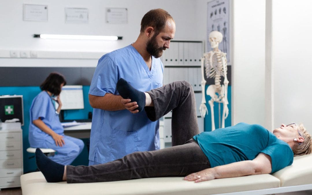
A common symptom of sciatica is radiating/spreading pain running down the leg. However, the leg pain could be something to do with the blood vessels. If the pain travels from the low back to the hip, through the buttocks, down the leg, and into the foot, then more than likely it is sciatica. However, sciatica is just one condition that causes leg pain; other causes of leg pain include:
Bone spurs
Herniated disc
Arthritis
All can irritate the sciatic nerve causing sciatica.
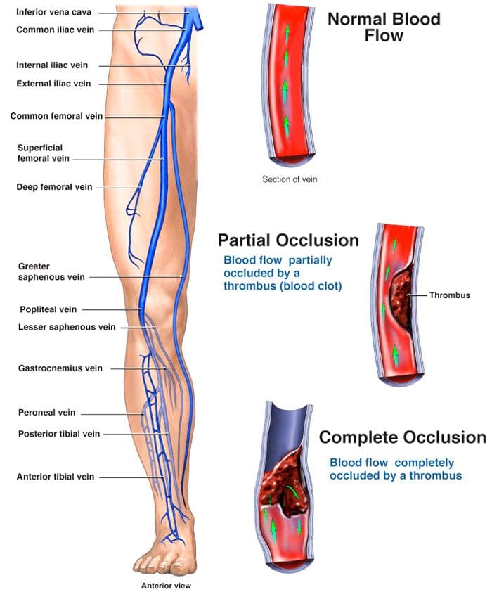
The vascular system, also called the circulatory system, comprises the vessels that circulate blood and lymph throughout the body. Problems with the vascular system are a less common cause of leg pain but can be severe. Therefore, it is vital to learn to tell the difference.
Deep Vein Thrombosis
Deep vein thrombosis – DVT happens when a blood clot forms in a deep vein in the body and not the superficial veins just under the skin. The legs’ deep veins are susceptible to clotting. The formation of a clot can happen:
After surgery
From an accident
When recovering, bed resting and not moving.
When the body is in the same position for a long time with little to no movement, like a long plane ride.
On long plane rides, try to get up and walk around every hour. If unable to walk, do three sets of 20 reps of heel-to-toe exercises every hour.
Deep vein thrombosis can cause leg pain or swelling but can also present without causing any symptoms. Other risk factors include:
Blunt or penetrating injury to the blood vessel and/or its walls.
Pain running down the leg from a blood clot feels like:
Tightness
Cramping soreness
Throbbing
Possible warmth
Swelling.
Blood clots and sciatica are reported to feel relatively different. The pain from a blood clot does not spread out and does not extend from or to the back. Sciatica does not cause swelling, redness, and warmth. If a doctor suspects a blood clot is causing the pain, they will order an ultrasound to confirm the diagnosis. If it is deep vein thrombosis, blood thinners could be recommended for three to six months.
A doctor may recommend aspirin, which can help in the prevention of blood clots.
In some cases, the clot may have to be surgically removed.
Vascular Conditions and Pain Running Down The Leg
Other blood vessel conditions that can cause individuals to believe they have sciatica include:
Peripheral artery disease – PAD
This often presents in individuals with diabetes or who smoke. It causes pain in the calf area but does not radiate throughout the leg. The pain usually presents with physical effort movement. If the pain occurs when at rest, this could be a serious medical emergency. Peripheral artery disease is a chronic condition that can worsen if lifestyle changes are not made to reduce risk factors.
Acute limb ischemia
This condition can cause leg pain, but not the same as sciatica. What happens is the leg is not receiving blood, causing:
Intense pain in the extremity
Change in the color of the skin
Numbness
Weakness
Loss of a pulse
This vascular condition is a medical emergency and requires immediate treatment.
Acute compartment syndrome
This can happen after some kind of trauma to the leg.
The pain is acute, with the leg swelling up and a building up of tight pressure.
It usually affects the lower part of the leg.
This condition can also cause:
Numbness
Tingling
Visible swelling
Bruising
It is considered a medical emergency and needs to be treated quickly to avoid complications.
Varicose veins
Varicose veins can cause some pain running down the leg and/or aching, but the discomfort is not as intense. Treatment has come a long way, is less invasive, and includes:
Compression stockings, including prescription socks/stockings
Laser treatments
Minimally invasive procedures
Not staying on the feet too much
Elevating the legs
Maintaining an ideal weight can help
Vascular Disorder Prevention
Healthy lifestyle habits are recommended to keep the vascular system operating correctly. This includes:
If it is sciatica, fortunately, most cases go away on their own, but if treatment is needed, it is recommended to start with conservative treatments such as:
Chiropractic
Physical therapy
Anti-inflammatory medication
Muscle relaxants
Corticosteroid injections
In severe cases, surgery like a microdiscectomy or laminectomy will be performed to relieve pressure on the sciatic nerve.
Body Composition
Why might blood pressure be different when measuring on each arm?
The heart sits just to the left of the midline in the chest cavity. The aorta is the largest blood vessel in the body. It leaves through the left side of the heart and transports blood to a network of blood vessels that branch out, supplying the body with oxygen and nutrients. The arteries that branch off the aorta and go to the left and right sides of the body are different.
On the right, the brachiocephalic trunk comes off the aorta and splits into the right common carotid artery and right subclavian artery. The left common carotid and left subclavian arteries branch directly off the aorta. The differences mean that the risk for arterial thrombosis is not the same for the right and left subclavian arteries. Arterial thrombosis causes the blood vessels to become stiff, causing obstruction over time and is more likely to happen in the left subclavian than in the right. The difference in arterial branching affects blood pressure measurements on the left and right arms. The blood vessels are surrounded by:
Muscle
Fat
Connective tissue
When muscles place pressure on the blood vessels around the heart, it can cause short-term turbulence changes that can affect blood pressure.
References
American Heart Association. Atherosclerosis and cholesterol. https://www.heart.org/en/health-topics/cholesterol/about-cholesterol/atherosclerosis
American Heart Association. What is excessive blood clotting (Hypercoagulation?) https://www.heart.org/en/health-topics/venous-thromboembolism/what-is-excessive-blood-clotting-hypercoagulation
Centers for Disease Control and Prevention. What is venous thromboembolism? https://www.cdc.gov/ncbddd/dvt/facts.html
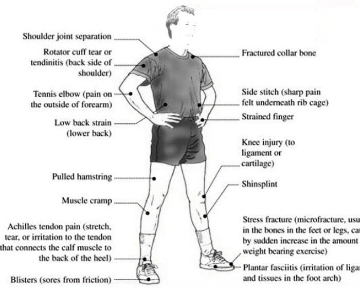
Acute and chronic sports injuries. Individuals who participate in sports or physical activities have an increased risk of experiencing an injury. These types of damages range from minor to severe and could require medical attention. Acute sports injuries happen suddenly and are usually the result of trauma to the area. A specific, identifiable incident is what causes an acute injury. Chronic sports injuries, also known as repetitive/overuse injuries, happen with time and are not caused by a single incident.
Acute and Chronic Sports Injuries Identification
Acute injuries can be identified by their cause. This could be a falling down during a run, sharp pain that presents in the shoulder after a throw, or a sprained ankle. The ability to focus on one cause usually means it’s acute. Acute injuries are characterized by:
Sudden pain in an area where there was none.
Swelling
Redness
Tenderness
Limited range of motion.
The inability of the injured area to support its weight.
A broken bone.
Dizziness
Headache
Nausea
Vomiting
Chronic injuries are different but are usually easy to identify. The pain begins gradually, usually over weeks or months. Repetitive activities like running, throwing, and swinging can exacerbate the pain. However, it is difficult to point to a specific issue that first caused the discomfort or pain. Chronic sports injuries are characterized by:
Pain and tenderness in the area, especially during and immediately after activity.
Minor swelling and limited range of motion.
Dull pain when resting.
These two types of injuries have different causes – trauma for acute and wear-and-tear for chronic – but they can both result in similar issues. For example, shoulder rotator cuff injuries are common, especially those that repeatedly use their shoulder to swing, throw, swim, etc. The individual needs to undergo a rotator cuff injury test to diagnose the injury correctly, whether the damage is acute or chronic. Chronic injuries can cause acute injuries, and acute injuries can lead to chronic injuries if left untreated.
Examples of Acute and Chronic Sports Injuries
Chronic and acute injuries are common in every type of sport. There’s an opportunity for both types of injuries. The most common include:
Other injuries from trauma, overuse, or both include:
Nonspecific Back Pain
Herniated Disc/s
Spondylolysis
Treatment
Minor acute injuries can be treated with rest, ice, compression, and elevation, aka R.I.C.E. Overuse injuries, are different as the injury has been gradually increasing in its severity, possibly causing scar tissue and ganglion cysts to develop. To prevent the injury from worsening, it’s recommended to see a sports injury chiropractor or physical therapist. These professionals can help heal the body and educate the individual on self-care and prevention.
Chiropractic
The musculoskeletal system takes a beating. Chronic injuries usually affect the bones, joints, muscles, or a combination. Chiropractic helps keep the musculoskeletal system limber and in proper alignment. Adjustments include:
Neck adjustments
Arm and hand adjustments
Shoulder adjustments
Knee adjustments
Hip adjustments
Foot adjustments
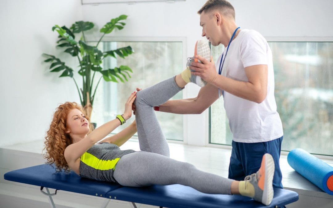
Physical Therapy
Physical therapy for a chronic injury can help prevent future injuries. A physical therapist helps:
Improve range of motion
Reduces pain and swelling
Increases strength
Whether an athlete or is just staying active and having some fun with sports, acute and chronic injuries can sneak up and worsen if they are not treated properly. Healing with the help of a professional can quicken recovery time and prevent future injuries.
Body Composition
Maintain Muscle Mass While Losing Fat
Individuals who want to lose weight should focus on losing excess fat tissue, not muscle mass. Studies have shown that diet and exercise are crucial to preserving Skeletal Muscle Mass while losing weight. Losing weight healthily includes:
A healthy balance of cardio and resistance training to burn calories and build muscle.
Wörtler, K, and C Schäffeler. “Akute Sportverletzungen und chronische Überlastungsschäden an Vor- und Mittelfuß” [Acute sports injuries and chronic overuse stress damage to the forefoot and midfoot]. Der Radiologe vol. 55,5 (2015): 417-32. doi:10.1007/s00117-015-2855-3
Yang, Jingzhen et al. “Epidemiology of overuse and acute injuries among competitive collegiate athletes.” Journal of athletic training vol. 47,2 (2012): 198-204. doi:10.4085/1062-6050-47.2.198
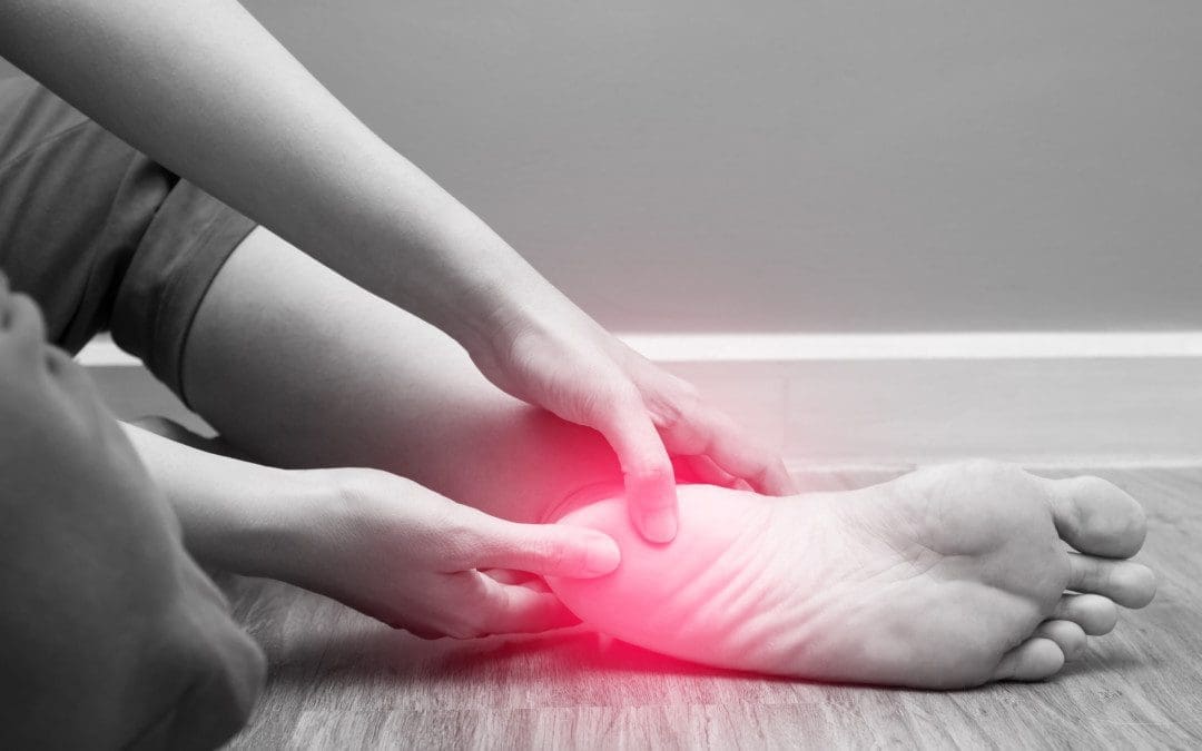
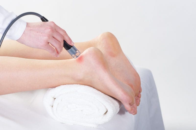
One of the most common tendons in the body that gets injured is the Achilles tendon, and this tendon tends to rupture when a person is doing recreational sports. Most people have opted for treatment for their Achilles tendon through surgery; however, low laser therapy can help the Achilles tendon recover a bit faster while providing beneficial properties during treatment. Low laser therapy has positive effects on the affected area where the pain resides and has helped progress the body’s natural healing process.
Achilles Tendon and Symptoms
The Achilles tendon is a strong fibrous cord connected at the back of the calf muscles to the heel bone. When a person does recreational sports, the Achilles tendon stretches during the activity. However, when the Achilles tendon is overly stretched during the exercise, it can rupture completely or partially depending on how strenuous the body is being put through.
Some of the symptoms of a ruptured Achilles include:
A feeling of being kicked in the calf
A popping or snapping sound where the injury occurred.
Pain and swelling near the heel.
The inability to bend the foot downwards
The inability to stand on the toes
When these symptoms occur in the Achilles tendon, it is due to the lack of blood flow that the body is not providing. Studies have found that when the Achilles tendon is ruptured, it is a severe injury due to the scarce blood supply, and it could take weeks or even months before it is completely healed.
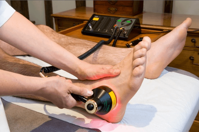
Low Laser Treatments and Benefits
Patients with a ruptured Achilles heel can get low laser therapy to help relieve the pain from the ruptured tendon. Studies found that when patients are being treated with low laser therapy has shown beneficial results. The results showed how the application of low laser treatment is very effective. The therapy provides a consequent relief from the motor function pain to the heel while also providing anti-inflammatory properties to the affected area. What this does is that the low-intensity laser concentrates on the inflammatory markers of the affected area, thus providing an increased blood flow (angiogenesis) in the treated area and decreasing inflammation. Low laser therapy can even help accelerate and enhance the repair of the injured Achilles tendon with frequent treatment sessions.
Conclusion
Overall, the Achilles tendon is one of the most frequent tendons that gets ruptured when a person is doing recreational sports. The healing process can take to about a week to a month for the tendon to properly heal. But through low laser therapy, the Achilles tendon can be repaired while providing relief from inflammation and enhancing the injured tendon recovery process.
References:
Ferreira, Rafaela, et al. Achilles Tendon Vascularization of … – Medical Laser. 2015, http://medical.summuslaser.com/data/files/79/1585169982_6Niglp3dbBeG7Cm.pdf.
Jesus, Julio Fernandes de, et al. “Low-Level Laser Therapy on Tissue Repair of Partially Injured Achilles Tendon in Rats.” Photomedicine and Laser Surgery, U.S. National Library of Medicine, 15 May 2015, https://pubmed.ncbi.nlm.nih.gov/24831690/.
Nogueira, Adelmário Cavalcanti, and Manoel de Jesus Moura Júnior. “The Effects of Laser Treatment in Tendinopathy: A Systematic Review.” Acta Ortopedica Brasileira, Sociedade Brasileira De Ortopedia e Traumatologia, 2015, https://www.ncbi.nlm.nih.gov/pmc/articles/PMC4544521/.
Staff, Mayo Clinic. “Achilles Tendon Rupture.” Mayo Clinic, Mayo Foundation for Medical Education and Research, 31 July 2020, https://www.mayoclinic.org/diseases-conditions/achilles-tendon-rupture/symptoms-causes/syc-20353234.
The body needs consistent energy to get through the day. The central nervous system runs from the brain, down through the spine, and then towards every organ and region of the body. The central nervous system works by sending signals to the organs or the body’s moving parts. But when there is a kink, misalignment, damage, or injury in the spine or other joints, the signals don’t get sent or received correctly.
Pressure builds on the nerves causing the signals/messages sent from the brain to the body to start to slow down and the body’s responses. This includes not just physical reactions but the body’s metabolism. Slow metabolism causes the body to become sluggish and slow. Regular chiropractic adjustments can remove the kinks restoring proper energy flow. When the body’s cells and organs are sending and receiving the signals correctly, the body gets the full force of the energy that the body has stored.
Energy Drain
Spinal misalignments can cause a multitude of symptoms. The most common problem is pain. Misalignments can also lead to energy-draining problems. These include:
Headaches
Difficulty concentrating
Joint pain
Inflammation
The body absorbs toxins from the air, water, food, or direct contact. A buildup of toxins can make the body sluggish. Chiropractic adjustments release these toxins so that the body can rid itself of them. Being out of alignment and balance requires the body to use more energy to get anything done. Even easy to do activities, chores, tasks, etc. When the body’s natural balance is restored, the result is more available energy.
Stress Management
Stress management is vital as chronic stress increases the risk of developing health problems. Learning to manage stress can help increase energy. Therapeutic massage can help with stress relief. A chiropractor will determine the best type of massage for each individual’s situation. The physiological effects of massage to reduce stress include:
Increased endorphins, serotonin, dopamine.
Decreased cortisol.
Increased tissue elasticity.
Endorphins, serotonin, and dopamine are neurotransmitters that are released by the autonomic system when stimulated.
Endorphins are responsible for relieving anxiety.
Serotonin prevents depression and gives a sense of well-being.
Dopamine increases motivation and prevents self-doubt.
When the body lacks these positive hormones, an individual can become stressed, anxious, and overwhelmed. A therapeutic massage stimulates the autonomic system, increasing the release of positive hormones. Increasing the level of positive hormones reduces stress and anxiety and improves overall mood.
When a decrease in cortisol occurs, stress is also reduced. Cortisol is a negative hormone released from the adrenal gland when stimulated by the brain’s hypothalamus region. The adrenal gland is located on top of the kidneys. Cortisol is released into the blood and is transported around the body. Cortisol increases:
Stress
Anxiety
Depression
Responsible for the fight or flight response.
When large amounts of cortisol are released in response to pain, stress levels increase, and the immune system is suppressed. A massage helps flush out the cortisol from the blood and replace it with the positive hormone endorphins, serotonin, and dopamine, decreasing stress and increasing relaxation.
Central Nervous System and Chiropractic
Chiropractic looks for the root cause of the problem and addresses that issue. Individuals can keep their bodies stay in balance by:
Chiropractic treatment can help improve quality of life and overall health.
Body Composition
Neglecting A Healthy Diet
Individuals might begin a weight loss journey by going to the gym, which is great, but neglecting a healthy diet is just wasting energy. Fat loss happens when the body is in a caloric/energy deficit. This means an individual has to take in fewer calories than the body is using. According to the CDC, individuals need to reduce food intake by at least 500 calories a day to lose around a pound of body fat a week.
Individuals that start increasing exercise/workouts make the body instinctively want to increase calorie intake.
Eating more calories than are burned means the individual is wasting the workout.
For example, the body needs a 2,100 calorie diet to maintain its weight, and on average, an individual eats 2,100 calories.
This means the weight won’t change much, if at all.
If an individual burns 300 calories from a workout, the body needs 2,400 calories to maintain weight.
If no changes are made to the diet, the individual will be in a negative -300 caloric deficit.
Suppose an individual starts increasing their caloric intake because they think their metabolism is speeding up, which is not how it works, then the individual negates any energy deficit that they worked for, leading to no fat loss.
References
Carlson, Linda E et al. “Integrative Approaches to Stress Management.” Cancer journal (Sudbury, Mass.) vol. 25,5 (2019): 329-336. doi:10.1097/PPO.0000000000000395
Kültür, Turgut et al. “Evaluation of the effect of chiropractic manipulative treatment on oxidative stress in sacroiliac joint dysfunction.” Turkish Journal of physical medicine and rehabilitation vol. 66,2 176-183. 18 May. 2020, doi:10.5606/tftrd.2020.3301
Salleh, Mohd Razali. “Life event, stress, and illness.” The Malaysian Journal of medical sciences: MJMS vol. 15,4 (2008): 9-18.
The body is a well-working machine that can endure anything that is thrown in its way. However, when it gets an injury, the body’s natural healing process will ensure that the body can get back to its daily activities. The healing process of an injured muscle varies throughout the body. Depending on how severe the damage is and how long the healing process will take, the body can recover to a mere few days to a few months. One of the most gruelly healing processes that the body has to endure is a ruptured calcaneal tendon.
The Calcaneal Tendon
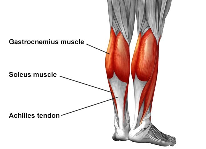
The calcaneal tendon or the Achilles tendon is a thick tendon that is located in the back of the leg. This muscle-tendon is what makes the body move while walking, running, or even jumping. Not only that, the calcaneal tendon is the strongest tendon in the body, and it connects the gastrocnemius and soleus muscles at the heel bone. When the calcaneal tendon is ruptured, the healing process can last from weeks to months until it is fully healed.
The Healing Effects of Low Laser Therapy
One of the ways that can help the damaged calcaneal tendons’ healing process is low laser therapy. Studies have shown that low laser therapy can speed up the damaged tendon repair after a partial lesion. Not only that but the combination of ultrasound and low laser therapy has been studied to be the physical agents for treating tendon injuries. The studies showed that the combination of low laser therapy and ultrasound has beneficial properties during the recovery process of treating calcaneal tendon injuries.
The study found that when patients are being treated for their calcaneal tendons, their hydroxyproline levels around the treated area are significantly increased with ultrasound and low laser therapy. The body’s natural biochemical and biomechanical structures on the injured tendon increase, thus affecting the healing process. Another study has shown that low laser therapy can help reduce fibrosis and prevent oxidative stress in the traumatized calcaneal tendon. The study even showed that after the calcaneal tendon is traumatized, inflammation, angiogenesis, vasodilation, and the extracellular matrix are formed in the affected area. So when patients are being treated with low laser therapy for about fourteen to twenty-one days, their histological abnormalities are alleviated, reducing collagen concentration and fibrosis; preventing oxidative stress from increasing in the body.
Conclusion
Overall, it is said that the effects of low laser therapy can help speed up the healing process of repairing the calcaneal tendon. The promising results have been proven since low laser therapy can help repair the damaged tendon, reducing oxidative stress and preventing fibrosis from escalating, causing more problems on the injured tendon. And with the combination of ultrasound, the calcaneal tendon can recover faster so the body can continue its everyday activities without any prolonged injuries.
References:
Demir, Huseyin, et al. “Comparison of the Effects of Laser, Ultrasound, and Combined Laser + Ultrasound Treatments in Experimental Tendon Healing.” Lasers in Surgery and Medicine, U.S. National Library of Medicine, 2004, https://pubmed.ncbi.nlm.nih.gov/15278933/.
Fillipin, Lidiane Isabel, et al. “Low-Level Laser Therapy (LLLT) Prevents Oxidative Stress and Reduces Fibrosis in Rat Traumatized Achilles Tendon.” Lasers in Surgery and Medicine, U.S. National Library of Medicine, Oct. 2005, https://pubmed.ncbi.nlm.nih.gov/16196040/.
Oliveira, Fla’via Schlittler, et al. Effect of Low Level Laser Therapy (830 Nm … – Medical Laser. 2009, http://medical.summuslaser.com/data/files/86/1585171501_uLg8u2FrJP7ZHcA.pdf.
Wood, Viviane T, et al. “Collagen Changes and Realignment Induced by Low-Level Laser Therapy and Low-Intensity Ultrasound in the Calcaneal Tendon.” Lasers in Surgery and Medicine, U.S. National Library of Medicine, 2010, https://pubmed.ncbi.nlm.nih.gov/20662033/.
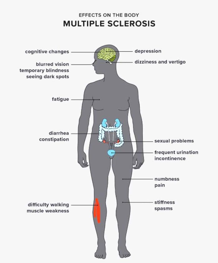
Multiple sclerosis and sciatica can exist side by side or have overlapping symptoms. The sciatic nerve begins at the lower back, then through the hips into the buttocks, and separates into both legs into the feet. Sciatica is a type of pain caused by a compressed/pinched or damaged/injured sciatic nerve. The sensation radiates across the nerve with frequency and severity at varying levels, depending on the individual’s body position and/or movement. Individuals with multiple sclerosis can also experience sciatica, believing it’s their multiple sclerosis. Neuropathic pain is a common symptom in multiple sclerosis or MS. It is caused by injury or damage to the nerves of the central nervous system and can cause burning, or sharp, stabbing sensations.
Multiple Sclerosis and Sciatic Nerve Pain Difference
MS is an autoimmune disorder where the immune system attacks the protective layer around nerve fibers known as myelin. This affects the central nervous system pathways that regulate feeling and sensation in the body. It can cause painful sensations that include:
Muscle spasms
Burning, tingling, or aching in the lower legs
Electrical shock-like sensations travel from the back toward the legs.
Migraines
The painful sensations result from the damaged nerve fibers creating interference in the brain’s neural pathways.
Sciatica works differently
An autoimmune response does not damage the sciatic nerve’s pathway, but an added stress/pressure compresses the sciatic nerve. The pain is usually caused by a quick, jerking, twisting, bending, reaching motion that pinches or twists the nerve. Herniated discs and bone spurs are another common cause, along with being overweight can place intense pressure on the sciatic nerve. The critical difference is that multiple sclerosis causes the central nervous system’s signaling pathways to malfunction.
MS and Sciatica
Most individuals, around 40%, will at some point experience some form of sciatica symptoms. This is from age, and all the wear and tear the low back goes through daily. This is why it’s not unusual for individuals with MS to experience sciatica as well. MS can cause body changes that affect activity levels.
Decreased mobility can lead to sitting for extended periods that can strain the muscles, tendons, and ligaments, causing sciatica.
There is evidence that the lesions that present from MS can extend to the sciatic nerve.
One study compared 36 individuals with MS to 35 individuals that don’t have it.
The research found that those with MS had slightly more lesions on the sciatic nerve than those without MS.
Sciatica Care
It can be challenging to figure out the types of pain being experienced. Sciatica travels down the length of the nerve uniquely and is often felt in only one leg. The pain, tingling, numbness, electrical sensations can present only in the lower back, the buttock, the back of the leg, hamstring, calf, and foot, or in a combination of all the areas. Treatments for sciatica depend on the severity. They include:
Medications – anti-inflammatories, muscle relaxants, tricyclic antidepressants, and antiseizure medications.
Steroid injections – corticosteroids
Surgery is a last resort reserved for severe cases that did not improve with other treatments and therapies.
It can be easy to mistake sciatica as a symptom or related condition of multiple sclerosis. Chiropractic can help alleviate sciatica, and although treatment cannot directly treat MS or its symptoms, it can relieve pain and discomfort.
Body Composition
Diabetic Nephropathy
Diabetic nephropathy or diabetic kidney disease is the result of mismanaged diabetes. Kidney failure is a severe medical emergency and can be fatal if not treated. Chronic low kidney function results in:
Fluid retention in the body.
Inability to filter out metabolites and waste from the blood.
Increased risk of infections.
Common symptoms of diabetic kidney disease include:
Increased blood pressure
This is the result of increased stress on the body.
The kidneys can no longer filter out all the metabolites and excess fluid needed to stabilize the blood pressure.
Chronic kidney damage results in the protein being excreted through urine.
Fatigue
Poor kidney function affects every organ in the body.
The organs have to work harder to compensate, leading to fatigue and low energy.
Lower extremity edema
Fluid retention usually presents in the lower extremities.
Puffy, swollen ankles and legs may appear shiny or waxy.
This is common in individuals that have severe diabetic nephropathy.
Shortness of breath
As the fluid builds up in the body, additional weight can get stored on and around the lungs.
This can make breathing very difficult when lying down or when engaged in physical activity.
Impaired cognition
Metabolites in the blood can cause brain damage when not filtered properly.
Memory loss
Mood changes
Loss of consciousness
References
Jende JME, et al. (2017). Peripheral nerve involvement in multiple sclerosis: Demonstration by magnetic resonance neurography. DOI:
10.1002/ana.25068
Mayo Clinic Staff. (2019). Sciatica.
mayoclinic.org/diseases-conditions/sciatica/symptoms-causes/syc-20377435
Murphy KL, et al. (2017). Chapter 4: Neuropathic pain in multiple sclerosis—current therapeutic intervention and future treatment perspectives.
ncbi.nlm.nih.gov/books/NBK470151/
Pain and itching. (n.d.).
nationalmssociety.org/Symptoms-Diagnosis/MS-Symptoms/Pain
Samson K. (2017). In the pipeline-multiple sclerosis neurography, MRI reveals peripheral nerve lesions in MS patients. DOI:
10.1097/01.NT.0000527861.27137.b0
Sciatica: Of all the nerves. (2016).
health.harvard.edu/pain/sciatica-of-all-the-nerve
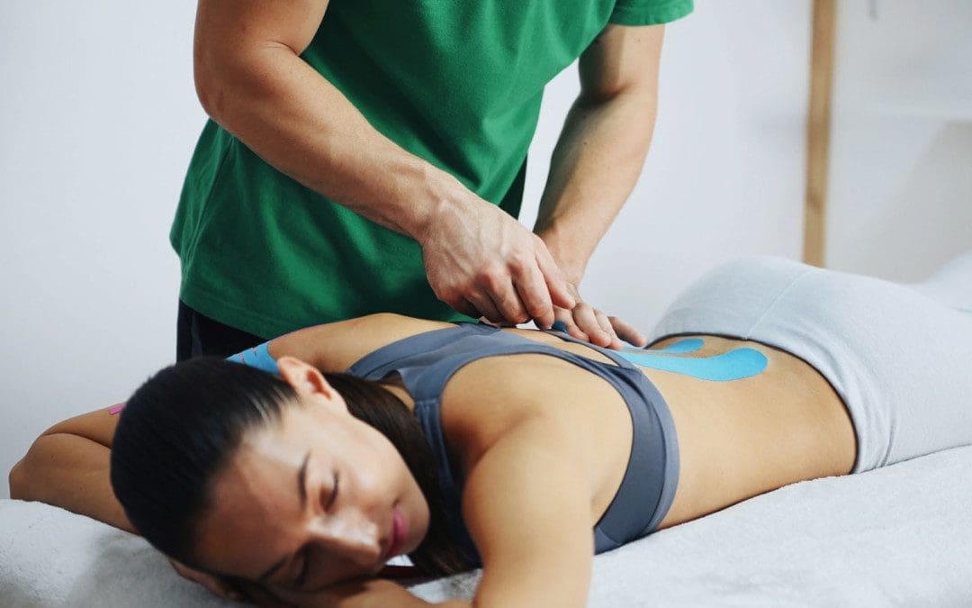
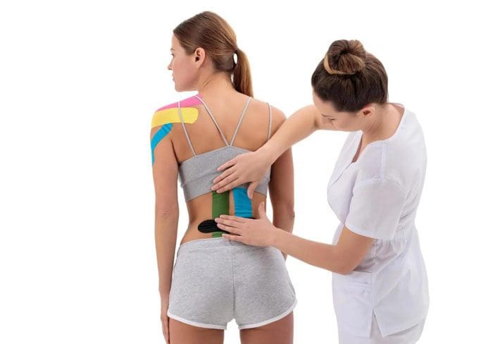
We see it on all types of athletes nowadays. They are wearing tape that looks like it’s for an injury. It is for injuries, but it can also be used as a preventative measure to avoid injuries. It is known as Kinesiology, Kinesio, KT, and elastic tape. It reduces swelling, increases mobility, and expedites recovery. It can be beneficial with back soreness/pain.
Tape
When it comes to Kinesio tape for back pain, medical professionals reported the tape is most effective when incorporated with other pain treatments. A study found that taping various areas of the body safely relieved knee pain and reduced the need for pharmacological treatment for knee osteoarthritis. It is applied to the body to support a joint, improve circulation, or provide proprioception feedback to the brain. The tape can help increase awareness of a specific painful area, reminding the individual to maintain proper posture and not move in a way that causes pain. Online videos can teach how to tape a particular area of the body. Examples include:
Each joint and muscle requires various tapings or different patterns and directions. Applying the tape to body areas that an individual can reach and access, like the knee and ankle, can be simple. But it can be a challenge to apply it to the shoulder or back. This is when a physical therapist, chiropractor, medical clinician, partner, family member, or friend can help with the application. Kinesiology tape is designed to adhere for an average of three to four days, even when bathing.
Benefits
Kinesio taping for low back pain with help should be done in a shortened muscle position, meaning the person helping should apply the tape while the person experiencing back pain stands up straight. The taping can be two stripes going up and down, or it can be done with strips fanning out towards the buttocks. This gives support to the spine/back muscles and decreases pain.
Recovery and Prevention
Recovery from a spine condition or injury prevention, kinesiology tape can be used without any risk. It can help reduce pain, improve circulation, and provide muscle support. For a minor sprain or strain, the tape could help on its own. But for an individual experiencing severe back pain, it is recommended to seek professional medical care along with a stretching and strengthening regimen. The tape is recommended to be used as part of a complete treatment plan.
Body Composition
Five-Day Training Plan
The idea of training five times a week can be pretty intimidating. This is not about pushing yourself to your breaking point Monday through Friday. The objective of working out this frequently is about exercising normally, not like a professional athlete. That’s why many individuals divide up the areas they work out each day. Particular attention is given to one muscle group or system, letting the others rest and recover. This workout strategy is called a split and is favored by the bodybuilding community. Five-day splits are utilized to target different major muscle group/s every day. A standard training plan includes:
This is just one of the many programs that trainers, athletes, and fitness individuals have developed. Some individuals replace shoulders day with cardio; some do abs every day; it depends on what fitness goal the individual is going for.
References
Journal of Bodywork and Movement Therapies. (April 2016) “Kinesio taping for chronic low back pain: A systematic review” https://pubmed.ncbi.nlm.nih.gov/27634093/
Therapeutic Advances in Musculoskeletal Disease. (August 2019) “The effectiveness of Kinesio Taping® for pain management in knee osteoarthritis: a randomized, double-blind, controlled clinical trial” https://journals.sagepub.com/doi/full/10.1177/1759720X19869135
What are the potential benefits for back pain?: Asian Journal of Sports Medicine. (June 2015) “Low Back Pain in Athletes” https://www.ncbi.nlm.nih.gov/pmc/articles/PMC4592766/
IFM's Find A Practitioner tool is the largest referral network in Functional Medicine, created to help patients locate Functional Medicine practitioners anywhere in the world. IFM Certified Practitioners are listed first in the search results, given their extensive education in Functional Medicine