ClickCease
+1-915-850-0900 [email protected]
Select Page
Lumbar Disc Nomenclature: Version 2.0

Lumbar Disc Nomenclature: Version 2.0

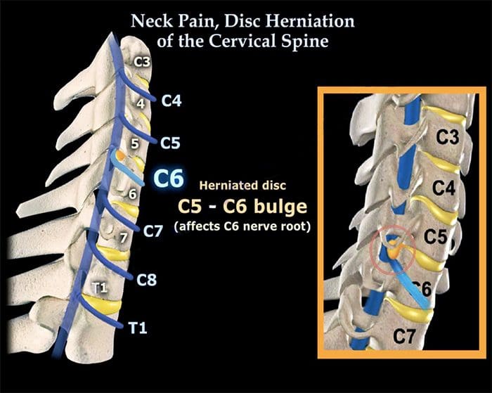
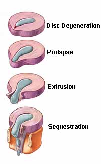
What is a Herniated Disc?

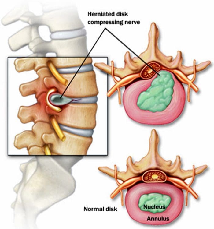
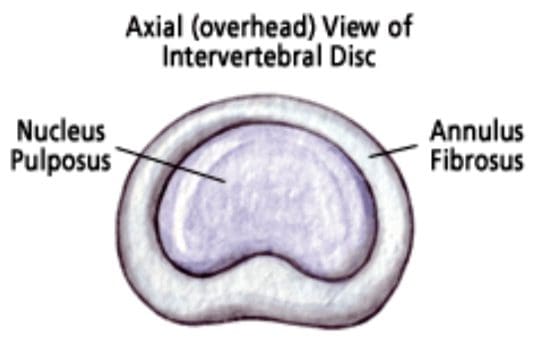
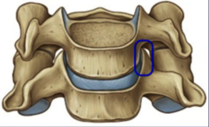
The spine is made up of 24 bones, called vertebrae, which are stacked on top of one another. These spinal bones are ultimately connected, creating a canal to protect the spinal cord. In between each vertebra are fluid-filled intervertebral discs which act as shock absorbers for the spine. Over time, however, these flexible, jelly donut-like discs can begin to herniate, where the nucleus of the intervertebral disc pushes against its outer ring, causing low back pain. Below, we will demonstrate the various types of herniated discs and discuss their causes, symptoms and treatment options.

Abstract

Background Context

The paper ��Nomenclature and classification of lumbar disc pathology, recommendations of the combined task forces of the North American Spine Society, the American Society of Spine Radiology and the American Society of Neuroradiology,�� was published in 2001 in Spine (� Lippincott, Williams & Wilkins). It was authored by David Fardon, MD, and Pierre Milette, MD, and formally endorsed by the American Society of Spine Radiology (ASSR), American Society of Neuroradiology (ASNR), and North American Spine Society (NASS). Its purpose was to promote greater clarity and consistency of usage of spinal terminology, and it has served this purpose well for over a decade. Since 2001, there has been sufficient evolution in our understanding of the lumbar disc to suggest the need for revision and updating of the original document. The revised document is presented here, and it represents the consensus recommendations of contemporary combined task forces of the ASSR, ASNR, and NASS. This article reflects changes consistent with current concepts in radiologic and clinical care.

Purpose

To provide a resource that promotes a clear understanding of lumbar disc terminology amongst clinicians, radiologists, and researchers. All the concerned need standard terms for the normal and pathologic conditions of lumbar discs that can be used accurately and consistently and thus best serve patients with disc disorders.

Study Design

This article comprises a review of the literature.

Methods

A PubMed search was performed for literature pertaining to the lumbar disc. The task force members individually and collectively reviewed the literature and revised the 2001 document. The revised document was then submitted for review to the governing boards of the ASSR, ASNR, and NASS. After further revision based on the feedback from the governing boards, the article was approved for publication by the governing boards of the three societies, as representative of the consensus recommendations of the societies.

Results

The article provides a discussion of the recommended diagnostic categories pertaining to the lumbar disc: normal; congenital/developmental variation; degeneration; trauma; infection/inflammation; neoplasia; and/or morphologic variant of uncertain significance. The article provides a glossary of terms pertaining to the lumbar disc, a detailed discussion of these terms, and their recommended usage. Terms are described as preferred, nonpreferred, nonstandard, and colloquial. Updated illustrations pictorially portray certain key terms. Literature references that provided the basis for the task force recommendations are included.

Conclusions

We have revised and updated a document that, since 2001, has provided a widely acceptable nomenclature that helps maintain consistency and accuracy in the description of the anatomic and physiologic properties of the normal and abnormal lumbar disc and that serves as a system for classification and reporting built upon that nomenclature.

Keywords

Annular fissure, Annular tear, Disc bulge (bulging disc), Disc degeneration, Disc extrusion, Disc herniation, Disc nomenclature, Disc protrusion, High-intensity zone, Lumbar intervertebral disc

Preface

The nomenclature and classification of lumbar disc pathology consensus, published in 2001, by the collaborative efforts of the North American Spine Society (NASS), the American Society of Spine Radiology (ASSR) and the American Society of Neuroradiology (ASNR), has guided radiologists, clinicians, and interested public for over a decade [1]. This document has passed the test of time. Responding to an initiative from the ASSR, a task force of spine physicians from the ASSR, ASNR, and NASS has reviewed and modified the document. This revised document preserves the format and most of the language of the original, with changes consistent with current concepts in radiologic and clinical care. The modifications deal primarily with the following: updating and expansion of Text, Glossary, and References to meet contemporary needs; revision of Figures to provide greater clarity; emphasis of the term ��annular fissure�� in place of ��annular tear��; refinement of the definitions of ��acute�� and ��chronic�� disc herniations; revision of the distinction between disc herniation and asymmetrically bulging disc; elimination of the Tables in favor of greater clarity from the revised Text and Figures; and deletion of the section of Reporting and Coding because of frequent changes in those practices, which are best addressed by other publications. Several other minor amendments have been made. This revision will update a workable standard nomenclature, accepted and used universally by imaging and clinical physicians.

Introduction and History

Physicians need standard terms for normal and pathologic conditions of lumbar discs [2, 3, 4, 5]. Terms that can be interpreted accurately, consistently, and with reasonable precision are particularly important for communicating impressions gained from imaging for clinical diagnostic and therapeutic decision-making. Although clear understanding of the disc terminology between radiologists and clinicians is the focus of this work, such understanding can be critical, also to patients, families, employers, insurers, jurists, social planners, and researchers.

In 1995, a multidisciplinary task force from the NASS addressed the deficiencies in commonly used terms defining the conditions of the lumbar disc. It cited several documentations of the problem [6, 7, 8, 9, 10, 11] and made detailed recommendations for standardization. Its work was published in a copublication of the NASS and the American Academy of Orthopaedic Surgeons [9]. The work had not been otherwise endorsed by major organizations and had not been recognized as authoritative by radiology organizations. Many previous [3, 7, 9, 10, 11, 12, 13, 14, 15, 16, 17, 18, 19] and some subsequent [20, 21, 22, 23, 24, 25] efforts addressed the issues, but were of more limited scope and none had gained a widespread acceptance.

Although the NASS 1995 effort was the most comprehensive at the time, it remained deficient in clarifying some controversial topics, lacking in its treatment of some issues, and did not provide recommendations for standardization of classification and reporting. To address the remaining needs, and in hopes of securing endorsement sufficient to result in universal standardizations, joint task forces (Co-Chairs David Fardon, MD, and Pierre Milette, MD) were formed by the NASS, ASNR, and ASSR, resulting in the first version of the document ��Nomenclature and classification of lumbar disc pathology�� [1]. Since then, time and experience suggested the need for revisions and updating of the original document. The revised document is presented here.

The general principles that guided the original document remain unchanged in this revision. The definitions are based on the anatomy and pathology, primarily as visualized on imaging studies. Recognizing that some criteria, under some circumstances, may be unknowable to the observer, the definitions of the terms are not dependent on or imply the value of specific tests. The definitions of diagnoses are not intended to imply external etiologic events such as trauma, they do not imply relationship to symptoms, and they do not define or imply the need for specific treatment.

The task forces, both current and former, worked from a model that could be expanded from a primary purpose of providing understanding of reports of imaging studies. The result provides a simple classification of diagnostic terms, which can be expanded, without contradiction, into more precise subclassifications. When reporting pathology, degrees of uncertainty would be labeled as such rather than compromising the definitions of the terms.

All terms used in the classifications and subclassifications are defined and those definitions are adhered to throughout the model. For a practical purpose, some existing English terms are given meanings different from those found in some contemporary dictionaries. The task forces provide a list and classification of the recommended terms, but, recognizing the nature of language practices, discuss and include in the Glossary, commonly used and misused nonrecommended terms and nonstandard definitions.

Although the principles and most of the definitions of this document can be easily extrapolated to the cervical and dorsal spine, the focus is on the lumbar spine. Although clarification of terms related to posterior elements, dimensions of the spinal canal, and status of neural tissues is needed, this work is limited to the discussion of the disc. While it is not always possible to discuss fully the definition of anatomical and pathologic terms without some reference to symptoms and etiology, the definitions themselves stand the test of independence from etiology, symptoms, or treatment. Because of the focus on anatomy and pathology, this work does not define certain clinical syndromes that may be related to lumbar disc pathology [26].

Guided by those principles, we have revised and updated a document that, since 2001, has provided a widely acceptable nomenclature that is workable for all forms of observation, that addresses contour, content, integrity, organization, and spatial relationships of the lumbar disc; and that serves a system of classification and reporting built upon that nomenclature.

Diagnostic Category & Subcategory Recommendations

These recommendations present diagnostic categories and subcategories intended for classification and reporting of imaging studies. The terminology used throughout these recommended categories and subcategories remains consistent with detailed explanations given in the Discussion and with the preferred definitions presented in the Glossary.

The diagnostic categories are based on pathology. Each lumbar disc can be classified in terms of one, and occasionally more than one, of the following diagnostic categories: normal; congenital/developmental variation; degeneration; trauma; infection/inflammation; neoplasia; and/or morphologic variant of uncertain significance. Each diagnostic category can be subcategorized to various degrees of specificity according to the information available and purpose to be served. The data available for categorization may lead the reporter to characterize the interpretation as ��possible,�� ��probable,�� or ��definite.��

Note that some terms and definitions discussed below are not recommended as preferred terminology, but are included to facilitate the interpretation of vernacular and, in some cases, improper use. Terms may be defined as preferred, nonpreferred, or nonstandard. Nonstandard terms by consensus of the organizational task forces should not be used in the manner described.

Normal

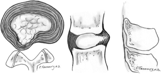
Normal defines discs that are morphologically normal, without the consideration of the clinical context and not inclusive of degenerative, developmental, or adaptive changes that could, in some contexts (eg, normal aging, scoliosis, spondylolisthesis), be considered clinically normal (Fig. 1).

Figure 1: Normal lumbar disc. (Top Left) Axial, (Top Right) sagittal, and (Bottom) coronal images demonstrate that the normal disc, composed of central NP and peripheral AF, is wholly within the boundaries of the disc space, as defined, craniad and caudad by the vertebral body end plates and peripherally by the planes of the outer edges of the vertebral apophyses, exclusive of osteophytes. NP, nucleus pulposus; AF, annulus fibrosus.

Congenital/Developmental Variation

The congenital/developmental variation category includes discs that are congenitally abnormal or that have undergone changes in their morphology as an adaptation of abnormal growth of the spine, such as from scoliosis or spondylolisthesis.

Degeneration

Degenerative changes in the discs are included in a broad category that includes the subcategories annular fissure, degeneration, and herniation.

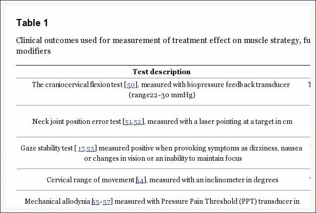
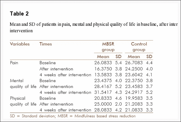
Annular fissures are separations between the annular fibers or separations of annular fibers from their attachments to the vertebral bone. Fissures are sometimes classified by their orientation. A ��concentric fissure�� is a separation or delamination of annular fibers parallel to the peripheral contour of the disc (Fig. 2). A ��radial fissure�� is a vertically, horizontally, or obliquely oriented separation of (or rent in) annular fibers that extends from the nucleus peripherally to or through the annulus. A ��transverse fissure�� is a horizontally oriented radial fissure, but the term is sometimes used in a narrower sense to refer to a horizontally oriented fissure limited to the peripheral annulus that may include separation of annular fibers from the apophyseal bone. Relatively wide annular fissures, with stretch of the residual annular margin, at times including avulsion of an annular fragment, have sometimes been called ��annular gaps,�� a term that is relatively new and not accepted as standard [27]. The term ��fissures�� describes the spectrum of these lesions and does not imply that the lesion is a consequence of injury.

Figure 2: Fissures of the annulus fibrosus. Fissures of the annulus fibrosus occur as radial (R), transverse (T), and/or concentric (C) separations of fibers of the annulus. The transverse fissure depicted is a fully developed, horizontally oriented radial fissure; the term ��transverse fissure�� is often applied to a less extensive separation limited to the peripheral annulus and its bony attachments.

Use of the term ��tear�� can be misunderstood because the analogy to other tears has a connotation of injury, which is inappropriate in this context. The term ��fissure�� is the correct term. Use of the term ��tear�� should be discouraged and, when it appears, should be recognized that it is usually meant to be synonymous with ��fissure�� and not reflective of the result of injury. The original version of this document stated preference for the term ��fissure�� but regarded the two terms as almost synonymous. However, in this revision, we regard the term ��tear�� as nonstandard usage.

Degeneration may include any or all of the following: desiccation, fibrosis, narrowing of the disc space, diffuse bulging of the annulus beyond the disc space, fissuring (ie, annular fissures), mucinous degeneration of the annulus, intradiscal gas [28], osteophytes of the vertebral apophyses, defects, inflammatory changes, and sclerosis of the end plates [15, 29, 30, 31, 32, 33, 34].

Herniation is broadly defined as a localized or focal displacement of disc material beyond the limits of the intervertebral disc space. The disc material may be nucleus, cartilage, fragmented apophyseal bone, annular tissue, or any combination thereof. The disc space is defined craniad and caudad by the vertebral body end plates and, peripherally, by the outer edges of the vertebral ring apophyses, exclusive of osteophytes. The term ��localized�� or ��focal�� refers to the extension of the disc material less than 25% (90�) of the periphery of the disc as viewed in the axial plane.

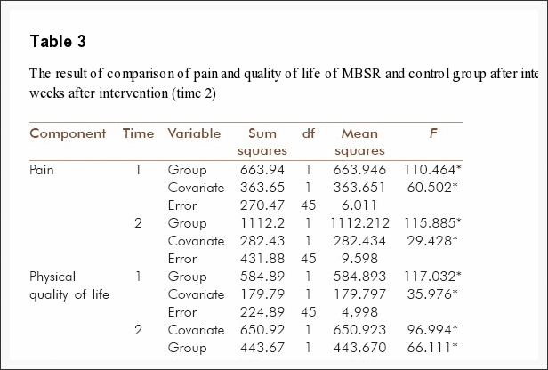
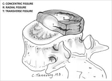
The presence of disc tissue extending beyond the edges of the ring apophyses, throughout the circumference of the disc, is called ��bulging�� and is not considered a form of herniation (Fig. 3, Top Right). Asymmetric bulging of disc tissue greater than 25% of the disc circumference (Fig. 3, Bottom), often seen as an adaptation to adjacent deformity, is, also, not a form of herniation. In evaluating the shape of the disc for a herniation in an axial plane, the shape of the two adjacent vertebrae must be considered [15, 35].

Figure 3: Bulging disc. (Top Left) Normal disc (for comparison); no disc material extends beyond the periphery of the disc space, depicted here by the broken line. (Top Right) Symmetric bulging disc; annular tissue extends, usually by less than 3 mm, beyond the edges of the vertebral apophyses symmetrically throughout the circumference of the disc. (Bottom) Asymmetric bulging disc; annular tissue extends beyond the edges of the vertebral apophysis, asymmetrically greater than 25% of the circumference of the disc.

Herniated discs may be classified as protrusion or extrusion, based on the shape of the displaced material.

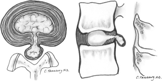
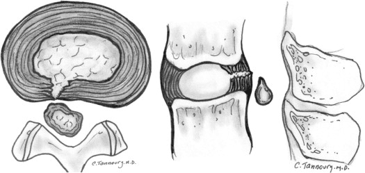
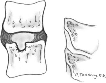
Protrusion is present if the greatest distance between the edges of the disc material presenting outside the disc space is less than the distance between the edges of the base of that disc material extending outside the disc space. The base is defined as the width of disc material at the outer margin of the disc space of origin, where disc material displaced beyond the disc space is continuous with the disc material within the disc space (Fig. 4). Extrusion is present when, in at least one plane, any one distance between the edges of the disc material beyond the disc space is greater than the distance between the edges of the base of the disc material beyond the disc space or when no continuity exists between the disc material beyond the disc space and that within the disc space (Fig. 5). The latter form of extrusion is best further specified or subclassified as sequestration if the displaced disc material has lost continuity completely with the parent disc (Fig. 6). The term migration may be used to signify displacement of disc material away from the site of extrusion. Herniated discs in the craniocaudad (vertical) direction through a gap in the vertebral body end plate are referred to as intravertebral herniations (Schmorl nodes) (Fig. 7).

Figure 4: Herniated disc: protrusion. (Left) Axial and (Right) sagittal images demonstrate displaced disc material extending beyond less than 25% of the disc space, with the greatest measure, in any plane, of the displaced disc material being less than the measure of the base of displaced disc material at the disc space of origin, measured in the same plane.
Figure 5: Herniated disc: extrusion. (Left) Axial and (Right) sagittal images demonstrate that the greatest measure of the displaced disc material is greater than the base of the displaced disc material at the disc space of origin, when measured in the same plane.
Figure 6: Herniated disc: sequestration. (Left) Axial and (Right) sagittal images show that a sequestrated disc is an extruded disc in which the displaced disc material has lost all connection with the disc of origin.
Figure 7:�Intravertebral herniation (Schmorl node). Disc material is displaced beyond the disc space through the vertebral end plate into the vertebral body, as shown here in sagittal projection

Disc herniations may be further specifically categorized as contained, if the displaced portion is covered by outer annulus fibers and/or the posterior longitudinal ligament, or uncontained when absent of any such covering. If the margins of the disc protrusion are smooth on axial computed tomography (CT) or magnetic resonance imaging (MRI), then the displaced disc material is likely contained by the posterior longitudinal ligament and perhaps a few superficial posterior annular fibers [21, 35, 36, 37]. If the posterior margin of the disc protrusion is irregular, the herniation is likely uncontained. Displaced disc tissue is typically described by location, volume, and content, as discussed later in this document.

An alternative scheme of distinguishing protrusion from extrusion is discussed in the Discussion section.

Trauma

The category of trauma includes disruption of the disc associated with physical and/or imaging evidence of violent fracture and/or dislocation and does not include repetitive injury, contribution of less than violent trauma to the degenerative process, fragmentation of the ring apophysis in conjunction with disc herniation, or disc abnormalities in association with degenerative subluxations. Whether or not a ��less than violent�� injury has contributed to or been superimposed on a degenerative change is a clinical judgment that cannot be made based on images alone; therefore, from the standpoint of description of images, such discs, in the absence of significant imaging evidence of associated violent injury, should be classified as degeneration rather than trauma.

Inflammation/Infection

The category of inflammation/infection includes infection, infection-like inflammatory discitis, and inflammatory response to spondyloarthropathy. It also includes inflammatory spondylitis of the subchondral end plate and bone marrow manifested by Modic Type I MRI changes [29, 30, 38] and usually associated with degenerative pathologic changes in the disc. To simplify the classification scheme, the category is inclusive of disparate conditions; therefore, when data permit, the diagnosis should be subcategorized for appropriate specificity.

Neoplasia

Primary or metastatic morphologic changes of disc tissues caused by malignancy are categorized as neoplasia, with subcategorization for appropriate specificity.

Miscellaneous Paradiscal Masses of Uncertain Origin

Although most intraspinal cysts are of meningeal or synovial origin, a minority arise from the disc and create a paradiscal mass that does not contain nuclear material. Epidural bleeding and/or edema, unrelated to trauma or other known origin may create a paradiscal mass or may increase the size of herniated disc material. Such cysts and hematomas may be seen acutely and unaccompanied by other pathology or may be a component of chronic disc pathology.

Morphologic�Variant of Unknown Significance

Instances in which data suggest abnormal morphology of the disc, but in which data are not complete enough to support a diagnostic categorization can be categorized as a morphologic variant of unknown significance.

Discussion of Nomenclature in Detail

This document provides a nomenclature that facilitates the description of surgical, endoscopic, or cadaveric findings as well as imaging findings; and also, with the caveat that it addresses only the morphology of the disc, it facilitates communication for patients, families, employers, insurers, and legal and social authorities and permits accumulation of more reliable data for research.

Normal Disc

Categorization of a disc as ��normal�� means the disc is fully and normally developed and free of any changes of disease, trauma, or aging. Only the morphology, and not the clinical context, is considered. Clinically ��normal�� (asymptomatic) people may have a variety of harmless imaging findings, including congenital or developmental variations of discs, minor bulging of the annuli, age-related desiccation, anterior and lateral marginal vertebral body osteophytes, prominence of disc material beyond one end plate as a result of luxation of one vertebral body relative to the adjacent vertebral body (especially common at L5�S1), and so on [39]. By this article�s morphology-based nomenclature and classification, however, such individual discs are not considered ��normal,�� but rather are described by their morphologic characteristics, independent of their clinical import unless otherwise specified.

Disc with Fissures of the Annulus

There is a general agreement about the various forms of loss of integrity of the annulus, such as radial, transverse, and concentric fissures. Yu et al. [40] have shown that annular fissures, including radial, concentric, and transverse types, are present in nearly all degenerated discs [41]. If the disc is dehydrated on an MRI scan, it is likely that there is at least one or more small fissures in the annulus. Relatively wide, radially directed annular fissures, with stretch of the residual annular margin, at times involving avulsion of an annular fragment, have sometimes been called ��annular gaps,�� although the term is relatively new and not accepted as a standard [27].

The terms ��annular fissure�� and ��annular tear�� have been applied to the findings on T2-weighted MRI scans of localized high intensity zones (HIZ) within the annulus [30, 42, 43, 44]. High intensity zones represent fluid and/or granulation tissue and may enhance with gadolinium. Fissures occur in all degenerative discs but are not all visualized as HIZs. Discography reveals some fissures not seen by the MRI, but not all fissures are visualized by discography. Description of the imaging findings is most accurate when limited to the observation of an HIZ or discographically demonstrated fissure, with the understood caveat that there is an incomplete concordance with the HIZs, discogram images, and anatomically observed fissures.

As far back as the 1995 NASS document, authors have recommended that such lesions be termed ��fissures�� rather than ��tears,�� primarily out of concern that the word ��tear�� could be misconstrued as implying a traumatic etiology [9, 30, 45, 46]. Because of potential misunderstanding of the term ��annular tear,�� and consequent presumption that the finding of an annular fissure indicates that there has been an injury, the term ��annular tear�� should be considered nonstandard and ��annular fissure�� be the preferred term. Imaging observation of an annular fissure does not imply an injury or related symptoms, but simply defines the morphologic change in the annulus.

Degenerated Disc

Because there is a confusion in the differentiation of changes of pathologic degenerative processes in the disc from those of normal aging [17, 31, 47, 48, 49], the classification ��degenerated disc�� includes all such changes, thus does not compel the observer to differentiate the pathologic from the normal consequence of aging.

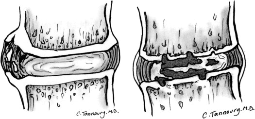
Perceptions of what constitutes the normal aging process of the spine have been greatly influenced by postmortem anatomic studies involving a limited number of specimens, harvested from cadavers from different age groups, with unknown past medical histories and the presumption of absence of lumbar symptoms [23, 50, 51, 52, 53, 54, 55, 56, 57]. With such methods, pathologic change is easily confused with consequences of normal aging. Resnick and Niwayama [31] emphasized the differentiating features of two degenerative processes involving the intervertebral disc that had been previously described by Schmorl and Junghanns [58]; ��spondylosis deformans,�� which affects essentially the annulus fibrosus and adjacent apophyses (Fig. 8, Left) and ��intervertebral osteochondrosis,�� which affects mainly the nucleus pulposus and the vertebral body end plates and may include extensive fissuring of the annulus fibrosus that may be followed by atrophy (Fig. 8, Right). Although Resnick and Niwayama stated that the cause of the two entities was unknown, other studies suggest that spondylosis deformans is the consequence of normal aging, whereas intervertebral osteochondrosis, sometimes also called ��deteriorated disc,�� results from a clearly pathologic, although not necessarily symptomatic, process [29, 31, 42, 59, 60].

Figure 8:�Types of disc degeneration by radiographic criteria. (Left) Spondylosis deformans is manifested by apophyseal osteophytes, with relative preservation of the disc space. (Right) Intervertebral osteochondrosis is typified by disc space narrowing, severe fissuring, and end plate cartilage erosion.

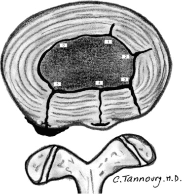
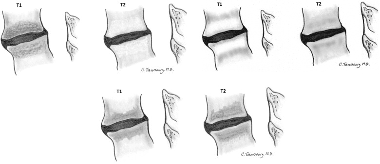
Degrees of disc degeneration have been graded based on gross morphology of midsagittal sections of the lumbar spine (Thompson scheme) [19]; postdiscography CT observations of integrity of the interior of the disc (Dallas classification) (Fig. 9) [42]; MRI observations of vertebral body marrow changes adjacent to the disc (Modic classification) [30], (Fig. 10); and MRI-revealed changes in the nucleus (Pfirrmann classification) [61]. Various modifications of these schemes have been proposed to suit specific clinical and research needs [17, 35, 62, 63].

Figure 9:�Internal disc integrity. The extent of radial fissuring, as visualized on postdiscography CT, graded 0 to 5 by the Modified Dallas Discogram classification, as depicted.
Figure 10:�Reactive vertebral body marrow changes. These bone marrow signal changes adjacent to a degenerated disc on magnetic resonance imaging. T1- and T2-weighted sequences are frequently classified as (Top Left) Modic I, (Top Right) Modic II, or (Bottom) Modic III.

Herniated Disc

The needs of common practices make necessary a diagnostic term that describes disc material beyond the intervertebral disc space. Herniated disc, herniated nucleus pulposus (HNP), ruptured disc, prolapsed disc (used nonspecifically), protruded disc (used nonspecifically), and bulging disc (used nonspecifically) have all been used in the literature in various ways to denote imprecisely defined displacement of disc material beyond the interspace. The absence of clear understanding of the meaning of these terms and the lack of definition of limits that should be placed on an ideal general term have created a great deal of confusion in clinical practice and in attempts to make meaningful comparisons of research studies.

For the general diagnosis of displacement of disc material, the single term that is most commonly used and creates least confusion is ��herniated disc.�� ��Herniated nucleus pulposus�� is inaccurate because materials other than nucleus (cartilage, fragmented apophyseal bone, and fragmented annulus) are common components of displaced disc material [64]. ��Rupture�� casts an image of tearing apart and therefore carries more implication of traumatic etiology than ��herniation,�� which conveys an image of displacement rather than disruption.

Though ��protrusion�� has been used by some authors in a nonspecific general sense to signify any displacement, the term has a more commonly used specific meaning for which it is best reserved. ��Prolapse,�� which has been used as a general term, as synonymous with the specific meaning of protrusion, or to denote inferior migration of extruded disc material, is not frequently used in a way to provide specific meaning and is best regarded as nonstandard, in deference to the more specific terms ��protrusion�� and ��extrusion.��

By exclusion of other terms, and by reasons of simplicity and common usage, ��herniated disc�� is the best general term to denote displacement of disc material. The term is appropriate to denote the general diagnostic category when referring to a specific disc and to be inclusive of various types of displacements when speaking of groups of discs. The term includes discs that may properly be characterized by more specific terms, such as ��protruded disc�� or ��extruded disc.�� The term ��herniated disc,�� as defined in this work, refers to localized displacement of nucleus, cartilage, fragmented apophyseal bone, or fragmented annular tissue beyond the intervertebral disc space. ��Localized�� is defined as less than 25% of the disc circumference. The disc space is defined, craniad and caudad, by the vertebral body end plates and, peripherally, by the edges of the vertebral ring apophyses, exclusive of the osteophyte formation. This definition was deemed more practical, especially for the interpretation of imaging studies, than a pathologic definition requiring identification of disc material forced out of normal position through an annular defect. Displacement of disc material, either through a fracture or defect in the bony end plate or in conjunction with displaced fragments of fractured walls of the vertebral body, may be described as ��herniated�� disc, although such description should accompany description of the fracture so as to avoid confusion with primary herniation of disc material. Displacement of disc materials from one location to another within the interspace, as with intraannular migration of nucleus without displacement beyond the interspace, is not considered herniation.

To be considered ��herniated,�� disc material must be displaced from its normal location and not simply represent an acquired growth beyond the edges of the apophyses, as is the case when connective tissues develop in gaps between osteophytes or when annular tissue is displaced behind one vertebra as an adaptation to subluxation. Herniation, therefore, can only occur in association with disruption of the normal annulus or, as in the case of intravertebral herniation (Schmorl node), a defect in the vertebral body end plate.

Details of the internal architecture of the annulus are most often not visualized by even the best quality MRIs [21]. The distinction of herniation is made by the observation of displacement of disc material beyond the edges of the ring apophysis that is ��focal�� or ��localized,�� meaning less than 25% of the circumference of the disc. The 25% cutoff line is established by way of convention to lend precision to terminology and does not designate etiology, relation to symptoms, or treatment indications.

The terms ��bulge�� or ��bulging�� refer to a generalized extension of disc tissue beyond the edges of the apophyses [65]. Such bulging involves greater than 25% of the circumference of the disc and typically extends a relatively short distance, usually less than 3 mm, beyond the edges of the apophyses (Fig. 3). ��Bulge�� or ��bulging�� describes a morphologic characteristic of various possible causes. Bulging is sometimes a normal variant (usually at L5�S1), can result from an advanced disc degeneration or from a vertebral body remodeling (as consequent to osteoporosis, trauma, or adjacent structure deformity), can occur with ligamentous laxity in response to loading or angular motion, can be an illusion caused by posterior central subligamentous disc protrusion, or can be an illusion from volume averaging (particularly with CT axial images).

Bulging, by definition, is not a herniation. Application of the term ��bulging�� to a disc does not imply any knowledge of etiology, prognosis, or need for treatment or imply the presence of symptoms.

A disc may have, simultaneously, more than one herniation. A disc herniation may be present along with other degenerative changes, fractures, or abnormalities of the disc. The term ��herniated disc�� does not imply any knowledge of etiology, relation to symptoms, prognosis, or need for treatment.

When data are sufficient to make the distinction, a herniated disc may be more specifically characterized as ��protruded�� or ��extruded.�� These distinctions are based on the shape of the displaced material. They do not imply knowledge of the mechanism by which the changes occurred.

Protruded Discs

Disc protrusions are focal or localized abnormalities of the disc margin that involve less than 25% of the disc circumference. A disc is ��protruded�� if the greatest dimension between the edges of the disc material presenting beyond the disc space is less than the distance between the edges of the base of that disc material that extends outside the disc space. The base is defined as the width of the disc material at the outer margin of the disc space of origin, where disc material displaced beyond the disc space is continuous with the disc material within the disc space (Fig. 4). The term ��protrusion�� is only appropriate in describing herniated disc material, as discussed previously.

Extruded Discs

The term ��extruded�� is consistent with the lay language meaning of material forced from one domain to another through an aperture [37, 64]. With reference to a disc, the test of extrusion is the judgment that, in at least one plane, any one distance between the edges of the disc material beyond the disc space is greater than the distance between the edges of the base measured in the same plane or when no continuity exists between the disc material beyond the disc space and that within the disc space (Fig. 5). Extruded disc material that has no continuity with the disc of origin may be characterized as ��sequestrated�� [53, 66] (Fig. 6). A sequestrated disc is a subtype of ��extruded disc�� but, by definition, can never be a ��protruded disc.�� Extruded disc material that is displaced away from the site of extrusion, regardless of continuity with the disc, may be called ��migrated,�� a term that is useful for the interpretation of imaging studies because it is often impossible from images to know if continuity exists.

The aforementioned distinctions between protrusion and extrusion and between contained and uncontained are based on common practice and wide acceptance of the definitions in the original version of this document. Another set of criteria, espoused by some respected practitioners, defines extrusion as uncontained and protrusion as a persistence of containment, regardless of the relative dimensions of the base to displaced portion of disc material. Per these criteria, a disc extrusion can be identified by the presence of a continuous line of low signal intensity surrounding the disc herniation. They state that current advanced imaging permits this basis of distinction and that the presence or absence of containment has more clinical relevance than the morphology of the displaced material [35].

Whether their method will prove superior to the currently recommended method will be determined by future study. The use of the distinction between ��protrusion�� and ��extrusion�� is optional and some observers may prefer to use, in all cases, the more general term ��herniation.�� Further distinctions can often be made regarding containment, continuity, volume, composition, and location of the displaced disc material.

Containment, Continuity, and Migration

Herniated disc material can be ��contained�� or ��uncontained.�� The test of containment is whether the displaced disc tissues are wholly held within intact outer annulus and/or posterior longitudinal ligament fibers. Fluid or any contrast that has been injected into a disc with a ��contained�� herniation would not be expected to leak into the vertebral canal. Although the posterior longitudinal ligament and/or peridural membrane may partially cover the extruded disc tissues, such discs are not considered ��contained�� unless the posterior longitudinal ligament is intact. The technical limitations of currently available noninvasive imaging modalities (CT and MRI) often preclude the distinction of a contained from an uncontained disc herniation. CT-discography does not always allow one to distinguish whether the herniated components of a disc are contained, but only whether there is a communication between the disc space and the vertebral canal.

Displaced disc fragments are sometimes characterized as ��free.�� A ��free fragment�� is synonymous with a ��sequestrated fragment,�� but not synonymous with ��uncontained.�� A disc fragment should be considered ��free�� or ��sequestrated�� only if there is no remaining continuity of the disc material between it and the disc of origin. A disc can be ��uncontained,�� with the loss of integrity of the posterior longitudinal ligament and the outer annulus, but still have continuity between the herniated/displaced disc material and the disc of origin.

The term ��migrated�� disc or fragment refers to the displacement of most of the displaced disc material away from the opening in the annulus through which the material has extruded. Some migrated fragments will be sequestrated, but the term ��migrated�� refers only to position and not to continuity.

The terms ��capsule�� and ��subcapsular�� have been used to refer to containment by an unspecified combination of annulus and ligament. These terms are nonpreferred.

Referring specifically to the posterior longitudinal ligament, some authors have distinguished displaced disc material as ��subligamentous,�� ��extraligamentous,�� ��transligamentous,�� or ��perforated.�� The term ��subligamentous�� is favored as an equivalent to ��contained.��

Volume and Composition of Displaced Material

A scheme to define the degree of canal compromise produced by disc displacement should be practical, objective, reasonably precise, and clinically relevant. A simple scheme that fulfills the criteria uses two-dimensional measurements taken from an axial section at the site of the most severe compromise. Canal compromise of less than one third of the canal at that section is ��mild,�� between one and two-thirds is ��moderate,�� and greater than two-thirds is ��severe.�� The same grading can be applied for foraminal involvement.

Such characterizations of volume describe only the cross-sectional area at one section and do not account for the total volume of displaced material; proximity to, compression, and distortion of neural structures; or other potentially significant features, which the observer may further detail by narrative description.

Composition of the displaced material may be characterized by terms such as nuclear, cartilaginous, bony, calcified, ossified, collagenous, scarred, desiccated, gaseous, or liquefied.

Clinical significance related to the observation of volume and composition depends on the correlation with clinical data and cannot be inferred from morphologic data alone.

Location

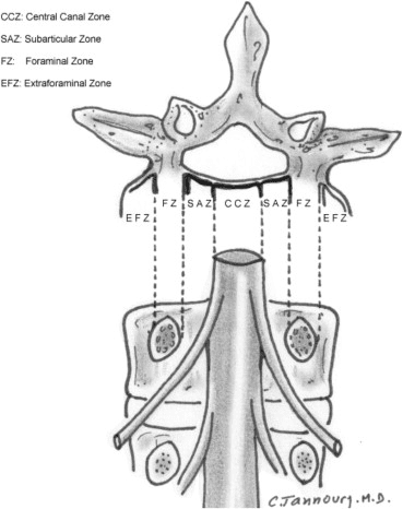
Bonneville proposed a useful and simple alphanumeric system to classify, according to location, the position of disc fragments that have migrated in the horizontal or sagittal plane [6, 13]. Using anatomic boundaries familiar to surgeons, Wiltse proposed another system [14, 67]. Anatomic ��zones�� and ��levels�� are defined using the following landmarks: medial edge of the articular facets; medial, lateral, upper, and lower borders of the pedicles; and coronal and sagittal planes at the center of the disc. On the horizontal (axial) plane, these landmarks determine the boundaries of the central zone, the subarticular zone (lateral recess), the foraminal zone, the extraforaminal zone, and the anterior zone, respectively (Fig. 11). On the sagittal (craniocaudal) plane, they determine the boundaries of the disc level, the infrapedicular level, the pedicular level, and the suprapedicular level, respectively (Fig. 12). The method is not as precise as the drawings depict because borderlines such as the medial edges of facets and the walls of the pedicles are curved, but the method is simple, practical, and in common usage.

Figure 11:�Anatomic zones depicted in axial and coronal projections.
Figure 12: Anatomic levels depicted in sagittal and coronal projections.

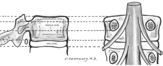
Moving from the central to right lateral in the axial (horizontal) plane, location may be defined as central, right central, right subarticular, right foraminal, or right extraforaminal. The term ��paracentral�� is less precise than defining ��right central�� or ��left central,�� but is useful in describing groups of discs that include both, or when speaking informally, when the side is not significant. For reporting of image observations of a specific disc, ��right central�� or ��left central�� should supersede the use of the term ��paracentral.�� The term ��far lateral�� is sometimes used synonymously with ��extraforaminal.��

In the sagittal plane, location may be defined as discal, infrapedicular, suprapedicular, or pedicular. In the coronal plane, anterior, in relationship to the disc, means ventral to the midcoronal plane of the centrum.

Glossary

Note:�some terms and definitions included in this Glossary are not recommended as preferred terminology but are included to facilitate the interpretation of vernacular and, in some cases, improper use. Preferred definitions are listed first. Nonstandard definitions are placed in brackets, and by consensus of the organizational task forces, should not be used in the manner described. Some terms are also labeled as colloquial, with further designation as to whether they are considered nonpreferred or nonstandard.

Acute disc herniation:�disc herniation of a relatively recent occurrence. Note: paradiscal inflammatory reaction and relatively bright signal of the disc material on T2-weighted images suggest relative acuteness. Such changes may persist for months, however. Thus, absent clinical correlation and/or serial studies, it is not possible to date precisely by imaging when a herniation occurred. An acutely herniated disc material may have brighter signal on T2-weighted MRI sequences than the disc from which the disc material originates [46,�59,�64,�68]. Note that a relatively acute herniation can be superimposed on a previously existing herniation. An acute disc herniation may regress spontaneously without specific treatment. See: chronic disc herniation.

Aging disc:�disc demonstrating any of the various effects of aging on the disc. Loss of water content from the nucleus occurs before MRI changes, followed by the progression of MRI manifested changes consistent with the progressive loss of water content and increase in collagen and aggregating proteoglycans. See Pfirrmann classification.

Annular fissure:�separations between annular fibers, separations of fibers from their vertebral body insertions, or separations of fibers that extend radially, transversely, or concentrically, involving one or many layers of the annular lamellae. Note that the terms ��fissure�� and ��tear�� have often been used synonymously in the past. The term ��tear�� is inappropriate for use in describing imaging findings and should not be used (tear: nonstandard). Neither term suggests injury or implies any knowledge of etiology, neither term implies any relationship to symptoms or that the disc is a likely pain generator, and neither term implies any need for treatment. See also: annular gap, annular rupture, annular tear, concentric fissure, HIZ, radial fissure, transverse fissure.

Annular gap�(nonstandard): focal attenuation (CT) or signal (MRI) abnormality, often triangular in shape, in the posterior aspect of the disc, likely representing widening of a radially directed annular fissure, bilateral annular fissures with an avulsion of the intermediate annular fragment, or an avulsion of a focal zone of macerated annulus.

Annular rupture:�disruption of fibers of the annulus by sudden violent injury. This is a clinical diagnosis; use of the term is inappropriate for a pure imaging description, which instead should focus on a detailed description of the findings. Ruptured annulus is�not�synonymous with ��annular fissure,�� or ��ruptured disc.��

Annular tear,�torn annulus�(nonstandard): see fissure of the annulus and rupture of annulus.

Anterior displacement:�displacement of disc tissues beyond the disc space into the anterior zone.

Anterior zone:�peridiscal zone that is anterior to the midcoronal plane of the vertebral body.

Anulus, annulus (abbreviated form of annulus fibrosus):�multilaminated fibrous tissue forming the periphery of each disc space, attaching, craniad and caudad, to end plate cartilage and a ring apophyseal bone and blending centrally with the nucleus pulposus. Note: either anulus or annulus is correct spelling. Nomina Anatomica uses both forms, whereas Terminologia Anatomica states �� anulus fibrosus�� [22]. Fibrosus has no correct alternative spelling; fibrosis has a different meaning and is incorrect in this context.

Asymmetric bulge:�presence of more than 25% of the outer annulus beyond the perimeter of the adjacent vertebrae, more evident in one section of the periphery of the disc than another, but not sufficiently focal to be characterized as a protrusion. Note: asymmetric disc bulging is a morphologic observation that may have various causes and does not imply etiology or association with symptoms. See bulge.

Balloon disc (colloquial, nonstandard):�diffuse apparent enlargement of the disc in superior-inferior extent because of bowing of the vertebral end plates due to weakening of the bone as in severe osteoporosis.

Base (of displaced disc):�the cross-sectional area of the disc material at the outer margin of the disc space of origin, where disc material beyond the disc space is continuous with disc material within the disc space. In the craniocaudal direction, the length of the base cannot exceed, by definition, the height of the intervertebral space. On axial imaging, base refers to the width at the outer margin of the disc space, of the origin of any disc material extending beyond the disc space.

Black disc�(colloquial, nonstandard): see dark disc.

Bulging disc, bulge (noun [n]), bulge (verb [v])

  1. A disc in which the contour of the outer annulus extends, or appears to extend, in the horizontal (axial) plane beyond the edges of the disc space, usually greater than 25% (90�) of the circumference of the disc and usually less than 3 mm beyond the edges of the vertebral body apophysis.
  2. (Nonstandard) A disc in which the outer margin extends over a broad base beyond the edges of the disc space.
  3. (Nonstandard) Mild, diffuse, smooth displacement of disc.
  4. (Nonstandard) Any disc displacement at the discal level.

Note:�bulging is an observation of the contour of the outer disc and is not a specific diagnosis. Bulging has been variously ascribed to redundancy of the annulus, secondary to the loss of disc space height, ligamentous laxity, response to loading or angular motion, remodeling in response to adjacent pathology, unrecognized and atypical herniation, and illusion from volume averaging on CT axial images. Mild symmetric posterior disc bulging may be a normal finding at L5�S1. Bulging may or may not represent pathologic change, physiologic variant, or normalcy. Bulging is not a form of herniation; discs known to be herniated should be diagnosed as herniation or, when appropriate, as specific types of herniation. See: herniated disc, protruded disc, extruded disc.

Calcified disc:�calcification within the disc space, not inclusive of osteophytes at the periphery of the disc space.

Cavitation:�spaces, cysts, clefts, or cavities formed within the nucleus and inner annulus from disc degeneration.

See vacuum disc.

Central zone:�zone within the vertebral canal between sagittal planes through the medial edges of each facet. Note: the center of the central zone is a sagittal plane through the center of the vertebral body. The zones to either side of the center plane are�right central�and�left central, which are preferred terms when the side is known, as when reporting imaging results of a specific disc. When the side is unspecified, or grouped with both right and left represented, the term�paracentral�is appropriate.

Chronic disc herniation:�a clinical distinction that a disc herniation is of long duration. There are no universally accepted definitions of the intervals that distinguish between acute, subacute, and chronic disc herniations. Serial MRIs revealing disc herniations that are unchanged in appearance over time may be characterized as chronic. Disc herniations associated with calcification or gas on CT may be suggested as being chronic. Even so, the presence of calcification or gas does not rule out an acutely herniated disc. Note that an acute disc herniation may be superimposed on a chronic disc herniation. Magnetic resonance imaging signal characteristics may, on rare occasion, allow differentiation of acute and chronic disc herniations [16,�59,�64]. In such cases, acutely herniated disc material may appear brighter than the disc of origin on T2-weighted sequences [46,�59,�61]. Also, see disc-osteophyte complex.

Claw osteophyte:�bony outgrowth arising very close to the disc margin, from the vertebral body apophysis, directed, with a sweeping configuration, toward the corresponding part of the vertebral body opposite the disc.

Collagenized disc or nucleus:�a disc in which the mucopolysaccharide of the nucleus has been replaced by fibrous tissue.

Communicating disc, communication (n), communicate (v)�(nonstandard): communication refers to interruption in the periphery of the disc annulus, permitting free passage of fluid injected within the disc to the exterior of the disc, as may be observed during discography. Not synonymous with ��uncontained.�� See ��contained disc�� and ��uncontained disc.��

Concentric fissure:�fissure of the annulus characterized by separation of annular fibers in a plane roughly parallel to the curve of the periphery of the disc, creating fluid-filled spaces between adjacent annular lamellae. See: radial fissures, transverse fissures, HIZ.

Contained herniation, containment (n), contain (v)

  1. Displaced disc tissue existing wholly within an outer perimeter of uninterrupted outer annulus or posterior longitudinal ligament.
  2. (Nonstandard) A disc with its contents mostly, but not wholly, within annulus or capsule.
  3. (Nonstandard) A disc with displaced elements contained within any investiture of the vertebral canal.

A disc that is less than wholly contained by annulus, but under a distinct posterior longitudinal ligament, is contained. Designation as ��contained�� or ��uncontained�� defines the integrity of the ligamentous structures surrounding the disc, a distinction that is often but not always possible by advanced imaging. On CT and MRI scans, contained herniations typically have a smooth margin, whereas uncontained herniations most often have irregular margins because the outer annulus and the posterior longitudinal ligament have been penetrated by the disc material [35,�37]. CT-discography also does not always allow one to distinguish whether the herniated components of a disc are contained, but only whether there is communication between the disc space and the vertebral canal.

Continuity:�connection of displaced disc tissue by a bridge of disc tissue, however thin, to tissue within the disc of origin.

Dallas classification�(of postdiscography imaging): commonly used grading system for the degree of annular fissuring seen on CT imaging of discs after discography. Dallas Grade 0 is normal; Grade 1: leakage of contrast into the inner one-third of the annulus; Grade 2: leakage of contrast into the inner two-thirds of the annulus; Grade 3: leakage through the entire thickness of the annulus; Grade 4: contrast extends circumferentially; Grade 5: contrast extravasates into the epidural space (See discogram, discography).

Dark disc�(colloquial, nonstandard): disc with nucleus showing decreased signal intensity on T2-weighted images (dark), usually because of desiccation of the nucleus secondary to degeneration. Also: black disc (colloquial, nonstandard). See: disc degeneration, Pfirrmann classification.

Degenerated disc, degeneration (n), degenerate (v)

  1. Changes in a disc characterized to varying degrees by one or more of the following: desiccation, cleft formation, fibrosis, and gaseous degradation of the nucleus; mucinous degradation, fissuring, and loss of integrity of the annulus; defects in and/or sclerosis of the end plates; and osteophytes at the vertebral apophyses.
  2. Imaging manifestation of such changes, including [35]�standard roentgenographic findings, such as disc space narrowing and peridiscal osteophytes, MRI disc findings (see Pfirrmann classification [61]), CT disc findings (see discogram/discography and Dallas classification [42]), and/or MRI findings of vertebral end plate and marrow reactive changes adjacent to a disc (see Modic classification [38]).

Degenerative disc disease�(nonstandard term when used as an imaging description): a condition characterized by manifestations of disc degeneration and symptoms thought to be related to those of degenerative changes. Note: causal connections between degenerative changes and symptoms are often difficult clinical distinctions. The term ��degenerative disc disease�� carries implications of illness that may not be appropriate if the only or primary indicators of illness are from imaging studies, and thus this term should not be used when describing imaging findings. The preferred term for description of imaging manifestations is ��degenerated disc�� or ��disc degeneration,�� rather than ��degenerative disc disease.��

Delamination:�separation of circumferential annular fibers along the planes parallel to the periphery of the disc, characterizing a concentric fissure of the annulus.

Desiccated disc

  1. Disc with reduced water content, usually primarily of nuclear tissues.
  2. Imaging manifestations of reduced water content of the disc, such as decreased (dark) signal intensity on T2-weighted images, or of apparent reduced water content, as from alterations in the concentration of hydrophilic glycosaminoglycans. See also: dark disc (colloquial, nonstandard).

Disc (disk):�complex structure composed of nucleus pulposus, annulus fibrosus, cartilaginous end plates, and vertebral body ring apophyseal attachments of annulus. Note: most English language publications use the spelling ��disc�� more often than ��disk�� [1,�20,�22,�69,�70]. Nomina Anatomica designates the structures as ��disci intervertebrales�� and Terminologia Anatomica as ��discus intervertebralis/intervertebral disc�� [22,�70]. (See ��disc level�� for naming and numbering of a particular disc).

Disc height:�The distance between the planes of the end plates of the vertebral bodies craniad and caudad to the disc. Disc height should be measured at the center of the disc, not at the periphery. If measured at the posterior or anterior margin of the disc on a sagittal image of the spine, this should be clearly specified as such.

Disc level:�Level of the disc and vertebral canal between axial planes through the bony end plates of the vertebrae craniad and caudad to the disc being described.

  1. A particular disc is best named by naming the region of the spine and the vertebra above and below it; for example, the disc between the fourth and fifth lumbar vertebral bodies is named ��lumbar 4�5,�� commonly abbreviated as L4�L5, and the disc between the fifth lumbar vertebral body and the first sacral vertebral body is called ��lumbosacral disc�� or ��L5�S1.�� Common anomalies include patients with six lumbar vertebrae or transitional vertebrae at the lumbosacral junction that require, for clarity, narrative explanation of the naming of the discs.
  2. (Nonstandard) A disc is sometimes labeled by the vertebral body above it; for example, the disc between L4 and L5 may be labeled ��the L4 disc��.
  3. Note: ��a motion segment,�� numbered in the same way, is a functional unit of the spine, comprising the vertebral body above and below, the disc, the facet joints, and the connecting soft tissues and is most often referenced with regard to the stability of the spine.

Disc of origin:�disc from which a displaced fragment originated. Synonym: parent disc. Note: since displaced fragments often contain tissues other than nucleus, disc of origin is preferred to nucleus of origin. Parent disc is synonymous, but more colloquial and nonpreferred.

Disc space:�space limited, craniad and caudad, by the end plates of the vertebrae and peripherally by the edges of the vertebral body ring apophyses, exclusive of osteophytes. Synonym: intervertebral disc space. See ��disc�� level for naming and numbering of discs.

Discogenic vertebral sclerosis:�increased bone density and calcification adjacent to the end plates of the vertebrae, craniad and caudad, to a degenerated disc, sometimes associated with intervertebral osteochondrosis. Manifested on MRI as Modic Type�III.

Discogram, discography:�a diagnostic procedure in which contrast material is injected into the nucleus of the disc with radiographic guidance and observation, often followed by CT/discogram. The procedure is often accompanied by pressure measurements and assessment of pain response (provocative discography). The degree of annular fissuring identified by discography may be defined by the Dallas classification and its modifications (See Dallas classification).

Disc-osteophyte complex:�intervertebral disc displacement, whether bulge, protrusion, or extrusion, associated with calcific ridges or ossification. Sometimes called a hard disc or chronic disc herniation (nonpreferred). Distinction should be made between ��spondylotic disc herniation,�� or ��calcified disc herniation�� (nonpreferred), the remnants of an old disc herniation; and ��spondylotic bulging disc,�� a broad-based bony ridge presumably related to chronic bulging disc.

Displaced disc�(nonstandard): a disc in which disc material is beyond the outer edges of the vertebral body ring apophyses (exclusive of osteophytes) of the craniad and caudad vertebrae, or, as in the case of intravertebral herniation, has penetrated through the vertebral body end plate.

Note: displaced disc is a general term that does not imply knowledge of the underlying pathology, cause, relationship to symptoms, or need for treatment. The term includes, but is not limited to, disc herniation and disc migration. See: herniated disc, migrated disc.

Epidural membrane:�See peridural membrane.

Extraforaminal zone:�the peridiscal zone beyond the sagittal plane of the lateral edges of the pedicles, having no well-defined lateral border, but definitely posterior to the anterior zone. Synonym: ��far lateral zone,�� also ��far-out zone�� (nonstandard).

Extraligamentous:�posterior or lateral to the posterior longitudinal ligament. Note: extraligamentous disc refers to displaced disc tissue that is located posterior or lateral to the posterior longitudinal ligament. If the disc has extruded through the posterior longitudinal ligament, it is sometimes called ��transligamentous�� or ��perforated�� and if through the peridural membrane, it is sometimes refined to ��transmembranous.��

Extruded disc, extrusion (n), extrude (v):�a herniated disc in which, in at least one plane, any one distance between the edges of the disc material beyond the disc space is greater than the distance between the edges of the base of the disc material beyond the disc space in the same plane or when no continuity exists between the disc material beyond the disc space and that within the disc space. Note: the preferred definition is consistent with the common image of extrusion, as an expulsion of material from a container through and beyond an aperture. Displacement beyond the outer annulus of the disc material with any distance between its edges greater than the distance between the edges of the base distinguishes extrusion from protrusion. Distinguishing extrusion from protrusion by imaging is best done by measuring the edges of the displaced material and the remaining continuity with the disc of origin, whereas relationship of the displaced portion to the aperture through which it has passed is more readily observed surgically. Characteristics of protrusion and extrusion may coexist, in which case the disc should be subcategorized as extruded. Extruded discs in which all continuity with the disc of origin is lost may be further characterized as ��sequestrated.�� Disc material displaced away from the site of extrusion may be characterized as ��migrated.�� See: herniated disc, migrated disc, protruded disc.

Note: An alternative scheme is espoused by some respected radiologists who believe it has better clinical application. This scheme defines extruded disc as synonymous with �uncontained disc� and does not use comparative measurements of the base versus the displaced material. Per this definition, a disc extrusion can be identified by the presence of a continuous line of low signal intensity surrounding the disc herniation. Future study will further determine the validity of this alternative definition. See: contained disc.

Far lateral zone:�the peridiscal zone beyond the sagittal plane of the lateral edge of the pedicle, having no well defined lateral border, but definitely posterior to the anterior zone. Synonym: ��extraforaminal zone.��

Fissure of annulus:�see annular fissure.

Foraminal zone:�the zone between planes passing through the medial and lateral edges of the pedicles. Note: the foraminal zone is sometimes called the ��pedicle zone�� (nonstandard), which can be confusing because pedicle zone might also refer to measurements in the sagittal plane between the upper and lower surfaces of a given pedicle that is properly called the ��pedicle level.�� The foraminal zone is also sometimes called the ��lateral zone�� (nonstandard), which can be confusing because the ��lateral zone�� can be confused with ��lateral recess�� (subarticular zone) and can also mean extraforaminal zone or an area including both the foraminal and extraforaminal zones.

Free fragment

  1. A fragment of disc that has separated from the disc of origin and has no continuous bridge of disc tissue with disc tissue within the disc of origin. Synonym: sequestrated disc.
  2. (Nonstandard) A fragment that is not contained within the outer perimeter of the annulus.
  3. (Nonstandard) A fragment that is not contained within the annulus, posterior longitudinal ligament, or peridural membrane.

Note: ��sequestrated disc�� and ��free fragment�� are virtually synonymous. When referring to the condition of the disc, categorization as extruded with subcategorization as sequestrated is preferred, whereas when referring specifically to the fragment, free fragment is preferred.

Gap of annulus:�see annular gap.

Hard disc (colloquial):�disc displacement in which the displaced portion has undergone calcification or ossification and may be intimately associated with apophyseal osteophytes. Note: the term ��hard disc�� is most often used in reference to the cervical spine to distinguish chronic hypertrophic and reactive changes at the periphery of the disc from the more acute extrusion of soft, predominantly nuclear tissue. See: chronic disc herniation, disc-osteophyte complex.

Herniated disc, herniation (n), herniated (v):�localized or focal displacement of disc material beyond the normal margin of the intervertebral disc space. Note: ��localized�� or ��focal�� means, by way of convention, less than 25% (90�) of the circumference of the disc.

Herniated disc material may include nucleus pulposus, cartilage, fragmented apophyseal bone, or annulus fibrosus tissue. The normal margins of the intervertebral disc space are defined, craniad and caudad, by the vertebral body end plates and peripherally by the edges of the vertebral body ring apophyses, exclusive of osteophytic formations. Herniated disc generally refers to displacement of disc tissues through a disruption in the annulus, the exception being intravertebral herniations (Schmorl nodes) in which the displacement is through the vertebral end plate. Herniated discs may be further subcategorized as protruded or extruded. Herniated disc is sometimes referred to as HNP, but the term ��herniated disc�� is preferred because displaced disc tissues often include cartilage, bone fragments, or annular tissues. The terms ��prolapse�� and ��rupture�� when referring to disc herniations are nonstandard and their use should be discontinued. Note: ��herniated disc�� is a term that does not imply knowledge of the underlying pathology, cause, relationship to symptoms, or need for treatment.

Herniated nucleus pulposus�(HNP, nonpreferred): see herniated disc.

High intensity zone (HIZ):�area of high intensity on T2-weighted MRIs of the disc, located commonly in the outer annulus. Note: HIZs within the posterior annular substance may indicate the presence of an annular fissure within the annulus, but these terms are not synonymous. An HIZ itself may represent the actual annular fissure or alternatively, may represent vascularized fibrous tissue (granulation tissue) within the substance of the disc in an area adjacent to a fissure. The visualization of an HIZ does not imply a traumatic etiology or that the disc is a source of pain.

Infrapedicular level:�the level between the axial planes of the inferior edges of the pedicles craniad to the disc in question and the inferior end plate of the vertebral body above the disc in question. Synonym: superior vertebral notch.

Internal disc disruption:�disorganization of structures within the disc. See intraannular displacement

Interspace:�see disc space.

Intervertebral chondrosis:�see intervertebral osteochondrosis.

Intervertebral disc:�see disc.

Intervertebral disc space:�see disc space.

Intervertebral osteochondrosis:�degenerative process of the disc and vertebral body end plates that is characterized by disc space narrowing, vacuum phenomenon, and vertebral body reactive changes. Synonym: osteochondrosis (nonstandard).

Intraannular displacement:�displacement of central, predominantly nuclear, tissue to a more peripheral site within the disc space, usually into a fissure in the annulus. Synonym: (nonstandard) intraannular herniation, intradiscal herniation. Note: intraannular displacement is distinguished from disc herniation, that is, herniation of disc refers to displacement of disc tissues beyond the disc space. Intraannular displacement is a form of internal disruption. When referring to intraannular displacement, it is best not to use the term ��herniation�� to avoid confusion with disc herniation.

Intraannular herniation (nonstandard):�see intraannular displacement.

Intradiscal herniation (nonstandard):�see intraannular displacement.

Intradural herniation:�disc material that has penetrated the dura so that it lies in an intradural extramedullary location.

Intravertebral herniation:�a disc displacement in which a portion of the disc projects through the vertebral end plate into the centrum of the vertebral body. Synonym: Schmorl node.

Lateral recess:�that portion of the subarticular zone that is medial to the medial border of the pedicle. It refers to the entire cephalad-caudad region that exists medial to the pedicle, where the same numbered thoracic or lumbar nerve root travels caudally before exiting the nerve root foramen under the caudal margin of the pedicle. It does not refer to the nerve root foramen itself. See also subarticular zone.

Lateral zone�(nonstandard): see foraminal zone.

Leaking disc�(nonstandard): see communicating disc.

Limbus vertebra:�separation of a segment of vertebral ring apophysis. Note: limbus vertebra may be a developmental abnormality caused by failure of integration of the ossifying apophysis to the vertebral body; a chronic herniation (extrusion) of the disc into the vertebral body at the junction of the fusing apophyseal ring, with separation of a portion of the ring with bony displacement; or a fracture through the apophyseal ring associated with intrabody disc herniation. This occurs in children before the apophyseal ring fuses to the vertebral body. In adults, a limbus vertebra should not be confused with an acute fracture. A limbus vertebra does not imply that there has been an injury to the disc or the adjacent apophyseal end plate.

Marginal osteophyte:�osteophyte that protrudes from and beyond the outer perimeter of the vertebral end plate apophysis.

Marrow changes (of vertebral body):�see Modic classification.

Migrated disc, migration (n), migrate (v)

  • 1.Herniated disc in which a portion of the extruded disc material is displaced away from the fissure in the outer annulus through which it has extruded in either sagittal or axial plane.
  • 2.(Nonstandard) A herniated disc with a free fragment or sequestrum beyond the disc level.

Note: migration refers to the position of the displaced disc material, rather than to its continuity with disc tissue within the disc of origin; therefore, it is not synonymous with sequestration.

Modic classification (Type I, II, and III)�[30]: a classification of degenerative changes involving the vertebral end plates and adjacent vertebral bodies associated with disc inflammation and degenerative disc disease, as seen on MRIs. Type I refers to decreased signal intensity on T1-weighted spin echo images and increased signal intensity on T2-weighted images, representing penetration of the end plate by fibrovascular tissue, inflammatory changes, and perhaps edema. Type I changes may be chronic or acute. Type II refers to increased signal intensity on T1-weighted images and isointense or increased signal intensity on T2-weighted images, indicating replacement of normal bone marrow by fat. Type III refers to decreased signal intensity on both T1-and T2-weighted images, indicating reactive osteosclerosis (See: discogenic vertebral sclerosis).

Motion segment:�the functional unit of the spine. See disc level.

Nonmarginal osteophyte:�an osteophyte that occurs at sites other than the vertebral end plate apophysis. See: marginal osteophyte.

Normal disc:�a fully and normally developed disc with no changes attributable to trauma, disease, degeneration, or aging. Note: many congenital and developmental variations may be clinically normal; that is, they are not associated with symptoms, and certain adaptive changes in the disc may be normal considering adjacent pathology; however, classification and reporting for medical purposes is best served if such discs are not considered normal. Note, however, that a disc finding considered not normal does not necessarily imply a cause for clinical signs or symtomatology; the description of any variation of the disc is independent of clinical judgment regarding what is normal for a given patient.

Nucleus of origin (nonpreferred):�the central, nuclear portion of the disc of reference, usually used to reference the disc from which the tissue has been displaced. Note: since displaced fragments often contain tissues other than the nucleus, disc of origin is preferred to nucleus of origin. Synonym: disc of origin (preferred), parent nucleus (nonpreferred).

Osteochondrosis:�see intervertebral osteochondrosis.

Osteophyte:�focal hypertrophy of the bone surface and/or ossification of the soft tissue attachment to the bone.

Paracentral:�in the right or left central zone of the vertebral canal. See central zone. Note: the terms ��right central�� or ��left central�� are preferable when speaking of a single site when the side can be specified, as when reporting the findings of imaging procedures. ��Paracentral�� is appropriate if the side is not significant or when speaking of mixed sites.

Parent disc�(nonpreferred): see disc of origin.

Parent nucleus�(nonpreferred): see nucleus of origin, disc of origin.

Pedicular level:�the space between the axial planes through the upper and lower edges of the pedicle. Note: the pedicular level may be further designated with reference to the disc in question as ��pedicular level above�� or ��pedicular level below�� the disc in question.

Perforated (nonstandard):�see transligamentous.

Peridural membrane:�a delicate, translucent membrane that attaches to the undersurface of the deep layer of the posterior longitudinal ligament, and extends laterally and posteriorly, encircling the bony spinal canal outside the dura. The veins of Batson plexus lie on the dorsal surface of the peridural membrane and pierce it ventrally. Synonym: lateral membrane, epidural membrane.

Pfirrmann classification:�a grading system for the severity of degenerative changes within the nucleus of the intervertebral disc. A Pfirrmann Grade I disc has a uniform high signal in the nucleus on T2-weighted MRI; Grade II shows a central horizontal line of low signal intensity on sagittal images; Grade III shows high intensity in the central part of the nucleus with lower intensity in the peripheral regions of the nucleus; Grade IV shows low signal intensity centrally and blurring of the distinction between nucleus and annulus; and Grade V shows homogeneous low signal with no distinction between nucleus and annulus.[61]

Prolapsed disc, prolapse (n, v)�(nonstandard): the term is variously used to refer to herniated discs. Its use is not standardized and the term does not add to the precision of disc description, so is regarded as nonstandard in deference to ��protrusion�� or ��extrusion.��

Protruded disc, protrusion (n), protrude (v):�1. One of the two subcategories of a ��herniated disc�� (the other being an ��extruded disc��) in which disc tissue extends beyond the margin of the disc space, involving less than 25% of the circumference of the disc margin as viewed in the axial plane. The test of protrusion is that there must be localized (less than 25% of the circumference of the disc) displacement of disc tissue and the distance between the corresponding edges of the displaced portion must not be greater than the distance between the edges of the base of the displaced disc material at the disc space of origin (See base of displaced disc). While sometimes used as a general term in the way herniation is defined, the use of the term ��protrusion�� is best reserved for subcategorization of herniation meeting the above criteria. 2. (nonstandard) Any or unspecified type of disc herniation.

Radial fissure:�disruption of annular fibers extending from the nucleus outward toward the periphery of the annulus, usually in the craniad-caudad (vertical) plane, although, at times, with axial horizontal (transverse) components. ��Fissure�� is the preferred term to the nonstandard term ��tear.�� Neither term implies knowledge of injury or other etiology. Note: Occasionally, a radial fissure extends in the transverse plane to include an avulsion of the outer layers of annulus from the apophyseal ring. See concentric fissures, transverse fissures.

Rim lesion (nonstandard): See limbus vertebra.

Rupture of annulus, ruptured annulus:�see annular rupture.

Ruptured disc, rupture�(nonstandard): a herniated disc. The term ��ruptured disc�� is an improper synonym for herniated disc, not to be confused with violent disruption of the annulus related to injury. Its use should be discontinued.

Schmorl node:�see intravertebral herniation.

Sequestrated disc, sequestration (n), sequestrate (v); (variant: sequestered disc):�an extruded disc in which a portion of the disc tissue is displaced beyond the outer annulus and maintains no connection by disc tissue with the disc of origin. Note: an extruded disc may be subcategorized as ��sequestrated�� if no disc tissue bridges the displaced portion and the tissues of the disc of origin. If even a tenuous connection by disc tissue remains between a displaced fragment and disc of origin, the disc is not sequestrated. If a displaced fragment has no connection with the disc of origin, but is contained within peridural membrane or under a portion of posterior longitudinal ligament that is not intimately bound with the annulus of origin, the disc is considered sequestrated. Sequestrated and sequestered are used interchangeably. Note: ��sequestrated disc�� and ��free fragment�� are virtually synonymous. See: free fragment. When referring to the condition of the disc, categorization as extruded with subcategorization as sequestered is preferred, whereas when referring specifically to the fragment, free fragment is preferred. See sequestrum.

Sequestrum (nonpreferred):�refers to disc tissue that has displaced from the disc space of origin and lacks any continuity with disc material within the disc space of origin. Synonym: free fragment (preferred). See sequestrated disc. Note: ��sequestrum�� (nonpreferred) refers to the isolated free fragment itself, whereas sequestrated disc defines the condition of the disc.

Spondylitis:�inflammatory disease of the spine, other than degenerative disease. Note: spondylitis usually refers to noninfectious inflammatory spondyloarthropathies.

Spondylosis:�1. Common nonspecific term used to describe effects generally ascribed to degenerative changes in the spine, particularly those involving hypertrophic changes to the apophyseal end plates and zygapophyseal joints. 2. (nonstandard) Spondylosis deformans, for which spondylosis is a shortened form.

Spondylosis deformans:�degenerative process of the spine involving the annulus fibrosus and vertebral body apophysis, characterized by anterior and lateral marginal osteophytes arising from the vertebral body apophyses, while the intervertebral disc height is normal or only slightly decreased. See degeneration, spondylosis.

Subarticular zone:�the zone, within the vertebral canal, sagittally between the plane of the medial edges of the pedicles and the plane of the medial edges of the facets and coronally between the planes of the posterior surfaces of the vertebral bodies and the anterior surfaces of the superior facets. Note: the subarticular zone cannot be precisely delineated in two-dimensional depictions because the structures that define the planes of the zone are irregular. The lateral recess is that portion of the subarticular zone defined by the medial wall of the pedicle, where the same numbered nerve root traverses before turning under the inferior wall of the pedicle into the foramen.

Subligamentous:�beneath the posterior longitudinal ligament. Note: although the distinction between outer annulus and posterior longitudinal ligament may not always be identifiable, subligamentous has meaning distinct from subannular when the distinction can be made. When the distinction cannot be made, subligamentous is appropriate. Subligamentous contrasts to extraligamentous, transligamentous, or perforated. See extraligamentous, transligamentous.

Submembranous:�enclosed within the peridural membrane. Note: with reference to the displaced disc material, characterization of a herniation as submembranous usually infers that the displaced portion is extruded beyond annulus and posterior longitudinal ligament so that only the peridural membrane invests it.

Suprapedicular level:�the level within the vertebral canal between the axial planes of the superior end plate of the vertebra caudad to the disc space in question and the superior margin of the pedicle of that vertebra. Synonym: inferior vertebral notch.

Syndesmophytes:�thin and vertically oriented bony outgrowths extending from one vertebral body to the next and representing ossification within the outer portion of the annulus fibrosus.

Tear of annulus, torn annulus�(nonstandard): see annular tear.

Thompson classification:�a five-point grading scale of degenerative changes in the human intervertebral disc, from 0 (normal) to 5 (severe degeneration), based on gross pathologic morphology of midsagittal sections of the lumbar spine.

Traction osteophytes:�bony outgrowth arising from the vertebral body apophysis, 2 to 3 mm above or below the edge of the intervertebral disc, projecting in a horizontal direction.

Transligamentous:�displacement, usually extrusion, of disc material through the posterior longitudinal ligament. Synonym: (nonstandard) (perforated). See also extraligamentous, transmembranous.

Transmembranous:�displacement of extruded disc material through the peridural membrane.

Transverse fissure:�fissure of the annulus in the axial (horizontal) plane. When referring to a large fissure in the axial plane, the term is synonymous with a horizontally oriented radial fissure. Often ��transverse fissure�� refers to a more limited, peripheral separation of annular fibers including attachments to the apophysis. These more narrowly defined peripheral fissures may contain gas visible on radiographs or CT images and may represent early manifestations of spondylosis deformans. See annular fissure, concentric fissure, radial fissure.

Uncontained disc:�displaced disc material that is not contained by the outer annulus and/or posterior longitudinal ligament. See discussion under contained disc.

Vacuum disc:�a disc with imaging findings characteristic of gas (predominantly nitrogen) in the disc space, usually a manifestation of disc degeneration.

Vertebral body marrow changes:�reactive vertebral body signal changes associated with disc inflammation and disc degeneration, as seen on MRIs. See Modic classification.

Vertebral notch (inferior):�incisura of the upper surface of the pedicle corresponding to the lower part of the foramen (suprapedicular level).

Vertebral notch (superior):�incisura of the under surface of the pedicle corresponding to the upper part of the foramen (infrapedicular level).

Supplementary Appendix

Appendix

A herniated disc most commonly develops as a result of age-related wear and tear or degeneration on the spine. In children and young adults, the intervertebral discs have a much higher water content. As we age, however, the water content of the intervertebral discs decreases and these begin to shrink while the spaces between the vertebra gets narrower, ultimately turning less flexible and becoming more prone to disc herniation. Proper diagnosis and treatment are essential to avoid further symptoms of low back pain. The scope of our information is limited to chiropractic as well as to spinal injuries and conditions. To discuss the subject matter, please feel free to ask Dr. Jimenez or contact us at�915-850-0900�.

Curated by Dr. Alex Jimenez

References

  1. Fardon, D.F. and Milette, P.C.�Nomenclature and classification of lumbar disc pathology: recommendations of the combined task forces of the North American Spine Society, the American Society of Spine Radiology and the American Society of Neuroradiology.�Spine.�2001;�26:�E93�E113
  2. Stadnik, T.W., Lee, R.R., Coen, H.L. et al.�Annular tear and disk herniation: prevalence and contrast enhancement on MR images in the absence of low back pain or sciatica.�Radiology.�1998;�206:�49�55
  3. Mink, J.H.�Terminology of lumbar spine disorders, the problem… and a solution.�California Managed Imaging Medical Group Publication,�Burlingame, CA;�1993
  4. Murtagh, F.R.�The importance of being Earnest-about disk nomenclature.�Am J Neuroradiol.�2007;28:�1�2
  5. in:�E.J. Nordby, M.D. Brown, E.D. Dawson, (Eds.)�A glossary on spinal terminology.�American Academy of Orthopaedic Surgeons,�Chicago;�1985:�31�32
  6. Bonneville, J.F. and Dietemann, J.L.�L�imagerie dans les sciatiques.�Rev Prat (Paris).�1992;�42:�554�566
  7. Brant-Zawadzki, M.N. and Jensen, M.C.�Imaging corner: spinal nomenclature. Inter- and intra-observer variability in interpretation of lumbar disc abnormalities: a comparison of two nomenclatures.�Spine.�1995;�20:�388�390
  8. Breton, G.�Is that a bulging disc, a small herniation, or a moderate protrusion?.�Can Assoc Radiol J.�1991;�42:�318
  9. Fardon, D.F., Herzog, R.J., and Mink, J.H.�Nomenclature of lumbar disc disorders.�in:�S.R. Garfin, A.R. Vaccaro (Eds.)�Orthopaedic knowledge update: spine.�American Academy of Orthopaedic Surgeons,Rosemont, IL;�1997:�A3�A14
  10. Milette, P.C.�The proper terminology for reporting lumbar intervertebral disc disorders.�Am J Neuroradiol.�1997;�18:�1859�1866
  11. Fardon DF, White AH, Wiesel S. Diagnostic terms and conservative treatments favored for lumbar disorders by spine surgeons in North America. Presented at the first annual meeting, North American Spine Society, Lake George, New York,�1986.
  12. Arana, E., Royuela, A., Kovacs, F.M. et al.�Lumbar spine: agreement in�the interpretation of 1.5T MR images by using the Nordic Modic�consensus group classification form.�Radiology.�2010;�254:�809�817
  13. Bonneville, J.F.�Plaidoyer pour une classification par l�image des hernies discales lombaires: la carte-image.�Rev Im Med.�1990;�2:�557�560
  14. Fardon, D.F., Pinkerton, S., Balderston, R. et al.�Terms used for diagnosis by English speaking spine surgeons.�Spine.�1993;�18:�1�4
  15. Farfan, H.F., Huberdeau, R.M., and Dubow, H.I.�Lumbar intervertebral disc degeneration: the influence of geometrical features on the pattern of disc degeneration: a post-mortem study.�J Bone Joint Surg [Am].�1972;�54:�492�510
  16. Milette, P.C., Fontaine, S., Lepanto, L. et al.�Differentiating lumbar disc protrusions, disc bulges, and discs with normal contour but abnormal signal intensity.�Spine.�1999;�24:�44�53
  17. Milette, P.C., Melancon, D., Dupuis, P. et al.�A simplified terminology for abnormalities of the lumbar disc.�Can Assoc Radiol J.�1991;�42:�319�325
  18. Taveras, J.M.�Herniated intervertebral disk. A plea for a more uniform terminology.�Am J Neuroradiol.�1989;�10:�1283�1284
  19. Thompson, J.P., Pearce, R.H., Schechter, M.T. et al.�Preliminary evaluation of a scheme for grading the gross morphology of the human intervertebral disc.�Spine.�1990;�15:�411�415
  20. Fardon, D.F., Balderston, R.A., Garfin, S.R. et al.�Disorders of the spine, a coding system for diagnoses.�Hanley and Belfus,�Philadelphia;�1991:�20�22
  21. Herzog, R.J.�The radiologic assessment for a lumbar disc herniation.�Spine.�1996;�21:�19S�38S
  22. International anatomical nomenclature committee approved by Eleventh International Congress of anatomists. Nomina anatomica.�5th ed.�Waverly Press,�Baltimore, MD;�1983:�A23
  23. Jarvik, J.G., Haynor, D.R., Koepsell, T.D. et al.�Interreader reliability for a new classification of lumbar disc abnormalities.�Acad Radiol.�1996;�3:�537�544
  24. Ketler, A. and Wilke, H.J.�Review of existing grading systems for cervical or lumbar disc and facet joint degeneration.�(with Erratum note in Eur Spine J 15(6); 729)Eur Spine J.�2006;�15:�705�718
  25. Kieffer, S.A., Stadlan, E.M., Mohandas, A., and Peterson, H.O.�Discographic-anatomical correlation of developmental changes with age in the intervertebral disc.�Acta Radiol [Diagn] (Stockholm).�1969;�9:�733�739
  26. Bundschuh, C.V.�Imaging of the postoperative lumbosacral spine.�Neuroimaging Clin N Am.�1993;�3:�499�516
  27. Bartynski, W.S., Rothfus, W.E., and Kurs-Lasky, M.�Post-diskogram CT features of lidocaine-sensitive and lidocaine-insensitive severely painful disks at provocation lumbar diskography.�AJNR.�2008;�29:�1455�1460
  28. Ford, L.T., Gilula, L.A., Murphy, W.A., and Gado, M.�Analysis of gas in vacuum lumbar disc.�AJR.�1977;�128:�1056�1057
  29. Modic, M.T. and Herfkens, R.J.�Intervertebral disc: normal age-related changes in MR signal intensity.�Radiology.�1990;�177:�332�334
  30. Modic, M.T., Masaryk, T.J., Ross, J.S., and Carter, J.R.�Imaging of degenerative disc disease.Radiology.�1988;�168:�177�186
  31. Resnick, D. and Niwayama, G.�Degenerative disease of the spine.�in:�D. Resnick (Ed.)�Diagnosis of bone and joint disorders.�3rd ed.�WB Saunders,�Philadelphia;�1995:�1372�1462
  32. Eckert, C. and Decker, A.�Pathological studies of intervertebral discs.�J Bone Joint Surg.�1947;�29:�447�454
  33. Marinelli, N.L., Haughton, V.M., and Anderson, P.A.�T2 relaxation times correlated with stage of lumbar disc degeneration and patient age.�AJNR.�2010;�31:�1278�1282
  34. Yasuma, T., Koh, S., Okamura, T. et al.�Histologic changes in aging lumbar intervertebral discs.�J Bone Joint Surg [Am].�1990;�72:�220�229
  35. Oh, K.-J., Lee, J.W., Kwon, E.T. et al.�Comparison of MR imaging findings between extraligamentous and subligamentous disk herniations in the lumbar spine.�AJNR.�2013;�34:�683�687
  36. United States Department of Health and Human Services. Publication no (PHS) 91-1260, International Classification of Diseases Ninth Revision, clinical modification fifth edition, Washington, DC, 1998; Adapted and published by Practice Management Information Corporation, Los Angeles, and by St. Anthony�s Publishing Company, Alexandria, Virginia,�1999.
  37. Williams, A.L., Haughton, V.M., Daniels, D.L., and Grogan, J.P.�Differential CT diagnosis of extruded nucleus pulposus.�Radiology.�1983;�148:�141�148
  38. Modic, M.T.�Degenerative disorders of the spine.�in:�Magnetic resonance imaging of the spine.�Yearbook Medical,�New York;�1989:�83�95
  39. Boden, S.D., Davis, D.O., Dina, T.S. et al.�Abnormal magnetic-resonance scans of the lumbar spine in asymptomatic subjects. A prospective investigation.�J Bone Joint Surg.�1990;�72:�403�408
  40. Yu, S., Haughton, V.M., Sether, L.A., and Wagner, M.�Anulus fibrosus in bulging intervertebral disks.Radiology.�1988;�169:�761�763
  41. Yasuma, T., Makino, E., Saito, S., and Inui, M.�Histologic development of intervertebral disc herniation.�J Bone Joint Surg.�1986;�68A:�1066�1073
  42. Sachs, B.L., Vanharanta, H., Spivey, M.A. et al.�Dallas discogram description. A new classification of CT/discography in low-back disorders.�Spine.�1987;�12:�287�294
  43. Carragee, E.J., Paragioudakis, S.J., and Khurana, S.�Lumbar high-intensity zone and discography in subject without low back problems.�Spine.�2000;�25:�2987�2992
  44. Schellhas, K.P., Pollei, S.R., Gundry, C.R. et al.�Lumbar disc high intensity zone. Correlation of magnetic resonance imaging and discography.�Spine.�1996;�21:�79�86
  45. Munter, F.M., Wasserman, B.A., Wu, H.M., and Yousem, D.M.�Serial MR imaging of annular tears in lumbar intervertebral disks.�Am J Neuroradiol.�2002;�23:�1105�1109
  46. Quencer, R.M.�The abnormal annulus fibrosus: can we infer the acuteness of an annular injury?.Am J Neuroradiol.�2002;�23:�1069
  47. Czervionke, L.F.�Lumbar intervertebral disc disease.�Neuroimaging Clin N Am.�1993;�:�465�485
  48. Rothman, S.L.G. and Chafetz, N.I.�An anatomic explanation for overreading disc herniations on MRI imaging studies of the lumbar spine: poster presentation.�American Society of Neuroradiology,Chicago, Illinois;�1995
  49. Twomey, L.T. and Taylor, J.R.�Age changes in lumbar vertebrae and intervertebral discs.�Clin Orthop.�1987;�224:�97�104
  50. Coventry, M.B., Ghormley, R.K., and Kernohan, J.W.�The intervertebral disc: its microscopic anatomy and pathology.�(233�7)J Bone Joint Surg.�1945;�27:�105�112
  51. Farfan, H.F.�Mechanical disorders of the low back.�Lea & Febiger,�;�1973:�141
  52. Hirsch, C. and Schajowicz, F.�Studies in structural changes in the lumbar annulus fibrosus.�Acta Orthop Scand.�1952;�22:�184�231
  53. Ito, T., Yamada, M., Ikuta, F. et al.�Histologic evidence of absorption of sequestration-type herniated disc.�Spine.�1996;�21:�230�234
  54. Liebscher, T., Haefeli, M., Wuertz, K. et al.�Age-related variation in cell density of human lumbar intervertebral disc.�Spine.�2011;�36:�153�159
  55. Nathan, H.�Osteophytes of the vertebral column. An anatomical study of their development according to age, race, and sex, with consideration as to their etiology and significance.�J Bone Joint Surg Am.�1962;�44:�243�268
  56. Sether, L.A., Yu, S., Haughton, V.M., and Fischer, M.E.�Intervertebral disk: normal age-related changes in MR signal intensity.�Radiology.�1990;�177:�385�388
  57. Twomey, L.T. and Taylor, J.R.�Age changes in lumbar intervertebral discs.�Acta Orthop Scand.�1985;56:�496�499
  58. Schmorl, G. and Junghanns, H.�(American Ed, 1971. Transl. by EF Besemann) (186�98)in:�The human spine in health and disease.�2nd.�Grune and Stratton,�New York;�1971:�141�148
  59. Pritzker, K.P.H.�Aging and degeneration in the lumbar intervertebral disk.�Orthop Clin North Am.�1977;�8:�65�77
  60. Ross, J.S.�Babel 2.0.�Radiology.�2010;�254:�640�641
  61. Pfirrmann, C.W., Metzdorf, A., Zanetti, M. et al.�Magnetic resonance classification of lumbar intervertebral disc degeneration.�Spine.�2001;�26:�1873�1878
  62. Griffith, J.F., Wang, W.X., and Antonio, G.E.�Modified Pfirrmann grading system for lumbar intervertebral disc degeneration.�Spine.�2007;�32:�E708�E712
  63. Yu, S., Haughton, V.M., Sether, L.A. et al.�Criteria for classifying normal and degenerated lumbar intervertebral disks.�Radiology.�1989;�170:�323�326
  64. Brock, M., Patt, S., and Mayer, H.M.�The form and structure of the extruded disc.�Spine.�1992;�17:�1457�1461
  65. Williams, A.L.�CT diagnosis of degenerative disc disease. The bulging annulus.�Radiol Clin North Am.�1983;�21:�289�300
  66. Masaryk, T.J., Ross, J.S., Modic, M.T. et al.�High-resolution MR imaging of sequestered lumbar intervertebral discs.�Am J Neuroradiol.�1988;�9:�351�358
  67. Wiltse, L.L., Berger, P.E., and McCulloch, J.A.�A system for reporting the size and location of lesions in the spine.�Spine.�1997;�22:�1534�1537
  68. Saal, J.A., Saal, J.S., and Herzog, R.J.�The natural history of lumbar intervertebral disc extrusions treated nonoperatively.�Spine.�1990;�15:�683�686
  69. Fardon DF. Disc nomenclature: current journal practices. Poster presentation, American Orthopaedic Association 110th annual meeting, Boca Raton, FL,�1997.
  70. Federative Committee on Anatomic Terminology.�Terminologia anatomica.�George Thieme Verlag,Struttgart;�1998:�27
Green Call Now Button H .png

Additional Topics: Acute Back Pain

Back pain�is one of the most prevalent causes of disability and missed days at work worldwide. Back pain attributes to the second most common reason for doctor office visits, outnumbered only by upper-respiratory infections. Approximately 80 percent of the population will experience back pain at least once throughout their life. The spine is a complex structure made up of bones, joints, ligaments, and muscles, among other soft tissues. Because of this, injuries and/or aggravated conditions, such as�herniated discs, can eventually lead to symptoms of back pain. Sports injuries or automobile accident injuries are often the most frequent cause of back pain, however, sometimes the simplest of movements can have painful results. Fortunately, alternative treatment options, such as chiropractic care, can help ease back pain through the use of spinal adjustments and manual manipulations, ultimately improving pain relief.
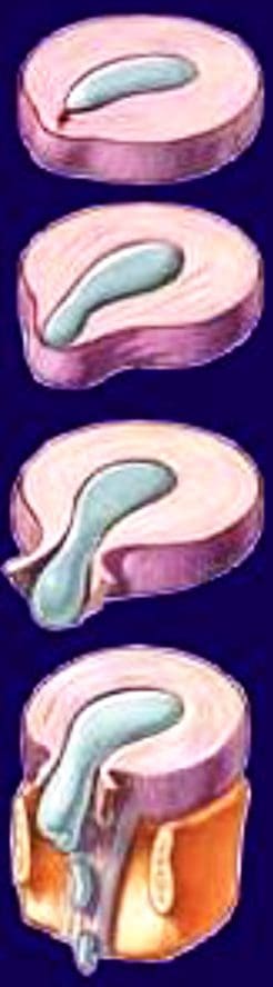
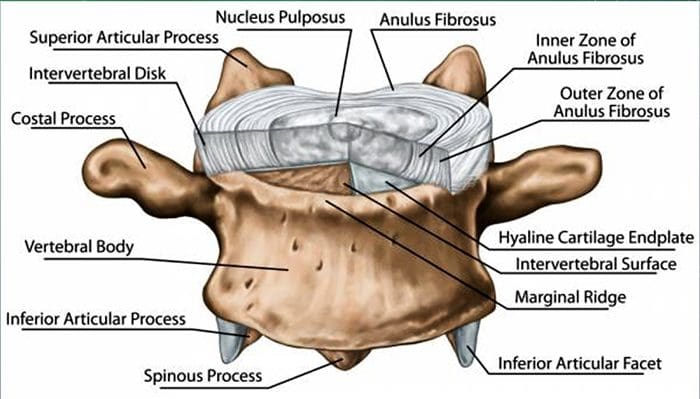
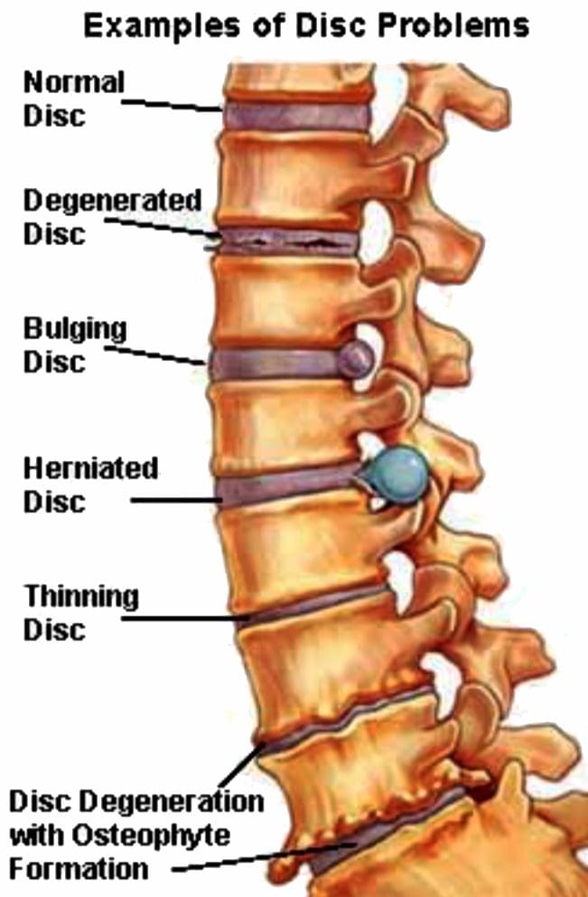
blog picture of cartoon paper boy

EXTRA IMPORTANT TOPIC: Sciatica Pain Chiropractic Therapy

Radiculopathies? What Are They?

Radiculopathies? What Are They?

The spine is made of bones called vertebrae, with the spinal cord running through the spinal canal in the center. The cord is made up of nerves. These nerve roots split from the cord and travel between the vertebrae into various areas of the body. When these nerve roots become pinched or damaged, the symptoms that follow are known as, radiculopathy. El Paso, TX. Chiropractor, Dr. Alexander Jimenez breaks down�radiculopathies,�along with their causes, symptoms and treatment.

The entire length of the spine, at each level, nerves exit through holes in the bone of the spine (foramen) on each side of the spinal column. These nerves are called nerve roots, or radicular nerves and�branch out from the spine and supply different parts of the body.

Nerves exiting the cervical spine travel down through the arms, hands, and fingers. This is where neck problems affecting a cervical nerve root can cause pain, as well as, other symptoms through the arms and hands, one form of (radiculopathy). Another is low back problems that affect a lumbar nerve root. This can radiate through the leg and into the foot, another form of (radiculopathy, or sciatica), which creates leg pain and/or foot pain.

The spinal cord does not go into the lumbar spine and because the spinal canal has space in the lower back, problems in the lumbosacral region often cause nerve root problems and not a spinal cord injury. Serious conditions i.e. disc herniation or fracture in the lower back are also not likely to cause permanent loss of motor function in the legs.

  • Cervical Spine – This nerve root is named according to the Lower spinal segment that the nerve root runs between.�
  • Example – The nerve at C5-C6 level is called the C6 nerve root.
  • It’s named like this because as it exits the spine, it passes Over the C6 pedicle (a piece of bone part of the spinal segment).
  • Lumbar Spine – These nerve roots are named according to the Upper spinal segment that the nerve runs between.
  • Example – The nerve at L4-L5 level is called the L4 nerve root.
  • The nerve root is named this way because as it exits the spine it passes Under the L4 pedicle.

Two Nerve Roots

Two nerves cross each disc level

Only one exits�the spine (through the foramen) at that level.

Exiting Nerve Root –�This is the nerve root exiting the spine at a certain level.

Example: L4 nerve root exits the spine at L4-L5 level.

Traversing Nerve Root –�This nerve root goes across the disc and exits the spine at the level below.

Example: L5 nerve is the traversing nerve root at L4-L5 level, and is the exiting nerve root at L5-S1 level.

There is some confusion when a nerve root is compressed by disc herniation or other cause to refer both to the intervertebral level (where the disc is) and to the nerve root that is affected. This depends on where the disc herniation or protrusion is happening. It could impinge upon either the exiting nerve�or the traversing nerve.

If The Traversing Nerve Is Affected

Lumbar Radiculopathy

In the lumbar spine, there is a weak area in the disc space right in front of the traversing nerve root, so lumbar discs tend to herniate or leak out and impinge on the traversing nerve.

If The Exiting Nerve Is Affected

Cervical Radiculopathy

The opposite is true in the neck. In the cervical spine, the disc tends to herniate to the side, rather than toward the back and the side. If the disc material herniates to the side, it will compress or impinge the exiting nerve root.

Radiculopathy & Sciatica

Nerve root goes by another name Radicular Nerve, and when a herniated or prolapsed disc presses on a radicular nerve, this is referred to as a radiculopathy. A medical physician might say there is herniated disc at L4-L5, which creates an L5 radiculopathy or an L4 radiculopathy. It all depends on where the disc herniation occurs (the side or the back of the disc) and which nerve is affected. And the term for radiculopathy in the low back is the ever famous Sciatica.

Radiculopathy

  • A pinched nerve can occur at different areas of the spine (cervical, thoracic or lumbar).
  • Common causes are narrowing of the hole where the� nerve roots exit, which can result from stenosis, bone spurs, disc herniation and other conditions.
  • Symptoms vary but often include pain, weakness, numbness and tingling.
  • Symptoms can be managed with nonsurgical treatment, but minimal surgery can also help.

Prevalence & Pathogenesis

radiculopathies chiropractic care el paso tx.

  • A herniated disc can be defined as herniation of the nucleus pulposus through the fibers of the annulus fibrosus.
  • Most disc ruptures occur during the third and fourth decades of life while the nucleus pulposus is still gelatinous.
  • The most likely time of day associated with increased force on the disc is the morning.
  • In the lumbar region, perforations usually arise through a defect just lateral to the posterior midline, where the posterior longitudinal ligament is weakest.

radiculopathies chiropractic care el paso tx.

Epidemology

radiculopathies chiropractic care el paso tx.Lumbar Spine:

  • Symptomatic lumbar disc herniation occurs during the lifetime of approximately 2% of the general population.
  • Approximately 80% of the population will experience significant back pain during the course of a herniated disc.
  • The groups at greatest risk for herniation of intervertebral discs are younger individuals (mean age of 35 years)
  • True sciatica actually develops in only 35% of patients with disc herniation.
  • Not infrequently, sciatica develops 6 to 10 years after the onset of low back pain.
  • The period of localized back pain may correspond to repeated damage to annular fibers that irritates the sinuvertebral nerve but does not result in disc herniation.

Epidemology

Cervical Spine:

  • The average annual incidence of cervical radiculopathies is less than 0.1 per 1000 individuals.
  • Pure soft disc herniations are less common than hard disc abnormalities (spondylosis) as a cause of radicular arm pain.
  • In a study of 395 patients with nerve root abnormalities, radiculopathies occurred in the cervical and lumbar spine in 93 (24%) and 302 (76%), respectively.

Pathogenesis

  • Alterations in intervertebral disc biomechanics and biochemistry over time have a detrimental effect on disc function.
  • The disc is less able to work as a spacer between vertebral bodies or as a universal joint.

Pathogenesis – LUMBAR SPINE

radiculopathies chiropractic care el paso tx.

  • The two most common levels for disc herniation are L4-L5 and L5-S1, which account for 98% of lesions; pathology can occur at L2-L3 and L3-L4 but is relatively uncommon.
    Overall, 90% of disc herniations are at the L4-L5 and L5-S1 levels.
  • Disc herniations at L5-S1 will usually compromise the first sacral nerve root, a lesion at the L4-L5 level will most often compress the fifth lumbar root, and herniation at L3-L4 more frequently involves the fourth lumbar root.

radiculopathies chiropractic care el paso tx.

radiculopathies chiropractic care el paso tx.

radiculopathies chiropractic care el paso tx.

  • Disc herniation may also develop in older patients.
  • Disc tissue that causes compression in elderly patients is composed of the annulus fibrosus and and portions of the cartilaginous endplate (hard disc.)
    The cartilage is avulsed from the vertebral body.
  • Resolution of some of the compressive effects on neural structures requires resorption of the nucleus pulposus.

radiculopathies chiropractic care el paso tx.

  • Disc resorption is part of the natural healing process associated with disc herniation.
  • The enhanced ability to resorb discs has the potential for resolving clinical symptoms more rapidly.
  • Resorption of herniated disc material is associated with a marked increase in infiltrating macrophages and the production of matrix metalloproteinases (MMPs) 3 and 7.
  • Nerlich and associates identified the origins of phagocytic cells in degenerated intervertebral discs.
  • The investigation identified cells that are transformed local cells rather than invaded macrophages.
  • Degenerative discs contain the cells that add to their continued dissolution.

radiculopathies chiropractic care el paso tx.

Pathogenesis – CERVICAL SPINE

  • In the early 1940s, a number of reports appeared in which cervical intervertebral disc herniation with radiculopathies was described.
  • There is a direct correlation between the anatomy of the cervical spine and the location and pathophysiology of disc lesion.

radiculopathies chiropractic care el paso tx.

  • The eight cervical nerve roots exit via intervertebral foramina that are bordered anteromedially by the intervertebral disc and posterolaterally by the zygapophyseal joint.
  • The foramina are largest at C2-C3 and decrease in size until C6-C7.
  • The nerve root occupies 25% to 33% of the volume of the foramen.
  • The C1 root exits between the occiput and the atlas (C1)
  • All lower roots exit above their corresponding cervical vertebrae (the C6 root at the C5-C6 interspace), except C8, which exits between C7 and T1.
  • A differential growth rate affects the relationship of the spinal cord and nerve roots and the cervical spine.

radiculopathies chiropractic care el paso tx.

  • Most acute disc herniations occur posterolaterally and in patients around the forth decade of life, when the nucleus is still gelatinous.
  • The most common areas of disc herniations are C6-C7 and C5-C6.
  • C7-T1 and C3-C4 disc herniations are infrequent ( less than 15 %).
  • Disc herniation of C2-C3 is rare.
  • Patients with upper cervical disc protrusions in the C2-C3 region have symptoms that include suboccipital pain, loss of hand dexterity, and paresthesias over the face and unilateral arm.
  • Unlike lumbar herniated discs, cervical herniated discs may cause myelopathy in addition to radicular pain because of the anatomy of the spinal cord in the cervical region.
  • The uncovertebral prominences play a role in the location of ruptured discs material.
  • The uncovertebral joint tends to guide extruded disc material medially, where cord compression may also occur.

radiculopathies chiropractic care el paso tx.

radiculopathies chiropractic care el paso tx.

  • Disc herniations usually affect the nerve root numbered most caudally for the given disc level; for example, the C3 � C4 disc affects the fourth cervical nerve root; C4- C5, the fifth cervical nerve root; C5 � C6, the sixth cervical nerve root; C6 � C7, the seventh cervical nerve root; and C7 � T1, the eighth cervical nerve root.

radiculopathies chiropractic care el paso tx.

  • Not every herniated disc is symptomatic.
  • The development of symptoms depends on the reserve capacity of the spinal canal, the presence of inflammation, the size of the herniation, and the presence of concomitant disease such as osteophyte formation.
  • In disc rupture, protrusion of nuclear material results in tension on the annular fibers and compress?on of the dura or nerve root causing pain.
  • Also important is the smaller size of the sagittal diameter, the bony cervical spinal canal.
  • Individuals in whom a cervical herniated disc causes motor dysfunction have a complication of cervical disc herniation if the spinal canal is stenotic.

Clinical History – LUMBAR SPINE

  • Clinically, the patient�s major complaint is a sharp, lancinating pain.
  • In many cases there may be a previous history of intermittent episodes of localized low back pain.
  • The pain not only in the back but also radiates down the leg in the anatomic distribution of the affected nerve root.
  • It will usually be described as deep and sharp and progressing from above downward in the involved leg.
  • Its onset may be insidious or sudden and associated with a tearing or snapping sensations of the spine.
  • Occasionally, when sciatica develops, the back pain may resolve because once the annulus has ruptured, it may no longer be under tension.
  • Disc herniation occurs with sudden physical effort when the trunk is flexed or rotated.
  • On occasion, patients with L4-L5 disc herniation have groin pain. In a study of 512 lumbar disc patients, 4.1% had groin pain.
  • Finally, the sciatica may vary in intensity; it may be so severe that patients will be unable to ambulate and they will feel that their back is “locked”.
  • On the other hand, the pain may be limited to a dull ache that increases in intensity with ambulation.
  • Pain is worsened in the flexed position and relieved by extension of the lumbar spine.
  • Characteristically, patients with herniated discs have increased pain with sitting, driving, walking, couching, sneezing, or straining.

Clinical History – CERVICAL SPINE

  • Arm pain, not neck pain, is the patient� s major complaint.
  • The pain is often perceived as starting in the neck area and then radiating from this point down to shoulder, arm and forearm and usually into the hand.
  • The onset of the radicular pain is often gradual, although it can be sudden and occur in association with a tearing or snapping sensation.
  • As time passes, the magnitude of the arm pain clearly exceeds that of the neck or shoulder pain.
  • The arm pain may also be variable in intensity and preclude any use of the arm; it may range from severe pain to a dull, cramping ache in the arm muscles.
  • The pain is usually severe enough to awaken the patient at night.
  • Additionally, a patient may complain of associated headaches as well as muscle spasm, which can radiate from the cervical spine to below the scapulae.
  • The pain may also radiate to the chest and mimic angina (pseudoangina) or to the breast.
  • Symptoms such as back pain, leg pain, leg weakness, gait disturbance, or incontinence suggest compression of the spinal cord (Myelopathy).

Physical Examination – LUMBAR SPINE

radiculopathies chiropractic care el paso tx.

  • Physical examination will demonstrated a decrease in range of motion of the lumbosacral spine, and patients may list to one side as they try to bend forward.
  • The side of the disc herniation typically corresponds to the location of the scoliotic list.
  • However, the specific level or degree of herniation does not correlate with the degree of list.
  • On ambulation, patients walk with an antalgic gait in which they hold the involved leg flexed so that they put as little weight as possible on the extremity.

radiculopathies chiropractic care el paso tx.

  • Neurologic Examination:
  • The neurologic examination is very important and may yield objective evidence of nerve root compression (We should evaluate of reflex testing, muscle power, and sensation examination of the patient).
  • In addition, a nerve deficit may have little temporal relevance because it may be related to a previous attack at a different level.
  • Compression of individual spinal nerve roots results in alterations in motor, sensory, and reflex function.
  • When the first sacral root is compressed, the patient may have gastrocnemius-soleus weakness and be unable to repeatedly raise up on the toes of that foot.
  • Atrophy of the calf may be apperent, and the ankle (Achilles) reflex is often diminished or absent.
  • Sensory loss, if present, is usually confined to the posterior aspect of the calf and the lateral side of the foot.

radiculopathies chiropractic care el paso tx.

  • Involvement of the fifth lumbar nerve root can lead to weakness in extension of the great toe and, in a few cases, weakness of the everters and dorsiflexors of the foot.
  • A sensory deficit can appear over the anterior of the leg and the dorsomedial aspect of the foot down to the big toe

radiculopathies chiropractic care el paso tx.

  • With compression of the fourth lumbar nerve root, the quadriceps muscle is affected; the patient may note weakness in knee extension, which is often associated with instability.
  • Atrophy of the thigh musculature can be marked. Sensory loss may be apparent over the anteromedial aspect of the thigh, and the patellar tendon reflex can be diminished.

radiculopathies chiropractic care el paso tx.


radiculopathies chiropractic care el paso tx.

  • Nerve root sensitivity can be elicited by any method that creates tension.
  • The straight leg-raising (SLR)test is the one most commonly used.
  • This test is performed with the patient supine.

Physical Examination – CERVICAL SPINE

Neurologic Examination:
  • A neurologic examination that shows abnormalities is the most helpful aspect of the diagnostic work-up, although the examination may remain normal despite a chronic radicular pattern.
  • The presence of atrophy helps document the location of the lesion, as well as its chronicity.
  • The presence of subjective sensory changes is often difficult to interpret and requires a coherent and cooperative patient to be of clinical value.

radiculopathies chiropractic care el paso tx.

  • When the third cervical root is compressed, no reflex change and motor weakness can be identified.
  • The pain radiates to the back of the neck and toward the mastoid process and pinna of the ear.
  • Involvement of the fourth cervical nerve root leads to no readily detectable reflex changes or motor weakness.
  • The pain radiates to the back of the neck and superior aspect of the scapula.
  • Occasionally, the pain radiates to the anterior chest wall.
  • The pain is often exacerbated by neck extension.
  • Unlike the third and the fourth cervical nerve roots, the fifth through eighth cervical nerve roots have motor functions.
  • Compression of the fifth cervical nerve root is characterized by weakness of shoulder abduction, usually above 90 degree, and weakness of shoulder extension.
  • The biceps reflexes are often depressed and the pain radiates from the side of the neck to the top of the shoulder.
  • Decreased sensation is often noted in the lateral aspect of the deltoid, which represents the autonomous area of the axillary nerve.

radiculopathies chiropractic care el paso tx.

  • Involvement of the sixth cervical nerve root produces biceps muscles weakness as well as diminished brachioradial reflex.
  • The pain again radiates from the neck down the lateral aspect of the arm and forearm to the radial side of hand (index finger, long finger, and thumb).
  • Numbness occurs occasionally in the tip of the index finger, the autonomous area of the sixth cervical nerve root.

radiculopathies chiropractic care el paso tx.

  • Compression of the seventh cervical nerve root produces reflex changes in the triceps jerk test with associated loss of strength in the triceps muscles, which extend the elbow.
  • The pain from this lesion radiates from the lateral aspect of the neck down the middle of the area to the middle finger.
  • Sensory changes occur often in the tip of the middle finger, the autonomous area for the seventh nerve.
  • Patients should also be tested for scapular winging, which may occur with C6 or C7 radiculopathies.

radiculopathies chiropractic care el paso tx.

  • Finally, involvement of the eighth cervical nerve root by a herniated C7-T1 disc produces significant weakness of the intrinsic musculature of the hand.
  • Such involvement can lead to rapid atrophy of the interosseous muscles because of the small size of these muscles.
  • Loss of the interossei leads to significant loss of fine hand motion.
  • No reflexes are easily found, although the flexor carpi ulnaris reflex may be decreased.
  • The radicular pain from the eighth cervical nerve root radiates to the ulnar border the hand and the ring and little fingers.
  • The tip of the little finger often demonstrates diminished sensation.

radiculopathies chiropractic care el paso tx.

  • Radicular pain secondary to a herniated cervical disc may be relieved by abduction of the affected arm.
  • Although these signs are helpful when present, their absence alone does not rule out a nerve root lesion.

Laboratory Data

radiculopathies chiropractic care el paso tx.

  • Medical screening laboratory test (blood counts, chemistry panels erythrocyte sedimentation rate [ESR]) are normal in patients with a herniated disc.
  • Electro diagnostic Testing
  • Electromyography(EMG)is an electronic extension of the physical examination.
  • The primary use of EMG is to diagnose radiculopathies in cases of questionable neurologic origin.
  • EMG findings may be positive in patients with nerve root impingement.

Radiographic Evaluation – LUMBAR SPINE

  • Plain x-rays may be entirely normal in a patient with signs and symptoms of nerve root impingement.
  • Computed Tomography
  • Radigraphic evaluation by CT scan may demonstrate disc bulging but may not correlate with the level of nerve damage.
  • Magnetic Resonance Imaging
  • MR imaging also allows visualization of soft tissues, including discs in the lumbar spine.
  • Herniated discs are easily detected with MR evaluation.
  • MR imaging is a sensitive technique for the detection of far lateral and anterior disc herniations.

Radiographic Evaluation – CERVICAL SPINE

  • X-rays
  • Plain x-rays may be entirely normal in patients wit han acute herniated cervical disc.
  • Conversely,�70% of asymptomatic women and 95% of asymptomatic men between the ages of 60 and 65 years have evidence of degenerative disc disease on plain roentgenograms.
  • Views to be obtained include anteroposterior, lateral, flexion, and extension.
radiculopathies chiropractic care el paso tx.

radiculopathies chiropractic care el paso tx.

  • Computed Tomography
  • CT permits direct visualization of compression of neural structures and is therefore more precise than myelography.
  • Advantages of CT over myelography include better visualization of lateral abnormalities such as foraminal stenosis and abnormalities caudal to the myelographic block, less radiation exposure, and no hospitalization.
  • Magnetic Resonance
  • MRI allows excellent visualization of soft tissues, including herniated discs in the cervical spine.
  • The test is noninvasive.
  • In a study of 34 patients with cervical lesions, MRI predicted 88% of the surgically proven lesions versus 81% for myelography-CT, 58% for myelography, and 50% for CT alone.

Differential Diagnosis – LUMBAR SPINE

  • The initial diagnosis of a herniated disc is ordinarily made on the basis of the history and physical examination.
  • Plain radiographs of the lumbosacral spine will rarely add to the diagnosis but should be obtained to help rule out other causes of pain such as infection or tumor.
  • Other tests such as MR, CT, and myelography are confirmatory by nature and can be misleading when used as screening tests.

Spinal Stenosis

  • Patient with spinal stenosis may also suffer from back pain that radiates to the lower extremities.
  • Patients with spinal stenosis tend to be older than those in whom herniated discs develop.
  • Characteristically, patients with spinal stenosis experience lower extremity pain (pseudoclaudication=neurogenic claudication) after walking for an unspecified distance.
  • They also complain of pain that is exacerbated by standing or extending the spine.
  • Radiographic evaluation is usually helpful in differentiating individuals with disc herniation from those with bony hypertrophy associated with spinal stenosis.
  • In a study of 1,293 patients, lateral spinal stenosis and herniated intervertebral discs coexisted in 17.7% of individuals.
  • Radicular pain may be caused by more than one pathologic process in an individual.

Facet Syndrome

  • Facet syndrome is another cause of low back pain that may be associated with radiation of pain to structures outside the confines of the lumbosacral spine.
  • Degeneration of articular structures in the facet joint causes pain to develop.
  • In most circumstances, the pain is localized over the area of the affected joint and is aggravated by extension of the spine (standing).
  • A deep , ill-defined, aching discomfort may also be noted in the sacroiliac joint, the buttocks, and the legs.
  • The areas of sclerotome affected show the same embryonic origin as the degenerated facet joint.
  • Patients with pain secondary to facet joint disease may have relief of symptoms with apophyseal injection of a long-acting local anesthetic.
  • The true role of facet joint disease in the production of back and leg pain remains to be determined.
  • Other mechanical causes of sciatica include congentenial abnormalites of the lumbar nerve roots, external compression of the sciatic nerve (wallet in a back pants pocket), and muscular compression of the nerve (piriformis syndrome).
  • In rare circumstances, cervical or thoracic lesion should be considered if the lumbar spine is clear of abnormalities.
  • Medical causes of sciatica (neural tumors or infections, for example) are usually associated with systemic symptoms in addition to nerve pain in a sciatic distribution.

Differential Diagnosis – CERVICAL SPINE

  • No diagnostic criteria exist for the clinical diagnosis of a herniated cervical disc.
  • The provisional diagnosis of a herniated cervical disc is made by the history and physical examination.
  • The plain x-ray is usually nondiagnostic, although occasionally disc space narrowing at the suspected interspace or foraminal narrowing on oblique films is seen.
  • The value of x-rays is to exclude other causes of neck and arm pain, such as infection and tumor.
  • MR imaging and CT-myelography are the best confirmatory examinations for disc herniation.
  • Cervical disc herniations may affect structures other than nerve roots.
  • Disc herniation may cause vessel compression (vertebral artery) associated with vertebrobasilar artery insufficiency and be manifested as blurred vision and dizziness.

radiculopathies chiropractic care el paso tx.

  • Other mechanical causes of arm pain should be excluded.
  • The most common is some form of compression on a peripheral nerve.
  • Such compression can occur at the elbow, forearm, or wrist. An example is compression of the median nerve by the carpal ligament leading to carpal tunnel syndrome.
  • The best diagnostic test to rule out these peripheral neuropathies is EMG.
  • Excessive traction on the arm secondary to heavy weights may cause radicular pain without disc compression of nerve roots.
  • Spinal cord abnormalities must be considered if signs of myelopathy are present in conjunction with radiculopathies.
  • Spinal cord lesions such as syringomyelia are identified by MRI, and motor neuron disease is identified by EMG.
  • Multiple sclerosis should be considered in a patient with radiculopathies if the physical signs indicate lesions above the foramen magnum (optic neuritis).
  • In very rare circumstances, lesions of the parietal lobe corresponding to the arm can mimic the findings of cervical radiculopathies.
Heal A Bulging Disc Through Chiropractic | El Paso, TX.

Heal A Bulging Disc Through Chiropractic | El Paso, TX.

Bulging disc is often thought of as a normal part of the aging process. It causes pain and decreases mobility. Athletes and people who have jobs that are very physical are often prone to bulging discs and other disc problems. Smoking tobacco can also be a contributing factor in spinal discs deteriorating and weakening. Chiropractic has been proven to be an effective treatment to heal a bulging disc and the associated pain.

What Is A Bulging Disc?

heal bulging disc el paso tx.

Bulging discs are often thought to be the same as herniated discs but that is incorrect. A herniated disc involves a crack in the disc�s outer layer. This is called an annulus. Typically, a small part of the disc is affected, allowing the soft material that makes up the nucleus pulposus to protrude. This is different from a bulging disc because, unlike a herniated disc, there is no crack. The disc bulges out of the space but it doesn�t crack and no material protrudes. It also affects more area of the disc than a herniated disc.

While a herniated disc is likely more painful, a bulging disc can also cause pain that can increase over time. Symptoms of a bulging disc include:

  • Tingling, numbness, or muscle weakness in one or both legs
  • Changes in bowel or bladder function
  • Hyper reflexivity in one or both legs
  • Paralysis below the waist
  • Deep pain over the shoulder blade or in the shoulder area
  • Pain when moving the neck
  • Radiating pain in the fingers, forearm, and upper arm

A bulging disc is often diagnosed by a combination of several methods. A physical exam, along with a full history of the problem will often lead to tests like MRI, x-ray, and myelogram with CT scan. From there, your doctor will work with you to find the best course of treatment.

Chiropractic To Help Heal A Bulging Disc

Chiropractic is a preferred treatment method for many patients with bulging disc because it is non-invasive and does not involve drugs or injections. Once you have your diagnosis, you and your chiropractor can work together to find the best way to treat your condition.

Your chiropractor will want to verify your diagnosis so you may go through questions about your medical history, a physical examination, and tests that involve nerve function, reflexes, and muscle tone. Your chiropractor may also order MRI or x-ray as well as other diagnostic testing in order to get a better picture of what is going on.

One of the most popular features of chiropractic care is the whole body approach to wellness. Your chiropractor will look at your entire spine, not just the area that is painful. They will treat your entire spine and provide self-care direction, exercise, and nutritional recommendations so that you can continue to progress and live pain free. Your pain and spinal problems could be the result of spinal misalignment so your chiropractor will seek to get to the root of the problem and treat your entire spine so that you have less pain, your spine can heal, and you have better mobility.

Through focused chiropractic adjustments, your chiropractor will gently use low force techniques to relieve the painful symptoms by manipulating your spine around and at the disc that is bulging. They may use other types of treatments depending on your specific condition and other issues that may be exacerbating your problem.

Chiropractic for bulging discs is safe, effective, and long lasting. If you are having back pain from a bulging disc, you owe it to yourself to seek quality chiropractic care so that you can enjoy less pain, improved mobility, and better quality of life.

Injury Medical Clinic: Non-Surgical Options

Herniated Disc Pain Treatment In El Paso, TX. | Video

Herniated Disc Pain Treatment In El Paso, TX. | Video

Herniated Disc Pain: Araceli Pizana started chiropractic care with Dr. Alex Jimenez due to chronic back pain symptoms associated with a herniated disc. Before finding the right alternative treatment option with Dr. Jimenez, Mrs. Pizana struggled to perform her everyday activities. Araceli Pizana describes how Dr. Alex Jimenez’s exceptional care for his patients ultimately reflects on his ability to improve her overall well-being. Mrs. Pizana recommends chiropractic care for health and wellness.

Herniated Disc Pain & Chiropractic Treatment

Most healthcare professionals agree that degeneration of the intervertebral discs is the main cause of spinal disc herniation, where trauma and/or injury is considered to be the least probable cause. Disc degeneration occurs both with degenerative disc disease and aging. When the degeneration of the intervertebral discs occurs, the soft gel-like center of the disc, known as the nucleus pulposus, pushes through the outer ring of the disc, known as the annulus fibrosus. A tear in the intervertebral disc is what’s known as a disc herniation. Furthermore, the chemical material released can irritate the surrounding structures of the spine causing herniated disc pain.

herniated disc pain in el paso tx.

Dr. Jimenez has teamed with the top surgeons, clinical specialist, medical researchers and�premiere rehabilitation�providers to bring El Paso the top clinical treatments to our community.��Providing the top non-invasive protocols is our priority.��The clinical insight is what our patients demand in order to give them the appropriate care required.

Our team has takes great�pride in bringing our families and injured patients only�clinically proven treatments protocols. �By teaching complete holistic wellness as a lifestyle,�we also change not only our patients lives but their families as well.� We do this so that we may reach as many El Pasoans who need us, no matter the affordability issues.

There is no reason we cannot help you.

Please Recommend Us: If you have enjoyed this video and/or we have helped you in any way please feel free to recommend us. Thank You & God Bless.

Recommend: Dr. Alex Jimenez � Chiropractor

Health Grades:�� http://www.healthgrades.com/review/3SDJ4

Facebook Clinical Page:� https://www.facebook.com/dralexjimenez/reviews/

Facebook Sports Page: https://www.facebook.com/pushasrx/

Facebook Injuries Page: https://www.facebook.com/elpasochiropractor/

Facebook Neuropathy Page: https://www.facebook.com/ElPasoNeuropathyCenter/

Yelp:�� http://goo.gl/pwY2n2

Clinical Testimonies: https://www.dralexjimenez.com/category/testimonies/

Information: Dr. Alex Jimenez � Chiropractor

Clinical Site: https://www.dralexjimenez.com

Injury Site: https://personalinjurydoctorgroup.com

Sports Injury Site: https://chiropracticscientist.com

Back Injury Site: https://elpasobackclinic.com

Linked In:�� https://www.linkedin.com/in/dralexjimenez

Pinterest:�� https://www.pinterest.com/dralexjimenez/

Twitter:�� https://twitter.com/dralexjimenez

Twitter: https://twitter.com/crossfitdoctor

Recommend: PUSH-as-Rx ��

Rehabilitation Center: https://www.pushasrx.com

Facebook:�� https://www.facebook.com/PUSHftinessathletictraining/

PUSH-as-Rx:�� http://www.push4fitness.com/team/

Chiropractic Clinic Extra: Herniated Disc Treatment

Mindfulness for Headache and Cervical Disc Herniation in El Paso, TX

Mindfulness for Headache and Cervical Disc Herniation in El Paso, TX

Stress is a result of the human body’s “fight or flight” response, a prehistoric defense mechanism triggered by the sympathetic nervous system (SNS). Stress is an essential component of survival. When stressors activate the fight or flight response, a mixture of chemicals and hormones are secreted into the blood flow, which prepare the body for perceived danger. Although short-term stress is helpful, however, long-term stress can lead to a variety of health issues. Furthermore, stressors in modern society have changed and it’s become more difficult for people to manage their stress and maintain mindfulness.


How Does Stress Affect the Body?


Stress can be experienced through three different channels: emotion; body and environment. Emotional stress involves adverse situations which affect our mind and decision making. Bodily stress includes improper nutrition and a lack of sleep. And finally, environmental stress occurs based on external experiences. When you experience any of these types of stressors, the sympathetic nervous system will trigger the “fight or flight” response, releasing adrenaline and cortisol to increase heart rate and heighten our senses to make us more alert in order to face the situation ahead of us.


However, if perceived stressors are always present, the SNS’s fight or flight response can remain active. Chronic stress can then lead to a variety of health issues, such as anxiety, depression, muscle tension, neck and back pain, digestive problems, weight gain and sleep problems as well as impaired memory and concentration. In addition, muscle tension along the spine due to stress can cause a spinal misalignment, or subluxation, which may in turn lead to disc herniation.


Headache and Disc Herniation from Stress


A herniated disc occurs when the soft, gel-like center of an intervertebral disc pushes through a tear in its outer, cartilage ring, irritating and compressing the spinal cord and/or the nerve roots. Disc herniation commonly occurs in the cervical spine, or neck, and in the lumbar spine, or low back. Symptoms of herniated discs depend on the location of the compression along the spine. Neck pain and back pain accompanied by numbness, tingling sensations and weakness along the upper and lower extremities are some of the most common symptoms associated with disc herniation. Headache and migraine are also common symptoms associated with stress and herniated discs along the cervical spine, as a result of muscle tension and spinal misalignment.


Mindfulness Interventions for Stress Management


Stress management is essential towards improving as well as maintaining overall health and wellness. According to research studies, mindfulness interventions, such as chiropractic care and mindfulness-based stress reduction (MBSR), among others, can safely and effectively help reduce stress. Chiropractic care utilizes spinal adjustments and manual manipulations to carefully restore the original alignment of the spine, relieving pain and discomfort as well as easing muscle tension. Additionally, a chiropractor may include lifestyle modifications to help further improve symptoms of stress.�A balanced spine can help the nervous system respond to stress more effectively. MBSR can also help reduce stress, anxiety and depression.


Contact Us Today


If you are experiencing symptoms of stress with headache or migraine as well as neck and back pain associated with disc herniation, mindfulness interventions such as chiropractic care can be a safe and effective treatment for your stress. Dr. Alex Jimenez’s stress management services can help you achieve overall health and wellness. Seeking the proper mindfulness interventions can get you the relief you deserve. The purpose of the following article is to demonstrate the effects of mindfulness-based stress reduction in patients with tension headache. Don’t just treat the symptoms, get to the source of the issue.


Effects of Mindfulness-Based Stress Reduction on Perceived Stress and Psychological Health in Patients with Tension Headache


Abstract


Background: Programs for improving health status of patients with illness related to pain, such as headache, are often still in their infancy. Mindfulness-based stress reduction (MBSR) is a new psychotherapy that appears to be effective in treating chronic pain and stress. This study evaluated efficacy of MBSR in treatment of perceived stress and mental health of client who has tension headache.


Materials and Methods: This study is a randomized clinical trial. Sixty patients with tension type headache according to the International Headache Classification Subcommittee were randomly assigned to the Treatment As Usual (TAU) group or experimental group (MBSR). The MBSR group received eight weekly classmates with 12-min sessions. The sessions were based on MBSR protocol. The Brief Symptom Inventory (BSI) and Perceived Stress Scale (PSS) were administered in the pre- and posttreatment period and at 3 months follow-up for both the groups.


Results: The mean of total score of the BSI (global severity index; GSI) in MBSR group was 1.63 � 0.56 before the intervention that was significantly reduced to 0.73 � 0.46 and 0.93 � 0.34 after the intervention and at the follow-up sessions, respectively (P < 0.001). In addition, the MBSR group showed lower scores in perceived stress in comparison with the control group at posttest evaluation. The mean of perceived stress before the intervention was 16.96 � 2.53 and was changed to 12.7 � 2.69 and 13.5 � 2.33 after the intervention and at the follow-up sessions, respectively (P < 0.001). On the other hand, the mean of GSI in the TAU group was 1.77 � 0.50 at pretest that was significantly reduced to 1.59 � 0.52 and 1.78 � 0.47 at posttest and follow-up, respectively (P < 0.001). Also, the mean of perceived stress in the TAU group at pretest was 15.9 � 2.86 and that was changed to 16.13 � 2.44 and 15.76 � 2.22 at posttest and follow-up, respectively (P < 0.001).


Conclusion: MBSR could reduce stress and improve general mental health in patients with tension headache.


Keywords: Mental health, tension headache, mindfulness-based stress reduction (MBSR), perceived stress, treatment as usual (TAU)


Dr Jimenez White Coat

Dr. Alex Jimenez’s Insight

Chiropractic care is an effective stress management treatment because it focuses on the spine, which is the base of the nervous system. Chiropractic utilizes spinal adjustments and manual manipulations to carefully restore the alignment of the spine in order to allow the body to naturally heal itself. A spinal misalignment, or subluxation, can create muscle tension along the spine and lead to a variety of health issues, including headache and migraine, as well as disc herniation and sciatica. Chiropractic care can also include lifestyle modifications, such as nutritional advice and exercise recommendations, to further enhance its effects. Mindfulness-based stress reduction can also effectively help with stress management and symptoms.


Introduction


Tension headache constitutes 90% of total headaches. About 3% of the population are suffering from chronic tension headache.[1] Tension headaches are often associated with lower quality of life and high levels of psychological discomforts.[2] In recent years, several meta-analyses evaluating the established pain treatments used today have shown that medical treatments, which may be effective in acute pain, are not effective with chronic pain and may, in fact, be causing further problems. Most of the pain treatments are designed for and useful for acute pain but if used in the long run may create more problems such as substance abuse and avoidance of important activities.[3] A common element in most of the pain treatments is that they emphasize on either avoiding pain or fighting to reduce pain. The pain in tension headache can be intolerable. Painkillers and pain management strategies can increase intolerance and sensitivity to pain. Therefore, the treatments that increase acceptance and tolerance to pain, especially chronic pain, are effective. Mindfulness-based stress reduction (MBSR) is a new psychotherapy that appears to be effective in improving physical performance and psychological well-being in patients with chronic pain.[4,5,6,7,8] In the past two decades, Kabat-Zinn et al. in the US successfully used mindfulness for the relief of pain and illness related to pain.[9] Recent studies on acceptance-based methods, such as mindfulness, show improved performance in patients with chronic pain. Mindfulness modulates the pain using nonelaborative awareness of thoughts, feelings and sensations, and an emotionally distanced relationship with internal and external experience.[10] Studies found that MBSR program can significantly alleviate medical illness related to chronic pains such as fibromyalgia, rheumatoid arthritis, chronic musculoskeletal pain, chronic low back pain, and multiple sclerosis.[7,11,12,13] MBSR has significant changes in pain intensity, anxiety, depression, somatic complaints, well-being, adaptation, quality of sleep, fatigue, and physical functioning.[6,14,15,16,17] But the programs for improving health status of patients with illness related to pain, such as tension headache, are often still in their infancy. Therefore, the study was conducted to assess the effects of MBSR on perceived stress and general mental health in patients with tension headache.


Materials and Methods


This randomized controlled clinical trial was performed in 2012 in Shahid Beheshti Hospital in Kashan City. The Research Ethics Committee of the Kashan University of Medical Sciences approved this study (IRCT No: 2014061618106N1). The participants of the study included adults with tension headache who were referred by the psychiatrists and neurologists in Kashan. The inclusion criteria were as follows: Having tension headache according to the International Headache Classification Subcommittee, willing to participate in the study, not having a medical diagnosis of organic brain disorder or psychotic disorder, and not having a history of psychological treatment during the preceding 6 months. The patients who did not complete the intervention and missed more than two sessions were excluded from the study. The participants, who signed an informed consent form, completed the measures as a pretest. For estimating the sample size, we referred to another study in which changes in mean of scores of fatigue was 62 � 9.5 in the pretreatment period and 54.5 � 11.5 in the posttreatment period.[18] Then, by utilizing the sample size calculation, 33 participants (with attrition risk) in each group with ? = 0.95 and 1 � ? = 0.9 were segregated. After sample size calculation, 66 patients with tension headache were selected via convenient sampling according to the inclusion criteria. Then, the patients were called and invited to participate in the study. If a patient agreed to participate, then he/she was invited to attend the study-briefing session and if not another patient was selected similarly. Then using a random number table, they were assigned either to the experimental group (MBSR) or to the control group that treated as usual. Finally, 3 patients were excluded from each group and 60 patients were included (30 patients in each group). The TAU group was treated only by antidepressant medication and clinical management. The MBSR group received MBSR training in addition to TAU. The patients in MBSR group were trained for 8 weeks by a clinical psychologist with PhD degree. The Brief Symptom Inventory (BSI) and Perceived Stress Scale (PSS) were administered before the first treatment session in the MBSR group, after the eighth session (posttest), and 3 months after the test (follow-up) in both groups. The TAU group was invited to Shahid Beheshti Hospital to fill out the questionnaires. Figure 1 shows a Consolidated Standards of Reporting Trials (CONSORT) diagram depicting the flow of study participants.


Figure 1 CONSORT Diagram Depicting Flow of Study Participants

Figure 1: CONSORT diagram depicting flow of study participants.


Intervention


The intervention group (MBSR) was trained in Shahid Beheshti Hospital. The eight weekly sessions (120 min) were held according to the standard MBSR protocol as developed by Kabat-Zinn.[11] Additional sessions were held for the participants who had missed one or two sessions. At the end of the training and 3 months later (follow-up), both MBSR and TAU groups were invited to Shahid Beheshti Hospital (the place of MBSR trial) and were instructed to complete the questionnaires. During the MBSR sessions, the participants were trained to be aware of their thoughts, feelings, and physical sensations nonjudgmentally. Mindfulness exercises are taught as two forms of meditation practices � formal and informal. Formal type exercises include trained sitting meditation, body scan, and mindful yoga. In informal meditation, attention and awareness are focused not only on daily activities, but also on thoughts, feelings, and physical sensation even they are problematic and painful. The overall content of the sessions were mentioned in Table 1.


Table 1 Agendas for Sessions of MBSR

Table 1: Agendas for sessions of mindfulness-based stress reduction.


Measurement Tools


International Headache Classification Subcommittee Diary Scale for Headache


Headache was measured by diary scale for headache.[19] The patients were asked to record the pain severity diary on a 0-10 rating scale. Absence of pain and the most intense disabling headache were characterized by 0 and 10, respectively. The mean of headache severity in a week was calculated by dividing the sum of the severity scores by 7. Moreover, the mean of headache severity in a month was calculated by dividing the sum of the severity scores by 30. The minimum and maximum scores of headache severity were 0 and 10, respectively. Headache diary was given to five patients and a neurologist and a psychiatrist confirmed the content validity of the instrument.[20] The reliability coefficient of Persian version of this scale was calculated as 0.88.[20]


Brief symptom Inventory (BSI)


Psychological symptoms were assessed with the BSI.[21] The inventory consist 53 items and 9 subscales that assess psychological symptoms. Each item scores between 0 and 4 (for example: I have nausea or upset in my stomach). BSI has a global severity index (GSI) achieved a total score of 53 items. The reliability of the test has reported a score of 0.89.[22] In our study, GSI test�retest estimate was .90 based on a sample of 60 patients with tension headache who completed the BSI.


Perceived Stress Scale (PSS)


Perceived stress was assessed using the PSS,[21,23] a 10-item scale that assesses the degree of uncontrollable and unpredictable situations of life during the past month (for example: Felt that you were unable to control the important things in your life?). Respondents report the prevalence of an item within the last month on a 5-point scale, ranging from 0 (never) to 4 (very often). Scoring is completed by reverse scoring of four positively worded items[4,5,7,8] and summing all item scores. The scale scores range from 0-40. Higher scores indicate higher levels of stress. It assumes that people depending on their coping resources evaluate level of threatening or challenging events. A higher score indicates a greater degree of perceived stress. Adequate test�retest reliability and convergent and discriminate validity have also been reported.[19] In our study, Cronbach’s alpha coefficients for assessing internal consistency of this scale were calculated to be 0.88.


The repeated measures analysis of variance was performed to compare the MBSR and TAU groups on measures of perceived stress and GSI at pretreatment, posttreatment, and 3-month follow-up. Also, Chi-square test was used to compare the demographics in the two groups. P value less than 0.05 was considered significant in all tests.


Results


Among 66 subjects, 2 participants from the MBSR group were excluded because of missing more than 2 sessions. Also, three participants were excluded because of did not complete the questionnaires in post-test or follow-up who one of them were from MBSR group and three participants from TAU group. Table 2 showed demographic characteristics of the subjects and results of the randomization check. The results of t-test for differences between the MBSR and TAU groups in age variable and Chi-square test in other variables showed that there was no significant difference between demographic variables in two groups and the subjects were randomly assigned to two groups.


Table 2 Demographic Characteristics of the Subjects

Table 2: Demographic characteristics of the subjects a,b.


Table 3 provides the mean scores and standard deviations of the dependent variables (perceived stress and GSI) and comparison of outcome measures at pretreatment period, post-treatment period, and 3-month follow-up.


Table 3 Means, Standard Deviations and Comparison of Outcome Measures

Table 3: Means, standard deviations, and comparison of outcome measures at pretreatment, posttreatment, and follow-up stages in the MBSR and TAU groups a,b.


Table 3 shows the more reduction in received stress and GSI in the intervention group (MBSR) compared to TAU group, while the reduction in received stress and GSI were not observed in the TAU group. The results revealed the significant effect of time and interaction between time and type of treatment on the changes of scores (P < 0.001).


Figures ?2 and ?3 present mean received stress and GSI scores for MBSR and TAU groups at posttest and follow-up stages.


Figure 2 CONSORT Diagram Depicting Flow of Study Participants

Figure 2: CONSORT diagram depicting flow of study participants.


Figure 3 Mean of Perceived Stress in MBSR and Control Groups

Figure 3: Mean of perceived stress in MBSR and control groups in pretest, posttest, and follow-up.


Discussion


This study compared efficacy of MBSR and Treatment As Usual (TAU) in perceived stress and mental health of patients with tension headache. Although MBSR is recognized as an effective treatment for stress symptoms and pain, there is a need to examine its efficacy for the treatment of mental health problems in patients with tension headache, which is one of the common complaints in the population.


The findings of our study demonstrate enhanced general mental health in the GSI index of BSI. In some study, significant improvements by MBSR intervention were reported on all indexes of the 36-item Short Form Health Survey (SF-36).[20,24] Studies showed significant reduction in psychological problems in the Symptom Checklist-90-Revised (SCL-90-R) subscale such as anxiety and depression by MBSR after intervention and 1-year follow-up.[5] Reibel et al. showed MBSR in patients with chronic pain reported a decrease in medical symptoms such as anxiety, depression, and pain.[5] It has been shown that tension headache and anxiety are accompanied with deficits in controlled cognitive processing such as sustained attention and working memory.[25] Negative emotions may amplify suffering associated with pain perception.


MBSR implements the following mechanisms to improve the patient’s mental status: First, mindfulness leads to increased awareness for what is happening in each moment, with an accepting attitude, without getting caught up in habitual thoughts, emotions, and behavior patterns. The increased awareness then gives rise to new ways to respond and cope in relation to oneself and the world around.[3] Mindfulness establishes a sense of self that is greater than one’s thoughts, feelings, and bodily sensation such as pain. Mindfulness exercises, learned clients develop an �observer�self�. With this ability, they can observe their thoughts and feelings in a nonreactive and nonjudgmental way that previously avoided, that previously avoided thoughts and feelings be observed in a nonreactive and nonjudgmental way. The clients learn to notice thoughts without necessarily acting on them, being controlled by them, or believing them.[3]


Second, mindfulness helps the client develop persistence in taking steps in valued directions that are important to them. Most clients with chronic pain want to become pain free rather than living the vital lives of their choice. But the MBSR program trained them to engage in valued action despite the the pain. Studies have shown attention and emotional reaction to pain has an important role in becoming persistent the pain.[26] Emotional and cognitive components can modulate attention to pain and worry about it that could intensify pain and disrupt the patients activities.[27,28]


Third, findings from some studies indicate that MBSR can alter the function of the brain that is responsible for affect regulation and the areas that govern how we react to stressful impulses, and this in turn may normalize body functions such as breathing, heart rate, and immune function.[29,30] Mindfulness practice reduces reactivity to distressing thoughts and feelings that comorbid and strengthen pain perception.[31] Also mindfulness may lessen psychophysiological activation related to stress and mood dysfunction by strengthening positive reappraisal and emotion regulation skills.[32]


The strength of this study is the use of a new effective psychotherapy in reducing the stress on a complaint that is less studied, but it is a common medical problem. The implications of our study are using a simple psychotherapy that does not make too much cognitive demand and is readily usable as a coping skill for the patient with tension headache. Therefore, the health-care professionals related to this complaint and the patient will be able to use this treatment. Also, MBSR will change the patient’s lifestyle who would be exacerbated by his/her problem. The main limitation of this study was the lack of comparison between MBSR and the gold standard psychotherapies such as cognitive behavior therapy (CBT). It is suggested that future studies need to compare the efficacy of MBSR and other traditional and newer cognitive behavioral therapies in patients with tension headache.


Conclusion


Our study supports the hypothesis that patients suffering from tension headache can enhance their general mental health by participating in the MBSR program. In summary, the results of the present study suggest that MBSR can reduce pain-related anxiety and interference in daily activities in the short term. The unique features of mindfulness exercises are easy training and no need to complex cognitive abilities.


Financial support and sponsorship: Nil.


Conflicts of interest: There are no conflicts of interest.


Author’s Contribution


AO contributed in the conception of the work, conducting the study, and agreed for all aspects of the work. FZ contributed in the conception of the work, revising the draft, approval of the final version of the manuscript and agreed for all aspects of the work.


Acknowledgments


Authors are grateful to the staff of Shahid Beheshti Hospital and participants. Authors also express their gratitude to Kabat-Zinn from the Center for Mindfulness (CFM) at the University of Massachusetts who graciously provided electronic copies of the MBSR guidelines.


In conclusion,�while short-term stress is helpful, long-term stress can eventually lead to a variety of health issues, including anxiety and depression as well as neck and back pain, headache and disc herniation. Fortunately, mindfulness interventions, such as chiropractic care and mindfulness-based stress reduction (MBSR) are safe and effective stress management alternative treatment options. Finally, the article above demonstrated evidence-based results that MBSR could reduce stress and improve general mental health in patients with tension headache. Information referenced from the National Center for Biotechnology Information (NCBI). The scope of our information is limited to chiropractic as well as to spinal injuries and conditions. To discuss the subject matter, please feel free to ask Dr. Jimenez or contact us at 915-850-0900 .


Curated by Dr. Alex Jimenez


Green-Call-Now-Button-24H-150x150-2-3.png


Additional Topics: Back Pain


According to statistics, approximately 80% of people will experience symptoms of back pain at least once throughout their lifetimes. Back pain is a common complaint which can result due to a variety of injuries and/or conditions. Often times, the natural degeneration of the spine with age can cause back pain. Herniated discs occur when the soft, gel-like center of an intervertebral disc pushes through a tear in its surrounding, outer ring of cartilage, compressing and irritating the nerve roots. Disc herniations most commonly occur along the lower back, or lumbar spine, but they may also occur along the cervical spine, or neck. The impingement of the nerves found in the low back due to injury and/or an aggravated condition can lead to symptoms of sciatica.


blog picture of cartoon paperboy big news


EXTRA IMPORTANT TOPIC: Managing Workplace Stress


MORE IMPORTANT TOPICS: EXTRA EXTRA: Car Accident Injury Treatment El Paso, TX Chiropractor


Blank
References
1.�Trkanjec Z, Aleksic-Shihabi A. Tension-type headaches.�Acta Med Croatica.�2008;62:205�10.[PubMed]
2.�Zirke N, Seydel C, Szczepek AJ, Olze H, Haupt H, Mazurek B. Psychological comorbidity in patients with chronic tinnitus: Analysis and comparison with chronic pain, asthma or atopic dermatitis patients.�Qual Life Res.�2013;22:263�72.�[PubMed]
3.�Dionne F, Blais MC, Monestes JL. Acceptance and commitment therapy in the treatment of chronic pain.�Sante Ment Que.�2013;38:131�52.�[PubMed]
4.�Cathcart S, Galatis N, Immink M, Proeve M, Petkov J. Brief mindfulness-based therapy for chronic tension-type headache: A randomized controlled pilot study.�Behav Cogn Psychother.�2013;42:1�15.[PubMed]
5.�Reibel DK, Greeson JM, Brainard GC, Rosenzweig S. Mindfulness-based stress reduction and health-related quality of life in a heterogeneous patient population.�Gen Hosp Psychiatry.�2001;23:183�92.[PubMed]
6.�Grossman P, Niemann L, Schmidt S, Walach H. Mindfulness-based stress reduction and health benefits. A meta-analysis.�J Psychosom Res.�2004;57:35�43.�[PubMed]
7.�Rosenzweig S, Greeson JM, Reibel DK, Green JS, Jasser SA, Beasley D. Mindfulness-based stress reduction for chronic pain conditions: Variation in treatment outcomes and role of home meditation practice.�J Psychosom Res.�2010;68:29�36.�[PubMed]
8.�Kerrigan D, Johnson K, Stewart M, Magyari T, Hutton N, Ellen JM, et al. Perceptions, experiences, and shifts in perspective occurring among urban youth participating in a mindfulness-based stress reduction program.�Complement Ther Clin Pract.�2011;17:96�101.�[PubMed]
9.�Kabat-Zinn J. New York: Dell Publishing; 1990. Full Catastrophe Living; p. 185.
10.�Hayes AM, Feldman G. Clarifying the construct of mindfulness in the context of emotion regulation and the process of change in therapy.�Clin Psychol-Sci Pr.�2004:255�62.
11.�Schmidt S, Grossman P, Schwarzer B, Jena S, Naumann J, Walach H. Treating fibromyalgia with mindfulness-based stress reduction: Results from a 3-armed randomized controlled trial.�Pain.�2011;152:361�9.�[PubMed]
12.�Pradhan EK, Baumgarten M, Langenberg P, Handwerger B, Gilpin AK, Magyari T, et al. Effect of Mindfulness-Based Stress Reduction in rheumatoid arthritis patients.�Arthritis Rheum.�2007;57:1134�42.[PubMed]
13.�Cramer H, Haller H, Lauche R, Dobos G. Mindfulness-based stress reduction for low back pain. A systematic review.�BMC Complement Altern Med.�2012;12:162.�[PMC free article][PubMed]
14.�Bazarko D, Cate RA, Azocar F, Kreitzer MJ. The impact of an innovative mindfulness-based stress reduction program on the health and well-being of nurses employed in a corporate setting.�J Workplace Behav Health.�2013;28:107�33.�[PMC free article][PubMed]
15.�Carlson LE, Garland SN. Impact of mindfulness-based stress reduction (MBSR) on sleep, mood, stress and fatigue symptoms in cancer outpatients.�Int J Behav Med.�2005;12:278�85.�[PubMed]
16.�Lengacher CA, Kip KE, Barta M, Post-White J, Jacobsen PB, Groer M, et al. A pilot study evaluating the effect of mindfulness-based stress reduction on psychological status, physical status, salivary cortisol, and interleukin-6 among advanced-stage cancer patients and their caregivers.�J Holist Nurs.�2012;30:170�85.�[PubMed]
17.�Simpson J, Mapel T. An investigation into the health benefits of mindfulness-based stress reduction (MBSR) for people living with a range of chronic physical illnesses in New Zealand.�N Z Med J.�2011;124:68�75.�[PubMed]
18.�Omidi A, Mohammadi A, Zargar F, Akbari H. Efficacy of mindfulness-based stress reduction on mood States of veterans with post-traumatic stress disorder.�Arch Trauma Res.�2013;1:151�4.�[PMC free article][PubMed]
19.�Cohen S, Kamarck T, Mermelstein R. A global measure of perceived stress.�J Health Soc Behav.�1983;24:385�96.�[PubMed]
20.�Roth B, Robbins D. Mindfulness-based stress reduction and health-related quality of life: Findings from a bilingual inner-city patient population.�Psychosom Med.�2004;66:113�23.�[PubMed]
21.�Brown KW, Ryan RM. The benefits of being present: Mindfulness and its role in psychological well-being.�J Pers Soc Psychol.�2003;84:822�48.�[PubMed]
22.�Astin JA, Shapiro SL, Lee RA, Shapiro DH., Jr The construct of control in mind-body medicine: Implications for healthcare.�Altern Ther Health Med.�1999;5:42�7.�[PubMed]
23.�Cohen S, Williamson G. Perceived stress in a probability sample of the United States. In: Spacapan S, Oskamp S, editors.�The Social Psychology of Health.�Newbury Park, CA: Sage; 1988. p. 185.
24.�Geary C, Rosenthal SL. Sustained impact of MBSR on stress, well-being, and daily spiritual experiences for 1 year in academic health care employees.�J Altern Complement Med.�2011;17:939�44.[PubMed]
25.�Dick BD, Rashiq S, Verrier MJ, Ohinmaa A, Zhang J. Symptom burden, medication detriment, and support for the use of the 15D health-related quality of life instrument in a chronic pain clinic population.�Pain Res Treat 2011.�2011:809071.�[PMC free article][PubMed]
26.�McCabe C, Lewis J, Shenker N, Hall J, Cohen H, Blake D. Don�t look now! Pain and attention.�Clin Med.�2005;5:482�6.�[PMC free article][PubMed]
27.�Bener A, Verjee M, Dafeeah EE, Falah O, Al-Juhaishi T, Schlogl J, et al. Psychological factors: Anxiety, depression, and somatization symptoms in low back pain patients.�J Pain Res.�2013;6:95�101.[PMC free article][PubMed]
28.�Lee JE, Watson D, Frey-Law LA. Psychological factors predict local and referred experimental muscle pain: A cluster analysis in healthy adults.�Eur J Pain.�2013;17:903�15.�[PMC free article][PubMed]
29.�Davidson RJ, Kabat-Zinn J, Schumacher J, Rosenkranz M, Muller D, Santorelli SF, et al. Alterations in brain and immune function produced by mindfulness meditation.�Psychosom Med.�2003;65:564�70.[PubMed]
30.�Lazar SW, Kerr CE, Wasserman RH, Gray JR, Greve DN, Treadway MT, et al. Meditation experience is associated with increased cortical thickness.�Neuroreport.�2005;16:1893�7.�[PMC free article][PubMed]
31.�McCracken LM, Jones R. Treatment for chronic pain for adults in the seventh and eighth decades of life: A preliminary study of Acceptance and Commitment Therapy (ACT)�Pain Med.�2012;13:860�7.[PubMed]
32.�McCracken LM, Guti�rrez-Mart�nez O. Processes of change in psychological flexibility in an interdisciplinary group-based treatment for chronic pain based on Acceptance and Commitment Therapy.�Behav Res Ther.�2011;49:267�74.�[PubMed]
Close Accordion

Cognitive-Behavioral Therapy for Auto Accident Injuries in El Paso, TX

Cognitive-Behavioral Therapy for Auto Accident Injuries in El Paso, TX

Being involved in an automobile accident is an undesirable situation which can result in a variety of physical trauma or injury as well as lead to the development of a number of aggravating conditions. Auto accident injuries, such as whiplash, can be characterized by painful symptoms, including chronic neck pain, however, recent research studies have found that emotional distress resulting from an auto collision could manifest into physical symptoms. Stress, anxiety, depression and post traumatic stress disorder, or PTSD, are common psychological issues which may occur as a result of an automobile accident.


The researchers of the research studies also determined that cognitive-behavioral therapy may be an effective treatment for emotional distress and psychological issues which may have developed as a result of the auto accident injuries. Additionally, auto accident injuries may also cause stress, anxiety, depression and even PTSD if left untreated for an extended amount of time. The purpose of the article below is to demonstrate the effects of cognitive-behavioral therapy, together with alternative treatment options like chiropractic care and physical therapy. for auto accident injuries, such as whiplash.


Neck Exercises, Physical and Cognitive Behavioural-Graded Activity as a Treatment for Adult Whiplash Patients with Chronic Neck Pain: Design of a Randomised Controlled Trial


Abstract


Background


Many patients suffer from chronic neck pain following a whiplash injury. A combination of cognitive, behavioural therapy with physiotherapy interventions has been indicated to be effective in the management of patients with chronic whiplash-associated disorders. The objective is to present the design of a randomised controlled trial (RCT) aimed at evaluating the effectiveness of a combined individual physical and cognitive behavioural-graded activity program on self-reported general physical function, in addition to neck function, pain, disability and quality of life in patients with chronic neck pain following whiplash injury compared with a matched control group measured at baseline and 4 and 12 months after baseline.


Methods/Design


The design is a two-centre, RCT-study with a parallel group design. Included are whiplash patients with chronic neck pain for more than 6 months, recruited from physiotherapy clinics and an out-patient hospital department in Denmark. Patients will be randomised to either a pain management (control) group or a combined pain management and training (intervention)group. The control group will receive four educational sessions on pain management, whereas the intervention group will receive the same educational sessions on pain management plus 8 individual training sessions for 4 months, including guidance in specific neck exercises and an aerobic training programme. Patients and physiotherapists are aware of the allocation and the treatment, while outcome assessors and data analysts are blinded. The primary outcome measures will be Medical Outcomes Study Short Form 36 (SF36), Physical Component Summary (PCS). Secondary outcomes will be Global Perceived Effect (-5 to +5), Neck Disability Index (0-50), Patient Specific Functioning Scale (0-10), numeric rating scale for pain bothersomeness (0-10), SF-36 Mental Component Summary (MCS), TAMPA scale of Kinesiophobia (17-68), Impact of Event Scale (0-45), EuroQol (0-1), craniocervical flexion test (22 mmHg – 30 mmHg), joint position error test and cervical range of movement. The SF36 scales are scored using norm-based methods with PCS and MCS having a mean score of 50 with a standard deviation of 10.


Discussion


The perspectives of this study are discussed, in addition to the strengths and weaknesses.


Trial registration


The study is registered in http://www.ClinicalTrials.gov identifier NCT01431261.


Background


The Danish National Board of Health estimates that 5-6,000 subjects per year in Denmark are involved in a traffic accident evoking whiplash-induced neck pain. About 43% of those will still have physical impairment and symptoms 6 months after the accident [1]. For Swedish society, including Swedish insurance companies, the economic burden is approximately 320 million Euros [2], and this burden is likely to be comparable to that of Denmark. Most studies suggest that patients with Whiplash-Associated Disorders (WAD) report chronic neck symptoms one year after the injury [3]. The main problems in whiplash patients with chronic neck pain are cervical dysfunction and abnormal sensory processing, reduced neck mobility and stability, impaired cervicocephalic kinaesthetic sense, in addition to local and possibly generalised pain [4,5]. Cervical dysfunction is characterised by reduced function of the deep stabilising muscles of the neck.


Besides chronic neck pain, patients with WAD may suffer from physical inactivity as a consequence of prolonged pain [6,7]. This influences physical function and general health and can result in a poor quality of life. In addition, WAD patients may develop chronic pain followed by sensitisation of the nervous system [8,9], a lowering of the threshold for different sensory inputs (pressure, cold, warm, vibration and electrical impulses) [10]. This can be caused by an impaired central pain inhibition [11] – a cortical reorganisation [12]. Besides central sensitisation, the group with WAD may have poorer coping strategies and cognitive functions, compared with patients with chronic neck pain in general [13-15].


Studies have shown that physical training, including specific exercises targeting the deep postural muscles of the cervical spine, is effective in reducing neck pain [16-18] for patients with chronic neck pain, albeit there is a variability in the response to training with not every patient showing a major change. Physical behavioural-graded activity is a treatment approach with a focus on increasing general physical fitness, reducing fear of movement and increasing psychological function [19,20]. There is insufficient evidence for the long-term effect of treatment of physical and cognitive behavioural-graded activity, especially in chronic neck pain patients. Educational sessions, where the focus is on understanding complex chronic pain mechanisms and development of appropriate pain coping and/or cognitive behavioural strategies, have shown reduced general pain [6,21-26]. A review indicated that interventions with a combination of cognitive, behavioural therapy with physiotherapy including neck exercises is effective in the management of WAD patients with chronic neck pain [27], as also recommended by the Dutch clinical guidelines for WAD [28]. However, the conclusions regarding the guidelines are largely based on studies performed on patients with either acute or sub-acute WAD [29]. A more strict conclusion was drawn for WAD patients with chronic pain in the Bone and Joint Decade 2000-2010 Task Force, stating, that ‘because of conflicting evidence and few high-quality studies, no firm conclusions could be drawn about the most effective non-invasive interventions for patients with chronic WAD” [29,30]. The concept of combined treatment for WAD patients with chronic pain has been used in a former randomised controlled trial [31]. The results indicated that a combination of non-specific aerobic exercises and advice containing standardised pain education and reassurance and encouragement to resume light activity, produced better outcomes than advice alone for patients with WAD 3 months after the accident. The patients showed improvements in pain intensity, pain bothersomeness and functions in daily activities in the group receiving exercise and advice, compared with advice alone. However, the improvements were small and only apparent in the short term.


This project is formulated on the expectation that rehabilitation of WAD patients with chronic neck pain must target cervical dysfunctions, training of physical function and the understanding and management of chronic pain in a combined therapy approach. Each single intervention is based upon former studies that have shown effectiveness [6,18,20,32]. This study is the first to also include the long-term effect of the combined approach in patients with chronic neck pain after whiplash trauma. As illustrated in Figure ?Figure1,1, the conceptual model in this study is based upon the hypothesis that training (including both individually-guided specific neck exercises and graded aerobic training) and education in pain management (based on a cognitive behavioural approach) is better for increasing the patients’ physical quality of life, compared with education in pain management alone. Increasing the physical quality of life includes increasing the general physical function and level of physical activity, decreasing fear of movement, reducing post-traumatic stress symptoms, reducing neck pain and increasing neck function. The effect is anticipated to be found immediately after the treatment (i.e. 4 months; short-term effect) as well as after one year (long-term effect).


Figure 1 Hypothesis of the Intervention Effect

Figure 1: Hypothesis of the intervention effect for patients with chronic neck pain after a whiplash accident.


Using a randomised controlled trial (RCT) design, the aim of this study is to evaluate the effectiveness of: graded physical training, including specific neck exercises and general aerobic training, combined with education in pain management (based on a cognitive behavioural approach) versus education in pain management (based on a cognitive behavioural approach), measured on physical quality of life’, physical function, neck pain and neck functions, fear of movement, post-traumatic symptoms and mental quality of life, in patients with chronic neck pain after whiplash injury.


Methods/Design


Trial Design


The study is conducted in Denmark as an RCT with a parallel group design. It will be a two-centre study, stratified by recruitment location. Patients will be randomised to either the Pain Management group (control) or the Pain Management and Training group (intervention). As illustrated in Figure ?Figure2,2, the study is designed to include a secondary data assessment 12 months after baseline; the primary outcome assessment will be performed immediately after the intervention program 4 months after baseline. The study utilises an allocation concealment process, ensuring that the group to which the patient is allocated is not known before the patient is entered into the study. The outcome assessors and data analysts will be kept blinded to the allocation to intervention or control group.


Figure 2 Flowchart of the Patients in the Study

Figure 2: Flowchart of the patients in the study.


Settings


The participants will be recruited from physiotherapy clinics in Denmark and from The Spine Centre of Southern Denmark, Hospital Lilleb�lt via an announcement at the clinics and the Hospital. Using physiotherapy clinics spread across Denmark, the patients will receive the intervention locally. The physiotherapy clinics in Denmark receive patients via referral from their general practitioners. The Spine Centre, a unit specialising in treating patients with musculoskeletal dysfunctions and only treating out-patients, receives patients referred from general practitioners and/or chiropractors.


Study Population


Two hundred adults with a minimum age of 18 years, receiving physiotherapy treatment or having been referred for physiotherapy treatment will be recruited. For patients to be eligible, they must have: chronic neck pain for at least 6 months following a whiplash injury, reduced physical neck function (Neck Disability Index score, NDI, of a minimum of 10), pain primarily in the neck region, finished any medical /radiological examinations, the ability to read and understand Danish and the ability to participate in the exercise program. The exclusion criteria include: neuropathies/ radiculopathies (clinically tested by: positive Spurling, cervical traction and plexus brachialis tests) [33], neurological deficits (tested as in normal clinical practice through a process of examining for unknown pathology), engagement in experimental medical treatment, being in an unstable social and/or working situation, pregnancy, known fractures, depression according to the Beck Depression Index (score > 29) [18,34,35], or other known coexisting medical conditions which could severely restrict participation in the exercise program. The participants will be asked not to seek other physiotherapy or cognitive treatment during the study period.


Intervention


Control


The Pain Management (control) group will receive education in pain management strategies. There will be 4 sessions of 11/2 hours, covering topics regarding pain mechanisms, acceptance of pain, coping strategies, and goal-setting, based upon pain management and cognitive therapy concepts [21,26,36].


Intervention


The Pain Management plus Training (intervention) group will receive the same education in pain management as those in the control group plus 8 treatment sessions (instruction in neck exercises and aerobic training) with the same period of 4 months length. If the treating physiotherapist estimates additional treatments are needed, the treatment can be extended with 2 more sessions. Neck training: The treatment of neck-specific exercises will be progressed through different phases, which are defined by set levels of neck function. At the first treatment session, patients are tested for cervical neuromuscular function to identify the specific level at which to start neck training. A specific individually tailored exercise program will be used to target the neck flexor and extensor muscles. The ability to activate the deep cervical neck flexor muscles of the upper cervical region to increase their strength, endurance and stability function is trained progressively via the craniocervical training method using a biopressure feedback transducer [18,37]. Exercises for neck-eye coordination, neck joint positioning, balance and endurance training of the neck muscles will be included as well, since it has been shown to reduce pain and improve sensorimotor control in patients with insidious neck pain [17,38]. Aerobic training: The large trunk and leg muscles will be trained with a gradually increasing physical training program. Patients will be allowed to select activities such as walking, cycling, stick walking, swimming, and jogging. The baseline for training duration is set by exercising 3 times at a comfortable level, that does not exacerbate pain and aims at a rated perceived exertion (RPE) level of between 11 and 14 on a Borg scale [39]. The initial duration of training is set 20% below the average time of the three trials. Training sessions are carried out every second day with a prerequisite that pain is not worsened, and that RPE is between 9 and 14. A training diary is used. If patients do not experience a relapse, and report an average RPE value of 14 or less, the exercise duration for the following period (1 or 2 weeks) is increased by 2-5 minutes, up to a maximum of 30 minutes. If the RPE level is 15 or higher, the exercise duration will be reduced to an average RPE score of 11 to 14 every fortnight [20,40]. By using these pacing principles, the training will be graded individually by the patient, with a focus on perceived exertion – with the aim of increasing the patient’ s general physical activity level and fitness.


Patients’ compliance will be administered by registration of their participation in the control and intervention group. The patients in the control group will be considered to have completed the pain management if they have attended 3 out of 4 sessions. The patiesnts in the intervention group will be considered to have completed if the patient has attended a minimum of 3 out of 4 pain management sessions and a minimum of 5 out of 8 trainings sessions. Each patient’s home training with neck exercises and aerobic training will be registered by him/her in a logbook. Compliance with 75% of the planned home training will be considered as having completed the intervention.


Physiotherapists


The participating physiotherapists will be recruited via an announcement in the Danish Physiotherapy Journal. The inclusion criteria consist of: being a qualified physiotherapist, working at a clinic and having at least two years of working experience as a physiotherapist, having attended a course in the described intervention and passed the related exam.


Outcome Measures


At baseline the participants’ information on age, gender, height and weight, type of accident, medication, development of symptoms over the last two months (status quo, improving, worsening), expectation of treatment, employment and educational status will be registered. As a primary outcome measure, Medical Outcomes Study Short Form 36 (SF36) – Physical Component Summary (PCS) will be used [41,42]. The PCS scales are scored using norm-based methods [43,44] with a mean score of 50 with a standard deviation of 10. The primary outcome with respect to having an effect, will be calculated as a change from baseline [45]. Secondary outcomes contain data on both clinical tests and patient-reported outcomes. Table ?Table11 presents clinical tests for measuring the intervention effect on neuromuscular control of the cervical muscles, cervical function and mechanical allodynia. Table ?Table22 presents the patient-related outcomes from questionnaires used to test for perceived effect of the treatment, neck pain and function, pain bothersomeness, fear of movement, post-traumatic stress and quality of life and potential treatment modifiers.


Table 1 Clinical Outcomes Used for Measurement of Treatment Effect

Table 1: Clinical outcomes used for measurement of treatment effect on muscle strategy, function and treatment modifiers.


Table 2 Patient Reported Outcomes Used for Measured of Treatment Effect

Table 2: Patient reported outcomes used for measured of treatment effect on pain and function.


Patients will be tested at baseline, 4 and 12 months after baseline, except for GPE, which will only be measured 4 and 12 months after baseline.


Power and Sample Size Estimation


The power and sample size calculation is based on the primary outcome, being SF36-PCS 4 months after baseline. For a two-sample pooled t-test of a normal mean difference with a two-sided significance level of 0.05, assuming a common SD of 10, a sample size of 86 per group is required to obtain a power of at least 90% to detect a group mean difference of 5 PCS points [45]; the actual power is 90.3%, and the fractional sample size that achieves a power of exactly 90% is 85.03 per group. In order to adjust for an estimated 15% withdrawal during the study period of 4 months, we will include 100 patients in each group. For sensitivity, three scenarios were applied: firstly, anticipating that all 2 � 100 patients complete the trial, we will have sufficient power (> 80%) to detect a group mean difference as low as 4 PCS points; secondly, we will be able to detect a statistically significant group mean difference of 5 PCS points with sufficient power (> 80%) even with a pooled SD of 12 PCS points. Thirdly and finally, if we aim for a group mean difference of 5 PCS points, with a pooled SD of 10, we will have sufficient power (> 80%) with only 64 patients in each group. However, for logistical reasons, new patients will no longer be included in the study 24 months after the first patient has been included.


Randomisation, Allocation and Blinding Procedures


After the baseline assessment, the participants are randomly assigned to either the control group or the intervention group. The randomisation sequence is created using SAS (SAS 9.2 TS level 1 M0) statistical software and is stratified by centre with a 1:1 allocation using random block sizes of 2, 4, and 6. The allocation sequence will be concealed from the researcher enrolling and assessing participants in sequentially numbered, opaque, sealed and stapled envelopes. Aluminium foil inside the envelope will be used to render the envelope impermeable to intense light. After revealing the content of the envelope, both patients and physiotherapists are aware of the allocation and the corresponding treatment. Outcome assessors and data analysts are however kept blinded. Prior to the outcome assessments, the patients will be asked by the research assistant not to mention the treatment to which they have been allocated.


Statistical Analysis


All the primary data analyses will be carried out according to a pre-established analysis plan; all analyses will be done applying SAS software (v. 9.2 Service Pack 4; SAS Institute Inc., Cary, NC, USA). All descriptive statistics and tests are reported in accordance with the recommendations of the ‘Enhancing the QUAlity and Transparency Of health Research’ (EQUATOR) network; i.e., various forms of the CONSORT statement [46]. Data will be analysed using a two-factor Analysis of Covariance (ANCOVA), with a factor for Group and a factor for Gender, using the baseline value as covariate to reduce the random variation, and increase the statistical power. Unless stated otherwise, results will be expressed as the difference between the group means with 95% confidence intervals (CIs) and associated p-values, based on a General Linear Model (GLM) procedure. All the analyses will be performed using the Statistical Package for Social Sciences (version 19.0.0, IBM, USA) as well as the SAS system (v. 9.2; SAS Institute Inc., Cary, NC, USA). A two-way analysis of variance (ANOVA) with repeated measures (Mixed model) will be performed to test the difference over time between the intervention and the control groups; interaction: Group � Time. An alpha-level of 0.05 will be considered as being statistically significant (p < 0.05, two- sided). The data analysts will be blinded to the allocated interventions for primary analyses.


The baseline scores for the primary and secondary outcomes will be used to compare the control and intervention groups. The statistical analyses will be performed on the basis of the intention-to-treat principle, i.e. patients will be analysed in the treatment group to which they were randomly allocated. In the primary analyses, missing data will be replaced with the feasible and transparent ‘Baseline Observation Carried Forward’ (BOCF) technique, and for sensitivity also a multiple imputation technique will apply.


Secondarily, to relate the results to compliance, a ‘per protocol’ analysis will be used as well. The ‘per protocol’ population he patients who have ‘completed’ the intervention to which they were allocated, according to the principles described in the intervention section above.


Ethical Considerations


The Regional Scientific Ethical Committee of Southern Denmark approved the study (S-20100069). The study conformed to The Declaration of Helsinki 2008 [47] by fulfilling all general ethical recommendations.


All subjects will receive information about the purpose and content of the project and give their oral and written consent to participate, with the possibility to drop out of the project at any time.


Dr Jimenez White Coat

Dr. Alex Jimenez’s Insight

Managing stress, anxiety, depression and symptoms of post traumatic stress disorder, or PTSD, after being involved in an automobile accident can be difficult, especially if the incident caused physical trauma and injuries or aggravated a previously existing condition. In many cases, the emotional distress and the psychological issues caused by the incident may be the source of the painful symptoms. In El Paso, TX, many veterans with PTSD visit my clinic after manifesting worsening symptoms from a previous auto accident injury. Chiropractic care can provide patients the proper stress management environment they need to improve their physical and emotional symptoms. Chiropractic care can also treat a variety of auto accident injuries, including whiplash, head and neck injuries, herniated disc and back injuries.


Discussion


This study will contribute to a better understanding of treating patients with chronic neck pain following a whiplash accident. The knowledge from this study can be implemented into clinical practice, as the study is based on a multimodal approach, mirroring the approach, which in spite of the current lack of evidence, is often used in a clinical physiotherapy setting. The study may also be included in systematic reviews thereby contributing to updating the knowledge about this population and to enhancing evidence-based treatment.


Publishing the design of a study before the study is performed and the results obtained has several advantages. It allows the design to be finalised without its being influenced by the outcomes. This can assist in preventing bias as deviations from the original design can be identified. Other research projects will have the opportunity to follow a similar approach with respect to population, interventions, controls and outcome measurements. The challenges of this study are related to standardising the interventions, treating a non-homogeneous population, defining and standardising relevant outcome measures on a population with long-lasting symptoms and having a population from two different clinical settings. Standardisation of the interventions is obtained by teaching the involved physiotherapists in an instructional course. Population homogeneity will be handled by strict inclusion and exclusion criteria and by monitoring the baseline characteristics of the patients, and differences between groups based on other influences than the intervention/control will be possible to analyse statistically. This research design is composed as an ‘add-on’ design: both groups receive pain education; the intervention group receives additional physical training, including specific neck exercises and general training. Today there is insufficient evidence for the effect of treatment for patients with chronic neck pain following a whiplash accident. All participating patients will be referred for a treatment (control or intervention), as we consider it unethical not to offer some form of treatment, i.e. randomising the control group to a waiting list. The add-on design is chosen as a pragmatic workable solution in such a situation [48].


For whiplash patients with chronic pain, the most responsive disability measures (for the individual patient, not for the group as a whole) are considered to be the Patient Specific Functional Scale and the numerical rating scale of pain bothersomeness [49]. By using these and NDI (the most often used neck disability measure) as secondary outcome measures, it is anticipated that patient-relevant changes in pain and disability can be evaluated. The population will be recruited from and treated at two different clinical settings: the out-patient clinic of The Spine Centre, Hospital Lilleb�lt and several private physiotherapy clinics. To avoid any influence of the different settings on the outcome measures, the population will be block randomised related to the settings, securing equal distribution of participants from each setting to the two intervention groups.


Competing Interests


The authors declare that they have no competing interests.


Authors’ Contributions


IRH drafted the manuscript. IRH, BJK and KS participated in the design of the study. All contributed to the design. RC, IRH; BJK and KS participated in the power and sample size calculation and in describing the statistical analysis as well as the allocation and randomization procedure. All authors read and approved the final manuscript. Suzanne Capell provided writing assistance and linguistic corrections.


Pre-Publication History


The pre-publication history for this paper can be accessed here: http://www.biomedcentral.com/1471-2474/12/274/prepub


Acknowledgements


This study has received funding from the Research Fund for the Region of Southern Denmark, the Danish Rheumatism Association, the Research Foundation of the Danish Association of Physiotherapy, the Fund for Physiotherapy in Private Practice, and the Danish Society of Polio and Accident Victims (PTU). The Musculoskeletal Statistics Unit at the Parker Institute is supported by grants from the Oak Foundation. Suzanne Capell provided writing assistance and linguistic correction.


The trial is registered in http://www.ClinicalTrials.gov identifier NCT01431261.


A Randomized Controlled Trial of Cognitive-Behavioral Therapy for the Treatment of PTSD in the context of Chronic Whiplash


Abstract


Objectives


Whiplash-associated disorders (WAD) are common and involve both physical and psychological impairments. Research has shown that persistent posttraumatic stress symptoms are associated with poorer functional recovery and physical therapy outcomes. Trauma-focused cognitive-behavioral therapy (TF-CBT) has shown moderate effectiveness in chronic pain samples. However, to date, there have been no clinical trials within WAD. Thus, this study will report on the effectiveness of TF-CBT in individuals meeting the criteria for current chronic WAD and posttraumatic stress disorder (PTSD).


Method


Twenty-six participants were randomly assigned to either TF-CBT or a waitlist control, and treatment effects were evaluated at posttreatment and 6-month follow-up using a structured clinical interview, self-report questionnaires, and measures of physiological arousal and sensory pain thresholds.


Results


Clinically significant reductions in PTSD symptoms were found in the TF-CBT group compared with the waitlist at postassessment, with further gains noted at the follow-up. The treatment of PTSD was also associated with clinically significant improvements in neck disability, physical, emotional, and social functioning and physiological reactivity to trauma cues, whereas limited changes were found in sensory pain thresholds.


Discussion


This study provides support for the effectiveness of TF-CBT to target PTSD symptoms within chronic WAD. The finding that treatment of PTSD resulted in improvements in neck disability and quality of life and changes in cold pain thresholds highlights the complex and interrelating mechanisms that underlie both WAD and PTSD. Clinical implications of the findings and future research directions are discussed.


In conclusion, being involved in an automobile accident is an undesirable situation which can result in a variety of physical trauma or injury as well as lead to the development of a number of aggravating conditions. However, stress, anxiety, depression and post traumatic stress disorder, or PTSD, are common psychological issues which may occur as a result of an automobile accident. According to research studies, physical symptoms and emotional distress may be closely connected and treating both physical and emotional injuries could help patients achieve overall health and wellness. Information referenced from the National Center for Biotechnology Information (NCBI). The scope of our information is limited to chiropractic as well as to spinal injuries and conditions. To discuss the subject matter, please feel free to ask Dr. Jimenez or contact us at 915-850-0900 .


Curated by Dr. Alex Jimenez

Green-Call-Now-Button-24H-150x150-2-3.png


Additional Topics: Back Pain


According to statistics, approximately 80% of people will experience symptoms of back pain at least once throughout their lifetimes. Back pain is a common complaint which can result due to a variety of injuries and/or conditions. Often times, the natural degeneration of the spine with age can cause back pain. Herniated discs occur when the soft, gel-like center of an intervertebral disc pushes through a tear in its surrounding, outer ring of cartilage, compressing and irritating the nerve roots. Disc herniations most commonly occur along the lower back, or lumbar spine, but they may also occur along the cervical spine, or neck. The impingement of the nerves found in the low back due to injury and/or an aggravated condition can lead to symptoms of sciatica.


blog picture of cartoon paperboy big news


EXTRA IMPORTANT TOPIC: Managing Workplace Stress


MORE IMPORTANT TOPICS: EXTRA EXTRA: Car Accident Injury Treatment El Paso, TX Chiropractor


Blank
References

1. The National Institute of Public H. Folkesundhedsrapporten, 2007 (engl: Public Health Report, Denmark, 2007) 2007. p. s.112.
2. Whiplash kommisionen och Svenska Lkl. Diagnostik och tidigt omh�ndertagande av whiplashskador (engl: Diagnostics and early treatment of Whiplash Injuries) Sandviken: Sandvikens tryckeri; 2005.
3. Carroll LJ, Hogg-Johnson S, van dV, Haldeman S, Holm LW, Carragee EJ, Hurwitz EL, Cote P, Nordin M, Peloso PM. et al. Course and prognostic factors for neck pain in the general population: results of the Bone and Joint Decade 2000-2010 Task Force on Neck Pain and Its Associated Disorders. Spine. 2008;12(4 Suppl):S75�S82. [PubMed]
4. Nijs J, Oosterwijck van J, Hertogh de W. Rehabilitation of chronic whiplash: treatment of cervical dysfunctions or chronic pain syndrome? ClinRheumatol. 2009;12(3):243�251. [PubMed]
5. Falla D. Unravelling the complexity of muscle impairment in chronic neck pain. ManTher. 2004;12(3):125�133. [PubMed]
6. Mannerkorpi K, Henriksson C. Non-pharmacological treatment of chronic widespread musculoskeletal pain. BestPractResClinRheumatol. 2007;12(3):513�534. [PubMed]
7. Kay TM, Gross A, Goldsmith C, Santaguida PL, Hoving J, Bronfort G. Exercises for mechanical neck disorders. CochraneDatabaseSystRev. 2005. p. CD004250. [PubMed]
8. Kasch H, Qerama E, Kongsted A, Bendix T, Jensen TS, Bach FW. Clinical assessment of prognostic factors for long-term pain and handicap after whiplash injury: a 1-year prospective study. EurJNeurol. 2008;12(11):1222�1230. [PubMed]
9. Curatolo M, Arendt-Nielsen L, Petersen-Felix S. Central hypersensitivity in chronic pain: mechanisms and clinical implications. PhysMedRehabilClinNAm. 2006;12(2):287�302. [PubMed]
10. Jull G, Sterling M, Kenardy J, Beller E. Does the presence of sensory hypersensitivity influence outcomes of physical rehabilitation for chronic whiplash?–A preliminary RCT. Pain. 2007;12(1-2):28�34. doi: 10.1016/j.pain.2006.09.030. [PubMed] [Cross Ref]
11. Davis C. Chronic pain/dysfunction in whiplash-associated disorders95. JManipulative Physiol Ther. 2001;12(1):44�51. doi: 10.1067/mmt.2001.112012. [PubMed] [Cross Ref]
12. Flor H. Cortical reorganisation and chronic pain: implications for rehabilitation. JRehabilMed. 2003. pp. 66�72. [PubMed]
13. Bosma FK, Kessels RP. Cognitive impairments, psychological dysfunction, and coping styles in patients with chronic whiplash syndrome14. Neuropsychiatry NeuropsycholBehavNeurol. 2002;12(1):56�65. [PubMed]
14. Guez M. Chronic neck pain. An epidemiological, psychological and SPECT study with emphasis on whiplash-associated disorders9. Acta OrthopSuppl. 2006;12(320):receding-33. [PubMed]
15. Kessels RP, Aleman A, Verhagen WI, van Luijtelaar EL. Cognitive functioning after whiplash injury: a meta-analysis5. JIntNeuropsycholSoc. 2000;12(3):271�278. [PubMed]
16. O’Sullivan PB. Lumbar segmental ‘instability’: clinical presentation and specific stabilizing exercise management. ManTher. 2000;12(1):2�12. [PubMed]
17. Jull G, Falla D, Treleaven J, Hodges P, Vicenzino B. Retraining cervical joint position sense: the effect of two exercise regimes. JOrthopRes. 2007;12(3):404�412. [PubMed]
18. Falla D, Jull G, Hodges P, Vicenzino B. An endurance-strength training regime is effective in reducing myoelectric manifestations of cervical flexor muscle fatigue in females with chronic neck pain. ClinNeurophysiol. 2006;12(4):828�837. [PubMed]
19. Gill JR, Brown CA. A structured review of the evidence for pacing as a chronic pain intervention. EurJPain. 2009;12(2):214�216. [PubMed]
20. Wallman KE, Morton AR, Goodman C, Grove R, Guilfoyle AM. Randomised controlled trial of graded exercise in chronic fatigue syndrome. MedJAust. 2004;12(9):444�448. [PubMed]
21. Hayes SC, Luoma JB, Bond FW, Masuda A, Lillis J. Acceptance and commitment therapy: model, processes and outcomes. BehavResTher. 2006;12(1):1�25. [PubMed]
22. Lappalainen R, Lehtonen T, Skarp E, Taubert E, Ojanen M, Hayes SC. The impact of CBT and ACT models using psychology trainee therapists: a preliminary controlled effectiveness trial. BehavModif. 2007;12(4):488�511. [PubMed]
23. Linton SJ, Andersson T. Can chronic disability be prevented? A randomized trial of a cognitive-behavior intervention and two forms of information for patients with spinal pain. Spine (Phila Pa 1976) 2000;12(21):2825�2831. doi: 10.1097/00007632-200011010-00017. [PubMed] [Cross Ref]
24. Moseley L. Combined physiotherapy and education is efficacious for chronic low back pain. AustJPhysiother. 2002;12(4):297�302. [PubMed]
25. Soderlund A, Lindberg P. Cognitive behavioural components in physiotherapy management of chronic whiplash associated disorders (WAD)–a randomised group study6. GItalMedLavErgon. 2007;12(1 Suppl A):A5�11. [PubMed]
26. Wicksell RK. Exposure and acceptance in patients with chronic debilitating pain – a behavior therapy model to improve functioning and quality of life. Karolinska Institutet; 2009.
27. Seferiadis A, Rosenfeld M, Gunnarsson R. A review of treatment interventions in whiplash-associated disorders70. EurSpine J. 2004;12(5):387�397. [PMC free article] [PubMed]
28. van der Wees PJ, Jamtvedt G, Rebbeck T, de Bie RA, Dekker J, Hendriks EJ. Multifaceted strategies may increase implementation of physiotherapy clinical guidelines: a systematic review. AustJPhysiother. 2008;12(4):233�241. [PubMed]
29. Verhagen AP, Scholten-Peeters GG, van WS, de Bie RA, Bierma-Zeinstra SM. Conservative treatments for whiplash34. CochraneDatabaseSystRev. 2009. p. CD003338.
30. Hurwitz EL, Carragee EJ, van dV, Carroll LJ, Nordin M, Guzman J, Peloso PM, Holm LW, Cote P, Hogg-Johnson S. et al. Treatment of neck pain: noninvasive interventions: results of the Bone and Joint Decade 2000-2010 Task Force on Neck Pain and Its Associated Disorders. Spine. 2008;12(4 Suppl):S123�S152. [PubMed]
31. Stewart MJ, Maher CG, Refshauge KM, Herbert RD, Bogduk N, Nicholas M. Randomized controlled trial of exercise for chronic whiplash-associated disorders. Pain. 2007;12(1-2):59�68. doi: 10.1016/j.pain.2006.08.030. [PubMed] [Cross Ref]
32. Ask T, Strand LI, Sture SJ. The effect of two exercise regimes; motor control versus endurance/strength training for patients with whiplash-associated disorders: a randomized controlled pilot study. ClinRehabil. 2009;12(9):812�823. [PubMed]
33. Rubinstein SM, Pool JJ, van Tulder MW, Riphagen II, de Vet HC. A systematic review of the diagnostic accuracy of provocative tests of the neck for diagnosing cervical radiculopathy. EurSpine J. 2007;12(3):307�319. [PMC free article] [PubMed]
34. Peolsson M, Borsbo B, Gerdle B. Generalized pain is associated with more negative consequences than local or regional pain: a study of chronic whiplash-associated disorders7. JRehabilMed. 2007;12(3):260�268. [PubMed]
35. Beck AT, Ward CH, Mendelson M, Mock J, Erbaugh J. An inventory for measuring depression. ArchGenPsychiatry. 1961;12:561�571. [PubMed]
36. Wicksell RK, Ahlqvist J, Bring A, Melin L, Olsson GL. Can exposure and acceptance strategies improve functioning and life satisfaction in people with chronic pain and whiplash-associated disorders (WAD)? A randomized controlled trial. Cogn BehavTher. 2008;12(3):169�182. [PubMed]
37. Falla D, Jull G, Dall’Alba P, Rainoldi A, Merletti R. An electromyographic analysis of the deep cervical flexor muscles in performance of craniocervical flexion. PhysTher. 2003;12(10):899�906. [PubMed]
38. Palmgren PJ, Sandstrom PJ, Lundqvist FJ, Heikkila H. Improvement after chiropractic care in cervicocephalic kinesthetic sensibility and subjective pain intensity in patients with nontraumatic chronic neck pain. JManipulative Physiol Ther. 2006;12(2):100�106. doi: 10.1016/j.jmpt.2005.12.002. [PubMed] [Cross Ref]
39. Borg G. Psychophysical scaling with applications in physical work and the perception of exertion. ScandJWork EnvironHealth. 1990;12(Suppl 1):55�58. [PubMed]
40. Wallman KE, Morton AR, Goodman C, Grove R. Exercise prescription for individuals with chronic fatigue syndrome. MedJAust. 2005;12(3):142�143. [PubMed]
41. McCarthy MJ, Grevitt MP, Silcocks P, Hobbs G. The reliability of the Vernon and Mior neck disability index, and its validity compared with the short form-36 health survey questionnaire. EurSpine J. 2007;12(12):2111�2117. [PMC free article] [PubMed]
42. Bjorner JB, Damsgaard MT, Watt T, Groenvold M. Tests of data quality, scaling assumptions, and reliability of the Danish SF-36. JClinEpidemiol. 1998;12(11):1001�1011. [PubMed]
43. Ware JE Jr, Kosinski M, Bayliss MS, McHorney CA, Rogers WH, Raczek A. Comparison of methods for the scoring and statistical analysis of SF-36 health profile and summary measures: summary of results from the Medical Outcomes Study. MedCare. 1995;12(4 Suppl):AS264�AS279. [PubMed]
44. Ware JE Jr. SF-36 health survey update. Spine (Phila Pa 1976) 2000;12(24):3130�3139. doi: 10.1097/00007632-200012150-00008. [PubMed] [Cross Ref]
45. Carreon LY, Glassman SD, Campbell MJ, Anderson PA. Neck Disability Index, short form-36 physical component summary, and pain scales for neck and arm pain: the minimum clinically important difference and substantial clinical benefit after cervical spine fusion. Spine J. 2010;12(6):469�474. doi: 10.1016/j.spinee.2010.02.007. [PubMed] [Cross Ref]
46. Moher D, Hopewell S, Schulz KF, Montori V, Gotzsche PC, Devereaux PJ, Elbourne D, Egger M, Altman DG. CONSORT 2010 Explanation and Elaboration: Updated guidelines for reporting parallel group randomised trials. JClinEpidemiol. 2010;12(8):e1�37. [PubMed]
47. Subjects WDoH-EPfMRIH. WORLD MEDICAL ASSOCIATION DECLARATION OF HELSINKI. WMA Declaration of Helsinki – Ethical Principles for Medical Research Involving Human Subjects. 2008.
48. Dworkin RH, Turk DC, Peirce-Sandner S, Baron R, Bellamy N, Burke LB, Chappell A, Chartier K, Cleeland CS, Costello A. et al. Research design considerations for confirmatory chronic pain clinical trials: IMMPACT recommendations. Pain. 2010;12(2):177�193. doi: 10.1016/j.pain.2010.02.018. [PubMed] [Cross Ref]
49. Stewart M, Maher CG, Refshauge KM, Bogduk N, Nicholas M. Responsiveness of pain and disability measures for chronic whiplash. Spine (Phila Pa 1976) 2007;12(5):580�585. doi: 10.1097/01.brs.0000256380.71056.6d. [PubMed] [Cross Ref]
50. Jull GA, O’Leary SP, Falla DL. Clinical assessment of the deep cervical flexor muscles: the craniocervical flexion test. JManipulative Physiol Ther. 2008;12(7):525�533. doi: 10.1016/j.jmpt.2008.08.003. [PubMed] [Cross Ref]
51. Revel M, Minguet M, Gregoy P, Vaillant J, Manuel JL. Changes in cervicocephalic kinesthesia after a proprioceptive rehabilitation program in patients with neck pain: a randomized controlled study. ArchPhysMedRehabil. 1994;12(8):895�899. [PubMed]
52. Heikkila HV, Wenngren BI. Cervicocephalic kinesthetic sensibility, active range of cervical motion, and oculomotor function in patients with whiplash injury. ArchPhysMedRehabil. 1998;12(9):1089�1094. [PubMed]
53. Treleaven J, Jull G, Grip H. Head eye co-ordination and gaze stability in subjects with persistent whiplash associated disorders. Man Ther. 2010. [PubMed]
54. Williams MA, McCarthy CJ, Chorti A, Cooke MW, Gates S. A systematic review of reliability and validity studies of methods for measuring active and passive cervical range of motion. JManipulative Physiol Ther. 2010;12(2):138�155. doi: 10.1016/j.jmpt.2009.12.009. [PubMed] [Cross Ref]
55. Kasch H, Qerama E, Kongsted A, Bach FW, Bendix T, Jensen TS. Deep muscle pain, tender points and recovery in acute whiplash patients: a 1-year follow-up study. Pain. 2008;12(1):65�73. doi: 10.1016/j.pain.2008.07.008. [PubMed] [Cross Ref]
56. Sterling M. Testing for sensory hypersensitivity or central hyperexcitability associated with cervical spine pain. JManipulative Physiol Ther. 2008;12(7):534�539. doi: 10.1016/j.jmpt.2008.08.002. [PubMed] [Cross Ref]
57. Ettlin T, Schuster C, Stoffel R, Bruderlin A, Kischka U. A distinct pattern of myofascial findings in patients after whiplash injury. ArchPhysMedRehabil. 2008;12(7):1290�1293. [PubMed]
58. Vernon H, Mior S. The Neck Disability Index: a study of reliability and validity. JManipulative Physiol Ther. 1991;12(7):409�415. [PubMed]
59. Vernon H. The Neck Disability Index: state-of-the-art, 1991-2008. JManipulative Physiol Ther. 2008;12(7):491�502. doi: 10.1016/j.jmpt.2008.08.006. [PubMed] [Cross Ref]
60. Vernon H, Guerriero R, Kavanaugh S, Soave D, Moreton J. Psychological factors in the use of the neck disability index in chronic whiplash patients. Spine (Phila Pa 1976) 2010;12(1):E16�E21. doi: 10.1097/BRS.0b013e3181b135aa. [PubMed] [Cross Ref]
61. Sterling M, Kenardy J, Jull G, Vicenzino B. The development of psychological changes following whiplash injury. Pain. 2003;12(3):481�489. doi: 10.1016/j.pain.2003.09.013. [PubMed] [Cross Ref]
62. Stalnacke BM. Relationship between symptoms and psychological factors five years after whiplash injury. JRehabilMed. 2009;12(5):353�359. [PubMed]
63. Rabin R, de CF. EQ-5D: a measure of health status from the EuroQol Group. AnnMed. 2001;12(5):337�343. [PubMed]
64. Borsbo B, Peolsson M, Gerdle B. Catastrophizing, depression, and pain: correlation with and influence on quality of life and health – a study of chronic whiplash-associated disorders4. JRehabilMed. 2008;12(7):562�569. [PubMed]

Close Accordion

Effectiveness of Mindfulness on Herniated Discs & Sciatica in El Paso, TX

Effectiveness of Mindfulness on Herniated Discs & Sciatica in El Paso, TX

Chronic low back pain is the second most common cause of disability in the United States. Approximately 80 percent of the population will experience back pain at least once throughout their lifetime. The most prevalent causes of chronic low back pain include: herniated discs, sciatica, injuries from lifting heavy objects or any other non-specific spine injury. However, people will often react differently to their symptoms. These differing responses are due to people’s psychological attitudes and outlooks.


Chronic Low Back Pain and the Mind


Stress has been associated with increased pain but your own personal health beliefs and coping strategies can influence your own perception of pain as well. That’s because psychological vulnerabilities can alter your brain and intensify the pain. Additionally, the pain itself can rewire the brain.�When pain first occurs, it impacts the pain-sensitivity brain circuits. When pain becomes persistent, the associated brain activity switches from the pain circuits to circuits that process emotions. That’s why it’s believed that stress, anxiety and depression can cause as well as worsen chronic low back pain.


Managing the Scourge of Chronic Low Back Pain


Fortunately, several stress management methods and techniques can help improve chronic low back pain. Mindfulness is the most common treatment with the best supporting evidence towards improving and managing chronic pain.�A recent study demonstrated that mindfulness-based stress reduction, or MBSR, including mindfulness meditation and other mindfulness interventions, can help reduce back pain and enhance psychological control by increasing brain blood flow to the frontal lobe. Practicing mindfulness involves activating a brain relaxation pathway by intentionally ignoring mental “chatter” and focusing on your breathing.�Cognitive behavioral therapy, or CBT can also be helpful for chronic low back pain. Cognitive behavioral therapy can prevent an acute injury from progressing to chronic low back pain. Hypnosis may also help relieve chronic low back pain. However, CBT and hypnosis have weaker evidence to support their effectiveness on back pain.


Mind Over Matter


So while it may seem that chronic low back pain is all “in your head”, research studies have demonstrated that stress can influence painful symptoms.��Mind� includes �matter,� especially when you consider that the physical �matter� of the brain plays a major role in mindset changes. This is especially true when it comes to the brain-based changes related to low back pain. The purpose of the article below is to demonstrate the effectiveness of mindfulness meditation on chronic low back pain.


Effectiveness of Mindfulness Meditation on Pain and Quality of Life of Patients with Chronic Low Back Pain


Abstract


  • Background and aim: Recovery of patients with chronic low back pain (LBP) is depended on several physical and psychological factors. Therefore, the authors aimed to examine the efficacy of mindfulness based stress reduction (MBSR) as a mind-body intervention on quality of life and pain severity of female patients with nonspecific chronic LBP (NSCLBP).
  • Methods: Eighty-eight patients diagnosed as NSCLBP by physician and randomly assigned to experimental (MBSR+ usual medical care) and the control group (usual medical care only). The subjects assessed in 3 times frames; before, after and 4 weeks after intervention by Mac Gil pain and standard brief quality of life scales. Data obtained from the final sample analyzed by ANCOVA using SPSS software.
  • Results: The findings showed MBSR was effective in reduction of pain severity and the patients who practiced 8 sessions meditation reported significantly lower pain than patients who only received usual medical care. There was a significant effect of the between subject factor group (F [1, 45] = 16.45, P < 0.001) and (F [1, 45] = 21.51, P < 0.001) for physical quality of life and (F [1, 45] = 13.80, P < 0.001) and (F [1, 45] = 25.07, P < 0.001) mental quality of life respectively.
  • Conclusion: MBSR as a mind-body therapy including body scan, sitting and walking meditation was effective intervention on reduction of pain severity and improvement of physical and mental quality of life of female patients with NSCLBP.
  • Keywords: Chronic low back pain, mindfulness based stress reduction, pain, quality of life, SF-12


Introduction


In nonspecific low back pain (NSLBP) the pain is not related to conditions such as fractures, spondylitis, direct trauma, or neoplastic, infectious, vascular, metabolic, or endocrine-related although it is a cause of limitation in daily activities due to actual pain or fear of pain.[1] Unfortunately, the majority of LBP patients (80�90%) suffers from nonspecific LBP which leads to considerable pain-related disability and limitation in daily activities.[1,2] Chronic LBP is not only prevalent, but is also a source of great physical disability, role impairment, and diminished psychological well-being and quality of life.[1]


Prior to the current accepted biopsychosocial model, the biomedical model dominated all illness conceptualizations for almost 300 years and still dominates in the popular imagination. First proposed by Engel (1977) the biopsychosocial model acknowledges biological processes but also highlights the importance of experiential and psychological factors in pain. The famous gate control theory of pain[3] also proposed that the brain plays a dynamic role in pain perception as opposed to being a passive recipient of pain signals. They suggested psychological factors can inhibit or enhance sensory flow of pain signals and thus influence the way brain ultimately responds to painful stimulation.[4] If mind processes can change the way the brain processes pain then this holds tremendous potential for psychological intervention to produce reduced pain signals from the brain.


Kabat-Zinn’s et al. (1986) described the process of pain reduction in his paper on mindfulness and meditation. The process of pain reduction occurred by �an attitude of detached observation toward a sensation when it becomes prominent in the field of awareness and to observe with similar detachment the accompanying but independent cognitive processes which lead to evaluation and labeling of the sensation as painful, as hurt.� Thus, by �uncoupling� the physical sensation, from the emotional and cognitive experience of pain, the patient is able to reduce the pain.[5] The patients� descriptions of distraction from pain, identifying maladaptive coping strategies toward pain and heightened awareness of pain sensation leading to behavioral changes are examples of how pain is unassociated with emotion, cognition, and sensation [Figure 1]. Therefore recently these theories attracted several researchers who are working on pain.


Figure 1 Consort Diagram

Figure 1: Consort diagram.


Mindfulness meditation has roots in Buddhist Vipassana philosophy and practice and has been independently adopted within clinical psychology in Western societies.[6,7,8,9] Recently in Netherlands Veehof et al. conducted a systematic review of controlled and noncontrolled studies on effectiveness of acceptance-based interventions such as mindfulness-based stress reduction program, acceptance and commitment therapy for chronic pain. Primary outcomes measured were pain intensity and depression. Secondary outcomes measured were anxiety, physical well-being and quality of life.[10] Twenty-two studies randomized controlled studies clinical controlled studies without randomization and noncontrolled studies were included totaling 1235 patients with chronic pain. An effect size on pain of (0.37) was found in the controlled studies. The effect on depression was (0.32). The authors concluded that ACT and mindfulness interventions had similar effects to other cognitive-behavioral therapy interventions and that these types of interventions may be a useful alternative or adjunct to current therapies. Chiesa and Serretti also conducted another systematic review on 10 mindfulness interventions.[11] The main findings were that these interventions produced small nonspecific effects in terms of reducing chronic pain and symptoms of depression. When compared to active control groups (support and education) no additional significant effects were noted.


In summary, there is a need for further studies into the specific effects of mindfulness studies on chronic pain. Regarding as the researcher knowledge efficacy of mindfulness has not been explored on quality of life of chronic pain patients in Iran. The authors aimed to examine the impact of mindfulness based stress reduction (MBSR) protocol designed for pain management on quality of life and pain of a homogeneous sample of females with nonspecific chronic LBP (NSCLBP) in comparison of the usual medical care group.


Methods


Sampling


Out of initial female samples aged 30�45 (n = 155) who diagnosed as chronic NSLBP by physicians in physiotherapy centers of Ardebil-Iran at least 6 months before. Only 88 met inclusion criteria and gave consent to participate in the research program. Patients were randomly assigned in small groups to receive MBSR plus medical usual care (experimental group) and medical usual care (control group). Some patients dropped during and after the treatment. The final sample of the study comprised of 48 females.


Inclusion Criteria


  • Age 30�45 years
  • Being under medical treatments like physiotherapy and medicine
  • Medical problem-history of NSCLBP and persisting pain for at least 6 months
  • Language – Persian
  • Gender – female
  • Qualification – educated at least up to high school
  • Consent and willingness to alternative and complementary therapies for pain management.


Exclusion Criteria


  • History of spine surgery
  • Combination with other chronic disease
  • Psychotherapy in the last 2 years excluded
  • Unavailability in next 3 months.


The proposal of study approved by the scientific committee of �Panjab University,� psychology department and all patients signed consent to participate in the present study. The study approved in India (in the university which researcher done her PhD), but conducted in Iran because researcher is from Iran originally and there was language and culture difference problem. Approval from Institutional Ethics Committee of physiotherapy center of Ardebil was obtained in Iran also to carry out the research.


Design


The study made use of the pre-post quasi time series experimental design to assess the efficacy of MBSR in 3 times frames (before-after-4 weeks after the program). A MBSR program administered one session per week for explaining techniques, practice, and feedback and share their experience for 8 weeks beside 30�45 min� daily home practice [Table 1]. The intervention was conducted in three groups included 7�9 participants in each group. The process of framing the program was based on the quid lines provided by Kabat-Zinn, Morone (2008a, 2008b and 2007)[6,12,13,14] and some adaptation done for the patients involved in the study. The control group was not offered any type of intervention in the research project. Consequently, they underwent the normal routines in healthcare including physiotherapy and medicine.


Table 1 Content of MBSR Sessions

Table 1: Content of MBSR sessions.


Intervention


The sessions conducted in a private physiatrist clinic near to physiotherapy centers. Sessions took 8 weeks, and each session lasted for 90 min. Meditation transformed the patients� awareness through the techniques of breathing and mindfulness. The intervention was conducted in small groups included 7�9 participants in each group. Table 1 for details of session’s content which prepared according books and previous studies.[6,12,13,14]


Assessments


The questionnaire completed by patients before the intervention, after intervention and 4 weeks after the interventions. The receptor of physiotherapy centers conducted the assessment. The receptors trained before conducting the assessment, and they were blind for the hypothesis of the study. The following are used for assessment of participants:


McGill Pain Questionnaire


The main component of this scale consists of 15 descriptive adjectives, 11 sensory including: Throbbing, Shooting, Stabbing, Sharp, Cramping, Gnawing, Hot-burning, Aching, Heavy, Tender, Splitting, and four affective including: Tiring-exhausting, Sickening, Fearful, Punishing-cruel, which are rated by the patients according to their severity on a four point scale (0 = none, 1 = mild, 2 = moderate, 3 = severe), yielding three scores. The sensory and affective scores are calculated by adding sensory and affective item values separately, and the total score is the sum of the two above-mentioned scores. In this study, we just used pain rating index with total scores. Adelmanesh et al.,[15] translated and validated Iran version of this questionnaire.


Quality of Life (SF-12)


The quality of life assessed by the validated SF-12 Health Survey.[16] It was developed as a shorter, quicker-to-complete alternative to the SF-36v2 Health Survey and measures the same eight health constructs. The constructs are: Physical functioning; role physical; bodily pain; general health; vitality; social functioning; role emotional; and mental health. Items have five response choices (for example: All of the time, most of the time, some of the time, a little of the time, none of the time), apart from two questions for which there are three response choices (for the physical functioning domain). Four items are reverse scored. Summed raw scores in the eight domains are transformed to convert the lowest possible score to zero and the highest possible score to 100. Higher scores represent better health and well-being. The standard form SF-12 uses a time frame of the past 4 weeks.[16]


The Iranian version of SF-12 in Montazeri et al. (2011) study showed satisfactory internal consistency for both summary measures, that are the Physical Component Summary (PCS) and the Mental Component Summary (MCS); Cronbach’s ? for PCS-12 and MCS-12 was 0.73 and 0.72, respectively. The known – group comparison showed that the SF-12 discriminated well between men and women and those who differed in age and educational status (P < 0.001) 2.5.[17]


Statistical Analysis


The SPSS 20 (Armonk, NY: IBM Corp) was used to analysis of data. For descriptive analysis mean, standard deviation (SD) used. For performing ANCOVA, the pretest scores were used as covariates.


Results


The mean age was 40.3, SD = 8.2. 45% of females were working and the rest were a house wife. 38% had two children, 55% one child and the rest did have children. All were married and from middle-income families. 9.8% of patients reported very low physical quality of life, and the rest were low (54.8%) and moderate (36.4%). This was 12.4%, 40% and 47.6% very low, low and medium levels of mental quality of life in patients participated in our study (n = 48). The mean and SD of patients in MBSR and control group showed a decrease in pain and increase in mental and physical quality of life [Table 2].


Table 2 Mean and SD of Patients

Table 2: Mean and SD of patients in pain, mental and physical quality of life in baseline, after intervention and 4 weeks after intervention.


Comparative Results


Pain. The results indicated that after adjusting for pretest scores, there was a significant effect of the between subject factor group (F [1, 45] =110.4, P < 0.001) and (F [1, 45] =115.8, P < 0.001). Adjusted post-test scores suggest that the intervention had an effect on increasing the pain scores of the NSCLBP patients who received the MBSR as compared to those who were in the control group and did not receive any mind-body therapy [Table 3].


Table 3 The Result of Comparison of Pain and Quality of Life

Table 3: The result of comparison of pain and quality of life of MBSR and control group after intervention (time 1) and 4 weeks after intervention (time 2).


Quality of life. The results shows that after adjusting for pretest scores, there was a significant effect of the between subject factor group (F [1, 45] =16.45, P < 0.001) and (F [1, 45] =21.51, P < 0.001). Adjusted post-test scores suggest that the intervention had an effect on increasing the physical quality of life scores of the NSCLBP patients who received the MBSR as compared to those who were in the control group and did not receive any mind-body therapy [Table 3].


The results also showed that after adjusting for pretest scores, there was a significant effect of the between subject factor group (F [1, 45] =13.80, P < 0.001) and (F [1, 45] =25.07, P < 0.001). Adjusted post-test scores suggest that the intervention had an effect on increasing the mental quality of life scores of the NSCLBP patients who received the MBSR as compared to those who were in the control group and did not receive any psychological therapy [Table 3].


Dr Jimenez White Coat

Dr. Alex Jimenez’s Insight

Mindfulness is the psychological process which involves�activating a brain relaxation pathway by intentionally ignoring mental “chatter”, bringing one’s attention to experiences occurring in the present moment and focusing on your breathing. Mindfulness can commonly be achieved through the practice of meditation and stress management methods and techniques. According to research studies, mindfulness is an effective treatment option which can help decrease chronic low back pain. Researchers have previously compared mindfulness-based stress reduction, or MBSR, with cognitive behavioral therapy to determine whether these mindfulness interventions could improve chronic low back pain. The following article was also conducted to determine if mindfulness meditation is an effective treatment option for chronic low back pain. The results of both research studies were promising, demonstrating that mindfulness can be more effective for chronic low back pain than traditional treatment options as well as the use of drugs and/or medication.


Discussion


The results showed that the experimental group who were subjected to the MBSR showed a significant improvement in their overall pain severity, physical and mental quality of life scores due to the training received as compared to the control group who received only usual medical care. The program reduced pain perception and enhanced both physical and mental quality of life and impacted on the experimental group clearly in comparison of the usual medical care. Baranoff et al., 2013,[18] Nykl�cek and Kuijpers, 2008,[19] and Morone (2) et al., 2008[20] reported the same results.


Kabat-Zinn et al. believed the process of pain reduction occurred by �uncoupling� the physical sensation, from the emotional and cognitive experience of pain, the patient is able to reduce the pain.[21] In the current study, the participants uncoupled the different components of the experience of pain. Breathing exercise distract their mind from pain to breathing and mindful living made them aware about maladaptive coping strategies.


In the first session, information given about the fundamentals of mindfulness, describing the mindfulness supporting attitudes included being nonjudgmental toward thought, emotions or sensations as they arise, patience, nonstriving, compassion, acceptance and curiosity gave them a wisdom and believe that they are suffering from painful thoughts more than the pain itself.


Furthermore, during body scan practice they learned to see their real body conditions, as it truly was, without trying to change the reality. Accepting their chronic illness condition helped them see the other possible abilities in their social and emotional roles. In fact the body scan practice helped them change the relationship with their body and pain. Through direct experience in body scan, one realizes the interconnection between the state of the mind and the body, and thereby increases patients� self-control over their life. Mindful living techniques also improved their quality of life by teaching them to pay more attention to their daily life necessities, which led to the experience of subtle positive emotions, like peace and joy, self-esteem and confidence. Furthermore, they appreciated positive things. Once they learned to see the persistent pain objectively and observe other sensations in their body, they applied the same principles through mindful living techniques in their everyday life. As a result, they learned how to manage their health and began to engage in their duties mindfully.


A number of research studies such as Plews-Ogan et al.,[22] Grossman et al.,[23] and Sephton et al., (2007)[24] showed effectiveness of mindfulness meditation program on quality of life of patients with chronic pain conditions.


Conclusion


All together the result of this study and previous studies highlighted the effectiveness of complementary and alternative treatment for patients with chronic LBP. Regarding the considerable role of quality of life in professional and personal life designing the effective psychotherapies especially for enhancement of quality of life of patients with chronic LBP strongly suggested by the authors.


This study involved with several limitations such as ununiformed usual care received by patients. The provided physiotherapy sessions or methods and medicine prescribed by different physicians in slightly different manner. Although some patients commonly dose not completed physiotherapy sessions. The sample size was small and it was only limited to three centers. This is suggested for future researchers to conduct study with considering physiologic variables such as MRI, NMR and neurologic signals to test the efficacy of MBSR to decrease pain sufferer.


In conclusion, more evidence-based larger scale researches with longer-term follow-up need to be done to increase the therapeutic weight and value of MBSR as a part of complementary alternative medicine being preventive and rehabilitation method among CLBP patients.


Acknowledgement


We are thankful from patients who were corporate with us. Dr. Afzalifard and staff of physiotherapy centers of Ardebil.


Footnotes


  • Source of support: Nil.
  • Conflict of interest: None declared.


In conclusion,�mindfulness�is the most prevalent treatment with the best supporting evidence towards improving and managing chronic low back pain. Mindfulness interventions, such as mindfulness-based stress reduction and cognitive behavioral therapy, have demonstrated to be effective for chronic low back pain. Furthermore, mindfulness meditation was also demonstrated to effectively help improve as well as manage chronic low back pain caused by stress. However, further research studies are still required to determine a solid outcome measure for mindfulness interventions and chronic pain. Information referenced from the National Center for Biotechnology Information (NCBI). The scope of our information is limited to chiropractic as well as to spinal injuries and conditions. To discuss the subject matter, please feel free to ask Dr. Jimenez or contact us at 915-850-0900 .


Curated by Dr. Alex Jimenez


Green-Call-Now-Button-24H-150x150-2-3.png


Additional Topics: Back Pain


According to statistics, approximately 80% of people will experience symptoms of back pain at least once throughout their lifetimes. Back pain is a common complaint which can result due to a variety of injuries and/or conditions. Often times, the natural degeneration of the spine with age can cause back pain. Herniated discs occur when the soft, gel-like center of an intervertebral disc pushes through a tear in its surrounding, outer ring of cartilage, compressing and irritating the nerve roots. Disc herniations most commonly occur along the lower back, or lumbar spine, but they may also occur along the cervical spine, or neck. The impingement of the nerves found in the low back due to injury and/or an aggravated condition can lead to symptoms of sciatica.


blog picture of cartoon paperboy big news


EXTRA IMPORTANT TOPIC: Managing Workplace Stress


MORE IMPORTANT TOPICS: EXTRA EXTRA: Choosing Chiropractic? | Familia Dominguez | Patients | El Paso, TX Chiropractor


Blank
References
1.�Waddell G. London, England: Churchill Livingstone; 1998. The Back Pain Revolution.
2.�Kovacs FM, Abraira V, Zamora J, Fern�ndez C. Spanish Back Pain Research Network. The transition from acute to subacute and chronic low back pain: A study based on determinants of quality of life and prediction of chronic disability.�Spine (Phila Pa 1976)�2005;30:1786�92.�[PubMed]
3.�Melzack R, Wall PD. Pain mechanisms: A new theory.�Science.�1965;150:971�9.�[PubMed]
4.�Beverly ET. USA: The Guilford Press; 2010. Cognitive Therapy for Chronic Pain: A Step-by-Step Guide.
5.�Kabat-Zinn J, Lipworth L, Burney R, Sellers W. Four-Year Follow-up of a meditation-based program for the self-regulation of chronic pain: Treatment outcomes and compliance.�Clin J Pain.�1986;2:159�73.
6.�Wetherell JL, Afari N, Rutledge T, Sorrell JT, Stoddard JA, Petkus AJ, et al. A randomized, controlled trial of acceptance and commitment therapy and cognitive-behavioral therapy for chronic pain.�Pain.�2011;152:2098�107.�[PubMed]
7.�Baer RA. Mindfulness training as a clinical intervention: A conceptual and empirical review.�Clin Psychol Sci Pract.�2003;10:125�43.
8.�Kabat-Zinn J. An outpatient program in behavioral medicine for chronic pain patients based on the practice of mindfulness meditation: Theoretical considerations and preliminary results.�Gen Hosp Psychiatry.�1982;4:33�47.�[PubMed]
9.�Glombiewski JA, Hartwich-Tersek J, Rief W. Two psychological interventions are effective in severely disabled, chronic back pain patients: A randomised controlled trial.�Int J Behav Med.�2010;17:97�107.[PubMed]
10.�Veehof MM, Oskam MJ, Schreurs KM, Bohlmeijer ET. Acceptance-based interventions for the treatment of chronic pain: A systematic review and meta-analysis.�Pain.�2011;152:533�42.�[PubMed]
11.�Chiesa A, Serretti A. Mindfulness-based interventions for chronic pain: A systematic review of the evidence.�J Altern Complement Med.�2011;17:83�93.�[PubMed]
12.�Morone NE, Greco CM, Weiner DK. Mindfulness meditation for the treatment of chronic low back pain in older adults: A randomized controlled pilot study.�Pain.�2008;134:310�9.�[PMC free article][PubMed]
13.�Kabat-Zinn J. New York: Dell Publishing; 1990. Full Catastrophe Living: Using the Wisdom of Your Body and Mind to Face Stress, Pain and Illness.
14.�Morone NE, Greco CM. Mind-body interventions for chronic pain in older adults: A structured review.�Pain Med.�2007;8:359�75.�[PubMed]
15.�Adelmanesh F, Arvantaj A, Rashki H, Ketabchi S, Montazeri A, Raissi G. Results from the translation and adaptation of the Iranian Short-Form McGill Pain Questionnaire (I-SF-MPQ): Preliminary evidence of its reliability, construct validity and sensitivity in an Iranian pain population.�Sports Med Arthrosc Rehabil Ther Technol.�2011;3:27.�[PMC free article][PubMed]
16.�Ware JE, Jr, Kosinski M, Turner-Bowker DM, Gandek B. Lincoln, RI: Quality Metric Incorporated; 2002. How to Score Version 2 of the SF-12� Health Survey (With a Supplement Documenting Version 1)
17.�Montazeri A, Vahdaninia M, Mousavi SJ, Omidvari S. The Iranian version of 12-item short form health survey (SF-12): A population-based validation study from Tehran, Iran.�Health Qual Life Outcomes.�2011;9:12.�[PMC free article][PubMed]
18.�Baranoff J, Hanrahan SJ, Kapur D, Connor JP. Acceptance as a process variable in relation to catastrophizing in multidisciplinary pain treatment.�Eur J Pain.�2013;17:101�10.�[PubMed]
19.�Nykl�cek I, Kuijpers KF. Effects of mindfulness-based stress reduction intervention on psychological well-being and quality of life: Is increased mindfulness indeed the mechanism?�Ann Behav Med.�2008;35:331�40.�[PMC free article][PubMed]
20.�Morone NE, Lynch CS, Greco CM, Tindle HA, Weiner DK. �I felt like a new person.� the effects of mindfulness meditation on older adults with chronic pain: Qualitative narrative analysis of diary entries.�J Pain.�2008;9:8 41�8.�[PMC free article][PubMed]
21.�Kabat-Zinn J, Lipworth L, Burney R. The clinical use of mindfulness meditation for the self-regulation of chronic pain.�J Behav Med.�1985;8:163�90.�[PubMed]
22.�Plews-Ogan M, Owens JE, Goodman M, Wolfe P, Schorling J. A pilot study evaluating mindfulness-based stress reduction and massage for the management of chronic pain.�J Gen Intern Med.�2005;20:1136�8.�[PMC free article][PubMed]
23.�Grossman P, Niemann L, Schmidt S, Walach H. Mindfulness-based stress reduction and health benefits. A meta-analysis.�J Psychosom Res.�2004;57:35�43.�[PubMed]
24.�Sephton SE, Salmon P, Weissbecker I, Ulmer C, Floyd A, Hoover K, et al. Mindfulness meditation alleviates depressive symptoms in women with fibromyalgia: Results of a randomized clinical trial.�Arthritis Rheum.�2007;57:77�85.�[PubMed]
Close Accordion

Mastodon