Individuals can develop a herniated, slipped or bulging disc in the neck or back.�Too much stress on the disc/s whether from poor posture, being overweight, injury, aging, and an unhealthy lifestyle can increase the risk for disc problems. Herniation can be caused by a combination of factors or physical injury. Several common questions about disc problems are answered.
Can Discs Slip
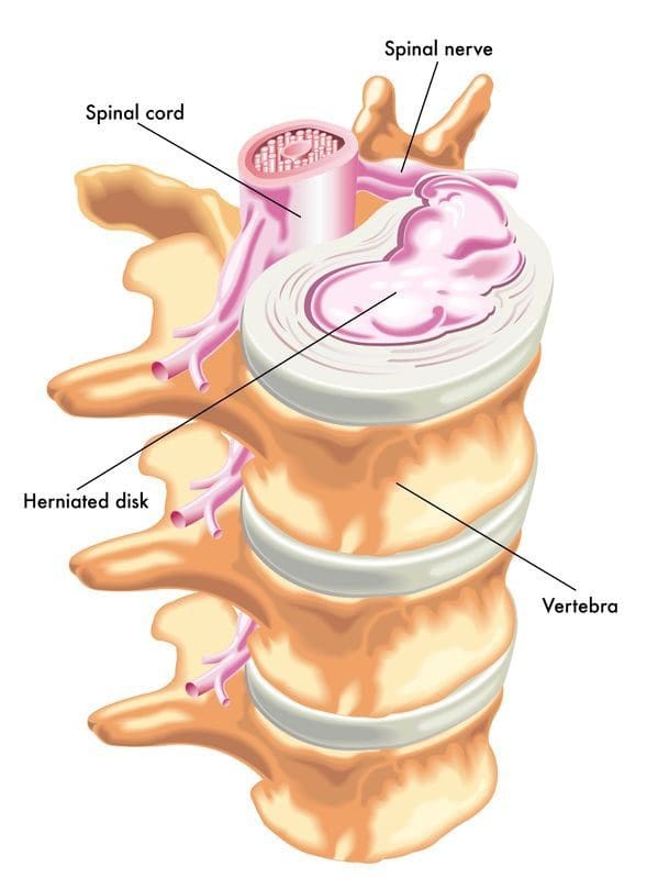
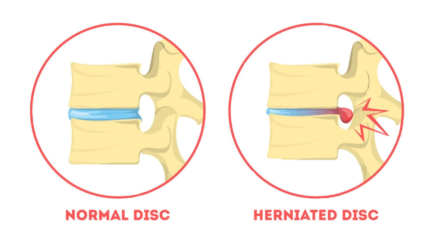
A slipped disc can mean a ruptured or herniated disc. We use the term slipped disc, however, the discs do not slip. Each disc is sandwiched between two vertebrae that are supported by a system of ligaments that hold the spine together. A bulging or herniated disc is the proper term.
Difference Between a Bulging and Herniated Disc
Disc disorders are categorized as contained or non-contained. Bulging disc is an example of a contained disc disorder.
Bulging
A bulging disc has not broken open meaning the nucleus is still contained inside the annulus fibrosus. The disc could protrude into the spinal canal without breaking open. The gel, the jelly interior does not leak out. The disc stays intact except a small bubble pops out.
Herniated/Ruptured
A non-contained disc has either partially or completely broken open, and that is a herniated/ruptured disc. Imagine a closed tube�being squeezed placed under pressure, which causes the contents to move wherever they can. If a portion of the tube is weak or there is too much pressure, the contents could leak or burst out. When a disc herniates the gel-like contents could spread out to the spinal cord and nerves.
Herniation Can Cause Pain
The disc’s gel-like nucleus has a chemical that irritates the nerves and causes them to become inflamed and swell. The chemical stays and continues to press on the irritated nerves. Sometimes fragments from the disc wall or the tube break off from the disc and drift into the spinal canal causing other nerves to inflame and swell. Based on the type of injury and condition of the discs, more than one disc could herniate, rupture, or bulge. Sometimes injury causes a combination of disc disorders.
Symptoms
Symptoms of a herniated disc can include:
Dull
Shooting pain
Muscle spasms
Cramping
Weakness
Tingling
Referred or radiating/traveling pain
Sometimes, however, a herniated disc does not cause any symptoms at all. When this happens it is called an asymptomatic herniated disc. Disc/s could be bulging or herniated, but if it or they are not applying pressure on the spinal nerve/sor the cord, symptoms like pain may not present. This makes a point about herniated disc symptoms that they are dependent on where you have a herniated disc.
Cervical Herniation Symptoms
With a herniated or bulging disc in the neck, then you could experience:
Neck soreness/pain
Muscle tightness
Cramping in the neck
Pain that travels down the arm/s
Tingling in the arm/s or hand/s
Weakness in the arm/s or hand/s
Lumbar Herniation Symptoms
With a herniated disc in the low back the following symptoms could happen:
Low back pain
Muscle tightness
Cramping in and around the low back
Radiating pain that travels down the leg/s
Tingling in your leg/s or foot/feet
Weakness in the leg/s or foot/feet
Referred Pain
Referred pain means that you have pain in another part of the body from the disc problem. An individual could have a bulging or herniated slipped disc in the low back and have pain in the leg. This is lumbar radiculopathy or sciatica. Usually, just one leg is affected. If you have a herniated disc in the neck, there could be referred pain going down the arm and into the hand.
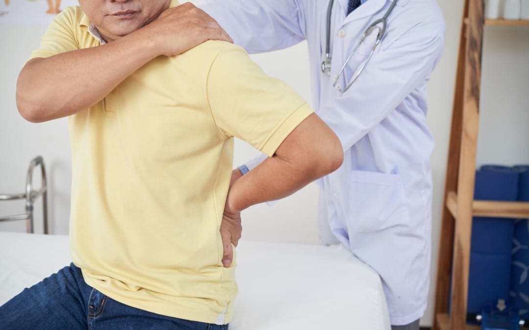
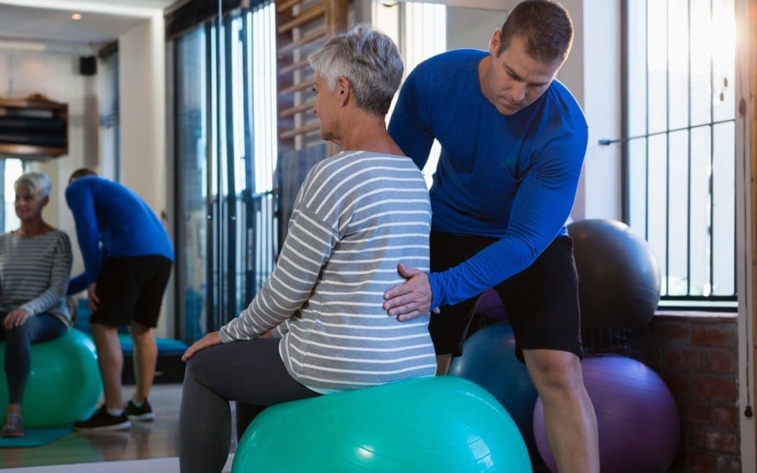
Chiropractic Cares
A chiropractor can help relieve back pain and other herniated disc symptoms. A chiropractor will go through your medical history, do a physical exam, and perform orthopedic and neurological tests. They are looking at several things. Orthopedic and neurological exams can help the chiropractor figure out what’s going on.
Are reflexes functioning properly?�Meaning are your nerves sending messages correctly. An example is a reflex test is when a doctor taps the knee with a hammer and the leg kicks.
Is there a loss of muscle strength?
Signs of muscle/s wasting away?
Is there a loss of sensation, tingling or numbness along the nerve/s path?
They will carefully look at posture, and will probably order an X-ray or MRI to help with the diagnosis.
Chiropractors evaluate the entire spine. Even if you only have lower back pain, your chiropractor will examine your neck, too. They want to see how well your spine is functioning overall. Remember what happens to one area of your spine can influence another part of the spine and/or body.
Pain from a herniated disc can make it difficult to enjoy daily life. Walking, sitting, and sleeping normally/comfortably can become a nightmare. You should make an appointment with a doctor or chiropractor if your herniated slipped disc symptoms last for more than two weeks.
Herniated Disc Treatment
NCBI Resources
In the United States alone, acute cases of lower back pain are the fifth most frequent reason for doctor visits and cause 40% of missed days off work. What�s more, it is the leading cause of disability worldwide.
Q: My primary healthcare provider recently diagnosed me with a herniated disc in the lumbar spine. They referred me to get chiropractic treatment, but I�m nervous because it’s new to me and I’m afraid of being adjusted wrong, paralyzed, etc. Can I trust chiropractic treatment to work?
A: It�s normal to be nervous about going to a chiropractic clinic.
If you’re not sure whether chiropractic is for you, there is scientific evidence that shows how chiropractic techniques like spinal manipulation/spinal adjustment and forms of manual/mechanical therapy are safe and effective for relieving pain and other musculoskeletal pain, conditions, and symptoms.
I encourage everyone to try chiropractic treatment as a non-surgical treatment option for a herniated disc.
It Is Your Decision
At the first appointment, a chiropractor will take a medical history and perform a thorough exam to determine the nature of the symptoms and their possible causes, which include a herniated disc.
Sometimes with a herniated disc, there may be no symptoms at all.
But usually a herniated disc causes:
Back pain
Referred pain or pain that is felt in other parts of the body like the legs, feet, etc.
An irritated spinal nerve can cause symptoms in the legs
This can lead to neurological symptoms like:
Tingling
Numbness
Weakness in the legs
Once the chiropractor determines your symptoms, they may use one or several techniques to relieve the back pain and other symptoms.
Techniques used by chiropractors for disc-related problems include:
Specific self-treatment exercises to improve motion & decrease back pain
Cox technique like spinal traction using special tables
Spinal manipulation
Hands-on techniques that relieve pain and restore movement to the spine and body
These techniques have been proven to be very safe. There are other techniques a chiropractor can recommend for various conditions, as each has their own style and method.
A chiropractic treatment plan will also include:
Education
Self-management instructions
This is to teach you how to control/eliminate pain with proper posture and proper body mechanics.
Whichever treatment the chiropractor recommends, he or she will discuss it with you, including the benefits and risks.
Although the treatments listed above will most likely be a part of your treatment plan, your chiropractor will answer your questions and work with you to select a treatment that meets your specific goals and preferences.
Don’t Be Nervous A Chiropractor Monitors Treatment Progress
If symptoms do not improve within a reasonable time frame, then a chiropractor may refer the patient to other treatments to manage disc-related pain, including:
Physical therapy
Acupuncture
Spinal injections
Surgery
Fortunately, self-management and time can be the best treatment. Allowing the body to heal itself is the way to go. But if rest is not enough then chiropractic treatment may be just what is needed to kick in the body’s self-healing function.
If you decide to give chiropractic treatment a try, don’t be nervous, as a chiropractor will monitor progress throughout the treatment.
In any case, chiropractors are qualified to discuss the benefits and risks of other treatments, depending on the condition.
Hopefully, this article has given you the basics of chiropractic medicine and how it works so you can make the best choice for your herniated disc/s.
Low Back Pain Management El Paso, TX Chiropractor
Denise suffered an auto accident injury which resulted in back pain. When she realized she could not sit, walk or sleep for lengthy periods of time without having painful symptoms, Denise found chiropractic care with Dr. Alex Jimenez at El Paso, TX. Once she received therapy for her automobile accident injuries, Denise experienced relief from her symptoms and she was able to execute her regular tasks once again. Thanks to the education and maintenance Dr. Alex Jimenez supplied, Denise regained her initial health and health.
Back pain is more most common, with roughly nine out of ten adults undergoing it at some time in their lifetime, and five functioning adults developing it annually. Some quote around 95 percent of Americans will experience back pain at some time in their lifetime. It is undoubtedly the typical cause of chronic pain since it’s also a substantial contributor to missed work and handicap. In the United States alone, acute cases of lower back pain are the fifth most frequent reason for doctor visits and cause 40% of missed days off work. What’s more, it is the leading cause of disability worldwide.
NCBI Resources
A herniated disc is a common spinal condition that typically affects the cervical spine (neck region) or the lumbar spine (lower back), although it can occur in any part of the spine. Most often, a herniated disc happens at the L4 � L5 and the L5 � S1.� This is because this portion of the spine, the lumbar region, bears the bulk of the body�s weight.
FDA recognizes and approves spinal decompression and its ability to eliminate herniated discs.
On the verge of back surgery, a mason discovered the non-surgical solution to work-related chronic back pain.
A new male patient who works in construction came to see me as a last resort to lessen his back pain brought on from damaged/herniated discs.
His primary caregiver recommended back surgery, but that would have put him on disability for months.
Fortunately, before saying yes to the surgery, a co-worker recommended chiropractic care.
Bricklayers/masons have the highest rate of back injuries with non-paid sick leave.
Constant bending over, even with a back brace, takes its toll on the spine, which in this case resulted in two herniated discs.
Pain medications helped in the beginning but with constant use, put him in a constant brain fog state, along with the expense, which took its toll on the family budget.
Disc Injury & Back Surgery
The doctor did not discuss spinal decompression therapy
�A non-surgical back treatment that slowly and gently stretches the spine.
This stretching lessens the pressure on the compressed nerve root (herniated disc) and results in less and even complete alleviation.
The patient came twice a week with myself and the team working on him over the course of a month, however, every case is different so treatments vary depending on the condition.
With each treatment, the two herniated discs were slowly reverted back to their natural position. This is able to be achieved with less pressure between the discs.
Towards the end of treatment, the patient’s pain was gone by about 90%.
With two weeks of rest, the patient was able to return to work.
The best part was that there was no surgery, pain medications, disability, and hospital bills.
Spine treatment alternative
Chiropractic/Decompression therapy is way less expensive than medication and surgery. It is:
Non-surgical
Recovery time is faster
Completely drug-free
People suffering every day with herniated/injured discs should consider the chiropractic decompression option. You do not have to learn to live with chronic back pain.
If you suffer from:
Herniated discs
Bulging discs
Degenerative disc disease
I encourage you to discuss the condition with an experienced chiropractor. There are many proven alternatives to back surgery and pain meds. People need to be aware of these alternatives for chronic back pain. The right-back pain treatment can definitely improve the quality of life.
Herniated Disc El Paso, TX
Sandra Rubio developed two herniated discs and a bulging disc after suffering from an accident at a young age, which caused her intense pain throughout her youth.
When she became a mother, her symptoms became severe.
After visiting doctors without results, Sandra found chiropractor Dr. Alex Jimenez and found relief from her sciatica and migraines.
The herniated disc treatment she received from Dr. Alex Jimenez was non-surgical.
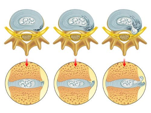
A herniated disc, also known as a slipped disc, is a medical condition in which:
Atear from the outer intervertebral disc allows the soft, central area to bulge out beyond the outer rings.
Disc herniation is usually a result of:
Degeneration (wear/tear)
Trauma (auto accident/sports injury)
Lifting injuries
Straining movement
The tear can release the compounds, which cause inflammation and can cause severe pain even if the nerve root not compressed.
A physical examination is usually the first step in diagnosing a herniated disc. The chiropractor will examine the spine while the patient is standing, and while they’re lying down. Depending on the severity and location of the herniation, they may note a decrease in spine curvature.
Radicular pain will be assessed, when the spine is:
Unmoving
In motion
With pressure applied
Other tests may be administered.
X-rays may also be taken, but an MRI is usually more accurate and shows more detail.
Chiropractic has been very effective in helping patients manage their pain and regain their mobility so they can return to their normal life. Therefore, it should be your first option for treatment before you go down the road with drugs or surgery.
NCBI Resources
It is often referred to as a ruptured disc or slipped disc and occurs when the disc moves or slips out of place. It can also be the result of a disc that has a small tear and is leaking the jelly-like substance that is inside. This can put pressure on the surrounding nerves, causing pain and discomfort.
The discs that cushion the vertebrae are made up of a tough outer layer and a softer inner layer. When the outer layer is damaged and the inner layer comes out into the spine, it is referred to as�disc herniation.
Often the symptoms of a herniated disc include back pain, as the inner layer of the disc puts pressure on nerves in the spine. A herniated disc can impact the sciatic nerve, leading to sciatica.
If you know that your sciatica was caused by a herniated disc then try these exercises and stretches to help reduce back and leg pain.
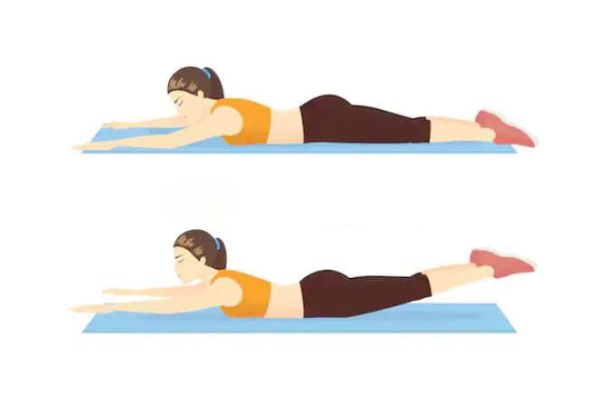
Exercises that help relieve sciatica from a herniated disc
If your spine specialist or chiropractor informed you that a herniated disc is what caused the pain also known as lumbar radiculopathy they may recommend� three sciatica exercises:
Prone on elbows into Press-up
Upper back extension
Opposite arm and leg extension
These stretches can help provide relief when the root cause of sciatica from herniated or bulging disc.
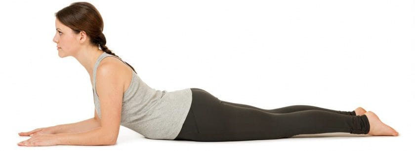
Prone Elbows/Press-Up
This exercise is to ease sciatica from herniated disc pain and pressure in the lumbar spine/low back.
How to:
Lay on stomach
Slowly push up until rested on forearms
Beginners hold for 30 seconds
Once strength is gained and you feel comfortable then hold for 3 to 5 minutes
Gently lower to the floor
Repeat 10 times
Once comfortable holding for 5 minutes then perform an extended arms version, which is like push-ups raising your arms to the point where your elbows lock
Upper Back Extension
This exercise is to strengthen and stabilize the low back muscles.
How to:
Lay on your stomach with a small pillow or rolled towel under your hips
Rest your arms at your sides
Slowly lift your upper body up off the floor, contracting your low back muscles as you rise
Hold the lifted position for 3 seconds
Slowly lower your body to the ground
Repeat 10 times
Throughout this exercise, keep movements fluid and controlled.
Opposite Arm and Leg Extension
This exercise is to stabilize your spine and strengthen your low back, hamstring and gluteus muscles.
How to:
Lay on your stomach with a small pillow or rolled towel under your abdomen
Extend both arms in front of you
Contract your abdominal muscles as you slowly lift both your right arm and left leg
Hold for 3 seconds. Lower your leg and arm down
Repeat with your left arm and right leg
Hold for 3 seconds
Repeat the exercise 5 to 10 times on each side
As you alternate the lifts, make sure to keep abdominal muscles contracted to get the full benefit.
How do these exercises relieve sciatica from herniated disc
These exercises and stretches are designed to move the pain from the leg and into the low back.
This is centralization/localization.
This is a good thing, as the goal is to get the pain centralized and back at the source.
When the leg pain goes away, it means the pressure on the sciatic nerve and related nerves has been removed.
If sciatic pain stretches down to the foot, you will feel these exercises, meaning that the pain and electrical sensations will move through the ankle and knee. Which means you’re doing it correctly.
This does not mean that the pain is immediately going to centralize to the low back, it does take time because you are trying to stretch and straighten out this long nerve.
But you will notice sciatica pain does not go as far down the leg.
Therefore�these exercises need to be done consistently and be made a part of your routine.
What to know before exercising
Before starting these stretches, consider three recommendations:
Get a doctor�s approval
A spine specialist should clear you to perform these stretches and exercises before you start.
While these exercises are safe, get a doctor�s permission before starting physical activity.
Know the cause so your exercise program helps and not makes the condition worse
Sciatica from herniated disc means a different type of exercise than sciatica caused by piriformis syndrome.
Knowing this information will help you choose an exercise plan that provides maximum relief.
Don�t push too hard
Don’t aggravate sciatica, listen to your body and go slow.
Experience any pain or symptoms:
Weakness
Tingling
Numbness
Contact a spine specialist immediately!
Sciatic nerve pain caused by a herniated or bulging disc is a common problem.
But incorporating these exercises and stretches can provide sustained relief.
The sciatic nerve is a large nerve that travels from the lower back down both of the legs and into the feet. When pressure is placed on the nerve, such as from a herniated disc, it can lead to the symptoms commonly referred to as sciatica.
The sciatic nerve can be impacted by a number of different things, including injury and degenerative diseases.
Difference Foot Orthotics Make to *REDUCE FOOT PAIN* & Correct Posture | El Paso, TX (2019)
Custom made foot orthotics can help control foot motion and posture. Healthcare professionals prescribe custom foot orthotics to help patients focus on their foot posture and mobility control. Research studies have ascertained that using custom foot orthotics for posture and mobility control can help fix excessive foot pronation and supination to prevent a variety of foot health problems. The subsequent video describes how custom foot orthotics will help control foot posture and mobility to improve health and wellness.
NCBI Resources
Sciatica is a common back ailment that affects approximately 1 in 10 adults in the United States. It is most prevalent in people between the ages of 25 and 45. Sciatica is characterized by a shooting pain that originates in the lower back and travels down through the hip, buttock, and back of the leg.
The pain can be so severe that it inhibits mobility and can prevent people from working, taking care of their homes, or just enjoying their life. Traditionally, doctors have treated the condition with medications and some invasive therapies, but chiropractic treatments have been found to be extremely effective in alleviating the pain and curing the condition.
A herniated disc is a common spinal disc issue. The spine is a very intricate structure, and when one component fails to function correctly, it can affect the entire body, causing pain and loss of mobility.
Tiny bones, called vertebrae, are stacked on each other to form the spine. They are joined in such a way to facilitate movement, flexibility, and a wide range of motion. There are small, fluid-filled discs that rest between each vertebra, providing a cushion between the bones. When one of these discs becomes damaged, it can affect the surrounding nerves, causing pain and making movement difficult.
What is It?
A herniated disc is a common spinal condition that typically affects the cervical spine (neck region) or the lumbar spine (lower back), although it can occur in any part of the spine. Most often, a herniated disc happens at the L4 � L5 and the L5 � S1. This is because this portion of the spine, the lumbar region, bears the bulk of the body�s weight.
It is often referred to as a ruptured disc or slipped disc and occurs when the disc moves or slips out of place. It can also be the result of a disc that has a small tear and is leaking the jelly-like substance that is inside. This can put pressure on the surrounding nerves, causing pain and discomfort.
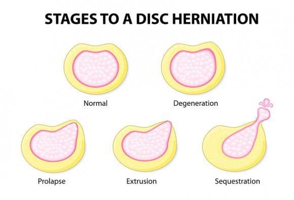
The first two stages are called incomplete herniations while the last two stages are called complete herniations.
Symptoms of a herniated disc may increase or worsen as the condition progresses although some patients do not experience any at all Typical symptoms include:
Pain in the affected area
Tingling
Numbness
Weakness
Leg or arm pain
Loss of reflex
Loss of mobility
Loss of flexibility
Decreased range of motion
What Causes It?
There are several causes. The most common are aging and degeneration, overuse, and normal wear and tear on the body.
A herniated disc resulting from an injury or trauma, such as a blow to the back, is less common, but it does happen. Because the back does bear most of the body�s weight, it can put a significant amount of pressure on the spine and discs. Over time, the discs may begin to weaken and a herniation can occur.
Injury or trauma that results in a herniation may include a car accident that involves sudden jerking, or incorrectly lifting heaving objects can put excessive pressure on the spine, causing it to herniate.
How is it Diagnosed?
A physical examination is usually the first step in diagnosing a herniated disc. The physician or chiropractor will examine the spine while the patient is standing, then while they are lying down. Depending on the severity and location of the herniation, they may note a decrease in spinal curvature.
Radicular pain will also be assessed, when the spine is unmoving, while in motion, and when pressure is applied. Other tests may also be administered. X-rays may also be taken, but an MRI is usually more accurate and provides greater detail.
What are the Treatments?
Medications may be recommended or prescribed, including NSAIDs, narcotics, muscle relaxers, and anticonvulsants. Some doctors may advise cortisone injections to reduce inflammation. Physical therapy may be recommended as a stand-alone treatment or in conjunctions with other treatments. Surgery for herniated discs is rare and usually reserved as a last resort option.
Chiropractic has been very effective in helping patients manage their pain and regain their mobility so they can return to their normal life. Therefore, it should be your first option for treatment before you go down the road with drugs or surgery.
Aracely Pisana saw Dr. Alex Jimenez, doctor of chiropractic in El Paso, Tx, for the very first time after many other treatment efforts were not able to supply her with the back pain relief she’d needed. Aracely Pisana describes how well Dr. Alex Jimenez and his staff have taken care of her and she adds that their services are what keeps her coming back to chiropractic care. Aracely Pisana has recovered her quality of life and she highly recommends Dr. Alex Jimenez as the non-surgical selection for back pain.
Chiropractic Treatment
�
We are blessed to present to you�El Paso�s Premier Wellness & Injury Care Clinic.
As El Paso�s Chiropractic Rehabilitation Clinic & Integrated Medicine Center,�we passionately are focused on treating patients after frustrating injuries and chronic pain syndromes. We focus on improving your ability through flexibility, mobility and agility programs tailored for all age groups and disabilities.
We want you to live a life that is fulfilled with more energy, positive attitude, better sleep, less pain, proper body weight and educated on how to maintain this way of life. I have made a life of taking care of every one of my patients.
I assure you, I will only accept the best for you�
If you have enjoyed this video and we have helped you in any way, please feel free to subscribe and recommend�us.
Spinal trauma consists of spine fractures, or spinal fractures, and spinal cord injuries. Approximately 12,000 spinal trauma cases are reported in the United States every year. While the most prevalent causes of spinal cord injuries and spine fractures are automobile accidents and falls, spinal trauma can also be attributed to assault, sports injuries, and work-related accidents. Diagnosis of spinal trauma includes imaging and assessment of nerve function, such as reflex, motor, and sensation. The following article discusses the role of emergency radiology in spinal trauma. Chiropractic care can help provide diagnostic evaluations for spinal trauma.
Abstract
Spinal trauma is very frequent injury with different severity and prognosis varying from asymptomatic condition to temporary neurological dysfunction, focal deficit or fatal event. The major causes of spinal trauma are high- and low- energy fall, traffic accident, sport and blunt impact. The radiologist has a role of great responsibility to establish the presence or absence of lesions, to define the characteristics, to assess the prognostic influence and therefore treatment. Imaging has an important role in the management of spinal trauma. The aim of this paper was to describe: incidence and type of vertebral fracture; imaging indication and guidelines for cervical trauma; imaging indication and guidelines for thoracolumbar trauma; multidetector CT indication for trauma spine; MRI indication and protocol for trauma spine.
Introduction
The trauma of the spine weighs heavily on the budget of social and economic development of our society. In the USA, 15�40 cases per million populations with 12,000 cases of paraplegia every year, 4000 deaths before admission and 1000 deaths during hospitalization are estimated. The young adult population is the most frequently involved in road accidents, followed by those at home and at work, with a prevalence of falls from high and sports injuries.1
Imaging has an important role in the management of spinal trauma. Quick and proper management of the patients with trauma, from diagnosis to therapy, can mean reduction of the neurological damage of vital importance for the future of the patient. Radiologists have a role of great responsibility to establish the presence or absence of lesions, defining the characteristics, assessing the prognostic influence and therefore treatment.
The aim of this paper was to describe:
incidence and type of vertebral fracture
imaging indication and guidelines for cervical trauma
imaging indication and guidelines for thoracolumbar trauma
multidetector CT (MDCT) pattern for trauma spine
MRI pattern for trauma spine.
Spinal trauma, including spine fractures and spinal cord injuries, represent about 3 percent to 6 percent of all skeletal injuries. Diagnostic assessments are fundamental towards the complex diagnosis of spinal trauma. While plain radiography is the initial diagnostic modality used for spine fractures and/or spinal cord injuries, CT scans and MRI can also help with diagnosis. As a chiropractic care office, we can offer diagnostic assessments, such as X-rays, to help determine the best treatment.
Dr. Alex Jimenez D.C., C.C.S.T.
Vertebral Fracture Management and Imaging Indication and Evaluation
The rationale of imaging in spinal trauma is:
To diagnose the traumatic abnormality and characterize the type of injury.
To estimate the severity, potential spinal instability or damaged stability with or without neurological lesion associated, in order to avoid neurological worsening with medical legal issue.
To evaluate the state of the spinal cord and surrounding structures (MR is the gold standard technique).
Clinical evaluation involving different specialities�emergency medicine, trauma surgery, orthopaedics, neurosurgery and radiology or neuroradiology�and trauma information is the most important key point in order to decide when and which type of imaging technique is indicated.2
A common question in patients with spine trauma is: is there still a role for plain-film X-ray compared with CT?
In order to clarify when and what is more appropriate for spinal trauma, different guidelines were published distinguishing cervical and thoracolumbar level.
Cervical Spinal Trauma: Standard X-Ray and Multidetector CT Indication
For cervical level, controversy persists regarding the most efficient and effective method between cervical standard X-ray with three film projections (anteroposterior and lateral view plus open-mouth odontoid view) and MDCT.
X-ray is generally reserved for evaluating patients suspected of cervical spine injury and those with injuries of the thoracic and lumbar areas where suspicion of injury is low. Despite the absence of a randomized controlled trial and thanks to the high quality and performance of�MDCT and its post-processing (multiplanar reconstruction and three-dimensional volume rendering), the superiority of cervical CT (CCT) compared with cervical standard X-ray for the detection of clinically significant cervical spine injury is well demonstrated.
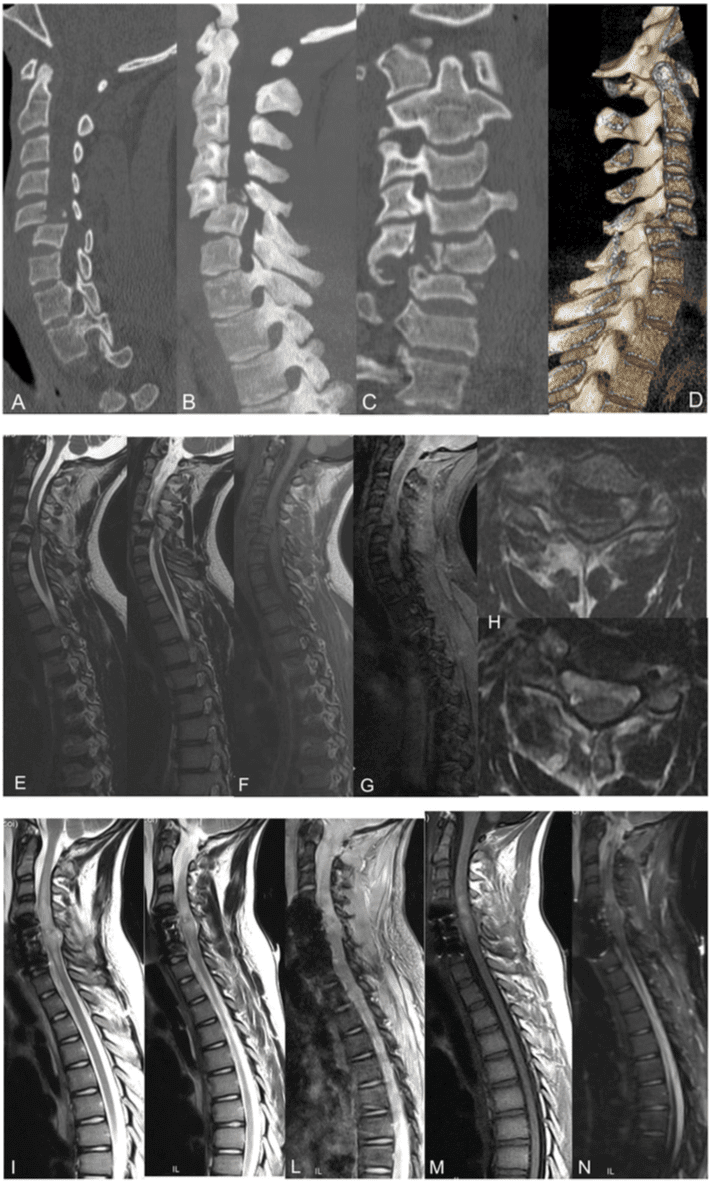
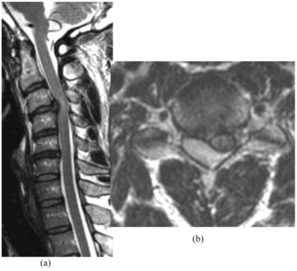
Figure 1. (a�l). A 20-year-old male involved in a motorbike accident. The multidetector CT with multiplanar reformatted and three- dimensional volume-rendering reconstructions (a�d) showed traumatic fracture of C6 with traumatic posterior spondylolisthesis grade III with spinal cord compression. The MRI (e�h) confirmed the traumatic fracture of C6 with traumatic posterior spondylolisthesis grade III with severe spinal cord compression. The post-surgical treatment MRI control (i�l) showed the sagittal alignment of cervical level and severe hyperintensity signal alteration of the spinal cord from C3 to T1.
In order to reduce the patient radiation exposure, it is important to determine and to select patients who need imaging and those who do not, through the clinical evaluation and probability of cervical spine injury, using only MDCT for the appropriate patient as is more cost-effective screening.3
First of all, it is necessary to distinguish the type of trauma:
minor trauma (stable patient, mentally alert, not under the influence of alcohol or other drugs and who has no history or physical findings suggesting a neck injury)
major and severe trauma (multitrauma, unstable patient with a simple temporary neurological dysfunction, with focal neurological deficit or with a history or mechanism of injury sufficient to have exceeded the physiologic range of motion).
Second, it is important to establish if trauma risk factors are presents, such as:
violence of trauma: high-energy fall (high risk) or low-energy fall (low risk)
age of the patient: <5years old, >65 years old�
associated lesions: head, chest, abdomen (multitrauma) etc.
clinical signs: Glasgow Coma Scale (GCS), neurological deficit, vertebral deformation.
Combining these elements, patients can be divided into �low risk� and �high risk� for cervical injury.
The first group consists of patients who are awake (GCS 15), alert, cooperative and non-intoxicated without any distract- ing injury.
The second group consists of unconscious, sedated, intoxicated or non-cooperative patients or those with a distracting injury or an altered mental state (GCS ,15) with a 5% chance of cervical spine injuries.3,4
CCT has a wider indication than X-ray for patients at very high risk of cervical spine injury (major trauma or multitrauma). No evidence suggests CCT instead of X-ray for a patient who is at low risk for cervical spine injury.5
Figure 2. (a�g). A 30-year-old male involved in a motorbike accident. The multidetector CT with multiplanar reformatted and three-dimensional volume-rendering reconstructions (a�d) showed traumatic burst fracture of L1 (A2-type Magerl class) with posterior bone fragment dislocation into spinal canal. The MRI (e�g) confirmed the burst fracture of L1 with moderate spinal cord compression.
Figure 3. (a�d) A 50-year-old male involved in a motorbike accident with acute spinal cord compression symptoms on anticoagulation treatment. The MRI showed an acute haemorrhagic lesion at the C2�C4 posterior epidural space, hypointense on sagittal T1 weighted (a) and hyperintense on T2 weighted (b) with spinal cord compression and dislocation on axial T2* (c) and T2 weighted (d).
In 2000, the National Emergency X-Radiography Utilization (NEXUS) study, analysing 34,069 patients, established low-risk criteria to identify patients with a low probability of cervical spine injury, who consequently needed no cervical spine�imaging. To meet the NEXUS criteria, a patient must have the following conditions:
no tenderness at the posterior midline of the cervical spine
no focal neurologic deficit
normal level of alertness
no evidence of intoxication
no clinically apparent painful injury that might distract the patient from the pain of a cervical spine injury.6
If all of these roles are present, the patient does not need to undergo X-ray because he has a low possibility of having a cervical spine injury with a sensitivity of 99% and a specificity of 12.9%.7
In 2001, the Canadian C-spine rule (CCSR) study developed a second decision rule using the risk factor of the trauma: three high-risk criteria (age $ 65 years, dangerous mechanism and paraesthesias in extremities), five low-risk criteria (simple rear-end motor vehicle collision, sitting position in emergency department, ambulatory at any time, delayed onset of neck pain and absence of midline cervical spine tenderness) and the ability of the patient to actively rotate his or her neck to determine the need for cervical spine radiography. In practice, if one of these risk factors is present, the patient needs to undergo imaging evaluation. On the other hand, if the risk factors are not present, the use of the NEXUS criteria plus a functional evaluation of the cervical spine is needed (left and right cervical spine rotation .45�); if this functional evaluation is possible, imaging is unnecessary. If an incomplete cervical movement is present, then the patient needs to be checked with imaging. The results showed the criteria to have a sensitivity of up to 100% and a specificity of up to 42.5%.8
Applying these criteria, before cervical spine imaging, the authors report a decrease of about 23.9% in the number of negative CCT, and applying a more liberal NEXUS criteria including the presence or absence of pain, limited range of motion or posterolateral cervical spine tenderness, they report a decrease of up to 20.2% in the number of negative studies.2
If these clinical criteria cannot be applied, CCT must be performed.
Major and severe traumas request a direct CCT screening, especially because there could be associated lesions, according to the high-risk criteria developed by Blackmore and Hanson to identify patients with trauma at high risk of c-spine injury who would benefit from CT scanning as the primary radiological investigation9 Figure 1.
Thoracolumbar Spinal Trauma: Standard X-Ray and Multidetector CT Indication
For thoracolumbar level, MDCT is a better examination for depicting spine fractures than conventional radiography. It has wider indication in the diagnosis of patients with thoracolumbar trauma for bone evaluation. It is faster than X-ray, more sensitive, thanks to multiplanar reformatted or volume-rendering reconstruction detecting small cortical fracture, and the sagittal alignment can be evaluated with a wide segment evaluation.10
It can replace conventional radiography and can be performed alone in patients who have sustained severe trauma.10
In fact, thoracolumbar spinal injuries can be detected during visceral organ-targeted CT protocol for blunt traumatic injury.
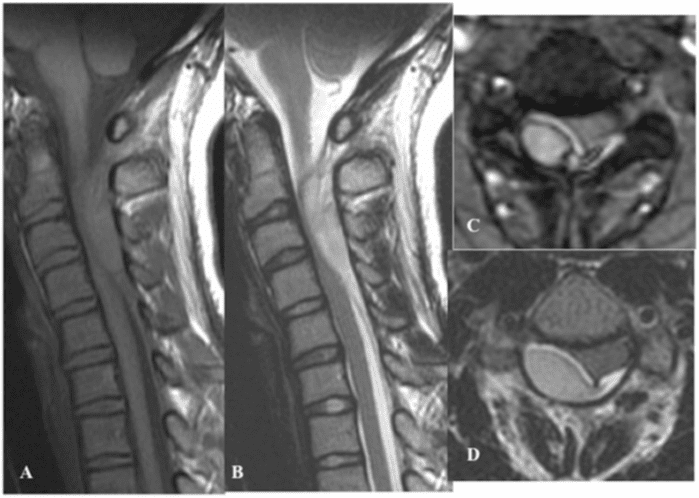
Figure 4. A 55-year-old female involved in a car accident with acute left cervical brachialgia. The sagittal T2 weighted (a) and axial T2 weighted (b) MRI showed a post-traumatic posterolateral herniated disc with spinal cord compression and soft hyper signal alteration on the C3�C4 spinal cord.
Thanks to multidetector technology, images reconstructed using a soft algorithm and wide-display field of view that covers the entire abdomen using a visceral organ-targeted protocol with 1.5-mm collimation are sufficient for the evaluation of spine fractures in patients with trauma, given that multiplanar reformatted images are provided without performing new CT study and without increasing radiation dose11 Figure 2.
With MDCT there is no information about spinal cord status or ligament lesion or acute epidural haematoma; it can only evaluate bone status. Spinal cord injury is suspected only by clinical data.
CCT is strictly recommended in patients affected by blunt cerebrovascular injuries. Both lesions can be strictly correlated and generally; contrast medium administration to exclude hemorrhagic brain lesion and cervical fracture is not needed.10
Magnetic resonance imaging, or MRI, is a medical diagnostic assessment technique utilized in radiology to create pictures of the anatomy and the physiological processes of the human body. Alongside radiography and CT scans, MRI can be helpful in the diagnosis of spinal trauma, including spine fractures and spinal cord injuries. Magnetic resonance imaging may not be necessary for all cases of spinal trauma. However, it could provide detailed information on the other soft tissues of the spine.�
Dr. Alex Jimenez D.C., C.C.S.T.
Spinal Trauma and MRI
Even if MDCT is the first imaging modality in a patient with trauma, MRI is essential for the soft assessment of the ligament, muscle or spinal cord injury, spinal cord, disc, ligaments and neural elements, especially using T2 weighted sequences with fat suppression or T2 short tau inversion recovery (STIR) sequence.12 MRI is also used to classify burst fracture, obtaining information about the status of the posterior ligamentous complex, a critical determinant of surgical indication even if the diagnosis of ligament injuries remains complex, and its grade is also underestimated using high-field MRI.13
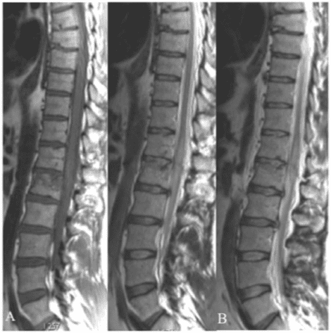
Figure 5. A 65-year-old female involved in domestic trauma with spinal cord symptoms. The sagittal T1 weighted (a) and T2 weighted (b) MRI showed a traumatic T12�L1 spinal cord contusion hypointense on T1 weighted and hyperintense on T2 weighted.
In the management of patients with polytrauma, MDCT total-body scan is necessary in an emergency condition, and�MRI whole-spine indication is secondary to the clinical status of the patient: spinal cord compression syndrome Figure 3�5�MRI protocols recommended for patients affected by spinal injury and trauma are the following:13,14
Sagittal T1 weighted, T2 weighted and STIR sequence for the�bone marrow and spinal cord injury or spinal cord compression evaluation owing to epidural haematoma or traumatic herniated disc
Sagittal gradient echo T2* sequence for haemorrhage evaluation of the spinal cord or into the epidural�subdural space
Sagittal diffusion-weighted imaging helpful when evaluating spinal cord injury, differentiating cytotoxic from vasogenic�oedema, assisting in detecting intramedullary haemorrhage. It can help to evaluate the degree of compressed spinal cord.
Axial T1 weighted and T2 weighted sequence for the right localization of the injury. Recently, for patients affected by acute blunt trauma and cervical spinal cord injury, the axial T2 weighted sequence has been shown to be important for trauma-predicting outcomes. On axial T2 weighted imaging, five patterns of intramedullary spinal cord signal alteration can be distinguished at the injury�s epicentre. Ordinal values ranging from 0 to 4 can be assigned to these patterns as Brain�and Spinal Injury Center scores, which encompassed the spectrum of spinal cord injury severity correlating with neurological symptoms and MRI axial T2 weighted imaging. This score improves on current MRI-based prognostic descriptions for spinal cord injury by reflecting functionally and anatomically significant patterns of intramedullary T2 signal abnormality in the axial plane.15
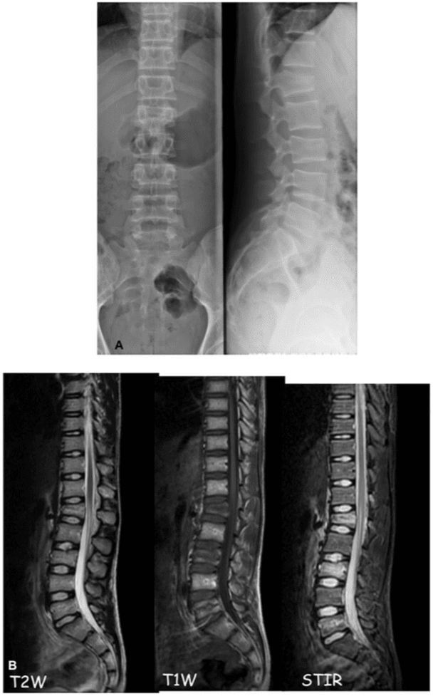
Figure 6. A 20-year-old female involved in domestic trauma with back pain resistance to medical therapy. The standard antero- posterior�laterolateral X-ray (a) showed no vertebral fractures. The MRI showed a bone marrow alteration at lumbar vertebral body hyperintense on T2 weighted (T2W) (a), hypointense on T1 weighted (T1W) (b) and short tau inversion recovery (STIR) (c).
MRI has also an important role in case of discordance between clinical status and CT imaging. In the absence of vertebral fracture, patients can suffer from back pain resistant to medical therapy owing to bone marrow traumatic oedema that can be detected only using STIR sequence on MRI Figure 6.
In spinal cord injury without radiologic abnormalities (SCI- WORA), MRI is the only imaging modality that can detect intramedullary or extramedullary pathologies or show the absence of neuroimaging abnormalities.16 SCIWORA refers to spinal injuries, typically located in the cervical region, in the absence of identifiable bony or ligamentous injury on complete, technically adequate, plain radiographs or CT. SCIWORA should be suspected in patients subjected to blunt trauma who report early or transient symptoms of neurologic deficit or who have existing findings upon initial assessment.17
Vertebral Fracture Type and Classification
The rationale of imaging is to distinguish the vertebral fracture type into two groups:
� vertebral compression fracture as vertebral body fracture compressing the anterior cortex, sparing the middle posterior columns associated or not with kyphosis � burst fracture as comminuted fracture of the vertebral body extending through both superior and inferior endplates with kyphosis or posterior displacement of the bone into the canal. and to distinguish which type of treatment the patient needs; by imaging, it is possible to classify fractures into stable or�unstable fracture, giving indication to conservative or surgical therapy.
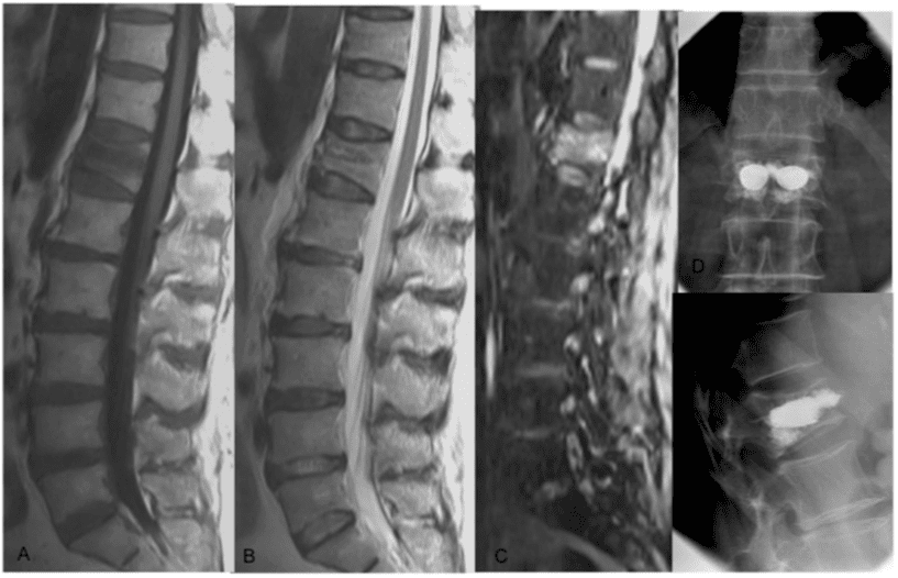
Figure 7. (a�f) A 77-year-old female involved in domestic trauma with back pain resistance to medical therapy. The multidetector CT (a) showed no vertebral fractures. The MRI showed a Magerl A1 fracture with bone marrow oedema at T12�L1 vertebral body hypointense on T1 weighted (b), hyperintense on T2 weighted (c) and short tau inversion recovery (d) treated by vertebroplasty (e�f).
Figure 8. (a�d) A 47-year-old male involved in a motorbike accident with back pain resistance to medical therapy. The MRI showed a Magerl A1 fracture with bone marrow oedema at T12 vertebral body hypointense on T1 weighted (a) hyperintense on T2 weighted (b) and short tau inversion recovery (c) treated by assisted-technique vertebroplasty�vertebral body stenting technique (d).
Using MDCT and MRI, thanks to morphology and injury distribution, various classification systems have been used for identifying those injuries that require surgical intervention, distinguishing among stable and unstable fractures and surgical and non-surgical fractures.1
Denis proposed the �three-column concept�, dividing the spinal segment into three parts: anterior, middle and posterior columns. The anterior column comprises the anterior longitudinal ligament and anterior half of the vertebral body; the middle column comprises the posterior half of the vertebral body and posterior longitudinal ligament; and the posterior column comprises the pedicles, facet joints and supraspinous ligaments. Each column has different contributions to stability, and their damages may affect stability differently. Generally, if two or more of these columns are damaged, the spine becomes unstable.18
Magerl divided the vertebral compression fracture (VCF) into three main categories according to trauma force: (a) compression injury, (b) distraction injury and (c) rotation injury. Type A has conservative or non-surgical mini-invasive treatment indication.19
The thoracolumbar injury classification and severity score (TLICS) system assigns numerical values to each injury based on the categories of morphology of injury, integrity of the posterior ligament and neurological involvement. Stable injury patterns (TLICS,4) may be treated non-operatively with�brace immobilization. Unstable injury patterns (TLICS.4) may be treated operatively with the principles of deformity correction, neurological decompression if necessary and spinal stabilization.20
The Aebi classification is based on three major groups: A = isolated anterior column injuries by axial compression, B = disruption of the posterior ligament complex by distraction posteriorly and C = corresponding to group B but with rotation. There is an increasing severity from A to C, and within each group, the severity usually increases within the subgroups from 1 to 3. All these pathomorphologies are supported by the mechanism of injury, which is responsible for the extent of the injury. The type of injury with its groups and subgroups is able to suggest the treatment modality.21
Thoracolumbar Fracture and Mini-Invasive Vertebral Augmentation Procedure: Imaging Target
Recently, different mini-invasive procedures called assisted- technique vertebroplasty (balloon kyphoplasty KP or kyphoplasty-like techniques) have been developed in order to obtain pain relief and kyphosis correction as alternative treatment for non-surgical but symptomatic vertebral fracture.
The rationale of these techniques is to combine the analgesic and vertebral consolidation effect of vertebroplasty with the restoration of the physiological height of the collapsed vertebral body, reducing the kyphotic deformity of the vertebral body, delivering cement into the fractured vertebral body with a vertebral stabilization effect compared with conservative therapy (bed rest and medical therapy).22
From interventional point of view, imaging has an important role for treatment indication together with clinical evaluation. Both MDCT and MRI are recommended Figure 7 and 8.
In fact, MDCT has the advantage of diagnosing VCF with kyphosis deformity easily, while MRI with STIR sequence is useful to evaluate bone marrow oedema, an important sign of back pain.
Patients affected by vertebral fracture without bone marrow oedema on STIR sequence are not indicated for interventional procedure.
According to imaging, Magerl A1 classification fractures are the main indication of treatment.
However, the treatment must be performed within 2�3 weeks from trauma in order to avoid sclerotic bone response: the younger the fractures, the better the results and easier the treatment and vertebral augmentation effect. To exclude sclerotic bone reaction, CT is recommended.
Conclusion
The management of spinal trauma remains complex. MDCT has a wide indication for bone evaluation in patients affected by severe trauma or patients with high risk of spine injury. MRI has a major indication in the case of spinal cord injury and the absence of bone lesion. Diagnostic assessment of spinal trauma, including radiography, CT scans, and MRI are fundamental towards the diagnosis of spine fractures and spinal cord injury for treatment. The scope of our information is limited to chiropractic as well as to spinal injuries and conditions. To discuss the subject matter, please feel free to ask Dr. Jimenez or contact us at�915-850-0900�.
Curated by Dr. Alex Jimenez
Additional Topics: Acute Back Pain
Back pain�is one of the most prevalent causes of disability and missed days at work worldwide. Back pain attributes to the second most common reason for doctor office visits, outnumbered only by upper-respiratory infections. Approximately 80 percent of the population will experience back pain at least once throughout their life. The spine is a complex structure made up of bones, joints, ligaments, and muscles, among other soft tissues. Because of this, injuries and/or aggravated conditions, such as�herniated discs, can eventually lead to symptoms of back pain. Sports injuries or automobile accident injuries are often the most frequent cause of back pain, however, sometimes the simplest of movements can have painful results. Fortunately, alternative treatment options, such as chiropractic care, can help ease back pain through the use of spinal adjustments and manual manipulations, ultimately improving pain relief.
Pneumaticos SG, Triantafyllopoulos GK, Gian- noudis PV. Advances made in the treatment of thoracolumbar fractures: current trends and future directions. Injury 2013; 44: 703�12. doi: 10.1016/j.injury.2012.12.005
Griffith B, Bolton C, Goyal N, Brown ML, Jain R. Screening cervical spine CT in a level I trauma center: overutilization? AJR Am J Roentgenol 2011; 197: 463�7.doi: 10.2214/ AJR.10.5731
Hanson JA, Blackmore CC, Mann FA, Wilson AJ. Cervical spine injury: a clinical decision rule to identify high-risk patients for helical CTscreening. AJR Am J Roentgenol 2000; 174: 713�17.
Saltzherr TP, Fung Kon Jin PH, Beenen LF, Vandertop WP, Goslings JC. Diagnostic imaging of cervical spine injuries following blunt trauma: a review of the literature and practical guideline. Injury 2009; 40: 795�800. doi: 10.1016/j.injury.2009.01.015
Holmes JF, Akkinepalli R. Computed to- mography versus plain radiography to screen for cervical spine injury: a meta-analysis. J Trauma 2005; 58: 902�5. doi: 10.1097/01. TA.0000162138.36519.2A
Hoffman JR, Wolfson AB, Todd K, Mower WR. Selective cervical spine radiography in blunt trauma: methodology of the National Emergency X-Radiography Utilization Study (NEXUS). Ann Emerg Med 1998; 32: 461�9. doi: 10.1016/S0196-0644(98)70176-3
Dickinson G, Stiell IG, Schull M, Brison R, Clement CM, Vandemheen KL, et al. Retro- spective application of the NEXUS low-risk criteria for cervical spine radiography in Canadian emergency departments. Ann Emerg Med 2004; 43: 507�14. doi: 10.1016/j. annemergmed.2003.10.036
Stiell IG, Wells GA, Vandemheen KL, Clem- ent CM, Lesiuk H, De Maio VJ, et al. The Canadian C-spine rule for radiography in
alert and stable trauma patients. JAMA 2001;
286: 1841�8. doi: 10.1001/jama.286.15.1841 9. Berne JD, Velmahos GC, El-Tawil Q, Deme- triades D, Asensio JA, Murray JA, et al. Value
of complete cervical helical computed to- mographic scanning in identifying cervical spine injury in the unevaluable blunt trauma patient with multiple injuries: a prospective study. J Trauma 1999; 47: 896�902. doi: 10.1097/00005373-199911000-00014
10. Wintermark M, Mouhsine E, Theumann N, Mordasini P, van Melle G, Leyvraz PF, et al. Thoracolumbar spine fractures in patients who have sustained severe trauma: depiction with multi-detector row CT. Radiology 2003; 227: 681�9. doi: 10.1148/radiol.2273020592
11. Kim S, Yoon CS, Ryu JA, Lee S, Park YS, Kim SS, et al. A comparison of the diagnostic performances of visceral organ-targeted ver- sus spine-targeted protocols for the evalua- tion of spinal fractures using sixteen-channel multidetector row computed tomography: is additional spine-targeted computed tomog- raphy necessary to evaluate thoracolumbar spinal fractures in blunt trauma victims? J Trauma 2010; 69: 437�46. doi: 10.1097/ TA.0b013e3181e491d8
12. Pizones J, Castillo E. Assessment of acute thoracolumbar fractures: challenges in mul- tidetector computed tomography and added value of emergency MRI. Semin Musculoskelet Radiol 2013; 17: 389�95. doi: 10.1055/s- 0033-1356468
13. Emery SE, Pathria MN, Wilber RG, Masaryk T, Bohlman HH. Magnetic resonance imag- ing of posttraumatic spinal ligament injury. J Spinal Disord 1989; 2: 229�33. doi: 10.1097/ 00002517-198912000-00003
14. Zhang JS, Huan Y. Multishot diffusion- weighted MR imaging features in acute trauma of spinal cord. Eur Radiol 2014; 24: 685�92. doi: 10.1007/s00330-013-3051-3
15. Talbott JF, Whetstone WD, Readdy WJ, Ferguson AR, Bresnahan JC, Saigal R, et al. The Brain and Spinal Injury Center score: a novel, simple, and reproducible method for assessing the severity of acute cervical spinal cord injury with axial T2-weighted MRI findings. J Neurosurg Spine 2015; 23: 495�504. doi: 10.3171/2015.1.SPINE141033
16. Boese CK, Oppermann J, Siewe J, Eysel P, Scheyerer MJ, Lechler PJ. Spinal cord injury without radiologic abnormality in children: a systematic review and meta-analysis. Trauma Acute Care Surg 2015; 78: 874�82. doi: 10.1097/TA.0000000000000579
17. Brown RL, Brunn MA, Garcia VF. Cervical spine injuries in children: a review of 103 patients treated consecutively at a level 1 pediatric trauma center. J Pediatr Surg 2001; 36: 1107�14. doi: 10.1053/jpsu.2001.25665
18. Denis F. The three column spine and its significance in the classification of acute thoracolumbar spinal injuries. Spine (Phila Pa 1976) 1983; 8: 817�31. doi: 10.1097/ 00007632-198311000-00003
19. Magerl F, Aebi M, Gertzbein SD, Harms J, Nazarian S. A comprehensive classification of thoracic and lumbar injuries. Eur Spine J 1994; 3: 184�201.
20. Patel AA, Dailey A, Brodke DS, Daubs M, Harrop J, Whang PG, et al; Spine Trauma Study Group. Thoracolumbar spine trauma classification: the Thoracolumbar Injury Classification and Severity Score system and case examples. J Neurosurg Spine 2009; 10: 201�6. doi: 10.3171/2008.12.SPINE08388
21. Aebi M. Classification of thoracolumbar fractures and dislocations. Eur Spine J 2010; 19(Suppl. 1): S2�7. doi: 10.1007/s00586-009-1114-6
22. Muto M, Marcia S, Guarnieri G, Pereira V. Assisted techniques for vertebral cementoplasty: why should we do it? Eur J Radiol 2015; 84: 783�8. doi: 10.1016/j.ejrad.2014.04.002
IFM's Find A Practitioner tool is the largest referral network in Functional Medicine, created to help patients locate Functional Medicine practitioners anywhere in the world. IFM Certified Practitioners are listed first in the search results, given their extensive education in Functional Medicine