Dr. Alex Jimenez at El Paso Back Clinic®: Beating Back Pain from Long Desk Hours
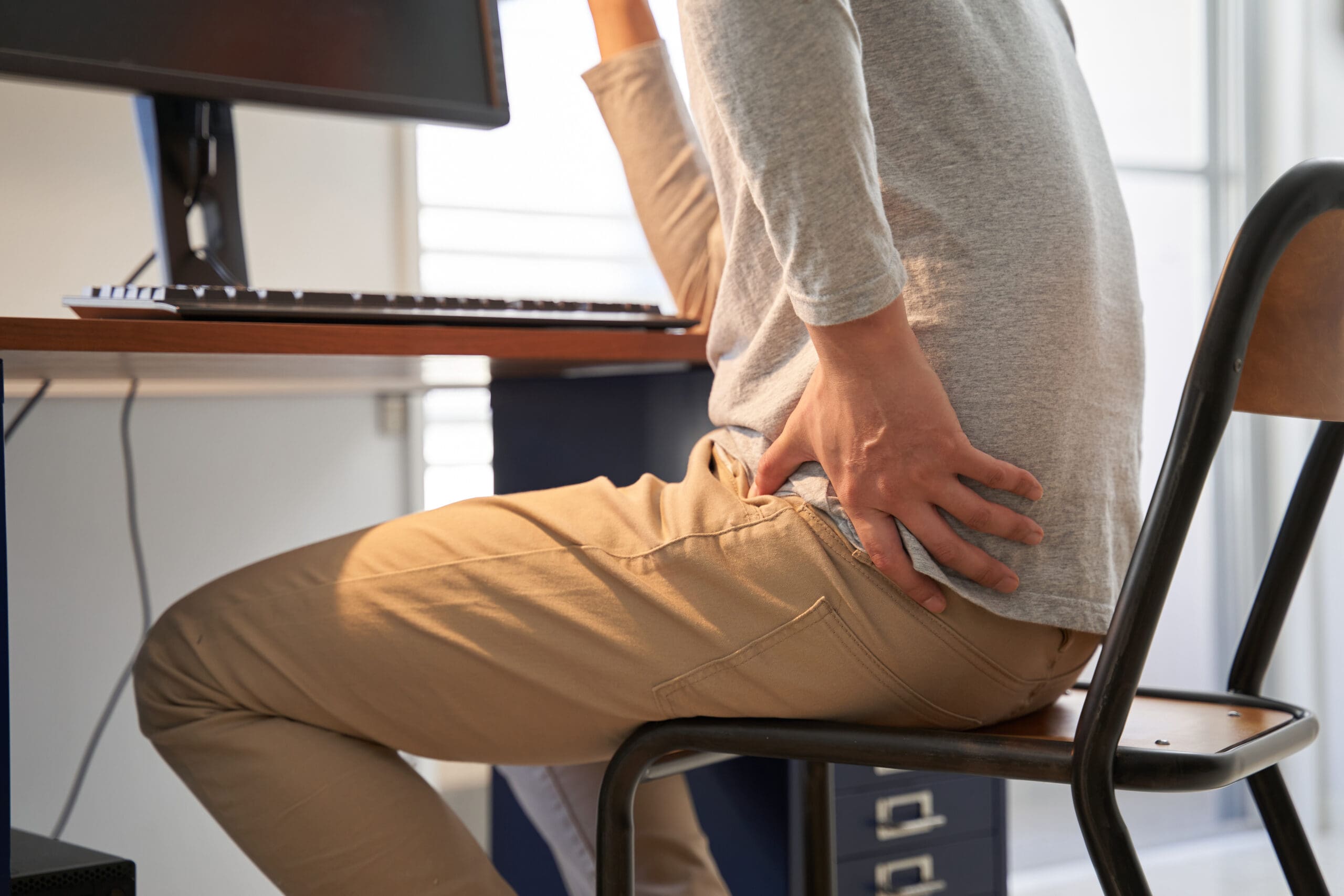
Businesswoman experiences worsening back pain while sitting at her desk.
If your back pain gets worse the longer you sit at your desk, you are not alone. Many people in El Paso face this issue due to long hours spent in sedentary jobs. Sitting for extended periods can put pressure on the spine, tighten muscles, and reduce blood flow, leading to stiffness, aches, and, in some cases, chronic problems (Colorado Pain Care, n.d.). The positive news is that you can take simple steps to reduce the pain and prevent it from worsening. At El Paso Back Clinic® in El Paso, TX, the wellness chiropractic care team, led by Dr. Alex Jimenez, DC, APRN, FNP-BC, focuses on helping people just like you find natural, long-term relief through personalized plans.
Prolonged sitting stresses the lower back by increasing disc pressure by up to 90% compared to standing. It flattens the spine’s natural curve, strains muscles, and creates imbalances (Colorado Pain Care, n.d.). Slouching or leaning forward adds extra load to the neck and upper back. Over time, this can lead to tight hips, weak core muscles, and ongoing discomfort that affects daily life.
At El Paso Back Clinic®, our experts understand these issues caused by sedentary work. They use a holistic approach that combines chiropractic adjustments, functional medicine, and rehab to address root causes like poor posture and muscle imbalances from desk jobs (Jimenez, n.d.-a).
Here are practical changes to start today:
Move often: Get up every 30 minutes to stand, walk, or shift positions. Short 1-2 minute breaks improve circulation and ease tension (Huntsville Hospital Health System, n.d.; Sydney West Physio, n.d.).
Use regular breaks: Set a timer for quick walks to get water or to stretch. This habit prevents stiffness from building up throughout the day.
Add dynamic movement: While sitting, shift weight, uncross legs periodically, or use a footrest to change angles. These small actions keep the spine mobile (Colorado Pain Care, n.d.).
A proper ergonomic setup supports optimal posture and reduces strain.
Follow these key tips:
Set your chair so that your feet are flat on the floor, your knees are at 90 degrees, and your hips are level with or above your knees.
Add lumbar support (a small pillow or rolled towel works) to maintain the lower back’s curve.
Place your screen at eye level to avoid looking down or up too much.
Keep the keyboard and mouse close so elbows bend at 90 degrees and shoulders stay relaxed.
Avoid crossing legs for long, as it can tilt the pelvis (Senara Chiropractic & Med Spa, n.d.; Huntsville Hospital Health System, n.d.).
Consider alternating between sitting and standing with a standing desk. Even partial standing reduces spinal pressure.
Stretches help loosen tight spots from sitting, such as the hips, shoulders, and neck.
Try these simple ones:
Hip flexor stretch: Kneel on one knee, gently push hips forward, and hold 20-30 seconds per side.
Chest and shoulder opener: Clasp hands behind your back or use a wall to stretch forward.
Neck tilts: Slowly tilt the head side to side or forward/back; hold for 10-15 seconds.
Upper back extension: Hands behind head, gently arch upper back (Sydney West Physio, n.d.).
Do them hourly or during breaks for better flexibility.
Strengthening the core supports the spine and improves posture long-term.
Include these:
Planks: Hold forearm plank 20-30 seconds.
Cat-camel: On hands and knees, arch and round back slowly.
Bridges: Lie back, lift hips while squeezing glutes.
Walking or gentle yoga: Build overall strength (Huntsville Hospital Health System, n.d.; Sydney West Physio, n.d.).
Aim for 20-30 minutes of activity most days.
For lasting relief, professional care targets alignment, mobility, and personalized fixes. At El Paso Back Clinic®, Dr. Alex Jimenez leads a team offering integrated chiropractic care. This includes spinal adjustments to correct misalignments, non-surgical spinal decompression for disc relief, acupuncture, functional medicine for nutrition and stress, and rehab exercises tailored to desk-related issues.
Dr. Jimenez, with dual expertise as a chiropractor and nurse practitioner, emphasizes posture correction, mobility training, and the prevention of sedentary pain through evidence-based methods. The clinic helps restore function without drugs or surgery, focusing on root causes like imbalances from prolonged sitting (Jimenez, n.d.-a; Jimenez, n.d.-b).
Other options in El Paso exist, but El Paso Back Clinic® stands out for its comprehensive wellness approach, advanced diagnostics, and patient-centered plans that go beyond basic adjustments.
If pain includes numbness, tingling, or weakness in the legs, or persists despite changes, seek evaluation to rule out serious conditions (University of Maryland Medical System, n.d.).
Start small: improve movement, setup, and stretches. If needed, contact El Paso Back Clinic® for expert help. Many in El Paso regain comfort and stay active with this care.
Make Your Health Goals Stick in 2026: How El Paso Back Clinic’s Integrative Team Supports Real Change
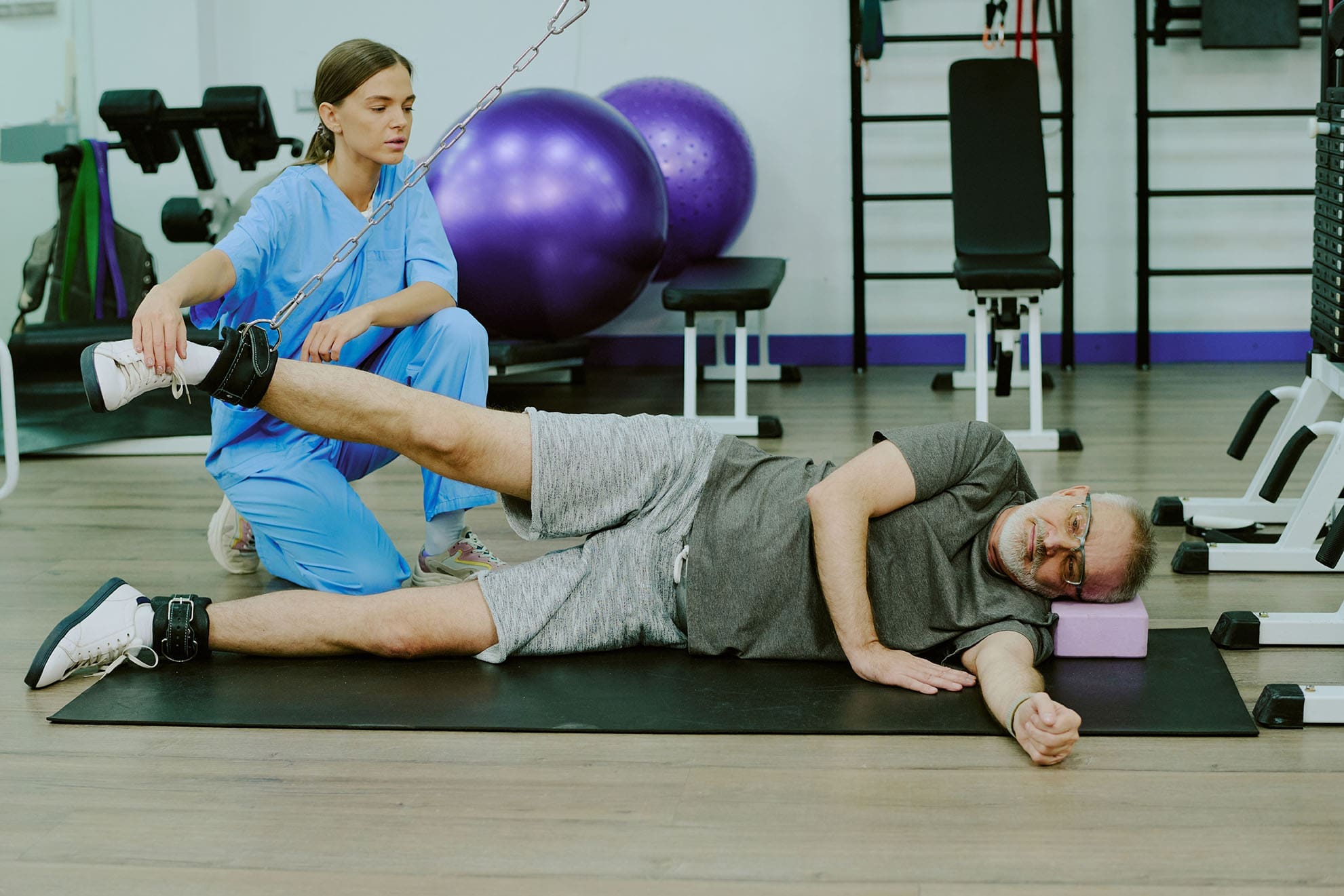
The patient uses a weight machine for injury rehabilitation under the supervision of a doctor of chiropractic and a nurse practitioner.
Most people don’t fail at New Year’s goals because they “don’t want it enough.” They fail because life gets busy, pain flares up, energy crashes, and stress piles on. When your body hurts or feels stiff, even simple plans—like walking more, lifting weights, or sleeping better—can feel harder than they should.
At El Paso Back Clinic, the goal is to make health changes easier to achieve and maintain through a team-based, integrative approach. That means bringing together the strengths of chiropractic care (movement, structure, mobility, and recovery) with the strengths of nurse practitioner care and wellness coaching (nutrition, sleep, stress, and whole-body support). The clinic describes this as a blend of injury care, wellness strategies, mobility programs, and integrated medicine designed to improve function and quality of life. El Paso Back Clinic® • 915-850-0900+2El Paso Back Clinic® • 915-850-0900+2
This kind of care supports common goals like:
increasing fitness and mobility
managing pain so you can stay active
improving energy and sleep
lowering stress and improving your stress response
“Integrative care” means your plan isn’t built around only one angle. Instead, it connects the pieces that usually get separated:
How you move
How you recover
How you eat
How you sleep
How you manage stress
How do you build habits that fit your real life
El Paso Back Clinic describes integrative chiropractic benefits as going beyond traditional adjustments by combining care approaches that support overall wellness and function. El Paso Back Clinic® • 915-850-0900
Why this matters for resolutions
Many resolutions are difficult to maintain because the plans ignore the real barriers. For example:
You want to exercise more—but your back pain spikes.
You want to lose weight—but your sleep is poor and your stress is high.
You want more energy—but your nutrition is inconsistent, and you’re not recovering.
An integrative plan helps because it aims to reduce the friction that makes healthy habits feel impossible.
The Team Approach: Chiropractor + Nurse Practitioner Mindset
Many clinics talk about how chiropractic care supports goals such as mobility, stress reduction, better sleep, and improved performance. gotcore.net+2Freedom Chiropractic+2 At El Paso Back Clinic, that support is often strongest when chiropractic care is paired with whole-person planning.
The chiropractor’s lane: move better with less strain
Chiropractic care commonly focuses on:
joint motion and spinal mechanics
posture and movement habits
mobility and flexibility
recovery support when you start working out again
helping reduce strain patterns that keep pain looping
The descriptions of services at El Paso Back Clinic emphasize spine-focused care and the restoration of function for back and musculoskeletal concerns. El Paso Back Clinic® • 915-850-0900+1
The NP/wellness lane: build a plan that supports your body from the inside out
A nurse practitioner and wellness-minded team approach can support:
nutrition planning that fits your schedule
sleep improvement routines
stress management strategies
health screening and medical risk review when appropriate
coaching that makes change more realistic to sustain
This matches the habit-focused guidance many health organizations recommend: set realistic goals, build routines, and avoid extreme “all at once” changes. Prism Health North Texas
Dr. Alexander Jimenez’s clinical observations (El Paso context)
Dr. Alexander Jimenez (DC, APRN, FNP-BC) frequently describes a dual-scope approach that connects biomechanics (how you move) with broader health planning (nutrition, functional assessments, and recovery strategies). His published clinic content also highlights the use of assessments and, when needed, imaging and integrated care planning to support recovery and function. LinkedIn+3El Paso, TX Doctor Of Chiropractic+3El Paso, TX Doctor Of Chiropractic+3
Why Resolutions Often Fail (And How an Integrative Plan Fixes That)
Here are common “resolution killers” and what a coordinated plan can do differently:
Pain blocks movement → Address mobility limits and movement mechanics so activity feels doable. National Spine & Pain Centers+1
Low energy → Improve sleep, nutrition consistency, and recovery structure. gotcore.net+1
Stress overload → Add stress skills and routines that calm the system and support follow-through. NIH News in Health+1
No accountability → Regular check-ins and plan adjustments keep you from quitting after a setback. drmmalone.com+1
A key idea in habit-based care is that early wins create a “positive feedback loop”—you feel better, so it becomes easier to keep going. drmmalone.com
1) Increase Fitness and Mobility (Without Getting Injured)
If your goal is to work out more, the priority is often moving well enough to train consistently.
Many chiropractic resources emphasize mobility, flexibility, and injury prevention as people increase activity at the start of the year. 5280 Balanced Health Center+2Freedom Chiropractic+2 El Paso Back Clinic also emphasizes flexibility, mobility, and agility programs to improve ability and quality of life. El Paso Back Clinic® • 915-850-0900
A simple evidence-based target
For general health, adults are commonly advised to aim for 150 minutes of moderate activity per week, plus 2 days of muscle-strengthening activities. CDC+1 That can be split into smaller chunks—like 30 minutes, 5 days a week.
What the integrative plan can look like
Assess mobility limits (hips, spine, shoulders) and address movement friction
Build a realistic weekly schedule
Progress intensity slowly, so you don’t crash or flare
Easy “start small” movement ideas:
10–20 minute walk after meals
2 strength sessions per week (basic full-body)
5-minute mobility routine daily
Progression rules that keep people consistent:
Add time before you add intensity
Keep at least 1–2 recovery days weekly
Measure consistency, not perfection
2) Manage Pain So You Can Stay Active
Pain goals often work better when you focus on function—not “zero pain tomorrow.” A pain-focused plan might aim to reduce flare-ups and increase what you can do safely. National Spine & Pain Centers
El Paso Back Clinic positions its care around helping people with frustrating injuries and chronic pain syndromes improve mobility and function. El Paso Back Clinic® • 915-850-0900
Practical pain goals that tend to stick
“Walk 20 minutes, 4 days/week without a flare.”
“Lift twice/week with pain staying under a 3–4/10.”
NP-style wellness support can focus on sleep, stress, consistency in nutrition, and pacing habits that support recovery. Prism Health North Texas+1
Helpful pacing ideas (simple but powerful):
Use shorter workouts more often
Stop just before your “flare threshold”
Build capacity gradually rather than “weekend warrior” bursts
3) Boost Energy the Smart Way
Energy is not just “motivation.” If you’re tired, your plan needs better recovery.
Many chiropractic sources link better sleep and reduced tension with feeling more capable and consistent over time. gotcore.net+1 El Paso Back Clinic also describes a wellness-focused approach aimed at improving energy, sleep, and overall function. El Paso Back Clinic® • 915-850-0900
It’s common to hear people say they want to “boost immunity.” A safe and practical way to think about this is:
You can support overall wellness by improving sleep, physical activity, and stress management—foundations that matter for health.
Regular physical activity is widely recommended for health. CDC
Mindfulness-based approaches have evidence supporting their effectiveness for stress, sleep, and pain management. NIH News in Health
So instead of chasing extreme detoxes or perfect diets, an integrative plan often focuses on steady basics:
sleep routine
movement most days
nutrition consistency
stress skills
That’s the kind of “quiet consistency” that makes resolutions last.
5) Lower Stress and Improve Stress Response
Stress shows up in the body: tight shoulders, headaches, jaw tension, shallow breathing, gut tension, and poor sleep.
Mindfulness-based treatments have evidence supporting reduced anxiety/depression symptoms and improved sleep, and may help people cope with pain. NIH News in Health Many chiropractic sources also connect care with stress reduction and better sleep as part of overall wellness. gotcore.net+1
Pick one main goal (fitness OR pain, energy, OR stress)
Add two support habits
Track consistency weekly
Adjust every 2–4 weeks
Examples of “support habits”:
protein at breakfast
20-minute walk 4x/week
5 minutes of mobility daily
bedtime routine 5 nights/week
A Simple 4-Week Plan (El Paso Back Clinic Style: Practical, Not Perfect)
This is a general example you can personalize with your provider team.
Week 1: Reduce friction
Identify mobility limits and pain triggers
Set one realistic activity goal
Begin a simple nutrition and sleep routine
Week 2: Build consistency
Add a second strength or mobility day
Keep intensity moderate
Track sleep and energy patterns
Week 3: Progress carefully
Increase walking time or training volume slightly
Add a stress routine you can repeat
Adjust the plan based on how your body responds
Week 4: Lock in your system
Keep what’s working
Simplify what isn’t
Create a “busy week version,” so you don’t fall off
This approach fits the clinic’s overall theme of improving function through mobility, recovery, and whole-person planning. El Paso Back Clinic® • 915-850-0900+1
When to Get Checked Right Away
If you have severe or unusual symptoms, don’t “push through.” Seek urgent medical care for red flags like:
chest pain, severe shortness of breath, fainting
sudden weakness, facial droop, confusion
loss of bowel/bladder control
fever with severe spine pain
major trauma with worsening symptoms
Bottom Line: Your Best Results Come From a Whole Plan
At El Paso Back Clinic, an integrative model supports real-life resolutions by combining:
Avoiding Common Christmas Accidents: Prevention and Recovery at El Paso Back Clinic®
After lying in an awkward position, the woman is suffering from back pain on the couch at home.
The Christmas season fills homes with lights, laughter, and loved ones. But it can also bring unexpected risks. From slips on icy paths to burns in the kitchen, holiday accidents happen more often than you might think. In El Paso, Texas, where winter weather can mix with the festive rush, these issues send many seeking help. Distracted or drunk driving spikes too, making roads risky. At El Paso Back Clinic®, we focus on wellness chiropractic care to help you prevent and heal from these mishaps. This article explains common Christmas accidents, their causes, and tips for prevention. It also shows how our integrative approach, led by Dr. Alexander Jimenez, DC, APRN, FNP-BC, offers holistic recovery. Using spinal adjustments, massage, nutritional guidance, and NP-partnered care, we support your body’s natural healing to help you have a pain-free holiday.
Common Christmas Holiday Accidents at El Paso Back Clinic®
At our clinic in El Paso, TX, we see a rise in holiday-related injuries each year. These range from home mishaps to road incidents. Here’s a list of the most common ones we treat.
Falls: Decorating ladders or icy El Paso sidewalks leads to slips. These cause sprains, fractures, or head trauma. Nationwide, about 160 decorating falls occur daily, accounting for half of decorating injuries. Kids might tumble from unstable trees or during outdoor fun.
Fires: Faulty lights, dry trees, or candles spark fires. In homes across Texas, Christmas tree fires average 155 per year, causing injuries and property damage. We advise checking decorations to avoid these dangers.
Burns: Holiday cooking with hot oil or deep fryers can result in scalds. Touching lit decorations adds risk. Turkey fryers alone cause 5 deaths and 60 injuries annually. Even hot foods like fried treats can burn mouths.
Cuts: Knife slips while wrapping or carving happen often. Broken glass ornaments or toy packaging lead to ER visits – about 6,000 yearly for gift-opening cuts.
Strains: Lifting decorations, gifts, or snow strains muscles. Back issues account for 15% of holiday accidents, and 11,500 ER visits are due to shoveling. In El Paso, our patients often come in after heavy lifting.
Alcohol-Related Incidents: Festive drinks cause falls or “holiday heart” – heart rhythm problems from overdrinking. This leads to dizziness and more.
Food Poisoning: Rushed meals with undercooked food or leftovers breed bacteria. About 48 million cases occur in the U.S. each year, peaking during holidays.
Injuries Related to Toys and Gifts: Choking on small parts injures 251,700 kids yearly. Faulty gifts cause cuts or trips.
Distracted or Drunk Driving: Busy El Paso roads see more crashes from texting or drinking. Drunk driving deaths rose to 1,013 in December 2021.
These issues increase ER visits by 5-12% in the U.S. and by over 80,000 in the UK during festivities. At El Paso Back Clinic®, we help locals recover quickly.
Causes of Holiday Injuries Seen at Our Clinic
Many injuries stem from everyday tasks gone wrong. To stop recurrences, we at El Paso Back Clinic® pinpoint these causes.
Overexertion: Heavy lifting, like trees or bags, strains backs. Bending incorrectly causes 80% of lower back pain. Travel luggage accounts for 72,000 doctor visits each year.
Cooking: Burns from oils or knives in busy kitchens. One in ten child injuries comes from cooking. Grease fires are frequent.
Decorating: Ladder falls, electrical shocks, or ornament cuts. Decorating sends 13,000 to ERs yearly. Cord trips cause 2,000 injuries.
Accidents on the Road or at Home: Distracted driving in El Paso’s traffic or at home. Stress slows reflexes.
Winter sports add 186,000 injuries, though they are less common here. Plants like mistletoe can poison if eaten.
Prevention Tips from El Paso Back Clinic®
Prevent accidents with simple steps. Our team at El Paso Back Clinic® shares these to keep your holidays safe.
For Falls: Use stable ladders and salt icy paths. Get help when climbing.
For Fires and Burns: Inspect wires, water trees, and use LED candles. Watch stoves closely.
For Cuts and Strains: Cut safely and lift with your knees. Team up for heavy items.
For Alcohol and Driving: Designate a driver or use a ride. Drink moderately.
For Food and Toys: Cook thoroughly and chill food fast. Pick safe, age-appropriate toys.
Keep a first aid kit handy and manage stress. Visit us for pre-holiday check-ups.
How Integrative Chiropractic Care at El Paso Back Clinic® Helps
If injured, turn to El Paso Back Clinic® for natural healing. Our integrative chiropractic care, in partnership with NPs, treats the whole person. Dr. Alexander Jimenez, with over 30 years in El Paso, observes that holiday injuries often stem from poor posture or stress, leading to misalignment of the spine. We use non-invasive techniques to ease pain without meds or surgery.
Adjustments for Spinal and Joint Pain: Realign the spine to relieve strain from falls or lifts. This boosts movement and cuts swelling.
Massage and Physiotherapy for Muscle Problems: Ease tension from overwork. Improves circulation for faster recovery.
NP-Led Care for Holistic Wellness: Our NPs manage overall health, including burn care and effects of poisoning, with a natural focus.
Nutrition Guidance: Counter rich holiday foods with diet tips to aid digestion and immunity. Fiber-rich choices help.
Managing Underlying Conditions: Reduce stress hormones for better sleep and mood. Prevents further harm.
Dr. Jimenez’s team uses functional medicine to develop personalized plans that address issues like sciatica from slips. Chiropractic enhances the nervous system for better health during the holidays.
Enjoy a Healthy Holiday with El Paso Back Clinic®
Make Christmas memorable for the right reasons. Know the risks, prevent them, and seek our care if needed. At El Paso Back Clinic®, we’re here for your wellness. Contact us in El Paso, TX, for expert chiropractic support. Happy holidays!
Discover the importance of a clinical approach to opioid use disorder in developing effective intervention strategies.
Overcoming Barriers in Managing Opioid Use Disorder: Strategies for Effective Care
Many people today have a serious health problem called opioid use disorder (OUD). It is part of a bigger group of problems called substance use disorders (SUD). Treating OUD can be hard because everyone has different problems, such as other health issues or pain. Plans should be made for each patient by doctors and other health care workers. They also have to keep up with the latest laws, ethics, and ways to keep patient information safe. The Health Insurance Portability and Accountability Act (HIPAA) of 1996 is an example of a general rule that applies to all patients. However, there are extra rules for people who are getting help with drug or alcohol problems.
This guide talks about how to deal with problems that come up when managing OUD. We talk about patient-centered care, how to talk to patients, stigma, team-based approaches, and the law. Health care providers can help patients get better faster by using these methods. Keywords like “managing opioid use disorder,” “overcoming stigma in OUD,” and “patient-centered care for SUD” bring out important points that make it easier to find and understand.
Learning Objectives
Explain treatment planning methods that use patient-focused choices and proven ways to talk.
Name the three kinds of stigma and how they affect people with mental health issues, SUD, and especially OUD.
Talk about legal, ethical, and privacy concerns in caring for people with OUD.
Effective Treatment Planning with Patient-Centered Decisions
People with complex issues, like mental health problems, SUD, and pain, need special care. Each person shows up differently, so health systems are now focusing on care that puts the patient first.
Patient-centered care means building teams with doctors, patients, and families. They work together to plan, give, and check health care. This way ensures the patient’s needs are met, and their wishes, likes, and family situations are respected. It focuses on shared choices about treatments while seeing the patient as a whole person in their daily life (Dwamena et al., 2012; Bokhour et al., 2018).
Studies show key steps for a good patient-centered plan:
Take a full patient history and a check-up, reviewing old and new treatments.
Find all available drug and non-drug options.
Check the patient’s current health, recent changes, and patterns.
Look at risks for misusing or abusing opioids.
If starting opioids or if the patient is already on them, think about opioid stewardship. This means checking harms, benefits, risks, side effects, pain control, daily function, drug tests, stop plans, and ways to spot OUD. These programs, sometimes called analgesia stewardship, help manage opioids safely (Harle et al., 2019; Coffin et al., 2022). Guides exist to set them up (American Hospital Association, n.d.; Shrestha et al., 2023).
Integrative chiropractic care can play a big role here. It uses spinal adjustments and targeted exercises to get proper spinal alignment. This helps reduce pain without relying only on drugs, making it a good fit for OUD patients with pain. For example, adjustments fix spine issues that cause pain, and exercises strengthen muscles to keep alignment right.
A Nurse Practitioner (NP) adds full management and ergonomic advice. They look at work setups to prevent pain, such as how to sit or lift. NPs coordinate care by reviewing options such as therapy, meds, and lifestyle changes, ensuring everything works together.
Dr. Alexander Jimenez, DC, APRN, FNP-BC, with over 30 years in chiropractic and as a family nurse practitioner, observes that blending these methods cuts opioid use. At his El Paso clinic, he uses functional medicine to address root causes through nutrition and non-invasive treatments. He notes that poor posture from modern life worsens pain, leading to OUD risks. His teams help patients with self-massage and VR for recovery, reducing drug needs (Jimenez, n.d.a; Jimenez, n.d.b).
Evidence-Based Ways to Communicate
Good talking skills are key to building a patient-centered plan (Schaefer & Block, 2009). There are proven methods for starting conversations and getting patients involved.
One method is BATHE:
Background: Ask, “How have things been since your last visit?”
Affect: Ask, “How does this make you feel?”
Trouble: Ask, “What bothers you most?”
Handling: Ask, “How are you coping?”
Empathy: Say, “That sounds hard.”
This uses open questions to let patients lead and feel supported (Stuart & Lieberman, 2018; Thomas et al., 2019).
Another is GREAT:
Greetings/Goals: Start with hello and set aims.
Rapport: Build trust.
Evaluation/Expectation/Examination/Explanation: Check and explain.
Ask/Answer/Acknowledge: Listen and respond.
Tacit agreement/Thanks: Agree and thank.
This guide talks well (Brindley et al., 2014).
Motivational interviewing is also useful. It’s a team-style talk to boost a patient’s desire to change. Build a bond, focus on the issue, spark a desire for change, and plan steps (Frost et al., 2018).
These methods emphasize listening, clear communication, and a structured approach to planning. For OUD patients with pain or mental issues, mix techniques for the best results.
Dr. Jimenez shares that in his practice, these talks help patients see non-drug options, such as chiropractic adjustments. He finds that empathy reduces stigma and fear, encouraging openness about OUD (Jimenez, n.d.a).
Understanding Stigma in Mental Health and Substance Use Disorders
Stigma blocks good talk for many with mental health or SUD. It’s attitudes, beliefs, actions, and systems that lead to unfair views and bad treatment (Cheetham et al., 2022).
Studies show stigmas like linking mental illness to violence (Perry, 2011). Media on shootings with mentally ill people strengthens this (McGinty et al., 2014; McGinty et al., 2016; Schomerus et al., 2022). For SUD, people think they’re more dangerous than those with schizophrenia or depression (Schomerus et al., 2011). Society blames people with SUDs more and avoids them (McGinty et al., 2015; Corrigan et al., 2012).
Views come from knowledge, contact with affected people, and the media. Public ideas are tied to norms on causes, blame, and danger. Race, ethnicity, and culture shape attitudes too (Giacco et al., 2014).
Health workers have biases. A survey of VA mental health providers showed awareness of race issues but avoidance of talks, using codes like “urban,” and thinking training stops racism (McMaster et al., 2021).
There are three stigma types:
Structural Stigma: The ways Society and institutions keep prejudice. In health, it’s worse care, less access to behavioral health. Less funding for mental vs. physical issues (National Academies of Sciences, Engineering, and Medicine, 2016).
Public Stigma: General or group attitudes, like police or church norms. Laws reinforce it, like broad mental illness rules implying all are unfit (Corrigan & Shapiro, 2010).
Self-Stigma: When people internalize stigmas, it leads to low self-worth and shame. “Why try” affects independent living (Corrigan et al., 2009; Clement et al., 2015).
Dr. Jimenez observes that stigma makes OUD patients hide symptoms, delaying care. In his integrative work, he addresses this through education on holistic options, showing that recovery is possible without judgment (Jimenez, n.d.b).
Overcoming Stigma and Addressing Social Factors
To fight stigma, use education, behavior changes, and better care. Laws like the ADA and MHPAEA help ensure equal coverage and prevent discrimination (U.S. Congress, 2009; U.S. Congress, 2008; U.S. Department of Health and Human Services, n.d.; Busch & Barry, 2008; Haffajee et al., 2019).
These address social determinants of health (SDOH), such as coverage, access, quality, education, and stability (Centers for Disease Control and Prevention, n.d.).
Community programs help too:
West Virginia’s Jobs and Hope: Training, jobs, education, transport, skills, record clearing for SUD people (Jobs and Hope, n.d.).
Belden’s Pathway: Rehab for failed drug tests, leading to jobs (Belden, n.d.).
Education boosts provider confidence in OUD meds, reducing barriers (Adzrago et al., 2022; Hooker et al., 2023; Campbell et al., 2021).
Overcoming stigma is key to success in mental health and SUD.
Interprofessional Team Work
Teams improve outcomes for patients with chronic pain and mental health or SUD (Joypaul et al., 2019; Gauthier et al., 2019).
Teams include doctors, nurses, NPs, pharmacists, PAs, social workers, PTs, therapists, SUD experts, and case managers.
Each helps uniquely:
Pharmacists watch meds, spot interactions.
Case managers link specialists, find resources, and support families (Sortedahl et al., 2018).
Teams set goals, max non-opioid treatments (Liossi et al., 2019).
Integrative chiropractic care includes adjustments and exercises for alignment, easing pain naturally.
NPs give full care, ergonomic tips to avoid pain triggers, and coordinate options.
Dr. Jimenez’s clinic shows this. As a DC and FNP-BC, he leads teams with therapists, nutritionists, and coaches. He observes interprofessional work cuts opioid use by addressing the roots with functional medicine, VR, and nutrition. For OUD, he blends chiropractic care for pain, NP coordination for plans, and stigma-fighting through team support (Jimenez, n.d.a; Jimenez, n.d.b).
The Power of Chiropractic Care in Injury Rehabilitation-Video
Legal and Ethical Issues in SUD Care
Providers must know laws and ethics for mental/SUD patients, like discrimination, aid, and privacy (Center for Substance Abuse Treatment, 2000).
Key Federal laws:
Americans with Disabilities Act (ADA) of 1990.
Rehabilitation Act of 1973.
Workforce Investment Act of 1998.
Drug-Free Workplace Act of 1988.
ADA and Rehabilitation ban discrimination in government and in business services like hotels, shops, and hospitals. Protect those with impairments limiting life activities (U.S. Department of Health and Human Services, n.d.).
Provisions:
Protect “qualified” people who meet the requirements.
Reasonable accommodations for jobs.
No hire/retain if there is a direct threat.
No denial of benefits, access, or jobs in funded places.
For SUD: Alcohol users are protected if qualified, no threat. Ex-drug users in rehab are the same. Current illegal drug users are protected for health/rehab, not others. Programs can deny if used during.
Workforce Act centralizes job programs; no refusal to SUD people (U.S. Congress, 1998).
Drug-Free Act requires drug-free policies for federal funds/contracts: statements, awareness, actions on violations (U.S. Code, n.d.).
States have their own laws; check the local laws.
Public Aid laws:
Contract with America Act (1996): No SSI/DI if SUD key factor (U.S. Congress, 1996).
Personal Responsibility Act (1996): Work after 2 years of aid, drug screens (U.S. Department of Health and Human Services, 1996).
These push work, sobriety.
Dr. Jimenez notes that legal awareness helps his practice by ensuring holistic plans comply and by reducing OUD risks through a non-drug focus (Jimenez, n.d.a).
Keeping Patient Info Private
Privacy is vital. Laws include:
HIPAA (1996): Protects PHI, sets use/disclosure rules (U.S. Department of Health and Human Services, n.d.).
42 CFR Part 2: Extra for SUD records. No disclosure of name or status without consent. Fines for breaks. Applies to federal-aided programs (Substance Abuse and Mental Health Services Administration, n.d.).
Consent needs: program name, receiver, patient name, purpose, info type, revoke note, expire date, signature, and date.
This fights discrimination fears, encouraging treatment (Center for Substance Abuse Treatment, 2000).
Wrapping Up
As we navigate the ongoing challenges of opioid use disorder (OUD), it’s clear that effective management requires a multifaceted approach that prioritizes patient well-being over quick fixes. From embracing patient-
It is clear that treating opioid use disorder (OUD) well requires a multi-faceted approach that puts the patient’s health and safety above quick fixes. Healthcare professionals play a pivotal role in transforming lives by implementing patient-centered decision-making and evidence-based communication, and by eradicating the three types of stigma—structural, public, and self—that hinder recovery. Interprofessional teams help people get the full treatment they need, and privacy laws like HIPAA and 42 CFR Part 2 make sure that people with disabilities can get help without being discriminated against.
Chiropractic therapy focuses on spinal adjustments and specific exercises to support proper alignment. It is a non-invasive way to ease pain and reduce dependence on opioids. Nurse Practitioners (NPs) make this better by providing comprehensive care, offering ergonomic advice to prevent injuries, and coordinating multiple treatment options, such as lifestyle changes and therapy. Dr. Alexander Jimenez, DC, APRN, FNP-BC, emphasizes in his clinical practice that these integrative approaches not only address physical symptoms but also empower patients through education and tailored strategies, leading to enduring recovery and diminished opioid consumption (Jimenez, n.d.a; Jimenez, n.d.b).
As we look ahead, new advancements in OUD therapy by 2025 show a trend toward making it easier to get and more tailored to each person. For instance:
Drugs like methadone, buprenorphine, and naltrexone that the FDA has approved are still the best way to treat OUD. They help with cravings and withdrawal symptoms and help people stay stable over time.
Precision medicine goes beyond one-size-fits-all methods by tailoring treatments to each person’s social, psychological, and genetic factors. This should lead to better results.
The World Health Organization’s 2025 updates put more emphasis on psychosocial support, with a focus on preventing overdoses in the community and making it easier for people to get care.
Declining Trends: The first yearly drop in opioid-related deaths since 2018 happened in 2023. This is a good sign because it shows that ongoing work in treatment, education, and lawmaking is having an effect.
We might be able to make OUD a treatable illness instead of a life sentence by combining these new ideas with collaborative care and reducing stigma. Policymakers, communities, and healthcare professionals must continue to advocate for equitable access to ensure that all individuals receive the evidence-based treatment they need. Overcoming problems in OUD management is about more than just getting better; it’s also about getting your dignity, hope, and a good quality of life back.
References
Adzrago, D., Paola, A. D., Zhu, J., et al. (2022). Association between prescribers’ perceptions of the utilization of medication for opioid use disorder and opioid dependence treatability. Healthcare, 10(9), 1733. https://doi.org/10.3390/healthcare10091733
Bokhour, B. G., Fix, G. M., et al. (2018). How can healthcare organizations implement patient-centered care? Examining a large-scale cultural transformation. BMC Health Services Research, 18(1), 168. https://doi.org/10.1186/s12913-018-2993-5
Brindley, P. G., Smith, K. E., Cardinal, P., & LeBlanc, F. (2014). Improving medical communication with patients and families: Skills for a complex (and multilingual) clinical world. Canadian Respiratory Journal, 21(2), 89-91. https://doi.org/10.1155/2014/789456
Campbell, C. I., Saxon, A. J., Boudreau, D. M., et al. (2021). Primary Care Opioid Use Disorders treatment (PROUD) trial protocol: A pragmatic, cluster-randomized implementation trial in primary care for opioid use disorder treatment. Addiction Science & Clinical Practice, 16(1), 9. https://doi.org/10.1186/s13722-021-00221-1
Center for Substance Abuse Treatment. (2000). Integrating Substance Abuse Treatment and Vocational Services. (Treatment Improvement Protocol (TIP) Series, No. 38.) Chapter 7—Legal Issues. https://www.ncbi.nlm.nih.gov/books/NBK64294/
Center for Substance Abuse Treatment. (2000). Substance Abuse Treatment for Persons with Child Abuse and Neglect Issues. (Treatment Improvement Protocol (TIP) Series, No. 36.) Appendix B –Protecting Clients’ Privacy. https://www.ncbi.nlm.nih.gov/books/NBK64900/
Cheetham, A., Picco, L., Barnett, A., et al. (2022). The impact of stigma on people with opioid use disorder, opioid treatment, and policy. Substance Abuse and Rehabilitation, 13, 1-12. https://doi.org/10.2147/SAR.S304256
Clement, S., Schauman, O., Graham, T., et al. (2015). What is the impact of mental health-related stigma on help-seeking? A systematic review of quantitative and qualitative studies. Psychological Medicine, 45(1), 11-27. https://doi.org/10.1017/S0033291714000129
Coffin, P. O., Martinez, R. S., Wylie, B., et al. (2022). Primary care management of long-term opioid therapy. Annals of Medicine, 54(1), 2451-2469. https://doi.org/10.1080/07853890.2022.2118597
Corrigan, P. W., Larson, J. E., & Rüsch, N. (2009). Self-stigma and the “why try” effect: Impact on life goals and evidence-based practices. World Psychiatry, 8(2), 75-81. https://doi.org/10.1002/j.2051-5545.2009.tb00218.x
Corrigan, P. W., Morris, S. B., Michaels, P. J., Rafacz, J. D., & Rüsch, N. (2012). Challenging the public stigma of mental illness: A meta-analysis of outcome studies. Psychiatric Services, 63(10), 963-973. https://doi.org/10.1176/appi.ps.201100529
Corrigan, P. W., & Shapiro, J. R. (2010). Measuring the impact of programs that challenge the public stigma of mental illness. Clinical Psychology Review, 30(8), 907-922. https://doi.org/10.1016/j.cpr.2010.06.004
Dwamena, F., Holmes-Rovner, M., Gaulden, C., et al. (2012). Interventions for providers to promote a patient-centred approach in clinical consultations. Cochrane Database of Systematic Reviews, 2012(12), CD003267. https://doi.org/10.1002/14651858.CD003267.pub2
Frost, H., Campbell, P., Maxwell, M., et al. (2018). Effectiveness of Motivational Interviewing on adult behavior change in health and social care settings: A systematic review of reviews. PLoS One, 13(10), e0204890. https://doi.org/10.1371/journal.pone.0204890
Gauthier, K., Dulong, C., & Argáez, C. (2019). Multidisciplinary treatment programs for patients with chronic non-malignant pain: A review of clinical effectiveness, cost-effectiveness, and guidelines – an update. Canadian Agency for Drugs and Technologies in Health. https://www.ncbi.nlm.nih.gov/books/NBK545496/
Giacco, D., Matanov, A., & Priebe, S. (2014). Providing mental healthcare to immigrants: Current challenges and new strategies. Current Opinion in Psychiatry, 27(4), 282-288. https://doi.org/10.1097/YCO.0000000000000070
Haffajee, R. L., Mello, M. M., Zhang, F., et al. (2019). Association of federal mental health parity legislation with health care use and spending among high utilizers of services. Medical Care, 57(4), 245-255. https://doi.org/10.1097/MLR.0000000000001076
Harle, C. A., DiIulio, J., Downs, S. M., et al. (2019). Decision-Centered design of patient information visualizations to support chronic pain care. Applied Clinical Informatics, 10(4), 719-728. https://doi.org/10.1055/s-0039-1696668
Hooker, S. A., Crain, A. L., LaFrance, A. B., et al. (2023). A randomized controlled trial of an intervention to reduce stigma toward people with opioid use disorder among primary care clinicians. Addiction Science & Clinical Practice, 18(1), 10. https://doi.org/10.1186/s13722-023-00366-1
Joypaul, S., Kelly, F., McMillan, S. S., et al. (2019). Multi-disciplinary interventions for chronic pain involving education: A systematic review. PLoS One, 14(10), e0223306. https://doi.org/10.1371/journal.pone.0223306
Liossi, C., Johnstone, L., Lilley, S., et al. (2019). Effectiveness of interdisciplinary interventions in paediatric chronic pain management: A systematic review and subset meta-analysis. British Journal of Anaesthesia, 123(2), e359-e371. https://doi.org/10.1016/j.bja.2019.01.024
McGinty, E. E., Goldman, H. H., Pescosolido, B., et al. (2015). Portraying mental illness and drug addiction as treatable health conditions: Effects of a randomized experiment on stigma and discrimination. Social Science & Medicine, 126, 73-85. https://doi.org/10.1016/j.socscimed.2014.12.010
McGinty, E. E., Kennedy-Hendricks, A., Choksy, S., et al. (2016). Trends in news media coverage of mental illness in the United States: 1995-2014. Health Affairs, 35(6), 1121-1129. https://doi.org/10.1377/hlthaff.2016.0011
McGinty, E. E., Webster, D. W., Jarlenski, M., et al. (2014). News media framing of serious mental illness and gun violence in the United States, 1997-2012. American Journal of Public Health, 104(3), 406-413. https://doi.org/10.2105/AJPH.2013.301557
McMaster, K. J., Peeples, A. D., Schaffner, R. M., et al. (2021). Mental healthcare provider perceptions of race and racial disparity in the care of Black and White clients. Journal of Behavioral Health Services & Research, 48(4), 501-516. https://doi.org/10.1007/s11414-021-00753-3
National Academies of Sciences, Engineering, and Medicine. (2016). Ending discrimination against people with mental and substance use disorders: The evidence for stigma change. National Academies Press. https://www.ncbi.nlm.nih.gov/books/NBK384923/
Perry, B. L. (2011). The labeling paradox: Stigma, the sick role, and social networks in mental illness. Journal of Health and Social Behavior, 52(4), 460-477. https://doi.org/10.1177/0022146511408913
Schaefer, K. G., & Block, S. D. (2009). Physician communication with families in the ICU: Evidence-based strategies for improvement. Current Opinion in Critical Care, 15(6), 569-577. https://doi.org/10.1097/ACQ.0b013e328332af31
Schomerus, G., Lucht, M., Holzinger, A., et al. (2011). The stigma of alcohol dependence compared with other mental disorders: A review of population studies. Alcohol and Alcoholism, 46(2), 105-112. https://doi.org/10.1093/alcalc/agq089
Schomerus, G., Schindler, S., Sander, C., et al. (2022). Changes in mental illness stigma over 30 years – Improvement, persistence, or deterioration? European Psychiatry, 65(1), e78. https://doi.org/10.1192/j.eurpsy.2022.2334
Shrestha, S., Khatiwada, A. P., Sapkota, B., et al. (2023). What is “Opioid Stewardship”? An overview of current definitions and a proposal for a universally acceptable definition. Journal of Pain Research, 16, 383-394. https://doi.org/10.2147/JPR.S389785
Sortedahl, C., Krsnak, J., Crook, M. M., et al. (2018). Case managers on the front lines of opioid epidemic response: Advocacy, education, and empowerment for users of medical and nonmedical opioids. Professional Case Management, 23(5), 256-263. https://doi.org/10.1097/NCM.0000000000000294
Thomas, C., Cramer, H., Jackson, S., et al. (2019). Acceptability of the BATHE technique amongst GPs and frequently attending patients in primary care: A nested qualitative study. BMC Family Practice, 20(1), 121. https://doi.org/10.1186/s12875-019-1011-1
Discover how a clinical approach to opioid therapy can transform pain management strategies for patients in a healthcare setting.
Key Points on Safe Pain Management with Opioids
Pain Affects Many People: Research suggests that about 100 million adults in the U.S. deal with pain, and this number might grow due to aging, more health issues like diabetes, and better survival from injuries. It’s important to address pain early to prevent it from becoming long-term (Institute of Medicine, 2011).
Non-Opioid Options First: Evidence leans toward starting with treatments like exercise, therapy, or over-the-counter meds before opioids, as they can be just as effective for common pains like backaches or headaches, with fewer risks (National Academies of Sciences, Engineering, and Medicine, 2019).
Team-Based Care Works Best: Studies show teams of doctors, nurses, and therapists can improve pain relief and daily life, though results vary. This approach seems likely to help more than solo care, especially for ongoing pain (Gauthier et al., 2019).
Opioids When Needed, But Carefully: Guidelines recommend low doses, short times, and regular check-ins to balance relief with risks like addiction. It’s complex, so talk openly with your doctor (Centers for Disease Control and Prevention, 2022).
Alternatives Like Chiropractic and NP Support: Integrative methods, such as chiropractic adjustments for spine alignment and ergonomic tips from nurse practitioners, can reduce reliance on meds. Clinical observations from experts like Dr. Alexander Jimenez highlight non-invasive approaches to managing pain effectively.
Understanding Pain Types
Pain can be short-term (acute), medium-term (subacute), or long-lasting (chronic). Acute pain often lasts less than three months and comes from injuries. If not treated well, it might turn chronic, affecting daily activities. Always respect someone’s pain experience—it’s personal and influenced by life factors (Raja et al., 2020).
Assessing Pain Simply
Doctors use tools like questions about when pain started, what makes it worse, and how it feels. Scales help rate it, from numbers (0-10) to faces showing discomfort. For kids or elders, special tools watch for signs like faster heartbeats (Wong-Baker FACES Foundation, 2022).
Treatment Basics
Start with non-drug options like rest, ice, or physical therapy. For chronic pain, meds like acetaminophen or therapies like yoga help. Opioids are for severe cases but come with risks—use them wisely (Agency for Healthcare Research and Quality, n.d.).
Role of Experts
According to clinical observations by Dr. Alexander Jimenez, DC, APRN, FNP-BC, who runs a multidisciplinary practice in El Paso, Texas (https://dralexjimenez.com/), combining chiropractic care with exercises targets root causes, such as misaligned spines, reducing opioid needs. As a nurse practitioner, he coordinates care and offers ergonomic advice to prevent pain from daily habits (LinkedIn Profile).
Comprehensive Guide to Safe and Effective Pain Management Using Opioid Therapy
Millions of people struggle with pain, which affects everything from hobbies to employment. Finding safe strategies to deal with pain is crucial, whether it’s a recent injury or persistent discomfort. This comprehensive handbook examines how to measure pain, available treatments, and responsible opioid use recommendations. We’ll discuss team-based treatment, non-opioid alternatives, and perspectives from professionals like Dr. Alexander Jimenez, who prioritizes holistic approaches. To help you locate trustworthy information online, keywords like “pain management strategies,” “opioid therapy guidelines,” and “non-opioid pain relief” are interwoven.
Introduction to Pain in America
The Institute of Medicine estimates that around 100 million American adults face acute or chronic pain daily. This number is expected to climb due to an aging population, rising rates of conditions like diabetes, heart disease, arthritis, and cancer, plus better survival from serious injuries and more surgeries that can lead to post-op pain (Institute of Medicine, 2011).
As people learn more about pain relief options and gain better access through laws like the Affordable Care Act (ACA), more folks—especially older ones—seek help. Passed in 2010, the ACA requires insurers to cover essential pain management benefits, including prescription drugs, chronic disease care, mental health support, and emergency services (111th Congress, 2009-2010). To use these effectively, healthcare providers need a solid grasp of pain assessment, classification, and treatment.
What Is Pain?
The International Association for the Study of Pain defines it as an unpleasant feeling associated with real or potential tissue damage. It’s subjective, shaped by biology, emotions, and social life. People learn about pain through experiences—some seek help right away, others try home remedies first. Respect their stories (Raja et al., 2020).
Pain falls into three main types, though definitions overlap:
Acute Pain: Lasts less than 3 months, or 1 day to 12 weeks; often limits daily activities for a month or less.
Subacute Pain: Sometimes seen as part of acute, or separate; lasts 1-3 months, or 6-12 weeks.
Chronic Pain: Persists over 3 months, or limits activities for more than 12 weeks (Banerjee & Argáez, 2019).
Poorly managed short-term pain can become chronic, so early action is important (Marin et al., 2017).
Assessing Pain Thoroughly
Pain is complex, influenced by body, mind, and environment. A full check includes history, physical exam, pain details, other health issues, and mental states like anxiety.
Basic pain evaluation covers:
When it started (date/time).
What caused it (injury?).
How does it feel (sharp, dull?)?
How bad it is.
Where is it?
How long does it last?
What worsens it (moving?).
What helps it?
Related signs (swelling?).
Impact on daily life.
Mnemonics help remember these. Here’s a table comparing common ones:
Pain scales provide information but aren’t diagnoses because they’re subjective. Single-dimensional ones focus on intensity:
Verbal: Mild, moderate, severe.
Numeric: 0 (none) to 10 (worst).
Visual: Like Wong-Baker FACES®, using faces for kids, adults, or those with barriers (Wong-Baker FACES Foundation, 2022). An emoji version works for surgery patients (Li et al., 2023).
Multi-dimensional scales check intensity plus life impact. The McGill Pain Questionnaire uses words like “dull” to rate sensory, emotional, and overall effects; shorter versions exist (Melzack, 1975; Main, 2016). For nerve pain, PainDETECT helps (König et al., 2021). Brief Pain Inventory scores severity and interference with mood/life (Poquet & Lin, 2016).
For babies, watch heart rate, oxygen, and breathing. Tools like CRIES rate crying, oxygen need, vitals, expression, sleep (Castagno et al., 2022). FLACC for ages 2 months-7 years checks face, legs, activity, cry, consolability (Crellin et al., 2015). Older kids use Varni-Thompson or draw pain maps (Sawyer et al., 2004; Jacob et al., 2014).
Elders face barriers like hearing loss or dementia. PAINAD assesses breathing, sounds, face, body, and consolability on a 0-10 scale (Malara et al., 2016).
The Joint Commission sets standards across various settings, which affect tool choice (The Joint Commission, n.d.).
Building Treatment Plans
Plans depend on pain type, cause, severity, and patient traits. For acute: meds, distraction, psych therapies, rest, heat/ice, massage, activity, meditation, stimulation, blocks, injections (National Academies of Sciences, Engineering, and Medicine, 2019).
Re-check ongoing acute pain to avoid chronic shift. Goals: control pain, prevent long-term opioids. Barriers: access to docs/pharmacies, costs, follow-ups.
For chronic: meds, anesthesia, surgery, psych, rehab, CAM. Non-opioids include:
Oral Meds:
Acetaminophen.
NSAIDs (celecoxib, etc.).
Antidepressants (SNRIs like duloxetine; TCAs like amitriptyline).
Anticonvulsants (gabapentin, etc.).
Muscle relaxers (cyclobenzaprine).
Memantine.
Topical: Diclofenac, capsaicin, lidocaine.
Cannabis: Medical (inhaled/oral/topical); phytocannabinoids (THC/CBD); synthetics (dronabinol) (Agency for Healthcare Research and Quality, n.d.).
Opioid use has risen, raising concerns (National Academies of Sciences, Engineering, and Medicine, 2019).
Key plan elements:
Quick recognition/treatment.
Address barriers.
Involve patients/families.
Reassess/adjust.
Coordinate transitions.
Monitor processes/outcomes.
Assess outpatient failure risk.
Check opioid misuse (Wells et al., 2008; Society of Hospital Medicine, n.d.).
Team Approach to Pain
Studies support the use of interprofessional teams for better results (Gauthier et al., 2019). Teams include docs, nurses, NPs, pharmacists, PAs, social workers, PTs, behavioral therapists, and abuse experts.
A 2017 report showed that teams improved pain/function from baseline, though not always compared with controls (Banerjee & Argáez, 2017). A meta-analysis found that teams were better at reducing pain after 1 month and sustained benefits at 12 months (Liossi et al., 2019).
Integrative chiropractic care fits here. It involves spinal adjustments—gentle manipulations to correct misalignments—and targeted exercises, such as core strengthening, to maintain alignment and reduce pressure on nerves/muscles. Dr. Alexander Jimenez observes that this helps sciatica/back pain without opioids, using tools like decompression (dralexjimenez.com).
Nurse Practitioners (NPs) provide comprehensive management, including ergonomic advice (e.g., better sitting postures) to prevent strain. They coordinate by reviewing options, referring to specialists, and overseeing plans, as seen in Dr. Jimenez’s practice, where his FNP-BC role includes telemedicine for holistic care (LinkedIn, n.d.).
Beyond Adjustments: Chiropractic and Integrative Healthcare- Video
Managing Opioids Safely
CDC’s 2022 guidelines cover starting opioids, dosing, duration, and risks (Centers for Disease Control and Prevention, 2022).
1. Starting Opioids:
Maximize non-opioids first—they match opioids for many acute pains (back, neck, etc.). Discuss benefits/risks (Recommendation 1, Category B, Type 3).
Review labels, use the lowest dose/shortest time. Set goals, exit strategy. For ongoing, optimize non-opioids (Recommendation 2, A, 2).
2. Choosing/Dosing Opioids:
Immediate-release (hydromorphone, etc.) over ER/LA (methadone, etc.). Studies show no edge for ER/LA; avoid for acute/intermittent (Recommendation 3, A, 4).
No rigid thresholds—guideposts. Risks rise with dose; avoid high if benefits dim (Recommendation 4, A, 3).
Taper slowly to avoid withdrawal (anxiety, etc.). Collaborate on plans; use Teams. If there is disagreement, empathize and avoid abandonment (Recommendation 5, B, 4).
3. Duration/Follow-Up:
For acute, prescribe just enough—often 3 days or less. Evaluate every 2 weeks. Taper if used for days. Avoid unintended long-term (Recommendation 6, A, 4).
Follow-up 1-4 weeks after start/escalation; closer for high-risk (Recommendation 7, A, 4).
4. Risks/Harms:
Screen for SUD/OUD. Offer naloxone for overdose risk (Recommendation 8, A, 4).
Check PDMPs for scripts/combos (Recommendation 9, B, 4).
Toxicology tests are performed annually to assess interactions (Recommendation 10, B, 4).
Caution with benzodiazepines (Recommendation 11, B, 3).
For OUD, use DSM-5 (2+ criteria/year); offer meds like buprenorphine (Recommendation 12, A, 1) (Hasin et al., 2013; American Psychiatric Association, 2013).
OUD signs: Larger amounts, failed cuts, time spent, cravings, role failures, social issues, activity loss, hazardous use, continued despite problems, tolerance, withdrawal.
Treatment: Meds, counseling, groups. Coordinate with specialists.
Conclusion
Finally, relying only on opioids is not necessary for efficient pain management. We can improve the lives of millions of people by giving priority to non-opioid alternatives like acetaminophen, physical therapy, or mindfulness and by taking opioids only when necessary under strict supervision. Teams of professionals, such as physicians, nurses, pharmacists, and specialists like chiropractors, collaborate to develop individualized strategies that lower dangers like addiction. By emphasizing spinal adjustments and targeted exercises, integrative chiropractic therapy may help restore normal alignment and reduce pain naturally, often eliminating the need for medication. Complete management, ergonomic guidance to prevent problems, and treatment coordination for optimal outcomes are all ways nurse practitioners provide value.
According to experts like Dr. Alexander Jimenez, these approaches target underlying issues using non-invasive treatments and functional medicine, promoting long-term well-being. Future developments in pain management seem promising, including FDA-approved non-opioid medications and distraction technologies such as virtual reality. In the end, everyone is empowered to address pain head-on, enhancing everyday activities and general health, when patients are included in decision-making and kept informed. Early evaluation and balanced treatment are crucial; discuss your options with your healthcare professional to determine what is best for you.
Banerjee, S., & Argáez, C. (2017). Multidisciplinary treatment programs for patients with chronic non-malignant pain: A review of clinical effectiveness, cost-effectiveness, and guidelines. Canadian Agency for Drugs and Technologies in Health. https://www.ncbi.nlm.nih.gov/books/NBK545496/
Banerjee, S., & Argáez, C. (2019). Multidisciplinary treatment programs for patients with acute or subacute pain: A review of clinical effectiveness, cost-effectiveness, and guidelines. Canadian Agency for Drugs and Technologies in Health. https://www.ncbi.nlm.nih.gov/books/NBK546002/
Castagno, E., Fabiano, G., Carmellino, V., et al. (2022). Neonatal pain assessment scales: Review of the literature. Prof Inferm, 75(1), 17-28. https://pubmed.ncbi.nlm.nih.gov/35837859/
Centers for Disease Control and Prevention. (2022). CDC clinical practice guideline for prescribing opioids for pain — United States, 2022. MMWR Recommendations and Reports, 71(3), 1-95. https://www.cdc.gov/mmwr/volumes/71/rr/rr7103a1.htm
Crellin, D. J., Harrison, D., Santamaria, N., et al. (2015). Systematic review of the Face, Legs, Activity, Cry, and Consolability scale for assessing pain in infants and children: Is it reliable, valid, and feasible for use? Pain, 156(11), 2132-2151. https://pubmed.ncbi.nlm.nih.gov/26218755/
Gauthier, K., Dulong, C., & Argáez, C. (2019). Multidisciplinary treatment programs for patients with chronic non-malignant pain: A review of clinical effectiveness, cost-effectiveness, and guidelines – an update. Canadian Agency for Drugs and Technologies in Health. https://www.ncbi.nlm.nih.gov/books/NBK545496/
Hasin, D. S., O’Brien, C. P., Auriacombe, M., et al. (2013). DSM-5 criteria for substance use disorders: Recommendations and rationale. American Journal of Psychiatry, 170(8), 834-851. https://pubmed.ncbi.nlm.nih.gov/23903334/
Jacob, E., Luck, A. K., Savedra, M., et al. (2014). Adolescent pediatric pain tool for multidimensional pain measurement in children and adolescents. Pain Management Nursing, 15(3), 694-706. https://pubmed.ncbi.nlm.nih.gov/24360399/
König, S. L., Prusak, M., Pramhas, S., et al. (2021). Correlation between the neuropathic PainDETECT screening questionnaire and pain intensity in chronic pain patients. Medicina (Kaunas), 57(4), 353. https://pubmed.ncbi.nlm.nih.gov/33918596/
Li, L., Wu, S., Wang, J., et al. (2023). Development of the Emoji Faces Pain Scale and its validation on mobile devices in adult surgical patients: a longitudinal observational study. Journal of Medical Internet Research, 25, e41189. https://pubmed.ncbi.nlm.nih.gov/37052994/
Liossi, C., Johnstone, L., Lilley, S., et al. (2019). Effectiveness of interdisciplinary interventions in paediatric chronic pain management: A systematic review and subset meta-analysis. British Journal of Anaesthesia, 123(2), e359-e371. https://pubmed.ncbi.nlm.nih.gov/30954242/
Main, C. J. (2016). Pain assessment in context: A state of the science review of the McGill pain questionnaire 40 years on. Pain, 157(7), 1387-1399. https://pubmed.ncbi.nlm.nih.gov/26901072/
Malara, A., De Biase, G. A., Bettarini, F., et al. (2016). Pain assessment in the elderly with behavioral and psychological symptoms of dementia. Journal of Alzheimer’s Disease, 50(4), 1217-225. https://pubmed.ncbi.nlm.nih.gov/26836181/
Marin, T. J., Van Eerd, D., Irvin, E., et al. (2017). Multidisciplinary biopsychosocial rehabilitation for subacute low back pain. Cochrane Database of Systematic Reviews, 6(6), CD002193. https://pubmed.ncbi.nlm.nih.gov/28664541/
National Academies of Sciences, Engineering, and Medicine. (2019). Framing opioid prescribing guidelines for acute pain: Developing the evidence. National Academies Press. https://www.ncbi.nlm.nih.gov/books/NBK554977/
Raja, S. N., Carr, D. B., Cohen, M., et al. (2020). The revised International Association for the Study of Pain definition of pain: Concepts, challenges, and compromises. Pain, 161(9), 1976-1982. https://pubmed.ncbi.nlm.nih.gov/32694387/
Sawyer, M. G., Whitham, J. F., Roberton, D. M., et al. (2004). The relationship between health-related quality of life, pain, and coping strategies in juvenile idiopathic arthritis. Rheumatology (Oxford), 43(3), 325-330. https://pubmed.ncbi.nlm.nih.gov/14623990/
Wells, N., Pasero, C., & McCaffery, M. (2008). Improving the quality of care through pain assessment and management. In R. G. Hughes (Ed.), Patient safety and quality: An evidence-based handbook for nurses. Agency for Healthcare Research and Quality. https://www.ncbi.nlm.nih.gov/books/NBK2658/
Discover the clinical approach for substance use disorder, a vital method in addressing challenges related to addiction treatment.
Integrative Management of Substance Use Disorder (SUD) and Musculoskeletal Health: A Collaborative Model for Chiropractors and Nurse Practitioners
Substance use disorder (SUD) is a chronic, treatable medical condition that affects the brain, behavior, and the entire body, including the musculoskeletal system. For many patients, SUD overlaps with chronic pain, injury, emotional distress, and functional limitations. An integrative care model that combines evidence‑based SUD screening and treatment with chiropractic care and nurse practitioner (NP)–led primary care can reduce risk, improve function, and support long‑term recovery (American Medical Association [AMA], n.d.; National Institute on Drug Abuse [NIDA], n.d.; National Institute of Mental Health [NIMH], 2025).
This article explains what SUD is, how it is identified and categorized, how clinicians can manage it using practical workflows, and how integrative chiropractic and NP care can address overlapping risk profiles and musculoskeletal consequences.
What Is Substance Use Disorder (SUD)?
SUD is a medical condition in which the use of alcohol, medications, or other substances leads to significant impairment or distress in daily life. It is not a moral failing or a lack of willpower; it is a chronic, brain‑ and body‑based disease that is treatable (NIDA, n.d.; NIMH, 2025).
SUD exists on a spectrum from mild to severe. People with SUD may:
Use more of the substance than they planned
Try and fail to cut down or stop
Spend a lot of time obtaining, using, or recovering from the substance
Continue to use even though it harms health, work, relationships, or safety (American Psychiatric Association, 2022; NIMH, 2025)
Person‑first, non‑stigmatizing language
Stigma can keep people from seeking care. Using respectful, person‑first language reduces shame and supports engagement. NIDA and the AMA recommend (NIDA, n.d.; AMA, n.d.):
Say “person with a substance use disorder,” not “addict” or “drug abuser.”
Say “substance use” or “misuse,” not “abuse.”
Focus on SUD as a chronic, treatable condition.
Categories and Diagnostic Features of SUD
DSM‑5‑TR framework: Mild, moderate, severe
Diagnostic criteria for SUD come from the Diagnostic and Statistical Manual of Mental Disorders, Fifth Edition, Text Revision (DSM‑5‑TR) (American Psychiatric Association, 2022; NIAAA, 2025). A diagnosis is based on the number of symptoms present over 12 months.
Typical criteria include (paraphrased):
Using more or for longer than intended
Unsuccessful efforts to cut down
Spending a lot of time obtaining, using, or recovering
Cravings or strong urges
Role failures at work, school, or home
Social or interpersonal problems caused or worsened by use
Giving up important activities
Using in physically hazardous situations
Continued use despite physical or psychological problems
Tolerance
Withdrawal
Severity is determined by symptom count (American Psychiatric Association, 2022; NIAAA, 2025):
Mild: 2–3 symptoms
Moderate: 4–5 symptoms
Severe: 6 or more symptoms
Substance‑specific categories
Clinically, SUD is further categorized by substance type (NIDA, n.d.; NIMH, 2025):
Alcohol use disorder (AUD)
Opioid use disorder (e.g., heroin, oxycodone, hydrocodone)
Stimulant use disorder (e.g., cocaine, methamphetamine)
Sedative, hypnotic, or anxiolytic use disorder (e.g., benzodiazepines)
Cannabis, tobacco, hallucinogen, or inhalant use disorders
Each category has similar behavioral criteria but unique medical risks, withdrawal profiles, and treatment options (NIDA, n.d.; NIAAA, 2025).
Risk and severity categories for clinical workflows
For practical care, validated screening tools classify risk that guide next steps (AMA, n.d.; NIDA, n.d.; NIAAA, 2025):
Low/no risk: Negative screen or very low scores
Moderate risk: At‑risk use with potential consequences (e.g., falls, crashes, future disease)
Substantial/severe risk: High scores suggest likely SUD and active harm
For example, adult risk zones using tools like AUDIT and DAST (AMA, n.d.):
Low risk/abstain: AUDIT 0–7; DAST 0–2
Moderate risk: AUDIT 8–15; DAST 3–5
Substantial/severe risk: AUDIT ≥16; DAST ≥6
These categories help teams decide when to give brief interventions, when to intensify care, and when to refer to specialty treatment.
Epidemiology and Public Health Impact
National surveys show that millions of people in the United States live with SUD, yet only a fraction receive treatment (Substance Abuse and Mental Health Services Administration [SAMHSA], 2023). The 2022 National Survey on Drug Use and Health reported high rates of both substance use and serious mental illness, often co‑occurring (SAMHSA, 2023).
Key points from recent federal data (SAMHSA, 2023; NIMH, 2025):
SUD commonly co‑occurs with depression, anxiety, and other mental disorders.
Co‑occurring conditions worsen medical outcomes and increase healthcare use.
Early identification and integrated treatment can improve function, reduce complications, and lower long‑term costs.
Identifying Patients With SUD: Screening and Assessment
Early, routine identification is critical. Primary care teams, NPs, and chiropractic clinics that integrate behavioral health can all play a role (AMA, n.d.; NIDA, n.d.; NIAAA, 2025).
Building a safe, trauma‑informed environment
Before asking about substance use, the team should (AMA, n.d.; NIDA, n.d.):
Explain that “we screen everyone” as part of whole‑person care.
Emphasize confidentiality within legal limits.
Use a calm, nonjudgmental tone and body language.
Offer patients the option not to answer any question.
Acknowledge that stress, trauma, pain, and life pressures often contribute to substance use.
This aligns with trauma‑informed care principles promoted by SAMHSA and helps patients feel safe enough to share (AMA, n.d.).
Validated screening tools
Evidence‑based tools are preferred over informal questioning. Common options include (AMA, n.d.; NIDA, n.d.; NIAAA, 2025):
For adults:
AUDIT or AUDIT‑C (Alcohol Use Disorders Identification Test) – screens for unhealthy alcohol use and risk of AUD.
DAST‑10 (Drug Abuse Screening Test) – screens for non‑alcohol drug use problems.
TAPS Tool (Tobacco, Alcohol, Prescription medication, and other Substances) – combined screen and brief assessment.
For adolescents:
CRAFFT 2.1+N – widely used for youth; captures risk behaviors and problems.
S2BI (Screening to Brief Intervention) and BSTAD – brief tools validated for ages 12–17 (NIDA, n.d.; AMA, n.d.).
For alcohol‑specific quick screens:
AUDIT‑C (3 questions) or full AUDIT
NIAAA Single Alcohol Screening Question (SASQ):
“How many times in the past year have you had 4 (for women) or 5 (for men) or more drinks in a day?” (NIAAA, 2025)
Results guide risk categorization and next steps.
Role of the care team
In integrated practices, roles can be divided (AMA, n.d.):
Medical assistants or nurses
Administer pre‑screens and full questionnaires.
Flag positive or concerning responses.
Nurse practitioners / primary care clinicians
Review screening results.
Deliver brief interventions using motivational interviewing.
Conduct or oversee further assessment.
Prescribe and manage pharmacotherapy for SUD when indicated.
Coordinate referrals and follow‑up.
Behavioral health clinicians (on‑site or virtual)
Perform biopsychosocial in-depth evaluations.
Provide psychotherapy and relapse‑prevention skills.
Support motivational enhancement and family engagement.
Chiropractors and physical‑medicine providers
Screen for substance misuse related to pain, function, and injury patterns.
Observe red flags (frequent lost prescriptions, inconsistent pain reports, sedation, falls).
Communicate concerns to the NP or primary medical provider.
Dr. Alexander Jimenez, DC, APRN, FNP‑BC, exemplifies this dual role. As both a chiropractor and a family practice NP, he combines neuromusculoskeletal assessment with medical screening and functional medicine evaluation to identify root causes of chronic pain and unhealthy substance use patterns (Jimenez, n.d.).
Clinical clues that may suggest SUD
Beyond formal tools, clinicians should stay alert for patterns such as (AMA, n.d.; NIMH, 2025):
Frequent injuries, falls, or motor vehicle accidents
Repeated missed appointments or poor adherence to treatment
Drowsiness, agitation, slurred speech, or odor of alcohol
Unexplained weight loss, infections, or liver abnormalities
Social and financial instability, job loss, or legal problems
In chiropractic and musculoskeletal settings, repeated injuries, delayed healing, inconsistent exam findings, or “pain behaviors” that do not match imaging or biomechanics may prompt gentle, supportive screening and medical referral.
Understanding Long Lasting Injuries- Video
Comprehensive Assessment and Risk Stratification
Once a screen is positive, the next level is a more detailed assessment. This should examine substance type, frequency, amount, impact, withdrawal, mental health, physical comorbidities, and function (AMA, n.d.; NIMH, 2025).
Structured assessment tools
Clinicians may use (AMA, n.d.; NIDA, n.d.; NIAAA, 2025):
Full AUDIT for alcohol
DAST‑10 for general drugs
CRAFFT or GAIN for adolescents
Checklists based directly on DSM‑5‑TR criteria to rate symptom count and severity (NIAAA, 2025).
These tools allow classification into mild, moderate, or severe SUD and support shared decision‑making regarding level of care.
Co‑occurring mental health conditions
SUD frequently co‑occurs with (NIMH, 2025):
Major depressive disorder
Anxiety disorders
Posttraumatic stress disorder (PTSD)
Bipolar disorder
Attention‑deficit/hyperactivity disorder
Co‑occurring disorders can:
Increased risk for self‑medication with substances
Worsen treatment outcomes if not recognized
Require integrated treatment plans (NIMH, 2025)
NPs, behavioral health clinicians, and chiropractors with integrative training should maintain a low threshold for mental health screening and referral.
Managing Patients With SUD: A Practical Clinical Process
Effective SUD care is chronic‑disease care: ongoing, team‑based, and tailored to readiness to change (AMA, n.d.; SAMHSA, 2023).
Core elements of management
Key components include (AMA, n.d.; NIDA, n.d.; NIMH, 2025):
Routine screening and re‑screening
Brief interventions and motivational interviewing
Harm‑reduction strategies
Medications for certain SUDs (when appropriate)
Evidence‑based behavioral therapies
Peer and family support
Long‑term follow‑up and relapse‑prevention planning
Brief intervention and motivational interviewing
For patients with low to moderate risk, brief intervention can be delivered in 5–15 minutes and often by NPs or primary care clinicians (AMA, n.d.; NIAAA, 2025). Using motivational interviewing, clinicians:
Ask open‑ended questions (“What do you enjoy about drinking? What concerns you about it?”)
Reflect and summarize the patient’s own statements
Ask permission before giving advice
Help patients set realistic, patient‑chosen goals (cutting down, abstaining, or seeking treatment)
This approach respects autonomy and builds internal motivation for change.
Determining level of care
The American Society of Addiction Medicine (ASAM) describes a continuum of care (AMA, n.d.; SAMHSA, 2023):
Prevention/early intervention
Brief interventions in primary care
Self‑management support and education
Outpatient services
Office‑based counseling and medications for AUD or opioid use disorder (OUD)
Integrated behavioral health visits
Intensive outpatient / partial hospitalization
Several therapy sessions per week, day or evening programs
Residential/inpatient services
24‑hour structured care for severe or complex cases
Medically managed intensive inpatient services
Medically supervised detoxification and stabilization
NPs and primary care teams decide the appropriate level based on risk severity, co‑occurring medical and psychiatric conditions, social supports, and patient preference (AMA, n.d.; NIMH, 2025).
Medications for SUD
For some patients, medications support recovery by reducing cravings, blocking rewarding effects, or stabilizing brain function (SAMHSA, 2020; AMA, n.d.; NIAAA, 2025). Examples include:
Alcohol use disorder
Acamprosate – supports abstinence after detox
Disulfiram – creates an unpleasant reaction to alcohol, discouraging use
Naltrexone blocks the rewarding effects of alcohol
Opioid use disorder
Buprenorphine – a partial opioid agonist that reduces cravings and overdose risk; often prescribed in primary care with appropriate DEA registration
Methadone – full agonist, dispensed in specialized opioid treatment programs
Naltrexone (extended‑release) – opioid antagonist that prevents relapse after detox
Overdose prevention
Naloxone – rapid opioid‑overdose reversal, recommended for anyone at risk (AMA, n.d.).
NPs managing patients with SUD work within state scope‑of‑practice rules and in collaboration with addiction specialists where needed.
Behavioral therapies and peer support
Evidence‑based therapies include (AMA, n.d.; NIDA, n.d.):
Cognitive behavioral therapy (CBT)
Dialectical behavior therapy (DBT)
Motivational enhancement therapy
The Matrix Model (especially for stimulants)
Family‑based therapy for adolescents
Peer support groups (Alcoholics Anonymous, Narcotics Anonymous, SMART Recovery) can reinforce coping skills, hope, and accountability.
Long‑term follow‑up
SUD is chronic; relapse risk can persist for years. Best practice includes (AMA, n.d.; NIMH, 2025):
Follow‑up within 2 weeks after treatment initiation
Monthly to quarterly visits as patients stabilize
Peer support and care management between visits
Rapid re‑engagement after any relapse or lapse
NASW, NIDA, and NIMH stress that relapse should be treated as a signal to adjust care—not as failure (NIDA, n.d.; NIMH, 2025).
How SUD Affects the Body and the Musculoskeletal System
SUD impacts nearly every organ system. Many effects directly or indirectly worsen neuromusculoskeletal health and pain.
General systemic effects
Common systemic consequences include (NIDA, n.d.; NIMH, 2025; SAMHSA, 2023):
Cardiovascular disease and hypertension
Liver disease and pancreatitis (especially with alcohol)
Respiratory disease (especially with tobacco and some drugs)
Endocrine and hormonal disruption
Immune dysfunction and higher infection risk
Sleep disturbances and fatigue
Worsening of mood, anxiety, and cognitive function
These changes affect healing capacity, resilience, and the way patients perceive pain.
Musculoskeletal and pain‑related effects
Substance use and SUD can influence the musculoskeletal system through several pathways:
Increased injury risk
Impaired judgment, coordination, and reaction time increase the risk of falls, motor vehicle accidents, and sports injuries.
Heavy alcohol use is associated with fractures, soft tissue injuries, and delayed healing (AMA, n.d.; SAMHSA, 2023).
Bone, joint, and muscle changes
Alcohol and some drugs can impair bone density and quality, increasing osteoporosis and fracture risk.
Nutritional deficiencies associated with SUDs weaken connective tissue and muscle function.
Sedentary behavior and deconditioning are common in people with long‑standing SUD.
Chronic pain and central sensitization
Chronic alcohol or opioid use can alter pain pathways in the central nervous system, raising pain sensitivity.
Opioid‑induced hyperalgesia can make pain seem worse even at stable or increasing doses.
Functional and ergonomic stress
Disrupted sleep, poor posture, and prolonged sitting or immobility (for example, in recovery environments or during unemployment) can lead to spinal stress, neck and low back pain, and muscle imbalance.
Clinically, Dr. Jimenez and similar integrative providers often see patients with combined profiles: chronic low back or neck pain, sedentary work, ergonomic strain, poor sleep, high stress, and escalating reliance on medications, including opioids or sedatives. Addressing both the mechanical and behavioral contributors can change the trajectory of pain and SUD risk (Jimenez, n.d.).
Integrative Chiropractic Care in the Context of SUD
Philosophy of integrative chiropractic care
Integrative chiropractic care focuses on restoring alignment, mobility, and neuromuscular control while considering lifestyle, nutrition, sleep, and emotional stress. In the model used by Dr. Jimenez, chiropractic adjustments are combined with functional medicine strategies, targeted exercise, and collaborative medical care (Jimenez, n.d.).
For patients with or at risk of SUD, this approach offers:
Non‑pharmacologic pain management
Improved movement, posture, and ergonomics
Education that empowers patients to self‑manage pain
Reduced reliance on habit‑forming medications
Spinal adjustments and targeted exercises
Spinal and extremity adjustments aim to:
Restore joint mobility
Reduce mechanical irritation of nerves and soft tissues
Improve segmental alignment and overall posture
Targeted exercises are prescribed to:
Strengthen deep stabilizing muscles (core, gluteal, cervical stabilizers)
Correct muscle imbalances and faulty patterns
Increase flexibility and joint range of motion
Enhance proprioception, balance, and movement control
Examples of targeted exercise strategies often used in integrative chiropractic and rehab clinics include (Jimenez, n.d.):
Lumbar stabilization and core‑strengthening sequences
Hip mobility and glute activation drills for low back and sciatica‑like pain
Cervical and scapular stabilization for neck and shoulder pain
Postural retraining, including ergonomic break routines for prolonged sitting
By reducing biomechanical stress and enhancing functional capacity, these interventions may decrease pain intensity, frequency, and flare‑ups, which in turn can lower the drive to self‑medicate with substances.
Reducing overlapping risk profiles
Many risk factors for SUD and for chronic musculoskeletal pain overlap, including (NIMH, 2025; NIDA, n.d.; Jimenez, n.d.):
Chronic stress and trauma
Poor sleep and circadian disruption
Sedentary lifestyle and obesity
Repetitive strain and poor ergonomics
Social isolation and low self‑efficacy
Integrative chiropractic care can help shift these shared risk profiles by:
Encouraging regular physical activity and graded movement
Coaching ergonomic and postural strategies at work and home
Teaching breathing, stretching, and relaxation routines that reduce muscle tension and sympathetic overdrive
Collaborating with NPs and behavioral health clinicians to align interventions with mental health and SUD treatment plans
In Dr. Jimenez’s practice, this often includes structured flexibility, mobility, and agility programs that are adapted to age and functional status, with close monitoring to avoid over‑reliance on medications, including opioids and sedatives (Jimenez, n.d.).
The Nurse Practitioner’s Role in Comprehensive SUD and Musculoskeletal Care
NPs are well-positioned to coordinate SUD care and integrate it with musculoskeletal and chiropractic treatment.
Comprehensive medical management
NP responsibilities typically include (AMA, n.d.; NIMH, 2025; NIAAA, 2025):
Conducting and interpreting SUD screening and risk stratification
Performing physical exams and ordering labs or imaging
Diagnosing SUD and co‑occurring conditions
Prescribing non‑addictive pain strategies and medications where indicated
Managing or co‑managing medications for AUD or OUD (per training and regulations)
Monitoring for drug–drug and drug–disease interactions
Coordinating with behavioral health and community resources
In integrative settings like Dr. Jimenez’s clinic, the NP role is blended with functional medicine principles, looking at nutrition, metabolic health, hormonal balance, and inflammation that influence both pain and SUD risk (Jimenez, n.d.).
Activity pacing and graded return to work or sport
Sleep hygiene and circadian rhythm support
Nutrition strategies that support musculoskeletal healing and brain health
These interventions lower the mechanical load on the spine and joints, reduce fatigue, and increase a patient’s sense of control—all of which help reduce triggers for substance use and relapse.
Care coordination and team communication
NPs often serve as the central coordinator who (AMA, n.d.; NIMH, 2025):
Ensures all team members (chiropractor, physical therapist, behavioral health, addiction medicine, primary care, or specialty providers) share a coherent plan
Tracks progress on pain, function, substance use, mood, and quality of life
Adjusts the plan as conditions change
Supports families and caregivers in understanding both SUD and musculoskeletal needs
In a model like Dr. Jimenez’s, this may involve regular case conferences, shared EHR notes, and integrated treatment plans that align spinal rehabilitation with SUD recovery goals (Jimenez, n.d.).
Practical Clinical Pathway: From First Contact to Long‑Term Recovery
For clinics that combine chiropractic and NP services, a practical, stepwise pathway for patients with possible SUD and musculoskeletal complaints can look like this (AMA, n.d.; NIDA, n.d.; NIAAA, 2025; NIMH, 2025; Jimenez, n.d.):
Step 1: Initial visit and global screening
Intake includes questions on pain, function, injuries, sleep, mood, and substance use.
Staff administer brief tools (for example, AUDIT‑C and DAST‑10 for adults, CRAFFT for adolescents).
The chiropractor documents neuromusculoskeletal findings; the NP reviews medical and behavioral health risks.
Step 2: Identification of SUD risk
Negative or low‑risk screens → brief positive health message and reinforcement of low‑risk behavior.
Moderate risk → NP provides brief intervention, motivational interviewing, and a follow‑up plan.
Substantial or severe risk → NP initiates comprehensive assessment, safety planning, and possible referral to specialized services.
Step 3: Integrated treatment planning
The team crafts a unified plan that may include:
Spinal adjustments and targeted exercises to correct alignment and biomechanics
Gradual increase in physical activity with pain‑sensitive pacing
Behavioral health referral for CBT, trauma‑informed treatment, or other modalities
Consideration of medications for AUD or OUD, if indicated
Harm‑reduction measures (for example, naloxone prescription for those at overdose risk)
Step 4: Ergonomics and lifestyle
NP and chiropractor jointly review workplace and home ergonomics, posture, and activity patterns.
Patients learn micro‑break routines, stretching, and strengthening sequences for high‑risk tasks (for example, lifting or prolonged sitting).
Nutrition, stress‑management, and sleep interventions are introduced or refined.
Step 5: Monitoring and long‑term follow‑up
Regular follow‑up visits evaluate:
Pain levels and functional capacity
Substance use patterns and cravings
Mood, sleep, and quality of life
Adherence to exercise and ergonomic plans
The team updates the treatment plan to respond to progress, setbacks, or new diagnoses.
Patients are coached to view flare-ups or lapses as opportunities to learn and adjust, not as failures.
This kind of coordinated, integrative approach can reduce repeated injuries, unnecessary imaging or surgeries, and long‑term dependence on medications, including opioids.
Clinical Insights from an Integrative Practice Model
Although each practice is unique, Dr. Alexander Jimenez’s clinic illustrates several principles that can guide others (Jimenez, n.d.):
Whole‑person assessment: History taking includes injuries, lifestyle, trauma, nutrition, environment, and psychosocial stressors.
Functional movement focus: Care plans emphasize flexibility, mobility, agility, and strength to restore capacity rather than just relieve symptoms.
Non‑invasive first: Chiropractic adjustments, functional exercise, and lifestyle interventions are prioritized before invasive procedures or long‑term controlled substances.
Integrated roles: As both DC and FNP‑BC, Dr. Jimenez unifies neuromusculoskeletal, primary care, and functional medicine perspectives in a single, coordinated plan.
Patient empowerment: Education, coaching, and accessible care options help patients take a proactive role in maintaining spinal health and reducing SUD risk.
This model aligns with national guidance on behavioral health integration and SUD management in medical settings while adding the musculoskeletal and ergonomic expertise of chiropractic care (AMA, n.d.; NIDA, n.d.; NIMH, 2025).
Key Takeaways
SUD is a chronic, treatable medical condition that often co‑occurs with mental disorders and chronic pain.
Validated screening tools and non‑stigmatizing, trauma‑informed communication are core to early identification.
Risk and severity categories (mild, moderate, severe) guide brief intervention, level of care, and referral decisions.
SUD significantly affects the body, including bone health, soft tissue integrity, injury risk, and chronic pain pathways.
Integrative chiropractic care—with spinal adjustments, targeted exercises, and ergonomic guidance—can reduce pain, improve function, and lower overlapping risk factors for SUD.
Nurse practitioners provide comprehensive SUD management, coordinate care, and deliver ergonomic and lifestyle counseling that complements chiropractic treatment.
A collaborative, long‑term, patient‑centered model—such as the one exemplified by Dr. Alexander Jimenez—offers a promising pathway to healthier spines, healthier brains, and healthier lives.
Conclusion
Substance use disorder is a complex medical condition that requires compassion, evidence‑based screening, and coordinated care across multiple disciplines. For healthcare professionals—whether chiropractors, nurse practitioners, primary care physicians, or behavioral health specialists—the opportunity to identify and support patients with SUD begins with understanding what it is, how to recognize it, and how to respond with respect and proven interventions.
The integration of chiropractic care and nurse practitioner-led primary care offers a distinctive advantage for patients struggling with both chronic pain and substance use. When a patient presents with a work injury, auto accident, or years of poor ergonomics, they may not volunteer that they are also wrestling with alcohol dependence, prescription opioid misuse, or stimulant use. Yet these challenges often coexist. The musculoskeletal system bears the weight of increased fracture risk, muscle wasting, poor healing, and heightened pain sensitivity. The mind and nervous system are equally affected, with sleep disruption, mood changes, and reduced resilience to stress all fueling the cycle of pain and substance use.
Clinics and practices that integrate screening, brief intervention, and coordinated treatment have a powerful tool to interrupt this cycle. Spinal adjustments restore mechanical function. Targeted exercises rebuild strength and proprioception. Ergonomic guidance prevents re‑injury. Nurse practitioners coordinate medications, monitor for drug interactions, and counsel on lifestyle factors that support both spine health and recovery from SUD. Behavioral health clinicians provide therapy, peer support, and relapse prevention. Together, this team addresses root causes, not just symptoms.
The clinical model exemplified by providers like Dr. Alexander Jimenez demonstrates that a single clinician with dual expertise—chiropractic and family practice nurse practitioner credentials—can seamlessly weave these threads into a coherent, patient‑centered plan. Patients benefit from continuity, alignment of goals, and a provider who understands both the biomechanics of a herniated disc and the neurobiology of addiction. Larger practices can achieve similar results through deliberate team communication, shared decision‑making, and a commitment to non‑stigmatizing, trauma‑informed care.
The evidence is clear: early identification saves lives and improves outcomes. Validated screening tools are quick and accurate. Motivational interviewing and brief interventions work. Medications for alcohol and opioid use disorders are safe and effective when used thoughtfully. Non‑pharmacologic approaches—exercise, manual therapy, stress management, social support—are powerful and underutilized. And when musculoskeletal and behavioral health care are woven together, patients heal faster, return to function sooner, and are far less likely to relapse into substance misuse.
For healthcare teams willing to expand their lens beyond isolated complaints—beyond “just” back pain or “just” anxiety—the reward is profound: patients who reclaim their health, their relationships, and their sense of purpose. This is the promise of integrative, collaborative, evidence‑based care for substance use disorder and musculoskeletal health.
References
American Medical Association. (n.d.). Substance use disorder treatment: How‑to guide for primary care integration [PDF]. American Medical Association.
American Psychiatric Association. (2022). Diagnostic and statistical manual of mental disorders (5th ed., text rev.). American Psychiatric Publishing.
Jimenez, A. D. (n.d.). Injury specialists: El Paso family practice nurse practitioner and chiropractor. Dr. Alex Jimenez. https://dralexjimenez.com/
Substance Abuse and Mental Health Services Administration. (2023). 2022 national survey on drug use and health: Annual national report (HHS Publication No. PEP23‑07‑01‑006). U.S. Department of Health and Human Services. https://www.samhsa.gov/data/report/2022-nsduh-annual-national-report
How Telemedicine Can Assist in the Management of Sciatica (with Integrative Chiropractic Care)
A man at home consults a chiropractor via telemedicine for back pain and sciatica.
Sciatica can make even simple tasks—like getting out of bed, sitting at a desk, or driving—feel almost impossible. When pain shoots down your leg or feels like burning, stabbing, or tingling, the idea of driving across town to sit in a waiting room can be overwhelming.
Telemedicine offers a way to get expert help for sciatica without leaving home. Telemedicine can significantly improve the quality of life for many individuals experiencing limited mobility or frequent flare-ups of pain. Spine specialists and integrative chiropractic teams now use secure video visits to evaluate symptoms, design treatment plans, and follow patients through recovery. UT Southwestern Medical Center+1
Dr. Alexander Jimenez, DC, APRN, FNP-BC, is a dual-licensed chiropractor and nurse practitioner in El Paso, Texas. His integrative model combines medical decision-making (such as imaging and prescriptions) with chiropractic and functional medicine. This blended approach fits perfectly with telemedicine because it allows him to assess nerve pain, guide movement, and adjust treatment plans over time—even when the patient is at home. El Paso, TX Doctor Of Chiropractic
What Is Sciatica?
Sciatica is not a disease by itself. It is a pattern of symptoms caused by irritation or compression of the sciatic nerve. This nerve starts in the lower back, runs through the hips and buttocks, and travels down each leg.
Common symptoms include:
Sharp or burning pain in the lower back, buttocks, and legs
Numbness, tingling, or “pins and needles” in the leg or foot
Weakness when trying to stand, walk, or lift the leg
Pain that worsens with sitting, coughing, or bending
Sciatica is usually caused by:
Herniated or bulging discs pressing on a nerve root
Spinal stenosis (narrowing of the spinal canal)
Degenerative disc disease
Muscle or joint dysfunction in the pelvis and lower back
Less commonly, tumors, infections, or serious conditions
Because sciatica can have many causes, proper evaluation and treatment planning are very important—this is where telemedicine can help you start sooner and stay on track.
What Is Telemedicine and How Does It Work for Back and Nerve Pain?
Telemedicine (also called telehealth) is health care delivered via secure video or phone rather than an in-person visit. You use a smartphone, tablet, or computer to speak with your provider, similar to a video call with family or friends.
Clinics that treat spine and nerve problems have made telemedicine a core part of their care model. They use it for first visits, follow-ups, second opinions, and surgical planning, especially for conditions like back pain, neck pain, and sciatica. UT Southwestern Medical Center+1
During a typical telemedicine visit for sciatica, your provider can:
Ask detailed questions about your pain pattern
Watch how you move on camera
Guide simple movement and strength tests
Review MRI, X-ray, or CT results
Explain treatment options, including chiropractic, physical therapy, injections, or surgery if needed
Many clinics report that they can accurately diagnose spine issues through video visits and that most telemedicine-based surgical plans do not require major changes after in-person exams. UT Southwestern Medical Center
Why Telemedicine Is Especially Helpful for Sciatica
People with sciatica often have trouble sitting, driving, or walking long distances. Telemedicine meets them where they are—literally.
Key benefits for sciatica patients
Less travel and less pain getting to care
No long car rides or sitting in waiting rooms
Easier for patients who have mobility issues or rely on others for transportation Southeast Texas Spine+1
Faster access to evaluation and treatment
Many clinics can schedule telemedicine visits sooner than in-person visits
You can start treatment earlier instead of waiting weeks to be seen
Better continuity of care
Telemedicine makes it easier to attend follow-ups, especially during long recovery plans
Providers can adjust medications, exercises, and activity limits in real time Southeast Texas Spine+1
Home-based evaluation of your real environment
Your provider can see your work setup, couch, bed, or home office
Straight-leg raise or seated leg raise while on camera
Heel and toe walking to assess nerve strength
Balance and gait observation
Imaging and tests
Your nurse practitioner or physician can order MRI, X-rays, or CT scans when needed
They may also recommend nerve tests (EMG/NCS) through in-person referrals
Spine centers and orthopedic clinics report that telemedicine visits can help determine when conservative care is sufficient and when urgent in-person care or surgery is needed. UT Southwestern Medical Center+1
Integrative Chiropractic Telemedicine for Sciatica
Integrative chiropractic telemedicine combines:
Medical care—history, diagnosis, imaging orders, prescriptions, and referrals
Chiropractic care—movement analysis, spinal and pelvic mechanics, and guided home-based therapies
Dr. Jimenez’s dual-scope role as a chiropractor and nurse practitioner is a strong example of this model. In his practice, he uses telemedicine to:
Review MRI and other imaging results with patients
Coordinate conservative care (chiropractic, physical therapy, massage, acupuncture, and functional medicine)
Monitor nerve symptoms and red flags that require fast in-person intervention
Looks for patterns of dysfunction in the lower back, pelvis, and hips
Guides you through gentle tests and movements
Designs a home exercise and stretching plan
Educates you about ergonomics, sleep positions, and movement habits
Even without hands-on adjustments, chiropractic expertise is used to understand mechanics and guide safe self-care at home. Evolve Chiropractic+2HealthCentral+2
Telemedicine and Medication Management for Sciatica
Telemedicine is also useful for medication oversight and pain management. Virtual pain management services can:
Review current medications and supplements
Start or adjust anti-inflammatory drugs, muscle relaxers, or nerve pain medications when appropriate
Help taper short-term medications to avoid long-term dependence
Coordinate with other therapies like physical therapy and chiropractic care Everlywell+1
This is important because the goal is not just to reduce pain for a few days but to manage it safely while addressing the underlying cause.
Guided Home Exercises and Self-Care for Sciatica via Telemedicine
A large part of sciatica management involves what you do every day at home. Telemedicine allows your integrative provider to coach you in real time.
Types of exercises a provider may guide over video
Always follow your own provider’s instructions. The list below is for education, not a personal prescription.
An integrative chiropractor, such as Dr. Jimenez, will often blend chiropractic reasoning (how joints and muscles are moving) with physical therapy-style exercise progressions to build strength and reduce nerve irritation over time. Integrative Medical of DFW+1
Telemedicine and Physical Therapy for Sciatica
Physical therapy is a key part of long-term sciatica care. Telemedicine makes it easier for your team to coordinate and supervise this care.
An NP–chiropractor team can:
Refer you to in-person physical therapy when you need hands-on manual work
Work with therapists to align goals: pain reduction, nerve mobility, strength, and posture
Review PT progress notes with you by video
Add or modify home exercises between in-person therapy visits
Modern integrative clinics describe physical therapy as treatment focused on your goals, your function, and your time—whether you are recovering from an acute episode of sciatica or managing long-term spine issues. Integrative Medical of DFW+1
Telemedicine for Office Workers and Remote Workers with Sciatica
Many people with sciatica sit for long periods at desks or work remotely at kitchen tables, couches, or beds. Poor ergonomics can worsen nerve pain.
Telemedicine allows providers to see your real work setup and give specific advice.
They may help you:
Adjust chair height, screen level, and keyboard position
Chiropractic-based telemedicine visits for office workers often focus on spinal alignment, hip position, and load sharing between joints — even if the provider cannot physically adjust the spine during the visit, they can teach you how to move better and reduce pressure on the sciatic nerve. tigardchiropracticautoinjury.com+1
How to Prepare for a Telemedicine Visit for Sciatica
Preparing well can make your telemedicine visit smoother and more helpful.
Before your appointment
Check your technology
Test your camera, microphone, and internet connection
Charge your device and have a backup (like a phone) ready
Choose your space
Find a quiet, private room
Make sure you have enough room to stand, walk, and lie down if needed
Gather information
List your current medications and supplements
Have your medical history and imaging reports handy
Dr. Jimenez’s clinical experience shows that when patients feel seen and supported—through regular check-ins, education, and coordinated care—they are more likely to stay consistent with their home program and achieve better long-term outcomes. El Paso, TX Doctor Of Chiropractic+1
Practical Tips for Getting the Most from Telemedicine for Sciatica
Here are some simple strategies to make telemedicine work for you:
Treat the visit like an in-person appointment
Show up on time and minimize distractions
Have a notebook handy for instructions
Be specific about your goals
“I want to sit for 30 minutes without pain”
“I want to walk around the block again”
Clear goals help your provider design better plans
Use photos or videos
Take a short video of how you walk or how you get out of a chair during painful times
Share this with your provider if their platform allows
Stay consistent with home exercises
Put reminders in your phone
Tie exercises to habits (after brushing teeth, after lunch, etc.)
Ask for a written or emailed summary
Many clinics send a visit summary through the patient portal
This can include your diagnosis, exercise plan, and red-flag symptoms
The Future: Telemedicine, Sciatica, and Integrative Care
Telemedicine is no longer just an emergency backup plan—it is a core part of modern spine and pain care. Spine centers, pain clinics, and integrative practices across the country use telemedicine to: UT Southwestern Medical Center+2NJ Spine & Orthopedic+2
Speed up diagnosis and treatment
Improve convenience for patients in pain
Coordinate care between specialists, therapists, and primary providers
Support long-term recovery with flexible follow-ups
For people with sciatica, this means you can:
Get expert guidance without leaving your home
Partner with an integrative chiropractor and nurse practitioner who can see both the nerve problem and the whole person
Combine remote consultations, at-home exercises, and lifestyle changes into a comprehensive plan
Under the care of a dual-licensed provider like Dr. Alexander Jimenez, telemedicine becomes more than a video call. It becomes a bridge between medical science, chiropractic biomechanics, and day-to-day life—helping you move from intense nerve pain toward safer movement, better function, and long-term relief. El Paso, TX Doctor Of Chiropractic+2Evolve Chiropractic+2
IFM's Find A Practitioner tool is the largest referral network in Functional Medicine, created to help patients locate Functional Medicine practitioners anywhere in the world. IFM Certified Practitioners are listed first in the search results, given their extensive education in Functional Medicine