Healing Long-Term Pain After a Car Accident: How Chiropractic Care and Regenerative Medicine Can Still Help Years Later
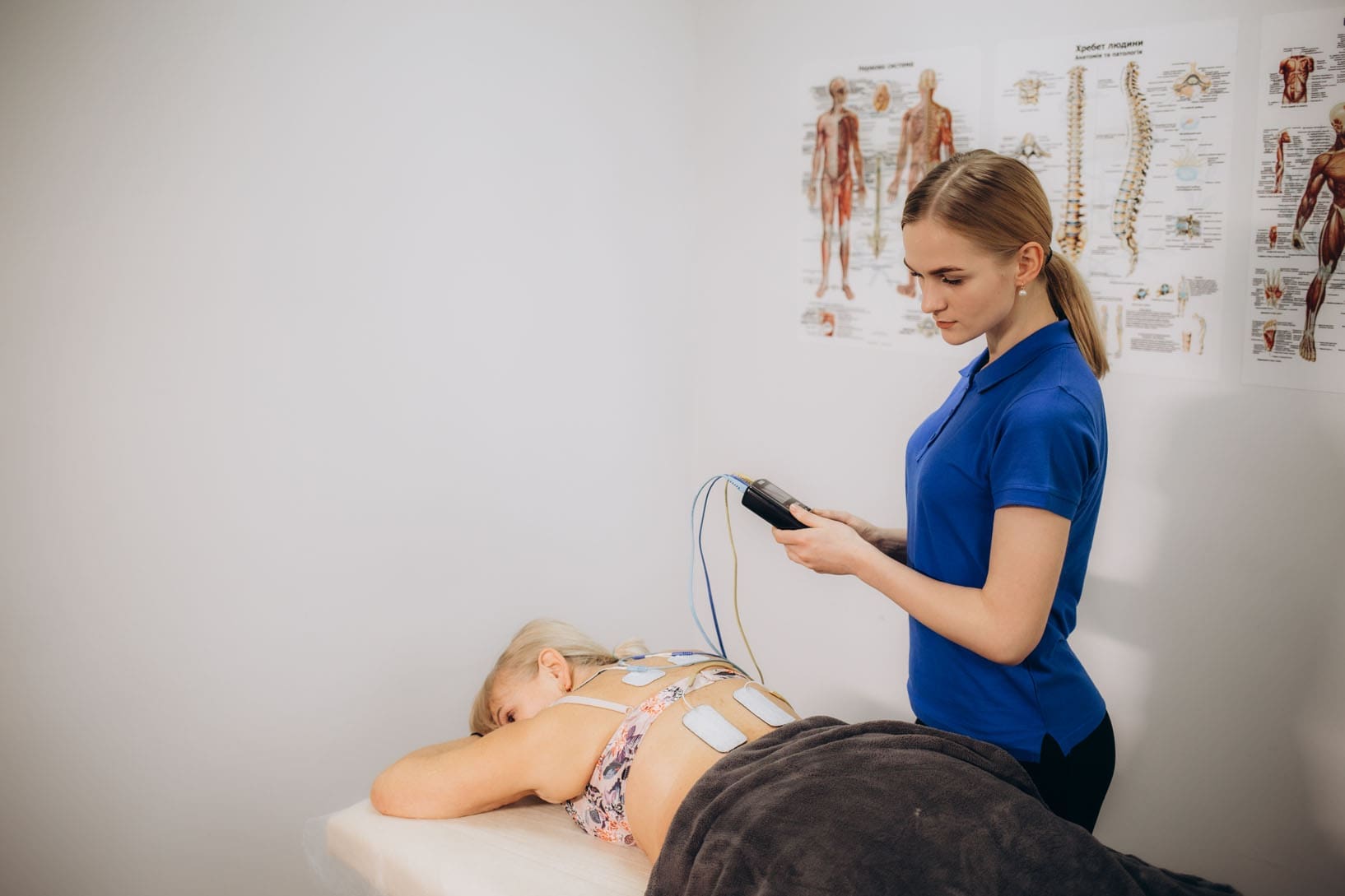
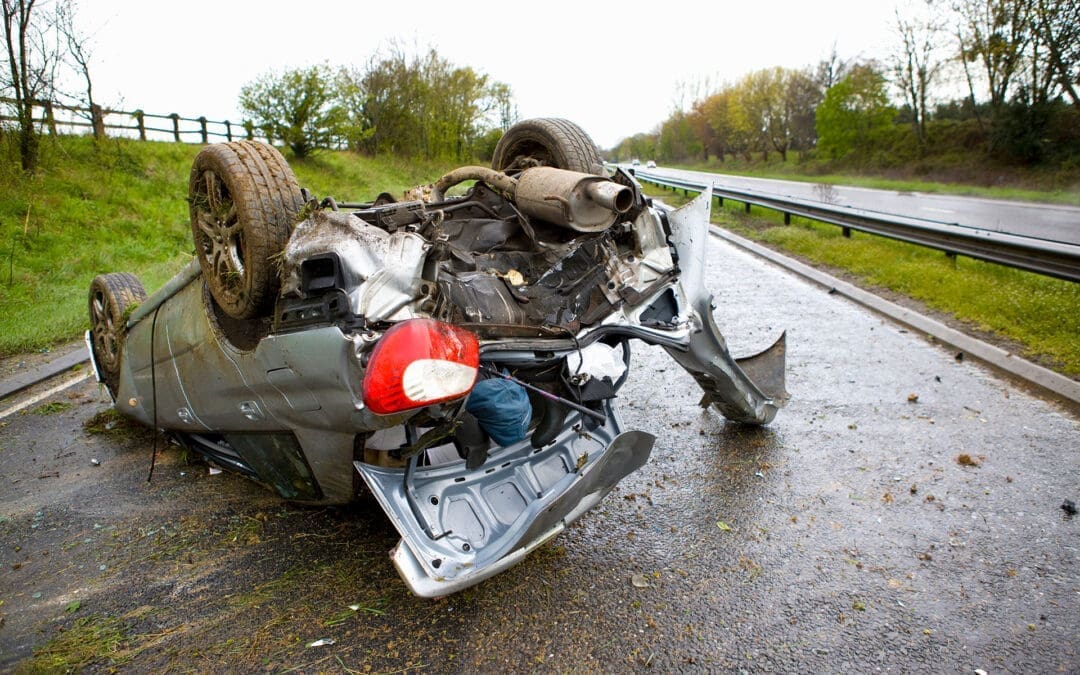
Have you ever walked away from a car crash thinking you were okay, only to feel stiff, sore, or in pain months or even years later? Many people do. A motor vehicle accident (MVA) can leave behind hidden damage that may not appear until long after the wreck. The good news? It is possible to feel better even if your crash happened a while ago. Integrative functional medicine and chiropractic care, combined with treatments such as platelet-rich plasma (PRP), microfragmented adipose tissue (MFAT), MLS laser therapy, and shockwave therapy, can address the underlying causes of ongoing pain rather than merely masking symptoms.
This article walks you through why old injuries continue to hurt, how these modern treatments work, and why they often work so well together. You will see clear steps from the problem to real relief.
Why Old Car Accident Injuries Turn Into Chronic Pain
Right after a crash, your body tries to fix sprains, strains, or torn ligaments. But occasionally the healing process does not finish the job. Months or years later, the area stays weak, inflamed, or stiff. Doctors call this a “latent” or hidden soft-tissue injury. The tissues never fully repaired, so small daily movements keep irritating them.
Cells in the damaged spot can act as if the injury just happened. Scar tissue builds up, blood flow drops, and nerves stay on high alert. This leads to ongoing joint pain, muscle tightness, or back and neck problems that feel like they will never go away. (Nob Hill Chiropractic, n.d.; Push as Rx, n.d.)
The key point is this: the body still wants to heal. Treatments that restart the repair process can make a big difference, even long after the accident.
How Chiropractic Care Helps Long After the Crash
Chiropractors gently adjust the spine and joints to realign them. This takes pressure off nerves, improves blood flow, and lets muscles relax. For people with old MVA injuries, chiropractic care can:
Reduce ongoing stiffness in the neck and back
Improve range of motion so daily tasks feel easier
Ease nerve irritation that causes tingling or shooting pain
Work with your body’s natural healing instead of forcing it
Even if you waited months or years to seek help, chiropractic adjustments can still correct the alignment issues that started in the crash. Many clinics note that proper documentation of your symptoms helps link the pain back to the accident for insurance or legal reasons. (Dallas Accident and Injury Rehab, n.d.)
Regenerative Medicine: PRP and MFAT Jump-Start Real Healing
Regenerative treatments use your body’s building blocks to fix damaged tissue. Two popular options are PRP and MFAT.
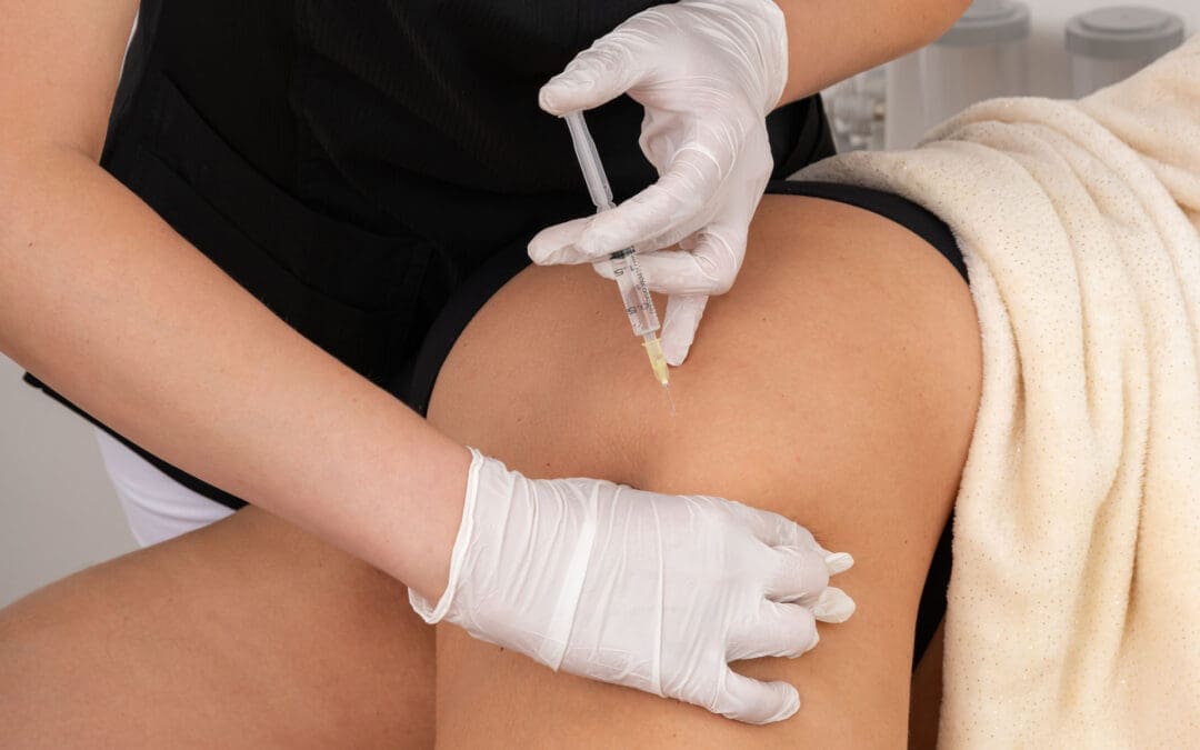
Platelet-Rich Plasma (PRP) Doctors draw a small amount of your blood, spin it to concentrate the platelets, and inject the PRP into the painful area. Platelets release growth factors that:
Fight inflammation
Bring in fresh blood and nutrients
Tell your cells to grow new, healthy tissue
Studies show that PRP helps with chronic tendon pain, ligament injuries, and joint problems that stem from trauma. One review found that it improved pain and function in knee, ankle, and back issues better than some traditional shots. People often feel relief that lasts months or longer because PRP treats the damaged tissue, not just the pain. (Thu, 2022; AABP Pain, n.d.)
Micro-Fragmented Adipose Tissue (MFAT) MFAT comes from a small amount of your own fat. The fat is processed into tiny fragments full of stem cells and healing signals, then injected where needed. It acts like a natural bandage, reducing swelling and supporting new tissue growth. MFAT works especially well for joints and ligaments that never healed right after a crash. (New Jersey Regenerative Institute, n.d.; Chiromed, n.d.)
Both PRP and MFAT are minimally invasive, use your own cells, and carry a low risk of side effects.
Cutting-Edge Modalities: MLS Laser and Shockwave Therapy
These painless, high-tech tools speed up repair without surgery or drugs.
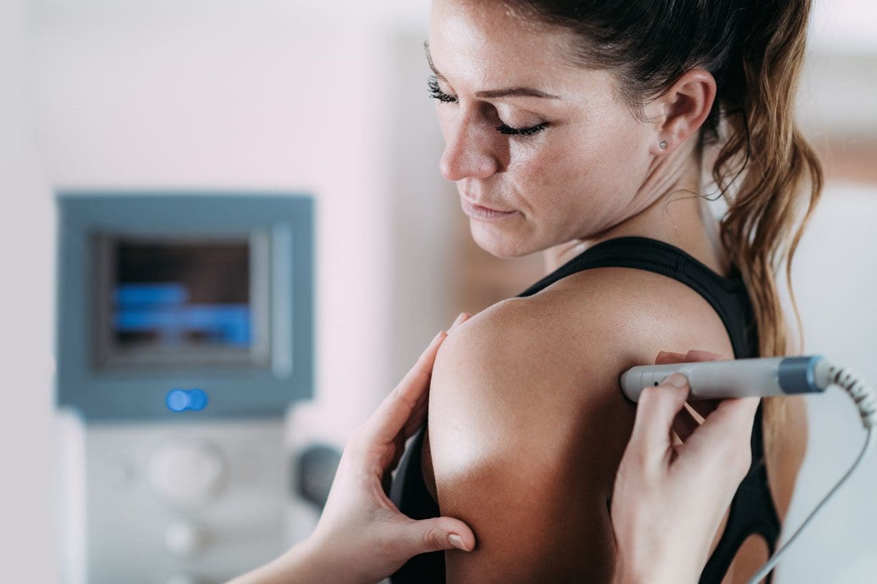
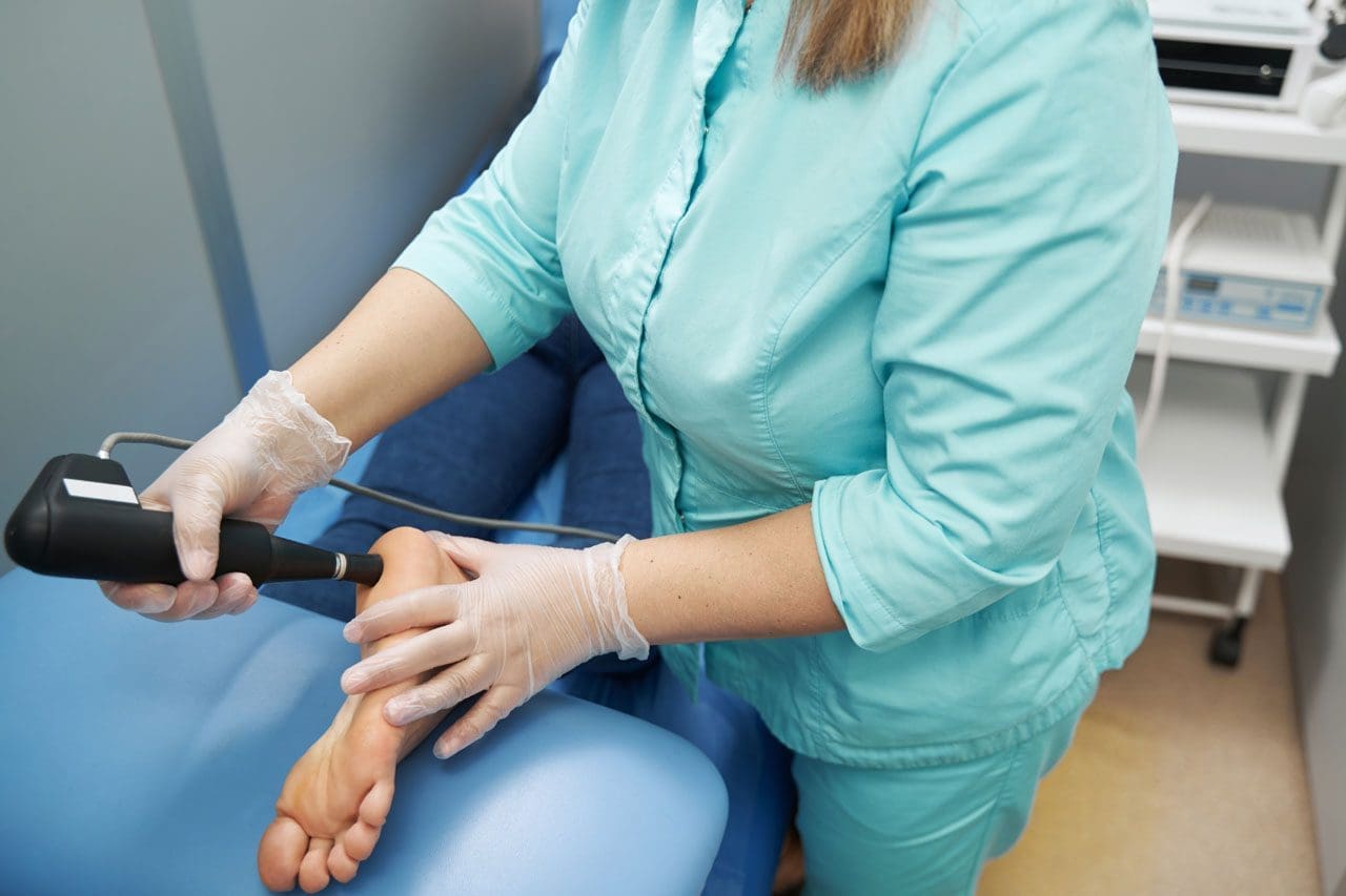
MLS Laser Therapy (Multiwave Locked System) uses specific light waves that penetrate deep into tissues. It:
Boosts cell energy so repairs happen faster
Lowers swelling and redness
Eases pain by calming overactive nerves
Improves blood flow to bring oxygen and nutrients
Patients with old whiplash, muscle strains, or ligament sprains often notice reduced stiffness and improved mobility after just a few sessions. The therapy is safe, relaxing, and works well alongside other treatments. (Nob Hill Chiropractic, n.d.; Drelham Nemat, n.d.; CARS Medical, n.d.)
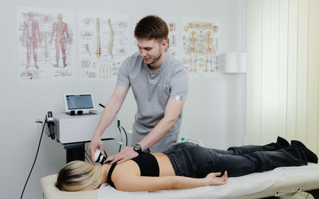
Shockwave Therapy: This uses sound waves to break up scar tissue and stimulate the growth of new blood vessels. When paired with PRP, it can provide faster pain relief and better long-term results in chronic tendon problems. (Jhan et al., 2024)
Why These Treatments Work Best Together
The real magic happens when chiropractic care, regenerative injections, and laser or shockwave therapy team up. Here is why:
Chiropractic aligns the body so the injected healing cells can reach the right spots.
Regenerative medicine (PRP and MFAT) rebuilds the damaged tissue at the cellular level.
MLS laser and shockwave reduce inflammation and scar tissue, so the new repairs can take hold.
Together they address the root cause—poorly healed soft tissue and ligament damage—rather than masking symptoms with pain pills. Many patients report less chronic pain, stronger joints, and a return to normal activities without surgery. (Push as Rx, n.d.; Chiromed, n.d.)
Clinical Observations from Dr. Alexander Jimenez
Dr. Alexander Jimenez, DC, APRN, FNP-BC, sees this pattern every day in his El Paso practice. As a chiropractor and family nurse practitioner trained in functional medicine, he treats hundreds of people with old MVA injuries. His clinical observations show that crashes often create a “chain reaction” of problems: one tight muscle pulls on another, nerves get irritated, and inflammation lingers for years if not fully addressed.
Dr. Jimenez uses a full evaluation—digital motion X-rays, nerve tests, and functional assessments—to identify the exact root causes. He then combines gentle chiropractic adjustments, PRP and shockwave therapy, MLS laser, and personalized nutrition plans. Patients with whiplash, chronic back pain, or unresolved ligament issues often regain mobility and feel stronger months after starting care. He stresses that even long-standing injuries respond when the whole body is supported, not just the painful spot. His approach aligns with research: early, or even delayed, integrative care can prevent arthritis and chronic disability. (Dr. Alexander Jimenez, n.d.)
Real Benefits You Can Expect
Here are some common improvements people notice:
Less daily pain and fewer pain pills needed
Better movement in the neck, back, shoulders, or knees
Stronger ligaments and tendons that feel more stable
Improved sleep because pain no longer keeps you awake
Faster return to work, sports, or family activities
Lower chance of needing surgery later
Results vary from person to person, but starting with a thorough exam helps create a plan that fits your exact needs.
Taking the First Step Toward Lasting Relief
If you have lived with pain from a car accident that happened months or years ago, you do not have to accept it as “just the way it is.” Integrative functional medicine and chiropractic care, paired with PRP, MFAT, MLS laser, and shockwave therapy, give your body the tools to finish the healing it started long ago. These approaches focus on the root cause—unresolved soft tissue and ligament damage—so you can move, work, and live with less pain.
Talk to a qualified provider who understands MVA injuries and regenerative options. A simple consultation can show whether these treatments are right for you. Many people discover that real relief is still possible, no matter how much time has passed.
MLS Laser and Chiropractic Care for Back and Joint Pain
Abstract
In this educational post, I walk you through how we integrate modern photobiomodulation (MLS laser therapy) with chiropractic care, manual therapy, and active rehabilitation for spinal and joint pain. You will learn how we set up treatment for low back facet pain, why patient comfort and precise dosing matter, and how we target both the painful site and the connective tissue network to drive better outcomes. I explain energy density (joules per cm²), the Arndt–Schulz dose-response principle, tissue optics, and how pulsed dual-wavelength lasers engage mitochondrial and neuroimmune pathways to reduce pain and enhance recovery. We will also explore how robotic and handheld delivery complement each other, how we schedule acute and chronic care plans, how we combine laser with shockwave, PRP, and movement therapy, and when this approach can delay surgery by improving pain and function. Throughout, I share clinical observations from our El Paso Back Clinic and highlight evidence from leading researchers using rigorous, evidence-based methods. The emphasis is on integrative chiropractic and physical therapy, with medications and hormones kept in the background.
At El Paso Back Clinic, our mission is to merge hands-on chiropractic care, targeted physical therapy, and precision technologies that safely accelerate healing. One modality we employ is MLS laser therapy, a form of photobiomodulation that uses synchronized near-infrared wavelengths to influence cellular energy, microcirculation, and neuroinflammatory signaling. In this post, I reframe a recent procedural walkthrough from my perspective and expand on the physiology, clinical reasoning, and practical protocols we use every day with patients presenting with low back pain, knee osteoarthritis, plantar fasciitis, and other musculoskeletal conditions. The star is not the device; it is the integrated plan that places your spine and movement at the center of care.
Optimizing patient comfort and precision: Why setup matters
Key concepts:
Patient positioning
Direct-to-skin contact when appropriate
Targeting by symptoms and anatomy
Stability during unattended robotic delivery
When I set up laser therapy—especially with a robotic head—my first priority is patient comfort and stability. If a patient shifts during an unattended cycle, the beam may drift from the intended target. For lumbar facet-mediated pain at L4–L5, I position the patient comfortably prone, ensure the treatment field is exposed with direct skin access when using a contact handpiece, and confirm the exact region of maximal tenderness and referral (e.g., right-sided zygapophyseal joint pain with proximal radiation).
To minimize error, I zero the device’s X and Y axes, center the beam over the primary pain generator, then expand the field to include adjacent connective tissue tracks. This is our clinical multimodal approach: treat the source, the site, and the surrounding soft tissue network. By caring for the paraspinal fascia, intermuscular septa, and periarticular tissues, we respect that pain is rarely a single-point phenomenon. Fascia transmits load and communicates mechanosensory signals; addressing it improves regional glide and reduces nociceptive drive.
Why direct skin contact? Tissue optics favor minimal reflection and refraction losses. Air-skin interfaces reflect more energy, especially at certain angles. When we must avoid contact—such as at post-surgical sites or in cases of allodynia—we employ a non-contact, collimated robotic head positioned at an optimal focal distance, measured with a calibrated ruler.
Robotic plus handheld delivery: Complementary tools
Robotic head:
Non-contact, collimated beam; ideal for broad areas, post-surgical sensitivity
Software auto-recalculates dose time when X-Y field size changes
Handheld contact piece:
Tactile feedback for focal trigger points and joint spaces
Allows dynamic, movement-based application during active care
In practice, I often run both channels simultaneously. The robot delivers a uniform, programmable energy density across a defined area while I probe and treat focal trigger points or facet capsules with the handheld. This mirrors how we layer manual therapy with exercise: a global reset paired with local precision.
Dosing by energy density: The language of photobiomodulation
Target dose: typically 4–10 joules/cm², depending on condition and depth
Why density matters more than total joules: tissue dose equals energy per unit area
Auto-time calibration: changing the field size while maintaining the same J/cm² adjusts the total joules and time automatically
We dose by energy density, not just total energy. For example, a lumbar facet region might be set to 6 J/cm². On a larger field, total joules increase, but the cellular dose per square centimeter remains constant, aligning with literature-supported ranges that optimize photobiomodulation responses without tipping into bioinhibition. This reflects the Arndt–Schulz principle: too little energy yields no change, optimal energy stimulates, and excessive energy can dampen biological activity.
The physiology behind pain relief and tissue recovery
Mitochondrial activation:
Photons at near-infrared wavelengths interact with cytochrome c oxidase, improving electron transport and boosting ATP production
Enhanced ATP supports ion pump function, cytoskeletal remodeling, and protein synthesis required for tissue repair
Nitric oxide and microcirculation:
Photo-dissociation of nitric oxide from cytochrome c oxidase and endothelial effects promotes vasodilation and microvascular perfusion, aiding oxygen delivery and metabolite clearance
Neuroinflammatory modulation:
Downregulation of pro-inflammatory cytokines and modulation of glial activity reduce peripheral and central sensitization
Neural effects and immediate analgesia:
Modulation of small-diameter nociceptive fibers and gate-control mechanisms can provide early symptom relief
Collagen and connective tissue remodeling:
Changes in fibroblast activity and collagen organization may improve tendon/ligament structure over time when paired with load-specific rehab
In our clinic, patients sometimes report warmth or a faint tingling, but with synchronized pulsed delivery and short pulse durations, surface heat remains low while energy is effectively absorbed at depth. When tissue temperature stays stable over time, we know we are within the desired window: enough photons to trigger biochemical cascades without superficial overheating.
Why pulsed, dual-wavelength delivery matters
Wavelength pairing:
808 nm: deeper penetration for mitochondrial and vascular effects
905 nm: high peak power in short pulses adds neuromodulatory and analgesic benefits while protecting against thermal buildup
Synchronized pulse trains:
High peak, short duration pulses deliver energy in “packets,” allowing absorption periods between bursts and reducing superficial heat accumulation
These engineering choices align with clinical goals: delivering energy to deeper targets, such as facet capsules or the posterior knee compartment, while preserving patient comfort.
Chiropractic integration: Adjustments, motor control, and fascia
Spinal adjustments:
Restoring joint play at hypomobile segments reduces aberrant mechanoreceptor input and reflex muscle guarding
Fascial glide and soft-tissue work:
Instrument-assisted or hands-on release improves shear planes; laser primes fibroblasts and microcirculation for better tissue response
We pair laser sessions with graded movement to convert biochemical gains into functional patterns
Laser does not replace chiropractic care; it helps us reach the dose of movement sooner by lowering pain and stiffness that otherwise block progress. For example, after an MLS session over L4–L5 facets and paraspinals, we cue diaphragmatic breathing and segmental stabilization to capitalize on reduced nociception and improved circulation.
Case walk-through: Low back facet pain (L4–L5)
Assessment:
Right-sided facet loading pain with limited extension and paraspinal tenderness
No red flags; neurological exam stable
Laser setup:
Patient prone, area exposed; robot field centered over right L4–L5 facet region
Density: 6 J/cm², field expanded to capture paraspinal fascia and myofascial referral zones
Handheld: contact sweeps over identified trigger points
Session length:
Robot 6–10 minutes, depending on field size; handheld 20–30 seconds per trigger point
Immediate follow-up:
Prone press-ups to reassess extension tolerance
Gentle lumbar stabilization exercises to lock in gains
Home plan:
Extension-biased mobility as tolerated, core endurance drills, ergonomic cues
What my patients often notice is not just pain relief within hours but improved ease of movement—the kind of change that allows us to progress from passive care to active loading.
Knee osteoarthritis: Accessing the joint intelligently
Beam access matters:
Anterior patella reflects substantial energy; flexing the knee opens the joint space and reduces reflection
Posterior and medial/lateral approaches improve delivery to synovium and periarticular tissues
Dosing strategy:
Target 4–8 J/cm² per compartment; treat multiple compartments in the same session by apportioning field time
Integration with PT:
Laser to modulate pain and effusion
Progressive quadriceps and hip strengthening, gait retraining, and balance work
Manual therapy for capsular mobility as indicated
While no laser regrows cartilage in advanced bone-on-bone disease, many of our patients experience reduced pain and swelling and better function, which can delay the need for surgery. The goal is to expand the movement envelope required for strength and neuromuscular control.
Acute vs. chronic protocols: Cumulative effects and scheduling
Acute conditions:
Six treatments delivered as close to daily as feasible (e.g., Monday–Wednesday–Friday pattern), aiming for rapid symptom control
Chronic conditions:
Twelve treatments, ideally within four weeks, to build cumulative neuroimmune and mitochondrial effects
Why packages:
Effects are additive; stopping after early relief risks relapse before tissue remodeling and motor reeducation are complete
Reassessment points:
After 3–4 sessions: evaluate pain and function
After 6–12 sessions: progress exercise intensity, reduce passive modalities
Our patients often report noticeable changes 4–6 hours after a session; we encourage them to “test” function later the same day (for example, stair climbing or walk tolerance) to anchor improvements to real-life tasks.
Combining laser with orthobiologics and shockwave
With PRP:
Two to three pre-injection laser sessions to improve local perfusion and tissue readiness
Day-of-injection: protocol tailored to avoid blunting intended inflammatory signaling while supporting analgesia
Six post-injection sessions to enhance microcirculation and cellular energy during proliferative phases
With shockwave:
Laser can reduce pain and prime tissues for mechanical signaling from shockwave
Sequence depends on goals; we often laser first for analgesia, then apply focused shockwave for mechanotransduction, followed by graded loading
Rationale:
Photobiomodulation and mechanotherapy act on complementary pathways—bioenergetics and microcirculation (laser) plus tenocyte activation and neovascular remodeling (shockwave)
Hormonal or medication considerations remain in the background for us; when appropriate, we coordinate with the patient’s prescribing providers to avoid interventions (e.g., routine NSAIDs immediately after PRP) that might dampen desired signaling. Our primary emphasis remains movement-based rehabilitation supported by laser and manual care.
Bone and postoperative considerations
Bone healing:
The evidence base for photobiomodulation in fracture healing exists but varies by device and parameters; in clinical experience, early application within 7–10 days post-fracture may support the inflammatory and early reparative phases. This is commonly considered off-label for certain devices and requires case-by-case judgment and collaboration with the treating orthopedic team
Post-surgical care:
Non-contact robotic delivery allows dosing without skin contact when sensitivity is high
Goals include edema control, pain reduction, and earlier initiation of therapeutic exercise
Dose ceilings and the bioinhibition paradox
Arndt–Schulz law:
Insufficient dose yields no effect; optimal dose stimulates; excessive dose may inhibit
Practical application:
If more time is desired, we distribute energy across multiple approaches (e.g., anterior-posterior or medial-lateral fields) instead of stacking excessive dose on one spot
Patients frequently report a “melting” of stiffness within the same day after an MLS session paired with extension-bias exercise; repeated sessions lower baseline pain and improve extension tolerance, allowing us to progress to anti-rotation and hip hinge training
Knee osteoarthritis:
Combining posterior-compartment laser dosing with patellar mobilization and quadriceps strengthening reduces pain during sit-to-stand and stair negotiation within two to three weeks; gains consolidate when patients adhere to home-based strength and balance work
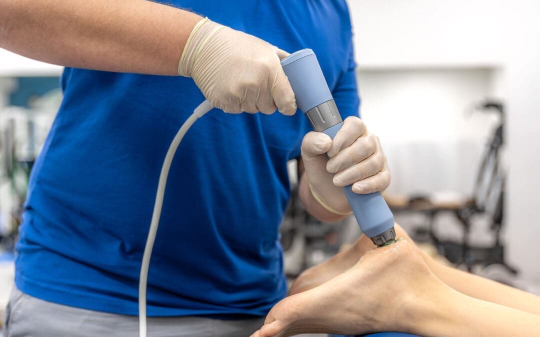
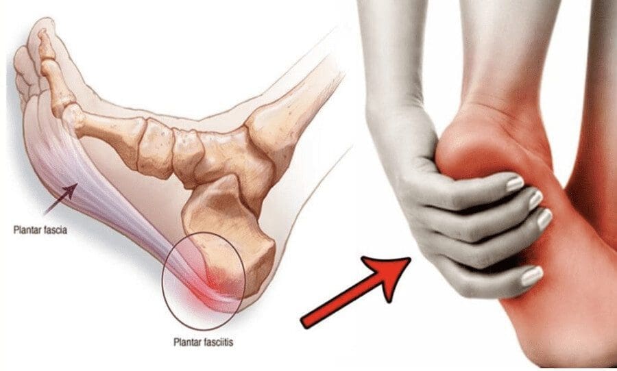
Plantar fasciitis:
Laser applied to the medial calcaneal region and along the plantar fascia with calf mobility and foot intrinsics training shortens the “first-step” pain window and speeds return to walking programs
Post-injection care:
In patients receiving PRP from collaborative providers, pre- and post-injection laser often reduces pain spikes and supports earlier initiation of controlled loading, which in turn improves functional outcomes at 6–12 weeks
Safety, reliability, and patient communication
Safety profile:
Proper eyewear, attention to reflective surfaces, and adherence to dosing ranges keep risk low
Device reliability:
Modern systems include field service support; routine calibration and training ensure consistent delivery
Expectations:
We counsel that pain did not develop in ten minutes and will not vanish in ten; however, many feel better within hours, see consistent improvement after three sessions, and sustain gains with a full plan of care
Putting it all together: A typical plan
Evaluation:
History, movement assessment, palpation, neurological screen, and imaging if indicated
Plan creation:
Define primary pain generators and movement deficits
Choose laser parameters (wavelengths, pulsing, J/cm²) and field geometry
Integrate manual therapy and exercise blocks within each visit
Visit flow:
Laser (robotic field + handheld focal points)
Manual therapy for joint and soft tissue restrictions
Targeted exercises (mobility, motor control, strength)
Education and home program
Progression:
Increase exercise intensity as pain decreases
Taper passive modalities
Reassess goals every 3–4 sessions
Why these techniques work, in plain terms
Pain is both chemical and mechanical. Laser modifies the chemical environment (reduces inflammatory signaling, increases ATP, improves microcirculation). Chiropractic and rehab address the mechanical side (joint motion, tissue glide, strength, coordination). Combining them tackles the problem from both angles
The nervous system adapts to pain by inhibiting movement. Rapid analgesia from laser helps unlock motor patterns so we can retrain stability and strength sooner
Tissues heal under the right load. Once pain is controlled and circulation improved, progressive loading guides collagen alignment and muscle conditioning for durable outcomes
Evidence-based grounding
Photobiomodulation has a growing body of research demonstrating analgesic, anti-inflammatory, and pro-recovery effects in musculoskeletal conditions. Rigorous, modern methodologies—randomized controlled trials, dose–response investigations, and consensus guidelines—support dosing in the 4–10 J/cm² range for many superficial-to-moderate-depth targets and highlight the importance of wavelength, pulse structure, and treatment frequency. Clinical effectiveness is maximized when photobiomodulation is embedded within active rehabilitation rather than used in isolation.
If you are considering care at El Paso Back Clinic, our team will assess your unique presentation and craft an integrative plan that prioritizes spinal mechanics, movement, and function—leveraging laser therapy where it adds value and always keeping the emphasis on your long-term resilience.
Chiropractic and Regenerative Care After Car Accidents
Motor vehicle accidents (MVAs) happen fast. One moment you are driving, and the next, sudden forces jolt your body. These impacts often cause soft tissue damage, ligament tears, joint injuries, and spinal trauma. Many people experience pain, stiffness, and limited mobility that can persist for months or years if not treated properly.
Fortunately, a growing number of patients find relief through a mix of regenerative therapies and integrative chiropractic care. Treatments such as Platelet-Rich Plasma (PRP), Platelet-Poor Plasma (PFP), Micro-Fragmented Adipose Tissue (MFAT), shockwave therapy, and chiropractic adjustments work together to support the body’s natural healing processes. These options are especially helpful for people who want to avoid surgery and reduce chronic pain from acute trauma.
Why Early Treatment Matters Most
Experts agree that starting care right after an accident gives the best results. Injuries from crashes can seem minor at first, but swelling, scar tissue, and poor movement patterns often lead to long-term problems. Acting quickly helps stop these issues before they become chronic.
Dr. Alexander Jimenez, DC, APRN, FNP-BC, a leader in El Paso, Texas, stresses this point in his clinical work. With dual training as a chiropractor and nurse practitioner, he sees how prompt integrative care helps patients recover function and avoid surgery. His approach combines detailed exams, advanced imaging, and personalized plans to address both the injury and overall health.
Common Injuries from Motor Vehicle Accidents
Crashes put tremendous stress on the body. Here are some frequent problems:
Soft tissue damage: Muscles, tendons, and ligaments stretch or tear.
Ligament tears: These stabilize joints but can become loose or painful.
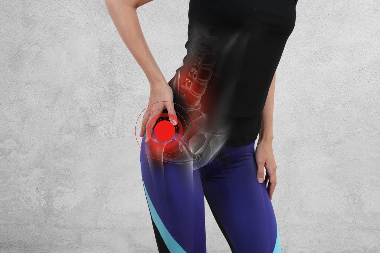
Joint injuries: Shoulders, knees, hips, and wrists often sustain impact injuries.
Spinal trauma: Whiplash, herniated discs, and misalignments affect the neck and back.
Nerve issues: Compression or irritation leads to pain, numbness, or tingling.
Without proper care, these injuries can cause ongoing pain, reduced mobility, and even problems with daily tasks like working or driving.
How Regenerative Therapies Support Healing
Regenerative medicine uses the body’s own materials to repair damage. These treatments deliver growth factors, stem cells, and healing signals exactly where they are needed.
Platelet-Rich Plasma (PRP) Therapy
Doctors draw a small amount of your blood and spin it in a machine to concentrate the platelets. These platelets contain growth factors that speed up tissue repair.
PRP helps with:
Whiplash and neck strains
Tendon and ligament injuries
Joint pain
Muscle tears
Patients often notice less pain and better movement after a few sessions. PRP is minimally invasive and uses your own blood, so the risk of reaction is low.
Platelet-Poor Plasma (PFP) and Related Options
PFP focuses on other helpful proteins in blood plasma. Clinics sometimes combine it with PRP for broader healing support. These concentrates create a strong healing environment without surgery.
Micro-Fragmented Adipose Tissue (MFAT)
MFAT uses a small sample of your own fat tissue. Doctors process it gently to keep helpful stem cells and growth factors, then inject it into injured areas.
MFAT offers:
Structural support for damaged tissue
Anti-inflammatory effects
Potential for longer-lasting repair
It shows promise for joint issues, partial tears, and chronic pain after accidents. The procedure is outpatient and involves minimal downtime.
The Power of Shockwave Therapy
Shockwave therapy sends acoustic waves into deep tissues. It breaks up scar tissue, improves blood flow, and stimulates healing cells. Many clinics use it alongside regenerative injections.
Benefits include:
Reduced swelling and pain
Better circulation
Faster recovery from soft tissue injuries
Help for whiplash, tendon problems, and lower back strain
Sessions are short, non-invasive, and require no downtime. Patients often feel relief within a few visits.
Integrative Chiropractic Care: Restoring Alignment and Function
Chiropractic adjustments correct spinal misalignments caused by crashes. Proper alignment takes pressure off the nerves, improves movement, and allows the body to heal more effectively.
Dr. Jimenez’s clinics combine chiropractic with medical oversight. This dual approach includes:
Gentle spinal adjustments
Soft tissue work
Rehabilitation exercises
Nutritional guidance to fight inflammation
Chiropractic care helps prevent chronic issues by fixing movement patterns early.
A Combined Treatment Journey
Many patients follow a clear path to recovery:
Immediate Evaluation – Get imaging and a full exam to understand the injuries.
Pain and Inflammation Control – Use shockwave or gentle therapies first.
Regenerative Injections – PRP, PFP, or MFAT to promote tissue repair.
Chiropractic and Rehab – Adjustments and exercises to restore strength and mobility.
Ongoing Support – Nutrition, lifestyle changes, and follow-up care.
This step-by-step plan helps patients return to normal activities faster and with less pain.
Real-World Benefits for Accident Victims
Avoid Surgery: Many people with ligament tears or joint damage avoid surgery.
Reduce Chronic Pain: Early regenerative care limits scar tissue and long-term issues.
Faster Return to Work and Life: Improved healing leads to quicker recovery of strength and mobility.
Natural Approach: Treatments use your body materials and avoid heavy drugs.
Dr. Jimenez often notes in his clinical observations that patients who receive integrated care report better outcomes in both physical function and quality of life. His focus on legal documentation also helps when building injury claims.
What to Expect During Treatment
Most procedures happen in an office setting. PRP or MFAT involves a quick blood draw or fat harvest under local numbing. Shockwave feels like firm taps but is tolerable. Chiropractic visits are comfortable and relaxing for most people.
Recovery times vary, but many patients resume light activities soon after. Full benefits build over weeks as tissues repair. Doctors tailor plans to each person’s needs, age, and injury severity.
Lifestyle Tips to Support Recovery
Eat anti-inflammatory foods like fruits, vegetables, and healthy fats.
Stay hydrated and get quality sleep.
Follow your exercise plan to rebuild strength safely.
Manage stress, which can slow healing.
Attend all follow-up visits to track progress.
When to Seek Help
See a qualified provider right after any accident, even if you feel okay at first. Delayed symptoms are common. Look for clinics that offer both regenerative options and chiropractic care for the best results.
Conclusion: A Smarter Path to Healing
Soft tissue damage, ligament tears, joint injuries, and spinal trauma from car accidents do not have to define your future. Combining PRP, PFP, MFAT, shockwave therapy, and integrative chiropractic care offers a powerful, natural way to heal. Starting treatment early gives your body the best chance to repair itself and prevents long-term problems.
Clinicians like Dr. Alexander Jimenez show how this whole-person approach works in real life—helping patients move better, feel better, and get back to living fully. If you or a loved one has been in a crash, explore these options with a knowledgeable provider. Recovery is possible, and modern regenerative care makes it more achievable than ever.
In this educational post, I share my personal journey from debilitating plantar fasciitis to lasting recovery, and how that experience led me to integrate true shockwave therapy into comprehensive chiropractic and physical therapy care at El Paso Back Clinic. I explain how shockwaves work at the cellular level, why electrohydraulic systems deliver stronger and more reliable therapeutic effects than radial pressure waves, and how focused and broad-focus energy can improve tissue healing, angiogenesis, and modulation of inflammation. I present outcome data from leading registries, highlight on-field recovery examples from professional athletes, and outline how integrative chiropractic care, manual therapy, and targeted rehabilitation protocols synergize with shockwave to accelerate return to sport and work. You will find clear explanations, clinical reasoning, and practical protocols for plantar fasciitis, adductor strains, bone edema, tendinopathies, and post-operative healing—prioritizing non-invasive, cash-pay, and workflow-friendly solutions that minimize reliance on injections or medications while keeping hormone and pharmacologic strategies in the background.
Introduction: Why I Brought Shockwave into Integrative Chiropractic Care
Seven years ago, a patient developed severe plantar fasciitis in both feet. They were told cortisone injections might help, but declined, hoping for a better way. A friend in the shockwave field saw them hobbling and suggested they try treatment. The first session was intense—the prevailing view back then was “push through pain”—and while they felt better that day, the pain returned within a few days. Two months later, without elaborate stretching or self-care, they woke up and realized they hadn’t felt foot pain for days. The relief lasted almost four years before they needed another round. This experience highlighted the potential of technologies that safely and effectively stimulate the body’s natural repair systems.
At El Paso Back Clinic, true shockwave therapy—specifically electrohydraulic technology—is integrated into a broader model of chiropractic care, physical therapy, and evidence-based rehabilitation. The goal is to make healing predictable, comfortable, and accessible for patients while reducing dependence on injections or medications unless truly necessary.
Shockwave Therapy Basics: What It Is and Why the Mechanism Matters
Core concept: A shockwave is a rapidly propagating acoustic wave with steep pressure gradients that exerts a mechanical force on cells and tissues.
Mechanotransduction: When a true shockwave hits a cell membrane, it causes a rapid pressure change that makes the membrane transiently more permeable. This mechanical stimulus triggers cellular signaling, promoting angiogenesis, modulation of inflammation, and tissue repair (Schmitz et al., 2015; Wang, 2012).
Therapeutic outcomes: Research shows increased microvascular density, improved perfusion, and activation of pathways such as VEGF, eNOS, and BMPs, thereby supporting bone and soft tissue regeneration (Wang, 2012; Schmitz et al., 2015).
Not all “shockwaves” are equal. In community advertising, the term “shockwave” often refers to radial pressure-wave devices, which are distinct from true shockwaves.
Radial pressure waves:
Mechanism: Generate surface-level pressure pulses that disperse broadly and do not reach the speeds or waveform required for a classic shockwave.
Effect: More superficial stimulation, often relying on tissue irritation to induce local blood flow. It can be beneficial for short-term analgesia but is less reliable for deep tissue regeneration, especially in the plantar fascia, adductors, hip, and deep tendons (Sorg et al., 2020).
Electrohydraulic shockwaves:
Mechanism: A submerged electrode ignites a plasma bubble; the leading edge of the bubble forms a true shockwave inside the applicator. Energy emerges already at shockwave speed and waveform, penetrating up to ~12 cm depending on settings and tissue properties.
Benefit: Consistent depth, strong mechanotransduction, and more uniform dosing across the therapeutic column of tissue (Schmitz et al., 2015).
Electromagnetic and Piezoelectric Alternatives: What’s Different
Both electromagnetic and piezoelectric sources can generate shockwaves, but their sound waves may converge into shockwave form outside the applicator, leaving a gap that requires careful tip selection and positioning for precise targeting (ISMS T guidelines; Schmitz et al., 2015).
Electrohydraulic energy begins as a shockwave, reducing sensitivity to positioning and providing a broader, more forgiving therapeutic column. This consistency matters in real-world clinics, where anatomy varies, scar tissue is present, and motion is restricted.
Focused Plus Broad-Focus Energy Delivery: Two-for-One Tissue Coverage
In electrohydraulic systems with parabolic reflectors, I utilize both:
The focused wave: like a laser pointer, ideal for pinpointing lesions (e.g., proximal plantar fascia origin, adductor insertion, enthesopathic changes).
The broad-focus column: distributes the reflected energy across a larger volume, covering diffuse pathology (e.g., fascial chains, kinetic-chain contributions, and regional interdependencies).
Why It Matters
Larger therapeutic zones shorten treatment times.
Less technician dependency: Patient biofeedback—gentle tapping in healthy tissue and heightened sensation over inflamed or damaged areas—guides us to the right spot without surgical precision.
Greater comfort: Broad dispersion reduces peak discomfort, allowing higher energy without anesthesia.
Patient Outcomes: What Registries and Clinical Data Show
Third-party registries have reported contrasting outcomes among radial pressure waves, focused shockwaves, and electrohydraulic systems.
Radial: Often effective for short-term pain relief; at ~6 months, many patients report a return to baseline symptoms.
Focused shockwave: Meaningful pain reduction by ~3 months, with some drift back by ~6 months.
Electrohydraulic with broad coverage: Sustained reduction in pain scores at 6 months, likely due to deeper, wider mechanotransduction and vascular changes supporting continued remodeling (Schmitz et al., 2015; Meta-analyses: Rompe et al., 2007; Wang, 2012).
Integrative Chiropractic Model: How We Fit Shockwave into Care
We blend shockwave therapy with chiropractic and physical therapy to support the body’s capacity to heal and move.
Chiropractic adjustments:
Correct regional joint restrictions contributing to overload of the plantar fascia, adductors, or hip stabilizers.
Myofascial techniques address fibrosis and improve interstitial fluid flow, supporting the vascular benefits of shockwave therapy.
Eccentric loading stimulates tenocyte activity and collagen realignment, capitalizing on shockwave’s activation of repair pathways (Wang, 2012).
Case Insight: Plantar Fasciitis Recovery and Long-Term Resilience
My personal case mirrored many patients at El Paso Back Clinic: initial plantar fascia pain responding to shockwave with subsequent delayed but sustained resolution. In the clinic, I see:
Patients reporting immediate pain relief after the first session due to nociceptive modulation.
Progressive improvements at 4–8 weeks consistent with vascular remodeling and matrix repair.
Enhanced durability when combined with foot intrinsic strengthening, hip stabilizer training, and chiropractic alignment work.
Sports Medicine Perspective: Adductor Strain and Return to Play
Professional teams increasingly use shockwave therapy for adductor strains and hip and groin injuries. The immediate benefits include reduced pain and accelerated tissue recovery compared with conventional timelines. In our practice:
We target the adductor longus origin and the fascial plane along the inner thigh while normalizing pelvic mechanics with sacroiliac and lumbar adjustments.
We integrate isometric-to-eccentric progressions and adductor-abductor balance training to reduce reinjury risk.
The combination improves tolerance to sport-specific loads and hastens return to competition.
Bone Edema and Post-Operative Healing: A Non-Invasive Boost
Electrohydraulic shockwave has supportive data in bone marrow edema and delayed union/slow healing states:
Mechanism: Induction of osteogenic signals (e.g., BMPs), increased neovascularization, and modulation of inflammatory mediators help restore homeostasis in bone and periosteum (Wang, 2012).
Clinical integration: We use shockwave alongside gentle mobilization and loading strategies, emphasizing safe progression while pain and function improve.
Radial vs. True Shockwave: Setting Patient Expectations
Many patients come in having tried “shockwave” elsewhere—usually radial pressure wave therapy—and feel wary because it was painful or ineffective long term. Education is essential:
We explain the difference between pressure waves and true shockwaves.
We demonstrate the biofeedback sensation: mild over healthy tissue, sharper over pathology.
We emphasize comfort: broader energy distribution allows higher therapeutic levels with better tolerability.
How We Deliver Care: Workflow and Patient Experience
Session length: 10–15 minutes for shockwave application, integrated into chiropractic and PT visits.
Immediate feedback: Often, we see same-day reductions in pain, which motivates adherence.
Training and delegation: The broad focus makes it safe for trained clinical assistants to apply my protocols, maintaining quality and efficiency.
Billing: Primarily cash-pay, with transparent packages; we discuss any local reimbursement possibilities if applicable.
Our Protocols: Practical Steps and Reasoning
Plantar Fasciitis
Assessment:
Foot posture, gait analysis, palpation of proximal fascia, and medial calcaneal tubercle.
Lower frequency, targeted dosing over the affected bone segment while respecting pain thresholds.
Chiropractic/PT:
Gentle mobilization for adjacent joints, graded weight-bearing, and circulation-enhancing strategies.
Rationale:
Supports osteogenesis and neovascularization; movement aids recovery without overloading.
Integrating Orthobiologics Carefully
While we focus on chiropractic and physical therapy first, shockwave can bridge the gap for patients reluctant to injections. When orthobiologics are warranted:
Same-day approach:
Shockwave first to reduce pain and improve tolerance; injection follows under improved comfort.
Staged approach:
Shockwave 48–72 hours before injection to enhance perfusion and microenvironment.
Evidence-building:
Biofeedback mapping demonstrates lesion localization to the care team and patient, supporting shared decision-making.
Comfort, Tolerance, and Safety
Electrohydraulic systems with broad-focus reflectors allow higher energy dosing with less discomfort:
Patients describe healthy tissue as gentle tapping.
Over lesions, they feel a clear but tolerable sensation guiding us to the target.
We avoid “torture” models—modern protocols prioritize comfort while achieving biologically meaningful dosing.
Real-World Implementation at El Paso Back Clinic
Training:
My team and I conduct device education, maintenance, and immediate patient trials so we can start treating day one.
Ongoing support:
We continue case reviews, update protocols, and refine integration with chiropractic and PT workflows.
Marketing:
We grow organically through patient word-of-mouth, outcome reporting, and community education.
What Patients Can Expect: Timeline and Milestones
First session:
Often a reduction in pain scores and improved movement due to nociceptive modulation.
2–4 weeks:
Vascular changes and early remodeling translate into improved function; PT progression intensifies.
6–12 weeks:
Collagen realignment and kinetic-chain improvements make gains more durable; return to sport or work accelerates.
Clinical Observations from My Practice
Sustained relief in plantar fasciitis with fewer recurrences when we address foot mechanics, hip stability, and load management alongside shockwave.
Faster return to play in adductor strains when pelvic corrections are included and eccentric programs are supervised.
Improved tolerance of loading in Achilles and patellar tendinopathies when shockwave precedes progressive rehab blocks.
Bone marrow edema cases respond well when shockwave is combined with graded load, alignment work, and patient-specific timelines.
Why This Model Works
We harness the body’s regenerative physiology—mechanotransduction, angiogenesis, osteogenesis—while restoring biomechanical balance through chiropractic adjustments and targeted rehab.
We keep injections and medications in the background, reserving them for cases that truly need them, and use shockwave to improve the microenvironment for all conservative strategies.
Call to Action: Experience Integrative Recovery
If you are dealing with plantar fasciitis, adductor strains, tendinopathies, or slow-healing injuries, we invite you to visit El Paso Back Clinic. We will evaluate your condition, map painful tissues using biofeedback, align your mechanics, and build a personalized plan that combines true electrohydraulic shockwave, chiropractic care, and physical therapy to help you recover efficiently and sustainably.
Unlocking the Body’s Healing Potential: El Paso Shockwave Therapy for Faster Pain Recovery
Abstract
Hello, I’m Dr. Alexander Jimenez, DC, APRN, FNP-BC, CFMP, IFMCP, ATN, CCST. In our ongoing commitment to bringing the most advanced and effective treatments to our community here at the El Paso Back Clinic, I want to share some exciting developments in regenerative medicine, specifically in Extracorporeal Shockwave Therapy (ESWT). This educational post will explore the profound science behind shockwave technology, explaining how we can harness high-pressure acoustic waves to stimulate the body’s own healing mechanisms. We will journey through the physiological processes of tissue regeneration, differentiate between the two primary types of shockwave therapy—Radial and Focused—and discuss their unique applications. We will also detail how these therapies are integrated into a comprehensive chiropractic and physical therapy treatment plan to address a wide range of musculoskeletal conditions, from chronic pain to acute injuries. My goal is to present the latest evidence-based findings from leading researchers, providing a clear and comprehensive understanding of how this innovative, non-invasive technology can dramatically improve patient outcomes, reduce pain, and restore function.
As a practitioner with a diverse background spanning chiropractic (DC), nursing (APRN, FNP-BC), and functional medicine (CFMP, IFMCP), my primary mission has always been to identify and implement the most effective, evidence-based treatments for my patients. The landscape of physical medicine is constantly evolving, and one of the most significant advancements I’ve observed is the application of Shockwave Therapy. This technology, backed by extensive research, represents a powerful convergence of physics and biology, offering a non-invasive means of triggering the body’s innate regenerative capabilities. At El Paso Back Clinic, we integrate advanced tools, such as shockwave therapy, into our patient-centered care, with a strong emphasis on chiropractic and physical rehabilitation.
Understanding Shockwave Therapy: A Deep Dive into Regeneration
To truly appreciate the power of shockwave therapy, we must first understand what “regeneration” means at a physiological level. When you sustain an injury, your body initiates a healing cascade. However, in cases of chronic conditions—think of long-standing plantar fasciitis or a stubborn tennis elbow—this natural healing process can stall. Scar tissue can form, blood flow may be compromised, and the body essentially “gives up” on repairing the area, leading to persistent pain and dysfunction.
So, how does shockwave therapy restart this process?
The technology works by delivering a series of high-energy acoustic (sound) waves to the targeted tissue. These are not electrical shocks; they are powerful mechanical pulses. This process, in essence, creates a controlled micro-trauma in the tissue. This might sound counterintuitive—why would we want to cause more trauma? The reason is strategic. This controlled micro-trauma sends a powerful signal to the brain, effectively saying, “This area is under stress and needs immediate attention!”
This signal triggers a cascade of profound biological responses:
Angiogenesis and Neovascularization: The shockwaves stimulate the release of growth factors, such as Vascular Endothelial Growth Factor (VEGF), which leads to the formation of new blood vessels. This increased vascularization is critical because blood carries the oxygen and nutrients necessary for tissue repair while also flushing out waste products.
Stimulation of Stem Cell Migration: The therapy activates and recruits the body’s own mesenchymal stem cells to the treatment site. These are the body’s master repair cells, capable of differentiating into various tissue types—like tendon, ligament, or muscle cells—to rebuild the damaged area.
Modulation of the Inflammatory Response: Shockwave therapy helps convert a chronic, non-healing inflammatory state into an acute, productive inflammatory response. This is the same type of healing response that occurs with a new injury, effectively tricking the body into mounting a fresh healing effort.
Release of Substance P: This neuropeptide is a key mediator of pain. Shockwave therapy has been shown to reduce the concentration of Substance P in the affected tissues, providing a powerful and immediate analgesic effect.
Breakdown of Calcifications and Scar Tissue: The mechanical force of shockwaves helps break down calcified deposits (such as those found in calcific tendinitis of the shoulder) and fibrous scar tissue, improving tissue elasticity and mobility.
Essentially, shockwave therapy takes a chronically injured area and reboots the healing process, pushing it from a stagnant state back into an acute healing phase. Over a series of treatments, we guide that process toward full regeneration and a pain-free state.
The Two Pillars of Shockwave Therapy: Radial vs. Focused
Shockwave technology is not a one-size-fits-all solution. There are two distinct types of devices—Radial and Focused—and understanding their differences is key to creating an effective treatment plan. Think of them as the yin and yang of shockwave therapy; they are opposites that work in perfect harmony.
Radial Shockwave Therapy: For Superficial Tissues and Large Muscle Groups
Radial Shockwave Therapy (rESWT), often delivered by devices such as the Storz Medical OrthoPulse Ultra 100, generates a pressure wave with its highest energy concentration at the skin’s surface. As the wave travels deeper into the body, it fans out and dissipates, much like the ripples from a stone dropped in a pond.
Mechanism: A projectile within the handpiece is accelerated by compressed air and strikes a metal applicator. This impact creates a pressure wave that radiates outwards into the tissue.
Treatment Depth: It effectively treats tissues up to about 6 centimeters deep.
Clinical Applications: Because of its divergent nature, radial shockwave is exceptionally effective for:
Large, superficial muscle groups (e.g., hamstrings, quadriceps, glutes).
Myofascial trigger points.
Superficial tendinopathies, such as Achilles tendinitis or patellar tendinitis.
Improving muscle tone and reducing muscle tightness surrounding an injured joint.
Focused Shockwave Therapy: Pinpoint Accuracy for Deeper Structures
Focused Shockwave Therapy (fESWT), delivered by advanced systems like the Storz Medical DuoLith SD1, is true to its name. It generates a sound wave that travels through the superficial tissues with minimal effect, converging at a precise, targeted focal point deep within the body.
Mechanism: Using an electromagnetic coil, the device generates a powerful acoustic pulse that is precisely focused on a small area, like a magnifying glass focusing sunlight.
Treatment Depth: It can reach depths of up to 12.5 centimeters, allowing us to target structures that are inaccessible to radial shockwave.
Clinical Applications: The pinpoint accuracy of focused shockwave makes it the gold standard for treating:
Deep tendon insertions (e.g., the origin of the hamstring or the rotator cuff).
Bone-related issues, such as stress fractures or delayed bone healing (non-union fractures).
Ligament injuries.
Chronic joint pain and deeper articular issues.
Chronic plantar fasciitis, for which it has specific FDA approval.
Integrating Shockwave into Chiropractic and Physical Therapy: A Synergistic Approach
The true power of this technology is unlocked when we combine the strengths of both radial and focused shockwaves and integrate them into a comprehensive rehabilitation program. This is where our integrative approach at El Paso Back Clinic truly shines.
Let’s consider a common and debilitating condition we see frequently: “tennis elbow” (lateral epicondylitis). The primary site of injury is the tendon insertion on the outside of the elbow. However, the pain and dysfunction are never isolated. The entire forearm is often tight and riddled with trigger points, and even the biceps and shoulder muscles may be compensating, leading to broader biomechanical issues.
Here is how we would structure an integrative treatment plan:
Initial Chiropractic Assessment: We begin with a thorough evaluation to identify not only the primary source of pain but also any related spinal misalignments (subluxations), particularly in the cervical and upper thoracic spine, that could be contributing to nerve interference and altered biomechanics down the arm.
Chiropractic Adjustments: Gentle, specific adjustments are performed to restore proper joint motion in the spine and extremities (e.g., wrists, elbows, shoulders). This helps improve nerve function, reduce mechanical stress on the injured area, and set the stage for effective soft-tissue therapy.
Radial Shockwave Application: We then use the radial shockwave device to treat the large muscle groups of the entire forearm and bicep. This helps release myofascial restrictions, break up trigger points, increase blood flow throughout the region, and prepare the tissue for more targeted therapy. The treatment feels like a deep, vibrating massage.
Focused Shockwave Application: Next, we switch to the focused shockwave device. Using our anatomical knowledge and patient feedback, we pinpoint the exact site of the tendon damage on the lateral epicondyle. We apply focused waves to this precise spot to stimulate deep regenerative processes—angiogenesis, stem cell migration, and tissue remodeling—right where they’re needed most.
Therapeutic Exercise and Physical Therapy: Following the shockwave session, we guide the patient through specific stretching and strengthening exercises. The immediate analgesic effect and increased range of motion from the shockwave treatment create a perfect window of opportunity to perform these exercises more effectively and with less pain, re-educating the muscles and reinforcing proper movement patterns.
This combined approach is far more effective than any single modality alone. We address the root cause of the problem from multiple angles: biomechanical (chiropractic), physiological (shockwave), and functional (physical therapy). Patients often experience a significant reduction in pain and an increase in range of motion immediately after the very first session. While some of the initial pain may return within 72 hours, each subsequent treatment builds upon the last, leading to progressively less pain and more durable healing until the tissue is fully regenerated.
Clinical Evidence and Practical Application
The efficacy of shockwave therapy is not anecdotal; it is supported by a vast body of scientific literature. The Storz Medical devices we use are considered the industry gold standard and are used in the vast majority of high-quality clinical studies. These studies, which I would be happy to share with any interested patient, consistently demonstrate high success rates for conditions that are notoriously difficult to treat.
A key indicator in research papers that they are using this gold-standard technology is the unit of energy measurement: millijoules per millimeter squared (mJ/mm²). This is characteristic of the electromagnetic technology used in Storz-focused shockwave devices, which differentiates them from less precise electrohydraulic systems.
A Note on FDA Status and Treatment Protocols
In the United States, the Storz Medical-focused shockwave device is FDA-approved for the treatment of chronic plantar fasciitis, while the radial device is FDA-cleared for managing both chronic and acute musculoskeletal pain.
At our clinic, we adhere to clinically validated protocols developed by experts worldwide. We have access to an exclusive online community of practitioners and leading researchers who share the latest treatment parameters—energy levels, frequencies, and pulse counts—for a wide variety of indications. This ensures we are always providing the most current and effective care possible. A typical combination treatment session lasts about 10 minutes, with approximately five minutes dedicated to each modality (radial and focused).
While shockwave therapy is an out-of-pocket expense, its efficiency and effectiveness often make it a more valuable and ultimately cost-effective solution than long-term co-pays for less effective treatments or the high costs and risks associated with surgery. We believe in providing our patients with all the information they need to make an informed decision about their health, including transparent pricing for treatment series.
If you are struggling with chronic pain, a nagging injury, or have been told that invasive procedures are your only option, I encourage you to explore the potential of integrative chiropractic care combined with advanced shockwave therapy. It is a testament to what is possible when we work with the body’s incredible capacity to heal itself.
References
Rompe, J. D., Furia, J., & Maffulli, N. (2009). Eccentric loading versus eccentric loading plus shock-wave treatment for midportion Achilles tendinopathy: a randomized controlled trial. The American Journal of Sports Medicine, 37(3), 463–470.https://doi.org/10.1177/0363546508326983
Schmitz, C., Császár, N. B., Milz, S., Schieker, M., Maffulli, N., Rompe, J. D., & Furia, J. P. (2015). Efficacy and safety of extracorporeal shock wave therapy for orthopedic conditions: a systematic review on studies listed in the PEDro database. British Medical Bulletin, 116(1), 115–138.https://doi.org/10.1093/bmb/ldv047
Theodore, G. H., Buch, M., Amendola, A., Bachmann, C., Fleming, L. L., & Zingas, C. (2004). Extracorporeal shock wave therapy for the treatment of chronic proximal plantar fasciitis. Foot & Ankle International, 25(5), 290–297.https://doi.org/10.1177/107110070402500503
Wang, C. J. (2012). Extracorporeal shockwave therapy in musculoskeletal disorders. Journal of Orthopaedic Surgery and Research, 7(1), 11.https://doi.org/10.1186/1749-799X-7-11
El Paso Parking Lot Crashes and Back Pain Recovery: Why They’re So Dangerous and How Integrative Chiropractic Care Can Help You Heal
Parking lots in El Paso feel like safe, everyday spots where you park your car, grab groceries, or drop off kids. But the truth is shocking—these areas are high-risk zones for vehicle accidents. Even though cars move slowly here, parking lots account for almost 20% of all vehicle crashes nationwide. That adds up to tens of thousands of injuries every year, and many happen right here in El Paso.
This article takes you on a clear journey: first, we’ll look at why El Paso parking lots create so many dangers, even at low speeds. Next, we’ll explore the extra headaches that come with accidents on private land, like tricky insurance claims and police response issues. Finally, we’ll show how integrative chiropractic care offers a simple, non-invasive path to real recovery for victims dealing with whiplash, back pain, and more. If you or someone you know has been in a parking-lot crash in El Paso, this guide explains the risks and the potential for healing.
The Shocking Risks in El Paso Parking Lots
You might think low speeds mean low danger. But parking lots mix cars, trucks, and people in tight spaces, and that creates big problems. Nationally, more than 50,000 collisions happen in parking lots and garages each year, leading to over 500 deaths and thousands of serious injuries. Nearly 40% of those fatalities involve pedestrians, especially kids and older adults.
El Paso drivers already face extra challenges. The city ranks 20th on Forbes’ list of U.S. cities with the worst drivers, based on crash rates, distracted driving, and other factors. Distracted behaviors—like texting or checking phones—happen a lot, making small mistakes turn into crashes.
Here are the top dangers you’ll find in any El Paso parking lot:
Lots of pedestrians everywhere: People walk between cars, push carts, or chase kids. Drivers often don’t see them until it’s too late.
Distracted drivers: More than half of people use phones for texts, calls, or social media while parking or backing out.
Poor visibility and blind spots: Tall SUVs, bad lighting, and crowded rows block views. Backing up creates huge “blind zones” where kids or shoppers disappear from sight.
“Blind” backing events: Drivers back out without full checks. These low-speed hits still cause painful injuries because of sudden jolts to the neck and back.
Even at 5–10 mph, the mix of moving cars and walking people makes parking lots riskier than many highways.
Why Accidents on Private Property Add Extra Stress in Texas
When a crash happens in an El Paso parking lot, it’s usually on private land—like at a mall, store, or apartment complex. That changes everything compared to a crash on a public street.
Texas law still applies certain traffic rules in these areas, but police often choose not to respond or file official reports unless someone is seriously injured. Without a police report, proving what happened gets harder. Insurance companies may argue over fault and offer lower settlements.
Texas follows a “modified comparative fault” rule. If you’re found 51% or more at fault, you can’t recover money for your injuries. Fault depends on who had the right of way—cars in the main lane usually win over someone backing out. But shared blame is common, and insurers sometimes split fault 50/50 by default.
Property owners can also share blame under “premises liability” if the lot has potholes, bad lighting, faded lines, or confusing signs that made the crash more likely.
Common crash types in El Paso lots include:
Two cars backing out at once
A forward-moving car hitting someone backing up
Drivers competing for the same spot
Pedestrians hit while crossing lanes
These details matter because they decide who pays for your medical bills and lost work time.
Common Injuries That Sneak Up After a Parking Lot Crash
Even a minor fender-bender in a parking lot can jolt your body. Soft-tissue injuries like whiplash happen when your neck snaps forward and back suddenly. Spinal misalignments press on nerves, causing pain, stiffness, and headaches. Many people feel fine at first because adrenaline hides the damage, but pain shows up hours or days later.
Without care, these issues can turn into long-term problems like chronic back pain or reduced mobility. That’s why quick action matters.
The Power of Integrative Chiropractic Care for Real Recovery
If you’ve been in a parking lot accident in El Paso, integrative chiropractic care offers a gentle, drug-free way to heal. Unlike pills or surgery, this approach treats the root cause—misaligned spine, tight muscles, and inflamed tissues—using natural methods.
Clinics in El Paso combine traditional spinal adjustments with massage, acupuncture, targeted exercises, and lifestyle tips. The goal? Reduce pain, restore movement, and stop small problems from becoming chronic.
Key benefits include:
Pain relief without medication: Adjustments ease pressure on nerves and cut inflammation naturally.
Better range of motion: Gentle techniques unlock stiff joints so you can turn your head or bend again.
Prevention of future issues: Fixing misalignments early stops wear-and-tear that leads to arthritis or ongoing pain.
Early care—within days of the crash—works best. Studies and clinical results show an 85–92% improvement in whiplash and neck pain within weeks when treatment starts early.
Dr. Alexander Jimenez: A Leader in El Paso Integrative Care
Dr. Alexander Jimenez, DC, APRN, FNP-BC, brings special insight to accident recovery. As a chiropractor and board-certified family nurse practitioner in El Paso, he leads a team at clinics like El Paso Back Clinic. His approach blends chiropractic adjustments with functional medicine, rehabilitation, and advanced diagnostics like X-rays and MRIs.
Dr. Jimenez’s clinical observations show that parking lot crashes often create hidden nerve and soft-tissue damage that standard check-ups miss. He notes that integrative care not only relieves immediate pain but also addresses whole-body effects—like stress on posture and energy levels. Patients regain mobility faster and avoid long-term complications through personalized plans that include spinal manipulation, soft-tissue therapy, nutrition guidance, and rehab exercises.
His dual training enables him to coordinate care with attorneys and insurance teams, ensuring that medical records clearly support your recovery needs.
Why Start Chiropractic Treatment Right After Your Accident
Waiting can allow scar tissue to form or joints to stiffen. Starting care early catches problems before they worsen. Many El Paso clinics accept personal injury cases and work with your insurance or PIP coverage (up to $2,500 in Texas for some plans).
Treatment plans usually include:
Spinal adjustments to realign vertebrae
Massage and myofascial release for tight muscles
Gentle exercises to build strength
Acupuncture or TENS therapy for extra pain relief
The result? Less pain, more movement, and a return to normal life without relying on pain pills.
Stay Safe and Take Action if You’re Hurt
Parking lots will always be part of daily life in El Paso. Simple habits help: look both ways, avoid using a phone while driving, and back in when possible to improve visibility. But if an accident happens, know your rights and your options for healing.
Integrative chiropractic care gives El Paso drivers a clear path from pain to progress. By addressing injuries at their source with safe, holistic methods, victims regain confidence and mobility faster.
If you’ve been in a parking lot crash, don’t wait for pain to settle in. Reach out to a qualified El Paso chiropractic team today. Recovery is possible—and it starts with the right care.
Evidence-Based Integrative Chiropractic Care for Hip Impingement and Hypermobility in Dancers: Ultrasound-Guided PRP, Rehabilitation, and Stability Strategies
Abstract
In this educational post, I present a comprehensive, step-by-step look at how integrative chiropractic care and targeted physical therapy support dancers with hip impingement, instability, and hypermobility. Using a real-world case of a young dancer with end-range pain and clicking, I explain the role of high-concentration platelet-rich plasma (PRP) delivered under ultrasound guidance to the intra-articular hip, and anchor it within a modern, multimodal care plan: precise manual therapy, neuromuscular control training, kinetic chain strengthening, and load-management strategies. I discuss why hip joints tolerate low-volume biologic injections, how labral irritation differs from labral tears, and why stabilizing the capsule, labrum, and deep rotators is essential for long-term outcomes. Throughout, I synthesize the latest evidence from leading researchers while sharing observations from my clinical practice at El Paso Back Clinic to help athletes return to pain-free performance with durable stability.
Introduction: Framing Hip Impingement and Hypermobility in Dancers
As Dr. Alexander Jimenez, DC, APRN, FNP-BC, CFMP, IFMCP, ATN, CCST, I routinely evaluate dancers and artistic athletes who present with hip impingement, hypermobility, end-range pain, and mechanical clicking. These individuals often possess an extraordinary range of motion, but their joint stability and neuromuscular control can lag behind their flexibility. In this post, I will:
Clarify the anatomy and pathophysiology of femoroacetabular impingement (FAI), hip instability, and labral irritation.
Explain why careful, low-volume PRP can be helpful in certain intra-articular hip cases and how ultrasound guidance improves accuracy and safety.
Detail how integrative chiropractic care and physical therapy anchor recovery through manual therapy, corrective exercise, motor control retraining, and graded load management.
Present a clear, staged plan for returning a dancer to durable performance while protecting the labrum and capsule.
Clinical Context: A Dancer with Hip Impingement and Hypermobility
The case involves a young dancer with hip impingement, clicking, and pain at end range. She has a history of hypermobility—meaning her passive tissue elasticity and joint laxity are high, but her dynamic control may be insufficient under load or at extreme positions. Ultrasound imaging shows the femoral head centrally, the acetabulum superior-lateral, and the triangular acetabular labrum hugging the joint margin. We have identified irritation and instability without a large labral tear.
Why this matters: Dancers often drive the hip into extremes of flexion, abduction, and external rotation. In FAI, bony morphology (cam or pincer) plus capsulolabral stress can irritate the labrum and capsule. In hypermobile athletes, the capsule may be lax, and repetitive end-range positions can produce shearing and clicking. The labrum acts as a suction seal and stabilizer; when irritated, it can become symptomatic even without a discrete tear.
Key Pathophysiology: Stability, Labrum, and the Capsule
The acetabular labrum increases the depth of the socket and contributes to joint pressurization—maintaining a negative intra-articular pressure for a “seal” that stabilizes the hip during rotational movements (Nepple et al., 2015).
The capsule (with ligaments like the iliofemoral ligament) provides passive restraint, especially in extension and external rotation. Hyperlaxity or micro-failure of capsular fibers can allow excessive translation, increasing labral stress (Domb et al., 2013).
The deep hip rotators (quadratus femoris, gemelli, obturator internus/externus) and gluteus medius/minimus provide dynamic stability, controlling femoral head position during motion. Weakness or delayed activation can lead to excessive femoral internal rotation and adduction, increasing anterosuperior labral load (Lewis & Sahrmann, 2006).
In FAI, altered bony contours cause abnormal contact between the femoral head-neck junction and the acetabular rim, particularly in flexion with internal rotation. Dancers with hypermobility may paradoxically experience impingement because lax passive structures permit unsafe end-range positioning.
Ultrasound-Guided PRP: Rationale, Technique, and Safety
For this dancer, we delivered a high-concentration PRP solution into the intra-articular space under ultrasound guidance. We used approximately 4 cc of concentrated PRP plus 2 cc of plasma protein concentrate to limit volume while maintaining bioactive content. Hips tolerate less injection volume than knees due to smaller capsular capacity and pressure sensitivity.
Why PRP in this setting:
Biologic modulation: PRP contains growth factors (e.g., PDGF, TGF-β, VEGF) that may promote healing responses, reduce synovial inflammation, and support matrix homeostasis in the labrum and capsule (Mautner et al., 2015; Fitzpatrick et al., 2017).
Symptom relief and function: Evidence suggests PRP can reduce pain and improve function in certain chronic tendinopathies and intra-articular conditions; in hips, results are mixed but promising in selected patients, especially when combined with a structured rehab plan (Smith, 2016).
Stability support: For irritative labral conditions without large tears, PRP may help calm the joint environment, enabling focused rehabilitation on motor control without persistent synovial irritation.
Technique principles emphasized in the procedure:
Use ultrasound to identify the femoral head, acetabulum, and labrum while avoiding neurovascular structures, such as the femoral artery, medially.
Maintain visualization of the needle at all times to confirm intra-articular positioning. If injection becomes painful and resistant, reassess to ensure you are not in soft tissue.
Employ an appropriate needle gauge (e.g., 23-gauge with PRP admixture; 21-gauge for more viscous concentrates) and thoroughly purge air to avoid echogenic artifacts and ensure smooth delivery.
Limit volume to protect capsular compliance and avoid pressure pain; hips typically do not tolerate large volumes well.
Importantly, PRP is an adjunct—not a stand-alone fix. The outcomes depend heavily on the quality of post-injection rehabilitation focused on stability and movement control.
Integrative Chiropractic Care: Building the Foundation for Hip Stability
At El Paso Back Clinic, our integrative approach blends chiropractic precision with physical therapy and sports rehabilitation. The goals are to:
Restore optimal joint centration and reduce aberrant motion.
Enhance neuromuscular control of the pelvis and hip through targeted activation.
Address regional interdependence—how spine, pelvis, foot, and thorax mechanics influence the hip.
Clinical observations from my practice:
Dancers with hypermobility often present with rib cage flare, anterior pelvic tilt, and lumbar extension bias. This pattern increases anterior hip joint load and narrows the clearance for hip flexion, exacerbating impingement.
Correcting breathing mechanics and pelvic positioning reduces hip flexor tone, improves diaphragmatic control, and normalizes intra-abdominal pressure, which stabilizes the lumbopelvic complex.
Manual Therapy: When, Why, and How
Manual therapy in hypermobile hips requires finesse: the aim is not to “loosen” lax joints but to normalize soft-tissue tone, improve joint mechanics, and facilitate motor learning.
Soft-tissue release for overactive muscles (iliopsoas, TFL, adductors): Reduces anterior shear and internal rotation bias, allowing the deep rotators to engage effectively. We use instrument-assisted techniques and targeted myofascial release to reduce nociceptive drive and guarding (Littlewood et al., 2013).
Joint mobilization: Low-amplitude, directional-specific mobilizations to improve posterior glide during flexion and enhance congruency without overstressing the capsule. In hypermobility, we avoid high-velocity thrusts directed at already lax segments and prioritize stabilization-oriented mobilizations (Kaltenborn, 2003).
Pelvic and lumbar adjustments: When segmental restrictions in the SI joint or lumbar spine increase compensatory hip motion, gentle, well-placed adjustments can restore symmetry. We carefully monitor for hypermobility and follow adjustments with stability drills to lock in motor control.
Why this matters physiologically:
Reducing myofascial tone can decrease abnormal compressive loads and nociceptive input, thereby improving the motor recruitment of stabilizers.
Improving arthrokinematics supports the labral seal by encouraging even femoral head loading rather than asymmetric rim stress.
Neuromuscular Control: Teaching the Hip to Stabilize
Rehabilitation for dancers hinges on motor control, not just strength. Our plan typically includes:
Deep rotator activation: Quadratus femoris and obturators provide transverse plane control, limiting excessive femoral internal rotation during flexion. Drills: prone hip external rotation isometrics, sidelying ER pulses with minimal ROM, and short-lever resisted ER in neutral. Rationale: These muscles act as local stabilizers, centering the femoral head and decreasing labral shear (Lewis & Sahrmann, 2006).
Gluteus medius/minimus re-education: These muscles resist pelvic drop and control frontal plane motion. Drills: lateral band walks with a neutral pelvis, isometric wall abductions emphasizing trunk stacking. Rationale: Better pelvis-on-femur control reduces end-range compensation and impingement mechanics (Semciw et al., 2013).
Adductor co-contraction: Balanced adductor activation with gluteals improves pelvic stability in turnout positions common in dance. Rationale: Adductors contribute to hip joint compression and stability when coordinated properly; imbalance leads to anterior shear.
Core sequencing and breathing: Diaphragm-first breathing with lateral rib expansion, followed by gentle pelvic floor and deep abdominal engagement. Rationale: Appropriate intra-abdominal pressure and rib-pelvis alignment stabilize the lumbopelvic complex, reducing hip overuse.
Programming details:
Early-phase isometrics minimize joint shear while enhancing proprioception.
Progress to short-range controlled articular rotations (CARs) in pain-free arcs to improve capsulolabral nutrition and synovial flow without end-range irritation.
Integrate perturbation training (elastic band pulls, multi-planar micro-perturbations) to build reflexive co-contraction.
Load Management: Protecting the Labrum While Building Resilience
We work closely with dancers and coaches to calibrate training loads:
Volume and intensity caps post-PRP: Initially reduce deep flexion and turnout volume; avoid prolonged end-range splits and extreme external rotation while the joint environment normalizes.
Temporal spacing of rehearsals: Micro-dosing technique works across the week rather than clustering high-intensity sessions. Rationale: Cartilage and labral tissue require time to recover; high-frequency end-range exposure elevates synovial irritation.
Landing mechanics: Soft landings with a neutral pelvis and stacked rib cage; reduce knee valgus and excessive hip internal rotation during jumps. Rationale: Limits combined shear-compression forces on the anterosuperior labrum.
Ultrasound Guidance: Visualizing Safety and Accuracy
In the procedure, we identified the femoral artery medially to avoid vascular puncture, then positioned the ultrasound to obtain a crisp, perpendicular view of the femoral head and joint space. As the needle advanced, we maintained visualization to confirm intra-articular placement. If injection caused disproportionate pain and resistance, we reassessed needle location to avoid extra-articular soft-tissue expansion.
Why ultrasound:
Real-time visualization improves accuracy of intra-articular delivery and reduces complications.
Dynamic scanning lets us confirm landmarks and adjust needle angle to achieve the safest trajectory.
For the hips, where depth and proximity to adjacent neurovascular structures increase risk, ultrasound offers a high-safety profile.
Rehabilitation Timeline: From PRP to Performance
While exact timelines vary, our structured approach commonly follows these phases:
Phase 1: Acute modulation (Weeks 0–2)
Goals: Calm irritation, protect the labrum, initiate motor control.
Actions: Relative rest from extremes; isometric deep rotator and gluteal activation; diaphragmatic breathing; gentle posterior chain mobility; low-load blood flow restriction (BFR) as appropriate to maintain conditioning while minimizing joint stress (Hughes et al., 2017).
Rationale: Minimize synovial irritation post-PRP; build a foundation for stability.
Phase 2: Controlled mobility and strength (Weeks 2–6)
Goals: Restore controlled ROM, increase strength without compromising stability.
Actions: Short-range CARs, band-resisted ER/abduction, controlled hinge patterns, foot tripod training to improve lower-chain mechanics.
Rationale: Gradual load promotes collagen remodeling and neuromuscular integration.
Phase 3: Dynamic control and return-to-technique (Weeks 6–12)
Goals: Build tolerance to dance-specific positions.
Actions: Turnout drills with strict pelvic control, landing pattern coaching, tempo progressions for leaps, proprioceptive perturbations.
Rationale: Bridge clinic gains to stage performance, ensuring capacity before exposure to extremes.
Rationale: Maintain the labral seal and capsular integrity under real-world demands.
Integrative Chiropractic and Physical Therapy Synergy
Our emphasis at El Paso Back Clinic is the synergy of manual care and movement retraining:
Chiropractic care targets alignment and segmental mobility that influence hip mechanics—especially in the lumbopelvic region. We emphasize precision adjustments when necessary, followed by stabilization drills to retain improved mechanics.
Physical therapy builds durable control and strength in the hip girdle through progressive overload, task-specific cues, and feedback-rich training environments.
Education ensures that athletes understand how habits such as deep lumbar extension and anterior pelvic tilt can compromise hip space. We coach sustainable alignment strategies for practice and performance.
Clinical Pearls from My Practice
In hypermobile dancers, prioritize strength and control over flexibility. A more passive range is rarely the answer; better control of the existing range is.
Pain during injection that is sharp and pressure-resistant often indicates extra-articular placement or capsular over-distension; reassess under ultrasound to confirm needle position.
Persistent clicking without a discrete tear may indicate a labral suction seal disruption. Focus on deep rotator activation and pelvic control to restore functional sealing.
Measuring progress: Use outcomes such as the Hip Outcome Score (HOS), return-to-technique benchmarks, and movement-quality metrics during controlled tasks.
When Surgery Is Considered—and Often Avoided
While hip arthroscopy for labral tears and FAI morphology can be beneficial in select cases, many dancers without large tears respond well to conservative care. If structural impingement is severe, surgical consultation may be warranted; however, careful rehab, load management, and biologic adjuncts like PRP can often provide significant relief and allow continued performance (Griffin et al., 2016).
Keeping Hormones and Medications in the Background
We maintain a primarily chiropractic and rehabilitation-centered approach. Hormonal factors, systemic inflammation, and medication considerations are reviewed as part of whole-person care, but they remain secondary to hands-on, movement-based strategies that directly influence hip stability and mechanics for dancers.
Putting It All Together: A Practical Plan for Dancers
Assess thoroughly with imaging and functional testing to differentiate between irritation and tear and to identify instability patterns.
Use ultrasound-guided PRP judiciously to modulate symptoms and support tissue healing in selected cases.
Apply manual therapy to normalize tone and mechanics—avoid overstretching lax joints.
Drive neuromuscular control of deep rotators, gluteals, and core with progressive, feedback-rich drills.
Implement load management and technique coaching to prevent end-range overuse.
Track objective outcomes and adjust the plan in response to functional and performance demands.
Conclusion: Durable Stability for High-Performance Hips
For dancers, the pathway back to pain-free, confident movement runs through stability, control, and smart loading. Biologic adjuncts like PRP, delivered safely under ultrasound guidance, can help create the conditions for successful rehabilitation. The heart of the solution, however, lies in integrative chiropractic care and physical therapy—precise manual techniques paired with targeted neuromuscular retraining, all tuned to the demands of dance. With this approach, many dancers move beyond pain and clicking to sustained performance, preserving the labral seal and protecting the capsule over the long term.
IFM's Find A Practitioner tool is the largest referral network in Functional Medicine, created to help patients locate Functional Medicine practitioners anywhere in the world. IFM Certified Practitioners are listed first in the search results, given their extensive education in Functional Medicine