Individuals who have sustained trauma to the knee area from work, physical activity, or a motor vehicle collision can experience significant pain and mobility impairment. Can physical therapy help heal and strengthen the PLC?
Posterolateral Corner Knee Injury
The posterolateral corner, or PLC, comprises muscles, tendons, and ligaments in the back of the knee that help support and stabilize the outside region. The primary role of the PLC is to prevent the knee from excessive amounts of rotation or bowing/turning outward. (Chahla J. et al., 2016) Posterolateral corner injuries can cause significant pain and can dramatically impact an individual’s ability to walk, work, or maintain independence. Treatment options will depend on the severity of the injury.
The Posterolateral Corner
The posterolateral corner comprises multiple structures that support and stabilize the outside of the knee. The structures are subdivided into primary and secondary stabilizers. The primary group includes:
The primary role is to prevent the knee from excessively turning outward, so the grouping provides secondary assistance in preventing the lower leg bone/tibia from shifting forward or backward on the thighbone/femur. Occasionally, one or several posterolateral corner structures can be sprained, strained, or torn.
How Injury Occurs
An injury occurs when a direct blow to the inner portion of the front of the knee causes the leg to bow outward. A posterolateral corner injury may also be sustained without contact, for example, if the knee hyperextends or buckles away from the other leg into a varus/bow leg position. Because the knee usually moves during a PLC, concurrent sprains or tears to the anterior cruciate ligament/ACL or posterior cruciate ligament/PCL are also common. (Chahla J. et al., 2016) Other situations that can also cause PLC injuries include automobile crashes and falls from elevated surfaces. (Shon O. J. et al., 2017) When this type of trauma causes a posterolateral corner injury, bone fractures are also common.
Symptoms
Depending on the severity of the injury, multiple symptoms may be present, including:
For individuals who suspect that they have sustained a PLC injury or have any of the symptoms listed, it is critical to be seen by an orthopedic specialist or emergency room physician. A healthcare provider will properly evaluate the leg and develop the appropriate treatment.
Diagnosis
Diagnosis begins with a comprehensive examination. In addition to looking for the symptoms noted, a healthcare provider will move the legs in different directions to assess for any instability. The dial test may be performed, which involves having the patient lie on their stomach while the healthcare professional assesses the side-to-side rotation in the leg to check for excessive motion. (Shon O. J. et al., 2017) Imaging is frequently ordered to determine which anatomical structures are affected more accurately. X-rays can help rule out concurrent fractures and check for excessive laxity in the knee area. MRIs are also useful for visualizing the various tendons and ligaments, helping the healthcare provider look closely at any sprains or tears that may have occurred. However, MRIs may be less accurate in diagnosing PLC injuries after 12 weeks, so they should be obtained as soon as possible. Based on this evaluation, the injury may be classified using the following system (Shon O. J. et al., 2017)
Grade 1
0 to 5 degrees of rotational or varus/bowing instability.
Incompletely torn posterolateral corner.
Grade 2
6 to 10 degrees of rotational or varus/bowing instability.
Incompletely torn posterolateral corner.
Grade 3
Eleven or more degrees of rotational or varus/bowing instability.
Completely torn posterolateral corner.
Treatment
The care received after a posterolateral corner injury can vary depending on the structures involved and the overall severity.
Nonsurgical
Nonsurgical treatment is typically reserved for isolated grade 1 or 2 PLC injuries. (Shon O. J. et al., 2017) Depending on which structures are affected, a stabilizing brace may be worn, and crutches are often needed to decrease the strain on the knee. Physical therapy is also commonly prescribed and focuses on the following goals:
Gradually reintroducing specific movements like running and jumping.
Surgery
Non-surgical treatment tends not to work with grade 3 injuries. If surgery is not performed, individuals may also suffer from chronic knee instability or develop long-term osteoarthritis. (Chahla J. et al., 2019) Surgical treatment is often recommended for grade 3 injuries. The damaged primary stabilizers are surgically reconstructed using a graft from another body region. Surgical repairs may also be performed on any secondary stabilizers to improve stability. (Chahla J. et al., 2019) Any other ligament injuries, such as ACL, PCL, or concurrent fractures, will also be addressed. Following the procedure, individuals immobilize their knee with a brace and do not place weight on the affected leg to protect the surgical area. Depending on the surgeon’s recommendations, this can last six weeks or more. Physical therapy is also initiated after a surgical procedure. Though rehabilitation progresses slowly, the goals are often the same as when treating milder PLC injuries. Returning to work, sports, and/or physical activity after surgery may take six months of therapy or more. (Shon O. J. et al., 2017)
Injury Medical Chiropractic and Functional Medicine Clinic works with primary healthcare providers and specialists to develop a personalized treatment program through an integrated approach to treat injuries and chronic pain syndromes, improve flexibility, mobility, and agility, relieve pain, and help individuals return to normal activities. If other treatments are needed, Dr. Jimenez has teamed up with top surgeons, clinical specialists, medical researchers, and rehabilitation providers to provide the most effective treatments.
Knee Injury Rehabilitation
References
Chahla, J., Moatshe, G., Dean, C. S., & LaPrade, R. F. (2016). Posterolateral Corner of the Knee: Current Concepts. The archives of bone and joint surgery, 4(2), 97–103.
Shon, O. J., Park, J. W., & Kim, B. J. (2017). Current Concepts of Posterolateral Corner Injuries of the Knee. Knee surgery & related research, 29(4), 256–268. https://doi.org/10.5792/ksrr.16.029
Chahla, J., Murray, I. R., Robinson, J., Lagae, K., Margheritini, F., Fritsch, B., Leyes, M., Barenius, B., Pujol, N., Engebretsen, L., Lind, M., Cohen, M., Maestu, R., Getgood, A., Ferrer, G., Villascusa, S., Uchida, S., Levy, B. A., Von Bormann, R., Brown, C., … Gelber, P. E. (2019). Posterolateral corner of the knee: an expert consensus statement on diagnosis, classification, treatment, and rehabilitation. Knee surgery, sports traumatology, arthroscopy : official journal of the ESSKA, 27(8), 2520–2529. https://doi.org/10.1007/s00167-018-5260-4
Competitive swimmers, recreational, and swimming enthusiasts who experience pinching and sharp shoulder pain while swimming may suffer from shoulder impingement. Can understanding symptoms can help healthcare providers develop an effective treatment program?
Swimmer’s Shoulder
Swimmer’s shoulder, medically known as rotator cuff impingement syndrome, is a common injury among swimmers. It can limit swimming ability and normal arm use for functional tasks. It is caused by persistent and abnormal rubbing and pinching of the structures in the shoulder, causing pain and irritation of the shoulder’s rotator cuff tendons and the bursa. The injury affects 40% to 90% of swimmers at some point. (Wanivenhaus F. et al., 2012) Self-care treatment involves rest, anti-inflammatory medication, and exercise to restore normal shoulder mobility. Most cases resolve within a few months, but physical therapy may be needed along with continued exercises and stretches to maintain pain relief.
Anatomy
The shoulder is a complex joint with extreme mobility. It is comprised of three bones:
The scapula or shoulder blade.
The clavicle or collar bone.
The humerus or upper arm bone.
These three bones combine at various places to make up the joint. Several muscles attach to and move the joint. (Kadi R. et al., 2017) The rotator cuff is one group of four muscles deep in the shoulder surrounding the joint. When lifting the arm, these muscles contract to hold the ball in the joint’s socket, allowing the arm to be raised in a fluid and smooth motion. Several ligaments hold the shoulder joint together and connect the various bones of the shoulder, giving the joint stability when moving. (Kadi R. et al., 2017)
Shoulder pain when bearing weight through the arm.
Symptoms tend to be worse during or immediately after swimming.
This is due to the position of the arms and upper extremities while swimming. (Wanivenhaus F. et al., 2012) Reaching overhead and turning the hand inward can cause the rotator cuff tendons or shoulder bursa to become pinched underneath the acromion process of the shoulder blade, similar to the motion that occurs during the crawl or freestyle stroke. When pinching/impingement occurs, the tendons or bursa can become inflamed, leading to pain and difficulty with normal arm use. (Struyf F. et al., 2017) The condition may also occur due to the laxity of the shoulder ligaments. (Wanivenhaus F. et al., 2012) It is theorized that the ligaments in swimmers become stretched and lax, leading to shoulder joint instability. This can cause the shoulder joint to become loose and compress the shoulder structures.
Diagnosis
A clinical examination can diagnose cases of swimmer’s shoulder. (Wanivenhaus F. et al., 2012) The exam can include:
Palpation
Strength test
Specialized tests
One shoulder test that is often used is called Neer’s test. A physician elevates the arm overhead to the maximum degree during this examination. If this results in pain, the rotator cuff tendons may be compressed, and the test is positive. Individuals may begin treatment after the examination, but a doctor may also refer them for diagnostic testing. An X-ray may be taken to examine the bone structures, and an MRI may be used to examine the soft tissue structures, such as the rotator cuff tendons and the bursa.
Treatment
Appropriate treatment of swimmer’s shoulder involves managing pain and inflammation in your shoulder and improving the way your shoulder moves so you avoid pinching structures inside the joint. (Wanivenhaus F. et al., 2012) There are various treatments available and can include:
Rest
Physical therapy
Acupuncture
Non-surgical decompression
Targeted exercises and stretches
Medications
Injections
Surgery for serious cases
Physical Therapy
A physical therapist can treat shoulder impingement. They can assess the condition and prescribe treatments and exercises to improve mobility and strength. (Cleveland Clinic, 2023) They may use various treatment modalities to decrease pain and improve circulation to facilitate and expedite healing. Physical therapy treatments can include:
Ice
Heat
Trigger point release
Joint mobilizations
Stabilization
Stretching
Exercise
Electrical stimulation
Ultrasound
Taping
Medication
Medication may include over-the-counter anti-inflammatory medicine to help decrease pain and inflammation. A physician may prescribe stronger medication to manage inflammation if the condition is severe. While taking medication, the shoulder will need rest, so avoiding swimming or other shoulder movements for a week or two may be necessary.
Injections
Cortisone is a powerful anti-inflammatory medicine. Individuals may benefit from cortisone injections into their shoulders. (Wanivenhaus F. et al., 2012) When injected, cortisone decreases pain, reduces swelling in the rotator cuff and bursa, and improves shoulder mobility.
Surgery
If symptoms are persistent and fail to be alleviated with conservative treatments, surgery may be recommended. An arthroscopic procedure called subacromial decompression may be performed. (Cleveland Clinic, 2023) This type of surgery is done with small incisions, inserting a camera, and tiny tools. During this procedure, inflamed tissue and bone spurs are removed from the underside of the acromion process of the shoulder blade, allowing more space to the shoulder joint. Post-surgery, individuals can gradually return to swimming and all other activities in about eight weeks.
Recovery
Most episodes last about eight to ten weeks, and severe cases last up to three months. (Struyf F. et al., 2017) Often, the symptoms slowly resolve with rest and gentle stretching. As symptoms improve, individuals can slowly return to normal activity and swimming. However, performing prescribed exercises two to three times a week may be necessary to maintain shoulder strength and mobility and help prevent future episodes of shoulder impingement. Individuals experiencing any of these symptoms should visit their physician for an accurate diagnosis of their condition to begin proper treatment. Discuss goals with a healthcare professional and physical therapist.
Sports Injuries Rehabilitation
References
Wanivenhaus, F., Fox, A. J., Chaudhury, S., & Rodeo, S. A. (2012). Epidemiology of injuries and prevention strategies in competitive swimmers. Sports health, 4(3), 246–251. https://doi.org/10.1177/1941738112442132
Kadi, R., Milants, A., & Shahabpour, M. (2017). Shoulder Anatomy and Normal Variants. Journal of the Belgian Society of Radiology, 101(Suppl 2), 3. https://doi.org/10.5334/jbr-btr.1467
Struyf, F., Tate, A., Kuppens, K., Feijen, S., & Michener, L. A. (2017). Musculoskeletal dysfunctions associated with swimmers’ shoulder. British journal of sports medicine, 51(10), 775–780. https://doi.org/10.1136/bjsports-2016-096847
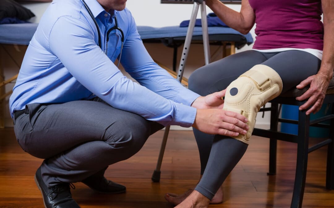
Can individuals with stiff person syndrome incorporate non-surgical treatments to reduce muscle stiffness and restore muscle mobility?
Introduction
The musculoskeletal system allows the body’s extremities to be mobile, provides stability to the host, and has an outstanding relationship with the other body systems. The musculoskeletal system’s muscles, tissues, and ligaments help protect the body’s vital organs from environmental factors. However, many individuals often deal with repetitive motions in the upper and lower body extremities that can cause pain and discomfort. Additionally, environmental factors, illnesses, and injuries can affect the musculoskeletal system and play a part in co-morbidities in overlapping risk profiles. These issues can cause the musculoskeletal system to develop a condition known as stiff person syndrome. Today’s articles focus on what stiff person syndrome is, the symptoms it is associated with, and how non-surgical treatments can help alleviate the symptoms of stiff person syndrome. We discuss with certified associated medical providers who consolidate our patients’ information to assess stiff person syndrome and its associated symptoms affecting the musculoskeletal system. We also inform and guide patients while asking their associated medical provider intricate questions to integrate non-surgical treatments to reduce the overlapping symptoms correlating with stiff person syndrome. Dr. Jimenez, D.C., includes this information as an academic service. Disclaimer.
What Is Stiff Person Syndrome
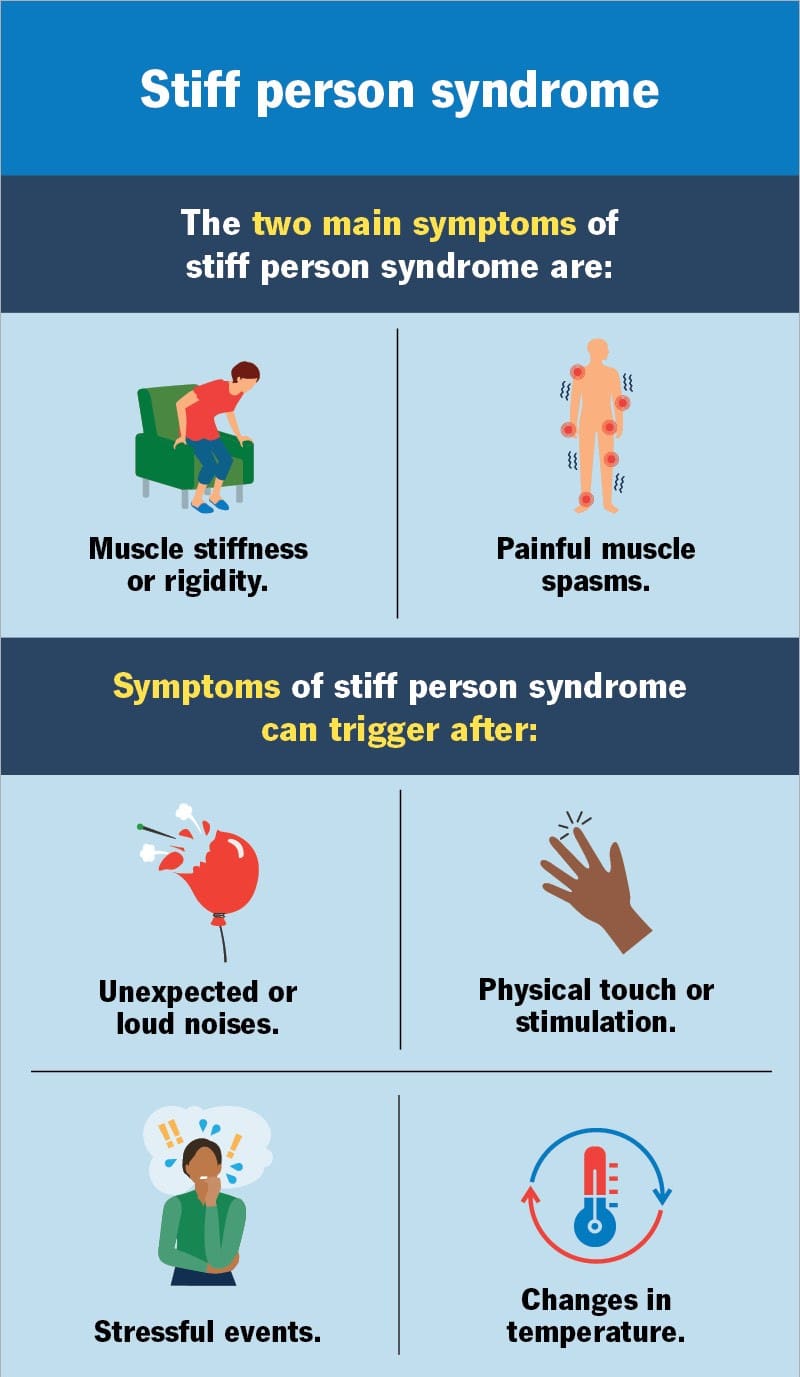
Have you been dealing with muscle stiffness in your lower extremities affecting your mobility? Have you noticed that your posture is rigid due to ongoing muscle spasms in your lower back? Or have you felt tightness in your back muscles? Many pain-like symptoms are associated with back pain, a common musculoskeletal condition; however, they can also correlate with a rare condition known as stiff person syndrome. Stiff person syndrome is a rare autoimmune disorder that is progressive and is characterized by rigidity and stimulus-triggered painful muscle spasms that affect the lower body and extremities. (Muranova & Shanina, 2024) There are three classifications that a person is experiencing with stiff person syndrome, and they are:
Classic Stiff Person Syndrome
Partial Stiff Person Syndrome
Stiff Person Syndrome Plus
Since stiff person syndrome is a rare condition, many individuals may not exhibit any objective findings early on, which then causes a delayed diagnosis that can impact a person’s quality of life (Newsome & Johnson, 2022). At the same time, since stiff person syndrome is a rare autoimmune disease, it can affect the musculoskeletal system with associated pain-like symptoms.
The Symptoms
Some symptoms associated with stiff person syndrome that can develop over time are muscle stiffness and painful muscle spasms. This is because the neuron receptors from the central nervous system can become haywire and cause non-specific somatic symptoms that make the individuals deal with comorbid chronic pain and myofascial tenderness in the muscles. (Chia et al., 2023) This is because stiff person syndrome can spread into different areas of the musculoskeletal system and can gradually develop over time. For muscle stiffness associated with stiff person syndrome, the muscles can become stiff over time, causing pain and discomfort, thus leading to many individuals developing abnormal posture, making it difficult to be mobile. Muscle spasms can affect the entire body itself or in a specific location and cause intense pain that lasts for hours. However, many individuals can incorporate non-surgical treatments to reduce the pain-like symptoms in the musculoskeletal system.
Movement Medicine: Chiropractic Care- Video
Non-Surgical Treatments For Stiff Person Syndrome
When it comes to reducing the musculoskeletal pain symptoms of stiff person syndrome, many individuals can begin to go to their primary doctor for early diagnosis and develop a customized treatment plan to manage the pain-like symptoms and provide a positive impact in creating awareness of this rare condition. (Elsalti et al., 2023) By assessing the pain-like symptoms of stiff person syndrome, many people can incorporate non-surgical treatments to manage the musculoskeletal pain symptoms and improve a person’s quality of life. Non-surgical treatments are cost-effective and can be combined with other therapies to restore mobility. One of the primary goals for managing stiff person syndrome is through pain management, symptom relief, and improved quality of life. (Cirnigliaro et al., 2021)
Chiropractic Care For Stiff Person Syndrome
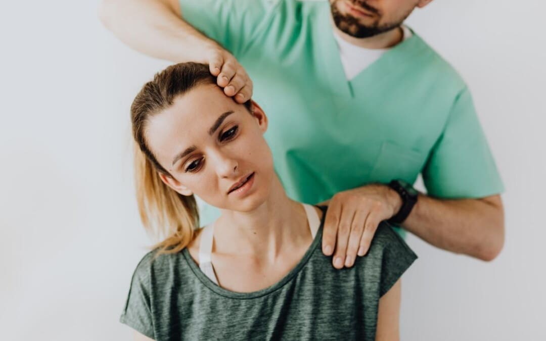
One of the non-surgical treatments that can help reduce symptoms of muscle spasms and muscle stiffness is chiropractic care. Chiropractic care incorporates mechanical and manual manipulation to stretch and mobilize the joint-muscle function while reducing pain and discomfort. (Coulter et al., 2018) For individuals suffering from stiff person syndrome, chiropractic care can help reduce muscle stiffness and muscle spasms in the upper and lower extremities and relieve the pain. Additionally, incorporating non-surgical treatments like chiropractic care and combined therapies can help manage the musculoskeletal pain associated with stiff person syndrome and improve a person’s quality of life.
References
Chia, N. H., McKeon, A., Dalakas, M. C., Flanagan, E. P., Bower, J. H., Klassen, B. T., Dubey, D., Zalewski, N. L., Duffy, D., Pittock, S. J., & Zekeridou, A. (2023). Stiff person spectrum disorder diagnosis, misdiagnosis, and suggested diagnostic criteria. Ann Clin Transl Neurol, 10(7), 1083-1094. https://doi.org/10.1002/acn3.51791
Cirnigliaro, F. A., Gauthier, N., & Rush, M. (2021). Management of refractory pain in Stiff-Person syndrome. BMJ Case Rep, 14(1). https://doi.org/10.1136/bcr-2020-237814
Coulter, I. D., Crawford, C., Hurwitz, E. L., Vernon, H., Khorsan, R., Suttorp Booth, M., & Herman, P. M. (2018). Manipulation and mobilization for treating chronic low back pain: a systematic review and meta-analysis. Spine J, 18(5), 866-879. https://doi.org/10.1016/j.spinee.2018.01.013
Elsalti, A., Darkhabani, M., Alrifaai, M. A., & Mahroum, N. (2023). Celebrities and Medical Awareness-The Case of Celine Dion and Stiff-Person Syndrome. Int J Environ Res Public Health, 20(3). https://doi.org/10.3390/ijerph20031936
Individuals experiencing pain, numbness, tingling, or a burning sensation in the front and outer thigh could have meralgia paresthetica, a nerve entrapment. Can understanding the condition help healthcare providers develop an effective treatment plan?
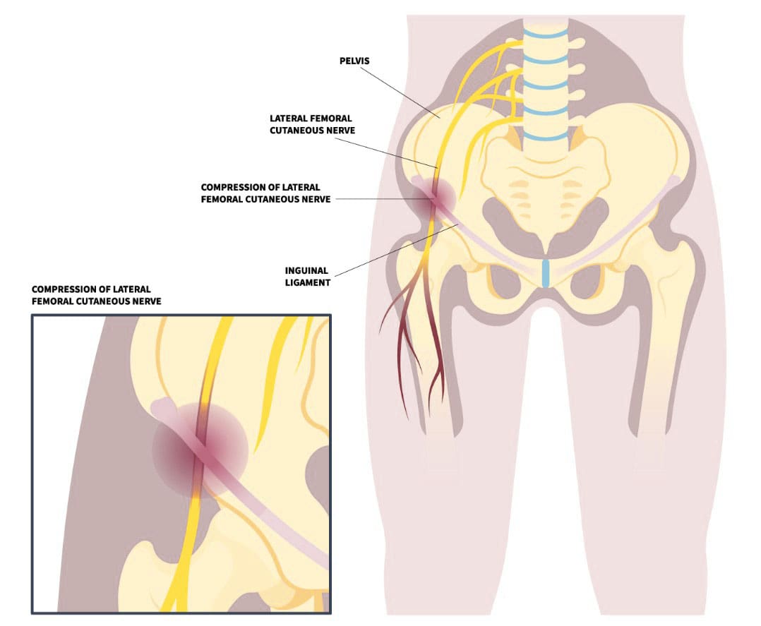
Meralgia Paresthetica
Meralgia paresthetica, or MP, is also known as Bernhardt-Roth syndrome, lateral femoral cutaneous nerve syndrome, or lateral femoral cutaneous neuralgia. It occurs when the lateral femoral cutaneous nerve, a sensory nerve that passes over the brim of the pelvis and down the front of the thigh, becomes compressed. The nerve supplies information about sensations over the front and outside of the thigh. This can happen for several reasons, including:
Recent hip injuries, such as from a motor vehicle collision/accident.
Repetitive hip activities, like cycling.
Pregnancy
Weight gain
Wearing tight clothing.
The nerve entrapment condition causes tingling, numbness, and burning pain in the front and/or outer thigh.
Causes
There can be several different causes of this condition, but it is frequently seen in pregnancy, sudden weight gain, wearing tight clothing or belts, and other conditions. (Ivins G. K. 2000) Sometimes, meralgia paresthetica can be caused by medical procedures. For example, the condition can present after an individual has surgery and is in an unusual position for a long period of time, where there is direct external pressure on the nerve. Also, the nerve can become damaged during a surgical procedure. (Cheatham S. W. et al., 2013) This can occur when a bone graft is obtained from the pelvis or anterior hip replacement surgery.
Sensitivity to lightly touching the outside of the thigh.
Worsening of symptoms with certain positions.
Increased symptoms when wearing belts, work belts, or tight-waist clothes.
The symptoms may come and go or be persistent. Some individuals are hardly noticeable and do not impact their lives or activities, while others can be very bothersome and cause significant pain. (Scholz C. et al., 2023)
Treatment
Treatment depends on how long the injury has been present and the frequency and severity of the condition.
Clothing Modifications
If the cause is due to tight clothing, belts, or work belts, then garment modification should alleviate symptoms.
If recent weight gain is thought to contribute to the condition, then a weight loss program may be recommended.
Cortisone Injections
If simple steps do not relieve symptoms, a cortisone injection around the nerve area may be recommended. The goal is to reduce inflammation that contributes to nerve pressure (Houle S. 2012) . Cortisone injections may be a definitive treatment or a temporary treatment.
Chiropractic
Chiropractic care can be an effective, natural, and safe treatment. Adjustments can help relieve pressure on the lateral femoral cutaneous nerve (LFCN) by realigning the spine and restoring nerve function. Chiropractors may also use soft tissue therapies, such as massage, to relieve muscle tension and support the body’s healing process. Other chiropractic techniques that may be used include:
A chiropractic treatment program may include 10–15 treatments over 6–8 weeks, but the number of treatments needed will vary from person to person. If there’s no noticeable progress after 3–4 weeks, it may be time to consult a specialist or surgeon.
Surgery
Surgery is rarely necessary. However, a surgical procedure may be considered when all conservative treatments fail to provide relief. (Schwaiger K. et al., 2018) A surgeon dissects and identifies the nerve, looks for compression locations, and tries to free the nerve from any areas where it may be pinched. Alternatively, some surgeons transect/cut the nerve so it no longer causes problems. If the transection procedure is performed, there will be a permanent area of numbness over the front of the thigh.
Injury Medical Chiropractic and Functional Medicine Clinic works with primary healthcare providers and specialists to develop a customized treatment plan to relieve pain, treat injuries, improve flexibility, mobility, and agility, and help individuals return to optimal function. If other treatments are needed, Dr. Jimenez has teamed up with top surgeons, clinical specialists, medical researchers, and rehabilitation providers to provide the most effective treatments.
Chiropractic Care for Leg Instability
References
Ivins G. K. (2000). Meralgia paresthetica, the elusive diagnosis: clinical experience with 14 adult patients. Annals of surgery, 232(2), 281–286. https://doi.org/10.1097/00000658-200008000-00019
Cheatham, S. W., Kolber, M. J., & Salamh, P. A. (2013). Meralgia paresthetica: a review of the literature. International journal of sports physical therapy, 8(6), 883–893.
Chung, K. H., Lee, J. Y., Ko, T. K., Park, C. H., Chun, D. H., Yang, H. J., Gill, H. J., & Kim, M. K. (2010). Meralgia paresthetica affecting parturient women who underwent cesarean section -A case report-. Korean journal of anesthesiology, 59 Suppl(Suppl), S86–S89. https://doi.org/10.4097/kjae.2010.59.S.S86
Scholz, C., Hohenhaus, M., Pedro, M. T., Uerschels, A. K., & Dengler, N. F. (2023). Meralgia Paresthetica: Relevance, Diagnosis, and Treatment. Deutsches Arzteblatt international, 120(39), 655–661. https://doi.org/10.3238/arztebl.m2023.0170
Hosley, C. M., & McCullough, L. D. (2011). Acute neurological issues in pregnancy and the peripartum. The Neurohospitalist, 1(2), 104–116. https://doi.org/10.1177/1941875211399126
Houle S. (2012). Chiropractic management of chronic idiopathic meralgia paresthetica: a case study. Journal of chiropractic medicine, 11(1), 36–41. https://doi.org/10.1016/j.jcm.2011.06.008
Schwaiger, K., Panzenbeck, P., Purschke, M., Russe, E., Kaplan, R., Heinrich, K., Mandal, P., & Wechselberger, G. (2018). Surgical decompression of the lateral femoral cutaneous nerve (LFCN) for Meralgia paresthetica treatment: Experimental or state of the art? A single-center outcome analysis. Medicine, 97(33), e11914. https://doi.org/10.1097/MD.0000000000011914
Can individuals with osteoarthritis can incorporate cycling to reduce joint pain and regain their joint mobility?
Introduction
The joints in the musculoskeletal system allow the individual to be mobile while allowing the extremities to do their jobs. Just like the muscles and ligaments of the body, the joints can also wear and tear through repetitive motions, leading to joint pain in the extremities. Over time, the wear and tear from the joints can lead to the potential development of osteoarthritis, which then can affect joint mobility and lead to a life of pain and misery for individuals. However, numerous ways exist to reduce osteoarthritis’s pain-like symptoms and help restore joint mobility through cycling. Today’s article looks at how osteoarthritis affects the joints, how cycling is incorporated for osteoarthritis, and how it can reduce joint pain. We discuss with certified associated medical providers who consolidate our patients’ information to assess osteoarthritis and its associated pain symptoms affecting the joints in the extremities. We also inform and guide patients while asking their associated medical provider intricate questions to integrate cycling into their personalized treatment plan to manage the pain correlated with osteoarthritis affecting their joints. Dr. Jimenez, D.C., includes this information as an academic service. Disclaimer.
Osteoarthritis Affecting Joint Mobility
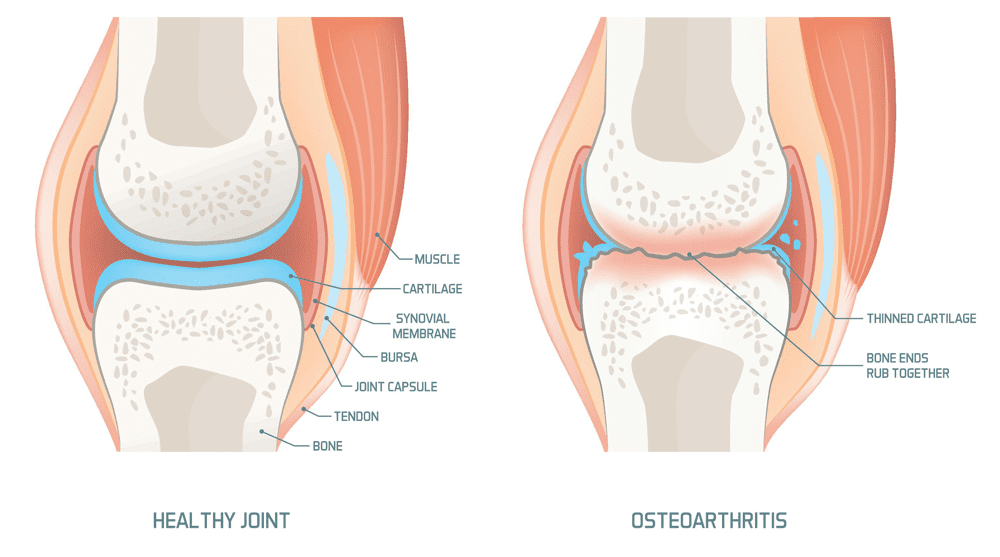
Do you feel pain and stiffness every morning in your joints only for it to feel better throughout the day? Do you experience pain in your knees, hips, and hands? Or have you noticed that your range of motion has decreased drastically? Many individuals, both young and old, can be affected by these pain-like issues and could be at risk of developing osteoarthritis in their joints. Osteoarthritis is the largest and most common musculoskeletal condition that causes a disturbance of the inflammatory cytokine balance, damaging the cartilage and other intra-articular structures surrounding the joints. (Molnar et al., 2021) This is because osteoarthritis develops over time, causing the cartilage to wear away and causing the connecting bones to rub against each other. This, in turn, can affect the extremity’s joint mobility, causing symptoms of stiffness, pain, swelling, and reduced range of motion to the joints.
Additionally, osteoarthritis is multifactorial as it can cause an imbalance in the joints due to genetics, environmental, metabolic, and traumatic factors that can contribute to its development. (Noriega-Gonzalez et al., 2023) This is because repetitive motions and environmental factors can impact the body and cause overlapping risk profiles to correlate with osteoarthritis. Some overlapping risk profiles associated with osteoarthritis are pathological changes in the joint structure that cause abnormal loading on the joints, which causes joint malalignment and muscle weakness. (Nedunchezhiyan et al., 2022) This causes many people to be in constant pain and trying to find relief from joint pain associated with osteoarthritis.
Chiropractic Solutions For Osteoarthritis-Video
Cycling For Osteoarthritis
Engaging in physical activities may seem daunting when managing osteoarthritis symptoms, but it can help restore joint mobility while reducing the pain associated with osteoarthritis. One of the physical activities that has little impact and does not impact the joints is cycling. Cycling for osteoarthritis has many beneficial properties as it can:
Strengthen surrounding muscles
Retain joint mobility
Improve range of motion
Weight management
Enhancing cardiovascular health
Cycling can help the individual focus on strengthening the lower extremity muscles surrounding the joints, which can help improve pain and functionality. (Katz et al., 2021) This, in turn, helps provide better support and stability to the joints, thus reducing overload on the body while minimizing the risk of injuries. Additionally, cycling can help improve many individuals looking for a healthier change and increase bone mineral density in the joints, thus decreasing the risk of fractures. (Chavarrias et al., 2019)
Cycling Reducing Joint Pain
Cycling is a safe and effective exercise for anyone, whether they’re just starting or haven’t been active for a while. The key to optimal recovery and joint functionality is to consult a doctor. This ensures that cycling is a safe option for you, helps you choose the right bike, and provides guidance on how to start slowly, warm up and stretch, maintain proper form, and stay consistent with the cycling sessions. This professional guidance is crucial, as it allows many individuals with joint pain to achieve complete functional recovery to their joints. (Papalia et al., 2020) Cycling is an excellent way to manage osteoarthritis and its associated symptoms. For many individuals with osteoarthritis, this low-impact exercise can be a game-changer, promoting muscle strengthening, improving joint range of motion, and helping alleviate osteoarthritis symptoms.
References
Chavarrias, M., Carlos-Vivas, J., Collado-Mateo, D., & Perez-Gomez, J. (2019). Health Benefits of Indoor Cycling: A Systematic Review. Medicina (Kaunas, Lithuania), 55(8). https://doi.org/10.3390/medicina55080452
Katz, J. N., Arant, K. R., & Loeser, R. F. (2021). Diagnosis and Treatment of Hip and Knee Osteoarthritis: A Review. JAMA, 325(6), 568-578. https://doi.org/10.1001/jama.2020.22171
Molnar, V., Matisic, V., Kodvanj, I., Bjelica, R., Jelec, Z., Hudetz, D., Rod, E., Cukelj, F., Vrdoljak, T., Vidovic, D., Staresinic, M., Sabalic, S., Dobricic, B., Petrovic, T., Anticevic, D., Boric, I., Kosir, R., Zmrzljak, U. P., & Primorac, D. (2021). Cytokines and Chemokines Involved in Osteoarthritis Pathogenesis. Int J Mol Sci, 22(17). https://doi.org/10.3390/ijms22179208
Nedunchezhiyan, U., Varughese, I., Sun, A. R., Wu, X., Crawford, R., & Prasadam, I. (2022). Obesity, Inflammation, and Immune System in Osteoarthritis. Front Immunol, 13, 907750. https://doi.org/10.3389/fimmu.2022.907750
Noriega-Gonzalez, D., Caballero-Garcia, A., Roche, E., Alvarez-Mon, M., & Cordova, A. (2023). Inflammatory Process on Knee Osteoarthritis in Cyclists. J Clin Med, 12(11). https://doi.org/10.3390/jcm12113703
Papalia, R., Campi, S., Vorini, F., Zampogna, B., Vasta, S., Papalia, G., Fossati, C., Torre, G., & Denaro, V. (2020). The Role of Physical Activity and Rehabilitation Following Hip and Knee Arthroplasty in the Elderly. J Clin Med, 9(5). https://doi.org/10.3390/jcm9051401
How do healthcare professionals provide a clinical approach in the role of nursing to reducing pain in individuals?
Introduction
The practice of Registered Nurses (RN), Advanced Practice Registered Nurses (APRN), and Licensed Practical Nurses (L.P.N.) is governed by the Nurse Practice Act. Nurses working in the specializations above must keep up their practice skills and knowledge, which includes familiarity with the rules and regulations that pertain to their profession. Practicing practical nursing is authorized for Licensed Practical Nurses (L.P.N.s). Today’s article looks at the role of nursing. We discuss with certified associated medical providers who consolidate our patients’ information to assess any pain or discomfort they are experiencing. We also inform and guide patients while asking their associated medical provider intricate questions to integrate into their personalized treatment plan to manage the pain. Dr. Jimenez, DC, includes this information as an academic service. Disclaimer.
The Roles In Nursing
The Nurse Practice Act describes practical nursing as “the performance of selected various actions, including the administration of numerous treatments and medications, in the care of the ill, injured, and providing the promotion of wellness, health maintenance and prevention of illnesses while following under the direction of a registered nurse, a licensed physician, osteopathic physician, podiatric physician, or a licensed dentist.” It was revised in 2014 and now teaches broad health and wellness concepts to non-nursing students and the public. The main goal for an RN is to complement the access to health care for individuals in pain or who are dealing with chronic issues. (Cassiani & Silva, 2019)
Many individuals are under the supervision of a registered nurse, doctor, or dentist, individuals who have completed a prelicensure practical nursing education program approved by the Board, a professional nursing education program, and graduate practical nursing students qualifying as professional nursing students; however, licensed practical nurses who have not completed the specified course under Rule 64 B9-12.005, FAC, may perform a limited scope of intravenous therapy. This range consists of:
Intravenous Therapy Within the Scope of the Practical Nurse:
Calculate and adjust the flow rate of IV therapy.
Observe and report both subjective and objective signs of various reactions to IV administration to the patient.
Must inspect the insertion site, change the dressing, and remove the intravenous needle or catheter from the peripheral veins
Hanging bags or bottles of hydrating fluid.
Intravenous Therapy Outside the Scope of the Practical Nurse:
Initiation of blood and blood products
Initiation or administration of cancer chemotherapy
Initiation of plasma expanders
Initiation of administration of investigational drugs
Making IV solution
IV pushes, except for heparin flushes and saline flushes
It is appropriate for licensed practical nurses to provide treatment for patients undergoing such therapy, even though this rule restricts the practice of licensed practical nurses. 64B-12.005 Requirements for Competency and Knowledge required for the LPN to be qualified to give IV therapy. If the IV Therapy Course Guidelines published by the National Federation of Licensed Practical Nurses Education Department are completed, an LPN may be certified to administer IV therapy. The LPN can take part in further training to provide IV therapy via central lines while supervised by an RN. “The Central Lines. The Board acknowledges that a Licensed Practical Nurse, as defined in subsection 64B9-12.002, FAC, may provide intravenous therapy via central lines under a registered professional nurse’s supervision with the necessary education and training. Four hours of instruction is the minimum required for appropriate education and training. The thirty hours of education for intravenous therapy needed for this rule’s subsection may include four hours of training. At the very least, didactic and clinical practicum instruction in the following areas must be included in the education and training mandated by this subsection:
Central venous anatomy and physiology
CVL site assessment
CVL dressing and cap changes
CVL flushing
CVL medication and fluid administration
CVL blood drawing
CVL complications and remedial measures
The Licensed Practical Nurse will be evaluated on clinical practice, competency, and theoretical knowledge and practice after completing the intravenous therapy course via central lines. A Registered Nurse must witness the clinical practice assessment and file a proficiency statement on a Licensed Practical Nurse. The Licensed Practical Nurse will be evaluated on clinical practice, competence, and theoretical knowledge and practice. A Registered Nurse who oversees the clinical practice assessment must sign a proficiency statement attesting to the Licensed Practical Nurse’s competence in administering intravenous treatment through central lines. The applicant’s Licensed Practical Nurse personnel file must contain the proficiency statement. 64B9-12.005 code.
Professional nursing is practiced by registered nurses (RNs). The Nurse Practice Act defines this as “the performance of those numerous acts requiring substantial specialized knowledge, judgment, and nursing skill based upon the applied principles of psychological, biological, physical, and social sciences.” Professional nursing goes beyond hands-on care to include nursing diagnosis, planning, supervision, and training other staff members in the theory and execution of any tasks mentioned above. Additionally, nurses must use numerous experiences to assist patients with an understanding of empathy to make them feel comfortable and safe. (Torres-Vigil et al., 2021)
Delegations & Certificates For Nursing
The delegation of responsibilities to another healthcare provider or a competent unlicensed individual is permitted by the Florida Nurse Practice Act. When assigning a task or activity, the registered nurse (RN) or licensed practical nurse (L.P.N.) must consider appropriateness. They had to consider the possibility of patient injury, the difficulty of the work, the outcome’s predictability or unpredictability, and the resources—including staff and equipment—available in the patient environment. The RN and the LPN may assign tasks outside the supervising or delegating nurse’s scope of practice. These tasks include determining the nursing diagnosis or interpreting nursing assessments, developing the plan of care, establishing the goals of nursing care, and assessing the progress of the care plan. The role of nursing is to promote advocacy and create a direct relationship with patients. (Ventura et al., 2020)
464.0205 Retired Volunteer Nurse Certificate
A retired practical or registered nurse may apply for a retired volunteer certificate from the Board of Nursing to work with underprivileged, impoverished, or critically ill populations. They are directly supervised by a physician, advanced practice registered nurse, registered nurse, director of a county health department, and:
Provides services under the certificate only in sponsored settings that the Board has approved
The scope of practice for a certified volunteer is limited to primary and preventive health care by the Board.
A retired volunteer nurse shall not:
Administer controlled substances
Supervise other nurses
Receive monetary compensation
464.012 Advanced Practice Registered Nurse (APRN)
“The Barbara Lumpkin Prescribing Act” was proposed towards the end of 2018. This Act helps many practitioners convert a certificate to a license, and it takes effect on October 1, 2018. This Act established a transition timeline and process for practitioners certified as advanced registered nurse practitioners or clinical nurse specialists as of September 30, 2018, to practice as advanced practice registered nurses (APRNs). Until the department and Board complete the transition from certification to licensure, established under this Act, an advanced registered nurse practitioner who is holding a certificate to practice on September 30, 2018, may continue to practice with all the rights, authorizations, and responsibilities under this licensure section as an advanced practice registered nurse. They may also use the applicable title under s.464.015 after this Act’s effective date.
The Board of Nursing requires the following to establish an APRN license:
A nurse who wants to become an advanced practice registered nurse must apply to the APRN department, provide documentation that they meet the requirements set out by the Board, and have a valid license to practice professional nursing or an active multistate license to practice professional nursing by s. 464.0095.
Accreditation by a relevant specialty board. To become a certified nurse in any nursing department and to renew your current state license, you must first obtain this certification. For a duration deemed suitable for preparing for and passing the national certification examination, the Board may, by rule, grant certified registered nurse anesthetists, clinical nurse specialists, certified nurse practitioners, psychiatric nurses, and certified nurse midwives provisional state licensure.
Completing a master’s program in a clinical nursing specialty field and training in particular practitioner skills. For candidates who will graduate on or after October 1, 1998, paragraph (4)(a) requires completion of a master’s degree program to be eligible for initial certification as a certified nurse practitioner.
The Board of Nursing defines APRN’s role/duties:
Prescribe, dispense, administer, or order any medication; however, an advanced practice registered nurse is only permitted to prescribe or dispense the controlled substance as specified in s.893.03 if they have completed a master’s or doctoral program that provides training in specialized practitioner skills and leads to a master’s or doctoral degree in clinical nursing.
Initiate appropriate therapies for certain conditions.
Performed additional functions as may be determined by rule under s.464.003.
Order diagnostic tests and physical and occupational therapy.
Order any medication for administration to a patient in a facility.
Beyond the general duties mentioned in subsection (3), an APRN is qualified to carry out the following tasks within their area of expertise:
Within the confines of established protocol, the certified nurse practitioner may carry out any or all of the following actions:
Manage selected medical problems.
Order physical and occupational therapy.
Initiate, monitor, or alter therapies for certain acute illnesses.
To monitor and manage patients with stable chronic diseases.
Established behavioral problems and diagnoses and made treatment recommendations.
The Stature goes on to define the functions of anesthetists and nurse midwives. Refer to the Statue for more details.
Obtaining & Maintaining Nursing License
A license may be acquired through testing, endorsement, or the Nurse Licensure Compact’s enactment. Upon application and a non-refundable payment fee determined by the Board, the department will grant the necessary license by endorsement to engage in professional or practical nursing to the applicant who can provide proof to the Board that they:
Possesses a valid license to practice professional or practical nursing in another state or territory in the United States, provided that the requirements for licensure in that state were either more stringent or substantially equivalent to those in Florida when the applicant obtained their original license.
Fulfills the requirements outlined in s.464.008 for licensing and has passed a state, regional, or national exam that is at least as difficult as the one administered by the department.
Has spent two of the previous three years actively practicing nursing in a different state, territory, or jurisdiction within the United States without having any action taken against their license by any jurisdiction’s licensing body. Under this paragraph, applicants who obtain a permit must finish a board-approved Florida laws and rules course within six months of receiving their license. After reviewing the findings of the national criminal background check, the applicant will be granted the relevant license by endorsement as soon as the department determines that the applicant has no criminal history.
It will be assumed that any exams and requirements from other US states and territories are roughly the same or more demanding than those from this state. This assumption will materialize on January 1, 1980. The Board may, however, establish rules designating some states and territories, the qualifications and exams for which shall not be deemed to be substantially similar to those of this state.
When an individual submission of the appropriate application and fees, as well as the successful completion of the criminal background check that is required under subsection (4), an applicant for licensure by endorsement who is relocating to this state due to the official military orders of their spouse with a military connection and who is a member of the Nurse Licensure Compact in another state will have all the requirements satisfied.
The applicant must submit a set of fingerprints to the department on a form and per departmental rules. The applicant must also pay the department a sum equal to the expenses the Department of Health paid for the applicant’s criminal background check. For a statewide criminal history check, the Department of Health will send the applicant’s fingerprints to the Florida Department of Law Enforcement, and the Florida Department of Law Enforcement will forward the fingerprints to the FBI for a nationwide criminal history check. When an applicant satisfies all other requirements for licensure and has no criminal record, the Department of Health will review the results of the criminal history check, issue a license, and refer all other applicants who have a criminal history back to the Board for a decision on whether or not to issue a permit and under what circumstances.
Until the investigation is finished, at which point the requirements of s.464.018 will take effect, the department will not grant an endorsement license to any applicant who is being investigated in another state, jurisdiction, or territory of the United States for an act that would violate this part or chapter 456. After completing all necessary data collection and verification, the department will issue a license within 30 days. It will also develop an electronic applicant notification process and provide electronic notifications upon application receipt and completion of background checks. Suppose the applicant must appear before the Board because of information on their application or because of screening, data gathering, and verification procedures. In that case, the 30-day license issuance time will be extended. The qualifications for licensure by endorsement in this section do not apply to an individual with an active multistate license in another state under s. 464.0095.
Licensure By Examination
Anyone who wants to take the licensing exam to become a registered nurse must apply to the department. The department will assess each candidate who:
The applicant has fulfilled the requirements by filling out the application form and paying the $150 fee set by the Board. Additionally, they have paid the $75 examination fee set by the Board and the actual cost per applicant to the department for purchasing the exam from the NCSBN (National Council of State Boards of Nursing) or a comparable national organization.
Possesses enough information as of October 1, 1989, or later, which the department needs to provide to conduct a statewide criminal records correspondence check with the Department of Law Enforcement.
Possesses a high school diploma or its equivalent, is in good mental and physical health, and has fulfilled the prerequisites for:
Graduation from an approved program
Graduation from a pre-licensure nursing education program equivalent to an approved program determined by the Board.
Graduated on or after July 1, 2009, from an accredited program
Graduation before July 1, 2009, from a pre-licensure nursing education program whose graduates were eligible for examination.
Completing courses in a professional nursing education program may satisfy the educational criteria for licensing as a licensed practical nurse. Possesses the ability to communicate in English, as assessed by a department exam. Unless rejected by s.464.018, any applicant who passes the exam and has completed the educational requirements listed in subsection (1) is eligible to become a licensed practical nurse or registered professional nurse, as the case may be.
Regardless of the jurisdiction in which the examination is administered, any applicant who fails the test three times in a row will need to finish a remedial course approved by the Board to be eligible for reexamination. The candidate may be permitted to attempt the test up to three times after completing the remedial course before being forced to undertake remediation. After the remedial process, the applicant has six months to petition for a reexamination. By regulation, the Board will set requirements for remedial education.
An applicant who completes an approved program must be enrolled in and complete a board-approved licensure examination preparing course if they choose not to take the license examination within six months of graduation. The applicant cannot use federal or state financial aid to cover any course-related expenses; they are solely responsible for covering them. The Board will set rules for the preparatory courses for licensing exams. Section 464.0095 exempts an individual from the licensure requirements if they currently have an active multistate license in another state (2).
Licensure Upon Enactment of the Nurse Licensure Compact
Florida passed the Nurse Licensure Compact into law. This allows nurses to participate in 26 states’ licensing compacts. The call to remove the burdensome and redundant system of duplicate licensure and to advance public safety and health advantages led to the enactment of this law. The official statement is as follows:
“This agreement becomes operative and legally binding on December 31, 2018, whichever comes sooner, or on the day it is enacted into law by at least 26 states. Within six months following the implementation date of this compact, any member states that were also parties to the previous Nurse Licensure Compact (“prior compact”) that this compact replaced are considered to have withdrawn from the previous compact.”
Until a party state is withdrawn from the prior compact, each party state to this one shall respect a nurse’s multistate licensure privilege to practice in that party state granted under the preceding compact. Any party state may opt out of the compact by passing a law canceling it. A party state’s departure becomes effective six months after the repealing Act is passed. Any cooperative arrangement, including nurse licensure agreements, between a party state and a nonparty state that complies with the other conditions of this compact remains valid and unaffected by this compact. The party states may alter this contract. Only when it is incorporated into the laws of every party, state a modification to this compact is binding on the party states and becomes effective. Before all party states adopt this compact, representatives of nonparty states to the agreement will be invited to engage in commission activities without being able to vote.
Unlocking Vitality: Chiropractic Wisdom & The Science of Functional Healing-Video
Continuing Nursing Education Requirement
Licenses need to be renewed every biennium or every two years. One contact hour must be completed for each calendar month of the licensure cycle in a given year. The hours stipulated in subsection (1) at the designated times must include the following continuing education courses as a necessary component:
A 2-hour course in prevention of medical errors must be completed each biennium.
A 1-hour course in HIV/AIDS in the first biennium only
A 2-hour course in Florida laws and rules in each biennium
Effective August 1, 2017, a 2-hour course in recognizing impairment in clinical approach and every other biennium after that.
On or after January 1, 2019, a 2-hour course on human trafficking and each biennium after that.
A 2-hour course in domestic violence is required every third biennium.
In addition, the Florida Board of Nursing requires general hours of continuing education to fulfill the requirement of one contact hour for each calendar month of the licensure cycle. These hour requirements are updated on their website. In addition to the courses mentioned above, they currently demand 16 hours of continuing education in general nursing.
Nurse Licensee With Two Licenses & CE Requirements
A licensee with an RN and an LPN license may fulfill CE requirements by completing the necessary RN-specific continuing education. Visit the Board of Nursing website for further information regarding the rules, as mentioned earlier, and the exceptions.
Standards For Continuing Education
Learner Objectives: The objectives should outline the anticipated behavioral outcomes of the learners and be measurable, reachable, and pertinent to the state of nursing practice today. The goals will dictate the curriculum, mode of instruction, and assessment strategy.
Subject Matter: The content must be specifically created to satisfy the participants’ learning needs, levels, and objectives. The information will be arranged logically and incorporate advice from subject-matter experts. Appropriate subject matter for continuing education offerings should include information from one or more of the following. It should represent the learner’s professional educational needs to address the consumer’s health care demands:
Nursing areas and special health care problems.
Biological, physical, behavioral, and social sciences.
Legal aspects of healthcare
Management/administration of health care personnel and patient care
Teaching/ learning process of health care personnel and patients
Evaluation: It must be demonstrated in a way that satisfies the Board that participants are given the chance to assess the educational opportunities, delivery strategies, facilities, and resources utilized in the offering. At the end of the learning process, self-directed learning activities—such as computer programs, web-based courses, internet research, and home study—must be used to assess student knowledge. There must be ten questions or more in the assessment. For the learner to be eligible for the contact hours, they must receive an evaluation score of at least 70%. The provider is required to grade the assessment.
References
Cassiani, S. H. B., & Silva, F. (2019). Expanding the role of nurses in primary health care: the case of Brazil. Rev Lat Am Enfermagem, 27, e3245. https://doi.org/10.1590/1518-8345.0000.3245
Torres-Vigil, I., Cohen, M. Z., Million, R. M., & Bruera, E. (2021). The role of empathic nursing telephone interventions with advanced cancer patients: A qualitative study. Eur J Oncol Nurs, 50, 101863. https://doi.org/10.1016/j.ejon.2020.101863
Ventura, C. A. A., Fumincelli, L., Miwa, M. J., Souza, M. C., Wright, M., & Mendes, I. A. C. (2020). Health advocacy and primary health care: evidence for nursing. Rev Bras Enferm, 73(3), e20180987. https://doi.org/10.1590/0034-7167-2018-0987
Can individuals with rheumatoid arthritis incorporate various exercises to reduce joint pain and inflammation in their hands and feet?
Introduction
The joints in the human help provide function, mobility, and flexibility to the upper and lower extremities. The joints are part of the musculoskeletal system and have an outstanding relationship with the muscles, ligaments, and soft tissues that give the body structure and support that lets the individual move around and protects the important organs to function normally. However, when a person is dealing with injuries or illnesses that affect the body’s musculoskeletal function, it can cause pain to the individual. One of the symptoms that often correlate in the joints is chronic inflammation, leading to the development of an autoimmune disease known as rheumatoid arthritis. Today’s article looks at how rheumatoid arthritis affects the joints in the musculoskeletal system and how various exercises can help manage and reduce the symptoms associated with rheumatoid arthritis. We discuss with certified associated medical providers who consolidate our patients’ information to assess rheumatoid arthritis and its associated pain symptoms affecting the joints. We also inform and guide patients while asking their associated medical provider intricate questions to integrate various exercises into their personalized treatment plan to manage the pain correlated with rheumatoid arthritis. Dr. Jimenez, D.C., includes this information as an academic service. Disclaimer.
How RA Affects The Joints
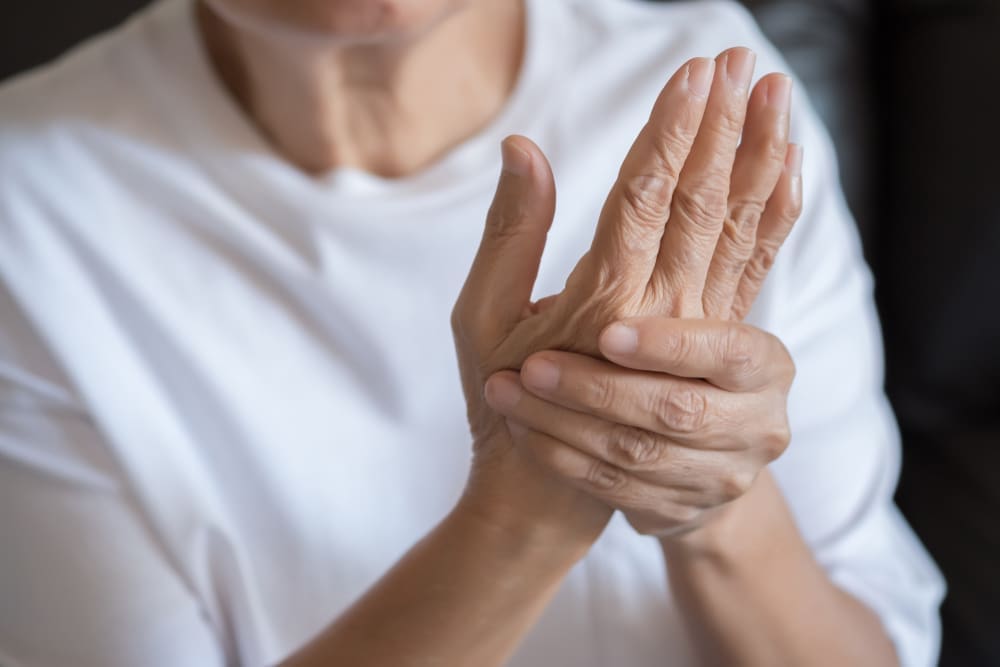
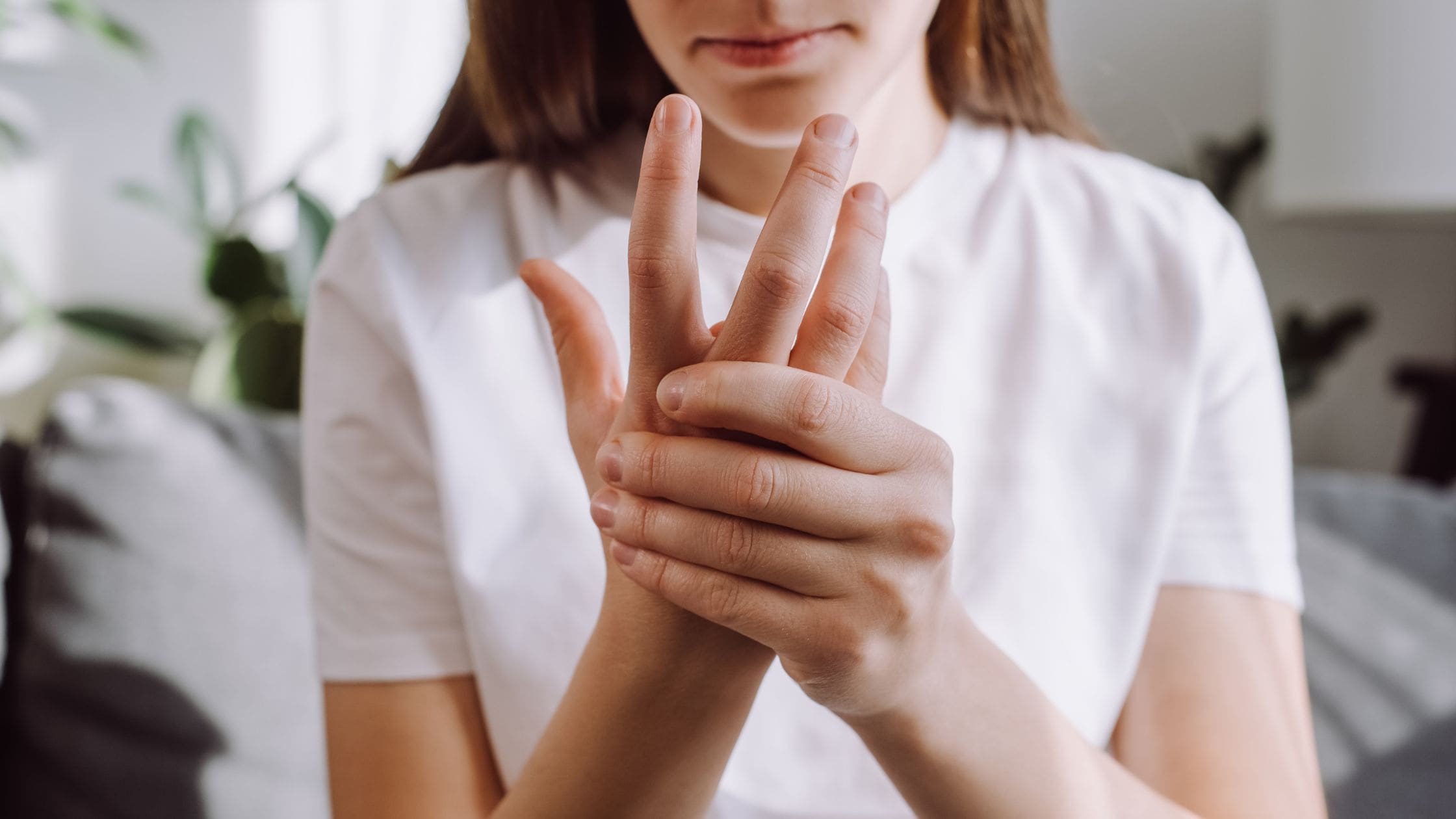
Do you feel pain and tenderness in your joints affecting your daily routine? Do you experience stiffness first thing in the morning, and it goes away throughout the day? Or do you feel fatigued throughout the day, even after a good night’s sleep? Many individuals with these symptoms could be dealing with early development of rheumatoid arthritis in their joints. Now, rheumatoid arthritis is a chronic inflammatory autoimmune disorder that affects the body’s joints but is more prominent on the hands, wrists, and feet. The symptoms of rheumatoid arthritis can develop early or slowly depending on the environmental factors contributing to the development. Since rheumatoid arthritis is categorized as a systemic autoimmune disease, genetic and environmental risk factors that can contribute to rheumatoid arthritis development can trigger overlapping risk profiles on the joints. (Jang et al., 2022) When a person is dealing with the symptoms of rheumatoid arthritis, one of the key pain symptoms that can affect the joints drastically is inflammation. Inflammation is associated with rheumatoid arthritis; it is reflected by joint pain, leading to swelling and subsequent destruction of the cartilage and bone. (Scherer et al., 2020) This causes many individuals to be in constant pain and prevents them from doing any activities.
Additionally, when a few joints are being affected by rheumatoid arthritis in the early stages, some of the symptoms include:
Fatigue
Flu-like symptoms
Swollen & tender joints
Stiffness
However, when rheumatoid arthritis reaches the later stages in the joints, the autoantigens that are specific to rheumatoid arthritis can lead to a self-perpetuating chronic inflammatory state on the joints, thus causing an expansion on the periarticular bone at the cartilage-bone junction, leading to bone erosion and cartilage degradation. (Lin et al., 2020) Luckily, there are therapeutic options to reduce the pain and inflammatory effects of rheumatoid arthritis and help manage the symptoms that are affecting the joints.
Arthritis Explained- Video
How Various Exercises Can Help With RA
When it comes to reducing the inflammatory effects of rheumatoid arthritis, many individuals can seek out therapeutic options to restore mobility, function, and flexibility. Many individuals can incorporate various physical activities to relieve stress on the inflamed tissues while slowing the progression of rheumatoid arthritis. (Radu & Bungau, 2021) When people with rheumatoid arthritis incorporate various physical activities, they can include a healthy diet and nutrition to suppress pro-inflammatory effects associated with rheumatoid arthritis, help provide symptomatic improvement, and restore bodily function to the joints. (Gioia et al., 2020)
When people with rheumatoid arthritis start exercising as part of their personalized treatment, it can have beneficial properties as they can help with the following:
Reduce joint pain & stiffness
Improve muscle strength around the joints
Enhance physical function
Boost mental health
Reduces inflammation
Increase energy levels
The main priority of incorporating exercises to reduce rheumatoid arthritis is choosing gentle exercises on the person’s joints while providing enough movement to keep the body flexible and strong. Below are some exercises to reduce rheumatoid arthritis.
Range of Motion Exercises
Range of motion exercises can help maintain normal joint function by improving flexibility and reducing stiffness for individuals with rheumatoid arthritis. Some examples include:
Finger Bends: Gently bend your fingers into a fist and straighten them. Repeat several times.
Wrist Stretch: Extend your arm with the palm facing down. Gently use your other hand to press the extended hand down and back for a stretch.
Shoulder Rolls: Roll the shoulders in a forward circular motion, then reverse the direction.
Strength Training Exercises
Strength training can help build the surrounding muscles around the joints. This allows many individuals with rheumatoid arthritis to provide better support and reduce stress on the joints. Some examples include:
Resistance Bands: Use resistance bands to perform bicep curls, leg extensions, and chest presses.
Light Weights: Incorporate light dumbbells to perform exercises like shoulder presses, tricep extensions, and squats.
Bodyweight Exercises: Engage in wall push-ups, seated leg lifts, and modified planks.
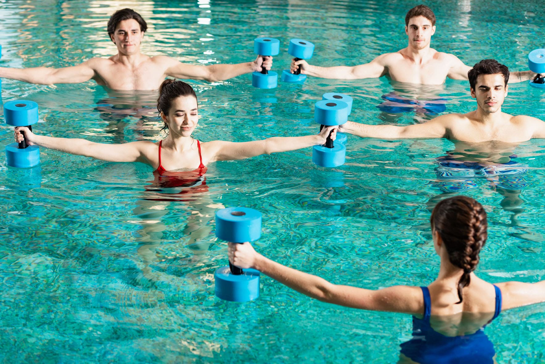
Water-Based Exercises
Water-based exercises provide resistance without impact on the joints, making it ideal for those with rheumatoid arthritis. The water helps cushion the joints by easing the stiffness, building strength, and helping relax sore muscles. Some examples of water-based exercises include:
Water Aerobics: Join a water aerobics class that offers structured routines in a supportive environment.
Aqua Jogging: Use a buoyancy belt to jog in the pool’s deep end.
Swimming: Perform laps or engage in gentle exercises like the backstroke or breaststroke.
Tips For Exercising With RA
It is important to remember that when exercising with rheumatoid arthritis, it is important to always start with a gentle warm-up and always end with a cool down to prepare the muscles and joints when beginning to exercise. Another thing to remember is to stay consistent and modify when needed. This allows many individuals to listen to their bodies and modify exercises to avoid pain and discomfort. Incorporating exercises is highly effective in reducing rheumatoid arthritis activity as it can help enhance the body’s immune function and help manage the inflammatory response associated with rheumatoid arthritis. (Li & Wang, 2022)
References
Gioia, C., Lucchino, B., Tarsitano, M. G., Iannuccelli, C., & Di Franco, M. (2020). Dietary Habits and Nutrition in Rheumatoid Arthritis: Can Diet Influence Disease Development and Clinical Manifestations? Nutrients, 12(5). https://doi.org/10.3390/nu12051456
Jang, S., Kwon, E. J., & Lee, J. J. (2022). Rheumatoid Arthritis: Pathogenic Roles of Diverse Immune Cells. Int J Mol Sci, 23(2). https://doi.org/10.3390/ijms23020905
Li, Z., & Wang, X. Q. (2022). Clinical effect and biological mechanism of exercise for rheumatoid arthritis: A mini review. Front Immunol, 13, 1089621. https://doi.org/10.3389/fimmu.2022.1089621
Lin, Y. J., Anzaghe, M., & Schulke, S. (2020). Update on the Pathomechanism, Diagnosis, and Treatment Options for Rheumatoid Arthritis. Cells, 9(4). https://doi.org/10.3390/cells9040880
IFM's Find A Practitioner tool is the largest referral network in Functional Medicine, created to help patients locate Functional Medicine practitioners anywhere in the world. IFM Certified Practitioners are listed first in the search results, given their extensive education in Functional Medicine