Chiropractic care and spinal manipulation offer a natural solution for back pain. Explore its effectiveness and benefits today.
Chiropractic Care for Back Pain Relief: A Holistic Approach to Healing
Back pain is a pervasive issue that affects millions globally, disrupting daily routines, work, and overall quality of life. From a nagging ache in the lower back to sharp, radiating pain down the leg, back pain can range from mildly bothersome to severely debilitating. Fortunately, nonsurgical treatments such as chiropractic care, spinal manipulation, targeted exercises, and integrative therapies like massage and acupuncture provide effective solutions for managing and alleviating back pain. This comprehensive guide explores the clinical rationale for chiropractic care and spinal manipulation, the factors contributing to back pain, and how a patient-centered, integrative approach can promote natural healing and prevent long-term complications. Drawing on scientific research and clinical expertise, this article offers actionable insights to help you understand and address back pain effectively.
The Global Impact of Back Pain
Back pain is one of the leading causes of disability worldwide, affecting approximately 11% of the global population (Gevers-Montoro et al., 2021). It ranks as the fourth leading cause of years lost to disability, posing significant personal, social, and economic challenges (Gevers-Montoro et al., 2021). For many individuals, back pain is not a one-time occurrence but a recurrent condition marked by periods of relief and flare-ups (Von Korff et al., 1996). Studies show that 66% to 75% of patients experience at least mild pain one month after seeking treatment, with about one in three reporting moderate to severe pain after a year (Von Korff et al., 1996).
Most back pain cases are classified as non-specific, meaning no single structural or pathological cause can be pinpointed (Gevers-Montoro et al., 2021). This complexity makes diagnosis and treatment challenging, as the pain often stems from a combination of musculoskeletal, lifestyle, and psychological factors. Chronic low back pain, defined as pain lasting beyond three months, can significantly impair mobility and daily activities, underscoring the need for effective, non-invasive interventions (Petrozzi et al., 2020).
Why Addressing Back Pain Matters
Back pain affects people across all age groups, though its prevalence increases with age, with fewer individuals over 65 experiencing it compared to younger adults (Borenstein, 2001). It can result from acute injuries, such as improper lifting, or chronic issues like poor posture or sedentary habits. Beyond physical discomfort, back pain can lead to emotional distress, reduced productivity, and increased healthcare costs. Understanding its causes and effective treatments is essential for managing symptoms and preventing long-term disability.
Factors Contributing to Back Pain
Back pain arises from a complex interplay of physical, lifestyle, psychological, and environmental factors. Below are the primary contributors to back pain, supported by research and clinical insights.
1. Musculoskeletal Factors
Muscle Imbalances and Weakness: Weak or imbalanced core and paraspinal muscles can compromise spinal stability, increasing the risk of pain and injury (Alrwaily et al., 2019). For instance, weak core muscles may fail to support the spine during movement, placing excessive stress on vertebrae and discs.
Herniated Discs: A herniated disc occurs when the soft inner material of an intervertebral disc protrudes, potentially compressing nerves and causing localized or radiating pain (sciatica) (Borenstein, 2001).
Spinal Misalignments: Misalignments or subluxations in the spine can disrupt normal biomechanics, leading to pain and restricted movement (Personal Injury Doctor Group, 2017).
Degenerative Conditions: Conditions such as spinal stenosis or osteoarthritis can narrow the spinal canal or degrade joint cartilage, resulting in pain and stiffness (Borenstein, 2001).
2. Lifestyle Factors
Sedentary behavior, particularly prolonged sitting with poor posture, weakens back muscles and increases spinal pressure, contributing to pain (Lis et al., 2015).
Physical Inactivity: A lack of regular exercise reduces muscle strength and flexibility, thereby heightening the risk of back pain (Alrwaily et al., 2019).
Obesity: Excess weight places additional strain on the spine, particularly the lower back, exacerbating pain (Borenstein, 2001).
Improper Lifting Techniques: Lifting heavy objects incorrectly can strain back muscles or cause acute injuries like sprains or disc herniations (Von Korff et al., 1996).
3. Psychological and Social Factors
Stress and Anxiety: Psychological stress can cause muscle tension, particularly in the back and neck, worsening pain (Pinheiro et al., 2016).
Fear-Avoidance Beliefs: Fear of pain or reinjury can lead to reduced activity, which may exacerbate symptoms and contribute to chronicity (Alrwaily et al., 2019).
Work-Related Factors: Jobs involving repetitive motions, heavy lifting, or prolonged sitting increase the risk of back pain. Lower work ability is also a predictor of worse outcomes in chronic low back pain (Petrozzi et al., 2020).
4. Medical and Genetic Factors
Previous Injuries: A history of back injuries can predispose individuals to recurrent pain or chronic conditions (Von Korff et al., 1996).
Genetic Predispositions: Genetic variations related to disc degeneration may increase susceptibility to back pain (Borenstein, 2001).
Comorbid Conditions: Conditions like depression or fibromyalgia can amplify pain perception and complicate recovery (Pinheiro et al., 2016).
5. Environmental and Occupational Factors
Poor Ergonomics: Inadequate workstation setups, such as non-ergonomic chairs or desks, can contribute to back strain (Lis et al., 2015).
High Physical Demands: Occupations involving heavy lifting, bending, or twisting elevate the risk of back pain and injury (Petrozzi et al., 2020).
Understanding these factors is crucial for developing personalized treatment plans that address the underlying causes of back pain. Chiropractic care, combined with spinal manipulation and integrative therapies, targets many of these contributors to promote healing and prevent recurrence.
Lower Back Pain Relief- Video
The Clinical Rationale for Chiropractic Care and Spinal Manipulation
Chiropractic care focuses on diagnosing and treating musculoskeletal disorders, particularly those affecting the spine, through manual techniques like spinal manipulation. Spinal manipulative therapy (SMT) involves applying controlled force to specific joints to restore mobility, reduce pain, and improve function. Below, we explore the clinical rationale for why chiropractic care and spinal manipulation are effective for back pain, supported by scientific evidence and clinical expertise.
How Spinal Manipulation Works
Spinal manipulation, often referred to as a chiropractic adjustment, involves high-velocity, low-amplitude thrusts or gentler mobilization techniques to realign the spine, reduce joint restrictions, and alleviate pain. According to the Personal Injury Doctor Group (2017), spinal manipulation restores proper alignment and motion to the spine, which can:
Reduce Nerve Irritation: Misaligned vertebrae or subluxations can compress or irritate spinal nerves, causing pain or radiating symptoms like sciatica. Manipulation relieves this pressure, reducing pain signals (Personal Injury Doctor Group, 2017).
Improve Joint Mobility: Restricted spinal joints can limit movement and cause stiffness. SMT restores range of motion, enhancing flexibility and function (Gevers-Montoro et al., 2021).
Decrease Muscle Tension: Manipulation can relax tight muscles and reduce spasms, which are common in back pain patients (Personal Injury Doctor Group, 2017).
Enhance Blood Flow: Improved circulation to the affected area supports tissue healing and reduces inflammation (Gevers-Montoro et al., 2021).
Scientific Evidence Supporting Chiropractic Care
Research supports the effectiveness of chiropractic care and spinal manipulation for managing back pain, particularly non-specific and chronic low back pain:
Effectiveness Compared to Other Treatments: A 2021 review found that spinal manipulative therapy is as effective as other recommended therapies, such as physical therapy or standard medical care, for managing non-specific and chronic primary spine pain (Gevers-Montoro et al., 2021). This positions chiropractic care as a viable first-line treatment for low back pain.
Clinical Practice Guidelines: Most clinical guidelines recommend SMT in combination with exercise for neck pain and as a frontline intervention for low back pain (Gevers-Montoro et al., 2021), reflecting its acceptance in evidence-based practice.
Short-Term Benefits: Patients with acute low back pain often experience significant improvement within the first four weeks of treatment, with chiropractic care contributing to reduced pain and improved function (Von Korff et al., 1996).
Prognostic Factors: Patients with lower work ability or recent consultations with medical specialists may have worse outcomes, indicating the need for tailored chiropractic interventions to address these risk factors (Petrozzi et al., 2020).
Clinical Insights for Holistic Care
Chiropractic care goes beyond symptom relief to address the underlying causes of back pain. A holistic approach includes:
Personalized Treatment Plans: Each patient’s condition is unique, requiring individualized assessments to identify specific musculoskeletal imbalances or lifestyle factors contributing to pain.
Integrative Care: Combining spinal manipulation with targeted exercises, nutritional guidance, and stress management enhances outcomes and promotes long-term wellness.
Patient Education: Clear communication empowers patients to understand their condition and actively participate in their recovery, reducing fear-avoidant behaviors and promoting adherence to treatment.
This approach aligns with research indicating that multimodal treatments, including SMT, exercise, and education, are effective for managing chronic low back pain (Petrozzi et al., 2020; Gevers-Montoro et al., 2021).
Mechanisms of Pain Relief
The clinical effectiveness of spinal manipulation can be attributed to several physiological mechanisms:
Neurophysiological Effects: SMT may modulate pain perception by influencing the central nervous system, reducing pain sensitivity, and altering pain processing pathways (Gevers-Montoro et al., 2021).
Biomechanical Corrections: By restoring proper spinal alignment, SMT reduces stress on surrounding muscles, ligaments, and discs, alleviating pain and improving function (Personal Injury Doctor Group, 2017).
Inflammation Reduction: Manipulation may decrease inflammatory markers in the affected area, promoting tissue healing (Gevers-Montoro et al., 2021).
Muscle Activation: SMT can enhance neuromuscular function, improving muscle coordination and strength, which supports spinal stability (Alrwaily et al., 2019).
These mechanisms collectively address the musculoskeletal and neurological components of back pain, making chiropractic care a comprehensive treatment option.
Nonsurgical Treatments for Back Pain
In addition to chiropractic care and spinal manipulation, other nonsurgical treatments can complement back pain management, promoting the body’s natural healing processes and preventing long-term issues.
1. Targeted Exercises
Stabilization Exercises: Exercises targeting the core and paraspinal muscles, such as abdominal, side support, and quadruped exercises, strengthen the spine’s supporting structures, reducing pain and preventing recurrence (Alrwaily et al., 2019).
Aerobic Exercise: Regular aerobic activities like walking or swimming are as effective as more complex exercise programs for chronic low back pain, improving fitness and reducing pain (Borenstein, 2001).
Stretching and Flexibility: Stretching exercises improve flexibility, reduce muscle tension, and enhance range of motion, complementing chiropractic adjustments.
2. Massage Therapy
Massage therapy, often integrated into chiropractic care, reduces muscle tension, improves circulation, and promotes relaxation. It is particularly effective when combined with SMT as part of a multimodal approach (Petrozzi et al., 2020).
Clinical evidence suggests that massage can alleviate pain and improve function in patients with chronic low back pain, especially when paired with other therapies.
3. Acupuncture
Acupuncture involves inserting thin needles into specific points to stimulate the body’s natural healing processes and reduce pain. It is thought to modulate pain signals and promote endorphin release (Borenstein, 2001).
While evidence for acupuncture’s efficacy is mixed, it can be a valuable complementary therapy for some patients, particularly those with chronic pain (Borenstein, 2001).
4. Integrative Medicine
Integrative medicine combines conventional and complementary approaches to address the whole person. Nutritional guidance, such as anti-inflammatory diets rich in omega-3 fatty acids and antioxidants, can reduce inflammation, while mindfulness practices can mitigate stress-related muscle tension (Pinheiro et al., 2016).
This holistic approach supports recovery by addressing lifestyle factors that contribute to back pain.
5. Patient Education and Communication
Educating patients about their condition, treatment options, and self-care strategies is critical for long-term success. Clear communication helps patients adhere to exercise regimens and lifestyle changes.
Addressing fear-avoidance beliefs through education can reduce the risk of chronicity and improve outcomes (Alrwaily et al., 2019).
Specialized Care for Motor Vehicle Accident (MVA) Injuries
Motor vehicle accidents (MVAs) can cause a range of musculoskeletal injuries, from whiplash-associated disorders (WAD) to spinal misalignments and soft tissue damage. Chiropractic care plays a crucial role in MVA recovery by:
Addressing Whiplash: Whiplash, a common MVA injury, involves rapid neck movement that can strain muscles and ligaments. Chiropractic adjustments and soft tissue therapies help restore alignment and reduce pain.
Rehabilitating Spinal Injuries: SMT corrects misalignments caused by the force of a collision, while targeted exercises strengthen supporting muscles.
Integrating Massage Therapy: Massage therapy complements chiropractic care by reducing muscle tension and promoting relaxation in MVA patients.
A comprehensive rehabilitation plan, including durable medical equipment like braces or supports, can enhance recovery and prevent long-term complications.
Nutrition for Recovery
Nutrition plays a crucial role in the healing of musculoskeletal injuries. A diet focused on reducing inflammation and supporting tissue repair can enhance chiropractic outcomes. Key recommendations include:
Anti-Inflammatory Foods: Foods rich in omega-3 fatty acids (such as salmon and walnuts), antioxidants (like berries and leafy greens), and anti-inflammatory spices (like turmeric) can help reduce inflammation and support healing.
Adequate Protein: Protein is essential for muscle repair and recovery, particularly after injuries like those sustained in MVAs.
Hydration: Proper hydration supports tissue health and reduces muscle stiffness.
Preventing Long-Term Problems
Preventing chronic back pain and long-term disability requires a proactive, multifaceted approach. Chiropractic care and nonsurgical treatments contribute by:
Promoting Natural Healing: Techniques like SMT and targeted exercises enhance the body’s ability to heal without invasive procedures or medications (Gevers-Montoro et al., 2021).
Addressing Root Causes: Correcting musculoskeletal imbalances, improving posture, and addressing lifestyle factors reduce the likelihood of recurrence (Personal Injury Doctor Group, 2017).
Empowering Patients: Education and clear communication enable patients to take control of their health, reducing their reliance on passive treatments and promoting self-management.
Tailoring Treatment to Risk Factors: Identifying patients with risk factors like low work ability or recent specialist consultations allows for customized interventions to improve prognosis (Petrozzi et al., 2020).
Challenges and Future Directions
While chiropractic care and spinal manipulation are effective for many patients, challenges remain:
Limited Evidence on Efficacy: The efficacy of SMT compared to placebo or no treatment is uncertain due to low-quality evidence, highlighting the need for further research (Gevers-Montoro et al., 2021).
Individual Variability: Not all patients respond equally to chiropractic care, and factors like psychological distress or comorbidities can influence outcomes (Pinheiro et al., 2016).
Access to Care: Ensuring access to qualified chiropractors and integrative care options is essential for widespread adoption of these treatments.
Future research should focus on identifying predictors of treatment success, optimizing multimodal approaches, and clarifying the specific effects of SMT to validate its role in back pain management (Gevers-Montoro et al., 2021).
Conclusion
Back pain is a complex condition with physical, lifestyle, and psychological contributors; however, nonsurgical treatments such as chiropractic care, spinal manipulation, targeted exercises, and integrative therapies offer effective solutions. Chiropractic care addresses the musculoskeletal and neurological components of back pain through spinal manipulation, which reduces nerve irritation, improves joint mobility, and promotes natural healing. Complementary approaches, such as massage, acupuncture, and nutrition, enhance outcomes, while patient education fosters long-term wellness. By addressing the root causes of back pain and tailoring treatments to individual needs, chiropractic care provides a holistic, patient-centered path to recovery.
References
Alrwaily, M., Timko, M., Schneider, M., Stevans, J., Bise, C., Hariharan, K., & Delitto, A. (2019). Stabilization exercises combined with neuromuscular electrical stimulation for patients with chronic low back pain: A randomized controlled trial. Brazilian Journal of Physical Therapy, 23(6), 506–515. https://doi.org/10.1016/j.bjpt.2018.10.003
Borenstein, D. G. (2001). Epidemiology, etiology, diagnostic evaluation, and treatment of low back pain. Current Opinion in Rheumatology, 13(2), 128–134. https://doi.org/10.1097/00002281-200103000-00006
Gevers-Montoro, C., Provencher, B., Descarreaux, M., Ortega de Mues, A., & Piché, M. (2021). Clinical effectiveness and efficacy of chiropractic spinal manipulation for spine pain. Frontiers in Pain Research, 2, 765921. https://doi.org/10.3389/fpain.2021.765921
Lis, A. M., Black, K. M., Korn, H., & Nordin, M. (2015). Association between sitting and occupational LBP. European Spine Journal, 26(2), 49–54. https://pubmed.ncbi.nlm.nih.gov/16736200/
Petrozzi, M. J., Rubinstein, S. M., Ferreira, P. H., Leaver, A., & Mackey, M. G. (2020). Predictors of low back disability in chiropractic and physical therapy settings. Chiropractic & Manual Therapies, 28(1), 41. https://doi.org/10.1186/s12998-020-00328-3
Pinheiro, M. B., Ferreira, M. L., Refshauge, K., Maher, C. G., Ordoñana, J. R., Andrade, T. B., … Ferreira, P. H. (2016). Symptoms of depression as a prognostic factor for low back pain: A systematic review. The Spine Journal, 16(1), 105–116. https://pubmed.ncbi.nlm.nih.gov/26523965/
Understanding Nerve Conditions of the Spine: Causes, Symptoms, and Treatments
The spine is a critical part of the body, serving as a highway for nerves that transmit signals between the brain and the rest of the body. When something goes wrong with these nerves—whether they’re compressed, irritated, or damaged—it can lead to a range of uncomfortable symptoms like pain, numbness, tingling, or weakness. These issues, known as nerve-related spine conditions, can affect the back, arms, or legs and stem from various causes, including injuries, degenerative conditions, or infections. In this article, we’ll explore these conditions, their symptoms, causes, and how they’re diagnosed and treated, with a special focus on integrative approaches like those used by Dr. Alexander Jimenez, a chiropractor and nurse practitioner in El Paso, Texas. We’ll also look at how chiropractic care, targeted exercises, massage therapy, acupuncture, and integrative medicine can promote healing and prevent long-term problems.
What Are Nerve-Related Spine Conditions?
Nerve-related spine conditions happen when the spinal nerves or spinal cord are compressed, irritated, or damaged. The spine is made up of bones called vertebrae, which protect the spinal cord—a bundle of nerves that carries messages to and from the brain. Between the vertebrae are intervertebral discs, which act as cushions, and small openings called foramina, where nerve roots exit the spinal cord to connect to other parts of the body. When these nerves or the spinal cord itself are affected, it can disrupt the signals, leading to symptoms like pain, numbness, tingling, or weakness (Mayo Clinic Health System, n.d.).
Some of the most common nerve-related spine conditions include:
Radiculopathy: Often referred to as a “pinched nerve,” this condition occurs when a nerve root is compressed or irritated as it exits the spine. It can cause pain, numbness, or weakness that radiates along the nerve’s path. For example, lumbar radiculopathy can lead to sciatica, a condition characterized by pain that shoots from the lower back down the leg (Cleveland Clinic, n.d.).
Spinal stenosis refers to the narrowing of the spinal canal, which puts pressure on the spinal cord or nerve roots. It’s often caused by aging or degenerative changes and can lead to symptoms like back pain, numbness, or difficulty walking (HSS Education, n.d.).
Herniated or Bulging Discs: Discs can bulge or herniate (when the inner gel-like material pushes out), pressing on nearby nerves. This can cause pain, tingling, or weakness in the arms or legs, depending on where the disc is located (Penn Medicine, n.d.).
Degenerative Conditions: Conditions like arthritis or bone spurs can narrow the spaces where nerves travel, causing compression and symptoms like pain or stiffness (Health Central, n.d.).
Trauma or Injury: Accidents, such as car crashes or falls, can damage the spine and compress nerves, leading to immediate or delayed symptoms (Verywell Health, n.d.).
Infections or Structural Abnormalities: Infections, tumors, or abnormal spine alignment (like scoliosis) can also press on nerves, causing similar symptoms (MSD Manuals, n.d.).
These conditions can range from mild annoyances to serious issues requiring immediate medical attention, especially if they cause severe symptoms like loss of bladder or bowel control, which may indicate cauda equina syndrome, a medical emergency (Verywell Health, n.d.).
Symptoms of Nerve-Related Spine Conditions
The symptoms of nerve-related spine conditions depend on where the nerve compression or damage occurs and the severity of the condition. Common symptoms include:
Pain: This can be sharp, burning, or aching and may stay in one spot (like the neck or lower back) or radiate to other areas, such as the arms, buttocks, or legs. For example, sciatica often causes burning pain that travels from the lower back to the legs (Penn Medicine, n.d.).
Numbness or Tingling: These sensations, often described as “pins and needles,” can occur in the hands, arms, feet, or legs, depending on the affected nerve (Cleveland Clinic, n.d.).
Weakness: Muscle weakness in the arms, hands, or legs can make it hard to lift objects, walk, or maintain balance. In severe cases, it can cause issues like foot drop, where a person struggles to lift their foot while walking (Johns Hopkins Medicine, n.d.).
Loss of Coordination: Compression of the spinal cord (myelopathy) can affect fine motor skills, making tasks like buttoning a shirt or writing difficult (Verywell Health, n.d.).
Balance Issues: Spinal stenosis or myelopathy can cause trouble walking or maintaining balance, sometimes described as feeling like “walking through mud” (Spine-health, n.d.).
Loss of Bladder or Bowel Control: This is a rare but serious symptom that requires immediate medical attention, as it may signal cauda equina syndrome (HSS Education, n.d.).
Symptoms can develop suddenly, like after an injury, or gradually, as with degenerative conditions like arthritis. If you experience severe or worsening symptoms, especially loss of bladder or bowel control, seek medical care right away.
Causes of Nerve-Related Spine Conditions
Nerve-related spine conditions can have many causes, ranging from natural aging to sudden injuries. Here are some of the main culprits:
Degenerative Changes: As people age, the spine can undergo wear and tear. Osteoarthritis can cause bone spurs, and degenerative disc disease can lead to bulging or herniated discs, both of which can press on nerves (Mayo Clinic Health System, n.d.).
Herniated or Bulging Discs: When a disc’s inner material bulges or herniates, it can push against nearby nerves, causing pain or numbness. This is a common cause of radiculopathy, including sciatica (Penn Medicine, n.d.).
Spinal Stenosis: The spinal canal can narrow due to thickened ligaments, bone spurs, or other changes, putting pressure on the spinal cord or nerve roots (Cleveland Clinic, n.d.).
Trauma: Car accidents, sports injuries, or falls can fracture vertebrae, dislocate joints, or cause swelling that compresses nerves, leading to severe consequences. For example, a car crash can lead to whiplash, which may cause nerve damage in the neck (Solomon Law, n.d.).
Infections: Spinal infections, like abscesses, can press on the spinal cord or nerves, causing pain and neurological symptoms (MSD Manuals, n.d.).
Structural Abnormalities: Conditions like scoliosis (abnormal spine curvature) or tumors can compress nerves, leading to symptoms like pain or weakness (Johns Hopkins Medicine, n.d.).
Inflammatory or Autoimmune Conditions: Diseases like rheumatoid arthritis can cause inflammation that compresses nerves, contributing to symptoms (OrthoTOC, n.d.).
Each cause can lead to different symptoms and requires specific diagnostic and treatment approaches to address the root issue.
Diagnosing Nerve-Related Spine Conditions
Diagnosing nerve-related spine conditions starts with a doctor asking about your symptoms and medical history, followed by a physical exam to check for numbness, weakness, reflexes, and posture. Depending on the findings, additional tests may be needed to pinpoint the cause (Penn Medicine, n.d.). Common diagnostic tools include:
Imaging tests, such as X-rays, CT scans, or MRIs, can reveal the spine’s structure, including bones, discs, and nerves, to identify compression or damage (Spine Info, n.d.).
Nerve Conduction Studies (NCS) and Electromyography (EMG): These tests assess the function of nerves and muscles, and can help confirm nerve damage (Spine Info, n.d.).
Myelogram: A special X-ray or CT scan with contrast dye can highlight pressure on the spinal cord or nerves (Spine Info, n.d.).
Dr. Alexander Jimenez, a chiropractor and nurse practitioner in El Paso, Texas, uses a dual-scope approach to diagnosis, combining his expertise in chiropractic care and advanced nursing. His clinic utilizes advanced neuromusculoskeletal imaging techniques, such as MRIs and CT scans, to obtain a clear picture of the spine’s condition. Dr. Jimenez correlates patient injuries—whether from work, sports, car accidents, or personal incidents—with clinical findings to create a precise diagnosis. This approach ensures that the treatment plan targets the specific cause of the nerve issue, whether it’s a herniated disc, spinal stenosis, or trauma-related damage (Jimenez, n.d.).
Treatment Options for Nerve-Related Spine Conditions
Treatment for nerve-related spine conditions depends on the cause, severity, and symptoms. Most doctors start with conservative (non-surgical) treatments, moving to surgery only if needed. Here’s an overview of common treatments:
Non-Surgical Treatments
Medications: Over-the-counter pain relievers, such as ibuprofen, or prescription medications, like gabapentin, can help manage pain and inflammation (Spine Info, n.d.).
Physical Therapy: Targeted exercises can strengthen muscles, improve posture, and reduce pressure on nerves. Physical therapy is often effective for radiculopathy and spinal stenosis (Cleveland Clinic, n.d.).
Epidural Steroid Injections: These deliver anti-inflammatory medication directly to the affected nerve root, reducing pain and swelling (Penn Medicine, n.d.).
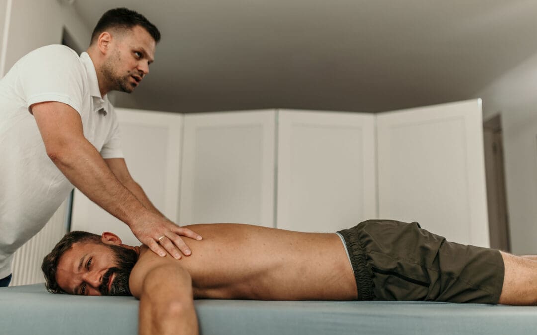
Chiropractic Care: Adjustments and manipulations can realign the spine, relieving pressure on nerves. Dr. Jimenez’s clinic utilizes chiropractic techniques to treat conditions such as sciatica and herniated discs, with a focus on restoring spinal alignment (Jimenez, n.d.).
Massage Therapy: This can relax tight muscles, improve blood flow, and reduce nerve irritation, especially for conditions caused by muscle tension or spasms (Inova, n.d.).
Acupuncture: By stimulating specific points, acupuncture can reduce pain and promote natural healing, often used alongside other treatments (Total Spine Ortho, n.d.).
Activity Modification: Avoiding activities that worsen symptoms, like heavy lifting, can help the spine heal (Penn Medicine, n.d.).
Surgical Treatments
If conservative treatments are not effective, surgery may be necessary. Common procedures include:
Laminectomy: Removes part of a vertebra to create more space for nerves, often used for spinal stenosis (Spine Info, n.d.).
Microdiscectomy: Removes part of a herniated disc that’s pressing on a nerve, commonly used for radiculopathy (Spine Info, n.d.).
Spinal Fusion: Fuses vertebrae together to stabilize the spine, used for severe degenerative conditions or trauma (Inova, n.d.).
Dr. Jimenez’s clinic takes an integrative approach, combining chiropractic care with targeted exercises, massage therapy, and acupuncture to treat nerve-related spine conditions. For example, a patient with sciatica resulting from a herniated disc may receive spinal adjustments to realign the spine, exercises to strengthen core muscles, and acupuncture to alleviate pain. This holistic approach addresses the root cause while promoting long-term healing and preventing future problems (Jimenez, n.d.).
Dr. Alexander Jimenez’s Integrative Approach in El Paso
Dr. Alexander Jimenez, a chiropractor and nurse practitioner in El Paso, Texas, has extensive experience treating nerve-related spine conditions caused by work, sports, personal, or motor vehicle accident injuries. His clinic uses a dual-scope approach, blending chiropractic expertise with advanced medical knowledge to provide comprehensive care. Here’s how his clinic handles these cases:
Treating Different Types of Injuries
Work Injuries: Repetitive motions or heavy lifting at work can lead to conditions like herniated discs or radiculopathy. Dr. Jimenez uses spinal adjustments, targeted exercises, and ergonomic advice to relieve nerve compression and prevent recurrence (Jimenez, n.d.).
Sports Injuries: Athletes may suffer nerve compression from trauma or overuse. The clinic employs chiropractic care, physical therapy, and massage to restore function and reduce pain, helping athletes return to their activities (Jimenez, n.d.).
Personal Injuries: Falls or other accidents can cause nerve damage. Dr. Jimenez’s team uses advanced imaging to assess the injury and creates personalized treatment plans, often including acupuncture and exercise (Jimenez, n.d.).
Motor Vehicle Accident (MVA) Injuries: Car crashes can cause whiplash or other trauma that compresses nerves. The clinic provides detailed diagnostic assessments, including MRIs, to identify nerve damage and offers treatments like spinal adjustments and massage to promote healing (Solomon Law, n.d.; Jimenez, n.d.).
Medical Care and Legal Documentation
Dr. Jimenez’s clinic is skilled in handling the medical and legal aspects of injury cases, especially for MVAs. They provide thorough documentation of injuries, diagnoses, and treatments, which is critical for insurance claims or legal cases. For example, if a patient has radiculopathy from a car accident, the clinic documents the injury’s impact on their daily life, the diagnostic findings (like MRI results), and the treatment plan. This detailed paperwork supports patients in legal proceedings while ensuring they receive proper medical care (Jimenez, n.d.).
Integrative Medicine for Healing and Prevention
Dr. Jimenez’s approach emphasizes integrative medicine, combining chiropractic care with other therapies to address the cause of nerve issues and enhance overall health. For instance:
Chiropractic Adjustments: Realign the spine to relieve nerve pressure, effective for conditions like sciatica or herniated discs.
Targeted Exercises: Strengthen muscles around the spine to improve stability and prevent future injuries.
Massage Therapy: Reduces muscle tension and improves circulation, aiding in nerve healing.
Acupuncture: Stimulates natural pain relief and promotes recovery, especially for chronic pain.
Lifestyle Changes: Advice on posture, ergonomics, and nutrition helps prevent long-term problems (Jimenez, n.d.).
This integrative approach not only treats the immediate symptoms but also focuses on long-term health, reducing the risk of chronic pain or recurring issues.
How Integrative Medicine Promotes Healing
Integrative medicine, as practiced by Dr. Jimenez, combines conventional medical treatments with complementary therapies to address the whole person, not just the symptoms. For nerve-related spine conditions, this approach offers several benefits:
Natural Healing: Chiropractic care and acupuncture stimulate the body’s natural healing processes, reducing reliance on medications (Total Spine Ortho, n.d.).
Pain Reduction: Therapies such as massage and acupuncture can help reduce pain levels, thereby improving quality of life (Inova, n.d.).
Improved Function: Exercises and adjustments restore mobility and strength, helping patients return to normal activities (Cleveland Clinic, n.d.).
Prevention: By addressing underlying causes, like poor posture or weak muscles, integrative medicine reduces the risk of future nerve problems (Jimenez, n.d.).
For example, a patient with spinal stenosis might receive adjustments to improve spinal alignment, exercises to strengthen their core, and massage to relax tight muscles. Over time, these treatments can reduce nerve compression, improve mobility, and prevent the condition from worsening.
Preventing Long-Term Problems
Preventing long-term nerve-related spine issues involves addressing the root causes and maintaining spinal health. Here are some strategies:
Maintain Good Posture: Proper posture reduces strain on the spine and nerves (Mayo Clinic Health System, n.d.).
Stay Active: Regular exercise, especially core-strengthening workouts, supports the spine and prevents injuries (Cleveland Clinic, n.d.).
Utilize ergonomics: Adjust workstations or lifting techniques to prevent repetitive strain (Jimenez, n.d.).
Manage Weight: Excess weight can put pressure on the spine, worsening nerve conditions (Health Central, n.d.).
Seek Early Treatment: Addressing symptoms early with chiropractic care or physical therapy can prevent conditions like radiculopathy from becoming chronic (Spine Info, n.d.).
Dr. Jimenez’s clinic emphasizes these preventive measures, educating patients on lifestyle changes to keep their spines healthy and reduce the risk of future nerve issues.
Conclusion
Nerve-related spine conditions, like radiculopathy, spinal stenosis, and herniated discs, can cause significant discomfort and disrupt daily life. These conditions stem from various causes, including degenerative changes, trauma, infections, or structural issues, and lead to symptoms like pain, numbness, tingling, and weakness. Through proper diagnosis using imaging and clinical assessments, doctors can pinpoint the cause and recommend treatments, ranging from medications and physical therapy to surgery in severe cases. Integrative approaches, like those used by Dr. Alexander Jimenez in El Paso, combine chiropractic care, targeted exercises, massage therapy, and acupuncture to treat injuries from work, sports, or accidents while promoting natural healing. By addressing the root cause and focusing on prevention, these methods can help patients recover and avoid long-term problems. If you’re experiencing symptoms of a nerve-related spine condition, consult a healthcare provider to explore your treatment options and start your journey to recovery.
Discover the role of chiropractic care in managing gastric distress and its relationship with spinal nerve compression.
Chiropractic Care for Gastric Distress and Spinal Health: A Comprehensive Guide
Introduction: The Gut-Spine Connection – More Than Meets the Eye
Imagine your body as a bustling city, with your spine as the main highway and your gut as the central food market. When traffic (or nerve signals) gets jammed on the highway, the market can start to feel the chaos, leading to tummy troubles that make you feel like you swallowed a grumpy cat. This isn’t just a quirky analogy—it’s a real connection! Your spine and gut are more linked than you might think, and when one’s out of whack, the other might throw a tantrum, too. In this blog, we’ll delve into how spinal nerve compression can trigger gastric distress, why chiropractic care is a game-changer, and how Dr. Alexander Jimenez, a renowned chiropractor in El Paso, helps people get back on track after injuries. Whether you’re dealing with bloating after a burrito or back pain from a fender-bender, we’ve got you covered with practical tips, clinical insights, and a sprinkle of humor to keep things light.
Chiropractic care isn’t just about cracking backs—it’s about restoring balance to your body’s communication system. Spinal nerve compression can mess with the signals that keep your gut happy, leading to issues like bloating, acid reflux, or even constipation that feels like your insides are staging a sit-in. Dr. Alexander Jimenez, DC, APRN, FNP-BC, at El Paso Back Clinic, uses advanced chiropractic techniques, combined with non-surgical treatments, to ease these problems. Additionally, his expertise in personal injury cases makes him a go-to resource for individuals recovering from car accidents or other mishaps in El Paso. Let’s explore how your gut and spine talk to each other, what environmental factors stir up trouble, and how small changes in your routine can make a big difference.
The Gut-Spine Connection: Why Your Back Might Be Messing with Your Belly
Your spine is like the body’s command center, housing the spinal cord and nerves that send messages to every part of you—including your gut. When spinal nerves get compressed, it’s like someone cutting the phone line during a crucial call. The gut, which relies on these nerve signals to digest food and keep things moving, can start acting up. This is where gastric distress—think bloating, gas, or that “ugh, I ate too much” feeling—comes into play. But how does this happen?
Spinal nerve compression often occurs when vertebrae (the bones in your spine) become misaligned or when discs (the cushions between them) become compressed or slip out of place. This can irritate or pinch the nerves that connect to your digestive system. For example, nerves in your lower back (lumbar spine) and mid-back (thoracic spine) directly influence organs like your stomach and intestines. If those nerves are under pressure, your gut might respond with symptoms like acid reflux, constipation, or even diarrhea. It’s like your body’s saying, “Houston, we have a problem!” but the message gets garbled.
Research backs this up. Studies suggest that a dysbiotic gut (when your gut bacteria are out of balance) might even contribute to back pain, creating a two-way street between your spine and your stomach (Wang et al., 2023). This means that not only can spinal issues mess with your gut, but gut problems might also make your back feel worse. It’s a vicious cycle, like when you argue with your sibling and nobody remembers who started it!
Why Chiropractic Care Helps
Chiropractic adjustments realign the spine, taking pressure off compressed nerves and restoring proper communication between your brain and gut. Dr. Jimenez uses gentle, precise techniques to correct misalignments, which can reduce gastric distress by improving nerve function. Think of it like untangling a knotted phone cord—suddenly, the call goes through clearly. His approach at El Paso Back Clinic also includes integrative therapies, such as massage and nutritional advice, to support overall gut health.
Environmental Factors That Stir Up Gastrointestinal Pain
Your gut doesn’t just react to what you eat—it’s also sensitive to the world around you. Environmental factors can throw your digestive system into a tizzy, and when combined with spinal nerve compression, it’s like adding fuel to an already cranky fire. Let’s break down some common culprits:
Stress: Ever feel “butterflies” in your stomach before a big test? That’s your gut responding to stress. Chronic stress disrupts your gut microbiota—the trillions of tiny bacteria living in your intestines that help digest food and maintain your health (Fukui et al., 2018). Stress can also tighten muscles around your spine, worsening nerve compression and making gastric distress feel like a daily soap opera.
Poor Diet: Eating too many processed foods, like chips or fast food, is like sending your gut a grumpy email with no clear instructions. Diets high in sugar or low in fiber can disrupt gut bacteria, leading to inflammation that might amplify back pain (Sender et al., 2014). This inflammation can also irritate spinal nerves, creating a double whammy of discomfort.
Sedentary Lifestyle: Sitting all day (hello, Netflix marathons!) can weaken your core muscles, misalign your spine, and compress nerves that connect to your gut. Lack of movement also slows digestion, making you feel bloated or sluggish, like a car stuck in traffic.
Environmental Toxins: Pollution, pesticides, or even chemicals in plastics can mess with your gut microbiota, throwing off the balance of good and bad bacteria (Jin et al., 2015). This imbalance, called dysbiosis, can lead to gastrointestinal pain and even contribute to spinal inflammation.
Sleep Issues: Not getting enough Z’s can disrupt your gut-brain axis, the two-way communication system between your stomach and your noggin (Smith et al., 2020). Poor sleep also tenses up your back muscles, increasing the risk of nerve compression.
Dr. Jimenez often sees patients whose gastric distress is tied to these environmental factors. At El Paso Back Clinic, he combines chiropractic adjustments with lifestyle advice to tackle these issues head-on. For example, he might suggest stress-busting techniques, such as deep breathing or making a dietary tweak to include more gut-friendly foods, like yogurt or leafy greens.
Smith, R. P., et al. (2020). From the gut to the brain and back: Therapeutic approaches for the treatment of network dysfunction in Parkinson’s disease. Frontiers in Neurology. https://www.ncbi.nlm.nih.gov/pubmed/XXXXXXX
How Chiropractic Care and Non-Surgical Treatments Ease the Pain
Chiropractic care is like the superhero of non-surgical treatments for spinal nerve compression and gastric distress. It doesn’t just mask the pain—it gets to the root of the problem. Here’s how Dr. Jimenez and his team at El Paso Back Clinic use a multi-pronged approach to help you feel like yourself again:
Spinal Adjustments: These are the bread and butter of chiropractic care. By gently realigning the spine, Dr. Jimenez relieves pressure on compressed nerves, improving communication between your brain and gut. This can reduce symptoms like bloating or acid reflux, making your stomach feel less like it’s hosting a rock concert.
Massage Therapy: After a car accident or long-term stress, your muscles can get tighter than a rubber band. Massage therapy loosens these muscles, reducing pressure on spinal nerves and promoting better blood flow to your gut, which helps with digestion.
Nutritional Counseling: Your gut thrives on a balanced diet, and Dr. Jimenez’s team offers personalized advice to support gut health. Think of it as giving your gut a VIP pass to the nutrient party, with foods like probiotics (found in yogurt) and fiber-rich veggies to keep things running smoothly.
Physical Therapy: Targeted exercises strengthen your core and improve posture, reducing the risk of spinal misalignments. It’s like giving your spine a personal trainer to keep it in tip-top shape.
Acupuncture: This ancient practice can reduce inflammation and calm overactive nerves, helping both your spine and gut chill out. It’s like a mini-vacation for your nervous system.
These treatments work together to tackle the overlapping risk factors of spinal nerve compression and gastric distress. For example, a misaligned spine may cause nerve irritation that slows digestion, while a poor diet can inflame the gut, exacerbating back pain. By addressing both issues, Dr. Jimenez helps break this cycle, giving you relief that lasts longer than a Netflix episode.
Small Changes, Big Impact: Dr. Jimenez’s Clinical Insights
You don’t need to overhaul your life to feel better—small tweaks can make a huge difference. Dr. Alexander Jimenez, with his decades of experience, shares practical tips to ease gastric distress and support spinal health. Here are some ideas straight from his playbook at El Paso Back Clinic:
Move More: Take a 10-minute walk after meals to boost digestion and keep your spine flexible. It’s like giving your body a high-five for eating that salad.
Hydrate Like a Pro: Drinking water helps your gut break down food and keeps your spinal discs cushy and happy. Aim for 8 glasses a day—your body will thank you.
Mind Your Posture: Slouching is like giving your spine a bad Yelp review. Sit up straight, especially at your desk, to reduce nerve compression.
Eat Gut-Friendly Foods: Add yogurt, bananas, or whole grains to your diet to support your gut microbiota. It’s like throwing a party for the good bacteria in your belly.
Manage Stress: Try deep breathing or meditation for 5 minutes a day. It’s like hitting the reset button on your nervous system, calming both your gut and spine.
Dr. Jimenez’s LinkedIn profile highlights his holistic approach, combining chiropractic care with lifestyle changes to help patients thrive (Jimenez, n.d.). These small steps can reduce inflammation, improve nerve function, and make you feel like you’re ready to take on the world—or at least that next Zoom meeting.
Dr. Jimenez: El Paso’s Go-To for Personal Injury Recovery
In El Paso, personal injury cases—like those from car accidents or slip-and-falls—are a big deal, and Dr. Alexander Jimenez is a trusted name for victims seeking recovery. Whether it’s a minor fender-bender or a serious 18-wheeler crash, injuries from these accidents can lead to spinal nerve compression and, you guessed it, gastric distress. Dr. Jimenez stands out because he doesn’t just treat the pain—he digs deeper to understand the injury’s impact on your whole body.
Using advanced imaging (like X-rays or MRIs) and diagnostic evaluations, Dr. Jimenez pinpoints exactly where spinal nerve compression is causing trouble. His dual-scope procedures—combining chiropractic adjustments with medical assessments—ensure a thorough approach. For example, if a car accident causes whiplash, it might misalign your cervical spine, irritating nerves that affect your stomach. Dr. Jimenez uses this data to create a tailored treatment plan, blending adjustments, massage, and nutritional advice to get you back on your feet.
What makes him unique is his role as a liaison between medical care and legal documentation. In personal injury cases, accurate records are crucial for insurance claims or legal proceedings. Dr. Jimenez’s detailed reports connect your injuries to the accident, helping lawyers build a strong case while ensuring you get the care you need. It’s like having a translator who speaks both “doctor” and “lawyer” fluently!
The Role of the Gastrointestinal System in Overall Health
Your gastrointestinal system is like the body’s kitchen, breaking down food, absorbing nutrients, and keeping everything running smoothly. But it’s not just about digestion—it’s a key player in your overall health. The gut houses trillions of bacteria (your microbiota) that do everything from boosting your immune system to regulating your mood (Sender et al., 2014). When spinal nerve compression disrupts the signals to your gut, it can throw this system out of balance, leading to symptoms like bloating, constipation, or even irritable bowel syndrome.
The gut also talks to your brain via the gut-brain axis, a communication highway that relies on healthy nerves (Smith et al., 2020). If spinal nerves are compressed, this highway can get congested, affecting both your digestion and your mood. Ever wonder why you feel cranky when your stomach’s upset? That’s the gut-brain axis at work! Chiropractic care helps by restoring nerve function, ensuring your gut and brain can chat without static on the line.
Smith, R. P., et al. (2020). From the gut to the brain and back: Therapeutic approaches for the treatment of network dysfunction in Parkinson’s disease. Frontiers in Neurology. https://www.ncbi.nlm.nih.gov/pubmed/XXXXXXX
Combining Chiropractic Care with Other Therapies for Maximum Relief
Dr. Jimenez’s approach at El Paso Back Clinic is like a perfectly blended smoothie—each ingredient (or therapy) works together to create something awesome. Here’s how chiropractic care teams up with other non-surgical treatments to tackle spinal nerve compression and gastric distress:
Chiropractic + Nutrition: Adjustments fix nerve compression, while a diet rich in probiotics and fiber supports gut health, reducing inflammation that can worsen back pain (Fukui et al., 2018).
Chiropractic + Massage: Massage loosens tight muscles, making adjustments more effective and improving blood flow to your gut for better digestion.
Chiropractic + Exercise: Core-strengthening exercises stabilize your spine, preventing future misalignments and keeping nerve signals clear.
Chiropractic + Acupuncture: Acupuncture calms overactive nerves, complementing adjustments to reduce both spinal and gastric inflammation.
This combo approach reduces overlapping risk factors, like inflammation or poor posture, that fuel both back pain and gut issues. It’s like assembling the Avengers to fight off discomfort from all angles!
Let’s drop the humor for a moment and get real. Spinal nerve compression and gastric distress aren’t just annoyances—they can seriously impact your quality of life. The good news? Chiropractic care, especially under the expertise of Dr. Alexander Jimenez at El Paso Back Clinic, offers a non-surgical, holistic way to address these issues. By combining spinal adjustments with lifestyle changes and other therapies, you can break the cycle of pain and discomfort, whether it’s from a car accident, poor posture, or environmental stressors. Small changes, such as adopting better posture, following a gut-friendly diet, and practicing stress management, can go a long way, but professional care is key to achieving lasting relief.
Disclaimer: This blog is intended for informational purposes only and should not be considered medical advice. Always consult a qualified healthcare provider, like Dr. Jimenez, before starting any treatment plan. Your health is serious business, and working with a professional ensures you get the care you need.
Smith, R. P., et al. (2020). From the gut to the brain and back: Therapeutic approaches for the treatment of network dysfunction in Parkinson’s disease. Frontiers in Neurology. https://www.ncbi.nlm.nih.gov/pubmed/XXXXXXX
Wang, X., et al. (2023). Insight into the Causal Relationship between Gut Microbiota and Back Pain: A Two-Sample Bidirectional Mendelian Randomization Study. Pain Medicine. https://pubmed.ncbi.nlm.nih.gov/38099244/
Exercise and Integrative Care: Healing the Spine and Supporting Gut Health
Why the Spine and Gut Are Connected
At El Paso Back Clinic, Dr. Alexander Jimenez, DC, APRN, FNP-BC, often explains to patients that the spine and gut influence each other. The nervous system links spinal alignment with digestive processes, while digestive stress can increase inflammation and tension in the back. When either system is disrupted—by injury, poor posture, or stress—the other often suffers.
This is why recovery programs at the clinic combine chiropractic care, functional medicine, and targeted exercises designed to restore both spinal stability and digestive balance.
Walking for Recovery and Digestive Energy
Walking is one of the simplest yet most effective tools for recovery. It improves circulation, strengthens spinal muscles, and encourages proper posture (Harvard Health, n.d.). Regular walking also promotes peristalsis, the natural movement of the intestines that keeps digestion flowing smoothly (Mission Health, n.d.).
Dr. Jimenez frequently recommends short daily walks for patients healing from motor vehicle accidents or work injuries. These gentle movements improve both spine and gut function while lowering stress.
Stretches That Heal Both Systems
Cat-Cow Mobility
Alternating between arching and rounding the spine with cat-cow relieves stiffness, improves flexibility, and massages abdominal organs (Institute of Living, n.d.).
Child’s Pose Relief
Child’s pose gently decompresses the spine and calms the nervous system while placing pressure on the abdomen to support digestion (St. Vincent’s, n.d.).
Knee-to-Chest Reset
The knee-to-chest stretch eases lumbar strain and stimulates gut motility, especially useful for patients who sit for long periods (Mayo Clinic, n.d.).
These stretches are easy to perform at home, making them ideal additions to chiropractic treatment.
Twists for Circulation and Core Flexibility
Seated twists rotate the spine, lengthen core muscles, and stimulate blood flow to abdominal organs. This “digestive massage” effect improves nutrient absorption and reduces bloating (SC Gastro, n.d.; NMRNJ, n.d.).
For patients recovering from accidents, Dr. Jimenez integrates twists into therapy sessions to restore spinal flexibility while encouraging smoother digestion.
Core Strengthening for Lasting Support
Bird-Dog Stability
This exercise trains spinal stabilizers and improves balance, reducing the risk of further injury (Harvard Health, n.d.).
Plank Endurance
Planks build deep abdominal strength and support the spine without added strain (Hackensack Meridian, 2023).
Core stability not only improves posture but also reduces abdominal pressure, helping digestive organs function more effectively.
Breathwork: Calming Stress and Stimulating Digestion
Diaphragmatic breathing lowers stress by activating the parasympathetic nervous system, which also stimulates healthy bowel movement (Healthline, n.d.).
Patients at El Paso Back Clinic often learn breathing exercises alongside chiropractic therapy to promote relaxation, enhance digestion, and speed up recovery from injury.
Dr. Jimenez’s Dual-Scope Approach
As both a chiropractor and nurse practitioner, Dr. Jimenez provides comprehensive injury care:
Chiropractic methods such as flexion-distraction therapy and spinal adjustments.
Functional medicine tools to improve systemic health.
Massage therapy and acupuncture to support healing naturally.
Detailed legal documentation for personal injury cases involving motor vehicle accidents or workplace injuries.
Advanced imaging and dual-scope evaluation for accurate diagnosis and treatment.
This integrated model prevents long-term complications and supports total wellness for spine and gut health.
Taking the Next Step
Exercises like walking, stretching, twisting, planking, and breathwork can transform your recovery journey. When combined with chiropractic and integrative care, they help patients live pain-free and improve digestive balance.
If you’re in pain or recovering from an accident, the team at El Paso Back Clinic can help. Contact Dr. Jimenez and start your path to natural healing.
Sport-Specific Chiropractic Care at El Paso Back Clinic: Building Strength, Restoring Balance, and Preventing Injuries
Moving Beyond One-Size-Fits-All Training
Every athlete faces unique physical demands. A sprinter’s body requires explosive power, while a baseball pitcher depends on shoulder rotation and stability. At El Paso Back Clinic, we emphasize that performance training should reflect those realities. Sport-specific chiropractic care addresses both the physical and neurological patterns behind athletic success, offering customized recovery and prevention strategies instead of generalized routines (Trainerize, n.d.; Seaver College, n.d.).
This approach enhances coordination, balance, and endurance by targeting the specific movements athletes use in their sport. More importantly, it builds resilience—protecting the musculoskeletal system against injuries that can derail progress (Physio-Pedia, n.d.).
Chiropractic Care as a Performance Tool
Chiropractic adjustments are often seen as a way to ease back or neck pain, but in athletics, they play a far greater role. Spinal and joint alignment improves nervous system efficiency, helping muscles fire correctly during sport-specific actions (Nansledan Chiropractic, n.d.).
At El Paso Back Clinic, chiropractic care goes hand-in-hand with soft tissue therapies, mobility exercises, and recovery strategies. Athletes benefit from:
This integrative approach makes chiropractic care a cornerstone for both rehabilitation and peak performance.
From Pain to Play: How Athletes Heal
Injury recovery is never just about repairing one area of the body; it’s about restoring overall function. At El Paso Back Clinic, we use chiropractic integrative care to restore overall function. For example, an athlete recovering from an ACL injury might receive adjustments for pelvic alignment alongside agility drills to re-train proper knee mechanics (Jag PT, n.d.).
Our recovery process follows clear steps:
Pain and Inflammation Control – through chiropractic adjustments, soft tissue work, and supportive therapies.
Strength and Mobility Restoration – using targeted, sport-specific rehabilitation exercises (HQPT, n.d.).
Neuromuscular Re-education – training the nervous system to move efficiently and avoid re-injury (ECU Research Online, n.d.).
Return-to-Sport Readiness – functional assessments ensure athletes are prepared for real-world demands (Marygrove Mustangs, n.d.).
By integrating rehabilitation and chiropractic strategies, athletes heal faster and more safely while regaining confidence on the field.
The El Paso Back Clinic Advantage: Integrative, Collaborative Care
What sets El Paso Back Clinic apart is the collaborative nature of care. Our providers don’t just focus on short-term relief; they build long-term health through a mix of therapies:
Chiropractic Adjustments – for alignment, pain reduction, and improved function
Acupuncture – reducing inflammation and supporting natural recovery
Nutritional Guidance – Promoting Anti-Inflammatory Eating to Accelerate Healing (Avance Care, n.d.)
Performance Training – customized sport-specific drills that build functional strength (Prevent PT, 2023)
Together, these therapies ensure athletes receive complete care that supports both the body and mind.
Prevention: Protecting Athletes Before Injuries Happen
Many injuries develop gradually, often due to poor posture, muscular imbalances, or repetitive stress. Regular chiropractic evaluations allow us to detect and correct these issues before they become painful setbacks (Hyperhealth, n.d.).
For instance, a runner may develop pelvic instability that, if untreated, leads to knee pain. At El Paso Back Clinic, chiropractic adjustments stabilize the pelvis while sport-specific training reinforces single-leg balance and stride efficiency. This proactive approach not only prevents injuries but also enhances performance (Essential Chiropractic, n.d.).
Legal and Medical Support for Injury Cases
Beyond athletics, El Paso Back Clinic also supports patients recovering from motor vehicle accidents, workplace injuries, and personal injury cases. Our providers deliver:
Thorough diagnostics using imaging and functional testing
Comprehensive injury documentation for legal cases
Collaborative care plans that integrate chiropractic treatment, exercise therapy, and functional medicine (Perrone Wellness, n.d.; RxWellness, n.d.)
By combining advanced care with precise documentation, we help patients heal physically while supporting them through legal processes that often follow accidents.
The Lasting Benefits of Sport-Specific Chiropractic Care
Athletes who embrace an integrated model of chiropractic care and tailored training experience benefit that extend well beyond the field. They gain:
Enhanced performance through better biomechanics
Faster and more complete recovery after injuries
Greater resilience against future injuries
A foundation for long-term musculoskeletal health
At El Paso Back Clinic, our mission is simple: to keep athletes, workers, and accident patients moving safely, confidently, and at their highest potential.
Rejuvenate After Travel: Chiropractic and Integrative Care at El Paso Back Clinic
Travel, whether for a weekend getaway or a long road trip, can leave you feeling drained, stiff, and mentally foggy. At El Paso Back Clinic, under the expertise of Dr. Alexander Jimenez, DC, APRN, FNP-BC, we specialize in addressing travel fatigue through chiropractic care and integrative therapies. This article explores how our holistic approach addresses the physical and neurological effects of travel, including muscle stiffness and nervous system imbalances, while promoting relaxation, stress relief, and enhanced sleep quality. Learn how Dr. Jimenez’s dual-scope expertise helps travelers, athletes, and injury victims recover naturally and prevent long-term issues.
What is Travel Fatigue?
Travel fatigue is a combination of physical and mental exhaustion resulting from prolonged sitting, time zone changes, or the stress of navigating unfamiliar environments (Kuoda Travel, 2023). It affects everyone, from high school students returning from a vacation to frequent flyers. At El Paso Back Clinic, we understand that travel fatigue can disrupt your quality of life, making it hard to bounce back to daily routines.
Physical Impacts: Muscle and Joint Stiffness
Sitting in cramped airplane seats or hunching over the steering wheel during a road trip can lead to muscle tightness and poor posture. This lack of movement reduces circulation, causing stiffness in the neck, back, and legs (Get Radiant Life, 2023). Over time, these issues can contribute to chronic pain if not addressed.
Neurological Impacts: Nervous System Disruptions
Travel disrupts the nervous system, affecting the body’s internal clock and leading to symptoms such as jet lag, irritability, and brain fog (Collective Chiro, 2023). These disturbances can weaken your immune system, leaving you vulnerable to illness post-travel.
Why Address Travel Fatigue?
Ignoring travel fatigue can prolong recovery and impact your health. At El Paso Back Clinic, we use chiropractic adjustments and integrative therapies to restore balance, helping you feel refreshed and ready to tackle your routine.
At El Paso Back Clinic, chiropractic care is the cornerstone of our approach to travel fatigue. Dr. Jimenez’s expertise as a chiropractor and nurse practitioner ensures a comprehensive recovery plan tailored to each patient.
Aligning the Spine for Optimal Health
Prolonged sitting during travel can misalign the spine, causing discomfort and restricted mobility. Chiropractic adjustments correct these misalignments, relieving nerve pressure and improving circulation (Desert Shadows Chiropractic, 2023). This process reduces stiffness and restores posture, helping you move freely again.
Supporting Nervous System Balance
A healthy spine supports a balanced nervous system, which is critical for overcoming travel-related fatigue. Adjustments enhance nerve communication, aiding recovery from jet lag and stress (Advantage Chiropractic, 2023). This balance boosts energy and mental clarity, essential for returning to school or work.
Clinical Benefits
Dr. Jimenez’s adjustments are evidence-based, reducing inflammation and improving blood flow to support recovery (Get Radiant Life, 2023). Regular chiropractic care helps prevent chronic issues, promoting long-term wellness.
Our integrative therapies at El Paso Back Clinic complement chiropractic care, addressing relaxation, stress, and sleep quality to ensure a full recovery.
Swedish Massage for Muscle Relaxation
Swedish massage uses gentle strokes to relax tight muscles and improve blood flow, ideal for easing travel-related tension (Red Mint, 2023). It also promotes endorphin release, reducing anxiety and improving mood after a long trip.
Acupuncture for Energy Restoration
Acupuncture stimulates specific points to enhance energy flow and balance the nervous system. This therapy improves circulation and sleep quality, helping you recover from fatigue and jet lag (Trinity Acupuncture, 2023; Acupuncture NE, 2023).
IV Therapy for Nutrient Replenishment
Travel can deplete essential nutrients, worsening fatigue. IV therapy delivers vitamins and hydration directly to your bloodstream, supporting muscle and nerve function (Austin MD Clinic, 2023). This rapid rehydration is ideal for students or athletes who need a quick recovery.
Massage for Jet Lag and Stress Relief
Massage therapy regulates the digestive system and promotes mental relaxation, countering jet lag’s effects (Spa Theory, 2023). It also flushes toxins from muscles, reducing post-travel soreness (Kaizen Health Group, 2023).
Dr. Jimenez’s dual licensure allows him to treat a wide range of injuries at El Paso Back Clinic, from travel fatigue to severe trauma caused by work, sports, personal incidents, or motor vehicle accidents (MVAs).
Work-Related Injuries
Repetitive strain or lifting injuries can cause chronic pain. Dr. Jimenez uses spinal adjustments and targeted exercises to restore mobility, while acupuncture and massage reduce inflammation (Jimenez, 2023, https://dralexjimenez.com/).
Sports Injuries
Athletes benefit from our spinal decompression and manual adjustments, paired with acupuncture to speed recovery and prevent re-injury. Nutritional guidance supports performance (Jimenez, 2023, https://www.linkedin.com/in/dralexjimenez/).
Personal Injuries
Falls or accidents at home can lead to neck or back pain. Dr. Jimenez employs advanced imaging and integrative therapies like IV therapy to promote healing (Jimenez, 2023, https://www.facebook.com/reel/24240689962228572).
Motor Vehicle Accident Injuries
MVAs often cause whiplash or soft tissue damage. Our clinic combines chiropractic care with acupuncture and massage, supported by detailed diagnostics for comprehensive recovery (Jimenez, 2023, https://www.instagram.com/reel/DMXxvgsiwAt/).
Dr. Jimenez’s expertise extends to medical care and legal documentation for personal injury cases, ensuring patients receive both healing and justice.
Holistic Medical Care
Using X-rays, MRIs, and dual-scope diagnostics, we identify the causes of injuries and create tailored treatment plans that incorporate chiropractic care, exercises, and integrative therapies (Jimenez, 2023, https://x.com/threebestrated/status/1947288030055678043).
Advanced Diagnostics and Treatment at El Paso Back Clinic
Our clinic’s approach integrates cutting-edge diagnostics with personalized treatment to address the root causes of injuries and fatigue.
Dual-Scope Diagnosis
Dr. Jimenez combines chiropractic and medical assessments to evaluate musculoskeletal and systemic issues, ensuring a thorough understanding of each patient’s condition (Jimenez, 2023, https://www.pinterest.com/pin/1132936850022111288/).
At El Paso Back Clinic, Dr. Alexander Jimenez and our team provide a comprehensive solution to address travel fatigue and injuries. Through chiropractic care, Swedish massage, acupuncture, IV therapy, and advanced diagnostics, we address muscle stiffness, nervous system imbalances, and more. Whether recovering from a vacation weekend or an MVA, our personalized treatments promote natural healing and prevent long-term issues. Contact El Paso Back Clinic at 915-850-0900 to start your recovery today!
Feeling drained? Relying on caffeine or sugar-packed energy drinks may give you a quick lift—but they don’t fix the real problem. These quick fixes can raise blood pressure, worsen sleep, and even contribute to more serious long-term health issues. El Paso Back Clinic takes a different approach: one that uncovers root causes and restores vitality naturally. (elpasobackclinic.com)
Your Nervous System = Your Power Source
The nervous system controls every important bodily function, from heartbeat to digestion. Spinal misalignment—due to poor posture, injuries, or birth trauma—can block nerve signals, leading to fatigue, pain, and even organ dysfunction. Chiropractic care realigns the spine and restores this vital communication. (elpasobackclinic.com)
Fatigue Fix: Circulation, Alignment, and Sleep
Chronic stress and misalignment often lead to poor sleep, brain fog, weakness, and digestive complaints. At El Paso Back Clinic, chiropractic + functional medicine work together to:
Align the spine for better nerve function
Improve circulation and oxygen delivery
Support restful sleep and reduce stress-related fatigue
Your body is like a car—it runs poorly on bad fuel. Proper nutrition makes a huge difference. El Paso Back Clinic emphasizes the importance of macronutrients and micronutrients that support recovery and energy. A poor diet can hinder healing and prolong fatigue. (elpasobackclinic.com)
Acupuncture: Energy Boost from Ancient Wisdom
Tired and worn out? Acupuncture may help. Studies show that it can improve both physical and mental fatigue, resulting in a better overall quality of life, reduced pain, improved sleep, and even enhanced gut health. For athletes, acupuncture aided faster metabolic recovery than rest alone. (elpasobackclinic.com)
Supplements That Help You Recover Smarter
Certain supplements speed recovery:
Protein & BCAAs rebuild muscle and reduce soreness
Fatty acids (MCTs, Omega-3s) deliver energy and reduce inflammation
Creatine, Citrulline Malate, and Magnesium support energy, muscle recovery, and blood flow
These can significantly shorten recovery and reduce fatigue. (elpasobackclinic.com)
Move Right: Exercise with Purpose
Routine activities like walking, stretching, and posture-aware movement strengthen the body and increase stamina. These simple practices improve endurance and productivity—especially when paired with chiropractic adjustments. (elpasobackclinic.com)
Dr. Jimenez’s Dual-Licensed Advantage
At El Paso Back Clinic, Dr. Alex Jimenez blends chiropractic precision with the diagnostic power of a board-certified nurse practitioner. This means:
Expert spinal alignment and pain relief
Comprehensive health diagnostics
Personalized treatment plans that address symptoms and root causes
Functional Medicine + Chiropractic = Full Recovery
Your pain relief plan includes more than adjustments. At El Paso Back Clinic, a functional medicine lens helps identify imbalances—such as inflammation or nutrient deficiencies—that interfere with recovery. Chiropractic adjustments, supplements, and targeted therapies then help the body heal and stay energized. (elpasobackclinic.com)
Beating Fatigue at El Paso Back Clinic
Here’s how the clinic restores vitality through an integrative approach:
Nervous system blockages → Bio-Chiropractic alignment → Improved nerve signaling
Poor circulation and sleep issues → Spinal realignment + functional support → Enhanced energy and mood
Stress and poor posture → Chiropractic adjustments + lifestyle coaching → Better rest and mental clarity
Inflammation → Nutrition plans, supplements, and acupuncture → Reduced fatigue and pain
Weak muscles → Movement therapy + targeted nutrients → Greater strength and endurance
Complex health problems → Functional medicine testing + chiropractic → Root-cause healing
Closing Thoughts
El Paso Back Clinic offers more than just a quick fix for fatigue. By combining nervous system alignment, functional nutrition, therapeutic movement, and Dr. Jimenez’s dual-licensed expertise, the clinic helps patients reclaim energy, vitality, and better health for the long haul.
IFM's Find A Practitioner tool is the largest referral network in Functional Medicine, created to help patients locate Functional Medicine practitioners anywhere in the world. IFM Certified Practitioners are listed first in the search results, given their extensive education in Functional Medicine