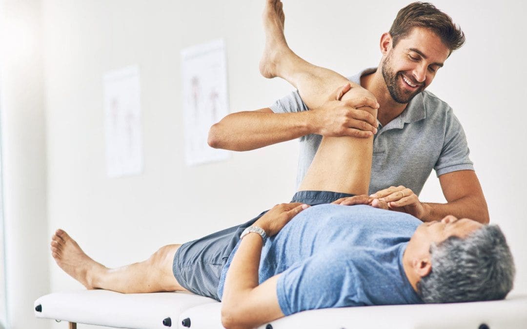
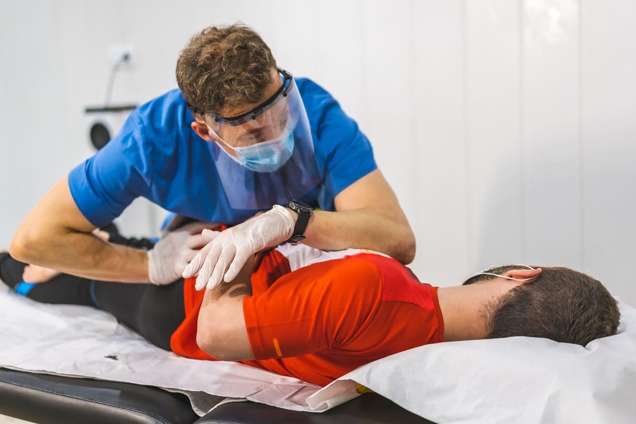
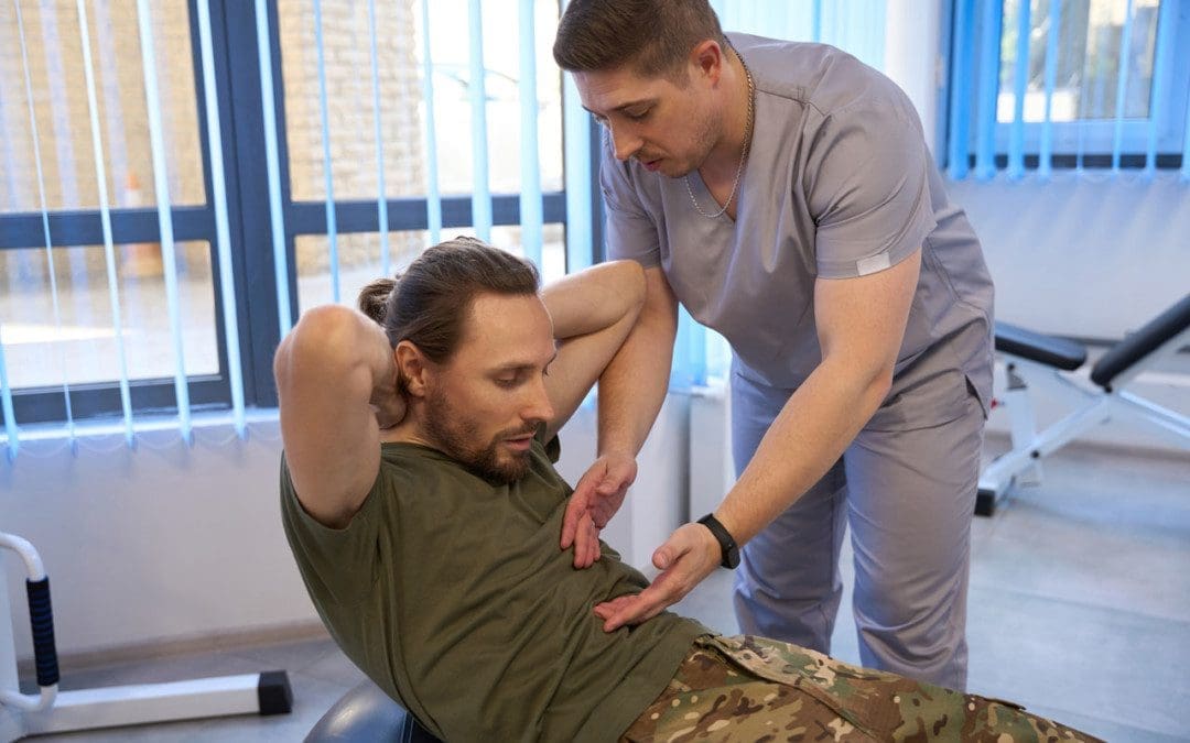
Back problems can affect everyone. The most healthy individuals can experience back issues from time to time because of unhealthy postures, standing or sitting for extended periods at a job or school, athletes, and individuals with previous injuries. Back soreness and pain symptoms are common in today’s active and inactive world. The pain can range from dull and constant aches to sharp and sudden piercing sensations. This causes pressure to build up against different regions of the spine. Individuals trying to reposition the body can cause an imbalance in the spine that often leads to more pain and problems. It is impossible to adjust one’s spine effectively and should be evaluated and treated by a professional chiropractor and therapy team.
Professional Chiropractor
Problems With DIY adjustments
Self-cracking or having a friend/family/spouse walk on their back or squeeze the body to achieve the crack or pop forces an increase in mobility that can lead to overexerting the spine.
Can loosen all of the joints instead of just the tight joints.
This can overly loosen already flexible joints that must compensate for the stiff joints.
The muscles have to work overtime to maintain stability, adding stress which increases muscle tension.
This can cause the health of compressed segments to continue to decline and worsen and/or cause further injury.
It can cause abnormal vertebral degeneration.
It can be dangerous if the individual has weak bones.
Injuries can include herniated and dislocated discs.
In the worst-case scenario, fractures and broken vertebrae.
Although rare, there is the possible risk of rib fracture or subluxation.
This can cause pain and symptoms to turn into a serious health or chronic condition.
Chiropractic Training
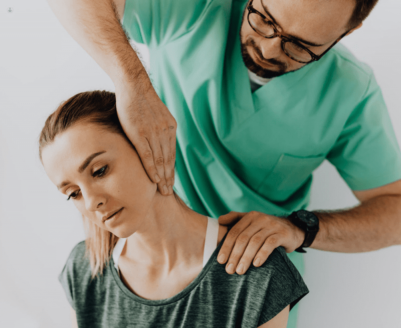
Chiropractors are trained medical specialists on the body’s structure and function. Chiropractors know the correct positioning and function of every area of the spine, from the neck to the tailbone. The most common injuries that chiropractic treats include:
Muscle Strains
Sciatica
Overuse/Repetitive Strains
Neck Sprains
Whiplash
Headaches
Herniated Discs
Dislocations
Fractures
Proper Realignment
Individuals think the popping sound is the goal; however, it’s not what relieves the pain or symptoms. The relief comes from the improved movement of the joints. The term for the popping sound is cavitation. Chiropractic manipulation is a specific force applied in a specific direction to a particular joint. During a professional realignment session, the pressure inside the joint decreases, releasing gasses within the synovial fluid into the joint space. That’s the popping sound. When a professional chiropractor performs adjustments/cavitations, they:
ImproveJoint function.
Relax the muscles.
Relieve nerve irritation.
There’s also the possibility of an unknown underlying cause for the pain symptoms. Therefore, friends and family should not attempt back adjustments unless they are professional chiropractors. When symptoms persist, treatment from a professional licensed chiropractic clinic is safer and more effective, and early treatment can prevent permanent damage caused by chronic inflammation. Professional chiropractor treatments include:
Adjustments
To gently realign joints to decrease pain and increase range of motion.
Soft-Tissue Therapy
Relaxes tight muscles, relieves spasms, and releases tension in the tissues that surround the muscle fascia.
Exercises and Stretches
Restore and maintain joint stability and mobility.
Joint Bracing and Kinesio Taping
Supports sprained joints or muscles during recovery.
Referrals to Medical Experts
For guidance on diet and nutrition to reduce inflammation and promote healthy weight.
The Path To Healing
References
Dunning, James, et al. “CAVITATION SOUNDS DURING CERVICOTHORACIC SPINAL MANIPULATION.” International Journal of sports physical therapy vol. 12,4 (2017): 642-654.
Evans, David W, and Nicholas Lucas. “What is manipulation? A new definition.” BMC musculoskeletal disorders vol. 24,1 194. 15 March 2023, doi:10.1186/s12891-023-06298-w
Hardy, Katie, and Henry Pollard. “The organization of the stress response, and its relevance to chiropractors: a commentary.” Chiropractic & osteopathy vol. 14 25. 18 October 2006, doi:10.1186/1746-1340-14-25
LaPelusa, Andrew. and Bruno Bordoni. “High-Velocity Low Amplitude Manipulation Techniques.” StatPearls, StatPearls Publishing, 6 February 2023.
Navid, Muhammad Samran, et al. “The effects of chiropractic spinal manipulation on central processing of tonic pain – a pilot study using standardized low-resolution brain electromagnetic tomography (sLORETA).” Scientific Reports vol. 9,1 6925. 6 May. 2019, doi:10.1038/s41598-019-42984-3
When it comes to our muscles, many of us often don’t stretch each muscle group at least two to three times per week. From waking up in the morning, we stretch our arms, legs, and back to relieve any stiffness or soreness from the previous day. However, many individuals deal with musculoskeletal issues that can affect not only the back and the neck but also the upper and lower extremities of the body, causing pain-like symptoms that can worsen throughout the entire day if not treated right away. When this happens, musculoskeletal pain can lead to overlapping risk problems that can cause the body to be misaligned and dysfunctional. Hence why numerous therapies help reduce the effects of musculoskeletal pain and help restore the body naturally. Today’s article looks at how musculoskeletal pain affects the body and how treatments like MET can be used as self-help methods to reduce musculoskeletal pain. We utilize valuable information about our patients to certified medical providers using MET therapy to relieve musculoskeletal pain by incorporating various exercises and stretches. We encourage and refer patients to associated medical providers based on their diagnosis while supporting that education is a remarkable and fantastic way to ask our providers the essential questions at the patient’s acknowledgment. Dr. Alex Jimenez, D.C., comprises this information as an educational service. Disclaimer
Musculoskeletal Pain Affecting The Body
Are you experiencing muscle stiffness or weakness in your back, neck, or shoulders? Do you feel pain when stretching or hunched over due to discomfort? Musculoskeletal pain is a common issue that can interfere with daily activities. Research studies show that this type of pain can cause symptoms that overlap with neuropathic or visceral pain. This means that problems with one muscle or organ in the body can lead to pain in other areas, causing significant discomfort.
Further research has shown that musculoskeletal pain can originate in the muscle tissues and persist for over three months, affecting many individuals’ social and emotional skills, work productivity, and independence. Several environmental factors like obesity, stress, poor sleep, inadequate nutrition, and lack of physical activity can overwork the muscles and joints, leading to trigger points and muscle strain in the musculoskeletal system, resulting in bodily misalignment.
Improving Athletic Performance Through Chiropractic- Video
Are you experiencing pain in specific areas of your body? Does the pain worsen when you are active or when you stretch? These pains are often associated with musculoskeletal issues, which can greatly affect your daily life. Research studies have revealed that musculoskeletal pain can significantly reduce a person’s productivity and job performance. Fortunately, several treatments are available to alleviate musculoskeletal pain and its symptoms. Many people seek chiropractic care or MET therapy to help realign their spine, stretch their muscles, and improve joint mobility. The video above demonstrates how chiropractic care uses manual manipulation to relieve musculoskeletal pain by stretching the muscles and realigning the spine.
MET Self-Help Methods For Musculoskeletal Pain
According to a book called “Clinical Applications of Neuromuscular Techniques” by Dr. Leon Chaitow, N.D., D.O., and Dr. Judith Walker DeLany, L.M.T., musculoskeletal pain can cause the soft tissues in the body to shorten and lead to disability. To alleviate the effects of musculoskeletal pain, people often seek the help of pain specialists such as chiropractors or massage therapists. These specialists often use muscle energy techniques (MET) to stretch the soft tissues and muscles and provide relief. Below are some exercises and techniques that are commonly used in MET therapy.
MET Neck Relaxation Techniques
The neck comprises soft tissues and is part of the musculoskeletal system. Two relaxation techniques can stretch out the shortened muscles to relieve soreness and stiffness in the scalene muscles. These techniques can help release tightness and improve the neck’s range of motion.
Phase 1:
Sit close to the table with the elbows and hands resting on the table surface on each side of the face.
Turn your head to the right or left as far as you can comfortably in one direction while letting your hands move with your face until you reach a pain-free rotation limit.
Afterward, use your left hand as resistance when turning your head to the left while using 25% or less of your strength to build up a force to match the resistance and start turning your head slowly.
Hold this push for 7-1o seconds and slowly stop turning your head to the left or right.
Return to a neutral position and turn to the right or left again to see how far you can stretch without pain.
You should notice the neck stretch is farther than it was previously.
This is known as post-isometric relaxation in MET therapy, as it allows the tight muscles to relax and stretch farther without pain than before.
Phase 2:
While lying on the table, the hands and elbows should be on the sides of the face.
Turn your head to the right to stretch as far as you can comfortably in one direction.
Use your right hand as resistance to attempt to turn without pain by using only 25% or less of your strength.
Slowly turn your head and maintain the turn and resistance for 7-10 seconds.
Slowly stop the resistance effort to see how far your neck can turn without pain. If you are experiencing pain, you use too much strength and reduce the contraction level where no pain is experienced.
This is known as reciprocal inhibition in MET therapy, as it achieves a different release for tight neck muscles.
Flexion Exercises Using MET
Flexion exercises in MET therapy help stretch the postural muscles and legs, feeling stiff. This allows spine flexibility while stretching out and reducing mechanical stresses in the surrounding muscles.
While sitting on the floor, your legs should be straight out, and your toes pointed towards the ceiling.
Bend comfortably as far as you can and grasp one leg with each hand.
Hold the position for 30 seconds and do four deep breathing cycles while allowing your head to hang down and relax into the stretch. *You will feel the stretch on your lower back and the back of your legs.
As you release during the fourth breathing cycle, ease yourself further down the legs and hold for another 30 seconds.
After 30 seconds, slowly return to an upright position by lightly pushing upwards from the hands.
Alternatively, you can bend one leg and do the same sequence on each leg to stretch out any leg muscles that are cramping or stiff. This flexion exercise help reduces pain and prevents trigger points from re-forming in the muscle fibers.
Extension Exercises Using MET
The extension exercises in MET therapy help the muscles and joints in the body group to increase movement without pain. This allows the body to be mobile and reduces the effects of musculoskeletal pain.
Lie on your stomach on a carpeted floor with a pillow to support your head and neck while your legs are together.
Bend your knees as comfortably as possible, and bring your heels towards your backside.
Now slowly grasp your legs and gently bend backward as far as possible without pain. Your back should be slightly arched.
Lift your head and shoulders gently to increase the arch in your back slowly and without pain.
Hold the position for four slow deep breaths, and hold your breath for 15 seconds on the last breathing cycle.
As you release, bring your body down slowly, from the legs to the stomach and finally, the shoulders and neck to rest.
This extension exercise, known as the boat position, helps lengthen and stretch the back and leg muscles while reducing pain and restoring mobility in the spine.
Conclusion
It is crucial to be aware of musculoskeletal pain in your body, whether in the morning or during work. This type of pain can lead to discomfort in other areas and even impair your ability to function. Fortunately, MET therapy can alleviate musculoskeletal pain by stretching the muscles and tissues and providing immediate relief. By utilizing stretching and physical therapy, you can help your body heal naturally and be more mindful of what triggers pain. Stretching can also prevent future injuries and promote a pain-free lifestyle.
References
Buck, Rhiannon, et al. “Working with Musculoskeletal Pain.” Reviews in Pain, June 2009, www.ncbi.nlm.nih.gov/pmc/articles/PMC4590039/.
Chaitow, Leon, and Judith Walker DeLany. Clinical Applications of Neuromuscular Techniques. Churchill Livingstone, 2003.
El-Tallawy, Salah N, et al. “Management of Musculoskeletal Pain: An Update with Emphasis on Chronic Musculoskeletal Pain.” Pain and Therapy, June 2021, www.ncbi.nlm.nih.gov/pmc/articles/PMC8119532/.
Puntillo, Filomena, et al. “Pathophysiology of Musculoskeletal Pain: A Narrative Review.” Therapeutic Advances in Musculoskeletal Disease, 26 Feb. 2021, www.ncbi.nlm.nih.gov/pmc/articles/PMC7934019/.
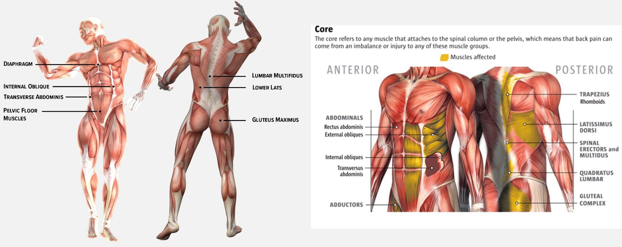
The body’s core muscles are used for stability, balance, lifting, pushing, pulling, and movement. Engaging the core muscles means bracing and tightening the abdominal muscles, which include the latissimus dorsi/lats, paraspinal muscles, gluteus maximus/glutes, and trapezius/traps. When engaged, the trunk muscles help maintain spinal stability, support the spine and pelvis in sitting and resting positions and during dynamic movements, and help prevent injury.
Engaging The Core
To know how to engage the core, individuals need to understand what the core is. The most important muscles for engaging the core include: These muscles are involved every time the body inhales and exhales, in posture control, and when using the bathroom, they start and stop the process.
Rectus Abdominis
The rectus abdominis muscle is responsible for the six-pack.
It’s a long, flat muscle that extends from the pubic bone to the sixth and seventh ribs.
The rectus abdominis is primarily responsible for bending the spine.
External Obliques
These are the muscles on either side of the rectus abdominis.
The external obliques allow the torso to twist, bend sideways, flex the spine, and compress the abdomen.
Internal Obliques
The internal obliques lie below the external obliques.
They work with the external obliques in the same functions.
Transverse Abdominis
This is the deepest layer of muscle in the abdomen.
It completely wraps around the torso and extends from the ribs to the pelvis.
The transverse abdominis are not responsible for spine or hip movement but for stabilizing the spine, compressing the organs, and supporting the abdominal wall.
Latissimus Dorsi
Commonly known as the lats, these muscles run along both sides of the spine from just below the shoulder blades to the pelvis.
The lats help stabilize the back, especially when extending the shoulders.
They also contribute to body ability when twisting from side to side.
Erector Spinae
The erector spinae muscles are on each side of the spine and extend down the back.
These muscles are responsible for extending and rotating the back and side-to-side movement.
These are considered postural muscles and are almost always working.
What Not To Do
Individuals learn from mistakes, which might make learning how to engage the core easier by understanding what not to do. Common examples of failing to or not engaging the core correctly.
The back slumps when sitting down – the upper body lacks strength and stability.
When bending, the stomach sticks out more.
Swaying or leaning far to one side when walking – lack of lower body strength causes balance and stability problems.
The lower abdomen and back present with discomfort and pain symptoms.
Training
Engaging the core decreases the chance of sustaining an injury at home, work, or exercising and can help with chronic back pain. It creates a stable musculature around the spine that keeps the vertebrae from over-flexing, over-extending, and bending too far to one side. Engaging the core muscles can mean different things, depending on what is trying to be achieved.
For example, if doing bending work, the muscles needed, and the order in which they contract differs from when trying to maintain balance while standing on one leg.
The muscles engaged will differ in their movement depending on whether an individual is:
Trying to move the spine or stabilize it.
Pushing or pulling weight.
Standing, sitting, or lying down.
For a strong and functional core, the objective is to be able to engage the core in any situation. Engaging the core can be challenging, but with training and practice, the body becomes stronger. Practice engaging the core throughout daily activities that include.
Bracing the core while standing, sitting at a workstation or desk, and walking.
Day-to-day activities, like reaching for something from a high shelf, grocery shopping, and taking the stairs.
Injury Medical Chiropractic and Functional Medicine Clinic can create a personalized program to address musculoskeletal issues, core training, targeted exercise, stretching, nutrition, massage, and adjustments to get the body to optimal health and maintain health.
The Non-Surgical Solution
References
Eickmeyer, Sarah M. “Anatomy and Physiology of the Pelvic Floor.” Physical Medicine and rehabilitation clinics of North America vol. 28,3 (2017): 455-460. doi:10.1016/j.pmr.2017.03.003
Lawson, Samantha, and Ashley Sacks. “Pelvic Floor Physical Therapy and Women’s Health Promotion.” Journal of Midwifery & Women’s Health vol. 63,4 (2018): 410-417. doi:10.1111/jmwh.12736
Seaman, Austin P et al. “Building a Center for Abdominal Core Health: The Importance of a Holistic Multidisciplinary Approach.” Journal of gastrointestinal surgery: official journal of the Society for Surgery of the Alimentary Tract vol. 26,3 (2022): 693-701. doi:10.1007/s11605-021-05241-5
Vining, Robert, et al. “Effects of Chiropractic Care on Strength, Balance, and Endurance in Active-Duty U.S. Military Personnel with Low Back Pain: A Randomized Controlled Trial.” Journal of Alternative and complementary medicine (New York, N.Y.) vol. 26,7 (2020): 592-601. doi:10.1089/acm.2020.0107
Weis, Carol Ann, et al. “Chiropractic Care for Adults With Pregnancy-Related Low Back, Pelvic Girdle Pain, or Combination Pain: A Systematic Review.” Journal of Manipulative and physiological therapeutics vol. 43,7 (2020): 714-731. doi:10.1016/j.jmpt.2020.05.005
Zachovajeviene, B et al. “Effect of the diaphragm and abdominal muscle training on pelvic floor strength and endurance: results of a prospective randomized trial.” Scientific Reports vol. 9,1 19192. 16 Dec. 2019, doi:10.1038/s41598-019-55724-4
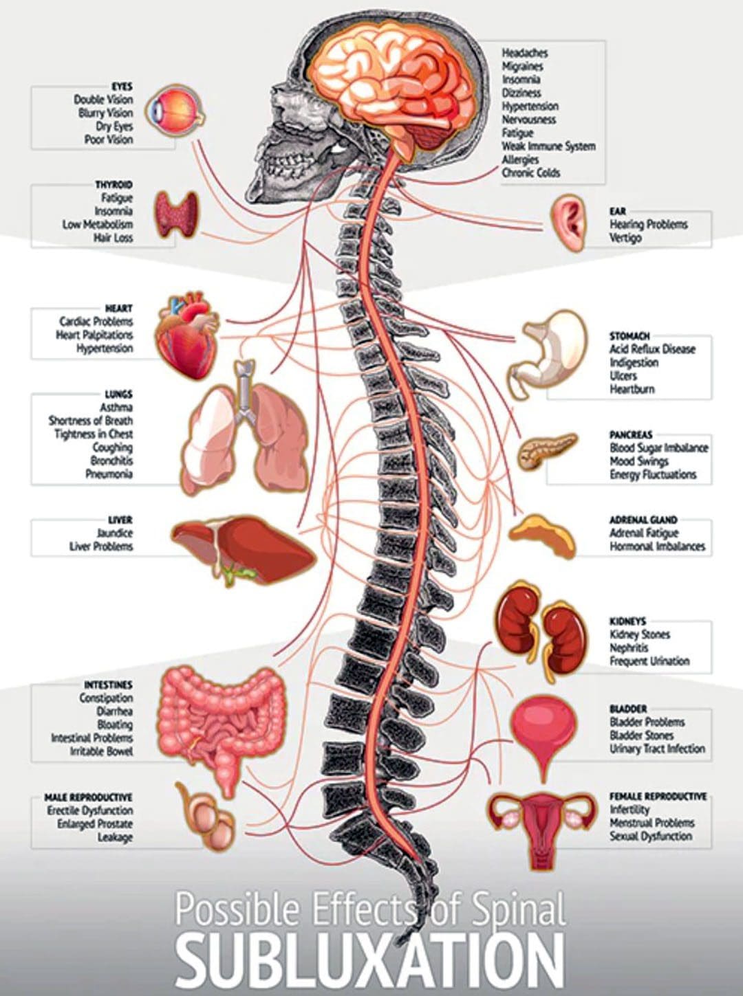
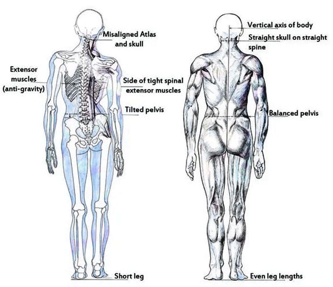
As humans, there are a variety of stressors experienced daily. Stress collects in various body areas, most commonly the upper back, jaw, and neck muscles. Stress leads to tension in the muscles. The built-up tension can cause the spinal bones to shift out of alignment, irritating the nerves between the spinal bones. A cycle begins as increased nerve tension causes the muscles to continue to contract/tighten. The extra muscle tension continues to pull the spinal bones out of alignment, making the spine stiff and less flexible affecting posture, balance, coordination, and mobility, causing the spine to become further unstable. Chiropractic treatment at regular intervals is recommended to help realign and maintain proper position.
Why The Spine Goes Out of Alignment
The nerves in the body are intricately linked to the spinal cord, and small distortions in the alignment can cause nerves to misfire and malfunction. When the spine goes out of alignment, the nervous system/brain and nerves get stuck in a stressed or tense state. Even a minor misalignment can cause a series of discomfort symptoms to travel throughout the body.
Causes
Causes of misalignment that creates tension in the nerves and muscles include:
A chiropractor will feel/palpate the spine to see if the bones are in alignment, move well, or are out of alignment and not moving correctly or moving at all.
Posture Exam
If the head, shoulders, and hips are uneven or the shoulders and head are pulling forward, the spinal bones are out of alignment/subluxations.
Balance and Coordination
Unhealthy balance and coordination can indicate the brain, nerves, and muscles are malfunctioning by spinal misalignment.
Range of Motion
A loss of spinal movement flexibility can show tension in the nerves, muscles, and misalignments.
Muscle Test
Loss of strength in a muscle can indicate the nerve signals are weak.
Orthopedic Tests
Tests that put the body in stressful positions focus on what tissue/s may be injured and the causes.
X-rays
X-rays look for abnormalities, dislocations, bone density, fractures, hidden/invisible injuries, and infections.
Injury Medical Chiropractic and Functional Medicine Clinic provide personalized treatment plans. These specific therapies are made to generate long-term spine benefits. Spinal manipulation, deep tissue massage, MET, and other manual therapy techniques, combined with exercise, help get the bones moving properly, the muscles functioning correctly, and the spine back into proper form. Treatment relieves muscle spasms, tension, and joint dysfunction, increases circulation, and retrains the muscles to remain relaxed.
The Natural Way to Heal
References
Ando, Kei et al. “Poor spinal alignment in females with obesity: The Yakumo study.” Journal of Orthopaedics vol. 21 512-516. 16 Sep. 2020, doi:10.1016/j.jor.2020.09.006
Le Huec, J C et al. “Sagittal balance of the spine.” The European spine journal: official publication of the European Spine Society, the European Spinal Deformity Society, and the European Section of the Cervical Spine Research Society vol. 28,9 (2019): 1889-1905. doi:10.1007/s00586-019-06083-1
Meeker, William C, and Scott Haldeman. “Chiropractic: a profession at the crossroads of mainstream and alternative medicine.” Annals of internal medicine vol. 136,3 (2002): 216-27. doi:10.7326/0003-4819-136-3-200202050-00010
Oakley, Paul A et al. “X-Ray Imaging is Essential for Contemporary Chiropractic and Manual Therapy Spinal Rehabilitation: Radiography Increases Benefits and Reduces Risks.” Dose-response: a publication of International Hormesis Society vol. 16,2 1559325818781437. 19 Jun. 2018, doi:10.1177/1559325818781437
Shah, Anoli A, et al. “Spinal Balance/Alignment – Clinical Relevance and Biomechanics.” Journal of biomechanical engineering, 10.1115/1.4043650. 2 May. 2019, doi:10.1115/1.4043650
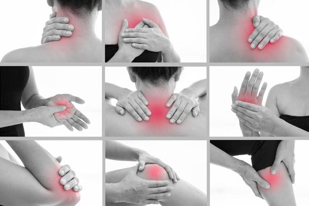
Body misalignment can cause various symptoms to be experienced, ranging from headaches, neck and back pain, sore feet, discomfort in the joints, muscles, or nerves, and digestive problems. Improperly aligned vertebrae can press against nerves, pinching or compressing them, causing the nerve signals of the digestive system, including those in the stomach and intestines, to misfire or fail to transmit at the appropriate moment. This can cause the organs to malfunction, resulting in heartburn, gas, constipation, cramping, diarrhea, and other symptoms. Chiropractic realignment adjustments are an effective treatment option for frequent stomachaches, reflux, constipation, and other gastrointestinal conditions.
Body Misalignment Digestive Problems
There are over a million nerve cells within the digestive system. A collection of nerves branch out from the lower part of the spinal cord and travels to the stomach and intestines. Nerve transmission plays an essential role in the following:
Digestion.
Movement of food through the gastrointestinal system.
Absorption of nutrients and minerals.
Removal of waste products.
Misalignments of the vertebrae are known as subluxations. Pressure on nerve roots caused by misalignment can interfere with the function of the bowel and other organs, which can lead to gastrointestinal issues. Muscle tension in the abdomen can also contribute to digestive problems, whether because of stress or sitting for long hours daily.
Misalignment Symptoms
When the body is out of alignment, symptoms of discomfort begin to appear. The most common include:
Fatigue.
Stiff neck.
Sore shoulders.
Chronic headaches.
Sore muscles.
Pain throughout the back.
Joint pain throughout the body.
Chronic aches.
Tight hips.
Difficulty walking.
Tingling, pins and needles, and numbness nerve sensations – sciatica.
Constantly getting sick.
Healthy Gut
A balanced healthy gut will have less difficulty processing food and eliminating waste, leading to reduced and eventually alleviated symptoms. The following show healthy gut function:
Regular, consistent energy levels.
Increased mental clarity.
Regular and healthy bowel movements.
No pain or discomfort symptoms.
A normal amount of gas and bloating.
Healthy stress levels.
Chiropractic
Chiropractic care will realign the body to its proper form, improving gastrointestinal issues. The chiropractic team will use various tools and techniques to guide and correct any subluxations, relax the muscles, and increase nerve and blood circulation.
Healthy Diet and Chiropractic
References
Ernst, Edzard. “Chiropractic treatment for gastrointestinal problems: a systematic review of clinical trials.” Canadian Journal of Gastroenterology = Journal canadien de Gastroenterologie vol. 25,1 (2011): 39-40. doi:10.1155/2011/910469
Hills, Ronald D Jr, et al. “Gut Microbiome: Profound Implications for Diet and Disease.” Nutrients vol. 11,7 1613. 16 Jul. 2019, doi:10.3390/nu11071613
Hornbuckle, William E., et al. “Gastrointestinal Function.” Clinical Biochemistry of Domestic Animals (2008): 413–457. doi:10.1016/B978-0-12-370491-7.00014-3
Leeming, Emily R et al. “Effect of Diet on the Gut Microbiota: Rethinking Intervention Duration.” Nutrients vol. 11,12 2862. 22 Nov. 2019, doi:10.3390/nu11122862
Li, Yuanyuan, et al. “The Role of Microbiome in Insomnia, Circadian Disturbance, and Depression.” Frontiers in psychiatry vol. 9 669. 5 Dec. 2018, doi:10.3389/fpsyt.2018.00669
Redwood, Daniel. “Chiropractic and visceral disorders.” Journal of Alternative and complementary medicine (New York, N.Y.) vol. 13,5 (2007): 479-80. doi:10.1089/acm.2007.7146
Valdes, Ana M et al. “Role of the gut microbiota in nutrition and health.” BMJ (Clinical research ed.) vol. 361 k2179. 13 Jun. 2018, doi:10.1136/bmj.k2179
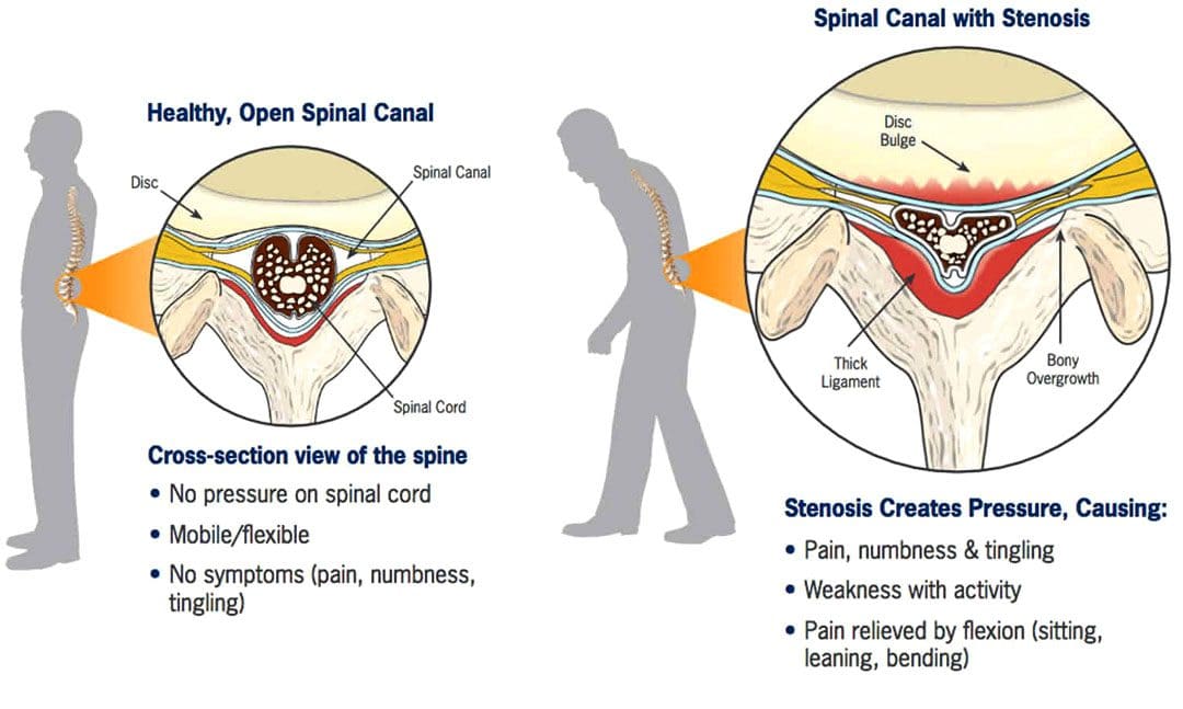
Spinal Stenosis Walking Issues:Stenosis means a narrowing. Spinal stenosis can happen in any spine region, but the neck and lower back are the most common locations. The spinal canal becomes narrower and can cause the nerves to become compressed, pinched, and irritated and can extend from the lumbar spine through the hips, buttocks, legs, and feet. Individuals with lumbar spinal stenosis may have difficulty walking caused by sensations of discomfort like numbness, electrical shocks, and pain, requiring the need to lean forward to relieve pressure and symptoms. Additionally, symptoms are likely to worsen the longer the walk. Chiropractic treatment can treat spinal stenosis because it corrects and re-aligns the spine, thus reducing pressure on the spinal cord, joints, and nerve roots.
Spinal Stenosis Walking Issues
The spine is made up of interlocking vertebrae. The regions are cervical, thoracic, lumbar, and sacral bones with a foramen opening. These openings form the protective tunnel/spinal canal surrounding the spinal cord. The spinal cord is a group of nerves that run through the tunnel. The narrowing suffocates the nerves supplying the lower extremities that can influence walking activity.
Symptoms
There may be no symptoms with early lumbar spinal stenosis. Most individuals develop symptoms gradually and may begin to notice them while walking or standing. These can include:
Lower back pressure sensations when standing upright or walking.
Leg numbness, tingling, weakness, burning, and/or cramping.
Muscle weakness.
Persistent pain in the back, hips, buttocks, or legs while walking.
Difficulty lifting the top part of the foot – known as drop foot.
Loss of sensation in the feet.
A weak foot that drops/slaps down when walking.
Loss of sexual ability.
In more serious cases, severe numbness, bladder problems, and inability to stand.
Individuals begin to lean forward when symptoms start, bringing relief by reducing the pressure on the nerves. However, constantly leaning forward leads to other posture and health problems.
Diagnosis
A doctor or chiropractor will ask questions about symptoms and medical history and perform a complete physical examination to diagnose lumbar spinal stenosis. During the physical examination, a healthcare provider will look for signs, such as loss of sensation, weakness, and abnormal reflexes.
Tests:
X-rays of the lumbar spine may show bone growths called spurs that push on spinal nerves and/or narrowing of the spinal canal.
Imaging tests – A CT or MRI scan can provide a detailed look at the spinal canal and nerve structures.
Other studies include – bone scans, myelogram, which is a CT scan that uses a color dye, and EMG, which is an electrical test of muscle activity.
Chiropractic Treatment
Chiropractic care combined with physical therapy is a tried-and-true treatment for spinal stenosis. A chiropractic treatment plan can include targeted and passive exercise programs. Targeted exercises involve strengthening the core and back muscles. Passive treatments include hot and cold therapy, massage, decompression, and electrical stimulation. The objective of chiropractic therapy is to:
Strengthen muscles in the core and legs
Correct posture and body mechanics.
Improve mobility.
Maintain ability to perform day-to-day activities.
Recommend stretches.
Educate on how to keep the spine and back muscles safe.
Train on using devices like a back brace, cane, or walker properly.
Advise about shoe inserts and splints.
Suggest work and home environment modifications, such as ergonomics and cushions.
Chiropractic Relief
References
Conway, Justin, et al. “Walking assessment in people with lumbar spinal stenosis: capacity, performance, and self-report measures.” The spine journal: official North American Spine Society journal vol. 11,9 (2011): 816-23. doi:10.1016/j.spinee.2010.10.019
Lurie, Jon, and Christy Tomkins-Lane. “Management of lumbar spinal stenosis.” BMJ (Clinical research ed.) vol. 352 h6234. 4 Jan. 2016, doi:10.1136/bmj.h6234
Macedo, Luciana Gazzi, et al. “Physical therapy interventions for degenerative lumbar spinal stenosis: a systematic review.” Physical therapy vol. 93,12 (2013): 1646-60. doi:10.2522/ptj.20120379
Tomkins-Lane, Christy C et al. “Predictors of walking performance and walking capacity in people with lumbar spinal stenosis, low back pain, and asymptomatic controls.” Archives of physical medicine and rehabilitation vol. 93,4 (2012): 647-53. doi:10.1016/j.apmr.2011.09.023
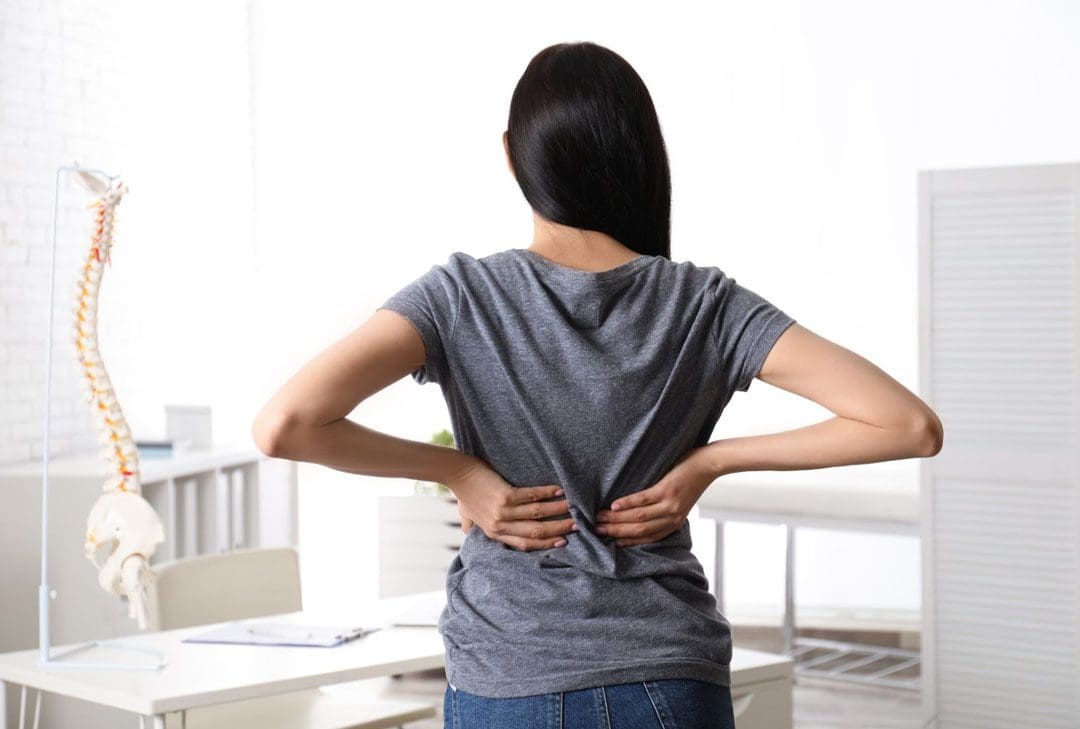
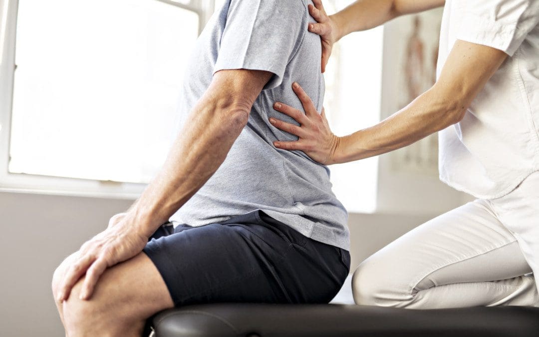
Prolonged standing can cause the pelvis to push backward, increasing the curve of the lower back/lumbar region. This increased pressure on the soft tissues surrounding the spine causes the lower back muscles to tighten and/or spasm, resulting in discomfort in the joints and nerves. Weakened core muscles and unhealthy posture/postural syndrome are the most common causes, but injury, aging, congenital malformations, or a disease/condition can also contribute to the symptoms. Injury Medical Chiropractic and Functional Medicine Clinic has a top team of professional therapists to evaluate the problem, diagnose the cause/s accurately, and develop a customized treatment and rehabilitation plan.
Prolonged Standing Back Discomfort
Back Structure
The lower back is one of the most used areas of the spine, moving around and bending during a normal day. When the body stands, the spine naturally curves both in and outwards.
The inward curve, called lordosis, curves towards the front of the body at the lower back and neck regions.
The outward curve, called kyphosis, curves towards the back of the body at the chest.
When bending over while standing, the five lumbar vertebrae of the lower back change position and shift from lordosis to kyphosis when bent completely.
When standing up from bending, the lumbar vertebrae change position again and return to the lordosis position.
Causes
The facet joints allow movement between each spine level. The standing spinal curvature can increase contact between the facet joints. As the body ages, the facet joints and discs begin to wear out, which can cause the discs and facet joints to become inflamed. Prolonged standing during normal daily activity combined with inflammation in these joints can aggravate the inflammation and cause symptoms. Regular routines and habits may contribute to low back discomfort during prolonged standing. These include:
Sleeping on a sinking or unsupportive mattress.
Practicing unhealthy postures that cause imbalances with proper weight distribution.
Not wearing proper footwear and/or supportive orthotics forces the lower spine into increased curvature and can compress the facet joints.
Not getting enough physical activity that strengthens the core.
Chiropractors are experts on the musculoskeletal system. They will:
Listen to the patient about symptoms, medical history, and occupation.
A physical examination of muscle tone, strength, and range of motion.
Therapeutic massage, electric muscle stimulation, and ultrasound therapy can help reduce muscle inflammation and increase circulation to injured soft tissues.
Chiropractic adjustments will reset joints, removing pressure from the surrounding muscles and nerves.
Targeted therapeutic strength training is recommended for core and leg muscles to improve hip flexibility.
Non-surgical decompression or traction, either with a machine or suspension, can reverse the pressure in spinal discs.
Standing Lower Back Relief Exercises
References
Hasegawa, Tetsuya, et al. “Association of low back load with low back pain during static standing.” PloS one vol. 13,12 e0208877. 18 Dec. 2018, doi:10.1371/journal.pone.0208877
Jo, Hoon, et al. “Negative Impacts of Prolonged Standing at Work on Musculoskeletal Symptoms and Physical Fatigue: The Fifth Korean Working Conditions Survey.” Yonsei medical journal vol. 62,6 (2021): 510-519. doi:10.3349/ymj.2021.62.6.510
Ognibene GT, Torres W, von Eyben R, Horst KC. Impact of a sit-stand workstation on chronic low back pain: randomized trial results. J Occup Environ Med. 2016;58(3):287-293. Abstract. https://www.ncbi.nlm.nih.gov/pubmed/26735316. Accessed March 2, 2017.
Parry, Sharon P et al. “Workplace interventions for increasing standing or walking for decreasing musculoskeletal symptoms in sedentary workers.” The Cochrane database of systematic reviews vol. 2019,11 CD012487. November 17, 2019, doi:10.1002/14651858.CD012487.pub2
Rodríguez-Romero, Beatriz, et al. “Thirty Minutes Identified as the Threshold for Development of Pain in Low Back and Feet Regions, and Predictors of Pain Intensity During 1-h Laboratory-Based Standing in Office Workers.” International journal of environmental research and public health vol. 19,4 2221. February 16, 2022, doi:10.3390/ijerph19042221
Smith, Michelle D et al. “The Influence of Using a Footstool during a Prolonged Standing Task on Low Back Pain in Office Workers.” International journal of environmental research and public health vol. 16,8 1405. April 18. 2019, doi:10.3390/ijerph16081405
IFM's Find A Practitioner tool is the largest referral network in Functional Medicine, created to help patients locate Functional Medicine practitioners anywhere in the world. IFM Certified Practitioners are listed first in the search results, given their extensive education in Functional Medicine


 Why The Spine Goes Out of Alignment