Spinal stenosis is the term used to describe a narrowing spine. Treatments vary because everybody’s case is different. Some individuals experience mild symptoms, while others experience severe symptoms. Can knowing treatment options help the patient and healthcare team customize and personalize a treatment plan to the individual’s condition?
Spinal Stenosis Treatments
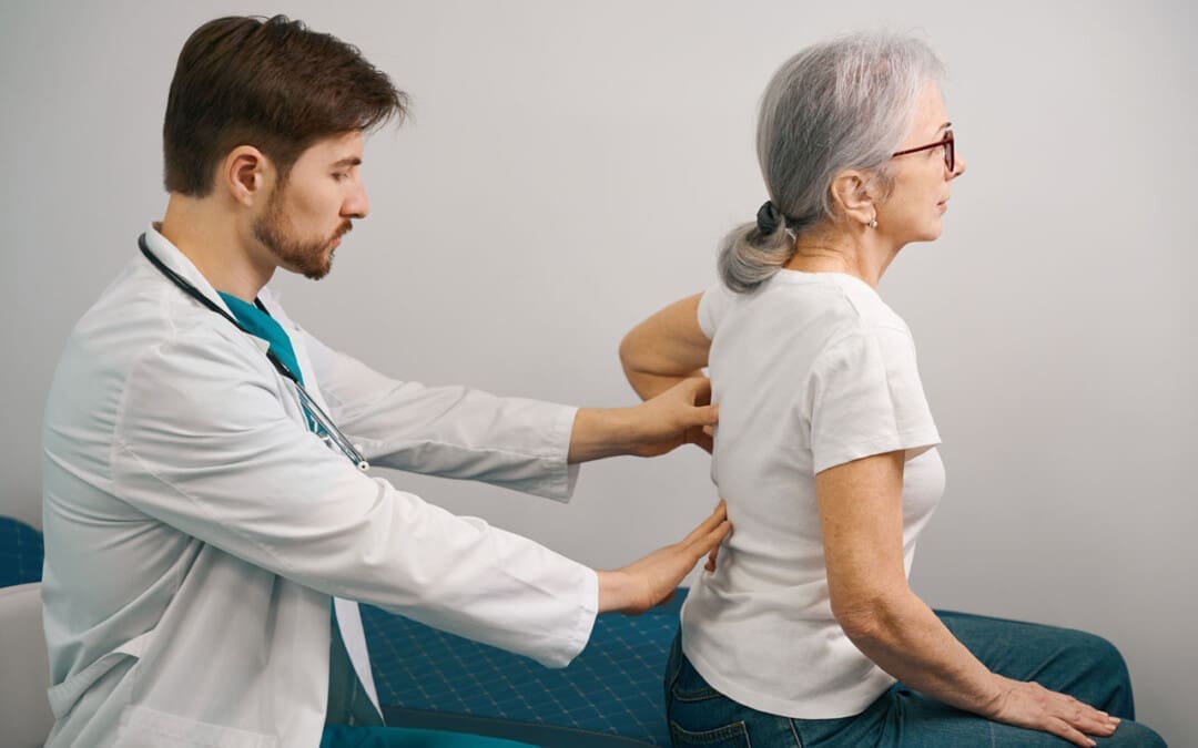
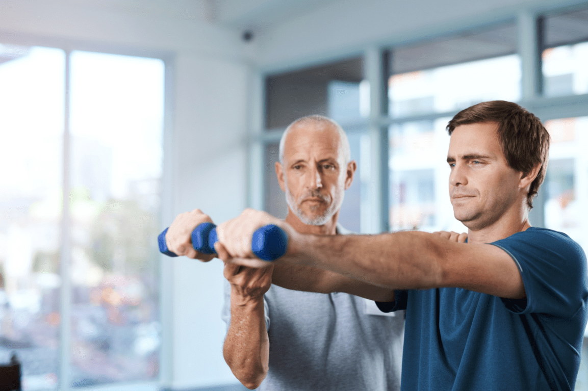
Spaces within the spine can become narrower than they’re supposed to be, which can cause pressure on nerve roots and the spinal cord. Anywhere along the spine can be affected. The narrowing can cause pain, burning, and/or aching in the back and weakness in the legs and feet. Spinal stenosis has several primary treatments. When working through spinal stenosis treatments, a healthcare provider will assess symptoms and start treatment with first-line therapy, such as pain medication and/or physical therapy. These are often the first among individuals with the disease.
Medication
Chronic pain is one of the main symptoms. The first-line treatment often involves using pain-relieving medication/s. Commonly prescribed medications are nonsteroidal anti-inflammatories or NSAIDs. These medications reduce pain and inflammation. However, NSAIDs are not recommended for long-term use, and other medications may need to be used to relieve pain that includes: (Sudhir Diwan et al., 2019)
Tylenol – acetaminophen
Gabapentin
Pregabalin
Opioids for severe cases
Exercise
Exercise can reduce spinal stenosis symptoms by taking pressure off the nerves, which can reduce pain and improve mobility. (Andrée-Anne Marchand et al., 2021) Healthcare providers will recommend the most effective exercises for the individual. Examples include:
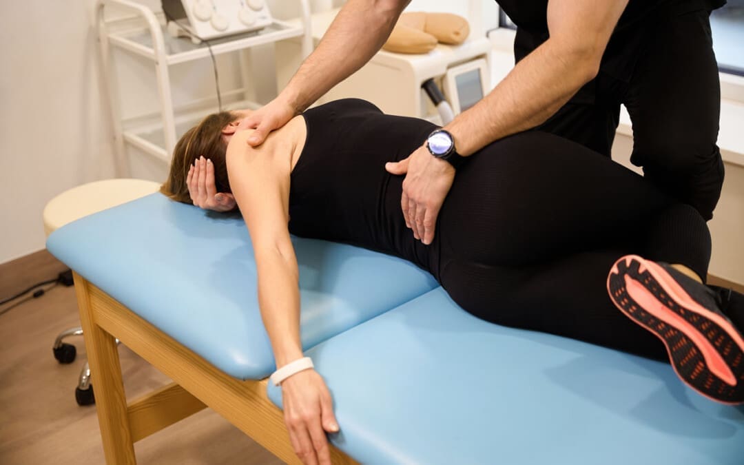
Another primary spinal stenosis treatment is physical therapy, which is often used alongside pain medications. Typically, individuals undergo six to eight weeks of physical therapy, with sessions two to three times a week. Utilizing physical therapy has been shown to (Sudhir Diwan et al., 2019)
Reduce pain
Increase mobility
Reduce pain medications.
Reduce mental health symptoms like anger, depression, and mood changes.
For severe cases, physical therapy following surgery can reduce recovery times.
Back Braces
Back braces can help reduce movement and pressure on the spine. This is helpful because even small spinal movements can lead to nerve irritation, pain, and worsening symptoms. Over time, the bracing can lead to a positive increase in mobility. (Carlo Ammendolia et al., 2019)
Injections
Epidural steroid injections may be recommended to relieve severe symptoms. Steroids act as anti-inflammatories to reduce pain and swelling caused by inflammation and irritation of the spinal nerves. They are considered nonsurgical medical procedures. According to research, injections can effectively manage pain for two weeks and up to six months, and some research has found that after a spinal injection, relief can last 24 months. (Sudhir Diwan et al., 2019)
Thickened Ligaments Decompression Procedure
Some individuals may be recommended to undergo a decompression procedure. This procedure involves using a thin needle tool inserted into the back. The thickened ligament tissue is removed to reduce the pressure on the spine and nerves. Research has found that the procedure can reduce symptoms and the need for more invasive surgery. (Nagy Mekhail et al., 2021)
Alternative Treatments
In addition to first-line treatments, individuals may be referred to alternative therapies for symptom management, including:
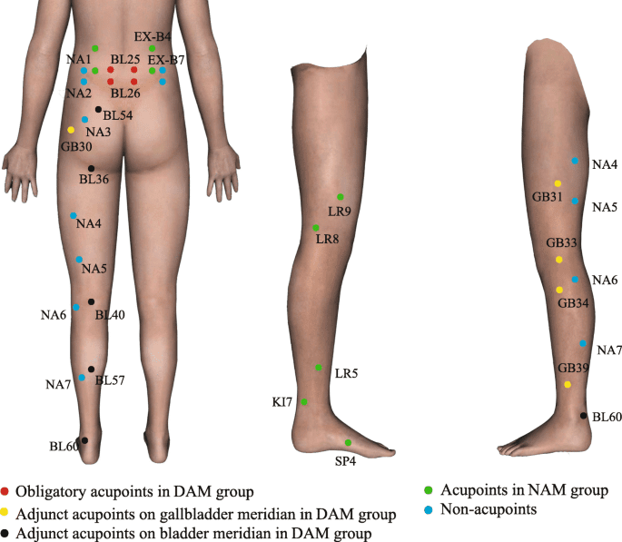
Acupuncture
This involves the insertion of thin-tipped needles into various acupoints to relieve symptoms.
Some research has found that acupuncture may be more effective at reducing symptoms than physical therapy alone. Both options are viable and can improve mobility and pain. (Hiroyuki Oka et al., 2018)
Chiropractic
This therapy reduces pressure on nerves, maintains spinal alignment, and helps to improve mobility.
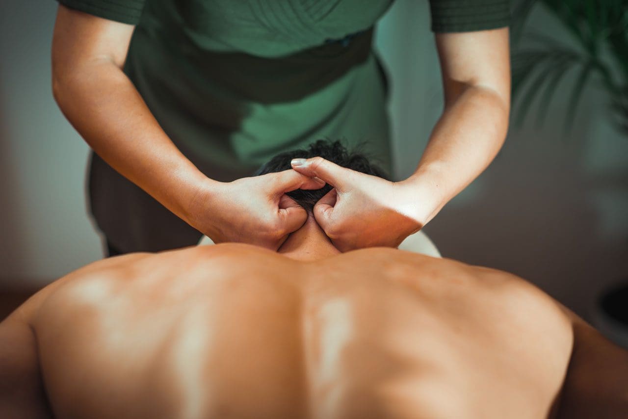
Massage
Massage helps to increase circulation, relax the muscles, and reduce pain and stiffness.
New Treatment Options
As spinal stenosis research continues, new therapies are emerging to help relieve and manage symptoms in individuals who don’t respond to traditional medicine or cannot partake in conventional therapies for various reasons. However, some evidence presented is promising; medical insurers may consider them experimental and not offer coverage until their safety has been proven. Some new treatments include:
Acupotomy
Acupotomy is a form of acupuncture that uses thin needles with a small, flat, scalpel-type tip to relieve tension in painful areas. Research on its effects is still limited, but preliminary data shows it could be an effective complementary treatment. (Ji Hoon Han et al., 2021)
Stem Cell Therapy
Stem cells are the cells from which all other cells originate. They act as the raw material for the body to create specialized cells with specific functions. (National Institutes of Health. 2016)
Individuals with spinal stenosis can develop soft tissue damage.
Stem cell therapy uses stem cells to help repair injured or diseased tissues.
Stem cell therapy can help repair or improve the damaged areas and provide symptom relief.
Clinical studies for spinal stenosis report that it could be a viable treatment option for some.
However, more research is needed to confirm whether the therapy is effective enough to be widely used. (Hideki Sudo et al., 2023)
Dynamic Stabilization Devices
LimiFlex is a medical device undergoing research and analysis for its ability to restore mobility and stability in the spine. It is implanted into the back through a surgical procedure. According to research, individuals with spinal stenosis who receive the LimiFlex often experience a higher reduction in pain and symptoms than with other forms of treatment. (T Jansen et al., 2015)
Lumbar Interspinous Distraction Decompression
Lumbar interspinous distraction decompression is another surgical procedure for spinal stenosis. The surgery is performed with an incision above the spine and places a device between two vertebrae to create space. This reduces movement and pressure on the nerves. Preliminary results show positive short-term relief from symptoms; long-term data is not yet available as it is a relatively new spinal stenosis treatment option. (UK National Health Service, 2022)
Surgical Procedures
There are several surgical procedures are available for spinal stenosis. Some include: (NYU Langone Health. 2024) Surgery for spinal stenosis is often reserved for individuals with severe symptoms, like numbness in the arms or legs. When these symptoms develop, it indicates a more notable compression of the spinal nerves and the need for a more invasive treatment. (NYU Langone Health. 2024)
Laminectomy
A laminectomy removes part or all of the lamina, the vertebral bone covering the spinal canal.
The procedure is designed to reduce pressure on nerves and the spinal cord.
Laminotomy and Foraminotomy
Both surgeries are used if an individual’s spinal stenosis negatively affects an opening in the vertebral foramen.
Ligaments, cartilage, or other tissues that constrict the nerves are removed.
Both reduce pressure on the nerves traveling through the foramen.
Laminoplasty
A laminoplasty relieves pressure on the spinal cord by removing parts of the spinal canal’s lamina.
This surgical procedure involves removing herniated or bulging discs that are placing pressure on the spinal cord and nerves.
Spinal fusion
Spinal fusion involves joining two vertebrae using metal pieces like rods and screws.
The vertebrae are more stable because the rods and screws act as a brace.
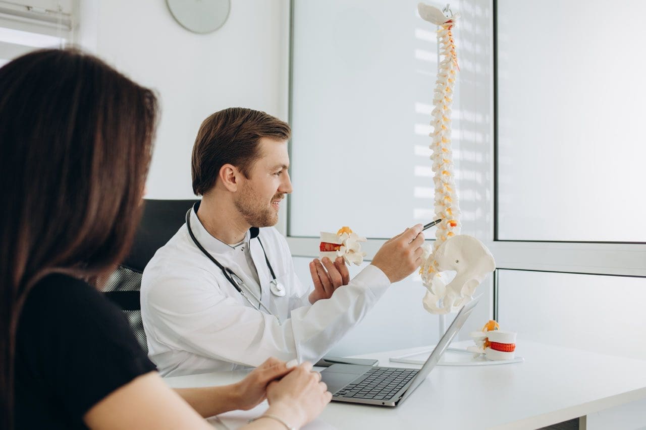
Which Treatment Is The Right One?
Because all treatment plans differ, determining the most effective is best suited for a healthcare provider. Each approach will be personalized to the individual. To decide what therapy is best, healthcare providers will assess: (National Institute of Arthritis and Musculoskeletal and Skin Diseases. 2023)
The severity of symptoms.
The current level of overall health.
The level of damage that’s occurring in the spine.
The level of disability and how mobility and quality of life are affected.
Injury Medical Chiropractic and Functional Medicine Clinic will work with an individual’s primary healthcare provider and/or specialists to help determine the best treatment options and concerns regarding medications or other forms of treatment.
Unlocking Wellness
References
Diwan, S., Sayed, D., Deer, T. R., Salomons, A., & Liang, K. (2019). An Algorithmic Approach to Treating Lumbar Spinal Stenosis: An Evidenced-Based Approach. Pain medicine (Malden, Mass.), 20(Suppl 2), S23–S31. https://doi.org/10.1093/pm/pnz133
Marchand, A. A., Houle, M., O’Shaughnessy, J., Châtillon, C. É., Cantin, V., & Descarreaux, M. (2021). Effectiveness of an exercise-based prehabilitation program for patients awaiting surgery for lumbar spinal stenosis: a randomized clinical trial. Scientific reports, 11(1), 11080. https://doi.org/10.1038/s41598-021-90537-4
Ammendolia, C., Rampersaud, Y. R., Southerst, D., Ahmed, A., Schneider, M., Hawker, G., Bombardier, C., & Côté, P. (2019). Effect of a prototype lumbar spinal stenosis belt versus a lumbar support on walking capacity in lumbar spinal stenosis: a randomized controlled trial. The spine journal : official journal of the North American Spine Society, 19(3), 386–394. https://doi.org/10.1016/j.spinee.2018.07.012
Mekhail, N., Costandi, S., Nageeb, G., Ekladios, C., & Saied, O. (2021). The durability of minimally invasive lumbar decompression procedure in patients with symptomatic lumbar spinal stenosis: Long-term follow-up. Pain practice : the official journal of World Institute of Pain, 21(8), 826–835. https://doi.org/10.1111/papr.13020
Oka, H., Matsudaira, K., Takano, Y., Kasuya, D., Niiya, M., Tonosu, J., Fukushima, M., Oshima, Y., Fujii, T., Tanaka, S., & Inanami, H. (2018). A comparative study of three conservative treatments in patients with lumbar spinal stenosis: lumbar spinal stenosis with acupuncture and physical therapy study (LAP study). BMC complementary and alternative medicine, 18(1), 19. https://doi.org/10.1186/s12906-018-2087-y
Han, J. H., Lee, H. J., Woo, S. H., Park, Y. K., Choi, G. Y., Heo, E. S., Kim, J. S., Lee, J. H., Park, C. A., Lee, W. D., Yang, C. S., Kim, A. R., & Han, C. H. (2021). Effectiveness and safety of acupotomy on lumbar spinal stenosis: A pragmatic randomized, controlled, pilot clinical trial: A study protocol. Medicine, 100(51), e28175. https://doi.org/10.1097/MD.0000000000028175
Sudo, H., Miyakoshi, T., Watanabe, Y., Ito, Y. M., Kahata, K., Tha, K. K., Yokota, N., Kato, H., Terada, T., Iwasaki, N., Arato, T., Sato, N., & Isoe, T. (2023). Protocol for treating lumbar spinal canal stenosis with a combination of ultrapurified, allogenic bone marrow-derived mesenchymal stem cells and in situ-forming gel: a multicentre, prospective, double-blind randomised controlled trial. BMJ open, 13(2), e065476. https://doi.org/10.1136/bmjopen-2022-065476
National Institutes of Health. (2016). Stem cell basics. U.S. Department of Health and Human Services. Retrieved from https://stemcells.nih.gov/info/basics/stc-basics
Jansen, T., Bornemann, R., Otten, L., Sander, K., Wirtz, D., & Pflugmacher, R. (2015). Vergleich dorsaler Dekompression nicht stabilisiert und dynamisch stabilisiert mit LimiFlex™ [A Comparison of Dorsal Decompression and Dorsal Decompression Combined with the Dynamic Stabilisation Device LimiFlex™]. Zeitschrift fur Orthopadie und Unfallchirurgie, 153(4), 415–422. https://doi.org/10.1055/s-0035-1545990
UK National Health Service. (2022). Lumbar decompression surgery: How It’s performed. https://www.nhs.uk/conditions/lumbar-decompression-surgery/what-happens/
NYU Langone Health. (2024). Surgery for spinal stenosis. https://nyulangone.org/conditions/spinal-stenosis/treatments/surgery-for-spinal-stenosis
Columbia Neurosurgery. (2024). Cervical laminoplasty procedure. https://www.neurosurgery.columbia.edu/patient-care/treatments/cervical-laminoplasty
National Institute of Arthritis and Musculoskeletal and Skin Diseases. (2023). Spinal stenosis: Diagnosis, treatment and steps to take. Retrieved from https://www.niams.nih.gov/health-topics/spinal-stenosis/diagnosis-treatment-and-steps-to-take
For individuals experiencing or managing low back pain and/or sciatica, can lumbar traction therapy help provide consistent relief?
Lumbar Traction
Lumbar traction therapy for lower back pain and sciatica could be a treatment option to help restore mobility and flexibility and safely support an individual’s return to an optimal level of activity. It is often combined with targeted therapeutic exercise. (Yu-Hsuan Cheng, et al., 2020) The technique stretches the space between the vertebrae in the lower spine, relieving lower back pain.
Lumbar or low back traction helps to separate the spaces between the vertebrae.
Separating the bones restores circulation and helps relieve the pressure on pinched nerves like the sciatic nerve, decreasing pain and improving mobility.
Research
Researchers say lumbar traction with exercise did not improve individual outcomes compared to physical therapy exercises on their own (Anne Thackeray et al., 2016). The study examined 120 participants with back pain and nerve root impingement who were randomly selected to undergo lumbar traction with exercises or simple exercises for pain. Extension-based exercises focused on bending the spine backward. This movement is considered effective for individuals with back pain and pinched nerves. The results indicated that adding lumbar traction to physical therapy exercises did not offer significant benefits over extension-based exercise alone for back pain. (Anne Thackeray et al., 2016)
A 2022 study found that lumbar traction is helpful for individuals with lower back pain. The study investigated two different lumbar traction techniques and found that variable-force lumbar traction and high-force lumbar traction helped to relieve lower back pain. High-force lumbar traction was also found to reduce functional disability. (Zahra Masood et al., 2022) Another study found lumbar traction improves the range of motion in the straight leg raise test. The study examined different forces of traction on herniated discs. All the levels improved the individuals’ range of motion, but the one-half body-weight traction setting was associated with the most significant pain relief. (Anita Kumari et al., 2021)
Treatment
For individuals with only low back pain, exercise, and postural correction may be all that is needed to provide relief. Research confirms physical therapy exercises can help decrease pain and improve mobility (Anita Slomski 2020). Another study revealed the importance of centralizing sciatic symptoms during repetitive movements. Centralization is moving the pain back to the spine, which is a positive sign that the nerves and discs are healing and occurs during therapeutic exercise. (Hanne B. Albert et al., 2012) A chiropractor and physical therapy team can educate patients on preventing back pain episodes. Chiropractors and physical therapists are body movement experts who can show which exercises are best for your condition. Starting an exercise program that centralizes symptoms can help individuals return to their normal lifestyle quickly and safely. Consult a healthcare provider before starting any exercise program for back pain.
Movement Medicine: Chiropractic
References
Cheng, Y. H., Hsu, C. Y., & Lin, Y. N. (2020). The effect of mechanical traction on low back pain in patients with herniated intervertebral disks: a systemic review and meta-analysis. Clinical rehabilitation, 34(1), 13–22. https://doi.org/10.1177/0269215519872528
Thackeray, A., Fritz, J. M., Childs, J. D., & Brennan, G. P. (2016). The Effectiveness of Mechanical Traction Among Subgroups of Patients With Low Back Pain and Leg Pain: A Randomized Trial. The Journal of orthopaedic and sports physical therapy, 46(3), 144–154. https://doi.org/10.2519/jospt.2016.6238
Masood, Z., Khan, A. A., Ayyub, A., & Shakeel, R. (2022). Effect of lumbar traction on discogenic low back pain using variable forces. JPMA. The Journal of the Pakistan Medical Association, 72(3), 483–486. https://doi.org/10.47391/JPMA.453
Kumari, A., Quddus, N., Meena, P. R., Alghadir, A. H., & Khan, M. (2021). Effects of One-Fifth, One-Third, and One-Half of the Bodyweight Lumbar Traction on the Straight Leg Raise Test and Pain in Prolapsed Intervertebral Disc Patients: A Randomized Controlled Trial. BioMed research international, 2021, 2561502. https://doi.org/10.1155/2021/2561502
Slomski A. (2020). Early Physical Therapy Relieves Sciatica Disability and Pain. JAMA, 324(24), 2476. https://doi.org/10.1001/jama.2020.24673
Albert, H. B., Hauge, E., & Manniche, C. (2012). Centralization in patients with sciatica: are pain responses to repeated movement and positioning associated with outcome or types of disc lesions?. European spine journal : official publication of the European Spine Society, the European Spinal Deformity Society, and the European Section of the Cervical Spine Research Society, 21(4), 630–636. https://doi.org/10.1007/s00586-011-2018-9
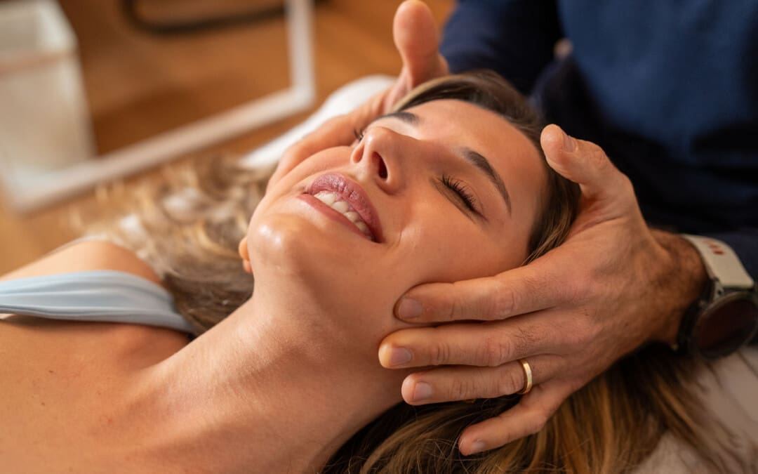
For individuals suffering from neck pain and headaches, can craniosacral head massage therapy help provide relief?
Craniosacral Therapy
Craniosacral therapy is a gentle massage to release fascia or connective tissue network tension. The therapy is not new but has gained new attention because of the public interest in natural pain treatments and therapies. Studies are limited, but clinical research is ongoing to see if the therapy can become a mainstream treatment option. The therapy aims to alleviate the symptoms of various health ailments and conditions, including:
By relieving compression in the lower back, head, and spinal column, cerebrospinal fluid circulation is restored, and the body rhythms within the nervous system are reset. This provides pain relief, lowers stress, and improves overall well-being.
The focus areas are those along the fascia, the connective tissue that holds organs, blood vessels, bones, nerve fibers, and muscles in place. By working this tissue through gentle-pressure massage, practitioners help to calm the fight-or-flight response by relaxing the sympathetic nervous system. The symptoms will determine what areas of the body necessitate craniosacral therapy. Individuals with headaches will be given a head or neck massage. Other areas involved in craniosacral therapy include: (Heidemarie Haller, Gustav Dobos, and Holger Cramer, 2021)
Back
Around the spinal column.
Other areas like the joints or muscles.
The pressure applied during craniosacral therapy is light and not the same as a deep tissue massage.
The parasympathetic and sympathetic nervous systems control various body responses.
The parasympathetic nervous system supports proper rest and digestive functions, and the sympathetic nervous system regulates the body’s fight-or-flight response. (Cleveland Clinic. 2022)
Therapy Techniques
The massage techniques used in craniosacral therapy rely on low pressure intended to be as gentle as possible. The fingertips are often used to avoid applying too much pressure. Healthcare providers work the areas between the skull and the bottom of the spine to identify and reset imbalances within the body and the cerebrospinal fluid. If there is an imbalance in cerebrospinal fluid, the massage therapist will reposition the individual or press on the area to release and/or increase circulation. The techniques work to improve the body’s ability to regulate physiological responses. (Heidemarie Haller et al., 2019) During and after the session, individuals may experience different sensations, including: (Biodynamic Craniosacral Therapy Association of North America, 2024)
Relaxation.
Feeling like being in a meditative state.
Sleepiness.
Energized.
Feeling a sense of warmth.
Deeper breathing.
Feeling the body is straighter and taller.
Individuals Who Should Not Receive Craniosacral Therapy
Craniosacral therapy is considered safe; however, some individuals should avoid it or consult a healthcare provider before trying it. Those recommended not to receive the treatment include individuals with the following ailments or disorders:
Concussion or other traumatic brain injuries.
Blood clots.
Brain swelling.
Brain aneurysm – a blood-filled bulge in a blood vessel in or around the brain.
Conditions that cause cerebrospinal fluid buildup.
Treatment
Craniosacral therapy is offered by several healthcare providers, including:
Craniosacral therapy licensed massage therapists
Physical therapists
Occupational therapists
Osteopaths
Chiropractors
These professionals know how to perform the massage technique correctly.
Tension Headaches
References
Haller, H., Lauche, R., Sundberg, T., Dobos, G., & Cramer, H. (2019). Craniosacral therapy for chronic pain: a systematic review and meta-analysis of randomized controlled trials. BMC musculoskeletal disorders, 21(1), 1. https://doi.org/10.1186/s12891-019-3017-y
Haller, H., Dobos, G., & Cramer, H. (2021). The use and benefits of Craniosacral Therapy in primary health care: A prospective cohort study. Complementary therapies in medicine, 58, 102702. https://doi.org/10.1016/j.ctim.2021.102702
Biodynamic Craniosacral Therapy Association of North America. (2024). What is a session like? https://www.craniosacraltherapy.org/what-is-a-session-like-
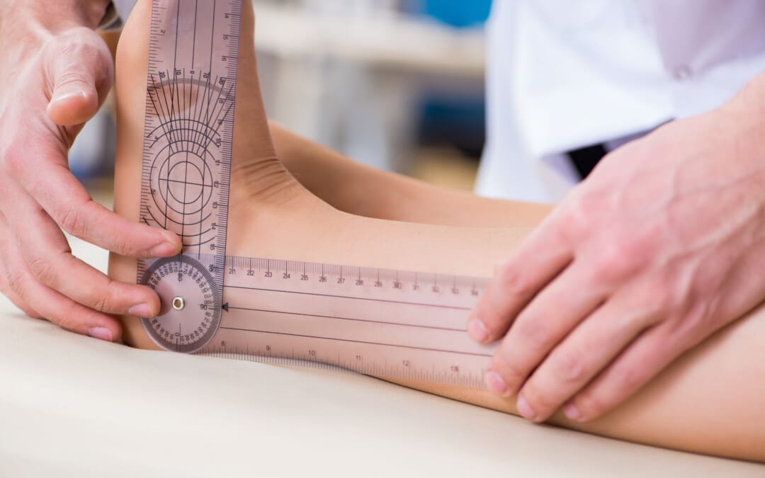
Can individuals with joint hypermobility find relief through nonsurgical treatments in reducing pain and restoring body mobility?
Introduction
When a person moves their body, the surrounding muscles, joints, and ligaments are incorporated into various tasks that allow them to stretch and be flexible without pain or discomfort. Many repetitive motions enable the individual to continue their routine. However, when the joints, muscles, and ligaments are stretched farther than normal in the upper and lower extremities without pain, it is known as joint hypermobility. This connective tissue disorder can correlate with other symptoms that affect the body and cause many people to seek treatment to manage joint hypermobility symptoms. In today’s article, we will look at joint hypermobility and how various non-surgical treatments can help reduce pain caused by joint hypermobility and restore body mobility. We talk with certified medical providers who consolidate our patients’ information to assess how their pain may be associated with joint hypermobility. We also inform and guide patients on how integrating various non-surgical treatments can help improve joint function while managing the associated symptoms. We encourage our patients to ask their associated medical providers intricate and insightful questions about incorporating non-surgical therapies as part of their routine to reduce pain and discomfort from joint hypermobility. Dr. Jimenez, D.C., includes this information as an academic service. Disclaimer.
What Is Joint Hypermobility?
Do you often feel your joints locked up in your hands, wrists, knees, and elbows? Do you experience pain and fatigue in your joints when your body feels constantly tired? Or when you stretch your extremities, do they extend farther than usual to feel the relief? Many of these various scenarios are often correlated with individuals experiencing joint hypermobility. Joint hypermobility is an inherited disorder with autosomal dominant patterns that characterize joint hyperlaxity and musculoskeletal pain within the body extremities. (Carbonell-Bobadilla et al., 2020) This connective tissue condition is often related to the flexibility of the connected tissues like ligaments and tendons in the body. An example would be if a person’s thumb is touching their inner forearm without feeling pain or discomfort, they have joint hypermobility. Additionally, many individuals dealing with joint hypermobility will often have a difficult diagnosis as they will develop skin and tissue fragility over time, causing musculoskeletal complications. (Tofts et al., 2023)
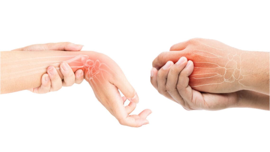
When individuals deal with joint hypermobility over time, many often have symptomatic joint hypermobility. They will present with musculoskeletal and systemic symptoms that lead to displaying skeletal deformities, tissue and skin fragility, and structural differences in the body’s system. (Nicholson et al., 2022) Some of the symptoms that joint hypermobility are shown in a diagnosis include:
Muscle pain and joint stiffness
Clicking joints
Fatigue
Digestive issues
Balance issues
Luckily, there are various treatments that many people can use to help restrengthen the surrounding muscles around the joints and reduce the correlating symptoms caused by joint hypermobility.
Movement As Medicine-Video
Nonsurgical Treatments For Joint Hypermobility
When dealing with joint hypermobility, many individuals need to seek treatments to reduce the correlating pain-like symptoms of joint hypermobility and help relieve the body’s extremities while restoring mobility. Some excellent treatments for joint hypermobility are non-surgical therapies that are non-invasive, gentle on the joints and muscles, and cost-effective. Various non-surgical treatments can be customized for the individual depending on how severe their joint hypermobility and comorbidities affect the person’s body. Non-surgical treatments can relieve the body from joint hypermobility by treating the causes of the pain through reduction and maximizing functional capacity and restoring a person’s quality of life. (Atwell et al., 2021) The three non-surgical treatments that are excellent for reducing pain from joint hypermobility and helping strengthen the surrounding muscles are below.
Chiropractic Care
Chiropractic care utilizes spinal manipulation and helps restore joint mobility in the body to reduce the effects of joint hypermobility by stabilizing the affected joints from the hypermobile extremities. (Boudreau et al., 2020) Chiropractors incorporate mechanical and manual manipulation and various techniques to help many individuals improve their posture by being more mindful of their bodies and work with multiple other therapies to emphasize controlled movements. With other comorbidities associated with joint hypermobility, like back and neck pain, chiropractic care can reduce these comorbidity symptoms and allow the individual to regain their quality of life.
Acupuncture
Another non-surgical treatment that many individuals can incorporate to reduce joint hypermobility and its comorbidities is acupuncture. Acupuncture utilizes small, thin, solid needles that acupuncturists use to block pain receptors and restore the body’s energy flow. When many individuals are dealing with joint hypermobility, their extremities in the legs, hands, and feet are in pain over time, which can cause the body to be unstable. What acupuncture does is help reduce the pain caused by joint hypermobility associated with the extremities and restore balance and functionality to the body (Luan et al., 2023). This means that if a person is dealing with stiffness and muscle pain from joint hypermobility, acupuncture can help rewire the pain by placing the needles in the body’s acupoints to provide relief.
Physical Therapy
Physical therapy is the last non-surgical treatment many people can incorporate into their daily routine. Physical therapy can help manage joint hypermobility that are tailored to help strengthen weak muscles that are surrounding the affected joints, improving a person’s stability and helping reduce the risk of dislocation. Additionally, many individuals can use low-impact exercise to ensure optimal motor control when doing regular exercises without putting excessive strain on the joints. (Russek et al., 2022)
By incorporating these three non-surgical treatments as part of a customized treatment for joint hypermobility, many individuals will begin to feel a difference in their balance. They will not experience joint pain by being more mindful of the body and incorporating small changes in their routine. Even though living with joint hypermobility can be a challenge for many individuals, by integrating and utilizing the right combination of non-surgical treatments, many can begin to lead active and fulfilling lives.
References
Atwell, K., Michael, W., Dubey, J., James, S., Martonffy, A., Anderson, S., Rudin, N., & Schrager, S. (2021). Diagnosis and Management of Hypermobility Spectrum Disorders in Primary Care. J Am Board Fam Med, 34(4), 838-848. https://doi.org/10.3122/jabfm.2021.04.200374
Boudreau, P. A., Steiman, I., & Mior, S. (2020). Clinical management of benign joint hypermobility syndrome: a case series. J Can Chiropr Assoc, 64(1), 43-54. https://www.ncbi.nlm.nih.gov/pubmed/32476667
Carbonell-Bobadilla, N., Rodriguez-Alvarez, A. A., Rojas-Garcia, G., Barragan-Garfias, J. A., Orrantia-Vertiz, M., & Rodriguez-Romo, R. (2020). [Joint hypermobility syndrome]. Acta Ortop Mex, 34(6), 441-449. https://www.ncbi.nlm.nih.gov/pubmed/34020527 (Sindrome de hipermovilidad articular.)
Luan, L., Zhu, M., Adams, R., Witchalls, J., Pranata, A., & Han, J. (2023). Effects of acupuncture or similar needling therapy on pain, proprioception, balance, and self-reported function in individuals with chronic ankle instability: A systematic review and meta-analysis. Complement Ther Med, 77, 102983. https://doi.org/10.1016/j.ctim.2023.102983
Nicholson, L. L., Simmonds, J., Pacey, V., De Wandele, I., Rombaut, L., Williams, C. M., & Chan, C. (2022). International Perspectives on Joint Hypermobility: A Synthesis of Current Science to Guide Clinical and Research Directions. J Clin Rheumatol, 28(6), 314-320. https://doi.org/10.1097/RHU.0000000000001864
Russek, L. N., Block, N. P., Byrne, E., Chalela, S., Chan, C., Comerford, M., Frost, N., Hennessey, S., McCarthy, A., Nicholson, L. L., Parry, J., Simmonds, J., Stott, P. J., Thomas, L., Treleaven, J., Wagner, W., & Hakim, A. (2022). Presentation and physical therapy management of upper cervical instability in patients with symptomatic generalized joint hypermobility: International expert consensus recommendations. Front Med (Lausanne), 9, 1072764. https://doi.org/10.3389/fmed.2022.1072764
Tofts, L. J., Simmonds, J., Schwartz, S. B., Richheimer, R. M., O’Connor, C., Elias, E., Engelbert, R., Cleary, K., Tinkle, B. T., Kline, A. D., Hakim, A. J., van Rossum, M. A. J., & Pacey, V. (2023). Pediatric joint hypermobility: a diagnostic framework and narrative review. Orphanet J Rare Dis, 18(1), 104. https://doi.org/10.1186/s13023-023-02717-2
Can individuals with herniated discs find the relief they are looking for from traction therapy or decompression to provide pain relief?
Introduction
The spine allows the individual to be mobile and flexible without feeling pain and discomfort when a person is on the move. This is because the spine is part of the musculoskeletal system that consists of muscles, tendons, ligaments, the spinal cord, and spinal discs. These components surround the spine and have three regions to allow the upper and lower extremities to do their jobs. However, the spine also ages when the body starts to age naturally. Many movements or routine actions can cause the body to be stiff and, over time, can cause the spinal disc to herniate. When this happens, a herniated disc can lead to pain and discomfort in the extremities, thus making individuals deal with a reduced quality of life and pain in three spinal regions. Luckily, there are numerous treatments, like traction therapy and decompression, to alleviate the pain and discomfort associated with herniated discs. Today’s article looks at why herniated discs cause issues in the spine and the effects of how these two treatments can help reduce herniated discs. We talk with certified medical providers who consolidate our patients’ information to assess how a herniated disc in the spine may be the issue causing musculoskeletal pain. We also inform and guide patients on how integrating spinal decompression and traction therapy can help realign the spine and reduce disc herniation that is causing spinal issues. We encourage our patients to ask their associated medical providers intricate and important questions about incorporating non-surgical treatments as part of their routine to reduce pain and discomfort in their bodies. Dr. Jimenez, D.C., includes this information as an academic service. Disclaimer.
Why Herniated Discs Causes Issues In The Spine?
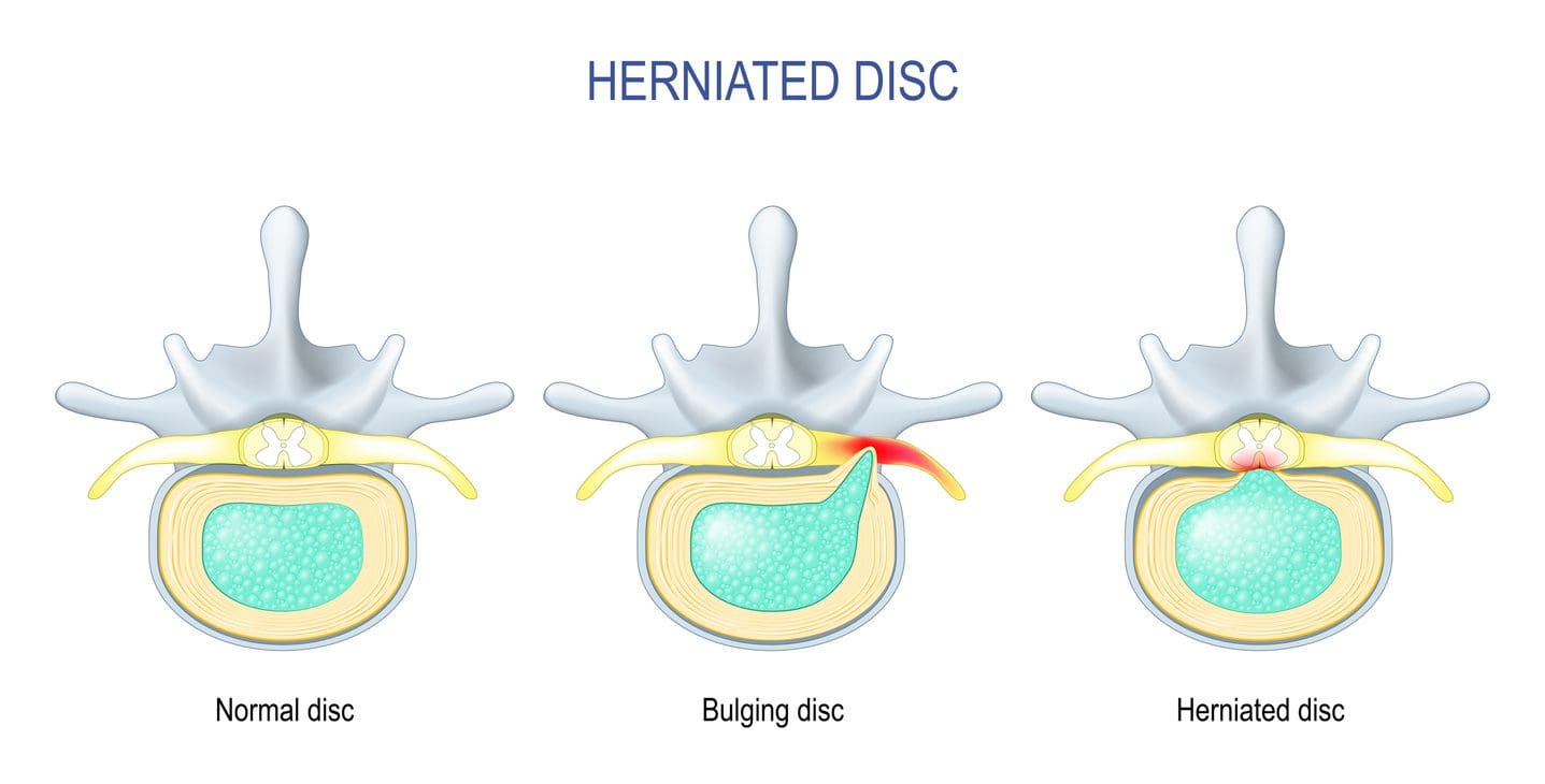
Have you been experiencing constant discomfort in your neck or back that doesn’t allow you to relax? Do you feel tingling sensations in your upper and lower extremities, making grasping objects or walking difficult? Or have you noticed that you are hunching over from your desk or standing and that stretching causes pain? As the spine keeps the body upright, its main components include the moveable vertebrae, the nerve root fibers, and spinal discs to help send neuron signals to the brain to allow movement, cushion the shocked forces on the spine, and be flexible. The spine allows the individual to perform various tasks without pain and discomfort through repetitive movements. However, when the body ages, it can lead to degenerative changes in the spine, causing the spinal disc to herniate over time. A herniated disc is a common degenerative musculoskeletal condition that causes the nucleus pulposus to break through any weak region of the annulus fibrosus and compress the surrounding nerve roots. (Ge et al., 2019) Other times, when repetitive motions start to cause a developing herniated disc, the inner portion of the disc can become desiccated and brittle. In contrast, the outer portion becomes more fibrotic and less elastic, causing the disc to shrink and be narrow. A herniated disc can affect young and old populations as they can have a multifactorial contribution that causes proinflammatory changes to the body. (Wu et al., 2020)
When many people are dealing with pain associated with a herniated disc, the disc itself goes through morphological change through the characterization of the disc being partial damage, which is then followed by the displacement and herniation of the inner disc portion in the vertebral canal to compress the spinal nerve roots. (Diaconu et al., 2021) This causes symptoms of pain, numbness, and weakness in the upper and lower body portions through nerve impingement. Hence why, many individuals are dealing with referred pain symptoms from their arms and legs that are radiating pain. When nerve compression associated with herniated discs starts to cause pain and discomfort, many individuals begin to seek out treatment to reduce the pain that the herniated disc is causing to provide relief for their bodies.
Spinal Decompression In Depth-Video
The Effects Of Traction Therapy In Reducing Herniated Disc
Many people who are suffering from pain that is being affected by herniated discs in their spines can seek out treatments like traction therapy to alleviate pain. Traction therapy is a non-surgical treatment that stretches and mobilizes the spine. Traction therapy can be mechanically or manually done by a pain specialist or with the help of mechanical devices. The effects of traction therapy can reduce the compression force on the spinal disc while reducing nerve root compression by expanding the disc height within the spine. (Wang et al., 2022) This allows the surrounding joints within the spine to be mobile and positively affect the spine. With traction therapy, intermittent or steady tension forces help stretch the spine, reduce pain, and improve functional outcomes. (Kuligowski et al., 2021)
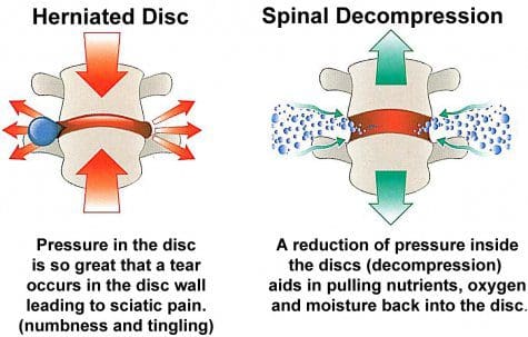
The Effects Of Spinal Decompression In Reducing Herniated Disc
Another form of non-surgical treatment is spinal decompression, a sophisticated version of traction that uses computerized technology to help apply controlled, gentle pulling forces to the spine. Spinal decompression does is that it can help decompress the spinal canal and help pull the herniated disc back to its original position while stabilizing the spine and keeping the vital bones and soft tissues safe. (Zhang et al., 2022) Additionally, spinal decompression can create negative pressure on the spine to allow the flow of nutritional fluids and blood oxygen back to the discs while creating an inverse relationship when tension pressure is introduced. (Ramos & Martin, 1994) Both spinal decompression and traction therapy can offer many therapeutic pathways to provide relief to many individuals dealing with herniated discs. Depending on how severe the herniated disc has caused issues to the person’s spine, many can rely on non-surgical treatments due to its customizable plan that is personalized to the person’s pain and can be combined with other therapies to strengthen the surrounding muscles. By doing so, many people can be pain-free over time while being mindful of their bodies.
References
Diaconu, G. S., Mihalache, C. G., Popescu, G., Man, G. M., Rusu, R. G., Toader, C., Ciucurel, C., Stocheci, C. M., Mitroi, G., & Georgescu, L. I. (2021). Clinical and pathological considerations in lumbar herniated disc associated with inflammatory lesions. Rom J Morphol Embryol, 62(4), 951-960. https://doi.org/10.47162/RJME.62.4.07
Ge, C. Y., Hao, D. J., Yan, L., Shan, L. Q., Zhao, Q. P., He, B. R., & Hui, H. (2019). Intradural Lumbar Disc Herniation: A Case Report and Literature Review. Clin Interv Aging, 14, 2295-2299. https://doi.org/10.2147/CIA.S228717
Kuligowski, T., Skrzek, A., & Cieslik, B. (2021). Manual Therapy in Cervical and Lumbar Radiculopathy: A Systematic Review of the Literature. Int J Environ Res Public Health, 18(11). https://doi.org/10.3390/ijerph18116176
Ramos, G., & Martin, W. (1994). Effects of vertebral axial decompression on intradiscal pressure. J Neurosurg, 81(3), 350-353. https://doi.org/10.3171/jns.1994.81.3.0350
Wang, W., Long, F., Wu, X., Li, S., & Lin, J. (2022). Clinical Efficacy of Mechanical Traction as Physical Therapy for Lumbar Disc Herniation: A Meta-Analysis. Comput Math Methods Med, 2022, 5670303. https://doi.org/10.1155/2022/5670303
Wu, P. H., Kim, H. S., & Jang, I. T. (2020). Intervertebral Disc Diseases PART 2: A Review of the Current Diagnostic and Treatment Strategies for Intervertebral Disc Disease. Int J Mol Sci, 21(6). https://doi.org/10.3390/ijms21062135
Zhang, Y., Wei, F. L., Liu, Z. X., Zhou, C. P., Du, M. R., Quan, J., & Wang, Y. P. (2022). Comparison of posterior decompression techniques and conventional laminectomy for lumbar spinal stenosis. Front Surg, 9, 997973. https://doi.org/10.3389/fsurg.2022.997973
Can individuals with spinal pain in their necks and back utilize decompression therapy to restore spinal disc height and find relief?
Introduction
Many people don’t realize that as the body ages, so does the spine. The spine is part of the musculoskeletal system and provides structural support, keeping the body upright. The muscles, ligaments, and tissues around the spine help with stability and mobility, while the spinal discs and joints provide shock absorption from the sheer vertical weight. When a person is on the move in their daily activities, the spine allows them to move without pain or discomfort. However, as time passes, the spine undergoes degenerative changes that can cause pain and discomfort, leaving the individual to contend with overlapping risk profiles affecting the neck and back. To that point, many people seek treatments to reduce pain in their spine and restore disc height. Today’s article looks at how spinal pain affects the neck and back, and how treatments like spinal decompression can reduce pain and restore disc height. We speak with certified medical providers who consolidate our patients’ information to assess how spinal pain can significantly impact a person’s well-being and quality of life in their bodies. We also inform and guide patients on how integrating spinal decompression can help reduce spinal pain and restore spinal disc height. We encourage our patients to ask their associated medical providers detailed, important questions about incorporating non-surgical treatments into a health and wellness routine to relieve spinal pain and improve their quality of life. Dr. Jimenez, D.C., includes this information as an academic service. Disclaimer.
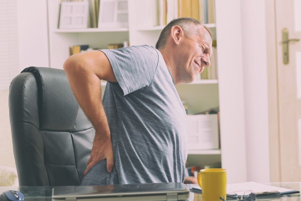
How Spinal Pain Affects A Person’s Neck & Back
Do you feel constant muscle aches and pains in your neck and back? Have you experienced stiffness and limited mobility when you are twisting and turning? Or do heavy objects cause muscle strain when moving from one location to another? Many individuals will be on the move and in weird positions without experiencing pain or discomfort in the spine. This is due to the surrounding muscles and tissues being stretched and the spinal discs taking on the vertical pressure on the spine. However, when environmental factors, traumatic injuries, or natural aging start to affect the spine, it can lead to the development of spinal pain. This is because the outer portion of the spinal disc is intact, while the inner portion is affected. When abnormal stresses reduce water intake within the disc, they can internally stimulate pain receptors without nerve root symptoms. (Zhang et al., 2009) This causes many individuals to deal with neck and back pain in their bodies and reduces their quality of life.
Spinal pain can lead to overlapping risk profiles that cause many individuals to deal with severe low back pain and neck pain, which then causes the surrounding muscles to become weak, tight, and overstretched. At the same time, the surrounding nerve roots are also affected, as the nerve fibers surround the outer and inner parts of the spinal disc, which causes nociceptive pain in the neck and back regions and leads to discogenic pain. (Coppes et al., 1997) When many individuals are dealing with muscle pain related to the spinal discs, it creates a pain-spasm-pain cycle that can affect their bodies due to reduced movement and lead to painful muscle activity when trying to be mobile. (Roland, 1986) When a person has limited mobility cause they are experiencing spinal pain, their natural disc height slowly degenerates, causing more issues to their bodies and socioeconomic burdens. Fortunately, when many individuals are dealing with spinal pain, numerous treatments can reduce spinal pain and restore their disc height.
How Spinal Decompression Reduces Spinal Pain
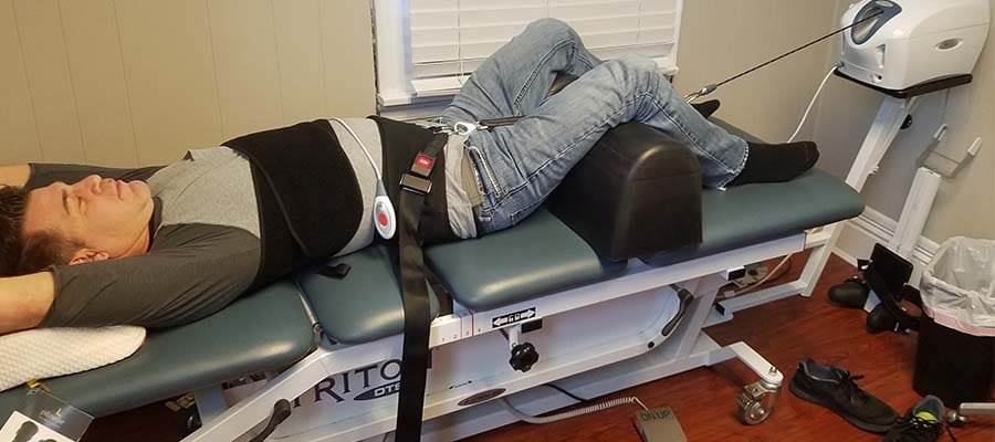
When people are seeking treatments for their spinal pain, many will seek surgical treatments to reduce their pain, but it can be a bit pricey. However, many individuals will opt for non-surgical treatments due to their affordability. Non-surgical treatments are cost-effective and customizable to a person’s pain and discomfort. From chiropractic care to acupuncture, depending on the severity of the person’s pain, many will find the relief they seek. One of the most innovative treatments for reducing spinal pain is spinal decompression. Spinal decompression allows the individual to be strapped to a traction table. This is because it gently pulls on the spine, realigning the spinal disc and reducing pressure to invoke the body’s natural healing process and relieve pain. (Ramos & Martin, 1994) Additionally, when many individuals undergo spinal decompression, the gentle traction provides a motorized distraction to the spine that may induce physical changes in the spinal disc and help restore a person’s range of motion, flexibility, and mobility. (Amjad et al., 2022)
Spinal Decompression Restoring Spinal Disc Height
When a person is strapped into the spinal decompression machine, the gentle traction helps the spinal disc return to its normal position, allowing fluids and nutrients to rehydrate the disc and increase disc height. This is because spinal decompression creates negative pressure on the spine, allowing the spinal disc to return to its original height and providing relief. Plus, the amazing thing about spinal decompression is that it can be combined with physical therapy to help stretch and strengthen the muscles surrounding the spine, providing more stability and flexibility. (Vanti et al., 2023) This allows the individual to be more mindful of their bodies and to start incorporating small habit changes to reduce the likelihood of pain returning. When many people begin to think about their health and wellness by going to treatment, they will regain their quality of life and return back to their daily routine without the issues affecting their spine.
References
Amjad, F., Mohseni-Bandpei, M. A., Gilani, S. A., Ahmad, A., & Hanif, A. (2022). Effects of non-surgical decompression therapy in addition to routine physical therapy on pain, range of motion, endurance, functional disability and quality of life versus routine physical therapy alone in patients with lumbar radiculopathy; a randomized controlled trial. BMC Musculoskelet Disord, 23(1), 255. https://doi.org/10.1186/s12891-022-05196-x
Coppes, M. H., Marani, E., Thomeer, R. T., & Groen, G. J. (1997). Innervation of “painful” lumbar discs. Spine (Phila Pa 1976), 22(20), 2342-2349; discussion 2349-2350. https://doi.org/10.1097/00007632-199710150-00005
Ramos, G., & Martin, W. (1994). Effects of vertebral axial decompression on intradiscal pressure. J Neurosurg, 81(3), 350-353. https://doi.org/10.3171/jns.1994.81.3.0350
Roland, M. O. (1986). A critical review of the evidence for a pain-spasm-pain cycle in spinal disorders. Clin Biomech (Bristol, Avon), 1(2), 102-109. https://doi.org/10.1016/0268-0033(86)90085-9
Vanti, C., Saccardo, K., Panizzolo, A., Turone, L., Guccione, A. A., & Pillastrini, P. (2023). The effects of the addition of mechanical traction to physical therapy on low back pain? A systematic review with meta-analysis. Acta Orthop Traumatol Turc, 57(1), 3-16. https://doi.org/10.5152/j.aott.2023.21323
Zhang, Y. G., Guo, T. M., Guo, X., & Wu, S. X. (2009). Clinical diagnosis for discogenic low back pain. Int J Biol Sci, 5(7), 647-658. https://doi.org/10.7150/ijbs.5.647
Can non-surgical treatments like acupuncture and spinal decompression provide relief to individuals dealing with sciatica?
Introduction
When many individuals start to feel pain running down their legs after a long day of activities, it causes them to have limited mobility and difficulty finding a place to rest. Many people think that they are just dealing with leg pain, but it can be more of an issue as they realize that it’s not just the leg pain they are experiencing but it is sciatica. While this long nerve comes from the lower back and travels down to the legs, it can succumb to pain and discomfort when herniated discs or muscles compress and aggravate the nerve. When this happens, it can impact a person’s mobility and quality of life, thus causing them to seek out treatment to reduce the pain from sciatica. Fortunately, alternative therapies like acupuncture and spinal decompression have been utilized to not only minimize sciatic pain but also provide positive, beneficial results. Today’s article looks at sciatica, how spinal decompression and acupuncture can relieve sciatica, and how integrating these two non-surgical treatments can lead to beneficial results. We talk with certified medical providers who consolidate our patients’ information to assess how sciatica can significantly impact a person’s well-being and quality of life. We also inform and guide patients on how integrating acupuncture therapy and spinal decompression can positively reduce sciatica. We encourage our patients to ask their associated medical providers intricate and important questions about incorporating non-surgical treatments into a wellness routine to relieve sciatica and its referred symptoms. Dr. Jimenez, D.C., includes this information as an academic service. Disclaimer.
Understanding Sciatica
Do you often experience numbness or tingling sensations from your lower back to your legs? Do you feel like your gait is feeling off balance? Or have you stretched your legs after being seated for a while, which provides temporary relief? While the sciatic nerve plays a pivotal role in motor function in the legs, when various factors, such as herniated discs and even pregnancy, start to aggravate the nerve, it can cause pain. Sciatica is a deliberating pain condition often mislabeled as low back pain or radicular leg pain due to these two musculoskeletal conditions. These are comorbidities and can be exacerbated by simple twists and turns. (Davis et al., 2024)
Additionally, when many individuals are doing repetitive motions or dealing with degenerative changes in the spine, the spinal discs are more prone to herniation. They may press on the spinal nerves, causing the neuron signals to invoke pain and discomfort in the lower extremities. (Zhou et al., 2021) At the same time, sciatica can be both spinal and extra-spinal sources in the lumbar spinal region, which causes many individuals to be in constant pain and looking for relief. (Siddiq et al., 2020) When sciatica pain starts to affect a person’s lower extremities, causing mobility issues, many people seek treatments to reduce the pain-like effects of sciatica.
The Science of Motion-Video
Acupuncture For Reducing Sciatica Pain
When it comes to treating sciatica, many people can look into non-surgical treatments due to its affordability and effectiveness in reducing sciatica and its associated pain-like symptoms. Non-surgical treatments can be customized to the individual’s pain and be combined to restore a person’s quality of life. Two non-surgical treatments that can help reduce sciatica are acupuncture and spinal decompression. Acupuncture has a long history of providing significant positive effects on lowering sciatic pain and improving a person’s quality of life. (Yuan et al., 2020) Highly trained professionals from China use acupuncture and incorporate small solid needles to provide instant relief from sciatica’s associated symptoms. This is because acupuncture exerts analgesic effects by regulating microglia activation, inhibiting the body’s natural inflammatory response, and modulating receptors along the pain pathway in the nervous system. (Zhang et al., 2023) To this point, acupuncture can stimulate the body’s acupoints to restore balance.
The Effects Of Acupuncture
One of the effects of acupuncture for relieving sciatica is that it can reduce pain intensity by changing the brain’s activity patterns when the pain receptors are disrupted. (Yu et al., 2022) Additionally, when acupuncturists start to stimulate the nerves in the muscles and tissues, they release endorphins and other neurohumoral factors that help change the pain process in the nervous system. Acupuncture helps reduce inflammation while improving muscle stiffness and joint mobility through increasing microcirculation to reduce swelling while blocking sciatica pain from affecting the lower extremities.
Spinal Decompression For Relieving Sciatica Pain
Another form of non-surgical treatment is spinal decompression, and it can help reduce the effects of sciatica and its associated pain symptoms. Spinal decompression utilizes a traction table to gently stretch the spine to create a negative pressure within the spinal disc and free up the affected nerves. For sciatica individuals, this non-surgical treatment relieves the sciatic nerve as spinal decompression helps reduce the pain intensity and improve mobility function in the lower extremities. (Choi et al., 2022) The main objective of spinal decompression is to create space within the spinal canal and neural structures to release the aggravated sciatic nerve from causing more pain. (Burkhard et al., 2022)
The Effects Of Spinal Decompression
Many individuals can begin to feel relief from incorporating spinal decompression in their wellness treatment. This non-surgical treatment promotes fluids and nutrients to the spinal disc to kick-start the body’s natural healing process. When the spine is being gently stretched, there is less pressure on the sciatic nerves, which can alleviate the pain and improve mobility. Additionally, many individuals will feel their flexibility and mobility back in their lumbar region.
Integrating Acupuncture and Spinal Decompression For Relief
So, when many people start to integrate spinal decompression and acupuncture as a holistic and non-surgical approach for relieving sciatica, the results and benefits are positive. While spinal decompression targets the mechanical healing of the spinal disc and reducing nerve pressure, acupuncture focuses on relieving the pain and reducing inflammation at a systemic level. This enhances the body’s natural healing process and offers a synergistic effect to improve treatment outcomes. Non-surgical treatments like acupuncture and spinal decompression can provide a hopeful outcome for many individuals seeking relief from their sciatic pain without resorting to surgical procedures. These treatments allow the individual to regain their mobility in their lower extremities, reduce pain, and improve their quality of life by making people more mindful of their bodies and reducing the chances of sciatica from returning. By doing so, many individuals can live a healthier and pain-free lifestyle.
References
Burkhard, M. D., Farshad, M., Suter, D., Cornaz, F., Leoty, L., Furnstahl, P., & Spirig, J. M. (2022). Spinal decompression with patient-specific guides. Spine J, 22(7), 1160-1168. https://doi.org/10.1016/j.spinee.2022.01.002
Choi, E., Gil, H. Y., Ju, J., Han, W. K., Nahm, F. S., & Lee, P. B. (2022). Effect of Nonsurgical Spinal Decompression on Intensity of Pain and Herniated Disc Volume in Subacute Lumbar Herniated Disc. International Journal of Clinical Practice, 2022, 6343837. https://doi.org/10.1155/2022/6343837
Siddiq, M. A. B., Clegg, D., Hasan, S. A., & Rasker, J. J. (2020). Extra-spinal sciatica and sciatica mimics: a scoping review. Korean J Pain, 33(4), 305-317. https://doi.org/10.3344/kjp.2020.33.4.305
Yu, F. T., Liu, C. Z., Ni, G. X., Cai, G. W., Liu, Z. S., Zhou, X. Q., Ma, C. Y., Meng, X. L., Tu, J. F., Li, H. W., Yang, J. W., Yan, S. Y., Fu, H. Y., Xu, W. T., Li, J., Xiang, H. C., Sun, T. H., Zhang, B., Li, M. H., . . . Wang, L. Q. (2022). Acupuncture for chronic sciatica: protocol for a multicenter randomised controlled trial. BMJ Open, 12(5), e054566. https://doi.org/10.1136/bmjopen-2021-054566
Yuan, S., Huang, C., Xu, Y., Chen, D., & Chen, L. (2020). Acupuncture for lumbar disc herniation: Protocol for a systematic review and meta-analysis. Medicine (Baltimore), 99(9), e19117. https://doi.org/10.1097/MD.0000000000019117
Zhang, Z., Hu, T., Huang, P., Yang, M., Huang, Z., Xia, Y., Zhang, X., Zhang, X., & Ni, G. (2023). The efficacy and safety of acupuncture therapy for sciatica: A systematic review and meta-analysis of randomized controlled trails. Front Neurosci, 17, 1097830. https://doi.org/10.3389/fnins.2023.1097830
Zhou, J., Mi, J., Peng, Y., Han, H., & Liu, Z. (2021). Causal Associations of Obesity With the Intervertebral Degeneration, Low Back Pain, and Sciatica: A Two-Sample Mendelian Randomization Study. Front Endocrinol (Lausanne), 12, 740200. https://doi.org/10.3389/fendo.2021.740200
IFM's Find A Practitioner tool is the largest referral network in Functional Medicine, created to help patients locate Functional Medicine practitioners anywhere in the world. IFM Certified Practitioners are listed first in the search results, given their extensive education in Functional Medicine