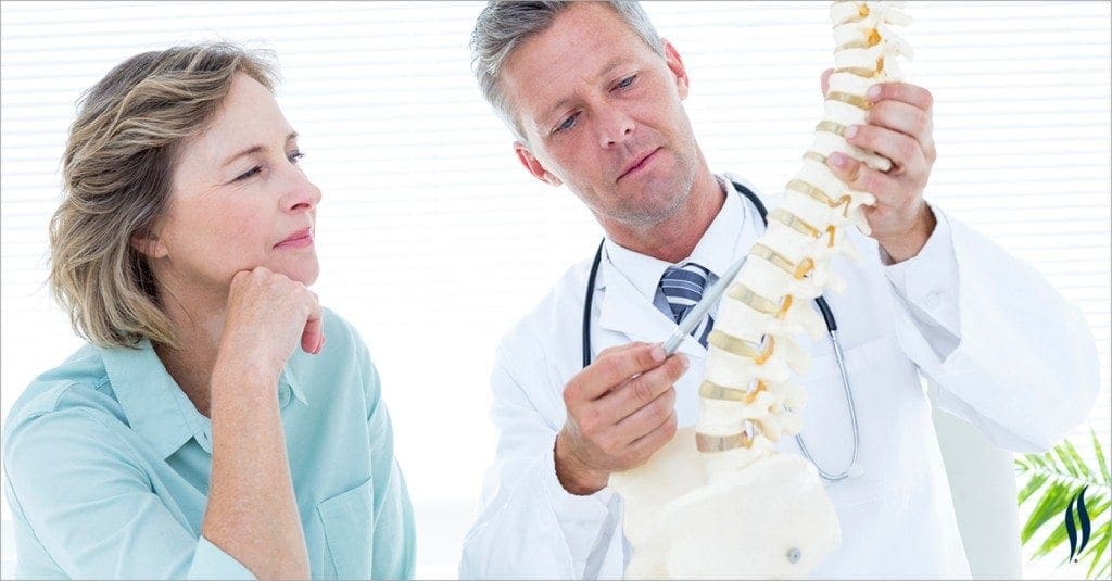
Case Report: The Assessment of Traumatic Cervical Spine Injury and Utilization of Advanced Imaging in a Chiropractic Office.
Abstract: the objective is to explore the standard of care regarding the assessment of cervical spine injuries in a setting of a chiropractic office. Diagnostic studies include physical examination, range of motion studies, orthopedic testing and cervical spine. MRI.
Introduction: On January 30, 2017 a 49 year old female presented in my office to a second opinion examination at the request of her attorney. She had been involved in a rear-end collision on 12/12/2015. (2) She was transported to a local hospital and arrived with complaints of headaches, disorientation, right-sided neck pain and right arm pain. At the hospital emergency department CAT scan was taken of her brain, which proved to be negative. She received prescriptions of muscle relaxers and pain relievers and instructed to visit her primary care physician if her symptoms persisted.
Initial Examination
She consulted a local Chiropractor on December 15, 2015. The initial examination included the following from my review of the doctor�s notes: Presenting complaints were right-sided neck pain that radiates to the right arm. The doctor�s records show a positive cervical compression test and a positive maximum cervical compression test. Both produced pain bilaterally worse on the right. Facet provocation tests were positive for facet disease. Right side radicular pain pattern includes the trapezius and deltoid. No x-ray studies were included in the doctor�s orders. The patient received 23 chiropractic treatments from 12/15/2015 through 4/5/2016 for a diagnosis of cervical sprain/strain. The treatments consisted of spinal manipulation and a variety of soft tissue therapies.
Around January 15, 2017 I received a phone call from a local attorney regarding this patient and asking if I would do a second opinion examination on her due to persistent neck pain and right upper extremity pain. The patient presented on January 30, 2017 for my evaluation. My clinical findings are as follows:
Vitals: Age 49, weight 170 lbs. height 5� 8�, B.P 126/82, pulse 64, Resp. 16/min.
Appearance: in pain
Orthopedic/Range of motion: All cervical compression tests produced pain with radiation bilaterally worse on the right. Range of motion studies revealed: 40 degrees of left rotation and 32 degrees of right rotation with radiating pain produced by both motions.
Palpation: cervical spine palpation produced centralized spine pain that radiates to the right shoulder with numbness in the right arm and hand.
The patient informed me during the examination that her pain made it difficult to sleep through the night. If she was on her right side her right arm and hand would go numb immediately. A big part of this patient�s life was riding and caring for her horse and she could not do either because it resulted in severe neck and arm pain.
My recommendation to her and her attorney was to obtain a cervical spine MRI with a 1.5 Tesla machine due to the high quality images it can produce. MRI is a highly sensitive tool to evaluation of neurologic tissue including the spinal cord and nerve roots. (1) I bypassed the x-ray at this time due to the clinical presentation and 12% of spinal cord with injuries having no radiographic abnormality. (3)
Imaging
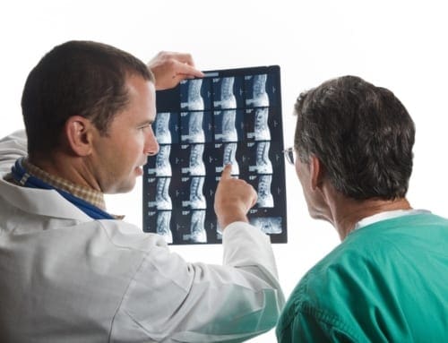
Figure 1: T2 Sagittal Cervical Spine MRI
Fig 2: T2 Axial Cervical Spine with Scout line through C3/4.
Radiology Report: The report and the images demonstrated a right paracentral disc extrusion measuring 9 mm and extending 8 mm cranial/caudal causing abutment of the spinal cord. (Fig 1)(2) Additionally the diameter of the central canal was reduced to 8.1mm and projected into the right lateral recess resulting in severe stenosis of the right neural canal. (Fig 2) Additional findings not pictured: C4/5 demonstrated a 2.5 mm bulging disc with facet hypertrophy with moderate stenosis of the left neural canal and severe stenosis of the right neural canal. C5/6 demonstrated a 1.5 mm posterior subluxation narrowing the central canal to 9.1 mm with unconvertebral joint hypertrophy resulting in moderate right and severe left neural canal stenosis. C6/7 revealed a broad based disc herniation worse on the left measuring 3.6 mm resulting in severe neural canal stenosis bilaterally complicated by unconvertebral joint hypertrophy. The MRI findings correlate with the patient�s clinical presentation. (4)
Discussion: When the patient returned to a consultation on the MRI findings my recommendation was to consult a neurosurgeon. (3) Her attorney asked me if the treating doctor acted incompetently. My only response was that I would have ordered the MRI immediately before treating the patient with manual manipulation. The case is likely to go to trial and there is a good chance that I will be called in as an expert witness. It is almost a guarantee that the defense attorney will ask me if I would have treated the patient for such a long period of time without an MRI or whether the treating doctor could have made the problem worse. The failure to accurately determine a diagnosis may result in malpractice action or a board hearing or both for this treating doctor and I would have ordered the MRI immediately considering the radicular findings and symptoms. After any myelopathic or significant radiculopathic symptoms a referral of advanced imaging needs to be performed in order to conclude and accurate diagnosis, prognosis and treatment plan prior to rendering care. Diagnostic appropriateness in the case of traumatic injury or with any etiology with neurologic symptoms or findings necessitates following triage protocols. In this case, an immediate 2-3mm MRI of the cervical spine is clinically indicated and proved integral to the safe care of this patient.
The scope of our information is limited to chiropractic and spinal injuries and conditions. To discuss options on the subject matter, please feel free to ask Dr. Jimenez or contact us at 915-850-0900 .
References:
Haris, A.M., Vasu, C., Kanthila, M., Ravichandra, G., Acharya, K. D., & Hussain, M. M. 2016. Assessment of MRI as a modality for evaluation of soft tissue injuries of the spine as compared to intraoperative assessment. Journal of Clinical and Diagnostic Research, 10(3), TC01-TC05
Schneider RC, Cherry G, Pantek H. The syndrome of acute central cervical spinal cord injury, with special reference to the mechanisms involved in hyperextension injuries of cervical spine. J Neurosurg 1954; 11: 546�577.
Tewari MK, Gifti DS, Singh P, Khosla VK, Mathuriya SN, Gupta SK et al. Diagnosis and prognostication of adult spinal cord injury without radiographic abnormality using magnetic resonance imaging: analysis of 40 patients. Surg Neurol 2005; 63: 204�209.
Miyanji F, Furian J, Aarabi B, Arnold PM, Fehlings MG. Acute cervical traumatic spinal cord injury: MR imaging Findings correlated with neurologic outcome-prospective study with 100 consecutive patients. Radiology 2007; 243: 820�827.
Additional Topics: Recovering from Auto Injuries
After being involved in an automobile accident, many victims frequently report neck or back pain due to damage, injury or aggravated conditions resulting from the incident. There’s a variety of treatments available to treat some of the most common auto injuries, including alternative treatment options. Conservative care, for instance, is a treatment approach which doesn’t involve surgical interventions. Chiropractic care is a safe and effective treatment options which focuses on naturally restoring the original dignity of the spine after an individual suffered an automobile accident injury.
Title: Conservative care and axial distraction therapy for the management of cervical and lumbar disc herniations and ligament laxity post motor vehicle collision.
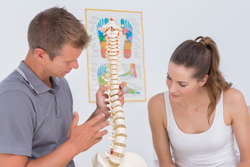
Dr. Alex Jimenez, doctor of chiropractic, focuses on the diagnosis, treatment and prevention of a variety of injuries and conditions associated with the musculoskeletal and nervous systems, utilizing several chiropractic methods and techniques. The following procedures may be similar to his own but can differ according to the specific issue and complications by which the individual is diagnosed.
Abstract: This middle-aged female was injured in a vehicle collision causing her to sustain disc and additional ligament injuries in the cervical and lumbar spine. Diagnostic studies included physical examination, orthopedic and neurological testing, lumbar MRI, multiple cervical MRI�s, CRMA with motion cervical radiographs and EMG studies. Typically, conservative care is initiated prior to interventional procedures, and this case study seeks to explore the usage of passive therapy for mechanical spine pain and noted anatomic disc lesions after failure of interventional procedures. She reported both short term and long term success regarding pain reduction along with improvement in her activities of daily living after initiating conservative care, and continued to report further reductions in pain with periodic pain management using conservative care.
Introduction: The 49-year-old married female (Spanish speaking patient) reported that on March 4th, 2014 she was the seat-belted driver of a truck that was struck by a much larger fuel truck changing lines, hitting her vehicle at the front passenger side (far side, side impact). The force of the impact caused her truck to be lifted up and the right wheel popped off. Her head hit the window after impact and the spinal pain and complaints started approximately 24 hours later. Two days after the crash she went to the emergency department. Occupant pictures were taken describing an out of position occupant injury. She did not report any additional significant trauma after the collision.
Initial Diagnosis and Treatment for Disc Herniations
Prior to her evaluation at our clinic, she utilized multiple providers for diagnosis and treatment over the course of 11 months. She went to the emergency department, utilized 3 pain management medical doctors, neuropsychologist and a cognitive rehabilitation therapist. Imaging included radiographs and MRI of the right shoulder revealing rotator cuff tear; radiographs of the lumbar and thoracic spine, and left hand; CT of the head and cervical spine were performed; MRI cervical (3) and lumbar spine. Medications prescribed included Fentanyl, Percocet, Naprosyn, Cyclobenzaprine, Norco, Hydrocodone-acetaminophen, Soma, and Carisoprodol. Physical therapy was provided for spinal injuries and she did not respond to treatment. The neurosurgeon recommended epidural steroid injections and facet blocks. Cervical nerve blocks and cervical trigger point injections, cervical and lumbar epidural steroid injections (ESI), lateral epicondyle steroid injections were performed, none of which were palliative. Post-concussion disorder and PTSD with major depressive disorder were diagnosed.
On February 12th, 2015, she presented to our office with neck pain (average 6/10 VAS) that affected her vision, with paresthesia�s in both upper extremities radiating to the hands with numbness. She had low back pain (average 6/10 VAS), and she additionally reported paresthesia at the plantar surface of feet bilaterally. She had left elbow pain, right shoulder pain, knee pain, headaches and �anxiety� along with anterior sternal pain.
Her injuries were causing significant problems with her activities of daily living. Summarily she had increased pain with lifting, increased pain and restricted movement with bending, walking and carrying. She had been unable to perform any significant physical activity from the time of the crash in March 2014 until March 2015. Her right hand was always hurting and her forearms. She was not able to clean windows or do laundry, difficulty using stairs, problems with mopping, ironing and cleaning. She had to limit her walking and jogging primarily due to neck pain and right arm pain. She was not able to sit for long periods of time and sleeping was disrupted due to numbness in her hands. She was only able to walk on a treadmill for 10 minutes before having to stop due to pain, prior to the crash she would exercise for an hour.
Prior History: No significant prior musculoskeletal or contributory medical history was reported.
Research Study Conclusions
Clinical Findings (2/12/15): She had a height of 5�2�, measured weight of 127 lbs.
Visual analysis of the cervical spine revealed pain in multiple ranges of motion including flexion, extension, bilateral rotation and bilateral side bending. On extension pain was noted in the upper back, on rotation pain was noted in the posterior neck, and on lateral flexion pain was noted contralaterally.
Visual analysis of the lumbar spine revealed pain in the low back on all active ranges of motion, including flexion, extension and side bending, pain primarily at L5/S1.
Dual inclinometer testing was ordered based on visual active range of motion limitations with pain.
Sensory testing was performed of the extremities, C5-T1 and L4-S1. No neurological deficits other than right sided C5 hypoesthesia.
Foraminal compression test produced pain in the cervical spine. Foraminal distraction test caused an increase in pain in the neck. Jackson�s test on the right produced pain bilaterally in the neck. Straight leg raise bilaterally produced low back pain, double Straight leg raise produce pain at L5/S1 at 30 degrees.
Muscle testing of the upper extremities was tested at a 5/5 with the exception of deltoid bilaterally tested at a 4/5. The patient�s deep tendon reflexes of the upper and lower extremities were tested including Triceps, Biceps, Brachioradialis, Patella, Achilles: all were tested at 2+ bilaterally, equal and reactive. No evidence of clonus of the feet and Hoffman�s test was unremarkable.
C3-C5 right sided segmental dysfunction was noted on palpation. T5-T12 spinous process tenderness on palpation. Low back pain on palpation, particularly L5/S1.
Imaging Results
MRI Studies:
I reviewed the cervical MRI images taken May 2014 with the following conclusions (images attached):
Dramatic reversal of the normal cervical curvature, apex C5/6.
C5/6 herniation, indentation of the spinal cord anteriorly. High signal posterior on STIR.
Due to the angular kyphosis of the cervical spine and axial slices performed, C6/7 slices did not render a pure diagnostic image for disc disruption.
Fig. 1 (A) T2 Axial C5/6, 2 months post injury Fig. 1 (B) Sag T2 C5/6
I reviewed cervical MRI images taken September 17th, 2014 approximately 6-months post injury, and rendered the following conclusions:
Reversal of the normal cervical lordosis.
C5/C6 herniation (extrusion type) with indentation of spinal cord, appropriate CSF noted posteriorly.
I reviewed the cervical MRI dated October 24th, 2015 (images attached):
C4/5 herniation, extrusion type, left oriented into the lateral recess and neural canal causing moderate neural canal stenosis
Fig. 2 (A) 3D Axial C4/5, 19 months post injury Fig. 2 (B) Sag T2 C4/5
IMPRESSIONS: C4/5 herniation noted on 10/24/15 was not noted on prior images. The patient reported no additional injury or symptoms between MRI studies, so it is postulated that initial slices revealed a false negative; or due to the severity of abnormal cervical biomechanics, it is possible that the C4/5 disc herniated between the pre/post MRI�s with no significant increase in symptomatology. There was improvement at C5/6 related to disc abnormality and cord involvement (see below).
Fig. 3 (A) 3D Axial C5/6, 19 months post injuryFig. 3 (B) Sag T2 C5/6, 19 months post injury
The cervical flexion/extension images were digitized February 2016 and interpreted by myself and Robert Peyster MD, CAQ Neuroradiology, revealing a loss of Angular Motion Segment Integrity at intersegment C6/C7 measured at 19.7 degrees (maximum allowed 11 degrees), indicating a 25% whole person impairment according to the AMA Evaluation of Permanent Impairment Guidelines 5th edition1. CRMA provided from Spine Metrics, independent analysis.
Evidence of significant ligament injury causing functional subfailure was measured at C3/4 at 10.4 degrees and at C4/5 measuring 10.9 degrees regarding angular motion. Abnormal paradoxical translation motion measured at C6/7 and C7/T1.
Functional Testing:
EMG of the upper extremity revealed bilateral C6 radiculopathy, December 16th, 2015.
Range of Motion Cervical Dual Inclinometry:
Initial Max 4 months later % Improvement
Cervical Extension 44 42 -5%
Flexion 40 62 55%
Cervical Left 25 41 64%
Lateral flexion Right 12 26 117%
Cervical Left 46 59 28%
Rotation Right 43 73 70%
Conservative treatment rendered: A neurosurgical referral was made for assessment and surgical options. Conservative care was initiated despite failure of other medical procedures since there is �further evidence that chiropractic is an effective treatment for chronic whiplash symptoms�2-3. The patient was placed on an initial care plan of 2-3x/week for 5 months, with a gap in passive care for 1 month.
23 chiropractic visits. Instrument adjusting cervical spine was utilized with Arthrostim. Non-rotatory HVLA (high velocity low amplitude) spinal adjustments were performed thoracic and lumbar spine, applied A-P. No HVLA spinal adjustments to the cervical spine.
Prior to being placed at maximum medical improvement she had persistent low back symptoms, continued tingling in the fingertips and occasional neck pain at a 4/10, with her upper extremity paresthesia�s improved 50%. She continued with pain management chiropractic care after MMI, approximately 1 visit every 3-4 weeks with axial distraction to the cervical and lumbar spine, chiropractic adjustments as needed (PRN). 2 years/9 months post collision, and 1 year/9 months after initiating conservative care at our clinic, she reports only slight (1-2/10 VAS) spinal complaints with her primary concern being a torn rotator cuff injury from the crash that still requires surgical intervention. After initiating care at our clinic, no other interventional procedures were performed, although medication usage persisted. Due to improvement in symptoms and functional status, spinal surgery was not considered. She still utilizes Aleve PRN, 1-2 tablets. No significant active spinal rehabilitation was utilized. The patient was given at home active care consisting only of cervical and lumbar stretches, walking, and ice to affected areas.
Conclusion:While chiropractic care is safe even in the presence of herniations and radicular symptoms, �the likelihood of injury due to manipulation may be elevated in pathologically weakened tissues�4. Due to cord involvement, the provider decided to utilize low force procedures although HVLA spinal adjustments to the cervical spine could be considered safe due to lack of cord compression. HVLA spinal adjustments A-P were utilized in the lumbar and thoracic spine not only for short term pain relief but also as part of managing the chronic low back pain secondary to ligament/disc damage. While previously theorized to be only episodic, low back pain can be a lifelong condition requiring patients to seek ongoing care5. This care can be active, passive, pharmaceutical, interventional, or conservative in nature, but ongoing pain management therapy is often required for permanent ligament conditions. There is clear benefit to the patient population to be able to avoid surgical intervention due to risks, costs, ongoing prescription medication usage and adjacent level degeneration in the future6. Avoiding opioid usage is also a high priority in today�s environment.
Long term conservative care utilizing instrument spinal adjusting and targeted axial distraction therapy significantly reduced subjective reporting of pain, increased activities of daily living, and allowed the patient to avoid further spinal injections or surgical intervention. Considering that various interventional procedures failed prior to conservative care, it is important that providers work in an interdisciplinary environment such that the safest, and in this case the most effective, therapies are utilized first to reduce risk to the patient and maximize benefit and reduce costs.
In this case study, the patient utilized multiple pain management physicians, cervical nerve blocks and epidural steroid injections, and was not directed to conservative care for 11 months post injury. Utilizing chiropractic as conservative care would have enabled this patient to regain function and decrease pain while reducing costs and risks that are associated with medications and interventional procedures.
Competing Interest: There are no competing interests in the writing of this case report.
De-Identification: All of the patient�s data has been removed from this case.
The scope of our information is limited to chiropractic and spinal injuries and conditions. To discuss options on the subject matter, please feel free to ask Dr. Jimenez or contact us at 915-850-0900 .
Cocchiarella L., Anderson G. Guides to the Evaluation of Permanent Impairment, 5th Edition, Chicago IL, 2001 AMA Press.
Khan S, Cook J, Gargan M, Bannister G. A symptomatic classification of whiplash injury and the implications for treatment. Journal of Orthopaedic Medicine 1999; 21(1):22-25.
Whedon J, Mackenzie T, Phillips R, Lurie J. Risk of traumatic injury associated with chiropractic spinal manipulation in Medicare Part B beneficiaries aged 66-99 years. Spine, 2015; 40:264�270.
Hestbaek L, Munck A, Hartvigsen L, Jarbol DE, Sondergaard J, Kongsted A: Low back pain in primary care: a description of 1250 patients with low back pain in Danish general and chiropractic practices. Int J Family Med, 2014.
Faldini C., Leonetti D., Nanni M. et al: Cervical disc herniation and cervical spondylosis surgically treated by Cloward procedure: a 10-year-minimum follow-up study. Journal of Orthopaedics and Traumatology, June 2010.Volume 11, Issue 2,pp 99-103.
Additional Topics: Recovering from Auto Injuries
After being involved in an automobile accident, many victims frequently report neck or back pain due to damage, injury or aggravated conditions resulting from the incident. There’s a variety of treatments available to treat some of the most common auto injuries, including alternative treatment options. Conservative care, for instance, is a treatment approach which doesn’t involve surgical interventions. Chiropractic care is a safe and effective treatment options which focuses on naturally restoring the original dignity of the spine after an individual suffered an automobile accident injury.
Leg length discrepancy is a condition in which the legs are not of equal length. This might give an appearance that one leg is shorter compared to the other. The reasons for leg length discrepancy can be many, including defects that are congenital or may be acquired, which might include certain medical conditions, fractures, infections or injuries impacting the bone.
Leg length discrepancy might be a result of accurate discrepancy, which can be caused by real distinctions in the leg lengths. In other instances, the causes of leg length discrepancy might be due to circumstances that result in change in the angle of the hip or pelvic bone. In such cases, as the hip gets tilted to the other side and one side gets raised, the leg on that side seems to be shorter.
However, it is important to understand the foundation and causes of leg length discrepancy to handle the condition properly. It is also crucial to understand the impact of leg length discrepancy on an individual health and overall performance just as the the reasons are important. Mental and physical health can be affected by leg length discrepancy health insurance and will also be connected to spinal issues like scoliosis.
Can Limb Length Discrepancy Trigger Scoliosis?
Leg length discrepancy, due to uneven leg lengths, can impact the normal gait of the person. The main perform that is noticeable is the way a person walks or performs human anatomy actions. These can get afflicted or be difficult because of leg size discrepancy. Changes in normal movements can more lead to certain issues of the muscles like soreness, discomfort, weak imbalances or muscles on either side of the physique. Leg duration discrepancy can impact the hip, knees and ankle, can cause pain and dysfunction.
The muscles on both sides of the physique and those related to the hip can get pulled due to tilting of the hip-bone. This can be one the major effects of leg duration discrepancy, where the muscles get pulled to one side, creating changes in the curvature of the backbone. In to side ways pulling of the spinal curvature, which is termed as scoliosis, such adjustments can eventually result. There is much concern whether leg length discrepancy can cause scoliosis and it is important to understand correct therapy to be planned by this and a void further complications.
Limb Length Discrepancy and Scoliosis
Many studies have already been conducted, which revolve round the chance of leg duration discrepancy being an underlying cause of scoliosis. In the same time, leg length discrepancy can also result in pulling of the muscles that are back to one facet, which can contribute to some extent to or worsen existing scoliosis.
It might result in scoliosis, which might be useful in the beginning as the curvature gets tilted to one aspect. In scoliosis that is functional there might be slight tilting or pulling of the muscles to one side, without adjustments or damage to the structure of the spine. However, if functional scoliosis, which is caused or aggravated by leg-length discrepancy isn’t treated in time, it might worsen, causing changes in the structure of the curvature. This may result in structural scoliosis, which may not be disturbing and only more painful but also difficult to manage.
Some studies have revealed that scoliosis in certain persons is the result of mechanism, to make up for the leg length discrepancy. Simply stated, in leg length discrepancy, the legs are of unequal lengths, so to match the lengths the individual pulls the aspect down along with the hip starts to tilt. This, when continued for a longer period of time, can result in pulling to one aspect, making changes in the curvature. Scoliosis is one such change in spinal curvature, at which spine gets curved to one side, comprising alternative activities.
Symptoms of Scoliosis from Limb Length Discrepancy
A person that has developed scoliosis due to leg size discrepancy, usually presents with tilting of the hip. Along with the signs of leg length discrepancy, the individual may possibly also encounter pain in the muscles that are again, imbalances of muscle power and function of the muscles that are again. Bending, twisting movements might be difficult and it could also be painful to maintain or raise objects.
The appearance of the shoulders may possibly be different on account of scoliosis and one-shoulder can happen elevated in relation to the other. This could cause problems in neck, arm and shoulder movements and also hurt. It could sometimes result into serious degrees of scoliosis, if the status is left unattended.
Treatment of Scoliosis from Limb Length Discrepancy
It is importance to comprehend if leg-length discrepancy can trigger scoliosis. The treatment options might have to be planned appropriately if scoliosis has been resulted in by complications of leg-length discrepancy.
In some cases, leg size discrepancy can contribute to or worsen existing scoliosis, therefore, correcting leg duration discrepancy with heel raise have to be in the offing cautiously. It’s important to thoroughly examine any circumstance with leg-length discrepancy, as they can cause scoliosis in some instances. Prescribing a heel raise to appropriate leg length discrepancy can boost the chances of worsening the scoliosis due to tilting if scoliosis is obvious.
Hence, it really is essential to to examine the bio mechanics of the hi-P, evaluate the modifications in the spinal curvature in scoliosis as well as the tilting due to leg duration discrepancy. Depending on the the reasons, some cases of leg length discrepancy might require procedure for surgical correction of leg lengths. When the symptoms, scoliosis and causes like complications of leg length discrepancy, are correctly evaluated a multi disciplinary treatment approach may be planned.
Limb Length Discrepancy Explained (Video)
The scope of our information is limited to chiropractic and spinal injuries and conditions. To discuss options on the subject matter, please feel free to ask Dr. Jimenez or contact us at 915-850-0900 .
By Dr. Alex Jimenez
Additional Topics: Scoliosis Pain and Chiropractic
According to recent research studies, chiropractic care and exercise can substantially help correct scoliosis. Scoliosis is a well-known type of spinal misalignment, or subluxation, characterized by the abnormal, lateral curvature of the spine. While there are two different types of scoliosis, chiropractic treatment techniques, including spinal adjustments and manual manipulations, are safe and effective alternative treatment measures which have been demonstrated to help correct the curve of the spine, restoring the original function of the spine.
Early onset scoliosis (EOS) is an abnormal sideways curvature of the spine found in children under the age of 10 years.
More than 100,000 kids are diagnosed with scoliosis each year in the USA and most have adolescent idiopathic scoliosis, or AIS. AIS is one of the most common types of scoliosis and it can affect kids between the ages of 10 to 18. EOS is significantly rarer and often more complex in character.
Types of Early Onset Scoliosis
Doctors have recognized several types of EOS. Most types of EOS have an obvious trigger and are associated with individual health issues. On the other hand, a general number of EOS cases are idiopathic, meaning they have no recognized cause and are identified based on the age at diagnosis.
Below are kinds of EOS:
Congenital scoliosis occurs when the bones of the spine do not form properly in the mother�s womb.
Neuromuscular scoliosis is caused by brain, spinal cord, or muscular system disorders (such as muscular dystrophy). These disorders prevent the back muscles from holding the spine straight.
Syndromicscoliosis develops as part of an underlying syndrome or disorder that affects numerous parts of the body (such as Prader-Willi
Syndrome; a rare disease affecting development).
Infantile idiopathic scoliosis is diagnosed in children ages birth to 3 years. It has no known cause.
Juvenile idiopathic scoliosis is diagnosed in children ages 4 to 10. It has no known cause.
Early Onset Scoliosis Symptoms
EOS can be difficult to identify, as some children don’t have a serious spinal curve and might not have pain that stops them from their typical exercise. The primary factor to keep in mind, however, is symmetry, as it could reveal an issue when all other indications point to a regular spine.
Below are the most frequent indicators of EOS:
The body appears to lean to one side
Shoulders look uneven, with one shoulder blade sticking out more
Waistline is uneven
Hip height appears off balance
Ribs protrude on one side more
Early Onset Scoliosis Diagnosis
Your child’s pediatrician, pediatric orthopedist, or spinal specialist can identify EOS utilizing a number of methods.
Physical exams including the Adam’s forward bend test, will expose a prominence, hump or deviation of the backbone, or spine, indicating an irregular curvature. But, it’ imaging scans, namely x-rays, that doctors count on most to validate EOS.
The doctor will simply take standing x-rays of your child’s spine to properly see the entire nature of the scoliosis. Typically, one x-ray is taken from back to front (called a posterior-anterior x-ray) and the second is from the side (called lateral x-ray).�Other x-rays may possibly contain bending from aspect-to-facet.
Your doctor may possibly also request a magnetic resonance imaging (MRI) test in order to rule out underlying involvement of the spinal-cord along with other buildings or CT scan to show 3 D views of the bone constructions.
Because x-rays are used throughout the monitoring process throughout therapy, and to identify scoliosis, individuals have raised concerns over radiation. With this consideration in mind, doctors limit the number of x-rays that a child may use direct shields to safeguard breast and thyroid tissue and wants, lower dose x-rays, as well as light-based scans of the physique form.
Early Onset Scoliosis Treatment
There are four general approaches for managing EOS:
Observation
Spinal bracing
Body casting
Spine surgery
Observation
Your physician may suggest an observation period prior to any active treatment is warranted, as some times the scoliosis even correct itself as your child grows especially with very little curves in really young kids and will stabilize. This generally indicates attending normal follow up appointments together with your doctor throughout the year to determine any adjustments in your child’s curve.
Spinal Bracing
Spinal bracing is a typical nonsurgical treatment for EOS. Your physician works with an orthotist to craft a custom spinal brace for your child. The objective of the brace is not necessarily to correct the scoliosis but to avoid the curve from progressing.
Body Casting
Body casting may be advised for kids between SIX MONTHS months and 6 years of age who have curves likely to to succeed. Body casts are custom made and placed while your child is asleep under general anesthesia. Casts can be in spot for up to 12 months, so that your child will require a sequence of casts throughout therapy. A cast may possibly be employed for more severe curves or in cases in which a brace fails to prevent the curve from getting worse. Often the forged is used to delay the need for spine surgery that is ideally performed after much of your child’s growth is complete. A brace is often used for the same purpose.
Spine Surgery
If your child has a severe curve of 50-levels or higher, spine surgery is considered but usually delayed before the curvature is significantly greater and the child is bigger and h-AS finished more development.
There are various surgical methods for EOS, including expanding rod surgery, VEPTR® (vertical expandable prosthetic titanium rib), vertebral physique tethering, growth guided gadgets, and spinal fusion.
Recovery Potential for Early Onset Scoliosis
It could be scary for each of you when your youngster is identified with early onset scoliosis. The remedies obtainable today are highly-successful at managing or even correcting the curve. Your encouragement and support along with the determination of your pediatric spine specialist will help your child respond well to treatment, and lead a pleased and full life.
Identifying Scoliosis in Children (Video)
The scope of our information is limited to chiropractic and spinal injuries and conditions. To discuss options on the subject matter, please feel free to ask Dr. Jimenez or contact us at 915-850-0900 .�
By Dr. Alex Jimenez
Additional Topics: Scoliosis Pain and Chiropractic
According to recent research studies, chiropractic care and exercise can substantially help correct scoliosis. Scoliosis is a well-known type of spinal misalignment, or subluxation, characterized by the abnormal, lateral curvature of the spine. While there are two different types of scoliosis, chiropractic treatment techniques, including spinal adjustments and manual manipulations, are safe and effective alternative treatment measures which have been demonstrated to help correct the curve of the spine, restoring the original function of the spine.
Be honest, you don’t know how your car works, do you? And despite spending most of the working day lashed to a QWERTY, if someone asked you how update their modem, you wouldn’t where to start (or what the modem even is).
And that’s fine. Other people do that stuff so you don’t have to. But the same can’t be said for your workout. You need to be okay with the specifics – do you honestly know what that dead lift is doing to your muscles? Or more importantly, the damage you could be doing to yourself if you’re getting it wrong.
Thankfully, experts are on hand. We’ve enlisted the help of Tim Walker, founder London’s Evolve Fitness to settle the form debate on five key exercises, once and for all.
First up, a pre-lift check list.
Breathing. Oxygen creates energy in the muscles, so don’t hold your breath.
Technical understanding. Understand which muscles you are about to engage, know the movement you’re about to make, and be deliberate with that movement.
Mental participation. Make sure you’re in the moment, and don’t think about what’s next. Connect your mind to your muscles, and aim for a full range of motion.
Load selection. Challenge yourself, but be realistic, your body will thank you in the long run. Go too heavy and you’ll fail to get a range of motion, too light and you won’t stimulate the muscle enough force growth.
1.Bicep Curls
The most common mistake: “Leaning back during the curl and bringing your elbows forward (rather than keeping them at your side).”
The damage it might be doing: You can incur bicep tendon injuries (tears, impingements and dislocations etc.) but the main reason you need to get your form right is so that the exercise actually has an effect. “Leaning too far backwards means that you’re not putting enough pressure on the bicep – you’re using your weight as momentum during the curl, rather than lifting only with the bicep muscles. And by lifting your elbows forwards, you’re shifting the focus of the exercise away from the bicep (you’ll be lifting with your shoulders and using the momentum from your body again), thus you won’t get the development you want.
Most Popular
How to fix it: “Focus on holding your posture more tightly; pull your shoulder blades back and down, and lift your chest up, lean forward slightly and keep your weight in your heels. Contract your abs at all times, too. To keep your elbow position, focus on keeping your elbows in line with your ears, and be forceful with that contraction in your abs when pulling the weight up.”
2. Bench Press
The most common mistake: “Elbow position. Most people have their elbows in line with their shoulders. It’s hampering your progress because it doesn’t target the chest. You’re looking for synergistic movement in the chest, shoulders and triceps.”
The damage it might be doing: The most common injuries are a Glenoid Labrum tear (front of upper arm), rotator cuff tears and shoulder impingement syndrome. Bench pressing is the kind of exercise that you want to keep increasing in weight, because the feeling of nailing that new three-rep max is unbeatable. But it only takes one lift with poor form for something to go wrong, so always think ‘form first, weight second’.
How you should be doing it: “I often ask my clients to lower their arms 20/25 degrees, so they are just above the nipple, and I always find it useful to keep my knuckles pointing to the ceiling, and my wrists straight.”
3. Deadlift
The most common mistake: “Rounding of the back, rather than keeping a natural arch.”
The damage it might be doing: “A slipped disc in the lower back is the main danger here.” You can also incur sprains and strains (different things), but if there’s any sharp pain at any point, you should stop.
How to fix it: “Try locking the upper body posture by keeping the chest high and arms long (aka fully extended, not bent). Keep your weight into your heels (make sure they don’t leave the ground, and you’re not feeling your full weight in your toes) concentrate on pressing through the legs and keep your core area strong by engaging your stomach muscles.”
4. Squat
The most common mistake: “For squats, there are several: bending forward too much, not squatting deep enough and allowing the knees to turn inwards.”
The damage it might be doing: “That mistake is damaging your body/hampering your progress because� Bending forward too much will put too much pressure on your back, and lead to the same kind of damage as an incorrect deadlift. If you’re not going deep enough you won’t be engaging the hamstrings and glutes as much as you could; if you’re aiming to build the muscles and boost metabolism you’ll be missing the mark. If you allow the knees to turn inwards you’re risking damage to the ligaments such as ACL.”
How to fix it: “For bending forward; this is commonly due to a general tightness in the chest and lats (latissimus dorsi muscles) and/or hip flexors, which is very common among office workers who spend a lot of time sitting. Fix it by stretching these muscles more regularly. For those not going deep enough, you need to man-up and understand the principles if fight-or-flight. Most people fear that when they go down deeper they won’t get back up, but you need to attack the movement with confidence and good technique. The worst that can happen is that the safety catches will stop the bar and you crawl out. For the knees, the best thing is to engage your brain. Think about what you are doing and what your knees are doing, you want your them to be in line with your second and third toes at all times.”
5. Single Arm Rows
The most common mistake: Rounding of the back, rotating too much as you pull the weight, and failing to achieve a full range of motion, i.e. not pulling the weight all the way into the body.
The damage it might be doing: “Rounding the back isn’t particularly dangerous, but it’ll prevent the most optimal development of your back. Over rotation when pulling the weight will mean you’re not working the back muscles as well as you could be, hampering your strength development. The same goes for not having a full range of motion; if you’re not pulling the weight all the way into your body, you’re not getting a full contraction of the muscles, which means you won’t be adequately stimulating them.”
How to fix it: “Stick your butt out and check your position in a mirror – your upper back should be flat, with a gentle/natural arch in your lower back. For over rotation, by more rigid in both your thinking and your positioning. When you hold the position more forcefully you will engage your abs and obliques better. This is one of my favourite back exercises – when done properly – it works and engages your core as well as the back.”
Tim Walker is the founder of Evolve Fitness,13-15 Bouverie Street, London, EC4Y 8DP
Scoliosis is an intricate illness. Experts nevertheless don’t know what causes 80 percent of scoliosis cases, and there’s no absolute cure. But nevertheless, there’s hope!
You can find proven techniques to handle scoliosis and lessen its symptoms. X-rays allow doctors to measure the unique, three-dimensional curve of each person’s backbone as a way to find out the best method of therapy. Chiropractic treatment for scoliosis involves normal adjustments, using the hands or a gadget. The aim will be to realign joints, bones and the muscles. There are two types to choose from: traditional and scoliosis specific.
Chiropractic Care for Scoliosis
Traditional treatment applies a common method, comparable to what the chiropractor would do for any other patient experiencing back complications. However, not all chiropractic doctors are qualified or experienced to treat scoliosis nor are they familiar with its intricacies, then, traditional chiropractic treatment is unlikely to have much of an influence on the Cobb angle. This approach is only recommended for patients within the age of 13 with very small Cobb angles of 20 degrees or less. Traditional care could be helpful for relieving discomfort but not for bodily straightening the Cobb angle in patients.
Aiming to mobilize the spine and straighten the curve, traditional chiropractors might press down on the spine and ribcage while the patient lies on their abdomen. However, the irregular curve of the spine occasionally develops pressure from the nerves. This stress may not be relieved by pushing down on the spine; instead, the nerves are further aggravated by it. The spine isn’t stuck, as it’s with most other issues, but rather it curves in the incorrect direction. You can’t mobilize a scoliotic backbone without also stabilizing and correcting it.
Chiropractic Methods and Techniques for Scoliosis
Chiropractic treatment for scoliosis goes outside of the traditional guidelines to stabilize the curve. Aiming to gradually correct the spine into a a classic curve, changes are precise and gentle. This technique can aid people who’ve currently had surgery and don’t want to have it again, people attempting to avoid surgery, teenagers who don’t want to wear a brace, and a variety of other situations.
Most people think of scoliosis as a sideways curve of the spine, but it’s a bit more difficult than that. A spine should have the lordosis that points ahead in the neck three curves, the kyphosis that points backward in the middle of the back and the lumbar lordosis that points forward in the low-back. Scoliosis forces the backbone in a different direction for one or more of these three natural curves.
People with scoliosis are, for all intents and purposes, double jointed in the neck. This puts them at a higher risk of dislocation and damage if not treated gently and hypermobility makes the joints unstable. There is absolutely no twisting or turning of the neck in scoliosis-particular adjustments. Specific treatments use a precision mechanical adjusting instrument to adjust the neck as well as joints of the body.
The first step to restore the curves in the spine is to recenter the the pinnacle. While the patient is sitting up, an adjusting instrument is utilized to deliver forces into the bones of the neck. These forces attempt to coax the neck to the best, most correct position. Adjustments may possibly also be done on the hips and the straight back, depending on the three dimensional measurements of the spine established from x-rays.
Many chiropractors claim to specialize in scoliosis, when in reality their information is constrained. It’s important to start a dialogue by means of your physician to ensure you’re receiving treatment from a chiropractor specializing in scoliosis. If your chiropractor is not providing you the results you want or modifying the treatment to yield them, it may be time to find a new doctor.
Outside of the adjustments in the doctor’s off ice, one to two hours of exercise a day is essential to achieve the most useful outcomes. Scoliosis exercises include the scoliosis traction chair, balance training, strength coaching and, for extreme cases of scoliosis to elongate the spine and uncoil the nerves. As your Cobb Angle decreases, the exercises can be changed as well. Make sure to maintain healthy habits to promote overall health and wellness.
Chiropractic Treatment
The scope of our information is limited to chiropractic and spinal injuries and conditions. To discuss options on the subject matter, please feel free to ask Dr. Jimenez or contact us at 915-850-0900 .�
By Dr. Alex Jimenez
Additional Topics: Scoliosis Pain and Chiropractic
According to recent research studies, chiropractic care and exercise can substantially help correct scoliosis. Scoliosis is a well-known type of spinal misalignment, or subluxation, characterized by the abnormal, lateral curvature of the spine. While there are two different types of scoliosis, chiropractic treatment techniques, including spinal adjustments and manual manipulations, are safe and effective alternative treatment measures which have been demonstrated to help correct the curve of the spine, restoring the original function of the spine.
Scoliosis is a disorder that causes an abnormal curve of the spine, or backbone. The backbone has regular curves when searching from the side, when looking from the front but nevertheless, it should appear straight. People with scoliosis create extra curves to both sides of the body, and also the bones of the spine twist on each other, forming a “C” or an “S” shape in the backbone.
Kyphosis is a curve in the spine seen in the side where the spine is bent. There exists a regular kyphosis in the middle (thoracic) spine. Lordosis is a curve observed from the side in which the spine is bent backward. There is a typical lordosis in the upper (cervical) spine along with the lower (lumbar) spine.
What type of healthcare professionals can treat scoliosis?
A person’s primary-care or pediatric doctor may first notice the problem and consults an orthopedic surgeon or neurosurgeon who specializes in spine surgery. Furthermore, a rehabilitation specialist or a physical therapist may be consulted. Some individuals might need a neurologist or an occupational therapist as part of the treatment team.
Most kids with scoliosis have curves that are gentle and probably will not require treatment with surgery or a brace. Children who have mild scoliosis might require check ups every four to to 6 months to determine if there there were modifications in the curvature of the spines.
Types of Treatments for Scoliosis
The decision to begin treatment is usually created on an individual basis while there are recommendations for gentle, moderate and severe curves.
An abnormality causes scoliosis else where in the human anatomy. This type of scoliosis is handled by treating that abnormality, like a difference in leg length. A little wedge may be put in the shoe to aid out the leg length and stop the spine from curving. There’s no direct remedy of the spine since the spine is typical in these people.
Neuromuscular scoliosis is triggered by an irregular advancement of the bones of the spine. These type s of scoliosis have the possibility for getting worse. Observation and bracing don’t normally perform well for these people. The bulk of these people will eventually need surgery to cease the curve from obtaining worse.
Treatment of idiopathic scoliosis is based on the age when it develops.
Oftentimes, infantile idiopathic scoliosis will enhance without any treatment. X-rays measurements and can be acquired compared on future visits to determine if the curve is getting worse. Bracing isn’t typically effective in these folks.
Juvenile idiopathic scoliosis has the highest-risk for getting worse of all the idiopathic type s of scoliosis. When the curve isn’t very severe bracing can be tried. The aim is to prevent the curve from getting worse before the person stops growing. They have a great deal of time left to grow, plus because these people are started early in by the curve, there exists a greater possibility for needing surgery or more aggressive treatment.
Idiopathic scoliosis is the most frequent type of scoliosis. When first identified if the curve is small, it can be observed and followed with program x rays and measurements. In case the curve or Cobb angle stays below about 20-25 levels (Cobb approach or angle, is a measurement of the diploma of curvature), no other treatment is needed. The patient might reunite to view the doctor every three to four months to test for almost any worsening of the curve. Additional X -rays could possibly be repeated each yr to acquire measurements and check for progression of the curve. Individual is still-growing, the in the event the curve is between 25-40 degrees and a brace may be recommended. Bracing isn’t suggested for folks that have finished growing. If the curve is better than 40 degrees, then surgery may be recommended.
Scoliosis isn’t an average of connected with again pain as explained above. However, in some patients with back pain, the symptoms can be lessened with physical treatment, massage, stretches, and workouts, including yoga (but refraining from twisting pressures on the backbone). These actions can assist to reinforce the muscles of the back. Medical remedy is mostly constrained to discomfort relievers like nonsteroidal anti-inflammatory drugs (NSAIDs) and anti-inflammatory injections. These remedies certainly will not be able to to improve the abnormal curve, a cure for scoliosis and aren’t, nevertheless.
Are there home remedies for scoliosis?
You will find numerous home remedies which have been described for scoliosis; some involve herbal herbal products, diet therapy, massage, physical treatment, stretches, particular exercises, and nutritional supplements like L-selenomethionine. A mattress which is composed of latex, memory foam, or cool gel (latex mattress infused with gel retains less heat than latex alone, also termed gel memory foam) and is adjustable (peak of head and foot of bed could be adjusted) is advised by some clinicians and patients. Patients are recommended to discuss these treatments, particularly exercises, making use of their doctor before starting any home solutions.
How to Treat Scoliosis (Video)
The scope of our information is limited to chiropractic and spinal injuries and conditions. To discuss options on the subject matter, please feel free to ask Dr. Jimenez or contact us at 915-850-0900 .
By Dr. Alex Jimenez
Additional Topics: Scoliosis Pain and Chiropractic
According to recent research studies, chiropractic care and exercise can substantially help correct scoliosis. Scoliosis is a well-known type of spinal misalignment, or subluxation, characterized by the abnormal, lateral curvature of the spine. While there are two different types of scoliosis, chiropractic treatment techniques, including spinal adjustments and manual manipulations, are safe and effective alternative treatment measures which have been demonstrated to help correct the curve of the spine, restoring the original function of the spine.
IFM's Find A Practitioner tool is the largest referral network in Functional Medicine, created to help patients locate Functional Medicine practitioners anywhere in the world. IFM Certified Practitioners are listed first in the search results, given their extensive education in Functional Medicine