Discover the connection between chiropractic care and bone health. Enhance your musculoskeletal system with expert insights.
Chiropractic Care: A Comprehensive Guide to Musculoskeletal Pain Relief and Bone Health
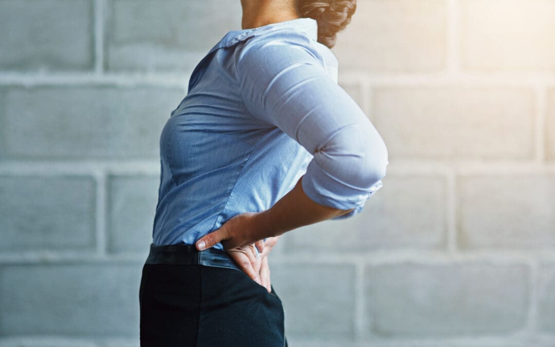
When you think about your body, it’s easy to take for granted the complex machinery that keeps you moving—your muscles, bones, and joints working together like a well-oiled (or sometimes creaky) machine. The musculoskeletal system is the unsung hero of daily life, allowing you to run, lift, dance, or even just scroll through your phone without toppling over. But when pain strikes—whether from a fender-bender, a clumsy tumble, or just the wear and tear of life—it can feel like your body’s betraying you. Enter chiropractic care, the hands-on hero that helps get you back on track. In this comprehensive guide, we’ll dive into how chiropractic care, led by experts like Dr. Alexander Jimenez, DC, APRN, FNP-BC, at El Paso Back Clinic, can reduce musculoskeletal pain, boost bone health, and help you reclaim your quality of life. Plus, we’ll sprinkle in a bit of humor to keep things light—because who said healing can’t come with a chuckle?
The Musculoskeletal System: Your Body’s Framework
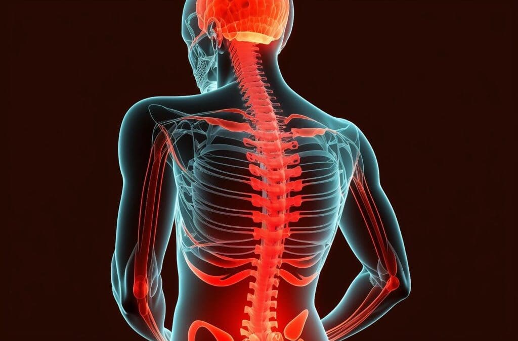
The musculoskeletal system is like the scaffolding of a building—it holds everything together, provides structure, and lets you move without collapsing into a heap. It’s made up of bones, muscles, tendons, ligaments, and other connective tissues that work in harmony. Bones provide the framework, muscles give you the power to move, and joints act like hinges, allowing flexibility. When everything’s working smoothly, you barely notice it. But when something goes wrong—like a strained muscle, a misaligned spine, or a bone that’s not as strong as it used to be—life can get uncomfortable fast.
Why Bone Health Matters
Bones aren’t just static structures; they’re living tissues that constantly remodel themselves. Strong bones are crucial for mobility, protecting vital organs, and even producing blood cells. Poor bone health, like osteoporosis, can lead to fractures, chronic pain, and reduced quality of life. According to research, maintaining bone health involves a balance of proper nutrition, exercise, and avoiding environmental factors that weaken bones (Baim, 2014).
Environmental factors—like poor posture, sedentary lifestyles, or repetitive stress—can wreak havoc on your musculoskeletal system. For instance, sitting at a desk all day can strain your spine, while heavy lifting without proper form can lead to joint injuries. Add in accidents like car crashes or slips, and you’ve got a recipe for musculoskeletal mayhem. Chiropractic care steps in to address these issues, not just by treating pain but by restoring balance and supporting bone health.
Reference:
Baim, S. (2014). Osteoporosis prevention, screening, and treatment: A review. Journal of Clinical Densitometry, 17(3), 371-378. https://pubmed.ncbi.nlm.nih.gov/24709112/
Chiropractic Care: The Science Behind the Snap, Crackle, Pop
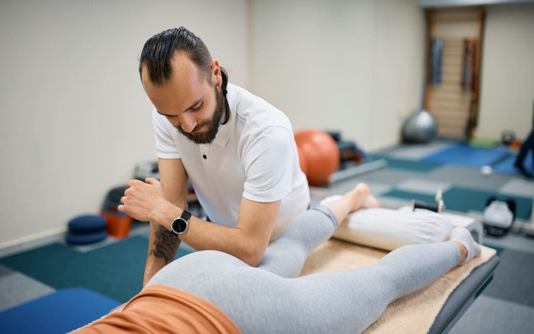
Chiropractic care is like giving your body a tune-up, but instead of a wrench, chiropractors use their hands (and sometimes a bit of tech) to adjust misaligned joints and relieve pressure on nerves. The goal? Restore proper alignment, reduce pain, and improve function. It’s not just about cracking your back—though, let’s be honest, that satisfying pop can feel like a mini-victory. The real magic happens when the musculoskeletal system is realigned, allowing your body to heal naturally.
Clinical Rationale for Pain Relief
Chiropractic care works by addressing misalignments (subluxations) in the spine and joints that can cause pain, inflammation, and reduced mobility. For example, spinal manipulative therapy (SMT) has been shown to reduce low back pain by improving spinal mobility and reducing muscle tension (Blanchette et al., 2016). A systematic review found that SMT is effective for acute low back pain, providing relief comparable to other treatments like medication but with fewer side effects (Paige et al., 2017).
Dr. Alexander Jimenez, a chiropractor at El Paso Back Clinic, emphasizes a holistic approach. His methods focus on restoring joint and biomechanical function, which can alleviate pain from conditions like sciatica, whiplash, or degenerative arthritis. By using advanced imaging and diagnostic evaluations, he identifies the root cause of pain, ensuring targeted treatment. For instance, a misaligned spine from a car accident can pinch nerves, causing radiating pain. Chiropractic adjustments realign the spine, relieving pressure and promoting healing.
Reference:
Blanchette, M. A., Stochkendahl, M. J., Borges Da Silva, R., Boruff, J., Harrison, P., & Bussières, A. (2016). Effectiveness and economic evaluation of chiropractic care for the treatment of low back pain: A systematic review of pragmatic studies. PLoS One, 11(8), e0160037. https://pubmed.ncbi.nlm.nih.gov/27487116/
Paige, N. M., Miake-Lye, I. M., Booth, M. S., Beroes, J. M., Mardian, A. S., Dougherty, P., … & Shekelle, P. G. (2017). Association of spinal manipulative therapy with clinical benefit and harm for acute low back pain: Systematic review and meta-analysis. JAMA, 317(14), 1451-1460. https://pubmed.ncbi.nlm.nih.gov/28399251/
Bone Health Benefits
Chiropractic care doesn’t just help with pain—it can also support bone health. By improving spinal alignment, chiropractic adjustments reduce stress on bones and joints, which can prevent wear and tear that contributes to conditions like osteoarthritis. Additionally, techniques like electrical stimulation, often used in chiropractic settings, can promote bone healing by stimulating osteogenesis (bone formation) (Ciombor & Aaron, 1996). This is particularly beneficial for individuals recovering from fractures or dealing with osteoporosis.
Dr. Jimenez incorporates therapies like massage and durable medical equipment (e.g., braces or supports) to enhance recovery. These tools stabilize joints, reduce inflammation, and support bone health, especially after injuries like those from motor vehicle accidents (MVAs).
Reference:
Ciombor, D. M., & Aaron, R. K. (1996). Stimulators of bone healing: Biologic and biomechanical. Clinical Orthopaedics and Related Research, 327, 12-20. https://pubmed.ncbi.nlm.nih.gov/8653943/
Five Common Musculoskeletal Issues and Chiropractic Solutions
Let’s break down five common musculoskeletal problems and how chiropractic care can help. Think of these as the “usual suspects” when it comes to body aches and pains.
1. Low Back Pain
Low back pain is the bane of many people’s existence—whether from sitting too long, lifting something heavy, or just sleeping in a weird position. It’s like your spine’s way of saying, “Hey, pay attention to me!” Chiropractic care, particularly spinal manipulative therapy, has been shown to reduce pain and improve function in active-duty military personnel with low back pain (Goertz et al., 2018).
Dr. Jimenez uses advanced imaging to pinpoint misalignments or disc issues, then applies targeted adjustments to restore mobility. He might also recommend exercises to strengthen core muscles, which act like a natural corset for your spine.
Reference:
Goertz, C. M., Long, C. R., Vining, R. D., Pohlman, K. A., Walter, J., & Coulter, I. (2018). Effects of chiropractic care on strength, balance, and endurance in active-duty U.S. military personnel with low back pain: A randomized controlled trial. Journal of Alternative and Complementary Medicine, 24(7), 669-676. https://pubmed.ncbi.nlm.nih.gov/29470104/
2. Neck Pain and Whiplash
Ever get a stiff neck from staring at your phone too long? Now imagine that pain amplified by a car accident. Whiplash-associated disorders (WAD) are common after MVAs, causing neck pain, headaches, and restricted movement. Chiropractic care can help by realigning the cervical spine and reducing muscle tension. Dr. Jimenez’s dual-scope approach—combining chiropractic adjustments with massage therapy—can speed up recovery by improving blood flow and reducing inflammation.
3. Sciatica
Sciatica is like an unwelcome guest that shoots pain down your leg, often caused by a pinched nerve in the lower back. Chiropractic adjustments can relieve pressure on the sciatic nerve, while exercises prescribed by Dr. Jimenez strengthen supporting muscles to prevent recurrence.
4. Joint Injuries
Whether from sports or a slip on a rainy day, joint injuries (like sprained ankles or shoulders) can limit mobility. Chiropractic care restores joint alignment and reduces inflammation, while integrative therapies like acupuncture (available at El Paso Back Clinic) enhance healing.
5. Degenerative Arthritis
Arthritis is like rust on your joints—it builds up over time, making movement painful. Chiropractic care can’t cure arthritis, but it can improve joint function and reduce pain through gentle adjustments and therapies like electrical stimulation, which supports bone health (Ciombor & Aaron, 1996).
Reference:
Ciombor, D. M., & Aaron, R. K. (1996). Stimulators of bone healing: Biologic and biomechanical. Clinical Orthopaedics and Related Research, 327, 12-20. https://pubmed.ncbi.nlm.nih.gov/8653943/
Understanding Ligamentous Injuries- Video
Environmental Factors and Their Impact
Your daily routine can be a minefield for your musculoskeletal system. Here are some common culprits:
Poor Posture: Slouching at your desk or hunching over your phone can misalign your spine, leading to pain. Chiropractic adjustments correct these misalignments, while Dr. Jimenez’s team offers ergonomic advice to prevent future issues.
Sedentary Lifestyle: Lack of movement weakens muscles and bones, increasing the risk of osteoporosis (Baim, 2014). Regular chiropractic care, combined with exercise plans, keeps your musculoskeletal system strong.
Accidents: MVAs, bicycle crashes, or 18-wheeler collisions can cause severe injuries. Dr. Jimenez’s expertise in personal injury cases ensures comprehensive care, from diagnostics to rehabilitation.
Repetitive Stress: Jobs involving repetitive motions (like typing or lifting) can strain joints and muscles. Chiropractic care reduces inflammation and restores function.
Reference:
Baim, S. (2014). Osteoporosis prevention, screening, and treatment: A review. Journal of Clinical Densitometry, 17(3), 371-378. https://pubmed.ncbi.nlm.nih.gov/24709112/
Dr. Alexander Jimenez: El Paso’s Personal Injury Expert
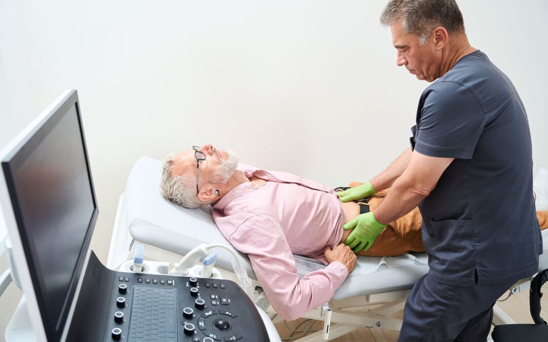
In El Paso, personal injury cases—like those from car accidents or workplace injuries—are a significant concern. Dr. Alexander Jimenez stands out as a distinguished practitioner, blending chiropractic expertise with advanced diagnostic skills. At El Paso Back Clinic, he uses tools like X-rays, MRIs, and dual-scope procedures to assess injuries accurately. His ability to connect medical findings with legal documentation makes him a vital liaison for patients navigating personal injury claims.
For example, after an 18-wheeler accident, Dr. Jimenez might use imaging to identify spinal misalignments or soft tissue damage, then create a tailored treatment plan combining adjustments, massage, and nutritional guidance. His holistic approach ensures patients recover physically while supporting their legal cases with detailed medical reports.
Chiropractic Care in Action: Real-World Applications
Let’s paint a picture: You’re driving home, and bam—a fender-bender leaves you with a sore neck and a grumpy spine. Or maybe you’re a cyclist who took a tumble, and now your shoulder’s acting like it’s auditioning for a horror movie. These scenarios are where chiropractic care shines.
Motor Vehicle Accidents (MVAs)
MVAs can cause everything from whiplash to gastrointestinal injuries. Dr. Jimenez’s team uses a combination of chiropractic adjustments, massage therapy, and durable medical equipment to address these issues. For example, trigger point therapy can release muscle knots caused by MVAs, while nutritional plans support internal healing.
Bicycle Accidents
Bicyclists face unique risks, like collisions with cars or falls. Chiropractic care restores joint function, while integrative medicine (like acupuncture) reduces inflammation. Dr. Jimenez’s comprehensive approach ensures cyclists get back on the road safely.
18-Wheeler Crashes
These accidents are no joke—think of an 18-wheeler as a giant rolling pin flattening your musculoskeletal system. Chiropractic care, combined with advanced diagnostics, helps address spinal and joint injuries, while therapies like electrical stimulation promote bone healing.
Nutrition and Lifestyle: The Unsung Heroes
You can’t talk about musculoskeletal health without mentioning nutrition. A diet rich in calcium, vitamin D, and anti-inflammatory foods (like leafy greens and fatty fish) supports bone health and reduces inflammation (Health Coach Clinic, n.d.). Dr. Jimenez’s team at El Paso Back Clinic creates personalized nutrition plans to aid recovery, especially after MVAs.
Lifestyle changes, like regular exercise and proper ergonomics, also play a role. Think of it like giving your body a daily pep talk—it needs movement and care to stay strong. Chiropractic care complements these efforts by ensuring your spine and joints are aligned, making exercise more effective.
The Chiropractic Identity: A Framework for Healing
Chiropractic care isn’t just a treatment—it’s a philosophy of health that emphasizes the body’s ability to heal itself when properly aligned. Research highlights spinal care as a cornerstone of chiropractic identity, focusing on restoring function and preventing chronic issues (Gliedt et al., 2015). Dr. Jimenez embodies this philosophy, combining evidence-based techniques with personalized care to address each patient’s unique needs.
Reference:
Gliedt, J. A., Hawk, C., Anderson, M., Ahmad, K., Bunn, D., Cambron, J., … & Schneider, M. J. (2015). Spine care as a framework for the chiropractic identity. Journal of Chiropractic Humanities, 22(1), 14-21. https://pubmed.ncbi.nlm.nih.gov/26770177/
A Dash of Humor: Keeping It Light
Let’s face it—talking about back pain and bone health can feel like a lecture from a grumpy science teacher. So, imagine your spine as a cranky old neighbor who just needs a little TLC to stop complaining. Chiropractic care is like inviting that neighbor over for a cup of tea and a good stretch—suddenly, they’re a lot less cranky! Whether it’s a quick adjustment or a full-on rehab plan, Dr. Jimenez’s team at El Paso Back Clinic knows how to make your musculoskeletal system smile again.
Conclusion
Chiropractic care is a powerful tool for managing musculoskeletal pain and supporting bone health. From low back pain to whiplash, Dr. Alexander Jimenez and his team at El Paso Back Clinic offer evidence-based solutions that address the root cause of discomfort. By combining spinal adjustments, advanced diagnostics, and integrative therapies like massage and nutrition, they help patients recover from injuries and improve their quality of life. For personal injury victims in El Paso, Dr. Jimenez’s expertise ensures comprehensive care and legal support, making him a trusted ally in recovery.
Disclaimer: This blog post is intended for informational purposes only and should not be taken as medical advice. Always consult a qualified healthcare professional, such as a chiropractor or physician, before starting any treatment. The information provided is based on current research and clinical insights, but does not replace professional medical evaluation.
Learn about the best poses for sciatica and the role of chiropractic care in managing pain and promoting wellness.
Chiropractic Care for Sciatica Pain Relief: A Comprehensive Guide to Understanding and Managing Sciatic Nerve Pain
Sciatica pain can feel like an uninvited guest who overstays their welcome, turning simple tasks like sitting, standing, or even sneezing into a wince-worthy ordeal. If you’ve ever felt a sharp, shooting pain radiating from your lower back down to your toes, you might be dealing with sciatica. This condition affects millions and can make daily life feel like a game of dodgeball with invisible opponents. Fortunately, chiropractic care offers a non-invasive, evidence-based solution to help manage and reduce sciatica pain, particularly when it’s linked to low back pain. In this comprehensive guide, we’ll dive into the clinical rationale behind chiropractic care for sciatica, explore the musculoskeletal system’s role in this condition, and highlight how Dr. Alexander Jimenez, a renowned chiropractor in El Paso, Texas, uses advanced techniques to help patients reclaim their mobility and quality of life. We’ll also sprinkle in some humor to keep things light—because who said learning about sciatica can’t be fun? Let’s get started!
Understanding Sciatica: What’s All the Fuss About?
Sciatica isn’t a single condition but a collection of symptoms caused by irritation or compression of the sciatic nerve, the longest and largest nerve in the human body. This nerve runs from the lower back, through the buttocks, and down the legs, controlling muscles and providing sensation to the thighs, calves, and feet. When something pinches or irritates this nerve, it can lead to a symphony of discomfort: pain, tingling, burning sensations, or numbness that radiates along its path (Ropper & Zafonte, 2015). Think of it like a cranky electrical wire sparking and short-circuiting your leg’s circuitry.
Common Causes of Sciatica
Sciatica can be triggered by various issues, including:
Herniated Discs: When the soft, jelly-like center of a spinal disc bulges or ruptures, it can press on the sciatic nerve roots in the lumbar spine. About 90% of sciatica cases stem from this issue (Ropper & Zafonte, 2015).
Piriformis Syndrome: The piriformis muscle in the buttocks can sometimes pinch the sciatic nerve, especially if it’s tight or inflamed. Imagine the piriformis as a grumpy bouncer at a club, refusing the sciatic nerve entry.
Spinal Stenosis: Narrowing of the spinal canal can compress nerve roots, leading to sciatica symptoms, particularly in older adults.
Spondylolisthesis: When a vertebra slips out of alignment, it can squeeze the sciatic nerve, causing pain.
Pregnancy: The added weight and shifting posture during pregnancy can irritate the sciatic nerve, making it a common complaint for expectant mothers.
Obesity: Excess weight can stress the spine, contributing to conditions like intervertebral disc degeneration that lead to sciatica (Cao et al., 2022).
How Sciatica Impacts Daily Life
Sciatica doesn’t just cause pain—it can disrupt your entire routine. Simple activities like sitting at a desk, driving, or even tying your shoes can become Herculean tasks. For some, the pain is a dull ache; for others, it’s a sharp, electric jolt that makes you question your life choices—like that time you tried to lift a couch solo. Studies show that low back pain, often linked with sciatica, affects 619 million people globally and is the leading cause of disability worldwide (Ferreira et al., 2023). Up to 30% of sciatica cases can persist for a year or more if untreated, making early intervention critical (Ropper & Zafonte, 2015).
Risk Factors and Overlapping Profiles
Certain factors increase your chances of developing sciatica, including:
Obesity: Excess body weight (BMI > 25) puts extra pressure on the spine, accelerating disc degeneration (Cao et al., 2022).
Occupational Stress: Jobs involving heavy lifting, prolonged sitting, or repetitive motions can strain the musculoskeletal system.
Poor Health Habits: Smoking, lack of exercise, and poor posture can exacerbate spinal issues.
Age: Degenerative changes in the spine, like disc wear and tear, become more common as we age.
These risk factors often overlap, creating a perfect storm for sciatica. For example, an obese individual with a sedentary job may be more prone to disc herniation, which in turn compresses the sciatic nerve. Chiropractic care, with its focus on spinal alignment and musculoskeletal health, can address these overlapping issues to provide relief.
References
Cao, S., Li, W., Wang, T., Li, Y., Xu, Y., & Zhang, J. (2022). Causal associations of obesity with the intervertebral degeneration, low back pain, and sciatica: A two-sample Mendelian randomization study. Frontiers in Endocrinology, 13, 882028. https://doi.org/10.3389/fendo.2022.882028
Ferreira, M. L., de Luca, K., Haile, L., Steinmetz, J., Culbreth, G. T., Cross, M., … & Woolf, A. D. (2023). Global, regional, and national burden of low back pain, 1990–2020, its attributable risk factors, and projections to 2050: A systematic analysis of the Global Burden of Disease Study 2021. The Lancet Rheumatology, 5(6), e316-e329. https://doi.org/10.1016/S2665-9913(23)00098-X
The Musculoskeletal System and the Sciatic Nerve: A Painful Partnership
The musculoskeletal system—your body’s framework of bones, muscles, joints, and connective tissues—plays a starring role in sciatica. When this system is out of whack, it can turn the sciatic nerve into a cranky toddler throwing a tantrum. Let’s break down how the musculoskeletal system and the sciatic nerve interact and why this relationship can lead to such a painful drama.
The Sciatic Nerve’s Journey
The sciatic nerve originates from the nerve roots (L4-S3) in the lumbar spine (lower back) and sacral plexus. It exits the pelvis through the sciatic notch, passes under or through the piriformis muscle, and travels down the back of the thigh, branching into smaller nerves that reach the feet. This nerve is like the body’s superhighway, carrying signals for movement and sensation. When it’s compressed or irritated—say, by a herniated disc or a tight piriformis muscle—it sends out distress signals in the form of pain, tingling, or numbness (Ropper & Zafonte, 2015).
Musculoskeletal Culprits
Several musculoskeletal structures can gang up on the sciatic nerve:
Spinal Discs: Herniated or bulging discs in the lumbar spine can press on nerve roots, triggering sciatica. Think of a disc as a jelly donut—when the jelly squeezes out, it can squish the nerve.
Piriformis Muscle: This small muscle in the buttocks can tighten or spasm, pinching the sciatic nerve. It’s like the nerve is stuck in a traffic jam with no exit.
Facet Joints: These joints connect vertebrae and can become inflamed or misaligned, contributing to nerve irritation.
Paraspinal Muscles: Tight or imbalanced muscles around the spine can alter posture and increase pressure on the sciatic nerve.
Impact on Daily Life
When the sciatic nerve is irritated, it doesn’t just cause pain—it can throw your whole body out of alignment. You might limp to avoid putting weight on the affected leg, which strains other muscles and joints. Prolonged sitting can feel like sitting on a bed of nails, and standing for too long might make you wish you could detach your legs. This can lead to reduced mobility, missed workdays, and even mood changes due to chronic discomfort. For instance, a truck driver with sciatica might struggle to sit for long hauls, while a pregnant woman might find walking unbearable (Jimenez, 2018).
Why Alignment Matters
The musculoskeletal system’s alignment is key to keeping the sciatic nerve happy. Misalignments, or subluxations, in the spine can compress nerve roots, while tight muscles can exacerbate the problem. Chiropractic care focuses on restoring this alignment through spinal adjustments, reducing pressure on the sciatic nerve, and improving overall function.
The Clinical Rationale for Chiropractic Care in Sciatica
Chiropractic care is like a superhero swooping in to save the day for sciatica sufferers. It’s a non-invasive, drug-free approach that targets the root causes of sciatica rather than just masking the pain with medications. Here’s why chiropractic care, particularly under the expertise of Dr. Alexander Jimenez, is a game-changer for sciatica and low back pain.
How Chiropractic Care Works
Chiropractors use spinal manipulation and mobilization techniques to restore proper alignment to the spine, reducing pressure on the sciatic nerve. These adjustments involve applying controlled force to specific vertebrae to correct subluxations (misalignments) that may be compressing nerve roots. A 2020 study found that spinal manipulative therapy significantly reduces pain and disability in patients with chronic low back pain, which often accompanies sciatica (Rubinstein et al., 2020). By realigning the spine, chiropractors help take the pressure off the sciatic nerve, allowing it to function without sending those pesky pain signals.
Specific Techniques for Sciatica
Dr. Alexander Jimenez, a board-certified chiropractor and family nurse practitioner in El Paso, Texas, employs a variety of techniques to address sciatica:
Spinal Adjustments: Precise manipulations to correct spinal misalignments, reducing nerve compression. These adjustments are like hitting the reset button on your spine’s alignment.
Spinal Decompression: A non-surgical technique that gently stretches the spine to relieve pressure on discs and nerves. It’s like giving your spine a much-needed stretch after a long day.
Manual Therapy: Techniques like myofascial release and trigger point therapy to relax tight muscles and reduce inflammation (Jimenez, 2025).
Electro-Acupuncture: Targeted electrical stimulation of acupuncture points to reduce pain and promote healing, shown to be effective for neuropathic pain (Li et al., 2018).
Rehabilitation Exercises: Customized programs to strengthen core and lumbar muscles, improving spinal stability, and preventing re-injury.
Why It’s Effective
Chiropractic care is effective because it addresses the underlying musculoskeletal issues causing sciatica, rather than just treating symptoms. For example, a herniated disc pressing on the sciatic nerve can be alleviated by adjusting the spine to create more space for the nerve roots. A 2021 systematic review confirmed that spinal manipulative therapy is effective for reducing spine pain, including sciatica, by improving mobility and reducing inflammation (Coulter et al., 2021). Unlike medications like gabapentin or pregabalin, which show limited efficacy for sciatica and carry risks of adverse events (Enke et al., 2022), chiropractic care offers a safer, non-invasive alternative.
Incorporating Poses and Stretches
Chiropractic care often integrates specific poses and stretches to complement spinal adjustments. Dr. Jimenez frequently incorporates yoga-inspired poses to enhance flexibility and reduce muscle tension, which can exacerbate sciatica. Here are seven poses inspired by yoga that can help soothe sciatica, as outlined by Yoga International (Rinehart, 2016):
Child’s Pose (Balasana): Relaxes the lower back and stretches the hips, reducing tension around the sciatic nerve.
Downward-Facing Dog: Lengthens the spine and strengthens core muscles, promoting better spinal alignment.
Pigeon Pose: Stretches the piriformis muscle, relieving pressure on the sciatic nerve.
Seated Forward Bend: Stretches the hamstrings and lower back, improving flexibility.
Thread the Needle: Targets the piriformis and glutes, releasing tension that may compress the sciatic nerve.
Supine Twist: Gently twists the spine to relieve pressure and improve mobility.
Bridge Pose: Strengthens the glutes and core, supporting spinal stability.
These poses, when guided by a chiropractor like Dr. Jimenez, can be tailored to a patient’s specific needs, ensuring safe execution and maximum benefit. Regular practice can reduce the overlapping risk factors like muscle tightness and poor posture, which contribute to sciatica.
Sciatica Secrets Revealed- Video
Dr. Jimenez’s Approach
Dr. Jimenez’s dual licensure as a chiropractor and nurse practitioner allows him to take a holistic approach, combining chiropractic adjustments with functional medicine. He uses advanced imaging (X-rays, MRIs) to pinpoint the exact cause of sciatica, such as a herniated disc or spinal stenosis. His diagnostic evaluations include thorough physical exams and functional assessments to identify biomechanical dysfunctions. By integrating these insights with spinal manipulation, acupuncture, and rehabilitation exercises, Dr. Jimenez creates personalized treatment plans that address both immediate pain and long-term recovery (Jimenez, 2025).
References
Coulter, I. D., Crawford, C., Vernon, C., Hurwitz, E. L., Khorsan, R., Booth, M. S., & Herman, P. M. (2021). Clinical effectiveness and efficacy of chiropractic spinal manipulation for spine pain. Frontiers in Pain Research, 2, 765921. https://doi.org/10.3389/fpain.2021.765921
Enke, O., New, H. A., New, C. H., Mathieson, S., McLachlan, A. J., Latimer, J., … & Lin, C. W. C. (2022). A systematic review and meta-analysis of the effectiveness and adverse events of gabapentin and pregabalin for sciatica pain. Atención Primaria, 54(2), 102144. https://doi.org/10.1016/j.aprim.2021.102144
Li, Y., Wu, F., & Wang, Y. (2018). Electro-acupuncture for the treatment of neuropathic pain: A systematic review and meta-analysis. Evidence-Based Complementary and Alternative Medicine, 2018, 1239467. https://doi.org/10.1155/2018/1239467
Rubinstein, S. M., de Zoete, A., van Middelkoop, M., Assendelft, W. J. J., de Boer, M. R., & van Tulder, M. W. (2020). Benefits and harms of spinal manipulative therapy for the treatment of chronic low back pain: Systematic review and meta-analysis of randomised controlled trials. BMJ, 368, m689. https://doi.org/10.1136/bmj.m689
Chiropractic Care in Personal Injury Cases: Dr. Jimenez’s Expertise in El Paso
In El Paso, Texas, personal injury cases—such as those from car accidents or workplace incidents—often involve musculoskeletal injuries like sciatica, whiplash, or chronic back pain. Dr. Alexander Jimenez stands out as a leading practitioner in this field, with over 25 years of experience and a unique dual licensure as a chiropractor (DC) and family nurse practitioner (FNP-BC). His practice, Injury Medical & Chiropractic Clinic, is a beacon of hope for victims of personal injuries seeking non-invasive solutions (Jimenez, 2025).
The Role of Chiropractic Care in Personal Injury
Personal injuries, especially from motor vehicle accidents (MVAs), can cause a range of musculoskeletal issues, including sciatica due to herniated discs or soft tissue damage. Chiropractic care is particularly effective in these cases because it addresses the biomechanical dysfunctions that result from trauma. For instance, a car accident might cause a vertebral misalignment that compresses the sciatic nerve, leading to radiating pain. Dr. Jimenez uses spinal adjustments to restore alignment, complemented by physical therapy to rebuild strength and prevent re-injury (Jimenez, 2024).
Advanced Imaging and Diagnostic Evaluations
Dr. Jimenez’s approach is grounded in precision. He utilizes advanced imaging techniques, such as X-rays and MRIs, to identify the exact source of sciatica, whether it’s a herniated disc, spinal stenosis, or piriformis syndrome. His diagnostic evaluations include:
Physical Exams: Assessing range of motion, muscle strength, and neurological function.
Functional Assessments: Evaluating how the injury affects daily activities, like walking or sitting.
Dual-Scope Procedures: Combining chiropractic and medical approaches to provide a comprehensive diagnosis.
These tools allow Dr. Jimenez to create detailed treatment plans tailored to each patient’s needs, ensuring both immediate pain relief and long-term recovery (Jimenez, 2025).
Legal and Medical Liaison
One of Dr. Jimenez’s standout qualities is his ability to serve as a liaison between medical care and legal documentation. In personal injury cases, accurate documentation is crucial for insurance claims and legal proceedings. Dr. Jimenez provides detailed reports that link a patient’s injuries to the accident, supported by advanced imaging and diagnostic findings. This dual-scope approach ensures that patients receive the medical care they need while also having the documentation required for legal success (Jimenez, 2024).
Patient Testimonials
Patients in El Paso rave about Dr. Jimenez’s care. Truide Torres, who suffered from sciatica during pregnancy, credits Dr. Jimenez’s chiropractic adjustments and lifestyle advice for helping her regain mobility. Edgar M. Reyes, a truck driver, found relief from sciatica that had made long drives unbearable, thanks to Dr. Jimenez’s tailored rehabilitation program (Jimenez, 2018). These testimonials highlight Dr. Jimenez’s commitment to personalized, patient-centered care.
Why El Paso Chooses Dr. Jimenez
With accolades as a top-rated chiropractor from 2015 to 2024, Dr. Jimenez’s practice is renowned for its holistic approach. His website, https://www.dralexjimenez.com, offers resources like patient testimonials and articles on spinal health, showcasing his expertise in treating complex conditions like sciatica. His LinkedIn profile (https://www.linkedin.com/in/dralexjimenez/) further underscores his credentials as a board-certified practitioner dedicated to holistic healing (Jimenez, 2025).
While chiropractic care shines in the spotlight for sciatica treatment, it’s worth comparing it to other options to understand its unique benefits. Let’s take a look at some common treatments and how they stack up.
Medications
Medications like gabapentin and pregabalin are often prescribed for sciatica. Still, a 2022 systematic review found little evidence of their effectiveness for sciatica pain, with potential side effects like dizziness and nausea (Enke et al., 2022). Non-steroidal anti-inflammatory drugs (NSAIDs) may reduce inflammation, but they don’t address structural issues like disc herniation or spinal misalignment. Chiropractic care, on the other hand, targets these root causes without the risks associated with long-term medication use.
Acupuncture
Acupuncture, including techniques like pestle needle (chu zhen), involves inserting needles into specific points to relieve pain. Some studies suggest acupuncture may help with sciatica by stimulating nerve pathways and reducing inflammation (Liu et al., 2019; Wang et al., 2019). However, its effects are often temporary, and it doesn’t correct spinal misalignments. Chiropractic care complements acupuncture by addressing structural issues, and Dr. Jimenez usually combines electro-acupuncture with adjustments for enhanced relief (Jimenez, 2025).
Physiotherapy
Physiotherapy focuses on exercises and manual therapies to improve strength and mobility. It’s effective for sciatica, particularly when combined with neural mobilization techniques that stretch and mobilize the sciatic nerve (Neto et al., 2023). However, physiotherapy alone may not fully address spinal misalignments. Chiropractic care integrates well with physiotherapy, as Dr. Jimenez often prescribes tailored exercises alongside adjustments to ensure comprehensive recovery (Jimenez, 2024).
Surgery
In severe cases, surgery like a discectomy or laminectomy may be recommended to relieve nerve compression. However, surgery carries risks like infection and long recovery times, and it’s typically a last resort. Chiropractic care is a non-invasive first line of defense, often preventing the need for surgery by addressing issues early (Coulter et al., 2021).
Why Chiropractic Stands Out
Chiropractic care is unique because it combines structural correction (spinal adjustments) with holistic approaches (exercises, lifestyle advice) to address both symptoms and causes. It’s cost-effective, too—a 2024 systematic review found that chiropractic care is associated with lower downstream costs compared to medical management, reducing the need for expensive procedures like surgery or advanced imaging (Andronis et al., 2024). Plus, it’s got that hands-on, personal touch that makes you feel like you’re in good hands—literally!
References
Andronis, L., James, J., Cairns, D., & Bhatt, S. (2024). Cost of chiropractic versus medical management of adults with spine-related musculoskeletal pain: A systematic review. Chiropractic & Manual Therapies, 32(1), 8. https://doi.org/10.1186/s12998-024-00529-9
Coulter, I. D., Crawford, C., Vernon, C., Hurwitz, E. L., Khorsan, R., Booth, M. S., & Herman, P. M. (2021). Clinical effectiveness and efficacy of chiropractic spinal manipulation for spine pain. Frontiers in Pain Research, 2, 765921. https://doi.org/10.3389/fpain.2021.765921
Enke, O., New, H. A., New, C. H., Mathieson, S., McLachlan, A. J., Latimer, J., … & Lin, C. W. C. (2022). A systematic review and meta-analysis of the effectiveness and adverse events of gabapentin and pregabalin for sciatica pain. Atención Primaria, 54(2), 102144. https://doi.org/10.1016/j.aprim.2021.102144
Liu, C. H., Kung, Y. Y., Lin, C. L., & Wu, H. C. (2019). Therapeutic efficacy and the impact of the “dose” effect of acupuncture to treat sciatica: A randomized controlled pilot study. Journal of Pain Research, 12, 351-360. https://doi.org/10.2147/JPR.S178510
Neto, T., Freitas, S. R., Marques, M. J., & Gomes, J. P. (2023). Neural mobilization in low back and radicular pain: A systematic review. Journal of Manual & Manipulative Therapy, 31(1), 4-13. https://doi.org/10.1080/10669817.2022.2065596
Wang, Y., Xu, L., & Zhang, X. (2019). Pestle needle (chu zhen) treatment for low-back pain and sciatica. Journal of Traditional Chinese Medicine, 39(2), 149-155.
Patient Education: Empowering Recovery
Educating patients about their condition is a cornerstone of Dr. Jimenez’s practice. A 2022 systematic review emphasized the importance of patient education materials for non-specific low back pain and sciatica, noting that informed patients are more likely to adhere to treatment plans and achieve better outcomes (Poitras et al., 2022). Dr. Jimenez provides comprehensive resources, such as videos and articles on his website (https://www.dralexjimenez.com), to help patients understand sciatica’s causes, symptoms, and treatment options.
Key Educational Points
Understanding the Condition: Patients learn that sciatica is often caused by musculoskeletal issues like herniated discs or piriformis syndrome, which can be managed non-invasively.
Lifestyle Modifications: Dr. Jimenez advises on weight management, posture correction, and regular exercise to reduce risk factors like obesity (Cao et al., 2022).
Home Exercises: Patients are taught specific stretches and poses, like those mentioned earlier, to maintain flexibility and prevent flare-ups.
When to Seek Help: Early intervention is key to preventing chronic sciatica, so patients are encouraged to seek care promptly.
This education empowers patients to take an active role in their recovery, reducing reliance on medications and improving long-term outcomes.
References
Cao, S., Li, W., Wang, T., Li, Y., Xu, Y., & Zhang, J. (2022). Causal associations of obesity with the intervertebral degeneration, low back pain, and sciatica: A two-sample Mendelian randomization study. Frontiers in Endocrinology, 13, 882028. https://doi.org/10.3389/fendo.2022.882028
Poitras, S., Tousignant, M., Maher, C. G., & Wong, J. J. (2022). Patient education materials for non-specific low back pain and sciatica: A systematic review and meta-analysis. PLoS ONE, 17(10), e0274521. https://doi.org/10.1371/journal.pone.0274521
The Science Behind Chiropractic Success
The effectiveness of chiropractic care for sciatica isn’t just anecdotal—science backs it up. Studies show that spinal manipulative therapy can reduce pain and disability in patients with low back pain and sciatica by improving spinal alignment, reducing inflammation, and enhancing mobility (Coulter et al., 2021). A 2010 study found that chiropractic care combined with standard medical treatment improved outcomes for acute back pain, including sciatica, compared to medical treatment alone (Goertz et al., 2013).
Biomechanical Benefits
Chiropractic adjustments restore proper biomechanics to the spine, reducing pressure on the sciatic nerve. For example, a case study demonstrated that spinal manipulation and mobilization forces significantly reduced sciatica symptoms by improving lumbar mobility (Hawk et al., 2022). This biomechanical correction is particularly effective for conditions like herniated discs, where misalignment directly contributes to nerve compression.
Cost-Effectiveness
Chiropractic care is not only effective but also cost-effective. A 2024 systematic review found that patients who started with chiropractic care for spine-related pain had lower rates of hospitalization, surgery, and advanced imaging, leading to reduced overall healthcare costs (Andronis et al., 2024). This makes chiropractic care an attractive option for those looking to avoid invasive procedures and their associated expenses.
Safety Profile
Unlike medications or surgery, chiropractic care has a low risk of adverse events when performed by a licensed professional like Dr. Jimenez. The most common side effects are mild soreness or discomfort, which typically resolve quickly. This safety profile, combined with its efficacy, makes chiropractic care a preferred choice for many sciatica sufferers.
References
Andronis, L., James, J., Cairns, D., & Bhatt, S. (2024). Cost of chiropractic versus medical management of adults with spine-related musculoskeletal pain: A systematic review. Chiropractic & Manual Therapies, 32(1), 8. https://doi.org/10.1186/s12998-024-00529-9
Coulter, I. D., Crawford, C., Vernon, C., Hurwitz, E. L., Khorsan, R., Booth, M. S., & Herman, P. M. (2021). Clinical effectiveness and efficacy of chiropractic spinal manipulation for spine pain. Frontiers in Pain Research, 2, 765921. https://doi.org/10.3389/fpain.2021.765921
Goertz, C. M., Pohlman, K. A., Vining, R. D., Brantingham, J. W., & Long, C. R. (2013). Integrating chiropractic care into the treatment of acute back pain: A randomized controlled trial. Spine, 38(7), 540-548. https://doi.org/10.1097/BRS.0b013e318275d0c9
Hawk, C., Whalen, W. M., Farabaugh, R. J., & Daniels, C. J. (2022). Spinal manipulation and mobilization forces delivered treating sciatica: A case report. Journal of Chiropractic Medicine, 21(3), 203-209. https://doi.org/10.1016/j.jcm.2022.02.006
Integrating Chiropractic Care with Lifestyle Changes
To maximize the benefits of chiropractic care, Dr. Jimenez emphasizes the importance of lifestyle changes to prevent sciatica flare-ups. Here are some key strategies:
Weight Management: Reducing excess weight can decrease spinal stress and lower the risk of disc degeneration (Cao et al., 2022).
Regular Exercise: Incorporating low-impact activities like walking, swimming, or yoga can improve spinal health and flexibility.
Proper Posture: Maintaining good posture while sitting, standing, and lifting can prevent undue pressure on the spine.
Ergonomic Adjustments: Using supportive chairs, mattresses, and proper lifting techniques can reduce musculoskeletal strain.
Dr. Jimenez often provides patients with tailored exercise plans and ergonomic advice to complement their chiropractic treatments, ensuring long-lasting relief (Jimenez, 2025).
References
Cao, S., Li, W., Wang, T., Li, Y., Xu, Y., & Zhang, J. (2022). Causal associations of obesity with the intervertebral degeneration, low back pain, and sciatica: A two-sample Mendelian randomization study. Frontiers in Endocrinology, 13, 882028. https://doi.org/10.3389/fendo.2022.882028
Sciatica can be a debilitating condition, but with the right care, it doesn’t have to control your life. Chiropractic care, under the expertise of professionals like Dr. Alexander Jimenez, offers a non-invasive, effective, and cost-efficient solution for managing sciatica and associated low back pain. By addressing the musculoskeletal root causes, incorporating targeted poses, and providing comprehensive patient education, chiropractic care empowers individuals to regain their mobility and quality of life. Dr. Jimenez’s unique dual-scope approach in El Paso, combining advanced diagnostics with holistic treatments, makes him a trusted choice for personal injury victims and those seeking lasting relief.
Disclaimer: The information provided in this blog post is for educational purposes only and does not constitute medical advice. Sciatica and low back pain can have various causes and severities, and treatment should be tailored to the individual. Always consult a qualified healthcare professional, such as a licensed chiropractor or physician, before starting any treatment plan. For personalized care in El Paso, contact Dr. Alexander Jimenez at Injury Medical & Chiropractic Clinic at 915-850-0900 or visit https://www.dralexjimenez.com for more information.
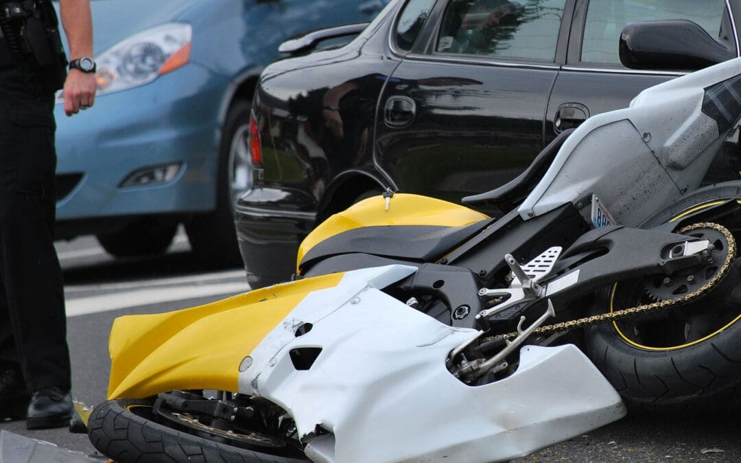
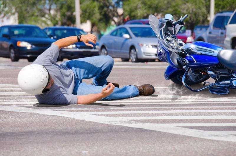
Recovering After a Motorcycle Accident: How Chiropractic Care Helps Heal the Spine and Body
Introduction: Motorcycle Accidents and Spinal Health Risks
Motorcycle accidents often lead to serious injuries—especially to the spine, neck, and musculoskeletal system. Riders are vulnerable because they lack the protection of a car’s frame, making them more likely to suffer from spinal misalignments, nerve compression, disc injuries, fractures, and soft tissue trauma. Even a low-speed collision can cause long-term pain if not treated properly.
At El Paso Back Clinic, Dr. Alexander Jimenez provides specialized chiropractic and medical care for patients recovering from motorcycle crashes. His dual-scope training in chiropractic care and advanced practice nursing enables him to assess, diagnose, and treat trauma injuries holistically and efficiently.
The Role of Chiropractic Care After a Motorcycle Accident
Chiropractic care offers a safe, effective, and drug-free approach to treating motorcycle-related injuries. At El Paso Back Clinic, chiropractic services focus on:
Spinal adjustments – Realign vertebrae to relieve nerve pressure and restore proper joint movement.
Meet Dr. Alexander Jimenez: El Paso’s Personal Injury Chiropractor
Dr. Alexander Jimenez, DC, APRN, FNP-BC, is one of El Paso’s leading experts in treating spinal injuries from motor vehicle accidents, including motorcycle crashes. With over 30 years of clinical experience, Dr. Jimenez offers a dual perspective as both a chiropractor and nurse practitioner.
His integrative approach includes:
Thorough medical and spinal assessments
Advanced imaging and neurological diagnostics
Chiropractic adjustments and therapeutic modalities
Case management for personal injury documentation
Functional medicine support for healing and inflammation control
Patients at El Paso Back Clinic benefit from individualized care that targets both symptoms and root causes.
Motorcycle injuries can be deceptive. A patient may feel mild soreness after an accident but develop significant spinal issues over time. That’s why Dr. Jimenez uses advanced diagnostics to identify injuries early:
MRI – Ideal for evaluating herniated discs, nerve root impingement, and ligament damage.
CT scans – Useful for detecting fractures and joint damage.
Digital motion X-rays – Show real-time spinal dysfunction during movement.
Neurological testing – Assesses motor control, reflexes, and nerve conduction.
These tools allow Dr. Jimenez to pinpoint injuries and tailor chiropractic treatments for the best possible outcomes.
Treating Road Rash, Muscle Trauma, and Ligament Sprains
While skin abrasions (road rash) may receive immediate medical attention, the deeper soft tissue trauma they accompany is often ignored. Dr. Jimenez offers therapies to restore muscle and ligament health, including:
Myofascial release – Breaks up scar tissue and restores mobility.
Electrical muscle stimulation (EMS) – Rebuilds strength and reduces spasms.
Ultrasound therapy – Promotes deep tissue healing.
A patient came to El Paso Back Clinic after a motorcycle accident, complaining of back stiffness, neck pain, and shoulder immobility. Dr. Jimenez conducted:
Spinal X-rays and MRI scans
Neurological testing for nerve involvement
Cervical and thoracic adjustments
Rehabilitation exercises to restore strength
Documentation for their personal injury attorney
Within weeks, the patient regained a full range of motion, experienced reduced pain, and was back to daily activities with improved posture and energy.
Personalized Chiropractic Recovery Plans in El Paso
At El Paso Back Clinic, no two care plans are the same. Every treatment protocol is based on:
Type of injury
Pain levels and movement limitations
Medical history and physical findings
Imaging results and response to care
Dr. Jimenez creates customized schedules that include chiropractic care, rehabilitation exercises, lifestyle support, and legal documentation. This comprehensive approach ensures patients recover safely, completely, and confidently.
Conclusion: Spinal Healing Starts with the Right Care
Motorcycle accidents are life-altering, but recovery doesn’t have to be overwhelming. Chiropractic care at El Paso Back Clinic offers a gentle, natural, and effective way to restore your spine, relieve pain, and return to normal life. Dr. Jimenez’s dual-scope training and personalized treatment plans ensure that your healing process is safe, complete, and well-documented.
If you’ve been in a motorcycle accident in El Paso, early chiropractic care can make all the difference in your recovery. Let El Paso Back Clinic be your partner on the path to healing, strength, and spinal wellness.
Discover effective treatments for peripheral neuropathy with chiropractic care to manage symptoms and enhance mobility.
Chiropractic Care for Peripheral Neuropathy: A Comprehensive Guide to Reducing Nerve Pain
Peripheral neuropathy is like that one friend who shows up uninvited and overstays their welcome, causing all sorts of chaos. It’s a condition where the peripheral nerves—the ones that carry messages between your brain, spinal cord, and the rest of your body—decide to throw a tantrum, leading to symptoms like tingling, numbness, or burning pain. If you’ve ever felt like your hands or feet are throwing a pins-and-needles party without your permission, you might be dealing with peripheral neuropathy. But don’t worry—there’s hope! Chiropractic care, particularly through the expertise of practitioners like Dr. Alexander Jimenez, DC, APRN, FNP-BC, in El Paso, Texas, can help manage this nerve-racking condition (pun intended). In this blog post, we’ll dive into the clinical rationale for why chiropractic care can reduce nerve pain associated with peripheral neuropathy, explore the musculoskeletal system’s role, and highlight how Dr. Jimenez’s unique approach makes him a go-to for personal injury cases in El Paso. Let’s get started!
What Is Peripheral Neuropathy? The Nerve of It All!
Imagine your body as a massive communication network, with your brain and spinal cord as the control center and your peripheral nerves as the Wi-Fi signals carrying messages to your limbs, organs, and muscles. Peripheral neuropathy happens when these signals get scrambled, damaged, or completely cut off. It’s like trying to stream your favorite show with a spotty internet connection—frustrating and disruptive.
Peripheral neuropathy refers to damage to the peripheral nervous system, which sends signals between the central nervous system (brain and spinal cord) and the rest of the body. This damage can cause symptoms like numbness, tingling, burning sensations, muscle weakness, or even loss of balance. It’s not just one condition but a group of disorders caused by various factors, including diabetes, chemotherapy, infections, autoimmune diseases, or physical trauma like motor vehicle accidents (MVAs) (National Institute of Neurological Disorders and Stroke, n.d.).
The prevalence of peripheral neuropathy is no small matter. It affects millions of people worldwide, with diabetic peripheral neuropathy being one of the most common forms, impacting up to 50% of people with diabetes (Hicks & Selvin, 2019). Chemotherapy-induced peripheral neuropathy (CIPN) is another major player, affecting 19-85% of cancer patients undergoing treatment (Seretny et al., 2014). These numbers show just how widespread this condition is, and for those dealing with it, the impact on daily life can be profound.
References
Hicks, C. W., & Selvin, E. (2019). Epidemiology of peripheral neuropathy and lower extremity disease in diabetes. Current Diabetes Reports, 19(10), 86. https://doi.org/10.1007/s11892-019-1212-8
Seretny, M., et al. (2014). Incidence, prevalence, and predictors of chemotherapy-induced peripheral neuropathy: A systematic review and meta-analysis. Pain, 155(12), 2461-2470. https://doi.org/10.1016/j.pain.2014.09.020
The Musculoskeletal System’s Role in Peripheral Neuropathy
Your musculoskeletal system—your bones, muscles, ligaments, tendons, and connective tissues—is like the scaffolding that keeps your body upright and moving. But when peripheral neuropathy enters the scene, it’s like someone’s shaking that scaffolding, causing all sorts of problems. The peripheral nerves are responsible for sending sensory and motor signals to your muscles and joints. When these nerves are damaged, the musculoskeletal system can take a hit, leading to symptoms that mess with your daily routine.
How Peripheral Neuropathy Affects the Musculoskeletal System
Peripheral neuropathy can disrupt the communication between your nerves and muscles, leading to:
Muscle Weakness: Damaged nerves may fail to send proper signals to muscles, causing weakness or difficulty moving. For example, you might struggle to grip a coffee mug or climb stairs without feeling like you’re auditioning for a slow-motion scene.
Loss of Coordination: Nerves help with balance and proprioception (knowing where your body is in space). Neuropathy can make you feel like you’re walking on a tightrope after a few too many spins.
Muscle Cramps and Spasms: Irritated or damaged nerves can cause muscles to contract involuntarily, leading to painful cramps or twitches.
Joint Instability: Weak muscles can’t support joints properly, increasing the risk of falls or injuries, especially in the ankles or knees.
Pain and Discomfort: Neuropathic pain, often described as burning, stabbing, or electric shocks, can radiate to muscles and joints, causing significant discomfort (personalinjurydoctorgroup.com, 2020).
These issues can turn simple tasks—like walking to the mailbox or tying your shoes—into a Herculean effort. For instance, someone with peripheral neuropathy might find their morning jog feels more like trudging through molasses, or they might drop their phone because their fingers have lost coordination.
Impact on Daily Routine
The musculoskeletal fallout from peripheral neuropathy can significantly disrupt daily life. Imagine trying to cook dinner when your hands feel like they’re wearing oven mitts, or attempting to drive when your feet can’t tell the difference between the gas and brake pedals. These symptoms can lead to:
Reduced Mobility: Difficulty walking or standing for long periods, limiting activities like shopping or socializing.
Decreased Independence: Tasks like dressing or bathing may require assistance, which can be a blow to self-esteem.
Increased Risk of Falls: Loss of sensation or balance can make falls more likely, especially for older adults.
Chronic Pain: Persistent nerve pain can sap energy, disrupt sleep, and even lead to mood changes like anxiety or depression.
Peripheral Neuropathy: A Successful Recovery Story- Video
Why Chiropractic Care? The Clinical Rationale
Now, let’s talk about the superhero of this story: chiropractic care. It’s not just about cracking backs and making you feel like a human pretzel—it’s a science-backed approach to improving nerve function and reducing pain. Chiropractic care focuses on the spine and musculoskeletal system to remove nerve interference, which is particularly relevant for peripheral neuropathy.
The Science Behind Chiropractic Care for Nerve Pain
The spine is like the central highway of your nervous system. If there’s a traffic jam—say, a misaligned vertebra or a compressed nerve—it can disrupt the signals traveling to and from your peripheral nerves. Chiropractic adjustments aim to clear these jams by realigning the spine and reducing pressure on nerves. Here’s why this matters for peripheral neuropathy:
Reducing Nerve Compression: Misalignments (subluxations) in the spine can compress nerve roots, exacerbating neuropathic symptoms like tingling or numbness. Adjustments restore alignment, relieving pressure on these nerves (elpasobackclinic.com, 2023).
Improving Blood Flow: Proper spinal alignment enhances blood circulation, which is crucial for nerve health. Damaged nerves need oxygen and nutrients to heal, and chiropractic care can help ensure they get it.
Modulating Pain Signals: Chiropractic adjustments can influence the central nervous system, reducing the perception of pain. Think of it like turning down the volume on a screaming nerve (Woolf & Salter, 2000).
Enhancing Autonomic Function: The autonomic nervous system, which controls involuntary functions like heart rate and digestion, can be affected by neuropathy. Chiropractic care may help regulate these functions by improving spinal health (Vagal, 2020).
Research supports these benefits. A study on spinal canal compression suggests that nerve root insults, whether chemical (from inflammation) or mechanical (from compression), can contribute to polyneuropathy-like symptoms. Chiropractic care addresses these insults by correcting spinal misalignments and reducing inflammation (Kulikov et al., 2016). Another study found that nonpharmacologic interventions, including manual therapies like chiropractic care, can reduce symptoms of chemotherapy-induced peripheral neuropathy (CIPN) by improving nerve function and reducing pain (Oh et al., 2023).
Dr. Alexander Jimenez’s Approach
Enter Dr. Alexander Jimenez, El Paso’s nerve-whisperer. With over 25 years of experience as a chiropractor and board-certified Family Nurse Practitioner (FNP-BC), Dr. Jimenez brings a unique, dual-scope approach to treating peripheral neuropathy. His practice at El Paso’s Premier Wellness and Injury Care Clinic combines chiropractic expertise with advanced medical diagnostics, making him a standout in managing nerve pain (Jimenez, 2025a).
Dr. Jimenez uses a holistic, evidence-based approach inspired by functional medicine. He doesn’t just slap a Band-Aid on symptoms—he digs deep to find the root cause. For example, suppose your neuropathy stems from a car accident. In that case, he might identify a spinal misalignment pinching a nerve while also checking for inflammation or metabolic imbalances that could slow healing (Jimenez, 2023b). His methods include:
Advanced Imaging: Using X-rays, MRIs, or CT scans to pinpoint issues like herniated discs or nerve compression (Jimenez, 2023c).
Diagnostic Evaluations: Neurological tests and motion studies to assess nerve function and biomechanical dysfunction.
Dual-Scope Procedures: Combining chiropractic adjustments with medical interventions like nutritional counseling or physical therapy to address both musculoskeletal and systemic factors.
Manual Therapies: Techniques like spinal decompression, joint mobilization, and myofascial release to relieve nerve pressure and improve mobility.
Kulikov, A. V., et al. (2016). Could spinal canal compression be a cause of polyneuropathy? Frontiers in Surgery, 3, 14. https://doi.org/10.3389/fsurg.2016.00014
Oh, P. J., et al. (2023). Prevention and treatment of chemotherapy-induced peripheral neuropathy (CIPN) with non-pharmacological interventions. Frontiers in Pain Research, 4, 1002967. https://doi.org/10.3389/fpain.2023.1002967
Vagal, V. (2020). Editorial: Understanding the role of the autonomic nervous system in health and disease. Frontiers in Neuroscience, 14, 615. https://doi.org/10.3389/fnins.2020.00615
Peripheral Neuropathy and Personal Injury Cases in El Paso
El Paso, Texas, is a bustling city with heavy traffic, which unfortunately means motor vehicle accidents (MVAs) are all too common. These accidents can cause nerve injuries, including peripheral neuropathy, especially when whiplash or spinal trauma is involved. If you’ve ever been rear-ended and felt like your nerves were playing a game of telephone with the wrong number, you know what I mean. This is where chiropractic care, and specifically Dr. Alexander Jimenez, shines.
The Link Between MVAs and Peripheral Neuropathy
MVAs can cause nerve damage through:
Mechanical Insults: The force of a collision can compress or stretch nerves, leading to symptoms like numbness or tingling. For example, whiplash can pinch nerves in the cervical spine, radiating pain to the arms or hands (Jimenez, 2025b).
Chemical Insults: Inflammation from soft tissue injuries can irritate nerves, contributing to neuropathic pain (Woolf & Thompson, 1991).
Spinal Canal Compression: Trauma can narrow the spinal canal, pressing on nerve roots and mimicking polyneuropathy symptoms (Kulikov et al., 2016).
These injuries don’t just hurt—they can disrupt your life, making it hard to work, drive, or even enjoy a Netflix binge without pain. In personal injury cases, proving the link between the accident and your symptoms is crucial for fair compensation, and that’s where Dr. Jimenez’s expertise comes in.
Dr. Jimenez: The Legal-Medical Liaison
Dr. Jimenez isn’t just a chiropractor—he’s a board-certified nurse practitioner with a knack for bridging medical care and legal documentation. His dual licensure allows him to:
Diagnose with Precision: Using advanced imaging (like MRIs) and neurological tests, he identifies the exact cause of nerve pain, whether it’s a herniated disc or a pinched nerve (Jimenez, 2023c).
Document for Legal Cases: He provides detailed reports that connect your injuries to the accident, strengthening your insurance or legal claims. Think of him as a translator who speaks both “doctor” and “lawyer” fluently (Jimenez, 2025a).
Deliver Holistic Care: His treatments combine spinal adjustments, physical therapy, and functional medicine to address both symptoms and underlying causes, helping you recover faster.
For example, if you’re dealing with post-accident neuropathy, Dr. Jimenez might use an MRI to spot a herniated disc, then apply spinal decompression to relieve nerve pressure. He’ll also check for metabolic issues (like vitamin deficiencies) that could worsen neuropathy, ensuring a comprehensive recovery plan (Jimenez, 2025b). His patients rave about his ability to get them back on their feet, as seen in testimonials on his social media (Jimenez, 2023f).
Kulikov, A. V., et al. (2016). Could spinal canal compression be a cause of polyneuropathy? Frontiers in Surgery, 3, 14. https://doi.org/10.3389/fsurg.2016.00014
Woolf, C. J., & Thompson, S. W. (1991). The induction and maintenance of central sensitization is dependent on N-methyl-D-aspartic acid receptor activation; implications for the treatment of post-injury pain hypersensitivity states. Pain, 44(3), 293-299. https://doi.org/10.1016/0304-3959(91)90100-C
Addressing Overlapping Risk Profiles
Peripheral neuropathy often comes with a side of extra baggage—overlapping risk factors that make symptoms worse. These include diabetes, chemotherapy, poor nutrition, or even stress from an injury. Chiropractic care, especially Dr. Jimenez’s integrative approach, can help manage these risks.
Common Risk Factors for Peripheral Neuropathy
Diabetic Peripheral Neuropathy: High blood sugar damages nerves over time, leading to numbness or pain, especially in the feet. Up to 50% of diabetic patients develop neuropathy (Hicks & Selvin, 2019).
Chemotherapy-Induced Peripheral Neuropathy (CIPN): Cancer treatments like platinum-based drugs can damage nerves, causing tingling or burning sensations (Seretny et al., 2014).
Trauma from MVAs: Physical injuries can compress or inflame nerves, contributing to neuropathic symptoms (Jimenez, 2025b).
Nutritional Deficiencies: Lack of B vitamins or other nutrients can impair nerve health, worsening symptoms (Oh et al., 2023).
How Chiropractic Care Helps
Dr. Jimenez’s approach tackles these risk factors head-on:
Diabetic Neuropathy: He combines spinal adjustments with nutritional counseling to stabilize blood sugar and support nerve repair. For example, he might recommend a diet rich in B vitamins to nourish nerves (Jimenez, 2025a).
CIPN: Chiropractic adjustments and therapies like acupuncture can reduce pain and improve nerve function, complementing nonpharmacologic interventions (Oh et al., 2023).
Post-Trauma Neuropathy: By addressing spinal misalignments and inflammation, Dr. Jimenez reduces nerve irritation from MVAs, helping patients regain mobility (Jimenez, 2025b).
Autonomic Nervous System Support: Chiropractic care can regulate the autonomic nervous system, which is often disrupted in neuropathy, improving symptoms like dizziness or digestive issues (Vagal, 2020).
His functional medicine approach also includes tools like the Neural Zoomer Plus, a blood test that analyzes neurological autoantibodies to pinpoint the causes of nerve damage (Jimenez, 2019). This allows for tailored treatments that address both symptoms and underlying risk factors.
References
Hicks, C. W., & Selvin, E. (2019). Epidemiology of peripheral neuropathy and lower extremity disease in diabetes. Current Diabetes Reports, 19(10), 86. https://doi.org/10.1007/s11892-019-1212-8
Oh, P. J., et al. (2023). Prevention and treatment of chemotherapy-induced peripheral neuropathy (CIPN) with non-pharmacological interventions. Frontiers in Pain Research, 4, 1002967. https://doi.org/10.3389/fpain.2023.1002967
Seretny, M., et al. (2014). Incidence, prevalence, and predictors of chemotherapy-induced peripheral neuropathy: A systematic review and meta-analysis. Pain, 155(12), 2461-2470. https://doi.org/10.1016/j.pain.2014.09.020
Vagal, V. (2020). Editorial: Understanding the role of the autonomic nervous system in health and disease. Frontiers in Neuroscience, 14, 615. https://doi.org/10.3389/fnins.2020.00615
The Chiropractic Process: What to Expect
So, what’s it like to visit a chiropractor like Dr. Jimenez for peripheral neuropathy? It’s not like walking into a magic show where someone waves a wand and poof—your pain’s gone. It’s a structured, evidence-based process that’s more like a well-choreographed dance between science and care.
Initial Consultation
Your first visit is like a detective mission. Dr. Jimenez will:
Take a detailed health history to understand your symptoms, lifestyle, and any trauma (like that fender-bender you thought was no big deal).
Perform a physical exam to assess nerve function, reflexes, and muscle strength.
Order advanced imaging (X-rays, MRIs) or tests like the Neural Zoomer Plus to get a clear picture of what’s going on (Jimenez, 2019).
Treatment Plan
Once the culprit is identified, Dr. Jimenez crafts a personalized plan, which might include:
Spinal Adjustments: Gentle manipulations to realign the spine and relieve nerve pressure.
Manual Therapies: Techniques like myofascial release or trigger point therapy to relax muscles and improve circulation.
Rehabilitation Exercises: Stretches and strength training to support muscles and joints affected by neuropathy.
Functional Medicine: Nutritional advice or supplements to address deficiencies that worsen nerve damage.
Legal Documentation: For personal injury cases, detailed reports linking your symptoms to the accident, ensuring you have the evidence needed for claims (Jimenez, 2025a).
Ongoing Care
Recovery isn’t a one-and-done deal. Dr. Jimenez monitors progress with regular check-ins, adjusting the plan as needed. You might start with weekly adjustments, then taper off as symptoms improve. It’s like training for a marathon—steady progress wins the race.
Benefits of Chiropractic Care for Peripheral Neuropathy
Chiropractic care isn’t just about feeling better—it’s about getting your life back. Here are some key benefits for neuropathy patients:
Pain Reduction: Adjustments and therapies can lower pain levels, making daily tasks more manageable (Oh et al., 2023).
Improved Mobility: By addressing musculoskeletal issues, chiropractic care helps you move more freely, whether it’s walking or picking up your grandkids.
Non-Invasive Approach: Unlike medications or surgery, chiropractic care is gentle and low-risk, avoiding side effects like those seen with intravenous lidocaine (Schwenk et al., 2023).
Holistic Healing: Dr. Jimenez’s integrative approach tackles both symptoms and causes, from spinal misalignments to nutritional deficiencies.
Legal Support: For MVA-related neuropathy, Dr. Jimenez’s documentation ensures your injuries are properly represented in legal claims, helping you secure fair compensation (Jimenez, 2025a).
Oh, P. J., et al. (2023). Prevention and treatment of chemotherapy-induced peripheral neuropathy (CIPN) with non-pharmacological interventions. Frontiers in Pain Research, 4, 1002967. https://doi.org/10.3389/fpain.2023.1002967
Schwenk, E. S., et al. (2023). Intravenous lidocaine for treatment of chronic pain: A retrospective cohort study. Pain Medicine, 24(6), 664-670. https://doi.org/10.1093/pm/pnac174
Real-Life Impact: Patient Stories
Let’s take a moment to hear from patients who’ve walked this path. One patient, after a car accident, described feeling like their feet were “on fire” from neuropathy. After working with Dr. Jimenez, they reported less pain and better balance, allowing them to return to their job as a delivery driver (Jimenez, 2023f). Another patient with CIPN said chiropractic care, combined with nutritional changes, helped them reduce tingling enough to enjoy gardening again. These stories highlight how Dr. Jimenez’s care can transform lives, one adjustment at a time.
In El Paso, Dr. Jimenez is a household name for personal injury and neuropathy care. His clinic, Injury Medical & Chiropractic Clinic, is a hub for holistic healing, equipped with advanced tools and a compassionate team. Voted a top chiropractor and wellness provider, Dr. Jimenez’s dual expertise as a chiropractor and nurse practitioner sets him apart (Jimenez, 2025a). His ability to integrate medical diagnostics with chiropractic care ensures patients get the best of both worlds—effective treatment and solid legal support for personal injury cases.
Peripheral neuropathy can be a challenging condition, but chiropractic care offers a promising, non-invasive solution to reduce nerve pain and improve quality of life. By addressing spinal misalignments, improving blood flow, and tackling underlying risk factors, chiropractors like Dr. Alexander Jimenez in El Paso, Texas, help patients regain mobility and independence. His dual-scope approach, combining advanced imaging, diagnostic evaluations, and holistic therapies, makes him a trusted ally for those dealing with neuropathy, especially from personal injuries like motor vehicle accidents. For El Paso residents, Dr. Jimenez’s expertise as a medical-legal liaison ensures that your recovery is supported both clinically and legally, paving the way for a pain-free, active life.
Disclaimer: This blog post is for informational purposes only and is not intended to replace professional medical advice. Always consult a qualified healthcare provider, such as Dr. Alexander Jimenez, DC, APRN, FNP-BC, for personalized diagnosis and treatment. The information provided is based on current research and clinical insights, but should not be used as a substitute for a one-on-one consultation with a licensed professional. For more information or to discuss your specific condition, contact Dr. Jimenez at 915-850-0900 or visit dralexjimenez.com.
El Paso Chiropractic Care After Car Accidents: Real Relief and Recovery
Introduction: Why Spinal Care Is Essential After an Accident
After a motor vehicle accident, your spine and nervous system can suffer hidden injuries—even when there are no broken bones. Pain may not show up until days later, but spinal misalignments, pinched nerves, and soft tissue damage are often already in progress.
At El Paso Back Clinic, led by Dr. Alexander Jimenez, chiropractic care plays a key role in recovery. With decades of experience treating auto injuries, Dr. Jimenez combines spinal adjustments, rehab therapies, and advanced diagnostics to help patients heal, restore motion, and avoid long-term complications.
Spinal Adjustments to Restore Movement and Reduce Pain
A sudden impact can cause the spine to shift out of its natural position. This misalignment can compress nerves, reduce joint mobility, and lead to pain in the neck, back, shoulders, or limbs. Chiropractic adjustments at El Paso Back Clinic help realign the spine, relieve pressure on nerves, and restore healthy motion.
Patients often experience:
Reduced inflammation
Improved flexibility
Less muscle tension
Better nerve communication
Spinal care after a crash can prevent minor injuries from developing into chronic pain.
Rather than using medications that only hide pain, Dr. Jimenez’s approach supports your body’s healing systems. Chiropractic adjustments reduce interference in the nervous system, which helps the body regulate inflammation and repair damaged tissues more effectively.
Delaying care after a crash increases the risk of:
Chronic back or neck pain
Degenerative disc disease
Loss of motion or function
Permanent nerve irritation
At El Paso Back Clinic, early chiropractic intervention helps stop these conditions before they worsen. Prompt diagnosis and treatment lead to faster recovery and better outcomes.
Recovery starts with a full exam to determine exactly what was injured and how severely. Dr. Jimenez performs:
Posture and gait analysis
Range of motion testing
Orthopedic and neurological exams
Imaging referrals (X-ray, MRI, digital motion studies)
After this detailed evaluation, a customized plan is created for your spine and muscles. Each step of your treatment is tracked to ensure steady improvement and complete recovery.
If spinal joints are misaligned or inflamed, the nerves exiting the spine can become compressed. This leads to symptoms like:
Burning pain
Tingling or numbness
Muscle weakness
Shooting leg or arm pain
Chiropractic adjustments at El Paso Back Clinic relieve pressure on these nerves, allowing them to function properly and reducing pain throughout the body.
Many people develop headaches or migraines after a crash, especially with whiplash. Chiropractic care can relieve pressure in the neck and upper spine, easing the nerves that contribute to pain.
Patients treated by Dr. Jimenez often report fewer headaches, improved sleep, and better concentration after consistent neck care.
Dr. Alexander Jimenez: El Paso’s Leader in Dual-Scope Injury Recovery
Dr. Alexander Jimenez is a chiropractor and nurse practitioner in El Paso who specializes in treating MVA injuries. His unique dual-scope approach allows him to:
Diagnose complex injuries
Order imaging like MRI or CT
Provide chiropractic and medical treatment
Support legal documentation for personal injury cases
His team at El Paso Back Clinic delivers integrative care that addresses spinal health, nerve function, and total body wellness—helping patients return to life with strength and confidence.
Conclusion: Your Recovery Starts at El Paso Back Clinic
Whether you’re experiencing back pain, whiplash, nerve issues, or soft tissue injuries after a car crash, chiropractic care can help you heal. El Paso Back Clinic, led by Dr. Alexander Jimenez, offers trusted, evidence-based care that restores spinal function, reduces pain, and supports your legal and personal recovery every step of the way.
Understand the role of chiropractic care for herniated discs in relieving pain and restoring function for a healthier spine.
Chiropractic Care for Low Back Pain: A Deep Dive into Herniated Discs, Spinal Decompression, and Recovery with Dr. Alex Jimenez
Mon cher, picture this: your spine, that elegant column of bones, is like a grand chandelier in the Addams Family mansion—beautiful, complex, but oh so prone to a flicker or two when things go awry! When a herniated disc sneaks into the lumbar spine, it’s like Gomez Addams tripping over a loose floorboard, sending chaos through the household. But fear not, for chiropractic care, led by the masterful Dr. Alexander Jimenez in El Paso, Texas, is here to restore harmony with a twirl and a flourish!
Low back pain is a common complaint, affecting millions worldwide, with herniated discs often playing the villain in this spine-tingling drama. This blog post explores the clinical rationale behind chiropractic care and spinal decompression as effective treatments for low back pain caused by herniated discs. We’ll dive into the anatomy of the lumbar spine, how herniated discs disrupt daily life, and why Dr. Alex Jimenez, DC, APRN, FNP-BC, stands out as a beacon of hope for personal injury victims in El Paso. With advanced imaging, diagnostic evaluations, and his unique dual-scope approach, Dr. Jimenez bridges the gap between medical care and legal documentation, ensuring patients recover while navigating the complexities of personal injury cases. So, grab a seat—preferably not on a wobbly one—and let’s unravel this tale of spinal recovery with a dash of Gomez Addams’ charm!
The Lumbar Spine: The Backbone of Your Daily Grind
The lumbar spine, or lower back, is the unsung hero of your body, supporting the weight of your upper torso while allowing you to bend, twist, and tango like Gomez with Morticia. It consists of five vertebrae (L1-L5), sturdy bones stacked like a tower of Gothic bricks, connected by intervertebral discs that act as shock-absorbing cushions. These discs, with their tough outer layer (annulus fibrosus) and jelly-like center (nucleus pulposus), are designed to handle pressure, much like a well-crafted torture device from the Addams Family—resilient but not invincible.
When a disc herniates, the nucleus pulposus bulges or ruptures through the annulus fibrosus, often pressing on nearby spinal nerves. This can happen due to aging, wear and tear, or sudden trauma, like lifting a heavy coffin or surviving a fender-bender in El Paso’s bustling streets. The result? Pain, numbness, or weakness that can radiate from the lower back into the buttocks, thighs, or calves, often mimicking the electric jolt Gomez feels when Morticia speaks French.
How Herniated Discs Affect Daily Life
A herniated disc in the lumbar spine, particularly at the L4-L5 or L5-S1 levels, can turn everyday activities into a comedy of errors—minus the laughs. Imagine trying to tie your shoes but feeling like Lurch is sitting on your back. Common symptoms include:
Low Back Pain: A dull ache or sharp, stabbing pain that worsens with movement, making bending or lifting as daunting as facing Uncle Fester’s experiments.
Sciatica: Pain radiating down the leg, caused by nerve root compression, often described as a burning or electric sensation. It’s like Gomez’s fencing foil zapping you unexpectedly.
Numbness or Tingling: A pins-and-needles feeling in the legs or feet, disrupting your ability to walk or stand without feeling like you’re on a bed of nails.
Weakness: Muscles served by affected nerves may weaken, causing stumbling or difficulty lifting objects, as if Pugsley swapped your weights for marshmallows.
These symptoms can severely limit daily routines. Sitting at a desk, driving to work, or even sleeping can become painful, leading to missed workdays, reduced productivity, and a dampened zest for life. For El Paso residents, who often lead active lifestyles and demanding jobs, a herniated disc can feel like a betrayal by their spine.
Chiropractic care, much like Gomez’s passionate dance moves, is all about restoring balance and flow. It focuses on the musculoskeletal system, particularly the spine, to correct misalignments (subluxations) that disrupt nerve function and cause discomfort. For herniated discs, chiropractic care offers a non-surgical, evidence-based approach to relieve pain, reduce nerve compression, and restore mobility. Here’s why it works:
Spinal Manipulation: The Chiropractic Tango
Spinal manipulation, also known as adjustments, involves the precise and controlled application of force to the spine to correct misalignments. Think of it as Gomez gently nudging Morticia back into step during a waltz. By realigning the vertebrae, chiropractors reduce pressure on the herniated disc and compressed nerves, alleviating pain and improving function. A 2020 study in the Spine Journal found that spinal manipulative therapy significantly reduces pain and disability in patients with chronic low back pain (Rubinstein et al., 2020, as cited in).
For patients with MRI-confirmed lumbar disc herniation and sacroiliac joint hypomobility, spinal manipulation has shown promising results. A quasi-experimental study in Chiropractic & Manual Therapies demonstrated that patients receiving spinal manipulation experienced significant pain reduction and improved mobility compared to the control group (Shokri et al., 2018). This is because adjustments restore joint function, reduce inflammation, and enhance blood flow, helping the body heal naturally.
Spinal Decompression: Stretching the Spine with Flair
Non-surgical spinal decompression is like stretching out a tightly wound Addams Family tapestry. This therapy uses a motorized table to gently elongate the spine, creating negative pressure within the disc. This negative pressure can help retract the herniated nucleus pulposus, reducing nerve compression and promoting disc healing. A 2017 study in the Journal of Physical Therapy Science found that spinal decompression significantly reduced pain and disability in patients with lumbar disc herniation (Choi et al., 2017, as cited in).
Dr. Alex Jimenez, a leading chiropractor in El Paso, emphasizes that spinal decompression not only alleviates pain but also rehydrates the disc by improving nutrient delivery. “It’s like giving your spine a refreshing sip of water after a long, dry day,” he notes on his website (El Paso Back Clinic, n.d.). By increasing disc height and reducing herniation volume, decompression therapy restores spinal flexibility, allowing patients to move without wincing.
Functional Medicine: A Holistic Twist
Dr. Jimenez’s practice extends beyond adjustments, incorporating functional medicine to address underlying issues such as inflammation and nutritional deficiencies. For instance, dietary changes can reduce systemic inflammation, accelerating recovery from disc injuries. A 2019 meta-analysis in Pain Physician confirmed that regenerative therapies, like platelet-rich plasma (PRP), can complement chiropractic care by reducing lumbar pain (Sanapati et al., 2019, as cited in). This holistic approach ensures that the body heals from the inside out, much like Gomez nurturing his beloved carnivorous plants.
Shokri, M., et al. (2018). Spinal manipulation in the treatment of patients with MRI-confirmed lumbar disc herniation and sacroiliac joint hypomobility: A quasi-experimental study. Chiropractic & Manual Therapies, 26, 16. https://chiromt.biomedcentral.com/articles/10.1186/s12998-018-0185-z
Rubinstein, S. M., et al. (2020). Spinal manipulative therapy for chronic low-back pain. Spine Journal, 20(4), 489–502.
Dr. Alex Jimenez: El Paso’s Chiropractic Maestro
In El Paso, Texas, Dr. Alexander Jimenez is the Gomez Addams of chiropractic care—passionate, skilled, and dedicated to his craft. With over 25 years of experience as a Doctor of Chiropractic (DC) and a board-certified Family Nurse Practitioner (APRN, FNP-BC), Dr. Jimenez brings a dual-scope approach to treating herniated discs and personal injury cases. His practice at El Paso Back Clinic (https://elpasobackclinic.com/) is a haven for those seeking relief from low back pain, sciatica, and other musculoskeletal woes.
Advanced Imaging and Diagnostics
Dr. Jimenez utilizes state-of-the-art imaging techniques, including MRI and CT scans, to precisely identify the location and severity of a herniated disc. These tools provide a clear picture of soft tissues, revealing disc bulges or nerve compression that X-rays might miss (Personal Injury Doctor Group, 2017). By combining imaging with physical exams, such as the straight leg raise test, he confirms diagnoses with precision, ensuring treatments are tailored to each patient’s individual needs.
Dual-Scope Procedures
What sets Dr. Jimenez apart is his ability to blend chiropractic and medical expertise. His dual-scope approach involves:
Chiropractic Assessments: Identifying spinal misalignments and nerve compression through hands-on evaluations.
Medical Evaluations: Assessing systemic factors, like inflammation or hormonal imbalances, that may hinder healing (Jimenez, 2023, as cited in).
This comprehensive method enables him to create personalized treatment plans that address both the biomechanical and physiological aspects of a herniated disc. For example, he might use spinal adjustments to relieve nerve pressure while recommending nutritional changes to reduce inflammation, ensuring a holistic recovery.
Bridging Medical and Legal Needs
In personal injury cases, such as those from auto accidents, Dr. Jimenez shines as a liaison between medical care and legal documentation. His detailed reports, backed by advanced diagnostics, provide critical evidence for insurance claims or court cases, ensuring patients receive fair compensation. “My goal is to help patients heal while protecting their rights,” Dr. Jimenez shares on his LinkedIn profile (Jimenez, n.d., https://www.linkedin.com/in/dralexjimenez/). His expertise in documenting injuries, from whiplash to complex herniated discs, makes him a trusted practitioner for El Paso’s personal injury victims.
Personal Injury in El Paso: Why Chiropractic Care Matters
El Paso, a vibrant city with a bustling economy, sees its fair share of personal injuries, particularly from motor vehicle accidents (MVAs). These incidents often result in herniated discs, whiplash, or nerve compression, leaving victims in pain and struggling to navigate insurance claims or legal battles. Chiropractic care, especially under Dr. Jimenez’s guidance, is a cornerstone of recovery for these individuals.
The Impact of MVAs
MVAs can cause sudden trauma to the lumbar spine, leading to disc herniation or nerve injuries. For instance, a rear-end collision might whip the spine, causing the nucleus pulposus to bulge and compress the sciatic nerve, resulting in debilitating pain. Dr. Jimenez’s clinic specializes in these cases, using non-invasive techniques like spinal decompression and adjustments to restore function without surgery.
Legal Documentation and Medical Care
Personal injury cases require meticulous documentation to prove the extent of injuries. Dr. Jimenez’s dual licensure as a chiropractor and nurse practitioner enables him to provide comprehensive medical reports that meet legal standards. His use of advanced imaging ensures that injuries are documented, strengthening patients’ cases while guiding their recovery. This dual role is particularly valuable in El Paso, where personal injury claims are common due to the high volume of traffic and industrial activity.
References
El Paso Back Clinic. (2016, September 29). El Paso, TX: Wellness Chiropractic Care Clinic. https://elpasobackclinic.com/
Spinal decompression is a star player in the chiropractic playbook, especially for herniated discs. By gently stretching the spine, this therapy creates a vacuum effect that pulls the herniated disc material back into its proper position, thereby reducing pressure on the nerves. It’s like coaxing a wayward bat back into the Addams Family attic—gentle but effective.
How It Works
During a decompression session, patients lie on a specialized table that alternates between traction and relaxation. This process:
Reduces Disc Pressure: Negative pressure within the disc helps retract the herniated material, relieving nerve compression.
Promotes Healing: Increased blood flow delivers oxygen and nutrients to the disc, aiding rehydration and repair.
Restores Mobility: By alleviating pain and stiffness, decompression allows patients to move freely again.
A 2022 study on PubMed found that non-surgical spinal decompression reduced pain and herniated disc volume in patients with subacute lumbar disc herniation, supporting its efficacy (Choi et al., 2022, https://www.ncbi.nlm.nih.gov/pmc/articles/PMC9473337/). Dr. Jimenez’s clinic leverages this therapy to help patients avoid surgery, with many reporting significant relief after a six-week course (El Paso Back Clinic, 2022).
Rehydration: The Disc’s Fountain of Youth
As we age, spinal discs lose water content, becoming less flexible and more prone to herniation. Spinal decompression counteracts this by improving nutrient exchange, effectively “rehydrating” the disc. Dr. Jimenez likens it to “watering a parched plant, bringing it back to life” (El Paso Back Clinic, n.d.). This process not only reduces pain but also enhances disc resilience, preventing future injuries.
References
Choi, J., et al. (2022). Effect of nonsurgical spinal decompression on intensity of pain and herniated disc volume in subacute lumbar herniated disc. International Journal of Clinical and Experimental Medicine, 15(4), 159–167. https://www.ncbi.nlm.nih.gov/pmc/articles/PMC9473337/
The effectiveness of chiropractic care for herniated discs is grounded in science, not just Gomez’s theatrical flair. Here’s a closer look at the mechanisms:
Nerve Root Compression Relief
Herniated discs often compress nerve roots, causing radiculopathy—pain, numbness, or weakness radiating along the nerve’s path. Chiropractic adjustments and decompression reduce this compression by realigning the spine and retracting disc material. A French study highlighted that nerve root compression due to lumbar disc herniation is a significant cause of sciatica, and non-surgical interventions, such as chiropractic care, can effectively address it (Valat et al., 2010, https://www.ncbi.nlm.nih.gov/pmc/articles/PMC2912793/).
Reducing Inflammation
Inflammation exacerbates disc-related pain. Chiropractic care, when combined with functional medicine, helps reduce inflammation through adjustments, targeted nutrition, and lifestyle modifications. Dr. Jimenez’s approach includes dietary plans to reduce systemic inflammation, which supports disc healing (Jimenez, 2023).
Enhancing Biomechanics
Misaligned vertebrae or sacroiliac joint hypomobility can worsen disc issues. Spinal manipulation corrects these misalignments, improving biomechanics and reducing stress on the disc. This is particularly effective for patients with both disc herniation and joint dysfunction (Shokri et al., 2018).
Shokri, M., et al. (2018). Spinal manipulation in the treatment of patients with MRI-confirmed lumbar disc herniation and sacroiliac joint hypomobility: A quasi-experimental study. Chiropractic & Manual Therapies, 26, 16. https://chiromt.biomedcentral.com/articles/10.1186/s12998-018-0185-z
Practical Tips for Managing Herniated Disc Pain
While chiropractic care is a powerful tool, patients can support their recovery with these practical tips, sprinkled with a touch of Addams Family mischief:
Stay Active (Carefully): Gentle movements, such as walking or stretching, keep the spine limber. Avoid heavy lifting—leave that to Lurch!
Mind Your Posture: Sit and stand like Gomez, proud and upright, to reduce spinal stress.
Apply Heat or Ice: Ice reduces inflammation, while heat soothes muscle spasms. Alternate them like Morticia’s mood swings.
Follow Dr. Jimenez’s Nutrition Advice: Anti-inflammatory foods, like berries or fatty fish, support healing. Avoid processed foods—they’re as harmful as Pugsley’s pranks.
Dr. Jimenez’s practice is a beacon for El Paso’s injury victims, offering a blend of compassion and expertise. His clinic, El Paso Back Clinic, provides:
Personalized Care: Tailored treatment plans based on advanced diagnostics.
Holistic Approach: Combining chiropractic, functional medicine, and rehabilitation.
Legal Support: Detailed documentation for personal injury claims, ensuring fair compensation.
Community Trust: Patient testimonials highlight his transformative impact (Jimenez, 2023).
His dual licensure and certifications (IFMCP, CFMP) make him uniquely qualified to address complex cases, from sciatica to chronic pain, with a focus on restoring function and quality of life.
My dear reader, we’ve danced through the shadowy halls of herniated discs and chiropractic care with the grace of Gomez Addams, but now it’s time to dim the candelabra and speak plainly. Low back pain from herniated discs is a serious condition that can disrupt daily life, but chiropractic care, including spinal manipulation and decompression, offers a proven, non-surgical solution. Dr. Alexander Jimenez, with his dual expertise and advanced diagnostic tools, stands out as a trusted practitioner in El Paso, particularly for personal injury cases. His ability to bridge medical care and legal documentation ensures patients recover physically and financially.
Disclaimer: This blog post is for informational purposes only and is not a substitute for professional medical advice. Always consult a qualified healthcare provider, such as Dr. Alex Jimenez, DC, APRN, FNP-BC, before starting any treatment. Individual results may vary, and chiropractic care may not be suitable for all conditions. For personalized guidance, contact El Paso Back Clinic at 915-850-0900 or visit https://elpasobackclinic.com/.
Choi, J., et al. (2022). Effect of nonsurgical spinal decompression on intensity of pain and herniated disc volume in subacute lumbar herniated disc. International Journal of Clinical and Experimental Medicine, 15(4), 159–167. https://www.ncbi.nlm.nih.gov/pmc/articles/PMC9473337/
Shokri, M., et al. (2018). Spinal manipulation in the treatment of patients with MRI-confirmed lumbar disc herniation and sacroiliac joint hypomobility: A quasi-experimental study. Chiropractic & Manual Therapies, 26, 16. https://chiromt.biomedcentral.com/articles/10.1186/s12998-018-0185-z
How Chiropractic Care Helps Gut Injuries After Car Accidents: Restoring Spinal Alignment for Digestive Relief
Car accidents often cause visible injuries like whiplash or back pain. But in some cases, they can also lead to unexpected symptoms, including digestive problems. Bloating, cramping, nausea, and constipation may not seem connected to an auto injury—but there’s a deeper link between the spine and the gut.
At El Paso Back Clinic, Dr. Alexander Jimenez, DC, APRN, FNP-BC, specializes in understanding how musculoskeletal injuries caused by car accidents can contribute to gut dysfunction. His integrative approach utilizes chiropractic adjustments, diagnostic imaging, and medical evaluation to help patients recover not only from back pain but also from internal symptoms that impact their quality of life.
Car Accidents and Digestive Problems: What’s the Link?
When a car accident occurs, the force of impact can damage the spine, muscles, and nerves. These injuries can lead to inflammation, spinal misalignment, and nerve compression—especially in the thoracic and lumbar spine, which are closely tied to digestion. Even if the abdomen wasn’t hit directly, nerve disruptions from the spine may still affect how the digestive system works.
The autonomic nervous system, which regulates internal organs, often becomes unbalanced following trauma. This can trigger symptoms like:
Nausea or vomiting
Constipation or diarrhea
Loss of appetite
Acid reflux or bloating
Misalignments in the spine—called subluxations—can also compress nerves that travel to the stomach and intestines, slowing down their normal function and increasing gut distress.
The spine doesn’t just support posture—it also protects the spinal cord and the nerves that control digestion. When an accident misaligns the vertebrae, it can pinch or irritate these nerves. For example:
T6–T9 vertebrae affect the stomach and small intestines
L1–L3 vertebrae impact the large intestine and colon
The vagus nerve, connected through the cervical spine, helps regulate digestion
When these nerves are compressed, the digestive system may not receive the right signals. This can lead to discomfort and dysfunction even without a direct abdominal injury.
Chiropractic adjustments aim to realign the spine, restore nerve flow, and reduce inflammation, thereby supporting the body’s natural ability to regulate digestion.
Dr. Alexander Jimenez’s Approach at El Paso Back Clinic
Dr. Alexander Jimenez brings a unique combination of chiropractic and medical expertise to patient care. As a Doctor of Chiropractic and a board-certified Family Nurse Practitioner (APRN, FNP-BC), he provides a dual-scope model that integrates:
Advanced spinal and musculoskeletal assessments
Diagnostic imaging (MRI, X-ray, CT scans)
Functional medicine support
Legal-medical documentation for personal injury claims
Nutritional and inflammatory evaluations
Dr. Jimenez and his team assess not just back and neck pain, but also internal systems that may be affected by spinal trauma. Patients reporting digestive issues after an accident often undergo evaluations that include spinal palpation, nerve testing, and postural analysis to uncover hidden contributors to their symptoms.
Whiplash is one of the most common injuries from rear-end car accidents. While it mainly affects the cervical spine, it can also disrupt the vagus nerve, which runs from the brainstem down into the digestive system. The vagus nerve plays a key role in the following functions:
Stomach acid production
Intestinal movement
Appetite regulation
Nutrient absorption
Misalignment of the neck can impair vagus nerve function, leading to symptoms such as reflux, indigestion, or slowed digestion. Realigning the cervical spine through chiropractic care may help relieve this nerve pressure and restore better digestive function.
Chiropractic Adjustments: Drug-Free Digestive Support
After a car accident, patients are often prescribed medications like antacids, laxatives, or anti-inflammatories to manage symptoms. While helpful in the short term, these don’t address the root cause of the digestive issues—especially when spinal misalignment is involved.
Chiropractic care is a drug-free, non-invasive solution that aims to correct the structural problems contributing to gut symptoms. At El Paso Back Clinic, chiropractic adjustments are tailored to each individual’s injuries and nervous system balance. Improvements in digestion may occur as the spine realigns and nerve signals are restored.
A Comprehensive Recovery Plan: Integrating Chiropractic and Functional Medicine
Dr. Jimenez takes a comprehensive approach to post-accident recovery. His patients often receive an individualized plan that includes:
Spinal and soft tissue adjustments
Anti-inflammatory nutritional guidance
Postural and core strengthening
Functional diagnostic testing
Emotional stress evaluation
By combining chiropractic care with functional medicine and injury rehabilitation strategies, El Paso Back Clinic helps patients heal from both external trauma and internal dysfunction. If gut issues are not resolving, additional imaging or laboratory tests may be recommended to rule out complications, such as internal bruising, hernias, or organ damage.
Legal and Documentation Support for Digestive Symptoms
Digestive issues related to car accidents are often harder to document than broken bones or visible injuries. But they still affect daily life—and they deserve attention in personal injury cases. Dr. Jimenez provides detailed documentation that supports legal claims and insurance coverage, including:
Injury diagnosis and symptom correlation
Imaging and nerve testing reports
Treatment progress updates
Functional impairment assessments
Collaborative notes for legal teams
This ensures that patients not only recover physically but also receive the support and recognition they need in their legal processes.
The sooner chiropractic care begins after a car accident, the better the outcomes—especially for preventing long-term gut issues. Delayed care can allow inflammation to worsen, nerves to become more irritated, and spinal misalignments to solidify into chronic dysfunction.
At El Paso Back Clinic, early evaluations include:
Spinal alignment assessments
Postural screenings
Neurological testing
Inflammatory marker checks
Digestive function reviews
Early detection and treatment help reduce the risk of chronic constipation, irritable bowel symptoms, or abdominal adhesions.
Conclusion: Restoring Digestive Health Through the Spine
Although chiropractors don’t treat the digestive system directly, they can help alleviate symptoms by addressing spinal misalignments that affect gut function. After a car accident, many patients at El Paso Back Clinic report digestive problems that may stem from nerve interference, muscle tension, or autonomic imbalance.
Dr. Alexander Jimenez, with his dual-scope expertise, provides comprehensive care that includes chiropractic adjustments, functional evaluations, and documentation of injuries. His integrative team assists patients in recuperating from car accidents, not only in terms of structure but also in terms of internal health.
If you’ve experienced digestive discomfort following a motor vehicle accident, don’t overlook the role of your spine. A proper evaluation could be the key to lasting relief.
IFM's Find A Practitioner tool is the largest referral network in Functional Medicine, created to help patients locate Functional Medicine practitioners anywhere in the world. IFM Certified Practitioners are listed first in the search results, given their extensive education in Functional Medicine