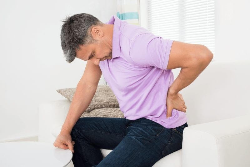
If you have lower back or buttocks pain which runs into your thigh or past the knee to one leg and foot, a healthcare professional may diagnose your symptoms as sciatica. Sciatica is a medical term used to describe painful sensations caused by the compression or impingement of the sciatic nerve. This compression is normally caused by a disc herniation or a bone spur pressing on one of the nerves in the lower back.
Sensations, or unusual feelings, could include numbness, tingling, pins and needles, and sometimes pain referred to as electric-shock-like. Determined by the individual nerve that is affected, pain may radiate only into the buttocks or all the way down to the foot.
Sciatica pain generally radiates along the length of the sciatic nerve, the longest and largest nerve in the human body, usually from the lower back, down the buttocks, and into the thigh and leg as well as the foot. One hallmark of classic sciatica is when the painful symptoms are felt beneath the knee and sometimes down into the foot and great toe. Usually, sciatica only affects one side of the body, however, it may occasionally affect both sides of the body.
Radicular Pain or Radiculopathy
Radicular pain, or radiculopathy, are different terms used to describe similar symptoms. Your healthcare professional may commonly utilize these terms interchangeably while discussing your sciatica. Radiculopathy is pain and/or an adverse sensation that travels past the affected site, along the length of a nerve. When a spinal nerve root is compressed, pinched or injured, it may become inflamed. Common conditions which could cause this kind of problem are spinal stenosis, foraminal stenosis or herniated discs.
What to Expect from a Sciatica Diagnosis
In order to determine the proper diagnosis of your sciatica symptoms, a healthcare professional may ask a series of questions, for instance:
When did the pain begin?
Where do you feel the pain?
What activities worsen or reduce pain and symptoms?
Does the pain go all the way down your leg or does it stop at the knee?
Is there weakness or tingling in your thighs or feet?
How severe is your pain, on a scale of 1 to 10? (10 being the worse pain possible)
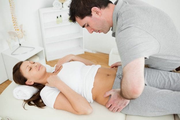
The healthcare professional may conduct a straight-leg test to find out whether you’ve got irritation or inflammation on a nerve. In order to perform this evaluations, you lie on your back while the doctor lifts each leg. When lifting a leg causes, or generates sciatic-like pain and sensations, you might have a bulging or ruptured disc, best known as a disc herniation.
Furthermore, the healthcare professional may ask you to walk as you normally do, then on your heels and next on your toes. This enables the physician to confirm your balance and aspects of lower-body strength. Compression or impingement of the sciatic nerve may cause muscle fatigue in the foot that will be revealed by these tests and evaluations. During your examination, your healthcare professional will:
Look at your position and range of movement
Note any movement that causes pain
Examine the curvature and alignment of your spine
Feel for muscle strain
Assess your sensation
Test your reflexes and muscle strength
Your doctor may order a plain x-ray, CT scan or MRI to help see the source of your sciatica more clearly. The CT scan or MRI provides the doctor with several snapshots of your spine, and will help confirm a suspected diagnosis. The findings of an imaging test are compared to the information that the doctor gathers during the taking of your medical history, and physical and neurological examination outcomes. An accurate identification is one of the very first steps in determining the best treatment options.
If it’s not Sciatica, What Else Could it Be?
Only a healthcare professional can tell for sure if your symptoms are sciatica or not. There are many complex structures in the spine which can result in similar kinds of pain. For instance, the joint between the pelvic and sacrum, or the sacroiliac joint, or SI joint, which is the smallest portion of the spine, may lead to pain from the buttock in the case of injury or due to an aggravated condition. You may also feel sciatica-like pain and discomfort if you sprain a very low back facet joint, which are the connecting joints at the back region of the spine. A tear in a disk can lead to pain down to the leg. The hip joint can occasionally trigger pain at the thigh as well. It’s essential to seek proper medical attention to assess the source of your symptoms.
Sciatica Treatment
Treatments for sciatica pain are diverse and there are lots of options to choose from. While sciatic nerve pain and radicular pain symptoms may resolve with the use of many traditional and alternative treatment options, severe cases may require surgery. Normally, some middle ground of these two extremes is the answer for curing sciatica.
Sciatica usually may be treated nonsurgically with short (24 to 48 hours) bed rest and pain relievers like aspirin or acetaminophen. In some cases, the physician may prescribe drugs and/or medications that relieves nerve pain, such as gabapentin. Oral steroids are another commonly used treatment to calm pain down. Typically, patients with sciatica feel better over time, generally in a few weeks. If pain persists, however, injections might be discussed. Muscle cramps, which might accompany sciatica symptoms, might be treated with heat or cold. Your physician will tell you to take brief walks, and might prescribe physical therapy. Once you recover, your doctor may also give you exercises to strengthen your back.
Can Chiropractic Care Treat Sciatica?
Chiropractic care is one of the top treatment options used for sciatica pain. Utilizing a variety of methods and techniques, chiropractic care doesn’t simply reduce the symptoms, it can ultimately fix the health issues associated with sciatica and prevent further circumstances of the collection of symptoms.
A good chiropractic care regimen might include spinal adjustments and manual manipulations, passive therapies, spinal decompression, massage therapy, and physical therapy to help reduce pain and correct the underlying problem causing it. A great chiropractic solution is going to be a plan which entails many or all of the above mentioned remedies as determined by your personal needs and recovery timeline. Furthermore, a chiropractor may recommend a series of appropriate stretches and exercises to help speed up the recovery process and promote a long-lasting recovery so you can live a pain-free life.
Dr. Alex Jimenez’s Insight
Sciatica is a medical term used to describe a collection of symptoms, including, pain, numbness or tingling sensations, caused by the compression or irritation of the sciatic nerve in the lower back. Although symptoms of sciatic nerve pain, or sciatica, are commonly concentrated in the lower back, radiating pain or radiculopathy may sometimes occur along the length of the sciatic nerve. A bulging or herniated disc is one of the most prevalent health issues which lead to sciatica. It’s essential to receive a proper diagnosis of any painful symptoms in order to follow-up with the best treatment options. Chiropractic care can help treat sciatica through the use of spinal adjustment and manual manipulations, among other treatment modalities, by carefully restoring the original alignment of the spine and reducing nerve compression and irritation associated with sciatic nerve pain.
If you believe that you are suffering from sciatic nerve pain, then consider the chiropractic care alternative solution. Many chiropractors can help by building a customized restoration plan around your requirements and goals. With years of experience, friendly employees, and innovative equipment, the proper chiropractor will get you back to normal the natural way. The scope of our information is limited to chiropractic as well as to spinal injuries and conditions. To discuss the subject matter, please feel free to ask Dr. Jimenez or contact us at 915-850-0900 .
Curated by Dr. Alex Jimenez
Additional Topics: Sciatica
Sciatica is medically referred to as a collection of symptoms, rather than a single injury and/or condition. Symptoms of sciatic nerve pain, or sciatica, can vary in frequency and intensity, however, it is most commonly described as a sudden, sharp (knife-like) or electrical pain that radiates from the low back down the buttocks, hips, thighs and legs into the foot. Other symptoms of sciatica may include, tingling or burning sensations, numbness and weakness along the length of the sciatic nerve. Sciatica most frequently affects individuals between the ages of 30 and 50 years. It may often develop as a result of the degeneration of the spine due to age, however, the compression and irritation of the sciatic nerve caused by a bulging or herniated disc, among other spinal health issues, may also cause sciatic nerve pain.
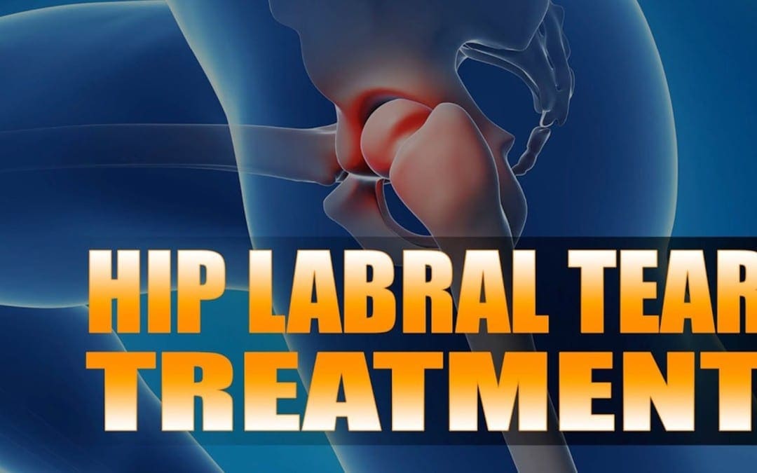
Hip Labral Tear: Andrew Hutchinson turned to chiropractic care and crossfit rehabilitation after suffering a high ankle sprain and a hip labrum tear for which he went through with surgery to repair it. After being bedridden for months in order to properly recover, Andrew Hutchinson transitioned to chiropractic care and crossfit rehabilitation to regain his strength, mobility and flexibility before returning to play. Although he has suffered other sports injuries, Andrew Hutchinson continues to trust in chiropractic care and crossfit rehabilitation to keep his spine properly aligned and maintain overall health and wellness.
Hip Labral Tear Treatment
Labrum tears in athletes can occur from a single event or recurring trauma. Running may cause labrum tears due to the labrum being utilized more for weight bearing and taking excess forces while at the end-range motion of the leg. Sporting activities are probable causes, specifically those that require frequent hip rotation or pivoting to a loaded femur as in ballet or hockey. Constant hip rotation places increased strain on the capsular tissue and harm to the iliofemoral ligament. This subsequently causes hip instability putting increased stress on the labrum and causing a hip labrum tear.
We focus on what works for you. We also strive to create fitness and better the body through researched methods and total wellness programs. These programs are natural, and use the body�s own ability to achieve goals of improvement, rather than introducing harmful chemicals, controversial hormone replacement, surgery, or addictive drugs.
We want you to live a life that is fulfilled with more energy, positive attitude, better sleep, less pain, proper body weight and educated on how to maintain this way of life. I have made a life of taking care of each and every one of my patients.
I assure you, I will only accept the best for you�
God Bless You & Your Health�?
If you have enjoyed this video and/or we have helped you in any way please feel free to subscribe and share us.
5 benefits of�walking in order to achieve better health is not new. Doctors and fitness experts have been touting its benefits for decades. When you walk, you engage more than 200 muscles � this includes your pelvis and spine. This makes it an exceptional complement to chiropractic treatment. However, if you aren�t convinced, these five compelling reasons that chiropractic patients should walk are sure to win you over.
5 Benefits Of Walking
Helps With Weight Loss & Weight Management
When you are carrying around excess weight it can lead to back pain and impaired mobility. Fat around your middle, especially in the stomach area, throws your body off balance. There is extra weight in front and it pulls that portion of your body forward, causing a swayback effect.
The pain in the lower back that is caused by this pressure can be excruciating. Over the long term this can cause damage to your spine and cause misalignment. While girdles or slings may help, the permanent remedy is to lose the weight. Walking is an excellent, low impact exercise that helps you lose weight, stay active, and stay healthy.
Improves Mobility & Flexibility
As we age we become less flexible and we don�t have the mobility of youth. As you walk, your circulation increases and that helps improve flexibility and mobility.
Add a little light stretching to the mix, along with regular chiropractic treatments and you will have a much better range of motion. Your posture will improve and you will reduce your chance of injury during physical activity. All this greatly enhances your spinal health making walking a great complement to chiropractic care.
Relieves Back Pain
Back pain is one of the top reasons that Americans miss work and worldwide it is the number one cause of disability. It is also expensive. Each year, Americans spend upwards of $50 billion trying to escape back pain.
Walking is recommended by the American Chiropractic Association (ACA) to help ease back pain. It is a very good, low impact exercise that helps you manage your weight and stay active � excess weight can cause your back to hurt. Walking helps relieve back pain, but it can help to prevent it as well. Even walking for just 30 minutes a day 3 to 5 times a week is beneficial.
Rehydrates Spinal Discs
There are small, fluid filled disks that lie between each vertebrae, acting as a cushion. As you move about during the day, gravity and certain movements cause your spinal disks to compress, squeezing the water out of them. This can lead to back pain and mobility issues.
The increased circulation from walking helps to force water into this area and the disks absorb that water and are rehydrated. This allows them to continue doing what they are supposed to � act as shock absorbers for your spine. It also helps if you drink plenty of water and stay hydrated throughout the day.
Improves Circulation
Good circulation is integral to spinal health as well as a properly functioning central nervous system. When you walk it increases your circulation allowing your blood to carry vital nutrients to your spine, organs, and your entire body. The soft tissues are enriched and nourished as toxins are flushed out.
Another benefit of this increased circulation is a decrease in blood pressure. It brings your body into balance so your muscles, ligaments, and joints are nourished. This, in turn, helps to make your chiropractic treatments more productive and beneficial.
5 benefits of walking is beneficial for whole body wellness. It can help you reduce your risk of many serious health conditions including diabetes, heart attack, stroke, and high blood pressure. It is also great for giving you a mental health boost and make you less prone to osteoporosis. So commit to walking just 30 minutes a day, 3 to 5 days a week. You will be astounded at the difference it will make.
Injury Medical Clinic: Elderly & Geriatric Fitness
Sciatic nerve pain, or sciatica, is a collection of symptoms caused by a wide array of underlying health issues. While there are numerous treatments for the treatment of sciatica, a doctor’s treatment plan for back pain and sciatica may also include alternative treatments, such as acupuncture, acupressure, biofeedback, and/or yoga. Many patients have reported that alternative treatments have helped relieve their sciatic nerve pain. If you’re experiencing symptoms of sciatica, you might want to try the following alternative treatment options.
Acupuncture
Acupuncture practitioners have the belief that your body has an energy force called Qi or Chi (pronounced “chee”). They believe that if Chi is blocked, it can create physical disease. Both acupuncture and acupressure (see below) function to restore a healthy, energetic flow of Chi. (All these Eastern approaches to healing are distinct from Western scientific concepts. That doesn’t make them better or worse; it only makes them different.)
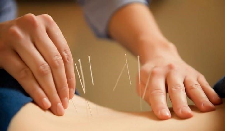
In order to perform acupuncture, acupuncturists insert very fine needles, which are sterilized and disposable, into specifically exact points on the body. These points are known as meridians. Each meridian is the same as a channel, referred to as an acupoint, or acupuncture point. Because meridians run near the skin’s surface, it is not necessary to insert needles deep into the tissue. Meridians correspond to specific sections of the human body or to a human body system like the nervous, musculoskeletal, cardiovascular, or lymphatic system. During an acupuncture treatment, the acupuncture practitioner will usually gently twirl or heat some or all of the needles.
It’s common for individuals to question whether acupuncture is uncomfortable, however, even patients who have been afraid of the needles at first, have found acupuncture to be a relaxing and painless experience. If you would like to pursue acupuncture treatment, please make sure to look for a licensed acupuncture practitioner who uses sterile and disposable equipment.
Acupressure
Acupressure has often been formerly compared to acupuncture. Acupressure is a non-invasive, secure, and gentle therapy which is believed to help unblocks Qi without using needles. The acupressure practitioner uses their thumbs, fingers, and elbows to put an exact quantity of pressure to specific points in the body. Acupressure therapy comprises the use of consistent pressure to one or more points and briskly rubbing against the acupressure point to stimulate it. Acupressure points and acupuncture points are identical.
Biofeedback
This type of alternative treatment option involves much more than simply telling your body to “stop feeling pain” Biofeedback is a mind-body therapy which rewires the brain by teaching you how to change or control a habitual reaction to pain or stress.
Could a person just “believe their pain away” with biofeedback? Unfortunately, it’s not that simple. As a matter of fact, it requires a much more demanding effort from the individual. Biofeedback frequently requires intensive patient participation and it is not an alternative treatment option for everybody. Some experts view biofeedback as a controversial therapy because its use in the treatment of low back pain or sciatica has not been adequately researched. In spite of this view, many individuals have benefited from biofeedback.
Biofeedback involves using special equipment to quantify and provide “feedback” to the individual about his or her physiological reactions to certain stimuli, like stress and muscle tension. By instructing the patient to perform deep breathing techniques, visualization, and mental and physical exercises, the individual learns how to control their response to stress and pain related to muscle tension, in this scenario, muscle tension that may contribute to sciatica.
Yoga
Yoga stretches can decrease symptoms of sciatica when the trigger is piriformis syndrome (sometimes a controversial diagnosis). Piriformis syndrome occurs when the piriformis muscle found in the lower portion of the spine compresses the sciatic nerve. This muscle aids in hip rotation. Gently stretching this muscle may help reduce sciatic pain. However, certain yoga stretches may aggravate symptoms of sciatica. Poses such as forward bending and twisting can irritate sciatic nerve pain. Any exercise which involves extending the back of the thighs (hamstrings) can irritate sciatica. As with any exercise, the patient must remember not to push their body beyond their pain limitations. Respect the body, and remember: Gently stretch.
As part of your treatment plan, you might want to try other sciatica treatment options. The following list are sciatica treatment, you may want to research to determine if they’re appropriate for you:
Chiropractic care
Physical therapy
Drugs/medications
Surgery
Chiropractic Care
Chiropractic care is a popular, alternative treatment option commonly utilized to help treat sciatica. The purpose of chiropractic care is to promote the body’s capacity to heal itself naturally, without the need for drugs/medications or surgery. It’s based upon the scientific principle that limited spinal movement leads to pain and decreased function and performance.
A chiropractor may use a variety of methods or techniques to improve sciatic nerve pain. The type of chiropractic care provided depends on the reason for the individual’s sciatica. A sciatica treatment program may include ice/cold treatments, ultrasound, TENS, and spinal adjustments or manual manipulations. Below are more details on these chiropractic care modalities.
Ice/cold treatment reduce inflammation and help improve sciatic nerve pain.
Ultrasound is gentle heat made by sound waves which penetrates deep into the soft tissues. Ultrasound increases circulation and helps to reduce muscle spasms, cramping, swelling, stiffness, and pain.
TENS unit (transcutaneous electrical nerve stimulation) is a small box-like, battery-powered, mobile muscle sculpting system. Variable intensities of electric current control acute pain and reduce muscle spasms. Bigger versions of this home-use TENS units are used by chiropractors, physical therapists and other rehabilitation professionals.
Spinal adjustments and manual manipulations are at the core of chiropractic care. Manual manipulation frees restricted movement of the spine and helps reestablish misaligned vertebrae in the spine. Spinal adjustments can help reduce nerve compression responsible for inducing pain, muscle spasm, inflammation and other symptoms associated with sciatica. Spinal adjustments and manual manipulations have been proven to be safe and effective.
Furthermore, a chiropractor may utilize physical therapy to help improve symptoms of sciatica. After a careful diagnosis, a doctor of chiropractic can recommend a series of appropriate stretches and exercises which, together some of the chiropractic care modalities mentioned above, can help speed up the recovery process. Chiropractic care focuses on healing through movement, helping to restore the proper connection between the brain and body in order to promote natural healing of sciatica nerve pain.
Dr. Alex Jimenez’s Insight
Because sciatica can be caused by a variety of underlying health issues, many different treatment modalities can be used to help relieve the common low back pain complaint. However, alternative treatment options, such as acupuncture, yoga and chiropractic care, have become increasingly popular in the treatment of sciatic nerve pain. Among the wide array of alternative treatment options, chiropractic care has become one of the most well-known modalities for improving symptoms of sciatica. Chiropractic care utilizes spinal adjustments and manual manipulations to carefully correct misalignments in the spine, or subluxations, which are often the most prevalent cause of sciatica. Other alternative treatment options can also efficiently help treat sciatic nerve pain without the need for drugs/medications or surgery.
Can Alternative Treatment Options Help Treat Sciatica?
There is no right or wrong answer to this question: several alternative treatment options can help relieve your sciatic nerve pain but they may not provide the same relief to another individual. If you’re considering trying alternative treatment options to address your own sciatica, discuss the options with your doctor. They might also have recommendations for healthcare professionals which can ultimately help improve your sciatica. The scope of our information is limited to chiropractic as well as to spinal injuries and conditions. To discuss the subject matter, please feel free to ask Dr. Jimenez or contact us at 915-850-0900 .
Curated by Dr. Alex Jimenez
Additional Topics: Sciatica
Sciatica is medically referred to as a collection of symptoms, rather than a single injury and/or condition. Symptoms of sciatic nerve pain, or sciatica, can vary in frequency and intensity, however, it is most commonly described as a sudden, sharp (knife-like) or electrical pain that radiates from the low back down the buttocks, hips, thighs and legs into the foot. Other symptoms of sciatica may include, tingling or burning sensations, numbness and weakness along the length of the sciatic nerve. Sciatica most frequently affects individuals between the ages of 30 and 50 years. It may often develop as a result of the degeneration of the spine due to age, however, the compression and irritation of the sciatic nerve caused by a bulging or herniated disc, among other spinal health issues, may also cause sciatic nerve pain.
There are many different treatments for cerebral palsy available today, however each case of cerebral palsy is as unique as the individual it affects. Because cerebral palsy can ultimately affect the normal functioning of the brain, treatment approaches which enhance the connection between the brain and the body are essential. Various treatments will work for different patients. A treatment known as physical therapy, or physiotherapy, is categorized as a non-medicinal treatment of cerebral palsy with the usage of massage, exercise, heat, and other external means of treatment.
Physiotherapy can be used to help cerebral palsy patients improve motion and motor abilities. Since cerebral palsy is a physical and movement disorder that disrupts the brain’s ability to correctly control muscle movement, physiotherapy can work wonders in helping cerebral palsy patients achieve mobility. Cerebral palsy physical therapy techniques are dependent on the degree of physical limitations of the person, and what’s going to be most beneficial to the cerebral palsy patient. Chiropractic care, can also include physical therapy techniques. Because the brain is believed to be lacking proper stimulation for functioning through cerebral palsy, chiropractic care can offer proprioception of touch for the assistance of mobility for the increased sensory stimulation of the brain through spinal adjustments and manipulations.
Physiotherapy for Cerebral Palsy
Cerebral palsy is the most common physical disability in children and it also represents the most frequent diagnosis in children who receive physical therapy. The harshness of limitations in gross motor function among children with cerebral palsy varies greatly, as some can walk without helping devices while some must use battery-powered wheelchairs. Physical therapists help children discover better ways to balance and move, as well as learn to walk, use their wheelchair, stand up with help, or go up and down stairs safely. The physical therapists engaged in physiotherapy reduce further growth of musculoskeletal problems by preventing muscle weakening, deterioration, and contracture during the suitable physiotherapy methods.
Physiotherapy usually consists of a couple kinds of treatment and helps a cerebral palsy patient to improve their gross motor abilities. Motor abilities that utilize the big muscles in the body, such as those in the arms and legs, are called gross motor abilities. This kind of physical therapy can help improve a cerebral palsy patient’s balance and motion.
Physical therapy for cerebral palsy patients consists of activities and education to enhance flexibility, strength, mobility, and function. A physical therapist also designs, modifies, and orders elastic gear to be used in the rehabilitation. Physical therapy can take place in clinics, hospitals, schools, and ought to continue in the home through a workout program. Physical treatment for cerebral palsy patients won’t be effective without an ongoing daily home program.
Physical Therapy Methods for Cerebral Palsy
A physical therapy program must include lots of exercises that include stretching, strengthening, and positioning. To elongate the muscles, the arms and legs must be transferred in ways that produce a slow, steady pull on the muscles to keep them loose. Because of the greater muscle tone of the cerebral palsy patient, they tend to have tight muscles. Therefore, it’s extremely important to perform daily stretches to maintain the arms and legs limber, allowing the child to continue to move and function. Strengthening exercises work specific muscle groups to enable them to encourage your system better and increase function. Positioning requires your system to be set in a particular position to achieve long stretches. Some places help minimize unwanted tone. Positioning can be achieved in many different ways. Bracing, abduction pillows, knee immobilizers, wheelchair inserts, sitting recommendations, and handling techniques are a part of placement techniques utilized in physical therapy for cerebral palsy patients.
New methods of physical therapy for cerebral palsy patients have taken into the water. Aquatic-based rehabilitation employs the physical properties of water to either resist or help in the operation of exercises. Cerebral palsy patients undergo muscle shortening in the majority of their involved extremities and it becomes a difficult job to lengthen the affected musculature with regular stretching while needing to manage the effects that gravity has on the spastic leg or arm. In earlier times there was clinical bias against strengthening activities for this population. But, recent study findings are revealing that kids with cerebral palsy may gain from strengthening applications and that strength is directly associated with motor function. Some of the recorded advantages are optimization of neuromuscular responses, improved motor unit contraction synchrony and facilitation of maximal muscle contraction combined with a wide available selection of motion.
Physical treatment for cerebral palsy patients does not heal spasticity but can improve impairments and limitations. Physical treatment for cerebral palsy patients is an important step towards an independent lifestyle. If these changes happen only in the therapy gym, the disability remains unchanged. Therapy must improve skills to carry out meaningful tasks in everyday life. Changing the level of handicap is the ultimate aim of physical therapy for cerebral palsy.
Occupational therapy is another element of physiotherapy used for cerebral palsy patients, and it’s used for aiding in the development of fine motor skills. Fine motor skills focus on the use of smaller muscles, such as those from the face, fingers, toes, palms, and feet. Fine motor skills have been used during daily living skills such as eating, dressing, writing, etc., and are fine tuned by occupational physiotherapy.
Physiotherapy also entails picking the right sort of adaptive equipment that could enhance a cerebral palsy patient’s motor abilities. Wheelchairs, walkers, special eating utensils and other adaptive equipment supply a patient with the liberty to accomplish some tasks on their own.
Additional types of physiotherapy like language and speech therapy might also be incorporated into a cerebral palsy patient’s program. Physiotherapy in the form of language and speech therapy that enables a cerebral palsy patient to communicate more easily with other people by developing the facial and jaw muscles, enhancing speech or sign language messages, and introducing communication resources such as computers and other visual aids.
Dr. Alex Jimenez’s Insight
Cerebral palsy is a lifelong group of movement disorders with no cure. However, several treatment options can help improve the quality of life of a patient with cerebral palsy. Chiropractic care and physical therapy, or physiotherapy, are some of the most common treatment approaches utilized to help restore strength, flexibility and mobility for individuals and children with cerebral palsy, without the need for drugs/medications and surgery. Chiropractic care can help improve many aspects associated with cerebral palsy due to the stimulation of the brain through touch, using spinal adjustment and manual manipulations to enhance sensory receptors in patients with this movement disorder. A physical therapist, as well as a chiropractor, may generally recommend a series of stretches and exercises which can improve range of motion in patients with cerebral palsy. Chiropractic care and physical therapy have the ability to develop self-sufficiency in cerebral palsy patients where it was previously absent.
Chiropractic Care for Cerebral Palsy
Several other physical therapy options can also help provide some form of relief from painful symptoms for cerebral palsy patients. Chiropractic care has become a popular, alternative treatment approach which focuses on maintaining as well as improving the overall health of the body through the use of spinal adjustments and manual manipulations. Because different areas of the body can be affected in individuals and children with cerebral palsy, chiropractic care can be beneficial towards helping those limbs regains some strength, flexibility and mobility. A chiropractor who specializes in patients with cerebral palsy can also offer several rehabilitation and physical therapy stretches and exercises to achieve the desired semblance of activity from the cerebral palsy patient. Because chiropractic care utilizes touch through spinal adjustments and manual manipulations to enhance the structure and function of the brain and the body, the sensory stimulation provided by a chiropractor can promote the migration of the brain in order to help change the receptors of the brain.
Furthermore, chiropractic care can help treat other, less noticeable aspects of the motor disorder. When used as a part of a rehabilitation and physical therapy program, chiropractic care can help improve some of the more problematic symptoms associated with cerebral palsy, including muscle spasms, seizures, and leg and arm issues through touch mobility protocols. The connection between the body and the brain has long been the center focus of chiropractic care, which is why spinal adjustments and manual manipulations are commonly utilized to enhance the stimulation of the brain, the spine, the nerves and the remaining structures of the body, especially in the case of patients with cerebral palsy, where the proper stimulation of the brain is necessary in order to restore function and improve quality of life. By carefully working to restore the natural alignment of the spine, a doctor of chiropractic, or chiropractor, can improve symptoms of back pain which may often be caused by the stress being placed on the spine�in patients with cerebral palsy. The purpose of chiropractic care and physical therapy is to improve physical movement and coordination, speech, vision and intellectual development�for patients with cerebral palsy.
Physiotherapy is an integral part in the vast majority of many cerebral palsy patients’ lives. Physiotherapy has the ability to develop self-sufficiency in cerebral palsy patients in which it was previously absent. A kid with cerebral palsy can start physiotherapy in just about any age. Speak with your child’s doctor about setting up physiotherapy program today. The scope of our information is limited to chiropractic as well as to spinal injuries and conditions. To discuss the subject matter, please feel free to ask Dr. Jimenez or contact us at 915-850-0900 .
Curated by Dr. Alex Jimenez
Additional Topics: Sciatica
Sciatica is medically referred to as a collection of symptoms, rather than a single injury and/or condition. Symptoms of sciatic nerve pain, or sciatica, can vary in frequency and intensity, however, it is most commonly described as a sudden, sharp (knife-like) or electrical pain that radiates from the low back down the buttocks, hips, thighs and legs into the foot. Other symptoms of sciatica may include, tingling or burning sensations, numbness and weakness along the length of the sciatic nerve. Sciatica most frequently affects individuals between the ages of 30 and 50 years. It may often develop as a result of the degeneration of the spine due to age, however, the compression and irritation of the sciatic nerve caused by a bulging or herniated disc, among other spinal health issues, may also cause sciatic nerve pain.
Sciatica is characterized by pain in the lower back and gluteal region. This pain can radiate down one or both legs into the thigh, calf, ankle, and foot. Genuine sciatica occurs when pain travels beneath the knee.
Sciatic nerve pain results when the base of the spine is compressed or irritated and/or if trauma from an injury or an aggravated condition have compressed the spinal segments surrounding the sciatic nerve. The sciatic nerve is located at the sacral areas of the spine and the lumbar spine. Sciatic nerve pain or sciatica can be described as sharp, dull, burning, tingly, numb, continuous, or intermittent and generally affects only one side of the body. It can radiate throughout the entire length of the nerve, in some cases all of the way down to the feet.
Sciatic nerve pain is most often the result of a bulging or herniated disc, spinal stenosis, or in extremely rare instances, infection or tumor. The cause of your pain determines what are your treatment options to relieve sciatica.
Sciatica Treatment Options
The following article lists several common sciatica treatment options. You may want to read about:
People who have lower back pain have been prescribed bed rest so as to offer relief for aching bones and joints. Research in recent years has suggested that bed rest alone won’t offer relief for those suffering from sciatica or sciatic nerve pain.
Staying active may be more beneficial for people who suffer with back pain due to the compression or irritation of their sciatic nerve. Not to say that you ought to be running marathons. Activity means being mobile and up for long periods of time which are not sufficient to cause further injury and aggravation for your back pain. Some healthcare professionals may prescribe certain exercises, or some could simply indicate walking.
Sciatic Nerve Pain Relief
Pain is often treated with a non-steroidal anti-inflammatory medication (NSAID) such as ibuprofen or codeine (in severe cases).
In some cases a cortisone-like drug could be injected into the epidural space surrounding the spinal column. This process is like the epidural used during childbirth, and it’s called an epidural steroid injection. A course of this type of treatment may offer temporary relief, however, it doesn’t address the root of the problem. Treatment options like chiropractic care can help treat symptoms of sciatica at the source.
Chiropractic Treatment for Sciatic Nerve Pain
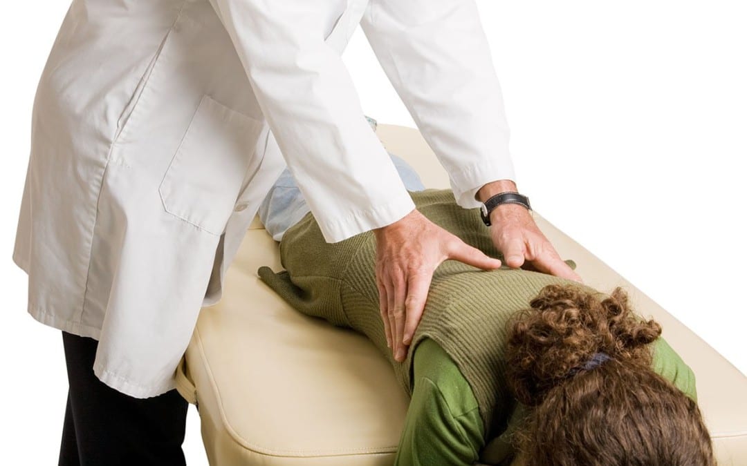
A chiropractor frequently treats people with sciatica. Their adjustments will aim to realign the spine, taking off the pressure of the sciatic nerve and often bringing rapid sciatica relief. When the stress is off, the body can begin to heal itself. While adjustments are probably most often used, other remedies may be given, especially if an adjustment is not recommended for the patient’s health issue. Other chiropractic treatment options might include using ice/cold treatment, ultrasound, a TENS (transcutaneous electric nerve stimulation) device or rehabilitative exercises. Ultrasound warms the region and increases circulation, which can reduce the swelling and muscular strain. A TENS device brings relief by using a slight electric current to relax muscle spasms and to increase endorphins. Additionally, a chiropractor might frequently consist of rehabilitative exercises so as to accelerate the recovery process. Massage therapy might also assist.
Ice/Cold Therapy reduces inflammation and aids restrain from sciatic nerve pain.
Ultrasound is gentle heat created by sound waves which penetrates deep into tissues. As mentioned previously, it increases circulation and reduces muscle spasms, cramping, swelling or inflammation, stiffness, and pain.
Adjustments (Spinal Manipulations). Spinal adjustments are in the heart of Chiropractic care. Manipulation supports restricted motion of the spine and helps to restore misaligned vertebral bodies with their proper position in the spinal column. Adjustment techniques differ from a swift high velocity thrust to those that combine minimal pressure and gentle pressure. Mastery of every technique is an art which requires great skill and precision. Spinal manipulation is the remedy that distinguishes chiropractic care from other medical areas.
Rehabilitative exercises. A combination of aerobics, strength training and stretching is commonly used to unleash pain-relieving endorphins in addition to relax the muscles which may be causing the nerve compression or irritation.
In some individuals, sciatica may fix itself, maybe occurring only once or a couple of times. However, if not treated properly, sciatica can worsen. A chiropractor can help bring relief, however, several alterations, will likely be required, especially if it has been occurring for a while. Letting a chiropractor treat sciatic nerve pain provides you a no surgery and no medication choice. Exercises will most likely be advisable to strengthen the muscles in the back to help prevent sciatica from recurring, and recovery is apt to take some time.
Surgery for Sciatic Nerve Pain?
If no other alternative treatment option has provided the patient relief from their symptoms, some patients with sciatica may discover substantial relief from surgery. In cases of herniated discs, a surgical procedure called a laminectomy may be performed. In this process, a portion of the posterior arch is removed to relieve pressure on pinched nerve tissues. In cases of spinal stenosis, the portion of bone that’s putting pressure on the sciatic nerve system could be taken off. Surgery is not for everybody. But for people who have shown no sign of progress in four to six weeks and that have had CT scans (computed tomography) or MRI that reveal a herniated disc or spinal stenosis, surgery may provide substantial relief.
Dr. Alex Jimenez’s Insight
Surgical interventions are frequently discussed as a possible treatment option for a variety of injuries and/or conditions which cause sciatica, however, surgery should only be considered as a last resort after all other treatment options have been utilized without improvement. Chiropractic care is a natural treatment approach which focuses on the diagnosis, treatment and prevention of sciatic nerve pain through the use of spinal adjustments and manual manipulations, among other commonly utilized treatment methods. Through the proper alignment of the spine, chiropractic care focuses on allowing the human body to naturally heal its sciatica without the need for surgery or drugs/medications.
The scope of our information is limited to chiropractic as well as to spinal injuries and conditions. To discuss the subject matter, please feel free to ask Dr. Jimenez or contact us at 915-850-0900 .
Curated by Dr. Alex Jimenez
Additional Topics: Sciatica
Sciatica is medically referred to as a collection of symptoms, rather than a single injury and/or condition. Symptoms of sciatic nerve pain, or sciatica, can vary in frequency and intensity, however, it is most commonly described as a sudden, sharp (knife-like) or electrical pain that radiates from the low back down the buttocks, hips, thighs and legs into the foot. Other symptoms of sciatica may include, tingling or burning sensations, numbness and weakness along the length of the sciatic nerve. Sciatica most frequently affects individuals between the ages of 30 and 50 years. It may often develop as a result of the degeneration of the spine due to age, however, the compression and irritation of the sciatic nerve caused by a bulging or herniated disc, among other spinal health issues, may also cause sciatic nerve pain.
Children with cerebral palsy have various needs. Some children have problems with motor skills and spasticity, but normally pick up things pretty fast. Others have a full range of issues from motor skills to esophageal and respiratory problems. Since so many kids with cerebral palsy have an array of different medical needs, there isn’t one particular type of treatment which may help each and every child. Luckily, there are a number of different therapeutic remedies to select from, which range from holistic care, water therapy, and much more.
Acupuncture
Whilst generally not adopted in Western Medicine, acupuncture has been used for centuries by Asian countries and is viewed as a medicinal art. Some families with children who have cerebral palsy take their kids to an acupuncturist to try and relieve the frequent pain related to the disorder. Other kids find relief in acupuncture for painful birth injuries such as spina bifida, Erb’s palsy, and brain damage. Acupuncture uses needles to ease pain, often instead of medication.
Aquatherapy
Aquatherapy is among the most popular and beneficials form of treatment for children with cerebral palsy, as they suffer from limb maladiess, but this might also be advantageous for children who suffer with Erb’s palsy and are trying to regain movement in their arm.
Under the supervision of a trained and experienced professional therapist, kids may gain from the strength exercise and training afforded by the anti-gravity character of a pool. In this soothing environment, a child can have a respite from some of the pain which comes with the disability (occasionally cerebral palsy causes stress on the musculoskeletal frame by simply gravity and body weight), and they can still work through the natural curative and restorative nature of water.
Behavioral Therapy (Psychotherapy)
Some birth injuries involve an intellectual disability that impacts how kids interact in social scenarios. Other children might have had physical constraints that included them being house-bound for a long time, causing them to have a deficiency in social skills or cues. Behavioral therapy, also known as psychotherapy, allows patients to work through problems they may have within their social and mental health with a behavioral health professional.
Chiropractic Care and Massage Therapy
Children with cerebral palsy may benefit from chiropractic care and massage therapy for a few different reasons. Because some children with cerebral palsy may have experienced lots of strain or stress on their musculoskeletal system as a result of the disorder, requiring chiropractic care may ultimately be fundamental towards their proper spinal alignment as well as for their overall health and wellness.
Chiropractic care is a well-known alternative treatment option which utilizes spinal adjustments and manual manipulations to treat a variety of injuries and/or conditions associated with the musculoskeletal and nervous system, including back pain.
Another reason that a patient with cerebral palsy might need chiropractic care or massage treatment is for the basic goal of extending and stretching muscles. When muscles relax as they perform through such therapies, they are more inclined to become stronger and healthier which is needed if they are going to correctly learn how to walk. This kind of treatment isn’t generally suggested for kids suffering from spina bifida because the raw exposed nerves could be inadvertently mishandled, causing more problems.
Furthermore, chiropractic care can be used to help treat other, less noticeable aspects of cerebral palsy. The theory of chiropractic care is that by healing the central area around the spine, the extremities and other parts of the body affected by the disorder can become more normalized, allowing for improved function and quality of life. Chiropractic care can also help improve strength, mobility and flexibility in children with cerebral palsy and its associated symptoms.
Conductive Education
Some children with neurological or mobility impairment found in almost any brain-related birth trauma need help performing activities that regular people learn through daily exercise, learning, and experience. Since these children don’t often have the same sorts of experiences that non-disabled people have, conductive education is a form of special education that functions as a kind of study group for life.
Conductive education provides opportunities of every day learning experiences so that kids can have the exact same general education that non-disabled individuals do.
Hippotherapy
Using equine motion and connections with horses, children with all kinds of birth injuries could learn basic occupational and speech therapy. Hippotherapy is not therapeutic horseback riding, but rather a trained practitioner introduces the child to the horse and uses the horse to access the child in ways which were previously thought of as unconventional.
Hyperbaric Oxygen Therapy
Normally short-term treatment and frequently only experienced once or twice, Hyperbaric Oxygen Therapy is a method of fast-healing for some kids that have suffered oxygen deprivation (anoxic, hypoxic, HIE, birth asphyxia, and perinatal asphyxia). If an infant is delivered and does not breathe for the upcoming instant minutes, hyperbaric oxygen treatment is a great way to introduce a lot of oxygen into the blood stream preventing or lessening the seriousness of birth injuries such as cerebral palsy.
Occupational Therapy
Occupational therapy’s main objective is to work on creating balance, strength, and gait. An occupational therapist might consult with an orthopedic surgeon to operate on strengthening and firming muscles, in which following the occupational therapist can delegate casts and orthopedic devices which also help strengthen and form muscles. These methods are to help patients learn how to walk, and also to create control and strength to stop spasticity.
The occupational therapist also trains patients to function on decision-making, abstract reasoning, problem-solving, perception, memory, sequencing, and much more.
Play Therapy
Utilizing play with a variety of different toys in various public places, kids with all kinds of birth injuries can learn to appreciate themselves. Often children with birth injuries can feel that they’re different or that they have health issues and end up stressing about their difficulties more than having fun.
While they’re having fun in play therapy, they can learn the way to interact with other kids, learn about themselves, and to construct self-confidence.
Physiotherapy and Physical Therapy
Physiotherapy and physical therapy both operate on the rehabilitation of muscle groups. This is extremely important for children with shoulder dystocia, Erb’s palsy, Klumpke’s palsy, or Brachial Plexus palsy, and, in fact, kids suffering from these birth injuries won’t regain use of their hand or arm without physical and physiotherapy. Through this type of treatment, therapists strive to receive the perfect movement from their patients through an assortment of different challenges and exercises.
This can be like occupational therapy, though the focus is mainly on what the muscle groups are doing, and not on so many different targets like occupational therapy. A physical therapist is often like a personal trainer in a gym, training, cheering, and challenging.
Respiratory, Digestive, and Dietician Therapy
Some kids with cerebral palsy encounter respiration and esophageal problems. Consequently they can experience issues with eating, breathing, and drinking, which divides into digestive and dietician treatment, addressing what foods and drinks should be consumed. Respiratory treatment may primarily tackle breathing exercises to strengthen and optimize lung development, but may also address these other concerns.
Speech and Language Therapy
Speech and language therapy can be very important for kids with cerebral palsy and other forms of brain-related birth harm. Approximately, 1 out of every 4 patients with cerebral palsy don’t have the capability to speak. Speech and language therapy helps them to work on exercises which progress the learning of speech and get kids closer to communicating effectively.
Some speech and language therapists utilize programs that help patients understand the operation of language inside individuals, and these programs also provide communication boards using pre-formed responses so that children can get in the habit of responding with particular answers until they consider trying to verbalize these answers.
Vocational Counseling
This has many different sorts of therapists, a few children could be confused or jeopardized by visiting a lot of individuals, or, worse, by having so many people invade their home. One way of approaching treatment is by using a vocational counselor, one individual who can master several distinct types of treatment.
As vocational counselors might not have exactly the exact same depth in all of these subjects as one therapist would have regarding one subject, this might be a great first step for treatment with your little one. By getting your child to adjust to only one person interacting within their lifetime, they’re more inclined to concentrate on the subjects at hand.
Afterwards, if more obstacles and more depth is required, your kid may have more assurance in different areas (and with a few social abilities from connecting with this particular counselor) and may be able to handle other therapists more efficiently.
Yoga Therapy
Normally prescribed under the direction of an occupational or physical therapist, yoga therapy is a fantastic alternative for kids whose muscles need to be loosened or lengthened. Children with cerebral palsy suffer from particularly tight muscles, so yoga therapy helps them to work on extending and on making the muscles more limb. This type of treatment might be incorporated to other sorts of therapy, and it might also be delegated as “homework” to kids with cerebral palsy for optimal flexibility and, ultimately, optimal freedom.
Dr. Alex Jimenez’s Insight
Cerebral palsy is a lifelong set of movement disorders with no cure. However, various types of treatment options can help provide some forms of relief for individuals and children with cerebral palsy as well as help restore some function and quality of life. Because cerebral palsy can affect patients differently, people with the disorder can benefit from many different therapies, including chiropractic care and physical therapy. Chiropractic care is a popular, alternative treatment option which focuses on the diagnosis and treatment of several kinds of injuries and/or conditions, including cerebral palsy. Through the use of spinal adjustments and manual manipulations, a chiropractor can help improve strength, mobility and flexibility in people with cerebral palsy.
The scope of our information is limited to chiropractic as well as to spinal injuries and conditions. To discuss the subject matter, please feel free to ask Dr. Jimenez or contact us at 915-850-0900 .
Curated by Dr. Alex Jimenez
Additional Topics: Sciatica
Sciatica is medically referred to as a collection of symptoms, rather than a single injury and/or condition. Symptoms of sciatic nerve pain, or sciatica, can vary in frequency and intensity, however, it is most commonly described as a sudden, sharp (knife-like) or electrical pain that radiates from the low back down the buttocks, hips, thighs and legs into the foot. Other symptoms of sciatica may include, tingling or burning sensations, numbness and weakness along the length of the sciatic nerve. Sciatica most frequently affects individuals between the ages of 30 and 50 years. It may often develop as a result of the degeneration of the spine due to age, however, the compression and irritation of the sciatic nerve caused by a bulging or herniated disc, among other spinal health issues, may also cause sciatic nerve pain.
IFM's Find A Practitioner tool is the largest referral network in Functional Medicine, created to help patients locate Functional Medicine practitioners anywhere in the world. IFM Certified Practitioners are listed first in the search results, given their extensive education in Functional Medicine


 Relieves Back Pain