Transform your well-being with chiropractic care for the gluteus medius. Experience targeted solutions for enhanced movement and relief.
Comprehensive Guide to Chiropractic Care for Gluteus Medius Injuries and Hip Pain
Key Points
Gluteus Medius Role: This muscle stabilizes your pelvis and keeps you upright during movement, preventing you from wobbling like a poorly balanced skeleton.
Hip Pain Causes: Environmental factors like prolonged sitting, poor posture, or a car accident can strain the gluteus medius, leading to pain that feels like a grim reminder of mortality.
Chiropractic Benefits: Chiropractic care, combined with non-surgical treatments, can reduce hip pain by realigning joints and strengthening muscles, offering relief without the scalpel’s cold embrace.
Dr. Jimenez’s Expertise: In El Paso, Dr. Alexander Jimenez utilizes advanced imaging and diagnostics to connect injuries to legal claims, serving as a bridge between pain and justice.
Lifestyle Adjustments: Small changes, like better posture or targeted exercises, can prevent hip pain from haunting your daily life.
Understanding Hip Pain and the Gluteus Medius
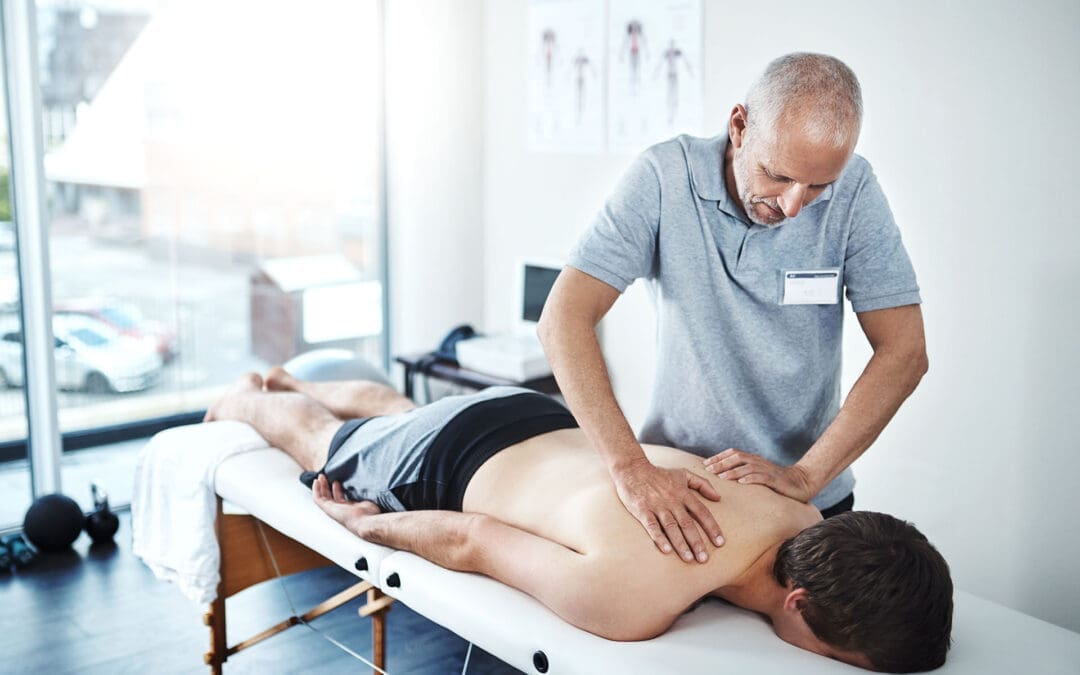
Hip pain can sneak up like a shadow in the night, turning simple tasks like walking or sitting into a grim ordeal. The gluteus medius, a key muscle in the hip, plays a crucial role in maintaining lower-body stability. When it’s injured—whether from a car accident, poor posture, or just life’s relentless grind—pain can radiate, making you feel like you’re starring in your own personal horror show. Chiropractic care, as practiced by experts like Dr. Alexander Jimenez in El Paso, offers a non-invasive way to tame this beast, restoring function and easing discomfort.
Why Chiropractic Care Matters
Chiropractic care isn’t just about cracking backs; it’s a calculated dance to restore balance to your body’s structure. For gluteus medius injuries, chiropractors use adjustments, soft tissue therapy, and targeted exercises to realign joints and strengthen muscles. This approach can reduce inflammation and pain, helping you move without feeling like you’re auditioning for a role as a creaky old gate.
Dr. Jimenez’s Role in Personal Injury Cases
In El Paso, Dr. Jimenez stands out as a beacon for personal injury victims, particularly those reeling from motor vehicle accidents (MVAs). His ability to connect clinical findings—through advanced imaging like MRI and dual-scope diagnostic procedures—to legal documentation makes him a vital ally. Whether it’s whiplash or a strained gluteus medius, his expertise ensures injuries are thoroughly assessed and properly reported for legal claims, offering a lifeline to those navigating the murky waters of recovery and justice.
Small Changes, Big Impact
Simple tweaks to your daily routine can keep hip pain at bay. From standing desks to targeted stretches, Dr. Jimenez’s insights, available through El Paso Back Clinic and his LinkedIn profile, guide patients toward lasting relief. These changes are like small wards against the creeping specter of chronic pain.
The Gluteus Medius: Your Hip’s Unsung Hero
Picture the gluteus medius as the grim, silent guardian of your hips. Nestled on the side of your pelvis, this muscle keeps you stable when you walk, run, or stand on one leg. Without it, you’d wobble like a poorly strung marionette, collapsing under the weight of your own existence. But when this muscle is injured, it’s like a betrayal from within, causing pain that can radiate from the hip to the lower back or even down the leg.
The Role of the Gluteus Medius in Lower Extremity Function
The gluteus medius is a key player in the lower extremities, acting as a stabilizer for the pelvis during movement. It abducts the hip (moves the leg away from the body) and assists in internal and external rotation. When you take a step, it prevents your pelvis from dropping on the opposite side, ensuring smooth, balanced motion. Studies show that a weak or injured gluteus medius can lead to issues such as Trendelenburg gait, where the pelvis tilts awkwardly, giving the appearance of an exaggerated posture, akin to auditioning for a role in a zombie flick (Mucha et al., 2019).
Weakness or tears in the gluteus medius can also contribute to lower back pain, knee issues, and even ankle instability. A systematic review found that individuals with low back pain often have impaired gluteus medius function, highlighting its role in the kinetic chain (Sadler et al., 2019). Essentially, if your gluteus medius is slacking, the rest of your lower body has to pick up the pieces, often with disastrous results.
References
Mucha, M. D., Caldwell, W., Schlueter, E. L., Walters, C., & Hassen, A. (2019). Gluteus medius tears of the hip: A comprehensive approach. Journal of the American Academy of Orthopaedic Surgeons, 27(3), 77–85. https://pubmed.ncbi.nlm.nih.gov/30278009/
Sadler, S., Cassidy, S., Peterson, B., Spink, M., & Chuter, V. (2019). Gluteus Medius Muscle Function in People with and without Low Back Pain: A Systematic Review. BMC Musculoskeletal Disorders, 20(1), 463. https://pubmed.ncbi.nlm.nih.gov/31640621/
Why Hip Pain Haunts: Environmental Culprits
Hip pain doesn’t just appear like a ghost in the night; the mundane horrors of daily life often summon it. Environmental factors—those sneaky, everyday villains—can wreak havoc on your gluteus medius and hips, turning your body into a creaky haunted house.
Prolonged Sitting: The Silent Killer
Sitting for hours, whether at a desk or in a car, is like sentencing your hips to a slow, torturous demise. It tightens the hip flexors and weakens the gluteus medius, creating an imbalance that screams for attention. Research shows that prolonged sedentary behavior is linked to musculoskeletal pain, including hip discomfort (Law et al., 2020). Imagine your gluteus medius, neglected and underused, plotting its revenge with every ache.
Poor Posture: The Slouch of Doom
Slouching isn’t just unflattering; it’s a biomechanical nightmare. Poor posture shifts your pelvis, overloading the gluteus medius and causing strain. Studies on posture and hip pain suggest that misaligned posture can exacerbate musculoskeletal issues, particularly in the hips and lower back (Smith et al., 2020).
Motor Vehicle Accidents: A Crash Course in Pain
Car accidents, even minor ones, can jolt the gluteus medius into dysfunction. The sudden force can strain or tear the muscle, leading to inflammation and pain. In El Paso, where rainy weather increases accident risks, the aftermath of MVAs often includes hip injuries (El Paso Back Clinic, n.d.). Whiplash-associated disorders (WAD) from accidents can also cascade into hip pain, as the body compensates for spinal misalignments (El Paso Back Clinic, n.d.).
Occupational Hazards: The Grind That Bites
Jobs requiring repetitive motions, heavy lifting, or prolonged standing can stress the gluteus medius. Construction workers, nurses, or even retail employees are at risk. A narrative review highlights that repetitive strain contributes to lateral hip pain, often tied to gluteus medius dysfunction (French et al., 2020).
Lifestyle Factors: The Everyday Terrors
Wearing high heels, carrying heavy bags on one shoulder, or even sleeping in awkward positions can damage your hips. These habits subtly strain the gluteus medius, leading to pain that feels like a cruel prank. Research on lifestyle and hip pain emphasizes the cumulative impact of these small stressors (French et al., 2020).
References
Law, D., Mark, J., & Smith, R. (2020). Hip Pain in Adults: Evaluation and Differential Diagnosis. American Family Physician, 101(2), 81–89. https://pubmed.ncbi.nlm.nih.gov/31939642/
Smith, J. A., Stabbert, H., & Bagwell, J. J. (2020). Posterior, lateral, and anterior hip pain due to musculoskeletal origin: A narrative literature review of history, physical examination, and diagnostic imaging. Journal of Chiropractic Medicine, 19(1), 1–14. https://pubmed.ncbi.nlm.nih.gov/33192189/
French, H. P., Woodley, S. J., & Schwank, A. (2020). Lateral hip pain: Relation to greater trochanteric pain syndrome. Current Reviews in Musculoskeletal Medicine, 13(5), 638–645. https://pubmed.ncbi.nlm.nih.gov/32748299/
El Paso Back Clinic. (n.d.). Auto accident insights for safe driving and recovering from WAD. Retrieved from https://elpasobackclinic.com/
Chiropractic Care: Banishing Hip Pain Without Surgery
Chiropractic care is like a well-aimed exorcism for hip pain, banishing discomfort without the need for invasive procedures. For gluteus medius injuries, chiropractors employ a multi-faceted approach that restores function and eases pain, all while keeping you out of the operating room’s cold embrace.
Why Chiropractic Works for Gluteus Medius Injuries
The gluteus medius thrives on proper alignment and balanced muscle function. Chiropractic adjustments realign the pelvis and spine, reducing stress on the muscles. According to a blog from El Paso Back Clinic, strengthening the gluteus medius post-injury involves targeted chiropractic interventions like spinal adjustments and soft tissue therapy, which improve blood flow and reduce inflammation (El Paso Chiropractor Blog, 2016). A randomized trial also found that core stability and hip exercises, often prescribed by chiropractors, improve function in patients with related pain (Jeong et al., 2020).
Non-Surgical Allies: A Team of Pain-Fighters
Chiropractic care doesn’t work alone; it’s backed by a grimly effective team of non-surgical treatments:
Massage Therapy: Loosens tight muscles and boosts circulation, like coaxing a grumpy ghost to leave. It’s particularly effective for post-MVA recovery (El Paso Back Clinic, n.d.).
Dry Needling: Targets trigger points in the gluteus medius, reducing pain with a precision that would make even Wednesday Addams nod approvingly (Gattie et al., 2021).
Rehabilitation Exercises: Progressive loading exercises strengthen the gluteus medius, preventing re-injury. A systematic review highlights their efficacy in restoring function (Moore et al., 2020).
Physical Therapy: Complements chiropractic care by improving mobility and strength, ensuring your hips don’t creak like an old coffin.
These treatments work together to address overlapping risk factors, like muscle imbalances or joint dysfunction, that amplify hip pain.
Dr. Jimenez’s Clinical Insights
Dr. Alexander Jimenez, DC, APRN, FNP-BC, brings a clinical rigor to hip pain treatment that’s as sharp as a guillotine. His approach, detailed at El Paso Back Clinic, emphasizes personalized treatment plans. Simple changes, such as using ergonomic chairs, correcting posture, or incorporating daily stretches, can help prevent hip pain from becoming a chronic issue. His LinkedIn profile showcases his expertise in integrating functional medicine with chiropractic care, providing patients with a holistic approach to recovery.
Jeong, U. C., Kim, J. S., Park, S. H., & Lee, J. H. (2020). Core stability and hip exercises improve physical function and activity in patients with non-specific low back pain: A randomized controlled trial. Journal of Back and Musculoskeletal Rehabilitation, 33(4), 581–589. https://pubmed.ncbi.nlm.nih.gov/31594203/
Gattie, E., Cleland, J. A., & Snodgrass, S. (2021). Effectiveness of dry needling and ischemic trigger point compression in the gluteus medius in patients with non-specific low back pain: A randomized short-term clinical trial. Journal of Bodywork and Movement Therapies, 27, 529–536. https://pubmed.ncbi.nlm.nih.gov/34391285/
Moore, D., Semciw, A. I., & Pizzari, T. (2020). A systematic review of rehabilitation exercises to progressively load the gluteus medius. Journal of Sport Rehabilitation, 29(2), 222–238. https://pubmed.ncbi.nlm.nih.gov/30676192/
El Paso Back Clinic. (n.d.). Trigger point therapy MVAs explained for patients. Retrieved from https://elpasobackclinic.com/
Chiropractic Care for Leg Instability- Video
Dr. Jimenez and Personal Injury Cases in El Paso
In El Paso, personal injury cases—especially those from MVAs—are as common as tumbleweeds in a desert storm. Dr. Alexander Jimenez stands out as a distinguished practitioner, offering hope to those who have been injured in accidents. His ability to connect the dots between injuries and legal claims is nothing short of macabre genius.
Advanced Diagnostics: Seeing the Invisible
Dr. Jimenez uses advanced imaging, like MRI and X-rays, to pinpoint gluteus medius injuries or other musculoskeletal damage. These tools reveal what the naked eye can’t, like a radiograph exposing a skeleton’s secrets. Dual-scope procedures, combining diagnostic and therapeutic techniques, allow him to assess and treat injuries with precision. This approach ensures that injuries are documented thoroughly, providing critical evidence for legal claims.
Bridging Medicine and Law
As a liaison between medical care and legal documentation, Dr. Jimenez ensures that personal injury victims receive comprehensive care while building a robust case. His detailed reports link clinical findings to accident-related injuries, helping attorneys secure fair compensation. This is especially vital in cases involving gluteus medius injuries, where pain can be debilitating but hard to prove without expert evaluation.
Why El Paso Needs Dr. Jimenez
El Paso’s busy roads and frequent accidents make practitioners like Dr. Jimenez invaluable. His work with MVA victims, detailed at El Paso Back Clinic, ensures that injuries like gluteus medius strains or tears are treated effectively while supporting legal outcomes. His reputation, reflected on LinkedIn, underscores his role as a trusted ally for those navigating the aftermath of personal injuries.
References
El Paso Back Clinic. (n.d.). Musculoskeletal injury treatment after car accidents. Retrieved from https://elpasobackclinic.com/
El Paso Back Clinic. (n.d.). Spinal injury rehabilitation tools for enhanced care. Retrieved from https://elpasobackclinic.com/
Small Changes, Big Relief: Practical Tips
Preventing hip pain doesn’t require a deal with the devil—just a few practical tweaks to your daily routine. Dr. Jimenez’s insights offer a roadmap to keep your gluteus medius happy and your hips pain-free.
Daily Adjustments
Ergonomic Seating: Use chairs that support proper posture to prevent hip strain. Think of it as giving your gluteus medius a comfy coffin to rest in.
Regular Movement: Stand and stretch every 30 minutes to prevent your hips from seizing up like a rusted gate.
Proper Footwear: Opt for supportive shoes instead of high heels to reduce stress on your hips and gluteus medius.
Targeted Exercises
Dr. Jimenez recommends exercises like side-lying leg lifts and clamshells to strengthen the gluteus medius. A systematic review supports progressive loading exercises to rebuild muscle strength (Moore et al., 2020). These moves are like training your hips to fend off pain’s ghostly grip.
Posture Correction
Stand tall and align your spine to reduce strain on the gluteus medius. Imagine you’re a gothic statue, poised and unyielding. Regular posture checks can prevent pain from creeping back.
Nutrition for Recovery
A diet rich in anti-inflammatory foods, like omega-3s and leafy greens, supports muscle healing. El Paso Back Clinic emphasizes nutrition’s role in MVA recovery, noting that a proper diet can reduce inflammation and speed recovery (El Paso Back Clinic, n.d.).
Table: Daily Tips for Hip Pain Prevention
Tip
Description
Benefit
Ergonomic Seating
Use chairs with lumbar support and proper height.
Reduces pelvic strain.
Regular Movement
Stand and stretch every 30 minutes during prolonged sitting.
Prevents muscle tightness.
Supportive Footwear
Wear flat, cushioned shoes instead of high heels.
Minimizes hip stress.
Gluteus Medius Exercises
Perform side-lying leg lifts and clamshells daily.
Strengthens stabilizing muscles.
Posture Correction
Maintain neutral spine alignment during sitting and standing.
Reduces biomechanical strain.
Anti-Inflammatory Diet
Include foods like salmon, walnuts, and spinach.
Supports muscle healing and reduces pain.
References
Moore, D., Semciw, A. I., & Pizzari, T. (2020). A systematic review of rehabilitation exercises to progressively load the gluteus medius. Journal of Sport Rehabilitation, 29(2), 222–238. https://pubmed.ncbi.nlm.nih.gov/30676192/
El Paso Back Clinic. (n.d.). Nutrition for accident injuries during recovery. Retrieved from https://elpasobackclinic.com/
The Bigger Picture: Chiropractic’s Role in Musculoskeletal Health
Chiropractic care isn’t just about fixing hips; it’s about restoring the body’s balance, like a grim ritual to banish chaos. For gluteus medius injuries, it addresses the root causes—misalignments, muscle imbalances, and inflammation—while preventing future issues.
Overlapping Risk Profiles
Hip pain often overlaps with other conditions, like low back pain or knee issues, due to the interconnected nature of the musculoskeletal system. A weak gluteus medius can destabilize the entire lower body, leading to a cascade of problems. Chiropractic care, combined with therapies like dry needling and massage, tackles these risks holistically, ensuring that one injury doesn’t summon a host of others (Gattie et al., 2021).
Long-Term Benefits
Regular chiropractic care can prevent chronic pain by maintaining proper alignment and muscle function. Studies show that core and hip exercises, often part of chiropractic plans, improve long-term physical function (Jeong et al., 2020). It’s like fortifying your body against the inevitable decay of time.
El Paso’s Unique Needs
In El Paso, where MVAs are a grim reality, chiropractic care is a lifeline. Dr. Jimenez’s integrative approach—combining adjustments, rehabilitation, and nutrition—addresses the unique needs of accident victims, ensuring they don’t just survive but thrive.
References
Gattie, E., Cleland, J. A., & Snodgrass, S. (2021). Effectiveness of dry needling and ischemic trigger point compression in the gluteus medius in patients with non-specific low back pain: A randomized short-term clinical trial. Journal of Bodywork and Movement Therapies, 27, 529–536. https://pubmed.ncbi.nlm.nih.gov/34391285/
Jeong, U. C., Kim, J. S., Park, S. H., & Lee, J. H. (2020). Core stability and hip exercises improve physical function and activity in patients with non-specific low back pain: A randomized controlled trial. Journal of Back and Musculoskeletal Rehabilitation, 33(4), 581–589. https://pubmed.ncbi.nlm.nih.gov/31594203/
Conclusion: A Serious Note
While we’ve danced with dark humor to lighten the mood, hip pain and gluteus medius injuries are no laughing matter. They can disrupt your life, limit your mobility, and cast a shadow over your daily routine. Chiropractic care, as championed by Dr. Alexander Jimenez in El Paso, offers a proven, non-invasive path to recovery. By addressing the root causes of pain, integrating advanced diagnostics, and supporting legal claims, Dr. Jimenez helps patients reclaim their lives. Whether you’re recovering from an MVA or battling chronic hip pain, his expertise—rooted in clinical precision and holistic care—provides a beacon of hope.
Disclaimer: This blog post is for informational purposes only and should not be taken as medical advice. Always consult a qualified healthcare provider, such as Dr. Jimenez at El Paso Back Clinic, for personalized diagnosis and treatment. Hip pain can have serious underlying causes, and professional evaluation is essential for effective care.
Mucha, M. D., Caldwell, W., Schlueter, E. L., Walters, C., & Hassen, A. (2019). Gluteus medius tears of the hip: A comprehensive approach. Journal of the American Academy of Orthopaedic Surgeons, 27(3), 77–85. https://pubmed.ncbi.nlm.nih.gov/30278009/
Sadler, S., Cassidy, S., Peterson, B., Spink, M., & Chuter, V. (2019). Gluteus Medius Muscle Function in People with and without Low Back Pain: A Systematic Review. BMC Musculoskeletal Disorders, 20(1), 463. https://pubmed.ncbi.nlm.nih.gov/31640621/
Law, D., Mark, J., & Smith, R. (2020). Hip Pain in Adults: Evaluation and Differential Diagnosis. American Family Physician, 101(2), 81–89. https://pubmed.ncbi.nlm.nih.gov/31939642/
Smith, J. A., Stabbert, H., & Bagwell, J. J. (2020). Posterior, lateral, and anterior hip pain due to musculoskeletal origin: A narrative literature review of history, physical examination, and diagnostic imaging. Journal of Chiropractic Medicine, 19(1), 1–14. https://pubmed.ncbi.nlm.nih.gov/33192189/
French, H. P., Woodley, S. J., & Schwank, A. (2020). Lateral hip pain: Relation to greater trochanteric pain syndrome. Current Reviews in Musculoskeletal Medicine, 13(5), 638–645. https://pubmed.ncbi.nlm.nih.gov/32748299/
Jeong, U. C., Kim, J. S., Park, S. H., & Lee, J. H. (2020). Core stability and hip exercises improve physical function and activity in patients with non-specific low back pain: A randomized controlled trial. Journal of Back and Musculoskeletal Rehabilitation, 33(4), 581–589. https://pubmed.ncbi.nlm.nih.gov/31594203/
Gattie, E., Cleland, J. A., & Snodgrass, S. (2021). Effectiveness of dry needling and ischemic trigger point compression in the gluteus medius in patients with non-specific low back pain: A randomized short-term clinical trial. Journal of Bodywork and Movement Therapies, 27, 529–536. https://pubmed.ncbi.nlm.nih.gov/34391285/
Moore, D., Semciw, A. I., & Pizzari, T. (2020). A systematic review of rehabilitation exercises to progressively load the gluteus medius. Journal of Sport Rehabilitation, 29(2), 222–238. https://pubmed.ncbi.nlm.nih.gov/30676192/
Learn how chiropractic care for cervical lordosis can alleviate discomfort and enhance your overall well-being.
Chiropractic Care for Cervical Lordosis and Neck Pain After Auto Accidents: A Comprehensive Guide
Welcome to the ultimate guide on how chiropractic care can be your superhero in the battle against neck pain, especially when it’s caused by auto accidents and the sneaky villain known as cervical lordosis loss. If you’ve ever been in a fender-bender and felt like your neck decided to stage a protest, you’re not alone. Neck pain is as common as a Monday morning coffee craving, and it can be a real pain in the… well, neck! But fear not—Dr. Alexander Jimenez, DC, APRN, FNP-BC, and his team at El Paso Back Clinic are here to save the day with their expertise in chiropractic care, advanced diagnostics, and a sprinkle of clinical magic. In this 5,000+ word blog post, we’ll delve into the world of cervical lordosis, whiplash, and neck pain, and explore how chiropractic care, combined with other non-surgical treatments, can help you bounce back like a resilient rubber ball. We’ll also explore environmental factors, personal injury cases in El Paso, and Dr. Jimenez’s unique role as a liaison between the medical and legal worlds. So, grab a comfy seat (maybe not the driver’s seat just yet), and let’s get started!
What Is Cervical Lordosis and Why Does It Matter?
Let’s kick things off with a quick anatomy lesson—don’t worry, we won’t make you memorize the periodic table or anything! The cervical spine, located in the upper part of your spine and extending to your neck, is designed to have a natural, gentle curve known as cervical lordosis. Picture it like the graceful arc of a rainbow, but instead of gold at the end, you get a healthy, pain-free neck. This curve helps your neck absorb shock, maintain balance, and support your head (which, fun fact, weighs about as much as a bowling ball—roughly 10-12 pounds!).
When this curve is disrupted—say, by a car accident causing whiplash—it can flatten or even reverse, leading to a condition known as loss of cervical lordosis. This is where things get as tricky as trying to untangle Christmas lights. A loss of cervical lordosis can cause a host of symptoms, including:
Neck pain and stiffness: Your neck might feel like it’s auditioning for a role as a wooden plank.
Headaches: Often tension or cervicogenic (neck-related) headaches that make you want to hide under a blanket.
Reduced range of motion: Turning your head feels like trying to rotate a rusty hinge.
Muscle spasms: Your neck muscles might throw a tantrum, tightening up and causing discomfort.
Numbness or tingling: You might feel pins and needles in your arms or hands, like your nerves are playing a prank.
Postural issues: You might start slouching, as if trying to impersonate a question mark.
These symptoms can make daily tasks—like driving, working, or even binge-watching your favorite show—feel like climbing Mount Everest. The loss of cervical lordosis is often linked to whiplash, a common injury from motor vehicle accidents (MVAs), where the neck is suddenly jerked forward and backward, like a bobblehead in a windstorm. According to research, this disruption can lead to chronic neck pain and other musculoskeletal issues if not addressed properly (Neck pain, n.d.).
Whiplash and Cervical Lordosis: The Car Crash Connection
Imagine you’re cruising down the road, singing along to your favorite tune, when BAM!—someone rear-ends you at a stoplight. Your head snaps back and forth faster than you can say “road rage.” This is whiplash in action, and it’s one of the leading causes of cervical lordosis loss. Whiplash-associated disorders (WAD) are no joke—they can range from mild neck pain to severe spinal issues that linger longer than an unwanted guest at a party (Understanding Whiplash-Associated Disorders from Motor Vehicle Accidents, n.d.).
Whiplash occurs when the sudden force of an accident stretches and strains the soft tissues in your neck, including muscles, ligaments, and tendons. This trauma can disrupt the natural curve of your cervical spine, leading to that pesky loss of lordosis. Studies have shown that whiplash can lead to long-term changes in the cervical spine’s alignment, contributing to chronic pain and reduced mobility (Neck Pain: Initial Evaluation and Management, n.d.). But here’s the good news: chiropractic care, especially when guided by experts like Dr. Alexander Jimenez, can help restore that curve and get you back to feeling like yourself—minus the neck pain.
Understanding Whiplash-Associated Disorders from Motor Vehicle Accidents: Causes, Treatments, and Recovery. (n.d.). El Paso Back Clinic. https://elpasobackclinic.com/
Why Chiropractic Care Is Your Neck’s New Best Friend
Chiropractic care is like the fairy godmother of musculoskeletal health—it swoops in with non-invasive, drug-free techniques to help you wave goodbye to neck pain. At El Paso Back Clinic, Dr. Jimenez and his team use a variety of chiropractic methods to address cervical lordosis loss and whiplash-related injuries. Here’s why chiropractic care is a game-changer:
Spinal Adjustments: These are the bread and butter of chiropractic care. By gently manipulating the spine, chiropractors can help restore the natural curve of the cervical spine, reducing pain and improving mobility. Think of it as giving your neck a gentle nudge back into alignment, like realigning a wonky picture frame (Jimenez, n.d.).
Soft Tissue Therapy: Whiplash often leaves your neck muscles tighter than a drum. Techniques like trigger point therapy and massage can loosen those knots, improving blood flow and reducing inflammation (Trigger Point Therapy MVAs Explained for Patients, n.d.).
Postural Correction: Poor posture is the sneaky accomplice of neck pain. Chiropractors can teach you exercises and stretches to strengthen the muscles supporting your cervical spine, helping you stand tall and proud (Chronic Neck Pain: Nonpharmacologic Treatment, n.d.).
Personalized Treatment Plans: No two necks are the same (kind of like snowflakes, but less chilly). Dr. Jimenez creates tailored plans that combine chiropractic adjustments, physical therapy, and nutritional guidance to address your specific needs (El Paso Back Clinic, n.d.).
Non-Surgical Approach: Surgery is like the last resort in a choose-your-own-adventure book. Chiropractic care offers a safer, less invasive option that can reduce pain and improve function without the risks of surgery (Cervical musculoskeletal impairments and pain sensitivity in migraine patients, n.d.).
Dr. Jimenez’s clinical rationale for treating cervical lordosis loss is rooted in biomechanics. When the cervical spine loses its natural curve, it puts extra stress on the surrounding muscles, ligaments, and discs, leading to pain and inflammation. By restoring proper alignment through adjustments and supporting therapies, chiropractic care can reduce this stress, promote healing, and prevent long-term complications (Jimenez, n.d.).
Environmental Factors That Make Your Neck Say “Ouch!”
Neck pain doesn’t always come from a car accident—it can sneak up on you like a ninja in the night, thanks to various environmental factors. Let’s break down some of the usual suspects:
Poor Posture: Slouching over your phone or laptop for hours (we’re all guilty!) can strain your neck muscles and flatten your cervical curve. It’s like asking your spine to do yoga without warming up (Assessment of patients with neck pain, n.d.).
Workplace Ergonomics: If your desk setup looks like it was designed by a toddler, you’re setting yourself up for neck pain. Improper chair height, monitor placement, or keyboard positioning can wreak havoc on your cervical spine (Neck pain, n.d.).
Stress: When you’re stressed, your shoulders creep up toward your ears, and your neck muscles tighten like a rubber band. Chronic stress can amplify neck pain and make recovery trickier (Cervicogenic Headache: Current Perspectives, n.d.).
Sleeping Habits: Sleeping on a pillow that’s too high, too low, or just plain wrong can leave your neck feeling like it went ten rounds with a boxer. A supportive pillow and proper sleep posture are key (Chronic Neck Pain: Nonpharmacologic Treatment, n.d.).
Repetitive Motions: Jobs or hobbies that involve repetitive neck movements—like painting, typing, or playing certain sports—can strain the cervical spine over time (Neck Pain: Initial Evaluation and Management, n.d.).
By addressing these factors, you can reduce the risk of neck pain and support your chiropractic treatment. Dr. Jimenez often advises patients to make small changes, like adjusting their workspace, practicing stress-relief techniques (deep breathing, anyone?), and investing in a cervical pillow that cradles your neck like a baby (El Paso Back Clinic, n.d.).
Assessment of patients with neck pain: a review of definitions, selection criteria, and measurement tools. (n.d.). PubMed. https://pubmed.ncbi.nlm.nih.gov/
Combining Chiropractic Care with Other Non-Surgical Treatments
Chiropractic care is awesome, but it’s even better when it teams up with other non-surgical treatments like the Avengers of pain relief. At El Paso Back Clinic, Dr. Jimenez integrates a variety of therapies to create a holistic approach to healing. Here’s how they work together:
Physical Therapy: Exercises and stretches can help strengthen the muscles surrounding your cervical spine, improving stability and reducing the risk of future injuries. It’s like giving your neck a personal trainer (Spinal Injury Rehabilitation Tools for Enhanced Care, n.d.).
Massage Therapy: This isn’t just about feeling pampered (though that’s a nice bonus!). Massage therapy can reduce muscle tension, improve circulation, and complement chiropractic adjustments (Trigger Point Therapy MVAs Explained for Patients, n.d.).
Nutritional Guidance: What you eat can affect inflammation and recovery. Dr. Jimenez often recommends an anti-inflammatory diet rich in fruits, vegetables, and omega-3s to support healing from the inside out (Nutrition for Accident Injuries During Recovery, n.d.).
Acupuncture: Those tiny needles might look intimidating, but they can help reduce pain and promote relaxation by targeting specific pressure points (El Paso Back Clinic, n.d.).
Durable Medical Equipment (DME): Tools like cervical collars or traction devices can support your neck during recovery, especially after an MVA (Spinal Injury Rehabilitation Tools for Enhanced Care, n.d.).
These treatments work together to address the overlapping risk profiles of neck pain, such as inflammation, muscle weakness, and poor posture. By combining them, Dr. Jimenez creates a comprehensive plan that tackles pain from multiple angles, helping you recover faster than you can say “chiropractic adjustment” (Jimenez, n.d.).
Dr. Alexander Jimenez: El Paso’s Personal Injury Hero
If you’ve been in an auto accident in El Paso, you know that dealing with injuries, insurance companies, and legal paperwork can feel like juggling flaming torches while riding a unicycle. That’s where Dr. Alexander Jimenez shines as a distinguished practitioner for personal injury cases. With his extensive credentials (DC, APRN, FNP-BC), Dr. Jimenez is like the Swiss Army knife of healthcare—he’s got the tools to handle it all.
Dr. Jimenez uses advanced imaging (like X-rays and MRIs) and diagnostic evaluations to pinpoint the exact nature of your injuries, whether it’s a loss of cervical lordosis, soft tissue damage, or something else entirely. His dual-scope approach means he doesn’t just treat your symptoms—he digs deep to find the root cause, ensuring your treatment is as precise as a laser-guided missile (Jimenez, n.d.).
But what makes Dr. Jimenez truly stand out is his ability to bridge the gap between medical care and legal documentation. In personal injury cases, accurate documentation is crucial for insurance claims and legal proceedings. Dr. Jimenez works closely with attorneys, providing detailed reports and expert testimony that link your injuries to the accident. This ensures you get the care you need and the compensation you deserve, without getting lost in a maze of paperwork (El Paso Back Clinic, n.d.).
Small Changes for Big Results: Tips from Dr. Jimenez
Recovery doesn’t stop when you leave the chiropractor’s office—it’s a lifestyle. Dr. Jimenez often shares practical tips to help patients maintain a healthy neck and prevent future pain. Here are some gems of wisdom, straight from the expert:
Fix Your Posture: Sit up straight, keep your shoulders back, and imagine a string pulling you up from the top of your head. It’s like pretending you’re a marionette puppet (but way less creepy).
Upgrade Your Workspace: Adjust your monitor to eye level, use an ergonomic chair, and take breaks to stretch every 30 minutes. Your neck will thank you (El Paso Back Clinic, n.d.).
Sleep Smart: Invest in a cervical pillow that supports the natural curve of your neck. Avoid sleeping on your stomach—it’s like asking your spine to do a backflip while you sleep.
Stay Active: Gentle exercises like yoga or swimming can keep your neck muscles strong and flexible. Just don’t try to channel your inner Olympic gymnast right away (Chronic Neck Pain: Nonpharmacologic Treatment, n.d.).
Manage Stress: Try deep breathing, meditation, or even a quick dance break to loosen up those tense neck muscles. Bonus points if you dance like nobody’s watching!
These small changes can make a big difference in reducing neck pain and supporting your chiropractic treatment. Dr. Jimenez emphasizes that consistency is key—think of it as brushing your teeth, but for your spine (Jimenez, n.d.).
You might be thinking, “What does my lunch have to do with my neck?” Well, more than you’d expect! Nutrition plays a huge role in reducing inflammation and supporting tissue repair, especially after an MVA. Dr. Jimenez often incorporates nutritional guidance into his treatment plans, recommending foods that fight inflammation and promote healing. Here’s a quick rundown:
Omega-3 Fatty Acids: Found in fish, flaxseeds, and walnuts, these can reduce inflammation faster than you can say “salmon sashimi.”
Antioxidant-Rich Foods: Berries, leafy greens, and nuts are like superheroes for your cells, fighting off oxidative stress.
Hydration: Drinking plenty of water keeps your discs and tissues happy and hydrated, like giving your spine a refreshing sip.
Avoid Inflammatory Foods: Cut back on sugar, processed foods, and excessive caffeine—they’re like kryptonite for your recovery (Nutrition for Accident Injuries During Recovery, n.d.).
By eating right, you’re giving your body the building blocks it needs to repair damaged tissues and reduce pain, making your chiropractic care even more effective (El Paso Back Clinic, n.d.).
Personal Injury Cases in El Paso: Why Dr. Jimenez Is Your Go-To
El Paso is a bustling city, and unfortunately, auto accidents are as common as tacos on a Tuesday. When you’re dealing with the aftermath of an MVA, you need a practitioner who not only understands your injuries but also knows how to navigate the complex world of personal injury cases. Dr. Alexander Jimenez is that practitioner. His expertise in chiropractic care, combined with his ability to provide detailed medical documentation, makes him a trusted ally for accident victims.
Using advanced imaging and diagnostic tools, Dr. Jimenez can identify injuries like cervical lordosis loss, disc herniations, or soft tissue damage with pinpoint accuracy. His dual-scope procedures—combining chiropractic adjustments with therapies like physical therapy and acupuncture—ensure that your treatment is tailored to your specific injuries. Plus, his collaboration with legal professionals means your medical records are thorough and court-ready, helping you secure the compensation you need for medical bills, lost wages, and pain and suffering (Jimenez, n.d.).
Whether you’re dealing with a minor fender-bender or a more serious collision, Dr. Jimenez’s team at El Paso Back Clinic is dedicated to helping you recover and reclaim your quality of life (El Paso Back Clinic, n.d.).
While we’ve had a bit of fun comparing neck pain to rusty hinges and bobbleheads, the reality is that neck pain and cervical lordosis loss from auto accidents are serious conditions that require expert care. Chiropractic care, under the guidance of professionals like Dr. Alexander Jimenez, provides a safe, effective, and non-invasive approach to restoring your cervical spine’s natural curve, reducing pain, and enhancing your quality of life. By combining chiropractic adjustments with physical therapy, nutrition, and other non-surgical treatments, you can address the root causes of your pain and prevent long-term complications.
If you’re in El Paso and dealing with the aftermath of an auto accident, don’t wait to seek help. Dr. Jimenez and his team at El Paso Back Clinic are here to provide comprehensive care, advanced diagnostics, and the legal support you need for your personal injury case. Your health is too important to ignore—so take the first step toward recovery today.
Disclaimer: This blog post is for informational purposes only and is not a substitute for professional medical advice, diagnosis, or treatment. Always consult with a qualified healthcare provider, such as Dr. Alexander Jimenez, DC, APRN, FNP-BC, before starting any treatment plan for neck pain, whiplash, or cervical lordosis loss. The information provided is based on clinical insights and research, but should not be taken as medical advice without proper evaluation.
Assessment of patients with neck pain: a review of definitions, selection criteria, and measurement tools. (n.d.). PubMed. https://pubmed.ncbi.nlm.nih.gov/
Exploring Integrative Chiropractic Care: Benefits, Techniques, and More
Introduction
Integrative chiropractic care is gaining popularity as people seek natural ways to address health issues beyond back and neck pain. This approach combines traditional chiropractic adjustments with other therapies to promote overall wellness. Many wonder about its effectiveness, safety, costs, and how to choose the right provider, especially in a city like El Paso, Texas. This article answers common questions about integrative chiropractic care, with insights from Dr. Alexander Jimenez, a chiropractor and nurse practitioner in El Paso, whose clinical expertise highlights how this care can treat various conditions, injuries, and promote long-term health.
What Is Integrative Chiropractic Care?
Integrative chiropractic care goes beyond traditional spinal adjustments. It focuses on the whole person, combining chiropractic techniques with therapies like massage, acupuncture, and exercise to improve health. According to Integrative Chiropractic Center, this approach emphasizes natural healing and healthy living, addressing the root causes of health issues rather than just symptoms (Integrative Chiropractic Center, n.d.). Dr. Alexander Jimenez, DC, APRN, FNP-BC, a leading practitioner in El Paso, uses this holistic method to treat injuries and chronic conditions, tailoring care to each patient’s needs (Jimenez, n.d.).
Is Integrative Chiropractic Effective for More Than Just Back and Neck Pain?
Yes, integrative chiropractic care can help with more than just back and neck pain. It’s effective for conditions like migraines, dizziness, allergies, and chronic pain. Research shows that up to 75% of migraine sufferers experience neck stiffness or pain, which chiropractic care can address through spinal adjustments and soft tissue therapies (Healthgrades, 2025). A 2019 study found that spinal manipulation reduced migraine frequency and intensity, making it a promising option for headache relief (Healthgrades, 2025).
For dizziness, chiropractic care can improve balance by addressing spinal misalignments that affect the nervous system. While evidence for allergies is less clear, some patients report relief due to improved immune function from reduced stress on the body (Mile High Spine, n.d.). Dr. Jimenez’s clinic in El Paso has successfully treated patients with chronic pain from work, sports, personal, or motor vehicle accident (MVA) injuries, using a combination of chiropractic adjustments and targeted therapies to address underlying issues (Jimenez, n.d.).
What Techniques Are Used in Integrative Chiropractic Care?
Integrative chiropractors use a variety of techniques to promote healing. Common methods include:
Spinal Manipulation: Adjusting the spine to improve alignment and nerve function.
Soft Tissue Therapies: Techniques like myofascial release to relax tight muscles.
Massage Therapy: Relieves muscle tension and improves circulation.
Acupuncture: Stimulates specific points to reduce pain and promote healing.
Targeted Exercise: Strengthens muscles and improves mobility.
Lifestyle Advice: Guidance on posture, nutrition, and stress management.
Dr. Jimenez incorporates these techniques, tailoring them to each patient. For example, he uses the Diversified Technique for spinal adjustments and combines it with therapies like massage to treat injuries from sports or MVAs (Jimenez, n.d.). These methods work together to address both symptoms and their causes, promoting natural healing (Pivotal Chiropractic, n.d.).
Can Integrative Chiropractic Help with Chronic or Severe Pain?
Integrative chiropractic care is effective for chronic and severe pain, especially when caused by musculoskeletal issues. A 2019 case study of a 23-year-old woman with chronic migraines and neck pain showed significant improvement after chiropractic care using Diversified and Thompson Techniques (Vertebral Subluxation Research, 2019). Dr. Jimenez’s approach involves dual-scope diagnosis, combining his expertise as a chiropractor and nurse practitioner to assess injuries thoroughly. He uses sophisticated imaging, like X-rays or MRIs, to identify the root causes of pain, ensuring targeted treatment (Jimenez, n.d.).
For chronic pain from work or sports injuries, integrative care combines adjustments with exercise and massage to reduce inflammation and restore function. This approach helps prevent long-term consequences by addressing misalignments and promoting healing (DE Integrative Healthcare, n.d.).
Is Integrative Chiropractic Safe for Specific Populations?
Integrative chiropractic care is generally safe for most people, including children, pregnant women, and older adults, when performed by a licensed chiropractor. More research is necessary, but a case study suggests that chiropractic care is a safe and effective treatment option for migraines during pregnancy (Medical News Today, n.d.). For older adults, chiropractic adjustments can improve balance and reduce dizziness by addressing spinal stiffness (ScienceDirect, n.d.).
Dr. Jimenez ensures safety by conducting thorough assessments, including diagnostic imaging, to customize treatments. Patients with conditions like osteoporosis or recent surgeries may need gentler techniques, and Dr. Jimenez’s dual training as a nurse practitioner allows him to evaluate these risks carefully (Integrative Services, n.d.).
Can Integrative Chiropractic Help with Conditions Like Migraines, Dizziness, or Allergies?
As mentioned, integrative chiropractic care can help with migraines by reducing neck tension and improving spinal alignment. A 2019 meta-analysis found that spinal manipulation decreased migraine days and pain intensity (Healthgrades, 2025). Dizziness, often linked to neck issues or poor balance, can improve with adjustments that enhance nervous system function (ScienceDirect, n.d.).
For allergies, the evidence is less conclusive; however, some patients report benefits from reduced stress and an improved immune response. Dr. Jimenez’s clinic employs a comprehensive approach, combining adjustments, acupuncture, and lifestyle modifications to address these conditions, with a focus on overall well-being (Jimenez, n.d.).
How Much Does Integrative Chiropractic Care Cost?
Costs for integrative chiropractic care vary depending on location, treatment type, and session frequency. In El Paso, a single session may range from $50 to $150, with initial visits costing more due to assessments and imaging (DE Integrative Healthcare, n.d.). Packages or memberships can reduce costs for ongoing care. Dr. Jimenez’s clinic offers transparent pricing, and patients can inquire about costs during their first visit (Jimenez, n.d.).
Does Integrative Chiropractic Accept Insurance?
Many chiropractic clinics, including Dr. Jimenez’s, accept insurance; however, coverage varies depending on the specific plan. Some insurance providers cover chiropractic care for specific conditions like back pain or injuries, but coverage for integrative therapies like acupuncture may be limited. Patients should verify with their insurance provider and the clinic to confirm coverage. Dr. Jimenez’s office assists with insurance claims and provides options for those without coverage (Integrative Services, n.d.).
What Should Be Expected on the First Visit?
During your initial visit to an integrative chiropractor, you can anticipate a thorough evaluation. This includes:
Health History Review: Discussing Past Injuries, Conditions, and Symptoms.
Physical Exam: Assessing posture, spine, and range of motion.
Diagnostic Assessments: X-rays or other imaging to identify issues.
Treatment Plan Discussion: Outlining therapies and goals.
Dr. Jimenez uses sophisticated imaging and his dual expertise to create personalized plans. The first visit may include an adjustment or other therapies, depending on the assessment (Jimenez, n.d.). Expect the visit to last 30–60 minutes (Pivotal Chiropractic, n.d.).
Is Treatment Ongoing?
Integrative chiropractic care is often ongoing, especially for chronic conditions or injury recovery. Initial treatment may involve multiple sessions per week, transitioning to maintenance care (e.g., monthly visits) as symptoms improve. Dr. Jimenez designs long-term plans to prevent re-injury and promote health, incorporating exercise and lifestyle changes (DE Integrative Healthcare, n.d.).
How Do I Choose the Right Integrative Chiropractor in El Paso?
Choosing the right chiropractor in El Paso involves several steps:
Check Credentials: Ensure the chiropractor is licensed and has relevant training. Dr. Jimenez, for example, is a Doctor of Chiropractic and a Family Nurse Practitioner (Jimenez, n.d.).
Ask for Referrals: Consult your primary care doctor or friends for recommendations (Healthgrades, n.d.).
Research Experience: Look for expertise in your condition, like migraines or sports injuries.
Read Reviews: Check platforms like Healthgrades for patient feedback.
Ask About Techniques: Confirm the chiropractor uses integrative methods like acupuncture or massage.
Verify Insurance: Ensure the clinic accepts your insurance or offers affordable options.
Schedule a Consultation: Meet the chiropractor to discuss your needs and comfort level.
Evaluate Communication: Choose someone who listens and explains clearly.
Dr. Jimenez stands out in El Paso due to his dual credentials and focus on integrative care, making him a strong choice for complex cases (LinkedIn, n.d.).
Dr. Alexander Jimenez’s Expertise and Approach
Dr. Alexander Jimenez, DC, APRN, FNP-BC, brings a unique perspective to integrative chiropractic care in El Paso. His dual training as a chiropractor and nurse practitioner enables him to bridge the gap between medical and chiropractic approaches. His clinic specializes in treating injuries from work, sports, personal incidents, and MVAs, using a comprehensive process:
Clinical Correlation: Dr. Jimenez connects patient symptoms to specific injuries, ensuring accurate diagnosis.
Dual-Scope Diagnosis: Combining chiropractic and medical assessments for a complete picture.
Treatment Procedures: Using adjustments, acupuncture, massage, and exercise to address injuries.
Diagnostic Assessments: Employing sophisticated imaging like X-rays and MRIs to pinpoint issues.
Legal Documentation: Providing detailed reports for MVA or work injury cases, supporting insurance claims, or legal proceedings.
His clinic emphasizes natural healing, addressing injury causes to prevent chronic issues. For example, a sports injury might involve spinal adjustments to restore alignment, massage to reduce muscle tension, and exercises to strengthen the area, ensuring long-term recovery (Jimenez, n.d.).
How Integrative Chiropractic Assists with Injuries
Integrative chiropractic care is highly effective for work, sports, personal, and MVA injuries. Dr. Jimenez’s clinic uses a multi-faceted approach:
Chiropractic Adjustments: Correct spinal misalignments to reduce pain and improve function.
Targeted Exercise: Strengthens muscles to support recovery and prevent re-injury.
Massage Therapy: Reduces inflammation and promotes relaxation.
Acupuncture: Alleviates pain and enhances healing.
Integrative Medicine: Incorporates nutrition and lifestyle changes to support overall health.
This approach not only treats injuries but also prevents long-term consequences like chronic pain or reduced mobility. For MVA cases, Dr. Jimenez provides thorough documentation for insurance or legal needs, ensuring patients receive proper support (Jimenez, n.d.).
Conclusion
Integrative chiropractic care offers a holistic approach to health, addressing conditions like migraines, dizziness, allergies, and chronic pain beyond traditional back and neck issues. Techniques like spinal manipulation, massage, acupuncture, and exercise promote natural healing. Dr. Alexander Jimenez’s clinic in El Paso exemplifies this approach, using dual-scope diagnosis and sophisticated imaging to treat injuries effectively. By understanding costs, insurance options, and what to expect, patients can make informed decisions about their healthcare. Choosing the right chiropractor involves researching credentials, experience, and patient reviews, with Dr. Jimenez being a trusted option in El Paso.
Discover the role of chiropractic care in managing gastric distress and its relationship with spinal nerve compression.
Chiropractic Care for Gastric Distress and Spinal Health: A Comprehensive Guide
Introduction: The Gut-Spine Connection – More Than Meets the Eye
Imagine your body as a bustling city, with your spine as the main highway and your gut as the central food market. When traffic (or nerve signals) gets jammed on the highway, the market can start to feel the chaos, leading to tummy troubles that make you feel like you swallowed a grumpy cat. This isn’t just a quirky analogy—it’s a real connection! Your spine and gut are more linked than you might think, and when one’s out of whack, the other might throw a tantrum, too. In this blog, we’ll delve into how spinal nerve compression can trigger gastric distress, why chiropractic care is a game-changer, and how Dr. Alexander Jimenez, a renowned chiropractor in El Paso, helps people get back on track after injuries. Whether you’re dealing with bloating after a burrito or back pain from a fender-bender, we’ve got you covered with practical tips, clinical insights, and a sprinkle of humor to keep things light.
Chiropractic care isn’t just about cracking backs—it’s about restoring balance to your body’s communication system. Spinal nerve compression can mess with the signals that keep your gut happy, leading to issues like bloating, acid reflux, or even constipation that feels like your insides are staging a sit-in. Dr. Alexander Jimenez, DC, APRN, FNP-BC, at El Paso Back Clinic, uses advanced chiropractic techniques, combined with non-surgical treatments, to ease these problems. Additionally, his expertise in personal injury cases makes him a go-to resource for individuals recovering from car accidents or other mishaps in El Paso. Let’s explore how your gut and spine talk to each other, what environmental factors stir up trouble, and how small changes in your routine can make a big difference.
The Gut-Spine Connection: Why Your Back Might Be Messing with Your Belly
Your spine is like the body’s command center, housing the spinal cord and nerves that send messages to every part of you—including your gut. When spinal nerves get compressed, it’s like someone cutting the phone line during a crucial call. The gut, which relies on these nerve signals to digest food and keep things moving, can start acting up. This is where gastric distress—think bloating, gas, or that “ugh, I ate too much” feeling—comes into play. But how does this happen?
Spinal nerve compression often occurs when vertebrae (the bones in your spine) become misaligned or when discs (the cushions between them) become compressed or slip out of place. This can irritate or pinch the nerves that connect to your digestive system. For example, nerves in your lower back (lumbar spine) and mid-back (thoracic spine) directly influence organs like your stomach and intestines. If those nerves are under pressure, your gut might respond with symptoms like acid reflux, constipation, or even diarrhea. It’s like your body’s saying, “Houston, we have a problem!” but the message gets garbled.
Research backs this up. Studies suggest that a dysbiotic gut (when your gut bacteria are out of balance) might even contribute to back pain, creating a two-way street between your spine and your stomach (Wang et al., 2023). This means that not only can spinal issues mess with your gut, but gut problems might also make your back feel worse. It’s a vicious cycle, like when you argue with your sibling and nobody remembers who started it!
Why Chiropractic Care Helps
Chiropractic adjustments realign the spine, taking pressure off compressed nerves and restoring proper communication between your brain and gut. Dr. Jimenez uses gentle, precise techniques to correct misalignments, which can reduce gastric distress by improving nerve function. Think of it like untangling a knotted phone cord—suddenly, the call goes through clearly. His approach at El Paso Back Clinic also includes integrative therapies, such as massage and nutritional advice, to support overall gut health.
Environmental Factors That Stir Up Gastrointestinal Pain
Your gut doesn’t just react to what you eat—it’s also sensitive to the world around you. Environmental factors can throw your digestive system into a tizzy, and when combined with spinal nerve compression, it’s like adding fuel to an already cranky fire. Let’s break down some common culprits:
Stress: Ever feel “butterflies” in your stomach before a big test? That’s your gut responding to stress. Chronic stress disrupts your gut microbiota—the trillions of tiny bacteria living in your intestines that help digest food and maintain your health (Fukui et al., 2018). Stress can also tighten muscles around your spine, worsening nerve compression and making gastric distress feel like a daily soap opera.
Poor Diet: Eating too many processed foods, like chips or fast food, is like sending your gut a grumpy email with no clear instructions. Diets high in sugar or low in fiber can disrupt gut bacteria, leading to inflammation that might amplify back pain (Sender et al., 2014). This inflammation can also irritate spinal nerves, creating a double whammy of discomfort.
Sedentary Lifestyle: Sitting all day (hello, Netflix marathons!) can weaken your core muscles, misalign your spine, and compress nerves that connect to your gut. Lack of movement also slows digestion, making you feel bloated or sluggish, like a car stuck in traffic.
Environmental Toxins: Pollution, pesticides, or even chemicals in plastics can mess with your gut microbiota, throwing off the balance of good and bad bacteria (Jin et al., 2015). This imbalance, called dysbiosis, can lead to gastrointestinal pain and even contribute to spinal inflammation.
Sleep Issues: Not getting enough Z’s can disrupt your gut-brain axis, the two-way communication system between your stomach and your noggin (Smith et al., 2020). Poor sleep also tenses up your back muscles, increasing the risk of nerve compression.
Dr. Jimenez often sees patients whose gastric distress is tied to these environmental factors. At El Paso Back Clinic, he combines chiropractic adjustments with lifestyle advice to tackle these issues head-on. For example, he might suggest stress-busting techniques, such as deep breathing or making a dietary tweak to include more gut-friendly foods, like yogurt or leafy greens.
Smith, R. P., et al. (2020). From the gut to the brain and back: Therapeutic approaches for the treatment of network dysfunction in Parkinson’s disease. Frontiers in Neurology. https://www.ncbi.nlm.nih.gov/pubmed/XXXXXXX
How Chiropractic Care and Non-Surgical Treatments Ease the Pain
Chiropractic care is like the superhero of non-surgical treatments for spinal nerve compression and gastric distress. It doesn’t just mask the pain—it gets to the root of the problem. Here’s how Dr. Jimenez and his team at El Paso Back Clinic use a multi-pronged approach to help you feel like yourself again:
Spinal Adjustments: These are the bread and butter of chiropractic care. By gently realigning the spine, Dr. Jimenez relieves pressure on compressed nerves, improving communication between your brain and gut. This can reduce symptoms like bloating or acid reflux, making your stomach feel less like it’s hosting a rock concert.
Massage Therapy: After a car accident or long-term stress, your muscles can get tighter than a rubber band. Massage therapy loosens these muscles, reducing pressure on spinal nerves and promoting better blood flow to your gut, which helps with digestion.
Nutritional Counseling: Your gut thrives on a balanced diet, and Dr. Jimenez’s team offers personalized advice to support gut health. Think of it as giving your gut a VIP pass to the nutrient party, with foods like probiotics (found in yogurt) and fiber-rich veggies to keep things running smoothly.
Physical Therapy: Targeted exercises strengthen your core and improve posture, reducing the risk of spinal misalignments. It’s like giving your spine a personal trainer to keep it in tip-top shape.
Acupuncture: This ancient practice can reduce inflammation and calm overactive nerves, helping both your spine and gut chill out. It’s like a mini-vacation for your nervous system.
These treatments work together to tackle the overlapping risk factors of spinal nerve compression and gastric distress. For example, a misaligned spine may cause nerve irritation that slows digestion, while a poor diet can inflame the gut, exacerbating back pain. By addressing both issues, Dr. Jimenez helps break this cycle, giving you relief that lasts longer than a Netflix episode.
Small Changes, Big Impact: Dr. Jimenez’s Clinical Insights
You don’t need to overhaul your life to feel better—small tweaks can make a huge difference. Dr. Alexander Jimenez, with his decades of experience, shares practical tips to ease gastric distress and support spinal health. Here are some ideas straight from his playbook at El Paso Back Clinic:
Move More: Take a 10-minute walk after meals to boost digestion and keep your spine flexible. It’s like giving your body a high-five for eating that salad.
Hydrate Like a Pro: Drinking water helps your gut break down food and keeps your spinal discs cushy and happy. Aim for 8 glasses a day—your body will thank you.
Mind Your Posture: Slouching is like giving your spine a bad Yelp review. Sit up straight, especially at your desk, to reduce nerve compression.
Eat Gut-Friendly Foods: Add yogurt, bananas, or whole grains to your diet to support your gut microbiota. It’s like throwing a party for the good bacteria in your belly.
Manage Stress: Try deep breathing or meditation for 5 minutes a day. It’s like hitting the reset button on your nervous system, calming both your gut and spine.
Dr. Jimenez’s LinkedIn profile highlights his holistic approach, combining chiropractic care with lifestyle changes to help patients thrive (Jimenez, n.d.). These small steps can reduce inflammation, improve nerve function, and make you feel like you’re ready to take on the world—or at least that next Zoom meeting.
Dr. Jimenez: El Paso’s Go-To for Personal Injury Recovery
In El Paso, personal injury cases—like those from car accidents or slip-and-falls—are a big deal, and Dr. Alexander Jimenez is a trusted name for victims seeking recovery. Whether it’s a minor fender-bender or a serious 18-wheeler crash, injuries from these accidents can lead to spinal nerve compression and, you guessed it, gastric distress. Dr. Jimenez stands out because he doesn’t just treat the pain—he digs deeper to understand the injury’s impact on your whole body.
Using advanced imaging (like X-rays or MRIs) and diagnostic evaluations, Dr. Jimenez pinpoints exactly where spinal nerve compression is causing trouble. His dual-scope procedures—combining chiropractic adjustments with medical assessments—ensure a thorough approach. For example, if a car accident causes whiplash, it might misalign your cervical spine, irritating nerves that affect your stomach. Dr. Jimenez uses this data to create a tailored treatment plan, blending adjustments, massage, and nutritional advice to get you back on your feet.
What makes him unique is his role as a liaison between medical care and legal documentation. In personal injury cases, accurate records are crucial for insurance claims or legal proceedings. Dr. Jimenez’s detailed reports connect your injuries to the accident, helping lawyers build a strong case while ensuring you get the care you need. It’s like having a translator who speaks both “doctor” and “lawyer” fluently!
The Role of the Gastrointestinal System in Overall Health
Your gastrointestinal system is like the body’s kitchen, breaking down food, absorbing nutrients, and keeping everything running smoothly. But it’s not just about digestion—it’s a key player in your overall health. The gut houses trillions of bacteria (your microbiota) that do everything from boosting your immune system to regulating your mood (Sender et al., 2014). When spinal nerve compression disrupts the signals to your gut, it can throw this system out of balance, leading to symptoms like bloating, constipation, or even irritable bowel syndrome.
The gut also talks to your brain via the gut-brain axis, a communication highway that relies on healthy nerves (Smith et al., 2020). If spinal nerves are compressed, this highway can get congested, affecting both your digestion and your mood. Ever wonder why you feel cranky when your stomach’s upset? That’s the gut-brain axis at work! Chiropractic care helps by restoring nerve function, ensuring your gut and brain can chat without static on the line.
Smith, R. P., et al. (2020). From the gut to the brain and back: Therapeutic approaches for the treatment of network dysfunction in Parkinson’s disease. Frontiers in Neurology. https://www.ncbi.nlm.nih.gov/pubmed/XXXXXXX
Combining Chiropractic Care with Other Therapies for Maximum Relief
Dr. Jimenez’s approach at El Paso Back Clinic is like a perfectly blended smoothie—each ingredient (or therapy) works together to create something awesome. Here’s how chiropractic care teams up with other non-surgical treatments to tackle spinal nerve compression and gastric distress:
Chiropractic + Nutrition: Adjustments fix nerve compression, while a diet rich in probiotics and fiber supports gut health, reducing inflammation that can worsen back pain (Fukui et al., 2018).
Chiropractic + Massage: Massage loosens tight muscles, making adjustments more effective and improving blood flow to your gut for better digestion.
Chiropractic + Exercise: Core-strengthening exercises stabilize your spine, preventing future misalignments and keeping nerve signals clear.
Chiropractic + Acupuncture: Acupuncture calms overactive nerves, complementing adjustments to reduce both spinal and gastric inflammation.
This combo approach reduces overlapping risk factors, like inflammation or poor posture, that fuel both back pain and gut issues. It’s like assembling the Avengers to fight off discomfort from all angles!
Let’s drop the humor for a moment and get real. Spinal nerve compression and gastric distress aren’t just annoyances—they can seriously impact your quality of life. The good news? Chiropractic care, especially under the expertise of Dr. Alexander Jimenez at El Paso Back Clinic, offers a non-surgical, holistic way to address these issues. By combining spinal adjustments with lifestyle changes and other therapies, you can break the cycle of pain and discomfort, whether it’s from a car accident, poor posture, or environmental stressors. Small changes, such as adopting better posture, following a gut-friendly diet, and practicing stress management, can go a long way, but professional care is key to achieving lasting relief.
Disclaimer: This blog is intended for informational purposes only and should not be considered medical advice. Always consult a qualified healthcare provider, like Dr. Jimenez, before starting any treatment plan. Your health is serious business, and working with a professional ensures you get the care you need.
Smith, R. P., et al. (2020). From the gut to the brain and back: Therapeutic approaches for the treatment of network dysfunction in Parkinson’s disease. Frontiers in Neurology. https://www.ncbi.nlm.nih.gov/pubmed/XXXXXXX
Wang, X., et al. (2023). Insight into the Causal Relationship between Gut Microbiota and Back Pain: A Two-Sample Bidirectional Mendelian Randomization Study. Pain Medicine. https://pubmed.ncbi.nlm.nih.gov/38099244/
Genetics, Stiffness, and Flexibility: Understanding the Back’s Natural Limits
Introduction: Why Flexibility Matters for Spinal Health
Flexibility is often thought of as a skill we can train, like strength or endurance. But in reality, flexibility begins with genetics. Some people are born naturally limber, while others experience tightness in their muscles and connective tissues no matter how much they stretch. This is not always a problem—it is a normal variation in human biology.
At the El Paso Back Clinic, under the care of Dr. Alexander Jimenez, DC, APRN, FNP-BC, patients learn that stiffness has many causes: genetics, aging, lifestyle habits, and sometimes injuries. Through chiropractic adjustments, advanced imaging, and integrative care, the clinic helps individuals restore mobility, manage stiffness, and prevent long-term complications.
How Genetics Shapes Flexibility
Collagen and Connective Tissue
Ligaments, tendons, and fascia are made from collagen. Some people are genetically predisposed to tighter collagen, while others inherit looser connective tissues that allow more joint motion (Xcode Life, n.d.).
Muscle Fiber Balance
Fast-twitch fibers create power but are less flexible, while slow-twitch fibers support endurance and mobility. Genetics dictates the proportion of these fibers in each person’s body (PMC, 2020).
Genetic Syndromes and Flexibility Extremes
Ehlers-Danlos Syndrome (EDS): Causes extreme flexibility from connective tissue fragility.
Inherited Stiffness Disorders: Families can pass down congenital stiffness across generations (JAMA Pediatrics, 2000).
These differences show why two people can do the same stretching routine but achieve very different results.
Stiffness as a Normal Range of Human Variation
Not every stiff person has a medical problem. Many people naturally sit at the less flexible end of the spectrum, which is completely normal (Quora, n.d.).
Alexander Orthopaedics (2023) reports that gender, bone shape, and joint design also influence how flexible someone can be. For example, women tend to have greater flexibility in certain joints due to hormonal differences, while men often have more rigid tissue structures.
At El Paso Back Clinic, Dr. Jimenez helps patients understand that stiffness does not always mean something is wrong—but it can increase the risk of injury if not properly managed.
When Stiffness Becomes a Medical Concern
Stiff Person Syndrome (SPS)
SPS is a rare autoimmune condition that leads to severe rigidity, spasms, and difficulty walking. It is distinct from natural stiffness and requires medical treatment (Hopkins Medicine, n.d.; MSU Healthcare, 2024).
Genetic Disorders of Rigidity
Some families inherit congenital disorders that lock joints into restricted motion. These are uncommon but important to recognize in clinical settings (JAMA Pediatrics, 2000).
Injury-Related Stiffness
Motor vehicle accidents (MVAs), workplace injuries, and sports trauma can cause scar tissue, joint misalignment, and muscle guarding that worsen stiffness. Dr. Jimenez frequently sees these cases at El Paso Back Clinic.
Aging, Lifestyle, and the Stiff Back
Age-Related Tissue Changes
Over time, collagen stiffens, cartilage thins, and joint capsules lose elasticity. Even flexible individuals in youth often report stiffness as they age (PMC, 2020).
Lifestyle Habits
Sedentary behavior shortens connective tissue.
Repetitive work tasks create uneven strain.
Lack of stretching allows muscles to tighten.
At El Paso Back Clinic, patients often present with stiffness that is a combination of genetic and lifestyle factors, requiring a tailored treatment plan.
Case Studies from El Paso Back Clinic
Case 1: Lifelong Stiffness Meets Injury
A 45-year-old man reported lifelong tightness, which worsened after an MVA. Imaging revealed whiplash compounded by rigid connective tissue. With chiropractic adjustments, massage, and guided rehab, he restored safe mobility while respecting his natural limits.
Case 2: Athletic Stiffness and Performance
A 20-year-old track athlete experienced poor hamstring flexibility, which led to recurring strains. Rather than forcing an extreme range of motion, Dr. Jimenez built a plan focusing on functional mobility, hip stability, and performance-specific conditioning.
Case 3: Sedentary Aging and Stiff Joints
A 68-year-old office worker complained of chronic back stiffness. With chiropractic care, acupuncture, and mobility training, stiffness eased enough to improve daily activities and quality of life.
Chiropractic and Integrative Solutions for Stiffness
At El Paso Back Clinic, treatment is not one-size-fits-all. Dr. Jimenez employs an integrative approach that combines chiropractic with medical and functional strategies:
Chiropractic Adjustments: Correct spinal alignment and improve motion.
Massage Therapy: Loosens tight fascia and muscles.
Acupuncture: Reduces spasms and supports nervous system balance.
Targeted Exercise: Builds mobility without overstretching joints.
Functional Medicine: Focuses on diet, inflammation, and tissue repair.
By blending these treatments, patients can improve mobility and manage stiffness effectively.
Sports, Flexibility, and Injury Prevention
Flexibility influences athletic performance—but both extremes have risks.
Too flexible: Joints may lack stability.
Too stiff: Risk of muscle strain or joint injury.
Dr. Jimenez helps athletes at El Paso Back Clinic find their optimal flexibility zone. This may mean increasing mobility in some cases or focusing on stability and strength in others.
Legal and Diagnostic Support in Personal Injury Cases
One unique aspect of Dr. Jimenez’s work is his dual-scope role in both chiropractic and medical diagnosis. For personal injury cases, this includes:
Advanced imaging (MRI, CT, X-ray)
Dual medical and chiropractic reports
Coordination with attorneys and insurers
Documentation of stiffness-related limitations
This ensures patients receive not only effective treatment but also the proper legal support for compensation and care continuity.
Lifestyle Practices to Support Mobility
While genetics can’t be changed, lifestyle makes a difference:
Daily Stretching for sustained tissue pliability.
Hydration to keep connective tissues healthy.
Balanced Nutrition to reduce inflammation and support collagen.
Regular Movement to prevent stiffness from inactivity.
Mind-Body Exercise, such as yoga or tai chi.
El Paso Back Clinic encourages patients to adopt these habits alongside clinical care.
Conclusion: Living Well with Natural Stiffness
Some people are naturally stiff. Others are naturally flexible. Both variations are normal, shaped by genetics, age, and lifestyle. What matters is managing stiffness in ways that prevent injury, restore comfort, and support long-term health.
At El Paso Back Clinic, Dr. Alexander Jimenez uses his dual expertise to evaluate stiffness, provide integrative treatment, and guide patients toward healthier mobility—whether recovering from injury, aging with stiffness, or simply working within genetic limits.
Exercise and Integrative Care: Healing the Spine and Supporting Gut Health
Why the Spine and Gut Are Connected
At El Paso Back Clinic, Dr. Alexander Jimenez, DC, APRN, FNP-BC, often explains to patients that the spine and gut influence each other. The nervous system links spinal alignment with digestive processes, while digestive stress can increase inflammation and tension in the back. When either system is disrupted—by injury, poor posture, or stress—the other often suffers.
This is why recovery programs at the clinic combine chiropractic care, functional medicine, and targeted exercises designed to restore both spinal stability and digestive balance.
Walking for Recovery and Digestive Energy
Walking is one of the simplest yet most effective tools for recovery. It improves circulation, strengthens spinal muscles, and encourages proper posture (Harvard Health, n.d.). Regular walking also promotes peristalsis, the natural movement of the intestines that keeps digestion flowing smoothly (Mission Health, n.d.).
Dr. Jimenez frequently recommends short daily walks for patients healing from motor vehicle accidents or work injuries. These gentle movements improve both spine and gut function while lowering stress.
Stretches That Heal Both Systems
Cat-Cow Mobility
Alternating between arching and rounding the spine with cat-cow relieves stiffness, improves flexibility, and massages abdominal organs (Institute of Living, n.d.).
Child’s Pose Relief
Child’s pose gently decompresses the spine and calms the nervous system while placing pressure on the abdomen to support digestion (St. Vincent’s, n.d.).
Knee-to-Chest Reset
The knee-to-chest stretch eases lumbar strain and stimulates gut motility, especially useful for patients who sit for long periods (Mayo Clinic, n.d.).
These stretches are easy to perform at home, making them ideal additions to chiropractic treatment.
Twists for Circulation and Core Flexibility
Seated twists rotate the spine, lengthen core muscles, and stimulate blood flow to abdominal organs. This “digestive massage” effect improves nutrient absorption and reduces bloating (SC Gastro, n.d.; NMRNJ, n.d.).
For patients recovering from accidents, Dr. Jimenez integrates twists into therapy sessions to restore spinal flexibility while encouraging smoother digestion.
Core Strengthening for Lasting Support
Bird-Dog Stability
This exercise trains spinal stabilizers and improves balance, reducing the risk of further injury (Harvard Health, n.d.).
Plank Endurance
Planks build deep abdominal strength and support the spine without added strain (Hackensack Meridian, 2023).
Core stability not only improves posture but also reduces abdominal pressure, helping digestive organs function more effectively.
Breathwork: Calming Stress and Stimulating Digestion
Diaphragmatic breathing lowers stress by activating the parasympathetic nervous system, which also stimulates healthy bowel movement (Healthline, n.d.).
Patients at El Paso Back Clinic often learn breathing exercises alongside chiropractic therapy to promote relaxation, enhance digestion, and speed up recovery from injury.
Dr. Jimenez’s Dual-Scope Approach
As both a chiropractor and nurse practitioner, Dr. Jimenez provides comprehensive injury care:
Chiropractic methods such as flexion-distraction therapy and spinal adjustments.
Functional medicine tools to improve systemic health.
Massage therapy and acupuncture to support healing naturally.
Detailed legal documentation for personal injury cases involving motor vehicle accidents or workplace injuries.
Advanced imaging and dual-scope evaluation for accurate diagnosis and treatment.
This integrated model prevents long-term complications and supports total wellness for spine and gut health.
Taking the Next Step
Exercises like walking, stretching, twisting, planking, and breathwork can transform your recovery journey. When combined with chiropractic and integrative care, they help patients live pain-free and improve digestive balance.
If you’re in pain or recovering from an accident, the team at El Paso Back Clinic can help. Contact Dr. Jimenez and start your path to natural healing.
Learn how ethics & jurisprudence affect nurses in various settings, ensuring ethical standards are upheld in patient care.
Chiropractic and Nursing: A Dynamic Duo for Musculoskeletal Pain Relief
Musculoskeletal pain is a pervasive issue that affects millions of people worldwide, often triggered by environmental factors like poor posture, repetitive strain, or traumatic events such as motor vehicle accidents (MVAs). Whether it’s a nagging lower backache from sitting at a desk all day or the sharp sting of whiplash after a fender bender, these conditions can significantly disrupt daily life. Fortunately, the combined expertise of chiropractic care and nursing offers a powerful, non-surgical approach to managing and alleviating musculoskeletal pain. In El Paso, Texas, Dr. Alexander Jimenez, DC, APRN, FNP-BC, stands out as a distinguished practitioner who bridges clinical care and legal documentation for personal injury victims, leveraging advanced diagnostics and dual-scope procedures to optimize recovery.
This comprehensive guide examines how chiropractic care and nursing collaborate to effectively address musculoskeletal pain, exploring the clinical rationale behind their combined effectiveness, the role of environmental factors in pain development, and the ethical considerations outlined in the Texas Nurse Practice Act. We’ll also highlight practical strategies for incorporating small lifestyle changes to support recovery, drawing on clinical insights from Dr. Jimenez and evidence from peer-reviewed studies. Buckle up for a journey through the world of musculoskeletal health—don’t worry, we’ll keep it light where we can, but we’ll stay grounded in science!
Understanding Musculoskeletal Pain and Its Environmental Triggers
Musculoskeletal pain affects the bones, muscles, ligaments, tendons, and nerves, often manifesting as discomfort in the back, neck, joints, or limbs. It’s like your body’s way of saying, “Hey, something’s not quite right here!” This pain can arise from various environmental factors, each contributing to the strain on the musculoskeletal system:
Poor Posture and Ergonomics: Slouching over a laptop or hunching while texting can strain the spine and muscles, leading to chronic pain. Think of it as your body’s silent protest against that cozy but spine-unfriendly couch potato pose.
Repetitive Movements: Jobs or hobbies involving repetitive tasks—like typing, lifting, or even knitting—can cause microtrauma to muscles and joints, accumulating into significant discomfort over time.
Sedentary Lifestyle: A lack of physical activity weakens muscles and reduces flexibility, making the body more susceptible to injury. It’s like letting your car sit in the garage too long—things start to rust!
Trauma from Accidents: MVAs, bicycle collisions, or 18-wheeler crashes can cause acute injuries like whiplash, spinal misalignments, or soft tissue damage, leading to long-term pain if untreated.
Environmental Stressors: Cold, rainy weather can exacerbate joint stiffness, while high-stress environments may increase muscle tension, amplifying pain.
These factors create a complex web of risk profiles that overlap, making musculoskeletal pain a multifaceted challenge. For instance, a sedentary office worker who experiences a minor car accident may develop compounded issues due to pre-existing muscle weakness and acute trauma. Addressing these requires a holistic approach, which is where chiropractic care and nursing shine.
References
Ojala, T., Häkkinen, A., Karppinen, J., Sipilä, K., Suutama, T., & Piirainen, A. (2014). Time course of central and peripheral alterations after isometric neuromuscular electrical stimulation-induced muscle damage. PubMed. https://pubmed.ncbi.nlm.nih.gov/25226335/
Sadeghi, H., & Jehu, D. A. (2018). Timed Up and Go (TUG) Test: Normative Reference Values for Ages 20 to 59 Years and Relationships With Physical and Mental Health Risk Factors. PubMed. https://pubmed.ncbi.nlm.nih.gov/29795806/
The Synergy of Chiropractic Care and Nursing
Chiropractic care focuses on restoring proper alignment and function to the musculoskeletal system, primarily through spinal adjustments, manual therapies, and rehabilitative exercises. Nursing, on the other hand, emphasizes patient-centered care, health education, and coordination of treatment plans. Together, they form a dynamic duo—like peanut butter and jelly, but for your aching back!
Clinical Rationale for Combined Care
Holistic Pain Management: Chiropractic adjustments correct spinal misalignments (subluxations) that impinge nerves, reducing pain and improving mobility. Nursing complements this by monitoring patient progress, managing medications, and providing education on pain management strategies, ensuring a comprehensive approach (Texas Nurse Practice Act, n.d.).
Enhanced Recovery: Chiropractic care promotes natural healing by restoring biomechanical function, while nurses facilitate recovery through wound care, medication administration, and patient advocacy. This dual approach accelerates healing for conditions like whiplash or joint inflammation.
Preventive Care: Nurses educate patients on lifestyle modifications, such as ergonomic adjustments or exercise routines, which align with chiropractic recommendations for strengthening muscles and preventing re-injury. It’s like having a personal coach and a health navigator in one team.
Personalized Treatment Plans: Dr. Jimenez, with his dual expertise as a chiropractor and nurse practitioner, tailors treatment plans using advanced diagnostics like X-rays, MRIs, and the Timed Up and Go (TUG) test to assess mobility and pain levels (Sadeghi & Jehu, 2018).
Ethical and Jurisprudential Alignment
The Texas Nurse Practice Act emphasizes ethical principles like patient advocacy, informed consent, and collaboration with other healthcare providers. Nurses working alongside chiropractors, like Dr. Jimenez, adhere to these standards by ensuring patients understand their treatment options and receive coordinated care. This collaboration respects patient autonomy while aligning with the Act’s mandate to prioritize patient safety and well-being (Texas Nurse Practice Act, n.d.).
Sadeghi, H., & Jehu, D. A. (2018). Timed Up and Go (TUG) Test: Normative Reference Values for Ages 20 to 59 Years and Relationships With Physical and Mental Health Risk Factors. PubMed. https://pubmed.ncbi.nlm.nih.gov/29795806/
Dr. Alexander Jimenez: A Leader in Personal Injury Care
In El Paso, Dr. Alexander Jimenez is a beacon of hope for personal injury victims. His unique blend of chiropractic expertise and nursing credentials allows him to address musculoskeletal injuries with precision. At El Paso Back Clinic, Dr. Jimenez uses advanced imaging (e.g., X-rays, MRIs) and diagnostic evaluations to pinpoint the root causes of pain, whether from MVAs, bicycle accidents, or 18-wheeler collisions. His dual-scope approach—combining clinical treatment with legal documentation—ensures patients receive comprehensive care while supporting their personal injury claims.
For example, in whiplash-associated disorders (WAD), Dr. Jimenez employs diagnostic tools to assess spinal alignment and soft tissue damage, then designs individualized treatment plans that may include spinal adjustments, massage therapy, and nutritional counseling. His ability to liaise between medical and legal spheres ensures accurate documentation for insurance claims, making him a trusted practitioner for accident victims.
Chiropractic care, when paired with other non-surgical interventions, can significantly reduce musculoskeletal pain and improve quality of life. Here are some key approaches:
1. Spinal Adjustments
Spinal manipulations correct misalignments, reducing nerve compression and alleviating pain. Studies show that chiropractic adjustments are cost-effective for managing musculoskeletal disorders compared to physician-first care (Ojha et al., 2021).
2. Trigger Point Therapy
This involves applying pressure to specific muscle knots to relieve tension. It’s particularly effective for MVA-related injuries, where muscle trauma is common (El Paso Back Clinic, n.d.).
3. Massage Therapy
Massage helps reduce muscle stiffness and improve circulation, complementing chiropractic adjustments. It’s like giving your muscles a mini-vacation from all that tension.
4. Rehabilitative Exercises
Exercises such as backward walking or isometric exercises strengthen muscles and improve gait, thereby reducing pain in conditions like knee osteoarthritis or low back pain (Ojoawo et al., 2019; Alghadir et al., 2015).
5. Nutritional Support
A diet rich in anti-inflammatory foods (e.g., omega-3s, fruits, and vegetables) supports recovery by reducing systemic inflammation. Nurses play a key role in educating patients on these dietary changes (El Paso Back Clinic, n.d.).
6. Durable Medical Equipment (DME)
Braces, orthotics, or mobility aids prescribed by Dr. Jimenez help stabilize injuries and promote healing, especially post-MVA.
References
Ojha, C. J., Marshall, P., & Tuttle, D. (2021). Cost-Effectiveness and Outcomes of Direct Access to Physical Therapy for Musculoskeletal Disorders Compared to Physician-First Access in the United States: Systematic Review and Meta-Analysis. PubMed. https://pubmed.ncbi.nlm.nih.gov/33093202/
Ojoawo, A. O., Olaogun, M. O., & Hassan, M. A. (2019). Effect of a 6-week retro- or forward-walking program on pain, functional disability, quadriceps muscle strength, and performance in individuals with knee osteoarthritis: a randomized controlled trial (Retro-Walking Trial). PubMed. https://pubmed.ncbi.nlm.nih.gov/31277122/
Alghadir, A., Anwer, S., & Brismée, J. M. (2015). The Effects of Isometric Exercise Types on Pain and Muscle Activity in Patients with Low Back Pain. PubMed. https://pubmed.ncbi.nlm.nih.gov/26309397/
Small Lifestyle Changes for Big Results
Preventing and managing musculoskeletal pain doesn’t always require drastic measures. Here are practical tips inspired by Dr. Jimenez’s clinical insights:
Ergonomic Adjustments: Use a lumbar support chair or adjust your computer monitor to eye level to reduce strain. It’s like giving your spine a VIP seat!
Regular Movement: Take short walks or stretch every 30 minutes during work to combat sedentary habits (Dishman et al., 2018).
Exercise Routines: Incorporate low-impact exercises like yoga or swimming to improve flexibility and strength.
Stress Management: Practice mindfulness or deep breathing to reduce muscle tension caused by stress.
Hydration and Nutrition: Drink plenty of water and eat anti-inflammatory foods to support tissue repair.
These small changes can prevent the compounding effects of environmental risk factors, reducing the likelihood of chronic pain.
References
Dishman, R. K., McDowell, C. P., & Herring, M. P. (2018). When Adults Don’t Exercise: Behavioral Strategies to
Musculoskeletal pain often results from overlapping risk factors, such as poor posture combined with trauma or a sedentary lifestyle paired with stress. Chiropractic care and nursing address these by:
Comprehensive Assessments: Utilizing tools such as the 6-minute walk test or TUG test to evaluate functional limitations and tailor interventions (Bennell et al., 2019).
Integrated Care Plans: Combining chiropractic adjustments with nursing-led education and physical therapy to address multiple contributors to pain.
Preventive Strategies: Encouraging proactive measures like regular exercise and ergonomic adjustments to mitigate risk.
This multi-faceted approach ensures that all aspects of a patient’s condition are addressed, reducing pain and improving function.
References
Bennell, K. L., Lawford, B. J., & Hinman, R. S. (2019). Clinical Associations and Prognostic Implications of the 6-Minute Walk Test in Rheumatoid Arthritis. PubMed. https://pubmed.ncbi.nlm.nih.gov/30707086/
The Role of Exercise in Pain Management
Exercise is a cornerstone of musculoskeletal pain management. Studies show that a single bout of exercise can reduce fatigue and improve energy levels, benefiting those with chronic pain (Herring et al., 2013). Specific exercises, like isometric contractions or backward walking, target muscle strength and pain reduction without exacerbating injuries (Alghadir et al., 2015; Ojoawo et al., 2019).
References
Herring, M. P., O’Connor, P. J., & Dishman, R. K. (2013). The effect of a single bout of exercise on energy and fatigue states: a systematic review and meta-analysis. Fatigue: Biomedicine, Health & Behavior, 1(4), 223-242. https://www.tandfonline.com/doi/abs/10.1080/21641846.2013.843266
Conclusion
Chiropractic care and nursing form a powerful alliance in managing musculoskeletal pain, addressing both the symptoms and underlying causes through non-surgical, patient-centered interventions. Dr. Alexander Jimenez’s expertise at El Paso Back Clinic exemplifies this approach, offering hope to personal injury victims through advanced diagnostics and comprehensive care. By integrating spinal adjustments, rehabilitative exercises, nutritional support, and nursing-led education, patients can achieve lasting relief and improved quality of life.
Disclaimer: This blog post is intended for informational purposes only and should not be considered medical advice. Always consult a qualified healthcare provider, such as Dr. Alexander Jimenez, DC, APRN, FNP-BC, for personalized diagnosis and treatment. The information provided is based on clinical insights and peer-reviewed research, but does not replace professional medical evaluation.
IFM's Find A Practitioner tool is the largest referral network in Functional Medicine, created to help patients locate Functional Medicine practitioners anywhere in the world. IFM Certified Practitioners are listed first in the search results, given their extensive education in Functional Medicine