Revive Your Gut Naturally at El Paso Back Clinic®: Chiropractic Care for Better Digestion, Diet, and Detox in El Paso, TX
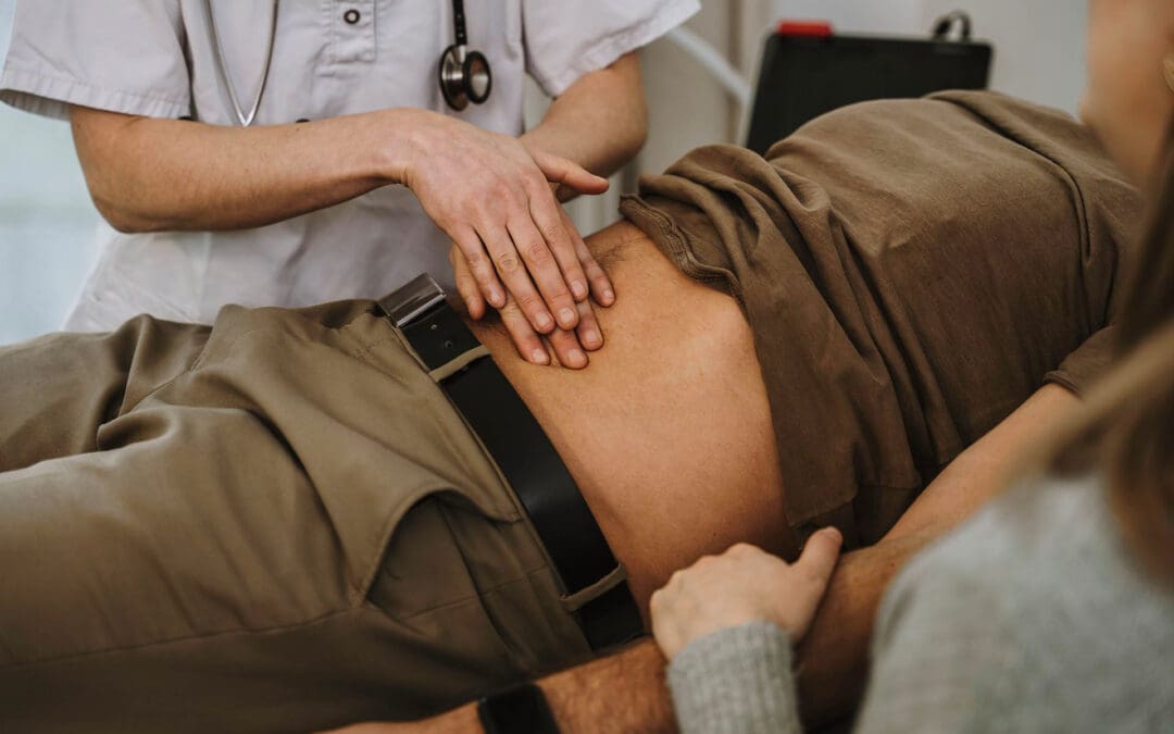
Stomach cramps, slow digestion, or constant puffiness can steal your joy in El Paso’s sunny days. These gut problems often stem from stress on the body’s control system. At El Paso Back Clinic® in El Paso, Texas, a trusted wellness team uses integrative chiropractic care to help them. Gentle spine adjustments, soft tissue therapy, smart eating plans, and natural detox steps work together. The focus is on the nervous system—it guides how food is broken down and waste is removed. Clear nerve paths mean less pain and smoother bowels. Fresh blood flow feeds organs. Simple diet and habit changes help the body clean itself. This full plan builds lasting gut strength for locals.
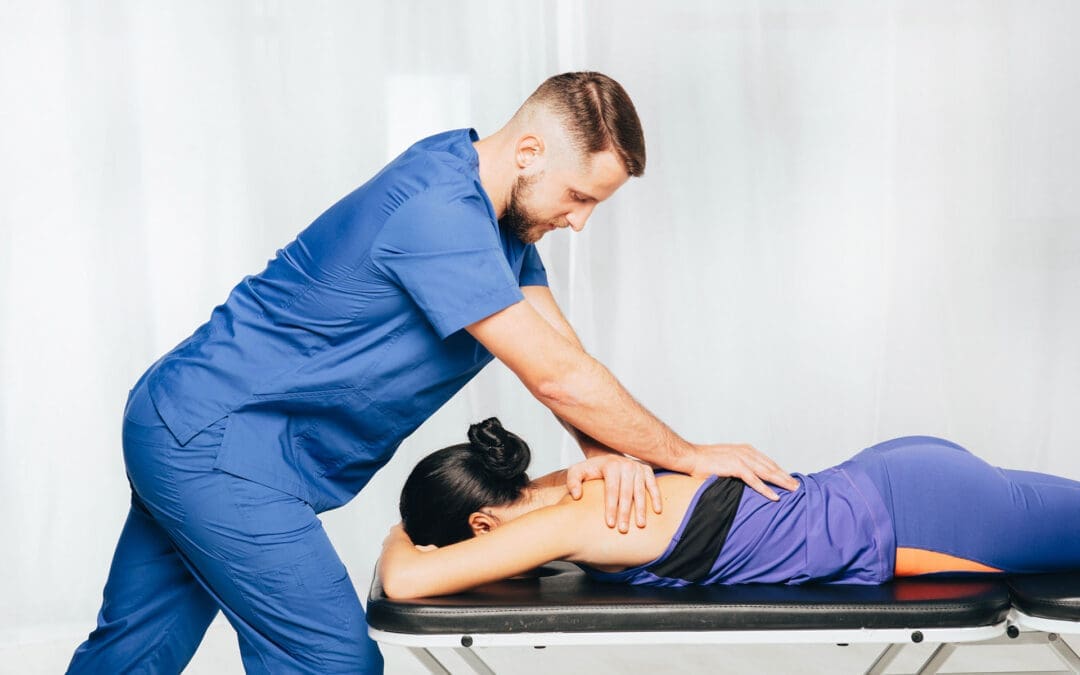
El Paso Back Clinic® sees the spine as the body’s main switchboard. A small bone shift can block signals to the stomach, liver, or intestines. Adjustments line up the spine to free nerves. The brain then sends clearer orders to enzymes and muscles. Patients feel less bloating and heartburn. Adding massage and food tips powers the body’s cleanup crew. The clinic helps thousands regain comfort without pills.
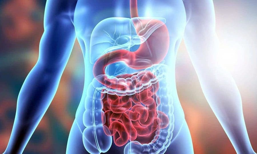
The gut and the liver work closely together. The gut moves waste; the liver filters it. Nerve blocks slow this pair, causing swelling and low energy. El Paso Back Clinic® restores the link with precise care. Dr. Alexander Jimenez, the clinic’s top chiropractor and nurse practitioner, sees quick wins daily. He notes that the spine helps reduce lower back swelling and supports the gut-liver axis (Jimenez, n.d.a.). Desk workers and crash survivors find their digestion steadies when nerves run clear.
Care here builds habits, not just quick relief. Guides cover tasty anti-inflammatory meals, water goals, and calm moves that fit El Paso life. These fuel detoxes ensure waste exits through bowels, skin, and breath—no harsh kits needed. A custom roadmap keeps changes simple and real.
Nervous System: The Gut’s Quiet Boss at El Paso Back Clinic®
Think of nerves as phone lines from the brain to the belly. They say when to mix acids, absorb vitamins, and push food along. The autonomic system automates this process. Poor posture or old injuries can pinch the spine. The middle back nerves tie to the stomach; the low back ones reach the colon. Pinches cause food to become stuck and waste to back up (Hyslop, 2023).
Clinic doctors examine by hand and with scans, then use soft thrusts to free spots. There is no force involved, only results. Nerves open; the vagus nerve—the gut leader—steps up. The body shifts to rest-and-digest calm. Gas drops ease cramps, and bowels move steadily (Parco of Ontario, n.d.).
Dr. Jimenez shares clinic wins. A driver with whiplash had daily bloat from thoracic pressure. Adjustments and simple stretches cleared both in weeks. Jimenez ties it to less stress juice that harms beneficial bugs (Jimenez, n.d.b.). Free signals keep the gut crew balanced and happy.
Spine Adjustments at El Paso Back Clinic®: Your Digestion Reset Button
Adjustments are the clinic’s star move—safe, no drugs, and proven. They realign the spine so the gut nerves work right. Focus lands on the thoracic and lumbar areas, home to the stomach and bowel wires.
Visits start with posture pics and light tests. A quick, gentle push may pop softly. Blood flows; nerves spark. Relief hits fast—easier stools, less reflux. Heartburn can drop 50% as diaphragm pressure lifts (Well Beings Medicine, n.d.). Colon nerves wake for wave motion, ending constipation (Abundant Life Chiropractor, n.d.).
Local athletes count on this. A soccer player with IBS saw patterns even out after lumbar care. Dr. Jimenez added planks to hold posture. Adjustments also address slouching that can crush organs. A tall spine gives guts space.
Circulation Power-Up: El Paso Back Clinic® Fuels Gut Healing
Strong blood flow brings oxygen and hauls trash. Spine stress or tight muscles clog it. Toxins stack; repair lags. The clinic clears the road.
Adjustments open the spine vessels, allowing blood to rush to the organs in the belly. This feeds gut walls and speeds junk to the liver (DC Labs, n.d.). Massage kneads knots; ultrasound warms deep for extra flow. Dr. Jimenez checks energy levels—patients see clear skin and pep as detox rolls (Jimenez, n.d.a).
Tips: strolls or home yoga. These move lymph that dispels waste. Less swelling stops gut leaks into the blood.
Nutrition Guidance at El Paso Back Clinic®: Meals That Heal
Food heals at the clinic. Three-phase plans.
Phase 1: Clear – Cut down on sweets and fast food to reduce harmful bacteria.
Phase 2: Build—load greens, beans, local salsa, and fermented foods.
Phase 3: Thrive – Mix proteins and fats for balance (Touch Chiropractic, n.d.).
Tests catch low beneficial bacteria. Dr. Jimenez suggests fish tacos with greens—tasty and anti-inflammatory. Hydration rule: clear urine means enough.
Detox Made Simple at El Paso Back Clinic®
The body cleans daily—liver, kidneys, skin, lungs. Clinic care speeds it. Adjustments push gut waste; massage moves lymph; diet traps toxins for exit (Spine and Joint Center, n.d.).
The El Paso sun helps—light sweat or a home sauna flushes skin. Mini-trampoline bounces junk out. Milk thistle supports liver health if medical tests indicate it is safe to use. Dr. Jimenez watches crash patients lose fog as toxins drop (Jimenez, n.d.c).
Dr. Jimenez Guides El Paso Back Clinic® with Real Results
Dr. Alexander Jimenez, DC, APRN, FNP-BC, IFMCP, leads with dual skills. Thousands of El Paso families trust his care yearly.
A factory worker, post-fall, had back pain and weeks of no bowel movements. Scans showed lumbar blocks. Adjustments, probiotics, and fiber were maintained for 6 weeks. Retests proved gut flora bounced back (Jimenez, n.d.a). Jimenez calls it spine-gut teamwork.
His plans use X-rays, stool checks, and food diaries—no guesses. As El Paso’s top wellness doctor, he hands patients the reins.
Begin Gut Wellness at El Paso Back Clinic® Today
El Paso Back Clinic® offers a clear path to gut ease. Start with a full spine and symptom review. Adjustments quiet nerve static; therapies and plans seal wins. Eat, move, and breathe for natural detox.
Call El Paso Back Clinic® in El Paso, TX—your gut deserves this care.
Unlock relief with chiropractic solutions aimed at treating and preventing spinal issues like disc herniation & disc bulging.
Understanding Disc Herniation and Disc Bulging: A Comprehensive Clinical Guide to Chiropractic Care and Spinal Decompression
Unlocking the Path to Recovery: Evidence-Based Chiropractic Solutions for Spinal Disc Disorders
Back pain represents one of the most pervasive health challenges affecting modern society, with approximately 80% of the population experiencing at least one episode during their lifetime (Al Qaraghli & De Jesus, 2023). Within this broad spectrum of spinal conditions, disc herniation and disc bulging emerge as two of the most common yet frequently misunderstood causes of debilitating pain. For patients experiencing persistent pain radiating through the neck, mid-back, or lower back, understanding the clinical distinctions between these conditions and the evidence-based treatment options available—particularly chiropractic care and nonsurgical spinal decompression therapy—can illuminate a pathway to lasting relief and functional restoration.
Understanding the Spinal Disc: Anatomy and Function
The human spine is a marvel of biological engineering, consisting of 24 vertebrae stacked on one another, separated by intervertebral discs that serve as sophisticated shock absorbers. These discs play multiple essential roles: they maintain height between vertebrae, absorb mechanical forces during movement and impact, facilitate spinal flexibility, and distribute biomechanical loads evenly throughout the spinal column (Al Qaraghli & De Jesus, 2023). Each intervertebral disc comprises two distinct structural components. The annulus fibrosus forms the tough, circular outer portion composed of 15 to 25 stacked sheets of highly organized fibrous connective tissue, predominantly type 1 collagen in the outer layers and type 2 collagen in the inner portions. Surrounding this protective shell lies the nucleus pulposus, a gel-like inner core consisting of a loose network of fibers suspended in a hydrophilic matrix. At birth, approximately 80% of disc composition consists of water, and proper hydration remains essential for optimal disc function throughout life (El Paso Chiropractor Blog, 2016). The structural integrity of healthy discs has often been compared to a jelly doughnut—a resilient outer ring containing a soft, gelatinous center. This unique composition enables discs to evenly distribute forces and pressures applied to the spine during daily activities, maintaining spinal stability while permitting controlled movement.
Disc Herniation vs. Disc Bulging: Critical Distinctions and Similarities
While disc herniation and disc bulging both involve displacement of disc material beyond normal anatomical boundaries, understanding their fundamental differences proves critical for appropriate clinical management and patient education.
Disc Bulging: Contained Disc Displacement
A disc bulge (also termed disc prolapse) occurs when the nucleus pulposus presses against the annulus fibrosus wall, causing the disc to protrude outward beyond its usual borders. Critically, in a bulging disc, the outer annular fibers remain intact—the gel-like nucleus stays fully contained within the disc structure, even though the entire disc extends beyond its normal space (Mayo Clinic, 2024). This condition typically affects at least 25% to 50% of the disc’s circumference and involves only the outer layer of tough cartilage (El Paso Chiropractor Blog, 2016).
The bulging disc can still compress surrounding neural structures, including spinal nerves and the spinal cord, potentially causing pain, numbness, tingling, and functional limitations. However, because the disc material remains contained, symptoms are often milder than with herniated discs, unless significant nerve compression occurs (Neurosurgery One, 2025).
Disc Herniation: Rupture and Extrusion
In contrast, a disc herniation (also called disc extrusion, ruptured disc, or slipped disc) develops when the tough outer annulus fibrosus develops a crack or tear, allowing the soft nucleus pulposus to squeeze through the opening and protrude into the spinal canal (Mayo Clinic, 2024). The herniated material can spread to adjacent structures, including the spinal cord and spinal nerve roots, often compressing these delicate tissues and triggering a cascade of symptoms (El Paso Chiropractor Blog, 2016).
When disc material herniates, two distinct pathological mechanisms contribute to pain generation. First, mechanical compression of neural structures directly irritates and damages nerve tissue. Second, the chemical composition of the nucleus pulposus itself proves highly inflammatory—when exposed to the immune system, these materials trigger significant inflammatory responses characterized by swelling, pain, and immune cell infiltration (Cosamalón-Gan et al., 2021).
Similarities Between Disc Conditions
Despite their structural differences, disc herniation and disc bulging share several important characteristics:
Common Symptom Patterns: Both conditions can produce identical or nearly identical symptoms, including localized back or neck pain, radiating pain into extremities (radiculopathy), numbness and tingling sensations, muscle weakness, and limited range of motion (Neurosurgeons of New Jersey, 2023).
Age-Related Degeneration: Both conditions typically arise from the spine’s natural degenerative process. As individuals age, spinal discs progressively dehydrate, becoming stiffer, more fragile, and less capable of adjusting to compression and mechanical stress. This degeneration represents the primary underlying cause for most disc complications (El Paso Chiropractor Blog, 2016).
Nerve Compression Mechanisms: Whether bulging or herniated, displaced disc material can impinge on spinal nerve roots or the spinal cord, triggering nerve irritation, inflammation, and the characteristic pain patterns associated with these conditions (Al Qaraghli & De Jesus, 2023).
Asymptomatic Presentations: Remarkably, many individuals harbor disc bulges or herniations without experiencing any symptoms whatsoever. These conditions are frequently discovered incidentally during imaging studies performed for unrelated medical issues (Mayo Clinic, 2024).
Regional Manifestations: How Disc Disorders Affect the Cervical, Thoracic, and Lumbar Spine
Disc herniation and bulging can develop throughout the spinal column, though certain regions are more vulnerable. The clinical presentation, symptom patterns, and functional impairments vary significantly depending on the spinal region affected.
Cervical Spine Disc Disorders
The cervical spine, comprising seven vertebrae in the neck, is the second most common site of symptomatic disc herniation. The most frequently affected levels are C4-C5, C5-C6, and C6-C7, with C6-C7 most likely to herniate in the cervical region (Spine-health, 2019).
Clinical Manifestations: Cervical disc herniation typically produces neck pain located toward the back or side of the neck, ranging from mild tenderness to sharp, burning sensations (Spine-health, 2019). Radicular pain—characterized by electric shock-like or hot sensations—commonly radiates from the neck down through the shoulder, arm, hand, and fingers. The specific distribution of symptoms depends on which nerve root suffers compression:
C5 nerve root (C4-C5 herniation): Pain and tingling radiating to the shoulder, with potential weakness in the deltoid muscle
C6 nerve root (C5-C6 herniation): Pain, tingling, and numbness affecting the thumb side of the hand, with weakness in the biceps and wrist extensors
C7 nerve root (C6-C7 herniation): Symptoms extending to the middle finger, with triceps weakness and finger extensor dysfunction
C8 nerve root (C7-T1 herniation): Pain and numbness in the pinky side of the hand, with handgrip weakness
Cervical herniated discs can also trigger cervical myelopathy when disc material compresses the spinal cord itself. This serious condition produces bilateral symptoms including numbness, weakness, balance disturbances (ataxia), hyperreflexia, and potential urinary incontinence. Chronic myelopathy may progress insidiously, sometimes delaying diagnosis as patients attribute symptoms to normal aging (Kamran Aghayev, 2025).
Thoracic Spine Disc Disorders
Thoracic disc herniations represent the rarest form of symptomatic disc pathology, with an estimated incidence of approximately one in one million per year, accounting for only 0.25% to 0.75% of total symptomatic spinal disc herniations (BCMJ, 2019). Despite this rarity, thoracic disc disorders present unique diagnostic challenges due to their atypical symptom presentations.
Clinical Manifestations: Thoracic herniated discs produce three distinct clinical patterns (Barrow Neurological Institute, 2025):
Radiculopathy (affecting approximately 52% of symptomatic patients): Mid-back pain that may wrap around the chest in a band-like distribution, corresponding to the dermatomal pattern of the affected nerve root. Patients often describe sensations of a strap tightening around their chest. Pain may also manifest as numbness, pressure sensations, or generalized discomfort rather than classic pain.
Myelopathy (affecting approximately 70% of symptomatic patients): Spinal cord compression producing difficulty walking, progressive lower extremity weakness and numbness, wide-based gait, increased muscle tone and clonus, hyperreflexia in lower extremities, and occasional bowel dysfunction.
Atypical extraspinal symptoms: Thoracic disc herniations frequently produce misleading symptoms, including nausea, emesis, chest tightness, gastrointestinal complaints, chronic constipation, buttock and leg burning pain, and urinary frequency—often leading to extensive workups for cardiac, pulmonary, or gastrointestinal disorders before the correct diagnosis emerges (Physio-pedia, 2023).
The most commonly affected thoracic levels include T7-T8, T8-T9, and T11-T12, with disc pathologies identified in approximately 18% of thoracic intervertebral disc levels among symptomatic patients (Turkish Journal of Medical Sciences, 2019).
Lumbar Spine Disc Disorders
The lumbar spine represents the most common location for disc herniation and bulging, with approximately 95% of lumbar disc herniations occurring at the L4-L5 or L5-S1 levels. Lumbar disc herniation affects 5 to 20 individuals per 1,000 adults annually, with peak prevalence occurring in the third to fifth decades of life and a male-to-female ratio of 2:1 (Al Qaraghli & De Jesus, 2023).
Clinical Manifestations: Lumbar disc disorders typically produce:
Low back pain: The primary symptom, arising from pressure exerted by herniated disc material on the posterior longitudinal ligament and local inflammation. The pain is often mechanical, worsening with movement, prolonged sitting, straining, coughing, and sneezing (Al Qaraghli & De Jesus, 2023).
Radiculopathy (sciatica): When disc material compresses lumbar nerve roots, pain radiates into the buttocks, thighs, calves, and feet, following specific dermatomal patterns:
L4 nerve root (L4-L5 herniation): Pain radiating to the anterior thigh and medial leg, with weakness in hip flexion/adduction and knee extension, plus diminished patellar reflex
L5 nerve root (L5-S1 herniation): Pain extending to the buttock, lateral thigh, lateral calf, dorsum of foot, and great toe, with weakness in foot dorsiflexion, great toe extension, and foot inversion/eversion
S1 nerve root (S1-S2 herniation): Sacral/buttock pain radiating to the posterolateral thigh, calf, and lateral/plantar foot, with weakness in plantar flexion and diminished Achilles reflex
Neurological deficits —sensory abnormalities (numbness, tingling), motor weakness, muscle atrophy in chronic cases, and altered reflexes — characterize nerve root compression. Severe central herniations may produce cauda equina syndrome, a surgical emergency characterized by saddle anesthesia, bowel/bladder incontinence, and progressive bilateral lower extremity weakness (Al Qaraghli & De Jesus, 2023).
Environmental and Occupational Risk Factors: Creating Overlapping Risk Profiles for Back Pain
While genetic factors contribute significantly to disc degeneration and herniation susceptibility, environmental and occupational exposures create substantial additional risk, often producing overlapping risk profiles that compound individual vulnerability to back pain across all spinal regions.
Occupational Physical Demands
Heavy physical workload and occupations requiring strenuous effort are associated most strongly with lumbar disc herniation risk. Research examining risk factors for lumbar disc herniation with radiculopathy identified occupation—particularly heavy labor—among the most robust risk factors, with certain professions showing risk ratios up to 6.0 (Dynamic Disc Designs, 2024).
Specific occupational activities that increase disc herniation risk include:
Repetitive lifting, bending, and twisting: Cumulative exposure to lifting heavy weights, forward bending, and rotational movements significantly increases lumbar disc herniation risk (Risk Factors Study, 2021)
Prolonged sitting: Sedentary work increases the risk of disc degeneration by exerting sustained compression loads on the spine during extended sitting. Sitting increases intradiscal pressure by approximately 40% compared to standing, intensifying mechanical stress on already vulnerable discs (Al Qaraghli & De Jesus, 2023)
Extended work hours: Working periods exceeding 8 hours consistently and experiencing high workplace stress levels are both associated with elevated disc herniation risk (Spine-health, 2024)
Whole-body vibration: Occupations involving prolonged exposure to vibration (truck drivers, heavy equipment operators) accelerate disc degeneration
Built Environment and Healthy Building Determinants
Emerging evidence indicates that indoor environmental quality and healthy building determinants significantly influence the risk of back and neck pain. A systematic review examining relationships between healthy building determinants and back/neck pain found evidence generally supporting that as healthy building determinants worsen—including poor air quality, inadequate ventilation, dust exposure, suboptimal lighting, moisture problems, excessive noise, thermal discomfort, and poor water quality—the risk of back and neck pain increases (PMC, 2022).
Given that people spend more than 90% of their time indoors, the built environment where most back and neck pain episodes occur deserves greater attention in prevention strategies. Poor environmental factors, including noise, dust, gases, fumes, and poor air quality, were significantly associated with increased back pain risk in both men and women across multiple studies (PMC, 2022).
Age and degeneration: While aging itself remains unavoidable, the natural degenerative cascade—characterized by reduced water content, increased type 1 collagen ratios in the nucleus pulposus, destruction of extracellular matrix, and upregulated inflammatory pathways—progresses throughout adult life, with disc herniation most prevalent between ages 30-50 (Al Qaraghli & De Jesus, 2023).
Obesity and excess weight: Elevated body mass index dramatically increases disc herniation risk by placing excessive mechanical load on the spine, accelerating disc degeneration and making herniation more likely. Excess body fat, particularly around the chest and abdomen, intensifies biomechanical stress on the lower back while promoting systemic inflammation (Spine-health, 2024).
Nicotine use: Smoking, vaping, and tobacco chewing disrupt nutrient flow to intervertebral discs, inhibit nucleus pulposus cell growth, and reduce collagen synthesis—all accelerating disc degeneration (Spine-health, 2024).
Sedentary lifestyle: Physical inactivity leads to weak core muscles, poor posture, and reduced flexibility, all of which increase stress on spinal discs. Regular low-impact exercise strengthens muscles supporting the spine and improves overall spinal health (Leucadia Chiropractic, 2025).
Improper lifting techniques: Using the back instead of legs when lifting, twisting while lifting, or attempting to carry excessive weight places dangerous pressure on the spine, potentially triggering acute herniation in susceptible individuals.
Cardiovascular risk factors: Surprisingly, high cholesterol, hypertension, diabetes, and family history of coronary disease all associate with higher lumbar disc herniation risk, particularly in women, suggesting metabolic health plays important roles in disc pathology (Dynamic Disc Designs, 2024).
Genetic Susceptibility and Gene-Environment Interactions
Twin studies demonstrate that both genetic and environmental factors contribute substantially to disc degeneration and back pain. Genetic factors appear to influence disc narrowing and degeneration—key pathways through which genes influence the development of back pain (FYZICAL, 2006). However, environmental factors interact with genetic predisposition, creating complex risk profiles where occupational exposures, lifestyle choices, and built environment quality either amplify or mitigate underlying genetic vulnerability.
Research on Finnish twins revealed that approximately 41% of the total variance in childhood low back pain could be attributed to shared environmental factors within families, while 59% stemmed from unique environmental factors, with genetic factors playing at most a minor role in pediatric populations (PMC, 2008). This underscores the critical importance of identifying and modifying environmental risk factors to prevent disc pathology across the lifespan.
The Inflammatory Cascade: Biochemical Mediators of Disc-Related Pain
Understanding disc herniation requires moving beyond purely mechanical models of nerve compression to appreciate the complex inflammatory processes that amplify and perpetuate pain. Until fairly recently, sciatic pain and radiculopathy associated with lumbar disc herniation were attributed exclusively to mechanical compression of nerve roots. However, mounting evidence from immunology, immunohistochemistry, and molecular biology studies indicates that herniated disc tissue is biologically active, expressing numerous inflammatory mediators that play central roles in pain generation (Cosamalón-Gan et al., 2021).
Pro-Inflammatory Cytokines
Herniated and degenerated discs demonstrate markedly elevated levels of pro-inflammatory cytokines, including:
Interleukin-1 beta (IL-1β): A master regulator of inflammatory responses that stimulates production of matrix metalloproteinases (MMPs), promoting extracellular matrix breakdown and disc degeneration. IL-1β also induces expression of additional inflammatory mediators and chemokines (PMC, 2013).
Tumor Necrosis Factor-alpha (TNF-α): Works synergistically with IL-1β to promote matrix degradation, increase production of catabolic enzymes, and stimulate inflammatory pathways. TNF-α directly sensitizes nociceptors, lowering pain thresholds and increasing pain sensitivity (PMC, 2013).
Interleukin-6 (IL-6): Elevated in degenerated and herniated discs, IL-6 contributes to chronic inflammatory states and correlates with pain intensity. Recent research demonstrates that disc herniation severity associates with circulating IL-6 levels, with this relationship particularly pronounced in patients with chronic symptoms (NYP Advances, 2020).
Interleukin-8 (IL-8): A potent chemotactic factor that recruits neutrophils to sites of disc herniation. Co-neutralization of IL-8 and TNF-α significantly improved mechanical hyperalgesia in experimental models (PMC, 2013).
Interleukin-17 (IL-17): Plays important roles in recruiting T-cells and macrophages and activating glial and astrocytic cells during nerve injury and subsequent neuropathic pain. IL-17 levels show significant elevation in herniated versus merely degenerated discs (PMC, 2013).
Chemokines and Immune Cell Recruitment
Beyond structural damage, inflammatory cytokines stimulate disc cells to produce chemotactic factors that recruit immune cells—including macrophages, neutrophils, and T cells—to the disc and surrounding tissues. Analysis of herniated discs reveals elevated levels of multiple chemokines, including:
Monocyte chemotactic protein-1 (MCP-1, CCL2)
CCL3, CCL4, CCL5
MCP-3, MCP-4
CXCL10
Expression of CCL3 correlates positively with degeneration grade and is higher in herniated tissue compared with degenerate but contained discs. By regulating chemokine expression, inflammatory cytokines promote C-C chemokine receptor type 1 (CCR1)-dependent macrophage migration, thereby establishing a self-perpetuating inflammatory cycle critical to pain-generating pathways (PMC, 2013).
Autoimmune Responses
Inflammation in disc herniation stems not only from chemical irritation by bioactive substances released from the nucleus pulposus but also from autoimmune responses against disc tissue itself. The nucleus pulposus, normally sequestered from the immune system, becomes recognized as foreign when herniation exposes it to immune surveillance. This triggers antibody production and T-cell-mediated responses that amplify local inflammation (Cosamalón-Gan et al., 2021).
Clinical Implications of Inflammatory Mechanisms
This biochemical understanding carries profound clinical implications. First, it explains why some patients experience severe pain despite relatively minor disc herniations—individual variations in inflammatory responses may prove more important than herniation size alone. Second, it validates treatment approaches targeting inflammation, including judicious use of anti-inflammatory medications and interventions like epidural steroid injections. Third, it suggests that therapies that promote the resolution of inflammation and support tissue healing—such as chiropractic care and spinal decompression—may address root causes rather than merely manage symptoms.
Spinal Decompression in Depth- Video
Clinical Rationale for Chiropractic Care in Disc Herniation and Bulging
Chiropractic care has emerged as a primary conservative treatment modality for patients suffering from disc herniation and bulge, supported by growing evidence demonstrating significant clinical benefits. The clinical rationale for chiropractic intervention in disc pathology rests on multiple therapeutic mechanisms that address both mechanical dysfunction and inflammatory processes.
Mechanisms of Chiropractic Spinal Manipulation
Chiropractic spinal manipulation—characterized by high-velocity, low-amplitude (HVLA) controlled forces applied to specific spinal segments—produces multiple beneficial effects in patients with disc disorders:
Restoration of spinal alignment and mobility: Spinal manipulation corrects vertebral misalignments (subluxations) that may contribute to abnormal biomechanical stress on intervertebral discs. By restoring proper spinal alignment, manipulation reduces asymmetric loading that accelerates disc degeneration (El Paso Chiropractor Blog, 2016).
Reduction of intradiscal pressure: Properly executed spinal manipulation may temporarily reduce pressure within affected discs, potentially facilitating retraction of herniated material and reducing compression on adjacent neural structures.
Improvement of spinal joint function: Manipulation increases range of motion in restricted spinal segments, reducing mechanical irritation of surrounding tissues and improving overall spinal biomechanics.
Modulation of pain perception: Spinal manipulation activates mechanoreceptors and produces neurophysiological effects that may modulate pain perception via gate-control mechanisms and descending pain-inhibition pathways.
Anti-inflammatory effects: Emerging evidence suggests that spinal manipulation may influence inflammatory processes, potentially reducing local cytokine production and promoting the resolution of inflammation.
Clinical Outcomes Evidence for Chiropractic Care
Multiple high-quality studies document the effectiveness of chiropractic spinal manipulation for disc herniation and bulging across spinal regions:
Lumbar Disc Herniation: A landmark prospective cohort study published in the Journal of Manipulative and Physiological Therapeutics followed 148 patients aged 18-65 with low back pain, leg pain, and MRI-confirmed lumbar disc herniation treated with high-velocity, low-amplitude spinal manipulation (Leemann et al., 2014). Outcomes proved remarkable:
At 3 months, 90.5% of patients reported “improvement” on global impression of change scales
At 1 year, 88.0% maintained “improved” status
Among chronic patients (symptoms >3 months), 81.8% reported improvement, increasing to 89.2% at 1 year
Both acute and chronic patients demonstrated significant improvements in numerical rating scale scores for low back pain, leg pain, and Oswestry Disability Index scores at all follow-up points (2 weeks, 1, 3, 6, and 12 months)
No adverse events were reported throughout the study period
The high success rates among chronic patients are particularly noteworthy, as this population typically shows poorer responses to conservative interventions. The sustained improvements at one-year follow-up indicate that chiropractic manipulation produces lasting benefits rather than merely temporary symptom relief.
Cervical Disc Herniation: Research from Zurich, Switzerland, examined 50 patients aged 18-65 with MRI-confirmed cervical disc herniation treated with chiropractic spinal manipulation at frequencies of 3-5 sessions weekly initially, reducing to 1-3 sessions weekly until symptom resolution (SSPT Chiropractic, 2024). Results demonstrated progressive improvement:
At 2 weeks, 55% of participants reported improvement
At 1 month, 68.8% showed improvement
At 3 months, 85.4% experienced favorable outcomes
Even among chronic cervical disc herniation patients, 76% reported beneficial effects, including reduced neck and arm pain
Another study specifically examining patients with MRI-confirmed lumbar disc herniation and concomitant sacroiliac joint hypomobility found that five sessions of lumbar and sacroiliac joint manipulation over a 2-week period produced significant improvements in both back and leg pain intensity and functional disability, as measured by the Oswestry Disability Index (Shokri et al., 2018).
Comparative Effectiveness: Research comparing chiropractic spinal manipulative therapy (CSMT) with other care modalities for newly diagnosed lumbar disc herniation and lumbar spinal radiculopathy found that patients receiving CSMT demonstrated significantly reduced odds of requiring lumbar discectomy surgery through 2-year follow-up compared to those receiving other care approaches (BMJ Open, 2022). This suggests that chiropractic care may help many patients avoid surgical intervention while achieving satisfactory functional outcomes.
Dr. Alexander Jimenez’s Integrative Approach
Dr. Alexander Jimenez, DC, APRN, FNP-BC, exemplifies the modern integrative chiropractic practitioner, combining advanced clinical expertise with comprehensive diagnostic evaluation to optimize patient outcomes. As both a board-certified Doctor of Chiropractic and Family Practice Nurse Practitioner practicing in El Paso, Texas, Dr. Jimenez brings a unique dual-scope perspective to treating complex spinal disorders, including disc herniation and bulging. Dr. Jimenez’s clinical approach emphasizes thorough diagnostic evaluation utilizing advanced imaging modalities—including MRI and other radiological studies—to precisely characterize disc pathology before initiating treatment. This imaging-guided approach ensures that manipulation techniques are appropriately tailored to each patient’s specific disc lesion type, location, and severity. As noted on his clinical website (dralexjimenez.com), Dr. Jimenez focuses on treating patients with “complex herniated discs” using evidence-based protocols that integrate chiropractic manipulation, functional medicine principles, nutritional optimization, and rehabilitation exercises. His dual training enables comprehensive evaluation of patients from both musculoskeletal and medical perspectives, identifying underlying metabolic, inflammatory, or systemic factors that may contribute to disc degeneration and impaired healing. Dr. Jimenez emphasizes that proper patient selection proves critical—when patients present with conditions better suited for alternative treatments or specialist referral, he ensures they receive appropriate care from the most qualified providers. The integration of functional medicine assessment tools, including detailed evaluations of genetics, lifestyle factors, environmental exposures, nutritional status, and psychological/emotional factors, enables Dr. Jimenez to address the root causes of disc pathology rather than merely treating symptoms. This comprehensive approach aligns with emerging evidence demonstrating that metabolic health, inflammatory status, and environmental factors significantly influence disc degeneration progression and healing potential.
Nonsurgical Spinal Decompression: Mechanism, Evidence, and Clinical Application
Nonsurgical spinal decompression therapy (NSDT) represents an advanced evolution of traditional traction therapy, utilizing sophisticated computer-controlled systems to create negative intradiscal pressure that facilitates disc healing and symptom resolution. Understanding the distinctions between NSDT and conventional traction proves essential for appreciating this intervention’s unique therapeutic potential.
Mechanism of Action: Creating Negative Intradiscal Pressure
NSDT operates through a precisely controlled biomechanical process fundamentally different from traditional traction:
Specialized positioning: Patients are positioned on a computer-controlled decompression table with the spine properly aligned and supported. Harnesses secure the upper body (chest and shoulders) while a separate harness attaches to the pelvis or lower body.
Computer-guided distraction: Unlike conventional traction that applies a constant pulling force, NSDT employs a sophisticated algorithm that gradually increases and decreases distraction force in cyclical patterns. This intermittent loading prevents reflexive muscle guarding, which limits the effectiveness of traditional traction (Hill DT Solutions, 2024).
Negative intradiscal pressure generation: The controlled distraction force creates a vacuum effect within targeted intervertebral discs. Research measuring intradiscal pressure during NSDT using pressure transducers inserted into the L4-L5 disc space demonstrated that decompression therapy can lower pressure in the nucleus pulposus to below -100 mmHg, compared to standard progressive traction achieving only -40 mmHg (compared to -75 mmHg resting supine) (Hill DT Solutions, 2024).
Disc material retraction: This sustained negative pressure may facilitate retraction of herniated or bulging nucleus pulposus material away from compressed neural structures. The vacuum effect theoretically “pulls” extruded disc material back toward its normal position within the disc space.
Enhanced nutrient influx: Negative intradiscal pressure promotes increased fluid exchange, drawing oxygen, nutrients, and hydration into degenerated discs. This enhanced nutrient delivery may support disc cell metabolism and tissue repair (Dr. DiGrado, 2024).
Spinal joint decompression: The distraction force increases the width of the intervertebral foramen, reducing pressure on exiting nerve roots and facet joints, thereby contributing to pain relief independent of effects on the disc itself.
Critical Distinction from Traditional Traction
The fundamental advantage of NSDT over conventional traction lies in its ability to overcome the muscle guarding reflex. When traditional traction applies sudden or sustained pulling forces, paraspinal muscles reflexively contract to protect the spine from perceived threat. This muscle contraction increases internal disc pressure and limits the therapeutic effect (Choi et al., 2022).NSDT systems employ gradual force application with intermittent relaxation phases that prevent this protective muscle contraction. The computer continuously monitors resistance and adjusts force application in real time, maintaining the spine in a relaxed state while delivering far greater decompressive forces than traditional traction can achieve. This creates what researchers describe as a “zero-gravitation” state in targeted discs (Choi et al., 2022).
Evidence for NSDT Effectiveness
A rigorous randomized controlled trial published in the International Journal of Clinical Practice provides compelling evidence for the effectiveness of NSDT in treating subacute lumbar disc herniation (Choi et al., 2022). This study enrolled 60 patients with subacute lumbar herniated intervertebral disc, randomizing them to either:
Decompression group (n=30): Received 10 NSDT sessions over 8 weeks (twice weekly for 2 weeks, then once weekly for 6 weeks), with distraction force starting at half body weight minus 5 kg and increasing by 1 kg per session
Nondecompression group (n=30): Received identical positioning and session frequency but with zero distraction force (sham treatment)
Results demonstrated significant advantages for the decompression group:
Pain outcomes:
Lower leg pain intensity at 2 months (p=0.028)
Significant reductions in low back and leg pain from baseline to 3 months in both groups (p<0.001), though between-group differences in back pain did not reach significance
Functional outcomes:
Significantly lower Korean Oswestry Disability Index scores at 2 months (p=0.023) and 3 months (p=0.019)
MRI-documented structural changes:
Herniation index decreased by 27.6±27.5% in the decompression group versus only 7.1±24.9% in the control group (p=0.017)
26.9% of decompression patients versus 0% of control patients achieved >50% reduction in herniation index (p=0.031)
42.3% of decompression patients achieved ≥30% herniation reduction versus 17.6% of controls
These findings prove groundbreaking—this study represents the first randomized controlled trial to document that NSDT produces measurable reductions in disc herniation volume as confirmed by follow-up MRI, while simultaneously improving pain and function. The fact that actual structural healing occurred rather than merely symptomatic improvement suggests that NSDT addresses the underlying pathology. Additional research supports these findings. A retrospective cohort study examining adults with chronic low back pain attributed to disc herniation or discogenic pain who underwent 6-week NSDT protocols via the DRX9000 system found significant correlations between disc height restoration and pain reduction (Apfel et al., 2010). Low back pain decreased from 6.2±2.2 to 1.6±2.3 (p<0.001) while disc height increased from 7.5±1.7mm to 8.8±1.7mm (p<0.001), with these variables showing significant correlation (r=0.36, p=0.044). Long-term follow-up studies demonstrate sustained benefits. Research tracking patients 4 years after completing NSDT protocols found that 52% maintained pain levels of zero, 91% resumed normal daily activities, and over 80% achieved 50% or greater pain reduction compared to pre-treatment baselines (Pain Free Charleston, 2004).
NSDT Treatment Protocols
Typical NSDT treatment courses involve:
Session frequency: 10-20 sessions over 4-8 weeks, with initial sessions typically scheduled 2-3 times weekly, reducing to 1-2 times weekly as improvement occurs
Session duration: 20-30 minutes per session
Force parameters: Distraction force is individualized based on patient body weight, disc level targeted, and tolerance, typically starting at conservative levels (40-50% body weight) and gradually progressing
Positioning: Supine positioning with flexed knees for lumbar protocols; prone or supine with cervical harness for cervical protocols
Cycle patterns: Alternating distraction and relaxation phases (commonly 60 seconds of tension, 30 seconds of relaxation) to prevent muscle guarding
Adjunctive therapies: Many protocols combine NSDT with complementary treatments, including cold/heat therapy, electrical stimulation, therapeutic exercise, and nutritional support to optimize outcomes
Safety and Contraindications
NSDT demonstrates excellent safety profiles when appropriately applied. The randomized controlled trial by Choi and colleagues reported zero adverse events throughout the study period (Choi et al., 2022). Similarly, the large prospective cohort study by Leemann and colleagues involving 148 patients receiving chiropractic manipulation for MRI-confirmed disc herniation reported no adverse events (Leemann et al., 2014).
However, certain contraindications to NSDT must be respected:
Absolute contraindications:
Pregnancy
Fracture
Tumor
Abdominal aortic aneurysm
Advanced osteoporosis
Cauda equina syndrome requiring emergency surgery
Severe spinal instability
Relative contraindications:
Prior spinal surgery with hardware
Severe disc degeneration with >50% height loss
Sequestrated disc fragments
Severe spinal stenosis
Extreme obesity is limiting proper positioning
Proper patient selection, thorough clinical examination, and careful review of imaging studies by qualified practitioners ensure NSDT is applied to appropriate candidates while avoiding potential complications.
Integrating Chiropractic Care and Spinal Decompression: Complementary Approaches
For many patients with disc herniation and bulging, optimal outcomes emerge from integrating multiple conservative therapies rather than relying on single interventions. Chiropractic spinal manipulation and NSDT offer complementary mechanisms that address different aspects of disc pathology:
Chiropractic manipulation primarily restores spinal joint mobility, corrects vertebral misalignments, modulates pain through neurophysiological mechanisms, and may influence local inflammatory processes. It proves particularly effective for acute presentations and when joint dysfunction accompanies disc pathology.
NSDT specifically targets the disc itself, creating negative intradiscal pressure that facilitates disc material retraction, promotes nutrient influx, and directly decompresses neural structures. It excels in cases where significant disc herniation or advanced degeneration requires sustained decompressive forces.
When combined, these approaches provide:
Comprehensive address of both joint dysfunction and disc pathology
Multiple mechanisms for pain relief and functional restoration
Options for tailoring treatment intensity to individual patient tolerance
Complementary effects that may accelerate healing beyond either therapy alone
Dr. Jimenez’s integrative approach exemplifies this comprehensive strategy, combining chiropractic adjustments with spinal decompression, functional medicine interventions, nutritional optimization, therapeutic exercise, and patient education to address all contributors to disc pathology and optimize healing potential.
Patient Selection and Prognostic Factors
Not all patients with disc herniation or bulging require or benefit equally from chiropractic care and spinal decompression. Understanding prognostic factors helps identify ideal candidates:
Favorable prognostic indicators:
Acute to subacute symptom duration (4 weeks to 3 months)
First episode of disc-related pain
Absence of progressive neurological deficits
Contained disc herniations (protrusions, extrusions) rather than sequestrations
Younger age (generally <65 years)
Absence of significant comorbidities
High motivation and compliance with treatment protocols
Adequate disc height preservation on imaging
Factors suggesting need for alternative or adjunctive interventions:
Significant psychological distress or catastrophizing
Major comorbidities affecting healing capacity
Even among chronic patients, evidence suggests substantial benefit from chiropractic care and NSDT, with the Leemann study demonstrating 89.2% of chronic lumbar disc herniation patients reporting improvement at 1-year follow-up after chiropractic manipulation (Leemann et al., 2014).
Conclusion: Evidence-Based Hope for Disc Pathology
The evidence presented throughout this comprehensive review supports a clear conclusion: chiropractic care and nonsurgical spinal decompression therapy are evidence-based, effective treatment options for patients with disc herniation and disc bulging across the cervical, thoracic, and lumbar spine regions. For patients experiencing the debilitating pain, functional limitations, and quality-of-life impairments associated with disc pathology, these conservative interventions offer hope grounded in rigorous scientific evidence. Studies consistently demonstrate that properly selected patients receiving chiropractic spinal manipulation achieve clinically meaningful improvements in pain, disability, and overall function, with success rates ranging from 76% to over 90% depending on patient characteristics and outcome measures. Remarkably, these benefits prove sustainable, persisting at one-year follow-up and beyond. NSDT adds a powerful tool capable of producing measurable structural improvements—actual reduction in disc herniation volume confirmed by MRI—alongside symptomatic relief. The ability to document disc healing, not merely symptom management, represents a paradigm shift in conservative disc care. The clinical insights provided by practitioners like Dr. Alexander Jimenez, DC, APRN, FNP-BC, who integrate advanced diagnostic evaluation, dual-scope clinical expertise, and comprehensive treatment protocols, demonstrate how modern chiropractic practice transcends historical limitations. By combining spinal manipulation, decompression therapy, functional medicine principles, nutritional optimization, and patient education, integrative approaches address root causes of disc pathology while supporting the body’s inherent healing capacities. Environmental and occupational risk factors create overlapping vulnerability profiles that compound genetic predisposition to disc degeneration. Recognition of these modifiable factors—including workplace ergonomics, physical demands, built environment quality, obesity, smoking, sedentary lifestyle, and metabolic health—enables comprehensive prevention and treatment strategies that extend beyond passive symptom management. Understanding the inflammatory cascade underlying disc-related pain—involving complex interactions among pro-inflammatory cytokines, chemokines, immune cell infiltration, and autoimmune responses—provides a mechanistic rationale for interventions targeting inflammation resolution and tissue healing rather than merely blocking pain signals. For the millions of individuals struggling with disc-related back and neck pain, the evidence reviewed herein offers legitimate hope for meaningful recovery through conservative, nonsurgical means. While not every patient will achieve complete resolution, and some will ultimately require surgical intervention, the substantial majority can expect significant improvement through properly applied chiropractic care and spinal decompression therapy.
Important Medical Disclaimer and Serious Note to Readers
THIS ARTICLE IS INTENDED FOR INFORMATIONAL AND EDUCATIONAL PURPOSES ONLY AND SHOULD NOT BE CONSTRUED AS MEDICAL ADVICE, DIAGNOSIS, OR TREATMENT RECOMMENDATION.
The information presented in this article, while based on peer-reviewed scientific literature and clinical evidence, does not substitute for professional medical evaluation, diagnosis, and treatment. Disc herniation, disc bulging, and related spinal conditions can produce serious complications, including permanent neurological damage, paralysis, bowel and bladder dysfunction, and chronic pain syndromes if inappropriately managed.
DO NOT ATTEMPT TO SELF-DIAGNOSE OR SELF-TREAT DISC-RELATED CONDITIONS. If you are experiencing back pain, neck pain, radiating pain into extremities, numbness, tingling, weakness, or any other symptoms potentially related to spinal disc pathology, seek immediate evaluation from qualified healthcare professionals.
CERTAIN SYMPTOMS CONSTITUTE MEDICAL EMERGENCIES requiring immediate emergency department evaluation, including:
Sudden onset of bowel or bladder incontinence or retention
Progressive lower extremity weakness or paralysis
Saddle anesthesia (numbness in the groin/inner thigh region)
Severe pain unresponsive to conservative measures
Symptoms following significant trauma
Chiropractic care and spinal decompression therapy, while generally safe when appropriately applied, carry potential risks and contraindications. These interventions should be performed only by licensed, qualified practitioners after thorough clinical examination and review of appropriate imaging studies. Improper application of spinal manipulation or decompression therapy can potentially worsen disc herniation, cause neurological damage, or result in other serious complications. The treatment outcomes and success rates cited in this article represent average findings from clinical studies and should not be interpreted as guarantees of individual outcomes. Individual results vary based on numerous factors, including age, overall health status, severity and duration of disc pathology, presence of comorbidities, lifestyle factors, and compliance with treatment protocols.
Before initiating any treatment for disc-related conditions, patients should:
Undergo a comprehensive evaluation by qualified healthcare providers
Obtain appropriate imaging studies (MRI, CT, or X-ray as indicated)
Discuss all treatment options, including risks, benefits, and alternatives
Ensure practitioners are properly licensed and credentialed
Verify that their specific condition is appropriate for conservative management
Understand when surgical intervention may be necessary
References to Dr. Alexander Jimenez and his clinical approaches are provided for illustrative purposes, demonstrating integrative treatment models and should not be construed as specific endorsements or treatment recommendations. Patients seeking care should independently research practitioners’ credentials, experience, and patient outcomes. The authors and publishers of this article disclaim all liability for any adverse outcomes, complications, or damages resulting from the application of information contained herein. Readers assume all responsibility and risk for decisions made regarding their healthcare and treatment choices. This article addresses complex medical conditions requiring individualized assessment and treatment planning. What proves safe and effective for one patient may be inappropriate or dangerous for another. Always consult qualified healthcare professionals for personalized medical advice specific to your individual circumstances. If you are currently experiencing a medical emergency, call emergency services (911 in the United States) immediately. Do not delay seeking emergency care while researching conservative treatment options. By continuing to read and apply information from this article, you acknowledge understanding and accepting this disclaimer and assume full responsibility for your healthcare decisions.
Apfel, C. C., Cakmakkaya, O. S., Martin, W., Richmond, C., Macario, A., George, E., Schaefer, M., & Pergolizzi, J. V. (2010). Restoration of disk height through non-surgical spinal decompression is associated with decreased discogenic low back pain: A retrospective cohort study. BMC Musculoskeletal Disorders, 11(1), 155. https://doi.org/10.1186/1471-2474-11-155
Choi, E., Gil, H. Y., Ju, J., Han, W. K., Nahm, F. S., & Lee, P. B. (2022). Effect of nonsurgical spinal decompression on intensity of pain and herniated disc volume in subacute lumbar herniated disc. International Journal of Clinical Practice, 2022, 6343837. https://doi.org/10.1155/2022/6343837
Cosamalón-Gan, I., Cosamalón-Gan, T., Mattos-Piaggio, G., Villar-Suárez, V., García-Cosamalón, J., & Vega-Álvarez, J. A. (2021). Inflammation in the intervertebral disc herniation. Neurocirugía (English Edition), 32(1), 21-35. https://doi.org/10.1016/j.neucir.2020.01.001
Gherscovici, E. D., & Mayer, J. M. (2022). Relationship of healthy building determinants with back and neck pain: A systematic review. International Journal of Environmental Research and Public Health, 20(1), 815. https://doi.org/10.3390/ijerph20010815
Leemann, S., Peterson, C. K., Schmid, C., Anklin, B., & Humphreys, B. K. (2014). Outcomes of acute and chronic patients with magnetic resonance imaging-confirmed symptomatic lumbar disc herniations receiving high-velocity, low-amplitude, spinal manipulative therapy: A prospective observational cohort study with one-year follow-up. Journal of Manipulative and Physiological Therapeutics, 37(3), 155-163. https://doi.org/10.1016/j.jmpt.2014.01.002
PMC. (2008). Genetic and environmental influences on non-specific low back pain in children: A twin study. European Spine Journal, 17(4), 502-508. https://pmc.ncbi.nlm.nih.gov/articles/PMC2295279/
PMC. (2022). Relationship of healthy building determinants with back and neck pain: A systematic review. International Journal of Environmental Research and Public Health, 20(1), 815. https://pmc.ncbi.nlm.nih.gov/articles/PMC9755707/
Risk Factors Study. (2021). Risk factors of intervertebral disc pathology—A point of view formerly and today—A review. International Journal of Environmental Research and Public Health, 18(2), 726. https://pmc.ncbi.nlm.nih.gov/articles/PMC7865549/
Shokri, E., Kamali, F., Sinaei, E., & Ghafarinejad, F. (2018). Spinal manipulation in the treatment of patients with MRI-confirmed lumbar disc herniation and sacroiliac joint hypomobility: A quasi-experimental study. Chiropractic & Manual Therapies, 26, 16. https://doi.org/10.1186/s12998-018-0185-z
Turkish Journal of Medical Sciences. (2019). The incidence and most common levels of thoracic degenerative disc disease. Acta Orthopaedica et Traumatologica Turcica, 52(3), 195-200. https://pmc.ncbi.nlm.nih.gov/articles/PMC6657757/
Find out about effective chiropractic care options for addressing hand numbness and enhancing your quality of life.
Understanding Hand Numbness and Carpal Tunnel Syndrome: How Chiropractic Care Offers Natural Relief
Hand numbness and tingling sensations affect millions of people worldwide, disrupting daily activities and diminishing quality of life. These uncomfortable symptoms often signal nerve compression issues, with carpal tunnel syndrome being the most common culprit. While many individuals immediately think surgery is their only option, research increasingly demonstrates that conservative, non-surgical approaches—particularly chiropractic care—can provide significant relief and lasting results. This comprehensive guide explores the causes, symptoms, and clinical rationale for using chiropractic treatment to address hand numbness and carpal tunnel syndrome. We’ll examine how environmental factors contribute to nerve compression, the critical connection between spinal health and hand symptoms, and evidence-based conservative treatments that can help you avoid surgery.
Understanding Hand Numbness: Causes and Symptoms
Hand numbness represents a sensory dysfunction involving the loss of normal sensation, including pain, temperature, touch, or vibratory perception. The severity varies considerably among individuals, ranging from mild intermittent tingling to constant numbness that significantly impairs hand function.
Common Symptoms of Hand Numbness
Individuals experiencing hand numbness typically report a constellation of symptoms that may include:
Paresthesia: The medical term for abnormal sensations, paresthesia manifests as numbness with loss of touch or temperature sensation. Some people describe feeling like they’re wearing gloves when they aren’t, while others experience gait and balance problems when numbness affects their ability to feel the ground beneath their feet.
Tingling and “Pins and Needles”: Often described as the sensation of limbs “falling asleep,” this symptom frequently occurs in the thumb, index, middle, and sometimes the ring finger. The tingling may start intermittently but can progress to become constant.
Burning Sensations: Many patients report a burning feeling along the affected nerve pathway, which can extend from the fingertips up through the hand and into the forearm.
Pain: Sharp, stabbing, or shooting pain often accompanies numbness, particularly at night when symptoms tend to worsen. This pain may radiate from the wrist up the forearm and sometimes as far as the shoulder.
Weakness: Muscle weakness accompanies numbness in the same location, making it difficult to grip objects, hold tools, or perform fine motor tasks like buttoning clothing.
Loss of Coordination: Decreased finger dexterity and hand clumsiness can make everyday activities challenging, from typing on a keyboard to opening jars.
What Causes Hand Numbness?
Hand numbness occurs when there is pressure, irritation, or damage to the nerves that supply sensation to the hands. The causes are varied and understanding the underlying mechanism is crucial for effective treatment:
Peripheral Neuropathy: This condition affects the very ends of nerves in the hands and feet. Diabetes is the most common cause of peripheral neuropathy, but alcoholism, vitamin deficiencies (especially B12), autoimmune conditions, liver or kidney disorders, and exposure to toxins can also damage peripheral nerves.
Nerve Compression Syndromes: Pressure on a nerve anywhere along its course from the neck to the fingertips can cause numbness. Common compression sites include the carpal tunnel at the wrist (carpal tunnel syndrome), the cubital tunnel at the elbow (cubital tunnel syndrome), and the cervical spine in the neck.
Cervical Radiculopathy: Compression or irritation of nerve roots exiting the cervical spine can send radiating pain, numbness, and weakness down through the shoulder, arm, and hand. This occurs when herniated discs, bone spurs, or degenerative changes put pressure on the nerve roots.
Thoracic Outlet Syndrome: Compression of nerves and blood vessels between the collarbone and first rib can cause symptoms similar to carpal tunnel syndrome.
Trauma and Injuries: Bone dislocations, fractures, and crushing injuries can cause swelling or direct nerve damage, resulting in numbness.
Double Crush Syndrome: This phenomenon occurs when a nerve is compressed at two distinct locations along its pathway—typically at both the cervical spine and the wrist. Compression at one site makes the nerve more vulnerable to symptoms from compression at a second site.
What is Carpal Tunnel Syndrome?
Carpal tunnel syndrome represents the most common peripheral nerve entrapment condition, affecting approximately one in ten adults at some point in their lifetime. For individuals with diabetes, the lifetime risk increases dramatically to 84 percent.
Anatomical Overview
The carpal tunnel is a narrow passageway in the wrist formed by the transverse carpal ligament at its upper boundary and the carpal bones at its lower boundary. This confined space accommodates nine flexor tendons and the median nerve, which must traverse through it to reach the hand.
The median nerve originates from nerve roots C5-T1 in the cervical spine and travels through the brachial plexus, down the arm, through the forearm, and ultimately through the carpal tunnel. The nerve provides both motor function (allowing movement) and sensory function (providing feeling) to the thumb, index finger, middle finger, and the thumb-side of the ring finger.
How Carpal Tunnel Syndrome Develops
Carpal tunnel syndrome develops when elevated pressure within the carpal tunnel compresses the median nerve. Normal pressure within the carpal tunnel ranges from 2 to 10 mmHg. However, extension or flexion of the wrist causes pressure to increase eight to ten times the normal level.
The pathophysiology involves a combination of mechanisms:
Mechanical Trauma: Repetitive compression and friction damage the nerve over time.
Increased Pressure: Elevated intracarpal pressure restricts blood flow to the endoneurial capillary system, causing ischemic damage to nerve tissue.
Inflammation: Swelling of the tendons and surrounding tissues within the confined space further compresses the median nerve.
Demyelination: Repeated compression can lead to demyelination (loss of the protective nerve covering) at the site of compression, impairing nerve signal transmission.
Symptoms Specific to Carpal Tunnel Syndrome
While carpal tunnel syndrome shares many symptoms with general hand numbness, it has distinctive characteristics:
Distribution Pattern: Numbness, tingling, and pain specifically affect the thumb, index, middle, and lateral half of the ring finger. The little finger is typically spared because it receives sensation from the ulnar nerve rather than the median nerve.
Nocturnal Symptoms: Symptoms frequently manifest or worsen at night while lying down. Many patients wake up shaking their hands to restore sensation—a phenomenon so common it’s considered pathognomonic for carpal tunnel syndrome.
Progressive Nature: Initially, symptoms come and go and tend to improve during the daytime. Over time, most patients begin to encounter symptoms during the day, particularly when engaged in repetitive activities such as typing, driving, or holding a phone.
Thenar Atrophy: In advanced cases, the muscles at the base of the thumb (thenar eminence) can atrophy and weaken, causing a flattened appearance and inability to oppose the thumb effectively.
Positive Provocative Tests: Clinical examination reveals positive Phalen’s test (symptoms reproduced by flexing the wrists for 60 seconds) and Tinel’s sign (tapping over the median nerve at the wrist reproduces symptoms).
Environmental and Occupational Risk Factors
Carpal tunnel syndrome is a multifactorial condition arising from a combination of patient-specific, occupational, social, and environmental factors. Understanding these risk factors is essential for both prevention and treatment.
Personal and Medical Risk Factors
Obesity: Being obese or overweight significantly increases carpal tunnel syndrome risk. Each unit rise in body mass index (BMI) increases the risk by approximately 7.4 percent. The association can be explained by accumulation of fat tissue inside the carpal tunnel or by increased hydrostatic pressure causing swelling that compresses the median nerve.
Diabetes Mellitus: Diabetes is strongly associated with carpal tunnel syndrome, with prevalence estimates suggesting that 60-70 percent of people with diabetes have mild to severe neuropathy. Diabetic polyneuropathy may render the median nerve more prone to entrapment, exemplifying the “double crush” phenomenon.
Thyroid Disorders: Hypothyroidism increases the risk of carpal tunnel syndrome with an odds ratio of 3.70. Thyroid disease was present in 7.8 percent of participants who developed acute carpal tunnel syndrome complicating distal radius fractures.
Pregnancy: Hormonal fluctuations and fluid retention during pregnancy commonly cause temporary carpal tunnel syndrome, which typically resolves after delivery.
Rheumatoid Arthritis and Inflammatory Conditions: Autoimmune diseases like rheumatoid arthritis, lupus, and Guillain-Barré syndrome increase susceptibility to nerve compression.
Age and Gender: Carpal tunnel syndrome is more common in women than men for unclear reasons, and incidence increases with age, particularly affecting individuals aged 45 to 64.
Genetics: Carpal tunnel syndrome tends to run in families, suggesting a genetic component. Certain physical characteristics like wrist shape (a square wrist ratio exceeding 0.7) increase risk.
Workplace and Environmental Factors
Repetitive Hand Movements: Occupations involving frequent repetitive hand and wrist activities significantly elevate carpal tunnel syndrome risk. Workers who assemble products, particularly in meat and poultry processing (incidence as high as 15 percent) and automobile manufacturing (affecting up to 10 percent of workers), face exceptionally high risk.
Forceful Exertion: Time spent in forceful exertion can be a greater risk factor for carpal tunnel syndrome than even obesity if job exposure is high. Research demonstrates that working with forceful exertion 20-60 percent of the time increases risk nearly threefold, while exertion more than 60 percent of the time increases risk nearly twentyfold.
Vibrating Tools and Equipment: Workers using hand-held vibratory tools such as rock drills, chainsaws, and power tools in quarry drilling and forestry operations face elevated risk. Hand-arm vibration syndrome can cause tingling and numbness that persist even after vibration stops.
Non-Neutral Wrist Postures: Positions of wrist flexion and extension during work activities increase carpal tunnel pressure and nerve compression risk.
Cold Temperature Exposure: Work performed in cold environments while performing repetitive wrist movements or using vibrating equipment significantly increases risk.
Computer and Keyboard Use: While traditionally associated with carpal tunnel syndrome, the evidence implicating computer use as a major cause is actually weak. Mouse use shows some association with carpal tunnel syndrome, but keyboard typing alone has not been definitively linked to the condition.
Psychosocial Workplace Factors: Job strain, intense deadlines, poor social work environment, and low job satisfaction are major contributors to carpal tunnel pain beyond just physical factors.
Chemical Exposure
Emerging research suggests that workers exposed to neurotoxic chemicals face increased carpal tunnel syndrome risk. Chemicals like n-hexane have potential neurotoxic effects, and frequent biomechanical and chemical co-exposure may create synergistic effects. Exposure to chemicals may generate diffuse subtle nerve damage, rendering the median nerve more prone to entrapment at the carpal tunnel—particularly when combined with biomechanical wrist stressors.
The Clinical Anatomy: How Nerve Compression Occurs
Understanding the anatomical pathway of the median nerve from the cervical spine through the carpal tunnel illuminates why symptoms can arise from compression at multiple sites and why addressing spinal health is crucial for treating hand numbness.
The Median Nerve Pathway
The median nerve begins its journey from nerve roots C5-T1 in the cervical spine. The anterior rami of these nerve roots merge to form the lateral and medial cords of the brachial plexus, which unite to create the median nerve proper.
Upper Arm Course: The median nerve descends through the arm lateral to the brachial artery, then crosses the artery (usually in front) to lie on its medial side at the elbow.
Forearm Course: At the elbow, the median nerve passes between the two heads of the pronator teres muscle and descends beneath the flexor digitorum superficialis. In the forearm, the median nerve supplies motor innervation to most flexor muscles including the pronator teres, palmaris longus, flexor digitorum superficialis, flexor carpi radialis, and through its anterior interosseous branch, the flexor pollicis longus and pronator quadratus.
Wrist Approach: Approximately 5 cm above the wrist, the median nerve becomes more superficial, lying between the tendons of the flexor digitorum superficialis and flexor carpi radialis. At this point, it gives off the palmar cutaneous branch, which passes over (not through) the carpal tunnel to provide sensation to the palm.
Carpal Tunnel Transit: The median nerve enters the carpal tunnel under the transverse carpal ligament, traveling alongside nine flexor tendons in this confined space. The median nerve is the most superficial structure within the carpal tunnel.
Hand Distribution: After exiting the carpal tunnel, the median nerve gives off the recurrent thenar motor branch to innervate the abductor pollicis brevis, opponens pollicis, and superficial head of the flexor pollicis brevis. It then divides into digital branches providing sensation to the palmar surface of the thumb, index, middle, and lateral half of the ring finger, while also innervating the first and second lumbrical muscles.
Multiple Compression Sites and Double Crush Syndrome
Nerve compression can occur at any point along the median nerve’s pathway from the cervical spine to the fingertips. The “double crush” hypothesis, formalized by Upton and McComas, suggests that compression of an axon at one location makes it more sensitive to effects of compression at another location because of impaired axoplasmic flow.
Cervical Spine Compression: Misalignments in the cervical vertebrae, herniated discs, bone spurs, or degenerative changes can compress nerve roots as they exit the spinal cord. A forward head posture can increase strain on the brachial plexus, and tight scalene or pectoralis minor muscles may compress nerves along their path.
Thoracic Outlet: Dysfunction in the thoracic outlet—located between the collarbone and first rib—can mimic or worsen carpal tunnel symptoms.
Elbow (Pronator Syndrome): The median nerve can be compressed at the elbow as it passes between the two heads of the pronator teres muscle.
Wrist (Carpal Tunnel): Finally, compression occurs at the carpal tunnel itself, the most common site of median nerve entrapment.
The double crush phenomenon is particularly relevant because in approximately 10 percent of carpal tunnel cases, there is also a cervical radiculopathy. Studies show that 65-75 percent of chronic lower arm injuries have a neck component, and treating the neck often produces much better and quicker results.
The clinical implication is profound: treating only the wrist may result in residual symptoms from uncorrected cervical compression, while addressing both sites of impingement offers the best outcomes.
Double Crush Syndrome: The Neck-Wrist Connection
Many patients diagnosed with carpal tunnel syndrome actually experience nerve compression originating not primarily at the wrist but at the cervical spine or multiple locations simultaneously. This concept—known as double crush syndrome—has important implications for treatment selection and outcomes.
Understanding Double Crush Physiology
Double crush syndrome occurs when a nerve is compressed at two distinct points along its pathway. The theory proposes that compression at one site renders the nerve more susceptible to dysfunction from compression at a second site, even when neither compression alone would produce significant symptoms.
Several mechanisms explain this increased vulnerability:
Impaired Axoplasmic Flow: Compression at one location disrupts the transport of nutrients and sustaining compounds along the length of the nerve, compromising overall nerve health.
Ion Channel Deregulation: Compression can disrupt the ion channels integral to the nerve’s ability to carry information to and from the spinal cord.
Restricted Nerve Mobility: Nerves normally glide along openings in the neck, muscles, and around joints during movement. Compression at one location may compromise this movement, creating increased pressure and tension in other parts of the nerve.
Clinical Presentation and Diagnosis
Patients with double crush syndrome often present with symptoms that extend beyond typical carpal tunnel distributions. They may experience:
Numbness and tingling not only in the first three-and-a-half fingers but also radiating up the forearm, past the elbow, into the upper arm, shoulder, and neck
Associated neck pain, cervical stiffness, or limited cervical range of motion
Positive cervical spine examination findings including hyperreflexia, sensory deficits, or motor weakness
Chiropractors and other clinicians trained in differential diagnosis can identify double crush syndrome through comprehensive examination that includes cervical spine assessment, postural evaluation, orthopedic testing at multiple sites, and neurological screening.
The Importance of Treating Both Sites
In double crush syndromes, recognizing and treating both compression sites is essential. Research demonstrates that addressing cervical spine dysfunction can completely resolve carpal tunnel symptoms in many cases—even without direct wrist treatment.
One case report documented complete resolution of carpal tunnel syndrome after improving cervical spine posture to remove the “first crush,” suggesting that treatment should be aimed at restoring normal cervical spine alignment. Another study found that when chronic carpal tunnel or arm pain cases failed to respond to traditional one-site treatments including physical therapy, chiropractic care, or even surgery, addressing the neck component led to successful resolution.
Discovering the Benefits of Chiropractic Care- Video
Clinical Rationale for Chiropractic Care
Chiropractic care offers a comprehensive, evidence-based approach to treating hand numbness and carpal tunnel syndrome by addressing the root causes of nerve compression rather than merely masking symptoms.
The Chiropractic Philosophy
Chiropractors recognize that the spine and nervous system are deeply interconnected. Misalignments in the spine—particularly in the cervical region—can interfere with nerve function throughout the body, including the median nerve that passes through the carpal tunnel.
Unlike conventional treatments that often focus on localized wrist pain, chiropractors take a holistic, full-body approach. They investigate and treat compression of nerves anywhere in the body, understanding that issues in the spine and musculoskeletal system can profoundly influence nerve function.
How Chiropractic Adjustments Address Nerve Compression
Spinal Realignment: Chiropractic adjustments gradually restore proper alignment of the cervical, thoracic, and lumbar spine. This realignment releases compression within nerve roots exiting the spinal cord, allowing nerve signals to flow normally to the extremities.
Improved Nerve Communication: By correcting spinal misalignments (subluxations), chiropractors restore proper nerve communication between the brain and body. When the upper cervical spine is properly aligned, nerve function improves, reducing pressure on nerves and restoring sensation and function to the hands.
Reduced Inflammation: Chiropractic care helps decrease inflammation around compressed nerves, reducing swelling that contributes to carpal tunnel pressure.
Enhanced Blood Flow: Adjustments promote improved circulation to nerve tissues, supporting healing and reducing ischemic damage.
Improved Biomechanics: Correcting postural dysfunctions like forward head carriage and protracted shoulders reduces strain on the brachial plexus and median nerve pathway.
Evidence Supporting Chiropractic for Carpal Tunnel Syndrome
Research increasingly supports the effectiveness of chiropractic care for carpal tunnel syndrome and related nerve compression conditions:
Manual Therapy Effectiveness: A 2024 systematic review and meta-analysis comparing manual therapy versus surgery found that manual therapy was more effective for short-term pain relief at one and three months compared with surgery. At six to twelve months, surgical intervention provided greater improvements, but quality-of-life improvements were similar in both groups. The researchers concluded that manual therapy offers effective short-term relief for mild to moderate carpal tunnel syndrome, making it a viable first-line option.
Conservative Treatment Success: A comprehensive 2018 European review of ten studies comparing surgery versus non-surgical care found that while results favored non-surgical approaches at three months and surgery at six months, there was no difference in outcome one year later. The research team concluded that conservative treatment should be preferred unless otherwise indicated.
Cochrane Review Findings: A Cochrane systematic review of exercise and mobilization interventions found that nerve mobilization, carpal bone mobilization, yoga, and chiropractic treatment provided symptom improvement for patients with carpal tunnel syndrome. While acknowledging limited evidence quality, the review supported these approaches as valid non-surgical treatment options.
Case Study Evidence: Multiple published case reports document successful chiropractic management of nerve compression syndromes. One case involving a 41-year-old woman with ulnar nerve compression demonstrated complete symptom resolution after 11 treatments consisting of chiropractic manipulation, myofascial therapy, and elastic therapeutic taping. Another case documented identification and successful treatment of cervical myelopathy by a chiropractor, leading to complete symptom resolution.
Comparison with Traditional Treatments: A 2003 Cochrane review found that chiropractic care and medical treatment provided similar short-term improvement in mental distress, vibrometry, hand function, and finger sensation. Importantly, chiropractic care achieved these results without medications or their associated side effects.
What Chiropractic Treatment Involves
Chiropractic care for carpal tunnel syndrome typically includes multiple treatment modalities:
Cervical Spine Adjustments: Gentle manipulations realign the neck to relieve pressure on nerve roots, improve posture, reduce forward head carriage, and restore proper nerve communication to the arm and hand.
Wrist and Hand Adjustments: Specific adjustments restore joint mobility in the carpal bones, reduce inflammation, increase circulation, and address biomechanical imbalances from overuse or improper motion.
Elbow and Shoulder Adjustments: Treatments resolve radial nerve entrapment, release restrictions in the shoulder girdle affecting nerve flow, and address thoracic outlet compression.
Myofascial Release: Soft tissue techniques ease tension in the forearm and hand muscles, target trigger points that radiate pain, and break up adhesions and scar tissue using active release technique or instrument-assisted mobilization.
Nerve Gliding Exercises: Patient education on specific exercises that help the median nerve move freely within surrounding tissues, reduce entrapment, and prevent scar tissue buildup.
Ergonomic Education: Guidance on proper workstation setup, posture correction, activity modification, and techniques to minimize repetitive stress.
Therapeutic Modalities: Additional treatments may include ultrasound therapy to reduce inflammation, cold laser therapy to accelerate healing, electrical stimulation, and massage therapy.
Dr. Alexander Jimenez’s Clinical Approach
Dr. Alexander Jimenez, DC, APRN, FNP-BC, represents a unique dual-credentialed practitioner who combines advanced medical expertise as a board-certified Family Practice Nurse Practitioner with specialized chiropractic training. His integrative approach exemplifies the evolution of conservative care for conditions like carpal tunnel syndrome and hand numbness.
Dual-Scope Practice Model
Operating El Paso’s premier wellness and injury care clinic, Dr. Jimenez offers comprehensive assessment and treatment capabilities that bridge traditional medical diagnosis with natural, non-invasive chiropractic interventions. As both a Doctor of Chiropractic and Advanced Practice Registered Nurse Practitioner, he can perform detailed clinical evaluations, order and interpret advanced imaging and diagnostic tests, and provide evidence-based treatment protocols inspired by integrative medicine principles.
Clinical Assessment Methodology
Dr. Jimenez’s approach to patients presenting with hand numbness or carpal tunnel symptoms includes:
Comprehensive Health History: Detailed evaluation of symptom onset, progression, aggravating and relieving factors, occupational exposures, medical conditions, and family history.
Functional Medicine Assessment: Utilizing the Institute for Functional Medicine’s assessment programs, Dr. Jimenez evaluates personal history, current nutrition, activity behaviors, environmental exposures to toxic elements, psychological and emotional factors, and genetics.
Advanced Imaging: When clinically indicated, Dr. Jimenez correlates patient injuries and symptoms with advanced imaging studies including X-rays, MRI, nerve conduction studies, and electrodiagnostic testing.
Physical Examination: Thorough orthopedic, neurological, and musculoskeletal examination assessing the cervical spine, thoracic outlet, shoulder, elbow, wrist, and hand.
Postural Analysis: Evaluation of forward head posture, shoulder protraction, and other biomechanical dysfunctions that contribute to nerve compression.
Individualized Treatment Plans
Dr. Jimenez emphasizes that treatment must be personalized based on each patient’s unique presentation, underlying causes, and health goals. His treatment protocols may include:
Chiropractic Adjustments: Targeted spinal and extremity manipulations to restore proper alignment and relieve nerve compression.
Functional Medicine Interventions: Root-cause analysis incorporating nutrition, lifestyle modifications, and environmental factor correction.
Acupuncture and Electro-Acupuncture: Traditional and modern techniques to reduce inflammation and promote healing.
Rehabilitation Programs: Customized flexibility, agility, and strength programs tailored for all age groups and abilities.
Nutritional Support: Personalized nutrition plans to optimize health, reduce inflammation, and support nerve function.
Collaborative Care Philosophy
A distinguishing feature of Dr. Jimenez’s practice is his commitment to collaborative care. When he believes another specialist is better suited for a patient’s condition, he refers to appropriate providers, ensuring patients receive the highest standard of care. He has established partnerships with top surgeons, medical specialists, and rehabilitation experts to bring comprehensive treatment options to his patients.
Focus on Non-Invasive Protocols
Dr. Jimenez’s practice prioritizes natural recovery, avoiding unnecessary surgeries or medications whenever possible. His treatments focus on what works for the patient, using the body’s inherent ability to heal rather than introducing harmful chemicals, controversial hormone replacement, unnecessary surgery, or addictive drugs.
Through his unique functional health approach to healing, Dr. Jimenez continues to be voted the best chiropractor in El Paso by reviewing sites, clinical specialists, researchers, and readers. This recognition reflects his compassionate, patient-centered approach and commitment to addressing the root causes of health issues through integrative care.
Non-Surgical Treatments and Conservative Management
Numerous non-surgical interventions have demonstrated effectiveness for carpal tunnel syndrome and hand numbness, offering patients alternatives to surgical intervention while providing significant symptom relief and functional improvement.
1. Wrist Splinting and Bracing
Wrist splints represent one of the most commonly prescribed and effective conservative treatments for carpal tunnel syndrome.
Mechanism of Action: Splints maintain the wrist in a neutral position, which results in the lowest carpal tunnel pressure compared with flexion or extension positions. Neutral positioning minimizes compression on the median nerve and prevents the excessive wrist flexion that commonly occurs during sleep—a primary contributor to nocturnal symptoms.
Optimal Splint Design: Recent research indicates that wrist splints incorporating the metacarpophalangeal (MCP) joints are more effective than traditional wrist-only splints. Active finger flexion causes lumbrical muscles to intrude into the carpal tunnel, elevating pressure and compressing the median nerve. Splints that limit both wrist and MCP joint motion yield better outcomes, with improvements persisting even after six months of intervention.
Wearing Schedule: Most doctors recommend wearing splints primarily at night, as symptoms like numbness and tingling tend to worsen during sleep when wrists naturally assume flexed positions. During the day, wearing the brace for a few hours while performing repetitive wrist movements can reduce strain on the median nerve. However, continuous wear is not recommended as overuse can lead to stiffness and weakness.
Evidence: A randomized controlled trial of 83 participants found that subjects wearing a soft hand splint at night for four weeks had decreased self-reported carpal tunnel symptoms and functional limitations compared to untreated controls. Another study comparing splinting with surgery found that while both groups improved, the differences at one-year follow-up were not statistically significant.
2. Therapeutic Ultrasound
Ultrasound therapy represents an evidence-based non-invasive treatment that has shown effectiveness for carpal tunnel syndrome relief.
Mechanism: Therapeutic ultrasound uses high-frequency sound waves (typically 1 MHz) to penetrate deep into wrist tissues, reducing inflammation, improving circulation, and promoting healing. The treatment creates gentle vibrations that increase blood flow, reduce swelling, help release pressure on the median nerve, and soften scar tissue in chronic cases.
Treatment Protocol: Effective protocols typically involve 20 sessions of ultrasound treatment (1 MHz, 1.0 W/cm², pulsed mode 1:4, 15 minutes per session) applied to the area over the carpal tunnel. Initial treatments are performed daily (five sessions per week), followed by twice-weekly treatments for five weeks.
Evidence: A landmark randomized, double-blind, sham-controlled trial found that ultrasound treatment had good short-term effectiveness and satisfying medium-term effects in patients with mild to moderate idiopathic carpal tunnel syndrome. At the end of treatment, 68 percent of wrists treated with active ultrasound showed satisfactory improvement or complete remission compared to 38 percent receiving sham treatment. At six-month follow-up, 74 percent of actively treated wrists maintained improvement compared to only 20 percent of sham-treated wrists. Both subjective symptoms and electroneurographic variables (motor distal latency and sensory nerve conduction velocity) showed significant improvement with active treatment.
Anti-Inflammatory Effect: Ultrasound therapy induces an anti-inflammatory effect that provides relief of carpal tunnel symptoms by enhancing blood flow, increasing membrane permeability, altering connective tissue extensibility, and affecting nerve conduction through thermal effects.
3. Low-Level Laser Therapy (Cold Laser)
Low-level laser therapy (LLLT), also called cold laser therapy, offers a non-invasive treatment option that has gained support from multiple systematic reviews and meta-analyses.
Mechanism: LLLT uses focused light at specific wavelengths and low intensities to stimulate healing without heating tissue. The light energy penetrates tissue and interacts with intracellular biomolecules to increase biochemical energy production, enhance oxygenated blood supply, increase collagen supply for tissue elasticity, accelerate nerve regeneration, and reduce swelling and inflammation.
Treatment Application: During treatment, low-intensity laser diodes are placed directly on the skin over the carpal tunnel and affected areas. Patients typically feel a warming sensation at the treatment site, and treatment is virtually painless with relief often experienced immediately.
Evidence: A 2016 meta-analysis of seven randomized clinical trials involving 531 participants found that LLLT improved hand grip strength, visual analog scale pain scores, and sensory nerve action potential after three months of follow-up for mild to moderate carpal tunnel syndrome. The researchers concluded that LLLT was more effective than placebo for both short-term and long-term symptom improvement.
Limitations: A 2017 Cochrane review noted that while some studies showed benefit, the risk of bias was moderate to low across studies, and more high-quality research using standardized laser intervention protocols is needed to confirm effects.
4. Nerve Gliding and Tendon Gliding Exercises
Nerve gliding (also called nerve flossing) and tendon gliding exercises help mobilize the median nerve and flexor tendons, improving their movement through the carpal tunnel and reducing compression.
Nerve Gliding Technique: Basic median nerve glides involve extending the affected arm straight out with the elbow extended and palm facing up, then bending the wrist downward toward the floor while tilting the head away from the arm. This position is held for two to five seconds, then released. More advanced versions involve extending the arm to the side, bending the wrist upward while tilting the head away, then bending the wrist downward while tilting the head toward the arm.
Tendon Gliding Exercises: These exercises involve sequential finger movements designed to glide the flexor tendons through the carpal tunnel. Starting with the wrist neutral and fingers straight, patients flex fingers at different joints in specific sequences, performing approximately 20 repetitions of each pattern.
Benefits: Nerve gliding improves median nerve mobility, reduces adhesions and tension along the nerve pathway, relieves symptoms associated with nerve compression (pain, tingling, numbness), enhances flexibility and range of motion, and supports the rehabilitation process. When combined with other conservative treatments, nerve gliding exercises significantly enhance outcomes.
Evidence: Studies incorporating nerve gliding as part of multi-component interventions have shown symptom improvement, though the independent effect of nerve gliding alone requires further research.
5. Oral Medications
Several oral medications have been studied for carpal tunnel syndrome treatment, with varying levels of evidence supporting their use.
Oral Corticosteroids: Short-term oral steroid treatment has demonstrated significant improvement in symptoms. Pooled data from randomized trials showed that two-week oral steroid treatment resulted in significant symptom improvement (weighted mean difference -7.23), with benefits maintained at four weeks. However, long-term use of steroids carries significant side effects and is not recommended.
NSAIDs (Non-Steroidal Anti-Inflammatory Drugs): Despite their anti-inflammatory properties and common prescription, NSAIDs have not demonstrated significant benefit compared to placebo for carpal tunnel syndrome in randomized trials.
Vitamin B6: The use of vitamin B6 (pyridoxine) for carpal tunnel syndrome remains controversial. While some early studies and clinical observations suggested benefit, the largest and most comprehensive study found no correlation between vitamin B6 status and carpal tunnel syndrome. A University of Michigan study of 125 workers found that 32 percent reported carpal tunnel symptoms and 8 percent had vitamin B6 deficiency, but there was no relationship between the deficiency, symptoms, or impaired nerve function. Vitamin B6 at doses less than 200 mg daily is unlikely to cause adverse effects, but excessive doses (200 mg or more) can be neurotoxic and cause sensory nerve damage.
Diuretics: Diuretics have not demonstrated significant benefit for carpal tunnel syndrome when compared to placebo.
6. Acupuncture
Acupuncture and electroacupuncture represent traditional and modern approaches to treating carpal tunnel syndrome that have shown promise in research studies.
Mechanism: Acupuncture involves inserting needles at specific points on the wrist, forearm, and hand. The needles are typically left in place for 15 to 30 minutes, with multiple sessions needed to alleviate pain.
Evidence: A 2013 study on acupuncture-evoked response in carpal tunnel syndrome found that electroacupuncture applied at local acupoints on the affected wrist and at distal acupoints on the contralateral ankle both produced reduced pain and paresthesia. Brain response to acupuncture in prefrontal cortex and other regions correlated with pain reduction following stimulation.
A multicenter randomized controlled trial examining acupuncture with complementary and integrative medicine modalities for chemotherapy-induced peripheral neuropathy (which shares mechanisms with carpal tunnel-related numbness) found significant improvement in hand numbness, tingling, discomfort, and physical functioning.
7. Yoga and Stretching
Yoga has been investigated as a treatment for carpal tunnel syndrome based on the theory that stretching may relieve compression in the carpal tunnel, better joint posture may decrease nerve compression, and improved blood flow may benefit the median nerve.
Evidence: A randomized trial involving 51 participants found that yoga significantly reduced pain after eight weeks when compared with wrist splinting alone. The yoga program focused on upper body postures, breathing, and relaxation techniques designed to improve strength, flexibility, and awareness in the joints from the shoulder to the hand.
8. Ergonomic Modifications
Activity and workstation modifications aim to position the wrist in a neutral position, provide maximum space within the carpal tunnel, and avoid forceful and repeated movements central to occupations associated with increased carpal tunnel risk.
Principles: Effective ergonomic interventions include adjusting chair height so feet rest flat with knees level with hips, positioning monitors at eye level to avoid neck strain, using ergonomic keyboards or mice to reduce wrist strain, ensuring proper wrist positioning during typing (wrists held up in line with backs of hands rather than resting), and investing in chairs with lumbar support.
Workplace Interventions: Research on ergonomic keyboards compared to controls has demonstrated equivocal results for pain and function. However, comprehensive ergonomic programs that include workstation modifications, job rotation, frequent microbreaks, and worker education show promise for preventing repetitive strain injuries including carpal tunnel syndrome.
Practical Tips and Home Remedies
In addition to professional treatment, numerous self-care strategies can help manage carpal tunnel symptoms and prevent progression.
Daily Hand Care Practices
Frequent Breaks: When performing repetitive hand activities, take breaks every 30-45 minutes to stretch and rest your hands. Set a timer as a reminder to prevent prolonged repetitive motions without rest.
Gentle Hand Shaking: When numbness occurs, particularly at night, gently shake your hands to restore circulation and sensation. Many carpal tunnel patients instinctively do this, and it can provide temporary relief.
Temperature Therapy: Some patients find relief alternating between cold and warm compresses on the wrist. Cold reduces inflammation, while warmth improves circulation.
Avoid Sleeping on Hands: Sleeping with hands under pillows or in bent positions increases carpal tunnel pressure. Try to maintain neutral wrist positions during sleep, and consider wearing wrist splints at night.
Hand Strengthening Exercises
Grip Strengthening: Use a stress ball or therapy putty to strengthen hand muscles. Compress the ball with your affected hand and repeat 10 times.
Wrist Curls: Hold a light weight (1-2 pounds) in your hand with your palm facing up. Curl your wrist up, then release and let the weight fall back down. Repeat 10 times.
Finger Opposition: Touch the tip of your thumb to the base of each finger on the same hand, moving from index finger to pinky. Repeat 10 times. This exercise helps maintain thenar muscle function.
Finger Abduction: Hold your hand out with fingers together. Slowly spread your fingers apart, then release and let them come back together. Repeat 10 times.
Stretching Exercises
Prayer Stretch: Place your hands together in front of your chest in a prayer position. Keeping palms together, slowly lower them toward your waist until you feel a moderate stretch in your wrists and forearms. Hold for 20-30 seconds and repeat 2-4 times.
Wrist Flexor Stretch: Extend your affected arm straight in front of you with your palm facing down. Bend your wrist back, pointing your fingers upward toward the ceiling. Use your opposite hand to gently pull the fingers back until you feel a stretch. Hold for 20-30 seconds and repeat 3 times.
Wrist Extensor Stretch: Extend your arm with palm facing down, then bend your wrist so fingers point toward the floor. Gently pull down with your opposite hand until you feel a stretch on top of your forearm. Hold for 20-30 seconds.
Thumb Stretch: Using your opposite hand, gently push your thumb backward until you feel a gentle stretch. Hold for 20 seconds and repeat 3-4 times.
Activity Modifications
Modify Grip: When possible, use tools and utensils with larger handles that require less grip force. Avoid pinch grips when a whole-hand grip will suffice.
Reduce Force: Type gently rather than pounding keys. Use a light touch on computer mice and avoid death-gripping steering wheels, tools, or phones.
Neutral Wrist Position: Keep wrists in neutral alignment rather than flexed or extended during activities. Use wrist rests appropriately—they’re for resting between typing, not supporting your wrists while typing.
Hand Position Variation: Alternate hand positions and tasks throughout the day to avoid sustained postures. If possible, switch between different types of work to vary the stress on your hands.
Nutritional Considerations
Anti-Inflammatory Diet: While specific dietary interventions for carpal tunnel syndrome lack extensive research, adopting an anti-inflammatory diet rich in omega-3 fatty acids, colorful fruits and vegetables, and whole grains may help reduce systemic inflammation.
Adequate Hydration: Proper hydration supports tissue health and may help reduce swelling that contributes to carpal tunnel pressure.
Limiting Pro-Inflammatory Foods: Reducing intake of processed foods, excess sugar, and trans fats may help minimize inflammation.
Vitamin B6 Consideration: While evidence is controversial, some practitioners recommend moderate vitamin B6 supplementation (50-100 mg daily) with zinc support. However, consult with a healthcare provider before starting supplements, as excessive B6 (over 200 mg daily) can cause nerve damage.
Lifestyle Modifications and Ergonomic Strategies
Preventing carpal tunnel syndrome progression and reducing symptoms requires addressing the lifestyle and environmental factors that contribute to nerve compression.
Workstation Ergonomics
Computer Setup: Position your monitor directly in front of you at arm’s length, with the top of the screen at or slightly below eye level. This prevents excessive neck flexion that contributes to cervical spine dysfunction and double crush syndrome.
Keyboard and Mouse Placement: Keep your keyboard directly in front of you at a height that allows your elbows to rest comfortably at a 90-degree angle. Position your mouse close to your keyboard at the same height to avoid reaching. Consider an ergonomic mouse that’s moved with finger motion rather than wrist motion.
Chair Adjustment: Select a chair with good lumbar support and adjust the height so your feet rest flat on the floor with knees at hip level. Armrests should support your elbows without elevating your shoulders.
Document Holder: If you frequently reference documents while typing, use a document holder positioned at the same height and distance as your monitor to avoid repetitive neck turning and flexion.
Posture Correction
Forward Head Posture: One of the most common postural dysfunctions contributing to upper extremity nerve compression is forward head carriage. For every inch your head moves forward from neutral alignment, it effectively weighs an additional 10 pounds, increasing strain on cervical structures. Conscious correction of forward head posture, combined with strengthening exercises for deep neck flexors and stretching of chest muscles, can significantly reduce nerve compression.
Overall Spinal Alignment: Maintaining neutral spinal curves throughout the day reduces stress on the nervous system. Regular breaks from sitting, standing desks used intermittently, and conscious attention to posture all contribute to better spinal health.
Activity Management
Job Rotation: If your work involves repetitive hand motions, advocate for job rotation that allows you to alternate between different types of tasks throughout the day. This variation prevents sustained stress on the same structures.
Microbreaks: Taking frequent short breaks (30-60 seconds every 20-30 minutes) to stretch and change position is more effective than infrequent long breaks. Use these microbreaks to perform wrist rotations, finger stretches, and shoulder rolls.
Pacing: Avoid marathon sessions of repetitive activities. Break large projects into smaller segments with rest periods between.
Tool Selection: When possible, choose ergonomically designed tools that require less grip force and allow neutral wrist positioning. Power tools with anti-vibration features reduce transmission of harmful vibrations to hands and wrists.
Weight Management
Obesity significantly increases carpal tunnel syndrome risk, with each BMI unit increase raising risk by approximately 7.4 percent. Weight loss through balanced nutrition and regular physical activity can reduce pressure within the carpal tunnel and improve symptoms.
Management of Underlying Conditions
Diabetes Control: Maintaining optimal blood glucose levels through medication, diet, and exercise helps protect nerves from diabetic neuropathy and reduces carpal tunnel risk.
Thyroid Management: If you have hypothyroidism, ensuring proper thyroid hormone replacement and regular monitoring can help reduce carpal tunnel risk.
Blood Pressure Control: Managing hypertension may reduce carpal tunnel syndrome risk, as arterial hypertension shows strong association with the condition.
Stress Management
Psychosocial factors including job strain, stress, and low job satisfaction contribute significantly to carpal tunnel symptoms. Incorporating stress management techniques such as mindfulness, meditation, adequate sleep, and work-life balance can help reduce overall symptom burden.
Conclusion and Disclaimer
Hand numbness and carpal tunnel syndrome affect millions of people, significantly impacting quality of life and functional capacity. However, as this comprehensive review demonstrates, numerous conservative treatment options—particularly chiropractic care—offer effective alternatives to surgery for many patients experiencing these conditions. The clinical rationale for chiropractic treatment is grounded in understanding the median nerve’s complete pathway from the cervical spine through the carpal tunnel. By recognizing that nerve compression can occur at multiple sites and that spinal dysfunction often contributes to hand symptoms through the double crush phenomenon, chiropractors can address root causes rather than merely treating symptoms. This whole-body approach, combined with specific adjustments, soft tissue therapy, patient education, and ergonomic guidance, provides comprehensive care that has been validated by research demonstrating effectiveness comparable to or exceeding more invasive interventions in the short to medium term. Dr. Alexander Jimenez’s integrative model, combining advanced nurse practitioner medical training with chiropractic expertise, exemplifies the evolution toward comprehensive, patient-centered care that utilizes advanced diagnostics while prioritizing conservative, natural treatment approaches. His emphasis on functional medicine assessment, personalized treatment plans, and collaborative care ensures patients receive optimal management tailored to their unique circumstances. The extensive non-surgical treatment options reviewed—including wrist splinting, therapeutic ultrasound, low-level laser therapy, nerve gliding exercises, acupuncture, and ergonomic modifications—provide patients and practitioners with an evidence-based toolkit for managing carpal tunnel syndrome and hand numbness. Combined with lifestyle modifications addressing underlying risk factors such as obesity, diabetes, and occupational exposures, these interventions offer real hope for symptom resolution without surgical intervention.
Important Medical Disclaimer
This article is intended for educational and informational purposes only and should not be construed as medical advice, diagnosis, or treatment recommendation. The information presented here represents a synthesis of current research and clinical practice patterns but does not replace individualized medical evaluation and care. If you are experiencing hand numbness, tingling, weakness, or other concerning symptoms, you should seek immediate evaluation by a qualified healthcare provider. These symptoms may indicate serious conditions including but not limited to nerve compression syndromes, peripheral neuropathy, cervical spine disorders, vascular insufficiency, or other medical conditions requiring prompt diagnosis and treatment. The treatments and interventions described in this article—including chiropractic care, physical therapy, exercises, and complementary approaches—should only be undertaken under the supervision and guidance of licensed healthcare professionals who can properly diagnose your specific condition, assess for contraindications, and monitor your progress. Not all treatments are appropriate for all patients, and individual results will vary based on numerous factors including symptom severity, underlying causes, patient compliance, and individual health status. Carpal tunnel syndrome and related nerve compression conditions can progress to cause permanent nerve damage if left untreated or if treatment is delayed. While conservative approaches are often successful, some cases require surgical intervention. Failure to seek appropriate medical evaluation or delaying necessary treatment can result in irreversible complications including permanent sensory loss, chronic pain, and loss of hand function. Dr. Alexander Jimenez and the practitioners mentioned in this article provide clinical services within their scope of practice and licensure. References to specific practitioners are for informational and illustrative purposes and do not constitute an endorsement or guarantee of specific outcomes. Patients should verify credentials, licensure, and appropriateness of care providers for their individual needs. Decisions regarding your healthcare should be made in consultation with qualified medical professionals who have personally examined you, reviewed your complete medical history, conducted appropriate diagnostic testing, and can provide individualized recommendations based on your specific circumstances. This article does not establish a doctor-patient relationship, and readers should not rely solely on the information presented here for making healthcare decisions. By reading and using the information in this article, you acknowledge that you have read and understood this disclaimer and agree to seek appropriate professional medical evaluation and treatment rather than relying solely on self-diagnosis or self-treatment based on information obtained from this or any other educational resource.
Ebenbichler, G. R., Resch, K. L., Nicolakis, P., Wiesinger, G. F., Uhl, F., Ghanem, A. H., & Fialka, V. (1998). Ultrasound treatment for treating the carpal tunnel syndrome: Randomised “sham” controlled trial. BMJ, 316(7133), 731-735. https://pubmed.ncbi.nlm.nih.gov/9529407/
Donati, D., Boccolari, P., & Tedeschi, R. (2024). Manual therapy vs. surgery: Which is best for carpal tunnel syndrome relief? Life, 14(10), 1286. https://pubmed.ncbi.nlm.nih.gov/39459587/
Genova, A., Dix, O., Saefan, A., Thakur, M., & Hassan, A. (2020). Carpal tunnel syndrome: A review of literature. Cureus, 12(3), e7333. https://pubmed.ncbi.nlm.nih.gov/32313774/
Illes, J. D., & Johnson, T. L., Jr. (2013). Chiropractic management of a patient with ulnar nerve compression symptoms: A case report. Journal of Chiropractic Medicine, 12(2), 66-73. https://pubmed.ncbi.nlm.nih.gov/24294148/
Page, M. J., O’Connor, D., Pitt, V., & Massy-Westropp, N. (2012). Exercise and mobilisation interventions for carpal tunnel syndrome. Cochrane Database of Systematic Reviews, 2012(6), CD009899. https://pubmed.ncbi.nlm.nih.gov/22696387/
Schmid, A. B., Brunner, F., Luomajoki, H., Held, U., Bachmann, L. M., Künzer, S., & Coppieters, M. W. (2009). Reliability of clinical tests to evaluate nerve function and mechanosensitivity of the upper limb peripheral nervous system. BMC Musculoskeletal Disorders, 10, 11.
Zhang, C., & Murrell, G. A. C. (2025). Prevalence of hand paresthesia and numbness in painful shoulders: A narrative review. Annals of Joint, 10, 6. https://pubmed.ncbi.nlm.nih.gov/39981434/
Explore effective functional wellness combined with methylation strategies for a healthier lifestyle and improved overall wellness.
The Clinical Synergy of Chiropractic and Acupuncture in Supporting Methylation: Pain Relief, Environmental Resilience, and Non-Surgical Strategies
Introduction
The integration of chiropractic care and acupuncture with targeted methylation strategies presents a promising clinical paradigm for pain relief, detoxification, and optimizing metabolic resilience—especially for individuals over 40. This post provides a comprehensive introduction to methylation, explores its essential functions in the body, analyzes how environmental factors and clinical risk profiles affect methylation, and explains why integrative therapies such as chiropractic and acupuncture can heighten healing and well-being. Clinical perspectives and rationale are strongly supported by evidence-based protocols, including those utilized by Dr. Alexander Jimenez, DC, APRN, FNP-BC.
Understanding Methylation: The Body’s Biochemical Backbone
What is Methylation?
Methylation is one of the body’s most vital biochemical processes. It involves the transfer of a methyl group (one carbon atom plus three hydrogen atoms, CH₃) from one molecule to another. This process occurs billions of times each second and directly impacts DNA regulation, neurotransmitter synthesis, detoxification, hormone production, and immune function (Dr. Axe, 2025).
Key Functions of Methylation in the Body
Genetic Expression and Repair
Methylation acts as a switch, turning genes on or off. It governs cell differentiation and repairs DNA, influencing health and disease risk (BodyBio, 2023; Thorne, 2025).
Neurotransmitter and Hormone Production
Methylation is required to produce melatonin (sleep), serotonin (mood), dopamine (motivation), norepinephrine (alertness), and glutathione (the body’s master antioxidant) (Food for the Brain, 2025).
Detoxification
The liver’s methylation pathways enable the body to eliminate heavy metals, environmental toxins, and metabolic waste (BodyBio, 2023).
Cardiovascular, Immune, and Stress Responses
Maintains cholesterol levels, inflammation control, proper immune function, and a balanced stress response via homocysteine conversion and nutrient metabolism.
Cellular Energy & Fat Metabolism
Supports mitochondrial activity and energy production for every cell (Dr. Axe, 2025).
Environmental Factors, Risk Profiles, and Methylation Disruption
How Environment Influences Methylation
Diet and Nutritional Status
Nutrient deficiencies (especially B vitamins, folate, methionine, choline, and betaine) can reduce methyl donor availability. This alters methylation, leading to impaired detoxification, chronic inflammation, and increased disease risk (Biomodal, 2025).
Environmental Toxicants
Exposure to air pollutants, heavy metals (lead, mercury, arsenic, cadmium), endocrine disruptors (BPA, PCBs), and pesticides can directly modify DNA methylation patterns, increasing susceptibility to neurodevelopmental disorders, cancer, and chronic pain (OUP, 2015; Nature, 2016).
Stress and Psychological Factors
Persistent stress alters methylation, impacting emotional regulation, increasing the risk of anxiety, depression, and exacerbating pain syndromes (Sciencedirect, 2025; Nature Communications, 2021).
Seasonality and Lifestyle
Changes in environment, physical inactivity, sleep deprivation, or chronic inflammation can modify methylation states and affect overall health (PLOS, 2014).
The Interplay Between Methylation and Pain
How Pain Influences Methylation
Chronic and neuropathic pain alters DNA methylation in both brain and peripheral tissues. These epigenetic changes can perpetuate pain, contribute to emotional and cognitive dysfunction, and create long-term health consequences (PMC, 2024; PMC, 2022). Targeting methylation changes offers new strategies for mitigating chronic pain.
Chiropractic and Acupuncture: Clinical Rationale for Methylation Optimization
Chiropractic Care
Chiropractic adjustments relieve mechanical stress and misalignments, decrease inflammation, enhance nervous system communication, and restore homeostatic balance. Spinal manipulation is shown to decrease inflammatory markers, modulate stress hormones, and improve neuropeptide expression that directly impacts pain perception and methylation pathways (The American Chiropractor, 2019; Nature, 2016; PMC, 2018).
Mechanisms Linking Chiropractic Care to Methylation
Reduces inflammation and oxidative stress, which otherwise impairs methylation
Restores nervous system regulation, supporting gene expression
Enhances bodily detoxification via improved lymphatic flow and metabolic function
Promotes pain relief and emotional well-being, central to normalizing methylation
Acupuncture
Acupuncture modulates pain and restores methylation by affecting DNA methylation in the central nervous system and pain-related genes. Experimental research confirms that acupuncture reverses pathological methylation seen in chronic pain syndromes. It normalizes gene expression linked to mitochondrial dysfunction, oxidative stress, inflammation, and neurogenesis, offering both analgesic and functional improvement (Jang et al., 2024; Jang et al., 2021).
Mechanisms Linking Acupuncture to Methylation
Restores abnormal DNA methylation in brain regions (prefrontal cortex, hippocampus) affected by pain
Reduces pain intensity and emotional comorbidities by regulating gene expression
Improves mitochondrial function and energy metabolism
Downregulates pathogenic gene pathways (e.g., Ras signaling), leading to long-term pain mitigation
Clinical Synergy: Chiropractic Plus Acupuncture
When chiropractic and acupuncture are combined with methylation-supportive protocols, pain relief, detoxification, improved metabolic resilience, and better emotional stability are achieved. Patients report improved recovery, less chronic pain, and enhanced well-being, particularly with age-related methylation decline (Jang et al., 2021; El Paso Back Pain Clinic, 2025).
Clinical Insights from Dr. Alexander Jimenez, DC, APRN, FNP-BC
Dual-Scope Integrative Clinical Approach
Dr. Jimenez’s protocols emphasize integrative assessment—combining chiropractic expertise with advanced diagnostics and medical management:
Advanced imaging and diagnostic evaluation (MRI, X-rays, CT scans) to pinpoint structural and systemic contributors to pain and dysfunction
Functional Medicine assessments examining genetics, lifestyle, nutrition, and environmental exposure to clarify methylation risk profiles (dralexjimenez.com)
Personalized, non-invasive treatment regimens blending manual therapy, acupuncture, nutrition, detoxification, and movement rehabilitation
Multidisciplinary team care integrating chiropractor, nurse practitioner, nutritionist, and physical performance trainers (LinkedIn, 2025)
Dr. Jimenez bridges physical medicine and metabolic optimization for patients with complex pain or metabolic disorders, promoting whole-body healing and resilience (El Paso Back Pain Clinic, 2025).
The Non-Surgical Approach To Wellness- Video
Non-Surgical Treatments and Tricks for Methylation-Related Pain
The intertwining of chiropractic care, acupuncture, and methylation optimization represents a powerful, non-surgical approach to pain relief, metabolic resilience, and whole-body health. When combined with environmental awareness and personalized integrative medicine, adults—especially those over 40—can experience enhanced healing, vitality, and quality of life.
However, this post is meant to be taken seriously as clinical information, not as a substitute for professional medical advice. Only a licensed healthcare provider can assess and tailor treatment to individual needs. Anyone considering major changes in health strategy, supplementation, or integrative therapies should consult an appropriate professional for guidance and safety.
Jang, J.-H. et al. (2024). The analgesic effect of acupuncture in neuropathic pain: regulatory mechanisms of DNA methylation in the brain. https://pubmed.ncbi.nlm.nih.gov/39450409/
Jang, J.-H. et al. (2021). Acupuncture alleviates chronic pain and comorbid conditions in a mouse model of neuropathic pain: the involvement of DNA methylation in the prefrontal cortex. https://pubmed.ncbi.nlm.nih.gov/32796318/
Disclaimer: This information is intended for educational and informational purposes. It should not be used as a substitute for individual medical judgment, diagnosis, or treatment. Always seek the advice of a qualified healthcare provider for personalized medical advice and care.
Massage Before or After Workout: Timing for Better Results and Pairing with Chiropractic at El Paso Back Clinic
Staying active with workouts keeps you feeling strong and energized. But sometimes, exercise leads to tight muscles or minor aches in the back or legs. A professional massage can help fix that. Before a workout, a gentle massage increases blood flow and makes moving easier. After a workout, it cuts down soreness and helps you unwind. Just don’t do deep tissue right before exercising—it might leave muscles feeling a bit weak for a short time.
At El Paso Back Clinic®, pairing massage with chiropractic care makes everything work even better. Chiropractors adjust your spine and joints to boost movement and flexibility and ease everyday discomforts. This combo leads to longer-lasting relief and smoother recovery. This article explains how to time your massages around workouts, the benefits of each, and how to get started at a place like El Paso Back Clinic® in El Paso, TX. You’ll also learn from Dr. Alex Jimenez, who uses smart, team-based care for active people.
How Massage Supports Your Active Lifestyle
Massage therapy uses simple rubs and presses to relax muscles and improve your feeling. It’s perfect for anyone who walks, runs, or lifts weights. Exercise makes muscles work hard, which can cause tightness. Massage boosts blood flow to bring fresh oxygen and remove waste, helping you recover faster (Northwich Foot Clinic, n.d.). Research shows it can reduce soreness by up to 30% after a tough session (Verywell Fit, 2023).
Choose the right type: Swedish for gentle calm, sports for targeted help, or deep tissue for stubborn spots—but save that for non-workout days. Studies prove it improves motion and eases stiffness (PureGym, n.d.). For folks in El Paso staying fit, getting the timing right keeps you going strong without extra aches.
Pre-Workout Massage: Warm Up Smart
Starting with a massage before exercise preps your body perfectly. It ramps up blood flow so muscles get more oxygen, letting you move better and stronger (One Peloton, n.d.). This lowers the chance of strains during runs or lifts.
Plus, it boosts flexibility. Light strokes loosen up areas like your lower back or thighs for smoother steps (Marathon Handbook, n.d.). It even calms your mind, cutting stress for better focus (Northwich Foot Clinic, n.d.). Keep it to 10-15 minutes of easy work. Evidence backs this: better range and less tightness (PureGym, n.d.).
If you’re aiming to feel ready and loose, try it before gym time. Local runners or weight trainers in El Paso often use it for legs and back.
Booking a Pre-Workout Massage at El Paso Back Clinic®
At El Paso Back Clinic®, licensed therapists make it simple. Call +1-915-850-0900 or book online at www.elpasobackclinic.com. Tell them it’s for workout prep—they’ll use light techniques on key spots like the back, legs, or arms.
Sessions last 30 minutes and cost $50-$80. You’ll relax on a table with oils for smooth strokes. Drink water afterward, then hit your workout. Share any tight areas so they can focus safely. Locations like 11860 Vista Del Sol Dr make it easy to stop by.
Post-Workout Massage: Bounce Back Quick
After your session, a massage is key for recovery. It clears out muscle waste, easing that delayed soreness (Verywell Fit, 2023). Studies show less pain and quicker strength return (Marathon Handbook, n.d.).
It also relaxes your nerves for deeper sleep and less tension (One Peloton, n.d.). Go for Swedish with soothing strokes (Verywell Fit, 2023). Schedule 24-48 hours after intense workouts (Prime Massage Chairs, n.d.). It aids rehab by softening scar tissue (Thai Kosai, n.d.).
Ideal for post-run or lift recovery—get back to feeling great sooner.
Getting a Post-Workout Massage at El Paso Back Clinic
El Paso Back Clinic® pros handle recovery sessions. Book via phone or site, sharing your workout and sore spots. 45-60 minute treatments cost $60-$100 at spots like 6440 Gateway East.
Breathe easily on the table as they target tightness. Hydrate and rest after—no heavy moves right away. Their team ensures you leave refreshed.
Don’t Do Deep Tissue Before Workouts
Deep tissue massages are great for deep knots but can temporarily decrease muscle power (Delta VAC, 2025). They also slow speed and strength (Marathon Handbook, n.d.). It is advisable to reserve deep tissue massages for rest days and allow 24 hours before engaging in weightlifting (Delta VAC, 2025).
Stick to light pre-workout rubs. If drained, switch timing.
Pick Based on What You Want
Prep and flexibility? Pre-massage. Soreness relief? Post. Many do both weekly (Quora, n.d.). Runners prep legs pre-race; lifters recover post-set (Thai Kosai, n.d.). Test for your routine.
Chiropractic Care: Fix Alignment for Easy Movement
At El Paso Back Clinic®, chiropractic adjusts the spine and joints for better flow. It improves mobility and flexibility and reduces aches (Neu Life Chiropractic, n.d.). About 20% of active adults use it for back or leg issues (Islip Chiropractic, n.d.).
Boosts blood for healing (Hanson Chiropractic, n.d.). Solves root problems for smoother workouts.
Massage + Chiropractic: Team Up for Real Relief
Massage softens muscles; chiropractic aligns bones. Together, better flow, less pain, more motion (Maverick Chiropractic, n.d.). Massage first prepares the body for adjustments, which are then locked in afterward (The Joint, 2025).
Speeds healing, stops issues, and ups performance (ChiroSports USA, n.d.). Research shows faster relief and a wider range (Yoder Chiropractic Center, n.d.). Perfect for an active El Paso life.
Dr. Alex Jimenez’s Team Approach at El Paso Back Clinic®
Dr. Alex Jimenez, DC, APRN, FNP-BC, leads El Paso Back Clinic®, which has been blending chiropractic and nurse care for 30+ years (Jimenez, n.d.a). His team treats workout tweaks, daily strains, sports hits, work injuries, and auto accidents.
He links muscle, bone, and nerve issues. Dual checks—adjustments plus full health scans—find causes like stress or diet (Jimenez, n.d.b). Non-surgical fixes include decompression, acupuncture, and rehab exercises.
For back tweaks from lifts, massage loosens, and adjustments align. Team therapies cut inflammation in auto whiplash. Advanced imaging spots nerve pinches exactly.
They handle medical care and legal documents for claims. Patients gain strength and less pain via custom plans. Their focus is natural healing for all ages.
Dr. Jimenez’s Clinical Insights on Active Recovery
Dr. Jimenez sees injury ties: workouts strain the spine, causing leg aches if unchecked. Dual diagnosis uses exams, labs, and MRIs for full views—like disc issues from poor form.
Treatments: pre-massage for adjustments, post for hold. Sports cases add agility drills; work strains build core. Documentation smooths insurance.
This method shortens downtime and boosts daily ease in El Paso.
Your Routine at El Paso Back Clinic®
Start weekly massages. Add monthly chiropractic if your muscles are tight. Track energy and soreness. Clinic coaches tailor plans for nutrition and stretches.
Virtual options are available. Call 915-850-0900 to begin.
Conclusion
The timing for massages is as follows: use pre-workout massages for warm-up and post-workout massages for healing. Skip deep pre-workout massages. Chiropractic at El Paso Back Clinic® adds alignment for pain-free moves. Dr. Jimenez’s integrated care keeps you thriving. Add this to stay active and strong.
Gut Neuropathies: Understanding Nerve Damage in Digestion and Integrative Relief Options
Damage to the nerves controlling the digestive system results in gut neuropathies, also known as enteric or autonomic neuropathies. These conditions disrupt the normal movement of food through the stomach and intestines, leading to symptoms like gastroparesis (delayed stomach emptying), chronic constipation, or frequent diarrhea. Often linked to underlying issues such as diabetes, autoimmune disorders, or physical trauma, gut neuropathies can significantly affect daily life with persistent digestive discomfort. This article reviews the causes, symptoms, diagnostic approaches, and management strategies for gut neuropathies, highlighting how integrative chiropractic care, as practiced at El Paso Back Clinic® in El Paso, TX, supports nerve health and overall recovery.
The Enteric Nervous System and Neuropathy
The digestive tract relies on the enteric nervous system (ENS), a complex network of neurons embedded in the gut wall. Often called the “second brain,” the ENS manages digestion by coordinating muscle contractions, enzyme secretion, and nutrient absorption. When these nerves are damaged, known as enteric neuropathy, digestion falters, causing food to move too slowly or too quickly. Autonomic neuropathy, which affects involuntary functions like heart rate and digestion, can make gut motility even worse by damaging nerves like the vagus nerve, which controls how quickly the stomach empties (Stanford Health Care, n.d.).
Damage to enteric neurons or glia—support cells in the ENS—alters signaling, leading to issues like bloating or malabsorption. Inflammation, oxidative stress, or immune attacks often drive this damage, weakening the gut barrier and increasing infection risks (McClurg et al., 2024). These changes can ripple outward, affecting overall health and complicating conditions like diabetes.
Causes of Gut Neuropathies
Gut neuropathies stem from various sources, with diabetes being a primary driver. Elevated blood sugar levels damage nerve fibers and their blood supply, particularly in the gastrointestinal tract, impairing motility and causing conditions like gastroparesis (Meldgaard et al., 2015). This nerve damage makes it hard for the stomach to contract properly, which slows down digestion.
Autoimmune disorders also play a significant role. The immune system may produce antibodies that attack gut nerves, resulting in motility issues or pseudo-obstruction—a condition mimicking a physical blockage (Camilleri et al., 2021). Diseases like Sjögren’s syndrome or paraneoplastic syndromes can trigger such responses, inflaming nerves and altering function.
Infections, including bacterial or viral gastroenteritis, can cause severe nerve damage. Post-infectious neuropathy may cause persistent diarrhea or constipation, as viruses like Epstein-Barr or toxins disrupt neural pathways (Caula et al., 2018). Medications, such as chemotherapy drugs or certain antibiotics, and environmental toxins like heavy metals further contribute by directly harming neurons.
Inflammatory conditions, such as Crohn’s disease or celiac disease, inflame the gut, leading to nerve dysfunction through malabsorption of nutrients like vitamin B12 or E (Zhang et al., 2024). Trauma or spinal misalignments from injuries can indirectly affect gut nerves by compressing autonomic pathways, exacerbating symptoms in vulnerable individuals (Kumar, n.d.). Aging also reduces enteric neuron density, increasing risks for constipation or motility issues (McClurg et al., 2024).
Symptoms of Gut Neuropathies
The symptoms of gut neuropathies depend on which part of the digestive system is affected. In the upper gut, gastroparesis is common, causing nausea, vomiting, bloating, and early fullness after meals. Patients may struggle with appetite loss or weight changes due to poor nutrient absorption (NIDDK, n.d.). Lower gut involvement leads to constipation, diarrhea, or alternating patterns, often with abdominal pain or urgency.
Small intestine dysfunction results in bloating, cramping, and malabsorption, which can cause fatigue, anemia, or vitamin deficiencies (Pathways Consult Service, n.d.). Severe cases may present as pseudo-obstruction, where the gut stops moving effectively, mimicking a blockage (Camilleri et al., 2021). Other signs include acid reflux, trouble swallowing, or fecal incontinence, which can get worse when you’re stressed or have a systemic condition like diabetes, because irregular digestion makes it harder to control blood sugar (NIDDK, n.d.).
Some patients experience systemic autonomic symptoms, such as dizziness upon standing or abnormal sweating, indicating broader nerve involvement (Stanford Health Care, n.d.). These symptoms often overlap with other neuropathic conditions, making diagnosis critical.
Diagnosing Gut Neuropathies
Diagnosis begins with a thorough medical history to identify risk factors like diabetes, autoimmune diseases, or recent infections. Physical exams assess abdominal symptoms, while blood tests check for glucose levels, autoantibodies, or nutrient deficiencies like B12.
Specialized tests evaluate gut motility. Gastric emptying scintigraphy uses a radioactive meal to measure digestion speed via imaging. Breath tests detect bacterial overgrowth or slow transit. Wireless motility capsules track movement through the digestive tract, providing detailed data (Meldgaard et al., 2015). Endoscopy or manometry measures muscle and nerve function, while biopsies, though less common, can confirm nerve damage.
For suspected autoimmune causes, antibody tests target specific markers like anti-Hu or ganglionic receptors (Camilleri et al., 2021). Advanced imaging, such as MRI or digital motion X-rays, assesses spinal contributions to autonomic dysfunction, especially in trauma cases (Jimenez, n.d.a). Skin biopsies may detect small fiber neuropathy linked to gut issues (Pathways Consult Service, n.d.).
Traditional Treatment Approaches
Managing gut neuropathies focuses on addressing causes and relieving symptoms. For diabetic patients, strict blood sugar control slows nerve damage progression (NIDDK, n.d.). Dietary changes—small, frequent meals for gastroparesis or high-fiber diets for constipation—support motility. Prokinetic drugs like erythromycin enhance stomach emptying, while laxatives or antidiarrheals regulate bowel habits (Stanford Health Care, n.d.).
Autoimmune neuropathies may respond to immunosuppressive therapies, such as corticosteroids or intravenous immunoglobulin (IVIG) (Caula et al., 2018). Nutritional supplements address deficiencies, improving nerve health. In severe cases, gastric electrical stimulation or feeding tubes manage intractable symptoms. Pain relief often involves medications targeting neuropathic discomfort (Kumar, n.d.).
Lifestyle adjustments, including hydration, stress management, and moderate exercise, support nerve repair and symptom control. Since many cases are chronic, long-term management is key (Piedmont Healthcare, n.d.).
Integrative Chiropractic Care for Nerve Health and Recovery
Integrative chiropractic care takes a whole-person approach to treating gut neuropathies by looking at the neuromusculoskeletal factors that affect nerve function. At El Paso Back Clinic® in El Paso, TX, practitioners combine spinal adjustments, soft tissue therapies, and rehabilitation exercises to enhance nerve communication, reduce inflammation, and promote recovery.
Spinal manipulations correct misalignments that exert pressure on autonomic nerves, such as the vagus nerve, which directly influences bodily functions. Soft tissue methods, like myofascial release or using tools to help with movement, reduce muscle tightness and increase blood flow to the nerves. Targeted exercises strengthen core and pelvic muscles, supporting digestive mechanics and preventing symptom flares.
Dr. Alexander Jimenez, DC, APRN, FNP-BC, at El Paso Back Clinic®, integrates his chiropractic and nurse practitioner expertise to provide comprehensive care. His clinic evaluates patients with advanced neuromusculoskeletal imaging, such as MRI or digital motion X-rays, to identify nerve compression from injuries like motor vehicle accidents (MVAs), workplace strains, sports impacts, or personal falls (Jimenez, n.d.a). These injuries can exacerbate gut neuropathy by disrupting spinal nerve pathways linked to digestion.
Dr. Jimenez’s clinical observations highlight connections between injuries and gut dysfunction. For example, MVAs causing whiplash may inflame cervical nerves, impairing vagus nerve function and worsening gastroparesis. Work-related repetitive strains misalign the thoracic spine, affecting enteric signals. Sports injuries compressing lumbar nerves contribute to lower gut dysmotility. Personal accidents, like slips, trigger systemic inflammation that mimics autoimmune neuropathy effects.
Treatment protocols at the clinic include chiropractic adjustments, therapeutic ultrasound, acupuncture, and functional rehabilitation tailored to the injury type. For people with diabetic neuropathy, nutritional counseling focuses on reducing inflammation and oxidative stress by giving them supplements like omega-3s or B vitamins. The clinic also provides detailed medical-legal documentation for insurance, workers’ compensation, or personal injury cases, which makes it easier for everyone to work together (Jimenez, n.d.b).
This integrative approach reduces pain, enhances mobility, and supports digestive health by optimizing nerve function. Patients say they can handle their symptoms better and are less likely to have problems again, which fits with the clinic’s focus on functional medicine and wellness.
The Spine-Gut Connection
The spine plays a critical role in gut health, as autonomic nerves, including the vagus, pass through spinal regions. Misalignments or trauma-induced inflammation can disrupt these pathways, aggravating neuropathy symptoms (Kumar, n.d.). Chiropractic care restores alignment, potentially easing gut motility issues by improving nerve signaling.
Dr. Jimenez notes that patients with spinal injuries often report digestive complaints, such as bloating or irregular bowels, which improve with targeted adjustments and rehab. Functional medicine tests at the clinic find signs of inflammation, helping create personalized plans that include nutrition, exercise, and chiropractic care to support the connection between the gut and brain.
Preventing Gut Neuropathies
Prevention involves managing risk factors: maintaining stable blood sugar, eating nutrient-rich foods, and avoiding neurotoxic substances like alcohol or certain medications. Regular exercise promotes nerve health, while stress reduction techniques, such as mindfulness, support autonomic balance (Piedmont Healthcare, n.d.).
Early intervention after injuries is crucial. Integrative care at El Paso Back Clinic® addresses trauma promptly, preventing chronic nerve dysfunction through holistic strategies that blend chiropractic, nutrition, and rehabilitation.
Conclusion
Gut neuropathies make it difficult to digest food because of nerve damage from diabetes, autoimmune issues, or trauma, but knowing how they work Symptoms like gastroparesis or dysmotility require precise diagnosis and tailored treatments. Integrative chiropractic care, as practiced at El Paso Back Clinic®, enhances recovery by addressing spinal and nerve health, offering a patient-centered path to relief. By combining medical insights with holistic methods, individuals can achieve better digestive function and overall wellness.
Caula, C., et al. (2018). Peripheral neuropathy and gastroenterologic disorders: An overview on an underrecognized association. European Journal of Gastroenterology & Hepatology. https://pmc.ncbi.nlm.nih.gov/articles/PMC6502186/
Achieve pain relief with heel pain chiropractic care targeting Achilles tendon concerns for a better quality of life.
Understanding Achilles Tendon Heel Pain: A Comprehensive Guide to Chiropractic Care and Natural Recovery
Heel pain affecting the Achilles tendon is one of the most common complaints among active individuals, weekend warriors, and even those with sedentary lifestyles. This debilitating condition can significantly impact your quality of life, limiting your ability to walk, run, or even stand comfortably. While many people immediately think of medications or surgery as solutions, chiropractic care offers a comprehensive, non-invasive approach to addressing the root causes of Achilles tendon pain and promoting natural healing. This guide explores the anatomy, biomechanics, causes, and evidence-based treatments for Achilles tendon heel pain, with a special focus on how chiropractic care can restore function and reduce discomfort.
Understanding the Achilles Tendon: The Body’s Strongest and Most Vulnerable Tendon
The Achilles tendon holds the distinction of being both the largest and strongest tendon in the human body, yet it remains paradoxically one of the most commonly injured structures in the lower extremity. This remarkable structure connects the powerful calf muscles to the heel bone, creating a critical link in the kinetic chain that allows us to walk, run, jump, and stand on our toes.
Despite its impressive strength, the Achilles tendon is uniquely vulnerable to injury. Research shows that this tendon can bear loads up to 12 times body weight during running and up to 3,500 Newtons of force before rupture. However, a hypovascular area exists approximately 2 to 6 centimeters proximal to the calcaneal insertion, where blood supply is significantly reduced. This zone of poor vascularity makes the tendon particularly susceptible to degenerative changes and injury.
Understanding the complexity of the Achilles tendon helps us appreciate why a comprehensive, whole-body approach like chiropractic care can be so effective. Rather than simply treating the symptoms at the site of pain, chiropractors evaluate the entire musculoskeletal system to identify biomechanical imbalances that may contribute to excessive stress on the tendon.
Anatomy and Biomechanics of the Achilles Tendon: A Marvel of Engineering
Structural Composition
The Achilles tendon, also known as the calcaneal tendon or triceps surae tendon, is formed by the confluence of three muscles: the gastrocnemius (with its medial and lateral heads) and the soleus muscle. The gastrocnemius originates from the posterior aspect of the femoral condyles, while the soleus arises from the posterior surface of the fibula and medial border of the tibia. These muscles coalesce distally to form the common Achilles tendon, which inserts onto the middle portion of the posterior calcaneal surface.
Compositionally, the Achilles tendon consists of approximately 95% type I collagen fibers, which provide exceptional tensile strength and flexibility. The remaining 5% includes type III collagen, elastin (accounting for up to 2% of dry mass), proteoglycans, and glycosaminoglycans. This hierarchical structure organizes into fibrils, fibers, and fascicles bound together by small matrix molecules.
A distinctive feature of the Achilles tendon is its spiral configuration. As the tendon descends toward its insertion, the fibers rotate approximately 90 degrees, causing the medial gastrocnemius fibers to become superficial while the lateral gastrocnemius and soleus fibers become deeper. This spiraling creates an area of concentrated stress but also confers a significant mechanical advantage during propulsion activities.
The Paratenon: A Unique Protective Sheath
Unlike many tendons, the Achilles does not possess a true synovial sheath. Instead, it is surrounded by a paratenon—a thin layer of loose connective tissue that provides a significant portion of the tendon’s blood supply and allows for gliding movement of up to 2-3 centimeters. The paratenon contains elastin and extends into the tendon, binding collagen bundles together while permitting movement among them.
Blood Supply and Vulnerability
The vascular supply to the Achilles tendon comes from three sources: the musculotendinous junction, vessels in the surrounding connective tissue (primarily the paratenon), and the osteotendinous junction. The vascular territories can be classified into three regions, with the midsection supplied by the peroneal artery and the proximal and distal sections supplied by the posterior tibial artery. This arrangement leaves a relatively hypovascular area in the mid-portion of the tendon—precisely where most pathology occurs.
Biomechanical Properties
The Achilles tendon demonstrates nonlinear mechanical properties at low strains, exhibiting what is known as a “toe region” in its force-displacement curve. This nonlinearity arises from the uncrimping of collagen fibers and an associated increase in collagen alignment as load is applied. Under polarized light, tendons exhibit periodic banding due to their waveform configuration known as “crimp,” which extends hierarchically from macro- to nano-structural scales.
At higher strains, the tendon deforms linearly prior to yield and rupture. While traditionally described as viscoelastic (containing both elastic and viscous components), recent evidence in humans suggests that its elastic properties dominate. These spring-like properties allow the Achilles tendon to store and release energy efficiently during ambulation, delivering explosive propulsion while protecting soft tissues from damage.
Functions of the Achilles Tendon in the Lower Body and Extremities
Primary Function: Plantarflexion
The primary function of the Achilles tendon is to enable plantarflexion of the foot—the movement that points the toes downward and lifts the heel off the ground. This action is fundamental to virtually all lower extremity movements, including walking, running, jumping, climbing stairs, and standing on tiptoes. The gastrocnemius muscle also contributes to knee flexion, adding another dimension to lower extremity function.
Force Transmission and Lever Action
The calcaneus (heel bone) acts as a lever arm for the triceps surae muscles, and the Achilles tendon serves as the critical link that transmits force from the calf muscles to the heel bone. This arrangement allows for efficient transfer of muscular force to the foot during the propulsive phase of gait. The heel bone projects posterior to the tibia and fibula, creating a mechanical advantage that amplifies the force generated by the calf muscles.
Shock Absorption and Energy Storage
During walking, the heel can absorb approximately 110% of body weight, and during running, this increases to 200% of body weight. The Achilles tendon, in conjunction with the plantar fascia and the specialized fat pad beneath the heel, functions as part of an integrated shock absorption system. The elastic properties of the tendon allow it to store mechanical energy during the loading phase of gait and release it during toe-off, improving efficiency and reducing metabolic cost.
Role in Postural Control and Balance
Vibration studies have demonstrated that the Achilles tendon plays a crucial role in postural orientation and balance. When the tendon is vibrated without visual input, subjects experience movement backwards and the illusion of forward body tilt. This occurs because vibrations stimulate muscle spindles in the calf muscles, alerting the brain to body position and initiating compensatory movements through the central nervous system.
Integration with the Kinetic Chain
The Achilles tendon does not function in isolation but rather as an integral component of the lower extremity kinetic chain. Problems with foot alignment, ankle mobility, knee position, hip alignment, or even spinal posture can alter the biomechanical forces acting on the Achilles tendon. This interconnected system explains why chiropractors examine the entire body when evaluating Achilles tendon pain, rather than focusing solely on the local area of discomfort.
Factors Leading to the Development of Heel Pain Associated with the Achilles Tendon
Achilles tendinopathy develops through a complex interplay of intrinsic and extrinsic factors that create an imbalance between the loading demands placed on the tendon and its capacity to adapt and recover.
Intrinsic Risk Factors
Muscle Strength and Weakness: Expert consensus identifies muscle strength, particularly plantarflexor weakness, as the primary modifiable risk factor for Achilles tendinopathy. Studies of military recruits have shown that plantarflexor strength is predictive of tendinopathy development. When the calf muscles are weak or fatigued, the Achilles tendon must bear disproportionate loads, increasing the risk of microtrauma and degeneration.
Previous Injuries and Incomplete Rehabilitation: A history of prior Achilles tendinopathy or incomplete recovery from previous injuries significantly increases the risk of recurrent problems. Residual strength deficits, altered neuromuscular control, and persistent structural changes may explain why previous injury is such a strong risk factor.
Age and Degenerative Changes: While age itself is not directly causal, age-related reductions in tendon vascularity, collagen quality, and muscle strength contribute to increased vulnerability. Achilles tendinopathy is most commonly seen in individuals aged 30-50 years, with middle-aged recreational athletes being particularly susceptible.
Anatomical Factors: Foot structure and alignment play crucial roles in tendinopathy development. Excessive pronation (rolling inward of the foot), high arches (pes cavus), flat feet (pes planus), limited ankle dorsiflexion, varus alignment with functional hyperpronation, leg length discrepancies, and excessive tibial torsion can all alter the distribution of forces through the Achilles tendon.
Systemic Conditions: Metabolic and systemic diseases can affect tendon health and increase vulnerability to injury. These include diabetes mellitus, thyroid and parathyroid disorders, gout, collagen deficiencies, hypercholesterolemia, and autoimmune conditions. Blood group O has also been associated with increased incidence of Achilles tendinopathy and rupture.
Genetic Factors: Family history appears to be a risk factor, with individuals who have a positive family history of Achilles tendinopathy having a five-fold greater risk for such injuries. Genetic factors may influence collagen structure, muscle fiber composition, and tendon morphology.
Extrinsic Risk Factors
Training Errors and Load Management: Changes in loading patterns represent the most consistently ranked extrinsic risk factor for Achilles tendinopathy. Sudden increases in training volume or intensity, particularly after layoffs or recovery periods, create a mismatch between tendon capacity and demands. Other problematic training errors include changes in training type (such as adding hill work), alterations in training due to events or competitions, excessive training intensity, inadequate recovery between sessions, and abrupt increases in weekly distance.
Overuse and Insufficient Recovery: The concept of “training errors” encompasses insufficient recovery periods between bouts of activity. Studies have shown that muscle weakness and fatigue may persist for up to 47 days after a single exercise session, even when individuals report feeling “recovered”. Continuing to train despite ongoing neuromuscular deficits inadvertently increases tendinopathy risk.
Footwear Issues: Improper footwear can contribute to Achilles problems through inadequate support, insufficient cushioning, worn-out shoes, heel counter pressure against the posterior heel, and inappropriate shoes for specific activities. For cyclists, low saddle height resulting in excessive ankle dorsiflexion during pedaling may be a causative factor.
Training Surface: Hard or uneven training surfaces can increase impact forces and alter biomechanics, contributing to overload of the Achilles tendon. Sudden changes in training surface (such as moving from a treadmill to outdoor pavement) can precipitate symptoms.
Medications: Certain medications, particularly fluoroquinolone antibiotics, corticosteroids, and anabolic steroids, have been associated with increased risk of Achilles tendinopathy and rupture. These medications may affect collagen synthesis, reduce tendon strength, or impair healing processes.
Pathophysiology: From Overload to Degeneration
The pathological process in Achilles tendinopathy represents failed healing or degenerative changes resulting from continuous overloading. Contrary to traditional understanding, chronic Achilles tendinopathy is not primarily an inflammatory condition (despite the term “tendinitis” suggesting inflammation). Histological examination reveals degenerative changes including collagen disorganization, increased type III collagen production, proteoglycan accumulation, neovascularization (new blood vessel formation), and nerve ingrowth.
The repetitive microtrauma hypothesis suggests that excessive stretching and loading create microscopic tears in the collagen matrix. When recovery time is insufficient, these microtears accumulate, overwhelming the tendon’s repair mechanisms. The resulting degenerative process involves tenocyte death, collagen fiber disruption, and areas filled with vessels and nerves that may contribute to pain.
How Heel Pain Can Mimic Sciatica Pain: Understanding Referred Pain Patterns
One of the most clinically challenging aspects of heel pain is its potential to mimic or coexist with sciatica, creating diagnostic complexity and potentially delaying appropriate treatment.
The Sciatic Nerve and Referred Pain
The sciatic nerve is the largest nerve in the human body, originating from the L4-S3 nerve roots in the lower spine and traveling through the buttocks and down the leg to the foot. When this nerve becomes compressed or irritated—a condition known as sciatica—it can produce pain, tingling, numbness, and weakness that radiates along its entire pathway.
Critically, sciatica can cause referred pain in the heel through several mechanisms. The L5-S1 nerve root provides segmental innervation to the posterior thigh, gluteal muscles, and the anterior, posterior, and lateral leg muscles, as well as sensory innervation to the heel. Compression or irritation of this nerve root can produce heel pain that patients may attribute to a local foot problem rather than a spinal origin.
Distinguishing Features
Understanding the differences between true Achilles tendinopathy and sciatica-related heel pain is essential for appropriate treatment:
Achilles Tendinopathy Characteristics:
Pain localized to the posterior heel and along the tendon
Tenderness to palpation of the Achilles tendon
Morning stiffness that improves with initial movement but may worsen with continued activity
Pain worsens with activities that load the tendon (walking, running, jumping)
No radiating pain up the leg
No neurological symptoms such as numbness or tingling
Sciatica-Related Heel Pain Characteristics:
Pain radiating from the lower back through the buttocks and down the leg to the heel
Numbness, tingling, or burning sensations in the leg or foot
Weakness in the leg or foot muscles
Pain may worsen with prolonged sitting, standing, or certain movements
Possible positive findings on nerve tension tests (straight leg raise)
Possible altered reflexes, particularly the ankle reflex
Chiropractic Care for Leg Instability- Video
The Double-Crush Phenomenon
Clinicians must also be aware of the “double-crush syndrome,” in which concomitant proximal and distal nerve entrapments occur simultaneously. A patient could have both lumbar radiculopathy causing sciatic symptoms and local nerve compression in the foot (such as tarsal tunnel syndrome), creating complex and overlapping pain patterns.
Clinical Implications for Chiropractic Care
The potential for sciatica to cause or contribute to heel pain underscores the importance of comprehensive evaluation by healthcare providers trained in whole-body assessment. Chiropractors are uniquely positioned to evaluate both spinal and peripheral sources of heel pain, ensuring that treatment addresses all contributing factors rather than focusing exclusively on local symptoms.
Clinical Rationale: Why Chiropractic Care Can Help Reduce Heel Pain in the Achilles Tendon
Chiropractic care offers a comprehensive, evidence-informed approach to managing Achilles tendon heel pain that addresses both local tissue dysfunction and systemic biomechanical imbalances.
Addressing Biomechanical Dysfunction in the Kinetic Chain
A fundamental principle of chiropractic philosophy is that the body functions as an integrated kinetic chain, where dysfunction in one area creates compensatory stress in others. The feet serve as the foundation for the entire skeletal system, and problems with foot alignment can cause cascading effects upward through the ankles, knees, hips, and spine.
Research confirms this interconnected relationship. Excessive pronation causes the arches to flatten and ankles to roll inward, forcing internal leg rotation, pelvic tilt, and lower back stress. Conversely, spinal misalignments, pelvic imbalances, or hip dysfunction can alter gait patterns and weight distribution, placing abnormal stress on the Achilles tendon.
Chiropractic adjustments to the spine, pelvis, hips, knees, ankles, and feet help restore proper alignment throughout the kinetic chain. By correcting misalignments (subluxations) in these areas, chiropractors reduce compensatory strain on the Achilles tendon and surrounding structures.
Improving Joint Mobility and Function
Limited ankle dorsiflexion and restricted foot and ankle joint mobility are established risk factors for Achilles tendinopathy. Chiropractic adjustments specifically directed at the foot and ankle can improve joint mobility, decrease pain, and enhance function.
A case series published in the Journal of Physical Therapy Science demonstrated that the addition of joint mobilization and manipulation to eccentric exercise programs produced immediate within-session improvements in pain, heel raise repetitions, and pressure pain thresholds in runners with chronic Achilles tendinopathy. At discharge and nine-month follow-up, patients showed improvements in self-reported function, pain levels, joint mobility, ankle motion, and single-leg heel raises.
The mechanisms underlying these improvements include decreased nociceptive reflex excitability, enhanced conditioned pain modulation, reduction of bilateral hyperalgesia following unilateral joint mobilization, and improved biomechanical function through restored joint alignment.
Enhancing Blood Flow and Tissue Healing
The Achilles tendon receives relatively poor blood supply, particularly in the vulnerable mid-portion region. Chiropractic care can assist in recovery from tendon injuries by improving circulation to the area. Manual techniques, including soft tissue mobilization and specific adjustments, may stimulate local blood flow, enhancing nutrient delivery and waste removal from the healing tendon.
Research on manual therapy for tendinopathy suggests that soft tissue and joint-directed techniques can decrease pain, improve mobility, and enhance muscle performance—all relevant for persons with Achilles tendinopathy. While manual therapy effects are typically short-lived, when performed prior to exercise and reinforced with subsequent activities, it can reduce pain sensitivity and increase range of motion, allowing individuals to participate more actively in tendon loading progressions.
Reducing Compensatory Muscle Tension
Muscle imbalances and excessive tension in the calf muscles (gastrocnemius and soleus) contribute to increased stress on the Achilles tendon. Chiropractors employ various soft tissue techniques, including myofascial release, trigger point therapy, instrument-assisted soft tissue mobilization (IASTM), and massage therapy to release tension in the calf muscles, plantar fascia, and surrounding structures.
These techniques not only provide pain relief but also improve tissue extensibility and flexibility, allowing for more normal tendon loading during movement. By addressing muscle dysfunction both locally and remotely (including muscles of the hip and trunk), chiropractors help reduce the overall stress on the Achilles tendon.
Neurological Effects and Pain Modulation
Chiropractic adjustments and manual therapy techniques produce neurophysiological effects that extend beyond simple mechanical realignment. Research demonstrates that spinal and extremity manipulation can increase stimulability of alpha motor neurons, enhance activity in corticospinal pathways, alter sensorimotor cortex activity, and modulate pain processing through effects on the central nervous system.
Joint mobilization has been associated with reduced sensitivity to pain, improved pain tolerance, and enhanced endogenous pain inhibition mechanisms. These neurological effects complement the biomechanical benefits of chiropractic care, providing multi-level pain relief for patients with Achilles tendinopathy.
Evidence Supporting Chiropractic Management
Multiple studies and case reports support the effectiveness of chiropractic care for Achilles tendinopathy:
A 2012 case study published in the Journal of Canadian Chiropractic Association reported successful management of midportion Achilles tendinopathy using chiropractic interventions, with rapid cessation of pain.
Research comparing chiropractic adjustments with daily stretching to custom orthotics alone found significant improvements in both groups, with the chiropractic care group showing advantages in pain ratings at the 15-day mark.
Case series combining manual therapy with eccentric exercise have demonstrated superior outcomes compared to exercise alone, particularly in the first four weeks of treatment.
Dr. Alexander Jimenez’s Clinical Approach: Integrating Advanced Diagnostics with Dual-Scope Care
Dr. Alexander Jimenez, DC, APRN, FNP-BC, CFMP, IFMCP, brings a unique dual-licensed perspective to the treatment of Achilles tendon heel pain and lower extremity injuries. As both a licensed chiropractor and a board-certified Family Practice Nurse Practitioner, Dr. Jimenez combines the biomechanical expertise of chiropractic care with advanced diagnostic capabilities and medical management.
Comprehensive Patient Evaluation
Dr. Jimenez’s approach begins with thorough patient assessment that examines not only the local site of pain but the entire musculoskeletal and neuromuscular system. His evaluation protocols include detailed medical history taking, comprehensive physical examination of the spine, pelvis, lower extremities, and feet, gait analysis to identify biomechanical abnormalities, orthopedic and neurological testing, and assessment of functional movement patterns.
This comprehensive evaluation allows Dr. Jimenez to identify all contributing factors to a patient’s heel pain, including spinal misalignments affecting the kinetic chain, muscle imbalances and weakness, joint restrictions in the foot and ankle, faulty foot mechanics (pronation/supination), and potential systemic factors influencing tissue health.
Advanced Imaging and Diagnostic Evaluation
One of Dr. Jimenez’s distinguishing characteristics is his ability to integrate advanced imaging and diagnostic evaluations into treatment planning. His dual licensure enables him to order and interpret diagnostic studies including musculoskeletal ultrasound for visualizing tendon structure and pathology, magnetic resonance imaging (MRI) to assess tendon integrity, inflammation, and surrounding structures, X-rays to evaluate bone alignment and rule out fractures or bone spurs, and specialized laboratory testing when systemic conditions are suspected.
This diagnostic capability ensures accurate diagnosis and allows for monitoring of treatment progress and tissue healing. Dr. Jimenez can differentiate between insertional and non-insertional Achilles tendinopathy, identify concurrent pathologies such as retrocalcaneal bursitis or Haglund’s deformity, assess the degree of tendon degeneration or partial tearing, and rule out other causes of heel pain that may require different treatment approaches.
Integrative Treatment Protocols
Dr. Jimenez’s treatment philosophy emphasizes integrative, patient-centered care that combines the best of chiropractic medicine, functional medicine, and evidence-based rehabilitation. His protocols for Achilles tendon heel pain typically include chiropractic adjustments to the spine, pelvis, and lower extremity joints to restore proper biomechanics, extremity-specific adjustments targeting the foot and ankle, soft tissue therapies including myofascial release and instrument-assisted techniques, customized rehabilitation exercises with progressive loading, functional medicine interventions addressing nutrition, inflammation, and tissue healing, and patient education on activity modification and injury prevention.
Dr. Jimenez recognizes that successful outcomes require addressing not just the symptoms but the underlying causes of tendon pathology. His functional medicine training allows him to consider factors such as nutritional deficiencies affecting collagen synthesis, metabolic conditions influencing tissue healing, inflammatory status and oxidative stress, hormonal imbalances that may affect musculoskeletal health, and lifestyle factors contributing to injury risk.
Collaborative Care Model
When necessary, Dr. Jimenez collaborates with orthopedic surgeons, physical therapists, podiatrists, and other specialists to ensure patients receive comprehensive care. If he determines that a patient would benefit from interventions outside his scope of practice, such as advanced imaging procedures, specialized injections, or surgical consultation, he refers to the appropriate providers while continuing to support the patient’s overall recovery.
This collaborative approach ensures that patients receive the most appropriate care for their specific condition while benefiting from Dr. Jimenez’s unique ability to bridge conventional medical evaluation with chiropractic and functional medicine interventions.
Different Nonsurgical Treatments for Heel Pain in the Lower Extremities
Effective management of Achilles tendon heel pain typically requires a multimodal approach combining various evidence-based interventions.
Eccentric Exercise Programs
Eccentric exercise has the highest level of evidence supporting its ability to reduce Achilles tendinopathy pain (Grade A recommendation). The most well-known protocol is the Alfredson protocol, which consists of a 12-week program where patients perform eccentric heel-drop exercises—three sets of 15 repetitions, twice daily, seven days a week.
The exercises are performed in two variations: with the knee straight (emphasizing the gastrocnemius muscle) and with the knee bent (emphasizing the soleus muscle). Patients start in a raised heel position, slowly lower the heel below the level of the step over three seconds (eccentric phase), then use the opposite leg to return to the starting position.
For insertional Achilles tendinopathy, the protocol is modified to reduce ankle dorsiflexion range of motion, avoiding compression of soft tissues at the tendon insertion. This modification has been shown to decrease pain from baseline to follow-up, with 67% of patients able to resume pre-injury activity levels.
Acupuncture for Achilles Tendinopathy
Acupuncture offers a holistic approach to treating Achilles tendinopathy by addressing both pain and underlying inflammation. A 2013 randomized controlled trial found that acupuncture intervention significantly improved pain and activity in patients with chronic Achilles tendinopathy compared to eccentric exercises alone.
In this study, the mean VISA-A score improved to 67.1 points in the acupuncture group versus 48.5 points in the control group after eight weeks—an 18.6-point advantage for acupuncture. Pain scores diminished by 3.7 cm after activity and 3.2 cm at rest in the acupuncture group, significantly more than the control group.
Mechanisms of Acupuncture for Tendinopathy:
Stimulates the body’s natural anti-inflammatory response, reducing pain and swelling
Enhances blood circulation to the Achilles tendon, promoting faster healing and recovery
Releases endorphins and neurochemicals that provide pain relief
Deactivates myofascial trigger points in the calf muscles contributing to tendon stress
Addresses underlying energy imbalances according to traditional Chinese medicine principles
Electroacupuncture, which adds electrical stimulation to traditional needle placement, has shown particular promise. A case series reported successful treatment using direct tendon needle insertion with electrostimulation at specific acupuncture points, resulting in symptomatic reduction and functional improvement.
Rest, Ice, Compression, and Elevation (RICE)
The RICE protocol remains a foundational component of acute Achilles tendon injury management. Rest or immobilization allows the tendon time to heal without continued mechanical stress. Doctors may recommend walking boots to keep the foot immobile for two to three weeks after an Achilles tendinitis diagnosis.
Ice application 2-3 times daily for 5-10 minutes helps alleviate pain and swelling. Compression with flexible bandaging during the first few days after injury reduces swelling. Elevation of the foot three times daily for 15 minutes also helps manage inflammation.
Extracorporeal Shock Wave Therapy (ESWT)
When exercise programs are unsuccessful, extracorporeal shock wave therapy appears to be the next best non-operative treatment option to reduce Achilles tendinopathy pain (Grade B recommendation). ESWT uses acoustic waves to stimulate healing processes in tendon tissue.
A randomized controlled trial by Rompe and colleagues found that ESWT was more effective at reducing pain than full-range eccentric exercise in patients who had not responded to other conservative treatments. The ESWT group experienced pain reduction from 7.0 to 3.0, while the eccentric exercise group improved from 6.8 to 5.0. Multiple clinical trials have demonstrated 2-point or greater decreases in pain with ESWT.
Physical Therapy and Manual Therapy
Physical therapy combines therapeutic exercise, manual techniques, and modalities to address Achilles tendinopathy comprehensively. Manual therapy techniques include joint mobilization to improve ankle and foot mobility, soft tissue mobilization targeting the calf muscles and surrounding tissues, trigger point release for myofascial dysfunction, nerve mobilization when neural tension is present, and massage therapy to improve circulation and reduce muscle tension.
Research demonstrates that adding manual therapy to eccentric exercise protocols can enhance rehabilitation outcomes, particularly in the first four weeks of treatment when manual therapy accelerates recovery compared to exercise alone.
Orthotic Interventions
Custom foot orthoses help correct biomechanical abnormalities contributing to Achilles stress. These devices support proper arch height, correct pronation or supination issues, balance leg length discrepancies, redistribute pressure across the foot, and improve overall foot alignment.
Heel lifts can temporarily reduce strain on the Achilles tendon by decreasing ankle dorsiflexion during walking and standing. While heel lifts do not address underlying pathology, they can provide symptomatic relief during the healing phase.
Nutritional Interventions
Emerging research supports nutritional supplementation to enhance tendon healing. Vitamin C plays a crucial role in collagen synthesis, and supplementation (particularly when combined with gelatin or hydrolysed collagen) may accelerate tendon recovery.
Studies suggest that 15 grams of gelatin containing 225mg of vitamin C taken one hour before resistance training increases collagen synthesis twofold. Vitamin C supplementation aids tendon healing through antioxidant activity, promotion of type I collagen fiber production, and reduction of oxidative stress parameters.
Various Stretches and Exercises to Stretch and Strengthen the Achilles Tendon
A comprehensive exercise program for Achilles tendinopathy should include eccentric strengthening, progressive loading, flexibility work, and neuromuscular training.
The Alfredson Eccentric Protocol
Exercise 1: Straight-Knee Heel Drops
Stand on the edge of a step on the affected leg, with the heel hanging over the edge. Rise up on the toes using both legs for assistance. Slowly lower the affected heel below the level of the step over 3 seconds (eccentric phase only). Use the unaffected leg to return to the starting position. Perform 3 sets of 15 repetitions, twice daily.
Exercise 2: Bent-Knee Heel Drops
Perform the same movement as above, but with the knee slightly bent (approximately 15-20 degrees). This variation emphasizes the soleus muscle. Slowly lower the heel below step level over 3 seconds. Return to starting position using the opposite leg. Perform 3 sets of 15 repetitions, twice daily.
Progression: As pain decreases and strength improves, add weight using a backpack or weighted vest to increase the load on the tendon. The exercises should be performed “into pain”—meaning some discomfort is acceptable and even desirable, as it indicates appropriate tendon loading.
Calf Stretching Exercises
Gastrocnemius Stretch: Stand facing a wall with the affected leg straight behind you and the unaffected leg bent in front. Keep the heel of the back leg on the ground and lean forward until a stretch is felt in the upper calf. Hold for 20-30 seconds, repeat 3 times, perform 3 times daily.
Soleus Stretch: Similar to the gastrocnemius stretch, but bend the knee of the back leg while keeping the heel on the ground. This targets the deeper soleus muscle. Hold for 20-30 seconds, repeat 3 times, perform 3 times daily.
Plantar Fascia Stretch: Sit with the affected leg crossed over the opposite knee. Pull the toes back toward the shin until a stretch is felt along the bottom of the foot and heel. Hold for 20-30 seconds, repeat 3 times, perform 3 times daily.
Progressive Strengthening Exercises
Double-Leg Heel Raises: Stand on a flat surface with feet hip-width apart. Rise up on both toes as high as possible. Lower back down with control. Perform 3 sets of 15-20 repetitions once daily.
Single-Leg Heel Raises (on flat surface): Progress to performing heel raises on the affected leg only when double-leg raises can be performed pain-free. Rise up on one toe as high as possible. Lower with control. Perform 3 sets of 10-15 repetitions once daily.
Calf Raises with Weight: Add progressive resistance using a backpack, weighted vest, or holding dumbbells to increase load through the Achilles tendon. Perform 3 sets of 8-12 repetitions 2-3 times per week.
Balance and Proprioception Training
Single-Leg Balance: Stand on the affected leg with eyes open for 30-60 seconds. Progress to eyes closed when able. Perform 3 sets once daily.
Wobble Board Training: Stand on an unstable surface (wobble board or balance pad) on the affected leg. Maintain balance for 30-60 seconds. Perform 3 sets once daily.
Return-to-Activity Progressions
Gradual return to running and jumping activities should follow a structured progression based on symptom response and functional testing:
Phase 1: Walking program—begin with 20 minutes daily, gradually increasing duration and pace.
Phase 3: Continuous jogging—transition to sustained jogging at comfortable pace.
Phase 4: Running with progressive intensity increases—gradually increase speed and distance.
Phase 5: Sport-specific training—incorporate movements specific to the athlete’s sport.
Throughout all phases, monitoring pain response is critical. The pain-monitoring model suggests that some discomfort during activity (rated 3-5 out of 10) is acceptable if pain returns to baseline within 24 hours and does not progressively worsen session to session.
Conclusion and Important Disclaimer
Achilles tendon heel pain represents a complex condition influenced by anatomical vulnerability, biomechanical dysfunction, training errors, and systemic factors. Understanding the intricate anatomy and biomechanics of this remarkable tendon helps us appreciate why comprehensive, whole-body approaches like chiropractic care can be so effective in promoting healing and preventing recurrence.
Chiropractic care addresses Achilles tendinopathy through multiple mechanisms: restoring proper alignment throughout the kinetic chain, improving joint mobility in the foot and ankle complex, enhancing blood flow to the relatively hypovascular tendon, reducing compensatory muscle tension and imbalances, and modulating pain through neurophysiological effects. When combined with evidence-based interventions such as eccentric exercise programs, acupuncture, appropriate rest and activity modification, progressive strengthening and stretching, nutritional support for tissue healing, and patient education, chiropractic care offers a comprehensive, non-invasive pathway to recovery.
The clinical insights from practitioners like Dr. Alexander Jimenez, DC, APRN, FNP-BC, CFMP, IFMCP, demonstrate the value of integrating advanced diagnostics with chiropractic and functional medicine approaches. His dual-scope practice allows for comprehensive evaluation of patient injuries, correlation with advanced imaging findings, and development of individualized treatment protocols that address both local tissue pathology and systemic contributing factors.
Serious Note and Medical Disclaimer
IMPORTANT: This article is intended for informational and educational purposes only and should be taken seriously as part of your health education. The information provided is not a substitute for professional medical advice, diagnosis, or treatment. You should NEVER disregard professional medical advice or delay seeking medical care because of information you have read in this article. Always consult with a qualified healthcare provider, such as a physician, chiropractor, or physical therapist, before beginning any new treatment program, exercise regimen, or making changes to your existing healthcare routine. If you are experiencing severe or worsening heel pain, sudden inability to bear weight, signs of tendon rupture (sudden “pop” with immediate severe pain and weakness), or symptoms that have not improved with conservative care, seek immediate medical evaluation. The exercises and treatments discussed in this article should only be performed under the guidance of a qualified healthcare professional who can assess your individual condition and provide personalized recommendations. Improper performance of exercises or inappropriate treatment can potentially worsen your condition or cause new injuries. Individual results may vary significantly based on the specific nature and severity of your condition, your overall health status, adherence to treatment recommendations, and other factors. No guarantees of specific outcomes can be made. The references to Dr. Alexander Jimenez and his clinical approach are provided for informational purposes to illustrate integrative treatment philosophies. They do not constitute an endorsement or recommendation of any specific provider. You should make your own informed healthcare decisions in consultation with providers you have selected. Please take this disclaimer seriously and prioritize your health by working with qualified healthcare professionals for the diagnosis and treatment of any musculoskeletal condition.
References
American Academy of Family Physicians. (2018). Heel pain: Diagnosis and management. American Family Physician, 97(2), 86-93. https://pubmed.ncbi.nlm.nih.gov/29365222/
Chimenti, R. L., & Dilger, C. P. (2019). Nonsurgical treatment options for insertional Achilles tendinopathy. Foot and Ankle Clinics, 24(3), 505-513. https://pmc.ncbi.nlm.nih.gov/articles/PMC7216975/
Dilger, C. P., & Chimenti, R. L. (2022). Successful treatment of Achilles tendinopathy with electroacupuncture: Two cases. Medical Acupuncture, 29(3). https://pmc.ncbi.nlm.nih.gov/articles/PMC5512315/
Dimou, E. S., Brantingham, J. W., & Wood, T. (2004). Conservative therapy for plantar fasciitis: A narrative review of randomized controlled trials. Journal of Canadian Chiropractic Association, 49(4). https://pmc.ncbi.nlm.nih.gov/articles/PMC1839987/
Doral, M. N., et al. (2010). Functional anatomy of the Achilles tendon. Knee Surgery, Sports Traumatology, Arthroscopy, 18(5), 638-643. https://pubmed.ncbi.nlm.nih.gov/20182867/
Jimenez, A. (2024). El Paso, TX, doctor of chiropractic. Injury Medical & Chiropractic Clinic. https://dralexjimenez.com
Martin, R. L., et al. (2021). Management of plantar heel pain: A best practice guide. British Journal of Sports Medicine, 55(19), 1106-1118. https://pubmed.ncbi.nlm.nih.gov/33785535/
Pearson, S. J., et al. (2013). Acupuncture for chronic Achilles tendinopathy: A randomized controlled study. Acupuncture in Medicine, 31(1), 142-146. https://pubmed.ncbi.nlm.nih.gov/23263998/
Reinking, M. F., et al. (2015). A Delphi study of risk factors for Achilles tendinopathy. International Journal of Sports Physical Therapy, 10(6). https://pmc.ncbi.nlm.nih.gov/articles/PMC5046962/
Silbernagel, K. G., et al. (2015). A proposed return-to-sport program for patients with midportion Achilles tendinopathy. Journal of Orthopaedic & Sports Physical Therapy, 45(11). https://www.jospt.org/doi/10.2519/jospt.2015.5885
IFM's Find A Practitioner tool is the largest referral network in Functional Medicine, created to help patients locate Functional Medicine practitioners anywhere in the world. IFM Certified Practitioners are listed first in the search results, given their extensive education in Functional Medicine