Diagnosing ankylosing spondylitis usually involves multiple tests. When doctors order blood tests to diagnose ankylosing spondylitis, an individual is experiencing worsening symptoms in their back and joints. Often, a blood test diagnosis means the doctor is looking for evidence of anything else that could be causing the symptoms. However, blood tests by themselves cannot definitively diagnose ankylosing spondylitis, but when combined with imaging and assessment, they can provide important clues that point to the answers.
Ankylosing Spondylitis Blood Test Diagnosis
Ankylosing spondylitis is arthritis that primarily affects the spine and hips. It can be difficult to diagnose as no single test can provide thorough information for a definitive diagnosis. A combination of diagnostic tests are utilized, including a physical exam, imaging, and blood tests. Doctors are not only looking for results that will point to ankylosing spondylitis, but they are looking for any results that might point away from the spondylitis results that might provide a different explanation for symptoms.
Physical Exam
The diagnostic process will begin with the individual’s medical history, family history, and physical exam. During the exam, the doctor will ask questions to help rule out other conditions:
How long have symptoms been presenting?
Do symptoms get better with rest or exercise?
Are the symptoms getting worse or staying the same?
Are the symptoms worse at a particular time of day?
The doctor will check for limitations in mobility and palpate tender areas. Many conditions can cause similar symptoms, so the doctor will check to see if the pain or lack of mobility is consistent with ankylosing spondylitis. The feature sign of ankylosing spondylitis is pain and stiffness in the sacroiliac joints. The sacroiliac joints are located in the lower back, where the base of the spine and pelvis meet. The doctor will look at other spinal conditions and symptoms:
Back pain symptoms caused by – injuries, posture patterns, and/or sleeping positions.
The HLA-B27 gene corresponds with ankylosing spondylitis; if an individual has it, one of their parents has it.
Imaging
X-rays often serve as the first step to a diagnosis.
As the disease progresses, new small bones form between the vertebrae, eventually fusing them.
X-rays work best at mapping the disease progression than the initial diagnosis.
An MRI provides clearer images in the early stages as smaller details are visible.
Blood Tests
Blood tests can help rule out other conditions and check for signs of inflammation, providing supportive evidence along with the results of imaging tests. It typically only takes about a day or two to get the results. The doctor may order one of the following blood tests:
Antinuclear antibodies, or ANA, go after the proteins in the cell’s nucleus, telling the body its cells are the enemy.
This activates an immune response that the body fights to eliminate.
A study determined that ANA is found in 19% of individuals suffering from ankylosing spondylitis and is higher in women than men.
Combined with other tests, the presence of ANA provides another clue to a diagnosis.
Gut Health
The gut microbiome plays an important role in triggering the development of ankylosing spondylitis and its treatment.
Tests to determine the gut’s health can give a doctor a complete picture of what is happening inside the body.
Blood test diagnoses for ankylosing spondylitis and other inflammatory conditions rely heavily on piecing together different tests alongside clinical exams and imaging.
Causes, Symptoms, Diagnosis, and Treatment
References
Cardoneanu, Anca, et al. “Characteristics of the intestinal microbiome in ankylosing spondylitis.” Experimental and therapeutic medicine vol. 22,1 (2021): 676. doi:10.3892/etm.2021.10108
Prohaska, E et al. “Antinukleäre Antikörper bei Spondylitis ankylosans (Morbus Bechterew)” [Antinuclear antibodies in ankylosing spondylitis (author’s transl)]. Wiener klinische Wochenschrift vol. 92,24 (1980): 876-9.
Sheehan, Nicholas J. “The ramifications of HLA-B27.” Journal of the Royal Society of Medicine vol. 97,1 (2004): 10-4. doi:10.1177/014107680409700102
Wenker KJ, Quint JM. Ankylosing Spondylitis. [Updated 2022 Apr 9]. In: StatPearls [Internet]. Treasure Island (FL): StatPearls Publishing; 2022 Jan-. Available from: https://www.ncbi.nlm.nih.gov/books/NBK470173/
Xu, Yong-Yue, et al. “Role of the gut microbiome in ankylosing spondylitis: an analysis of studies in the literature.” Discovery medicine vol. 22,123 (2016): 361-370.
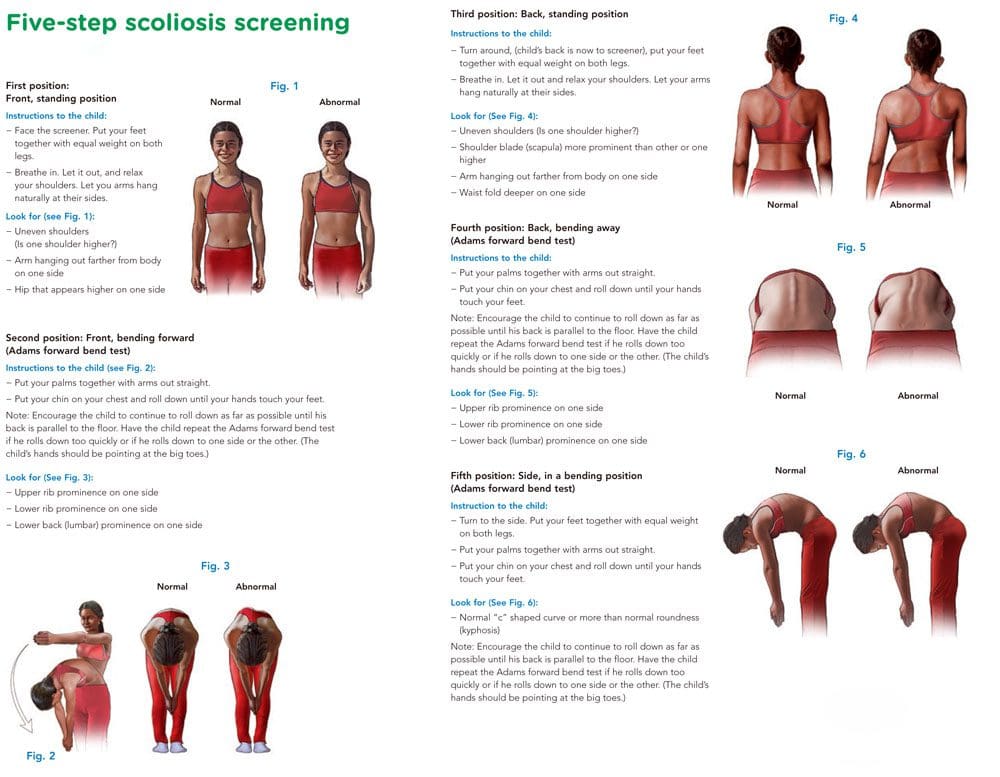
The Adams forward bend test is a simple screening method that can help with scoliosis diagnosis and help in developing a treatment plan. The exam is named after the English physician William Adams. As part of an examination, a doctor or chiropractor will look for an abnormal side-to-side bend in the spine.
Scoliosis Diagnosis
The Adams forward-bend test can help determine if there are indicators for scoliosis.
It is not an official diagnosis, but the results can be used as a starting point.
The Adams test will reveal signs of scoliosis and/or other potential deformities like:
Uneven shoulders
Uneven hips
Lack of symmetry between the vertebrae or the shoulder blades.
The head does not line up with a rib hump or the pelvis.
Detection of Other Spinal Issues
The test can also be used to find spinal curvature issues and conditions like:
Kyphosis or hunchback, where the upper back is bent forward.
Scheuermann’s disease is a form of kyphosis where the thoracic vertebrae can grow unevenly during a growth spurt and cause the vertebrae to develop into a wedge-like shape.
The Adams test by itself is not enough to confirm scoliosis.
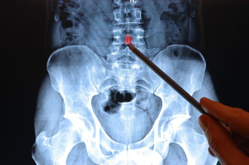
A standing X-ray with Cobb angle measurements above 10 degrees is required for diagnosing scoliosis.
The Cobb angle determines which vertebrae are tilted the most.
The higher the angle, the more severe the condition and the more probable it will produce symptoms.
Computed tomography or CT and magnetic resonance imaging or MRI scans can also be used.
Forward Bend Test
References
Glavaš, Josipa et al. “The role of school medicine in the early detection and management of adolescent idiopathic scoliosis.” Wiener klinische Wochenschrift, 1–9. 4 Oct. 2022, doi:10.1007/s00508-022-02092-1
Grossman, T W et al. “An evaluation of the Adams forward bend test and the scoliometer in a scoliosis school screening setting.” Journal of pediatric orthopedics vol. 15,4 (1995): 535-8. doi:10.1097/01241398-199507000-00025
Letts, M et al. “Computerized ultrasonic digitization in the measurement of spinal curvature.” Spine vol. 13,10 (1988): 1106-10. doi:10.1097/00007632-198810000-00009
Senkoylu, Alpaslan, et al. “A simple method for assessing rotational flexibility in adolescent idiopathic scoliosis: modified Adam’s forward bending test.” Spine deformity vol. 9,2 (2021): 333-339. doi:10.1007/s43390-020-00221-2
Low back pain is one of the most common ailments for people visiting a doctor or an urgent care clinic. When the back pain becomes intense, it can get you thinking something is seriously wrong with your back. The doctor might offer an x-ray or MRI scan to put your concerns at ease.
Fortunately, most cases of low back pain, even acute pain, improve within days or a few weeks. Most cases are remedied with chiropractic, physical therapy, heat/ice therapy, and rest. And a lot of these cases do not require any form of spinal imaging. However, those are why X-ray, MRI, and CT scans are necessary to figure out what’s happening.
Strained muscle
Sprained ligament
Poor posture
These typical causes of low back pain can be painful and limit activities.
Back Pain Lasting Longer Than 2/3 Weeks
Subacute pain lasts between 4 and 12 weeks, while chronic back pain lasts three months or longer. These are not indications of a severe lower back spinal condition.
Less than 1% of people with low back pain are diagnosed with the condition that may require spine surgery:
Doctors may recommend an x-ray or MRI if the low back pain is from a traumatic injury, like a:
Slip
Fall
Automobile accident
Other potential causes of low back pain may warrant medical imaging immediately or later.
The diagnostic process starts with the evaluation of the low back symptoms and how they relate to what was found during the:
Physical exam
Neurological exam
Medical history
A doctor utilizes these results to determine whether spinal imaging is necessary, along with the type of imaging test, x-ray, or MRI and the timing to confirm a diagnosis.
A Low Back X-Ray/MRI
X-ray spinal imaging best detects bony structural problems but is not so great with soft tissue injuries. X-ray series may be performed to diagnose vertebral compression fractures.
Anterior
Posterior
Lateral views
MRI is a radiation-free test. MRIs create 3-D anatomical views of the spinal bones and soft tissues. A contrast dye like gadolinium is used to enhance and improve the quality of the images. The contrast is injected through an intravenous line in your hand or arm before or during the test. An MRI can evaluate neurological symptoms, like radiating pain or pain that develops after a cancer diagnosis.
Symptoms, Co-existing Medical Diagnoses, and Conditions that may Require Spine Imaging
Neurological symptoms
Low back pain that radiates, fans out, or downward into the buttocks, legs, and feet
Abnormal reflexes in the lower body can indicate nerve disruption
Radiation to your entire body is measured through the millisievert (mSv), also known as the effective dose. The radiation dose is the same amount every time you experience an x-ray. When undergoing an x-ray, the radiation not absorbed by the body creates the image.
The effective dose helps a doctor measure the risk for possible side effects of radiographic imaging:
CT scans use radiation as well
Specific body tissues and organs in the lower back are sensitive to radiation exposure, like the reproductive organs.
MRI Radiation-Free Why Not Just Use This Test All The Time
MRI cannot be used on all patients because of its powerful magnet technology. Pregnant women or individuals with metal inside their body, like a spinal cord stimulator, heart pacemaker, etc., cannot be scanned with an MRI.
MRI testing is also expensive; doctors do not want to prescribe unnecessary tests that increase costs. Or because of the fine detail that MRIs provide, sometimes a spinal issue can look severe but is not.
Example: An MRI of the lower back reveals a herniated disc in a patient with no back/leg pain or other symptoms.
This is why doctors bring all their findings like the symptoms, physical exam, and medical history to confirm a diagnosis and create a custom treatment plan.
Imaging Test Takeaways
If low back pain takes its toll, listen to what the doctor recommends. They might not order a lumbar x-ray or MRI immediately but remember the issues mentioned above, like neurological symptoms and co-existing medical conditions. But these tests help discover the cause or causes of the pain. Remember this is to help get patients to their optimal health and pain-free.
How to eliminate Back Pain naturally | (2020) Foot Levelers |El Paso, Tx
NCBI Resources
Imaging diagnostics is an essential element in the evaluation of spine trauma. The rapid evolution of imaging technology has tremendously changed the assessment and treatment of spine injuries. Imaging diagnostics utilizing CT and MRI, among others, are helpful in acute and chronic settings. Spinal cord and soft-tissue injuries are best evaluated by magnetic resonance imaging, or MRI, whereas computed tomography scanning or CT scans best evaluate spinal trauma or spine fracture.
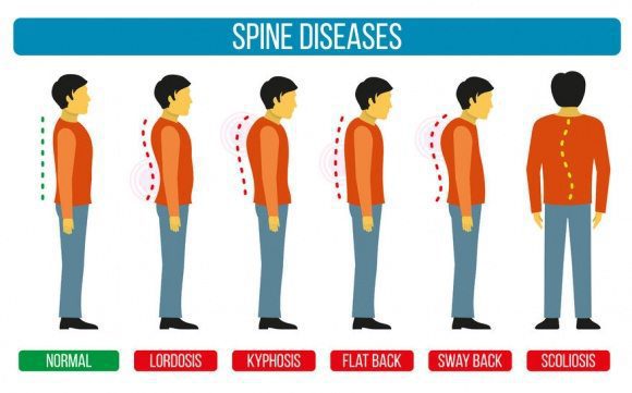
Sometimes there are abnormalities of the spine and it causes a misalignment of the natural curvatures or some curvatures may be exaggerated. These unnatural curvatures of the spine are characterized by three health conditions called lordosis, kyphosis, and scoliosis.
It is not intended to be naturally bent, twisted, or curved. The natural state of a healthy spine is somewhat straight with slight curves running front to back so that a side view would reveal them.
Viewing the spine from the back, you should see something completely different � a spine that runs straight down, top to bottom with no side to side curves. This doesn�t always happen though.
The spine is comprised of vertebrae, small bones that are stacked on top of each other with impact cushioning discs between each one. These bones act as joints, allowing the spine to bend and twist in a variety of ways.
They gently curve, sloping slightly inward at the small of the back, and again slightly at the neck. The pull of gravity, combined with body movement, can put a great deal of stress on the spine and these slight curves help absorb some of the impact.
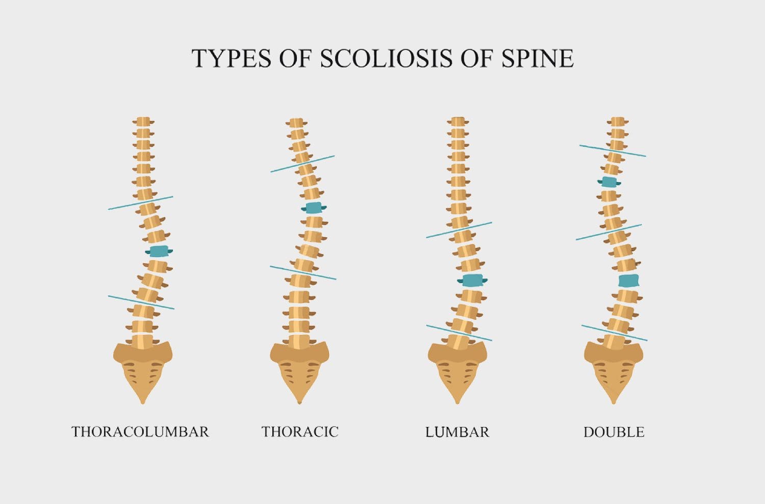
Different conditions for different types of spinal curvatures
Each of these three spinal curvature disorders affects a certain area of the spine in a very specific way.
Hyper or Hypo Lordosis � This spinal curvature disorder affects the lower back, causing the spine to curve inwards or outwards significantly.
Hyper or Hypo Kyphosis � This spinal curvature disorder affects the upper back, causing the spine to bow, resulting in that area rounding or flattening abnormally.
Scoliosis � This spinal curvature disorder can affect the entire spine, causing it to curve sideways, forming a C or S shape.
What are the symptoms?
Each type of curvature exhibits its own set of symptoms. While some symptoms may overlap, many are unique to the specific curvature disorder.
Lordosis
A �swayback� appearance where the buttocks stick out or are more pronounced.
Discomfort in the back, typically in the lumbar region
When lying on a hard surface on the back, the lower back area does not touch the surface, even when attempting to tuck the pelvis and straighten the lower back.
Difficulty with certain movements
Back pain
Kyphosis
A curve or hump to the upper back
Upper back pain and fatigue after sitting or standing for long periods (Scheuermann�s kyphosis)
Leg or back fatigue
The head bends far forward instead of being more upright
Scoliosis
Hips or waist are uneven
One shoulder blade is higher than the other
The person leans to one side
What are the causes?
Many different health issues can cause the spine to become misaligned or to form a spinal curvature. Each of the spinal conditions mentioned is affected by different conditions and situations.
Lordosis
Osteoporosis
Achondroplasia
Discitis
Obesity
Spondylolisthesis
Kyphosis
Kyphosis
Arthritis
Tumors on or in the spine
Congenital kyphosis (abnormal development of the vertebrae while the person is in utero)
Spina bifida
Scheuermann’s disease
Spine infections
Osteoporosis
Habitual slouching or poor posture
Scoliosis is still a bit of a mystery to doctors. They are not certain what exactly causes the most common form of scoliosis that is typically seen in children and adolescents. Some of the causes that they have pinpointed include:
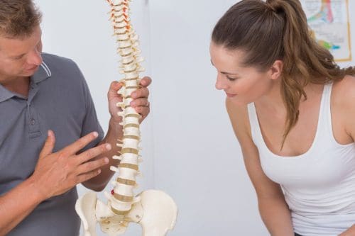
There are screenings available for both children and adults to identify any spinal curvatures in their early stages through your chiropractor. Early detection of these disorders is crucial in identifying them before they become too serious.
Personalized Spine & *SCIATICA TREATMENT* | El Paso, TX (2019)
It is estimated that scoliosis affects anywhere from 2 to 3 percent of children and adults in the United States. That is roughly six to nine million people. While it seems to develop most commonly within specific age ranges for boys and girls, it can also develop in infancy. Every year, approximately 30,000 children are fitted with a scoliosis back brace while 38,000 people have spinal fusion surgery to correct the problem. Scoliosis screenings can have tremendous benefits by identifying both risk factors for scoliosis and allowing for early treatment.
The earlier you detect scoliosis, the easier it is to treat.
Scoliosis typically develops in childhood. For girls, it usually occurs between 7 and 14 years of age. Boys develop it a little later, between 6 and 16 years of age.
Getting a scoliosis screening each year during these critical age ranges allow doctors to identify the condition early and begin treating it before it gets serious. Advanced scoliosis can require extensive treatments, bracing, and even surgery.
Chiropractic has been shown to help scoliosis, as do stretching, special exercises, and physical therapy. There are spinal adjustments that chiropractors do that are specific to the treatment of scoliosis.
When addressing the condition early on, the Cobb angle can be stopped from progressing and even reduced so that the spine has a more natural curve. Non-surgical treatments tend to be much more effective in the earlier stages of scoliosis, so early detection and early diagnosis are critical.
Identifying high-risk cases early can address current issues and prevent future ones.
Chiropractors can identify certain scoliosis risk factors in children before the condition even develops. A scoliosis screening allows them to spot tension in a child�s spinal cord � a common sign that they will develop scoliosis.
When parents are aware that their child is in a high-risk category for developing scoliosis, they can take proactive measures with home monitoring for the signs of scoliosis as well as keeping up with the course of recommended screenings. They will know to look for the signs and can address them quickly so that treatment can be started at the earliest possible time.
Help researchers and doctors become more effective in treating scoliosis.
The early stages and development of scoliosis are still shrouded in mystery for researchers and doctors. While there have been great strides made in better understanding the condition, there is still much left to learn.
There have been many studies that have aided doctors in identifying high-risk children and making early stage diagnoses, such as how the�angle of the ankle and foot are linked to scoliosis. However, screening, diagnosis, and treatment are vital to maintaining the flow of data for more studies to be conducted and more research to be done.
More mainstream screenings mean�identifying more cases of scoliosis at the early stages. This would have a two-prong effect on research. It would provide more data to be reviewed and studied, and it would increase interest in the condition as more cases of early stage scoliosis is found. This would further spur research.
Avoid the �waiting game� of seeing if scoliosis will progress.
Any parent who has had to wait for the results of a test or to see if a condition will develop or worsen knows well the anxiety of playing that waiting game. A family is usually the first person to discover scoliosis in a child.
While they may suspect a problem, or know that a problem exists, they may take a �wait and see� approach in getting treatment. If the curve worsens they may eventually seek treatment, but the constant nagging of not knowing if the curve will get worse � and the anxiety it produces � can impact not only the parents� peace of mind�but the child�s as well.
Scoliosis screenings provide peace of mind and monitor the child�s development so that if their scoliosis does progress or become a problem it can be addressed in the quickest, most efficient way possible.
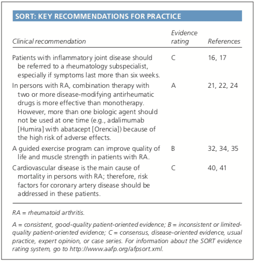
About 1.5 million people in the United States have rheumatoid arthritis. Rheumatoid arthritis, or RA, is a chronic, autoimmune disease characterized by pain and inflammation of the joints. With RA, the immune system, which protects our well-being by attacking foreign substances like bacteria and viruses, mistakenly attacks the joints. Rheumatoid arthritis most commonly affects the joints of the hands, feet, wrists, elbows, knees and ankles. Many healthcare professionals recommend early diagnosis and treatment of RA.
Abstract
Rheumatoid arthritis is the most commonly diagnosed systemic inflammatory arthritis. Women, smokers, and those with a family history of the disease are most often affected. Criteria for diagnosis include having at least one joint with definite swelling that is not explained by another disease. The likelihood of a rheumatoid arthritis diagnosis increases with the number of small joints involved. In a patient with inflammatory arthritis, the presence of a rheumatoid factor or anti-citrullinated protein antibody, or elevated C-reactive protein level or erythrocyte sedimentation rate suggests a diagnosis of rheumatoid arthritis. Initial laboratory evaluation should also include complete blood count with dif- ferential and assessment of renal and hepatic function. Patients taking biologic agents should be tested for hepatitis B, hepatitis C, and tuberculosis. Earlier diagnosis of rheumatoid arthritis allows for earlier treatment with disease-modifying antirheumatic agents. Combinations of medications are often used to control the disease. Methotrexate is typically the first-line drug for rheumatoid arthritis. Biologic agents, such as tumor necrosis factor inhibitors, are generally considered second-line agents or can be added for dual therapy. The goals of treatment include minimiza- tion of joint pain and swelling, prevention of radiographic damage and visible deformity, and continuation of work and personal activities. Joint replacement is indicated for patients with severe joint damage whose symptoms are poorly controlled by medical management. (Am Fam Physician. 2011;84(11):1245-1252. Copyright � 2011 American Academy of Family Physicians.)
Rheumatoid arthritis (RA) is the most common inflammatory arthritis, with a lifetime prevalence of up to 1 percent worldwide.1 Onset can occur at any age, but peaks between 30 and 50 years.2 Disability is common and significant. In a large U.S. cohort, 35 percent of patients with RA had work disability after 10 years.3
Etiology and Pathophysiology
Like many autoimmune diseases, the etiology of RA is multifactorial. Genetic susceptibility is evident in familial clustering and monozygotic twin studies, with 50 percent of RA risk attributable to genetic factors.4 Genetic associations for RA include human leukocyte antigen-DR45 and -DRB1, and a variety of alleles called the shared epitope.6,7 Genome-wide association studies have identified additional genetic signatures that increase the risk of RA and other autoimmune diseases, including STAT4 gene and CD40 locus.5 Smoking is the major environmental trigger for RA, especially in those with a genetic predisposition.8 Although infections may unmask an autoimmune response, no particular pathogen has been proven to cause RA.9
RA is characterized by inflammatory pathways that lead to proliferation of synovial cells in joints. Subsequent pannus formation may lead to underlying cartilage destruction and bony erosions. Overproduction of pro-inflammatory cytokines, including tumor necrosis factor (TNF) and interleukin-6, drives the destructive process.10
Risk Factors
Older age, a family history of the disease, and female sex are associated with increased risk of RA, although the sex differential is less prominent in older patients.1 Both current and prior cigarette smoking increases the risk of RA (relative risk [RR] = 1.4, up to 2.2 for more than 40-pack-year smokers).11
Pregnancy often causes RA remission, likely because of immunologic tolerance.12 Parity may have long-lasting impact; RA is less likely to be diagnosed in parous women than in nulliparous women (RR = 0.61).13,14 Breastfeeding decreases the risk of RA (RR = 0.5 in women who breastfeed for at least 24 months), whereas early menarche�(RR = 1.3 for those with menarche at 10 years of age or younger) and very irregular menstrual periods (RR = 1.5) increase risk.14 Use of oral contraceptive pills or vitamin E does not affect RA risk.15
Diagnosis
Typical Presentation
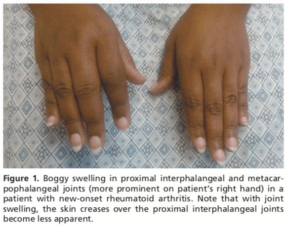
Patients with RA typically present with pain and stiffness in multiple joints. The wrists, proximal interphalangeal joints, and metacarpophalangeal joints are most commonly involved. Morning stiffness lasting more than one hour suggests an inflammatory etiology. Boggy swelling due to synovitis may be visible (Figure 1), or subtle synovial thickening may be palpable on joint examination. Patients may also present with more indolent arthralgias before the onset of clinically apparent joint swelling. Systemic symptoms of fatigue, weight loss, and low-grade fever may occur with active disease.
Diagnostic Criteria
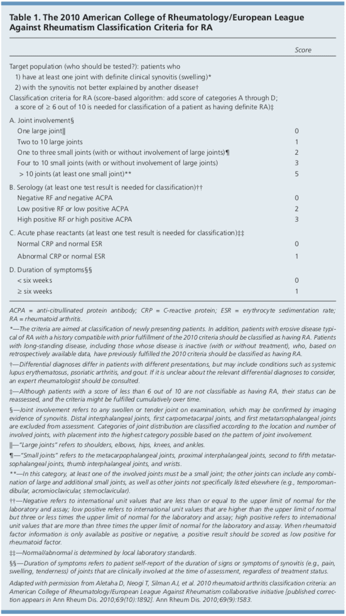
In 2010, the American College of Rheumatology and European League Against Rheumatism collaborated to create new classification criteria for RA (Table 1).16 The new criteria are an effort to diagnose RA earlier in patients who may not meet the 1987 American College of Rheumatology classification criteria. The 2010 criteria do not include presence of rheumatoid nodules or radiographic erosive changes, both of which are less likely in early RA. Symmetric arthri- tis is also not required in the 2010 criteria, allowing for early asymmetric presentation.
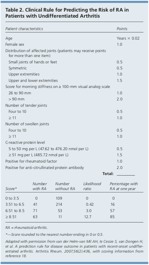
In addition, Dutch researchers have developed and validated a clinical prediction rule for RA (Table 2).17,18 The purpose of this rule is to help identify patients with undifferentiated arthritis that is most likely to progress to RA, and to guide follow-up and referral.
Diagnostic Tests
Autoimmune diseases such as RA are often characterized by the presence of autoanti- bodies. Rheumatoid factor is not specific for RA and may be present in patients with other diseases, such as hepatitis C, and in healthy older persons. Anti-citrullinated protein antibody is more specific for RA and may play a role in disease pathogenesis.6 Approxi- mately 50 to 80 percent of persons with RA have rheumatoid factor, anti-citrullinated protein antibody, or both.10 Patients with RA may have a positive antinuclear antibody test result, and the test is of prognostic impor- tance in juvenile forms of this disease.19 C-reactive protein levels and erythrocyte sedimentation rate are often increased with active RA, and these acute phase reactants are part of the new RA classification criteria.16 C-reactive protein levels and erythrocyte sedimentation rate may also be used to follow disease activity and response to medication.
Baseline complete blood count with differential and assessment of renal and hepatic function are helpful because the results may influence treatment options (e.g., a patient with renal insufficiency or significant thrombocytopenia likely would not be prescribed a nonsteroidal anti-inflammatory drug [NSAID]). Mild anemia of chronic disease occurs in 33 to 60 percent of all patients with RA,20 although gastrointestinal blood loss should also be considered in patients taking corticosteroids or NSAIDs. Methotrexate is contraindicated in patients with hepatic disease, such as hepatitis C, and in patients with significant renal impairment.21 Biologic therapy, such as a TNF inhibitor, requires a negative tuberculin test or treatment for latent tuberculosis. Hepatitis B reactivation can also occur with TNF inhibitor use.22 Radiography of hands and feet should be performed to evaluate for characteristic periarticular erosive changes,�which may be indicative of a more aggressive RA subtype.10
Differential Diagnosis
Skin findings suggest systemic lupus erythematosus, systemic sclerosis, or psoriatic arthritis. Polymyalgia rheumatica should be considered in an older patient with symptoms primarily in the shoulder and hip, and the patient should be asked questions related to associated temporal arteritis.
Chest radiography is helpful to evaluate for sarcoidosis as an etiology of arthritis.�Patients with inflammatory back symptoms, a history of inflammatory bowel disease, or inflammatory eye disease may have spondyloarthropathy. Persons with less than six weeks of symptoms may have a viral process, such as parvovirus. Recurrent self-limited episodes of acute joint swelling suggest crystal arthropathy, and arthrocentesis should be performed to evaluate for monosodium urate monohydrate or calcium pyrophosphate dihydrate crystals. The presence of numerous myofascial trigger points and somatic symptoms may suggest fibromyalgia, which can coexist with RA. To help guide diagnosis and determine treatment strategy, patients with inflammatory arthritis should be promptly referred to a rheumatology subspecialist.16,17
Rheumatoid arthritis, or RA, is the most common type of arthritis. RA is an autoimmune disease, caused when the immune system, the human body’s defense system, attacks its own cells and tissues, particularly the joints. Rheumatoid arthritis is frequently identified by symptoms of pain and inflammation, often affecting the small joints of the hands, wrists and feet. According to many healthcare professionals, early diagnosis and treatment of RA is essential to prevent further joint damage and decrease painful symptoms. Dr. Alex Jimenez D.C., C.C.S.T. Insight
Treatment
After RA has been diagnosed and an initial evaluation performed, treatment should begin. Recent guidelines have addressed the management of RA,21,22 but patient preference also plays an important role. There are special considerations for women of childbearing age because many medications have deleterious effects on pregnancy. Goals of therapy include minimizing joint pain and swelling, preventing deformity (such as ulnar deviation) and radiographic damage (such as erosions), maintaining quality of life (personal and work), and controlling extra-articular manifestations. Disease-modifying antirheumatic drugs (DMARDs) are the mainstay of RA therapy.
DMARDs
DMARDs can be biologic or nonbiologic (Table 3).23 Biologic agents include monoclonal antibodies and recombinant receptors to block cytokines that promote the inflammatory cascade responsible for RA symptoms. Methotrexate is recommended as the first- line treatment in patients with active RA, unless contraindicated or not tolerated.21 Leflunomide (Arava) may be used as an alternative to methotrexate, although gastrointestinal adverse effects are more common. Sulfasalazine (Azulfidine) or hydroxychloroquine (Plaquenil) pro-inflammatory as monotherapy in patients with low disease�activity or without poor prognostic features (e.g., seronegative, non-erosive RA).21,22
Combination therapy with two or more DMARDs is more effective than monotherapy; however, adverse effects may also be greater.24 If RA is not well controlled with a nonbiologic DMARD, a biologic DMARD should be initiated.21,22 TNF inhibitors are the first-line biologic therapy and are the most studied of these agents. If TNF inhibitors are ineffective, additional biologic therapies can be considered. Simultaneous use of more than one biologic therapy (e.g., adalimumab [Humira] with abatacept [Orencia]) is not�recommended because of an unacceptable rate of adverse effects.21
NSAIDs and Corticosteroids
Drug therapy for RA may involve NSAIDs and oral, intramuscular, or intra-articular corticosteroids for controlling pain and inflammation. Ideally, NSAIDs and corticosteroids are used only for short-term management. DMARDs are the preferred therapy.21,22
Complementary Therapies
Dietary interventions, including vegetarian and Mediterranean diets, have been�studied in the treatment of RA without convincing evidence of benefit.25,26 Despite some favorable outcomes, there is a lack of evidence for the effectiveness of acupuncture in placebo-controlled trials of patients with RA.27,28 In addition, thermotherapy and therapeutic ultrasound for RA have not been studied adequately.29,30 A Cochrane review of herbal treatments for RA concluded that gamma-linolenic acid (from evening primrose or black currant seed oil) and Tripterygium wilfordii (thunder god vine) have potential benefits.31 It is important to inform patients that serious adverse effects have been reported with use of herbal therapy.31
Exercise and Physical Therapy
Results of randomized controlled trials sup- port physical exercise to improve quality of life and muscle strength in patients with RA.32,33 Exercise training programs have not been shown to have deleterious effects on RA disease activity, pain scores, or radiographic joint damage.34 Tai chi has been shown to improve ankle range of motion in persons with RA, although randomized trials are limited.35 Randomized controlled trials of Iyengar yoga in young adults with RA are underway.36
Duration of Treatment
Remission is obtainable in 10 to 50 percent of patients with RA, depending on how remission is defined and the intensity of therapy.10 Remission is more likely in males, nonsmokers, persons younger than 40 years, and in those with late-onset disease (patients older than 65 years), with shorter duration of disease, with milder disease activity, without elevated acute phase reactants, and without positive rheumatoid factor or anti-citrullinated protein antibody findings.37
After the disease is controlled, medication dosages may be cautiously decreased to the minimum amount necessary. Patients will require frequent monitoring to ensure stable symptoms, and prompt increase in medication is recommended with disease flare-ups.22
Joint Replacement
Joint replacement is indicated when there is severe joint damage and unsatisfactory control of symptoms with medical management. Long-term outcomes are support, with only 4 to 13 percent of large joint replacements requiring revision within 10 years.38 The hip and knee are the most commonly replaced joints.
Long-Term Monitoring
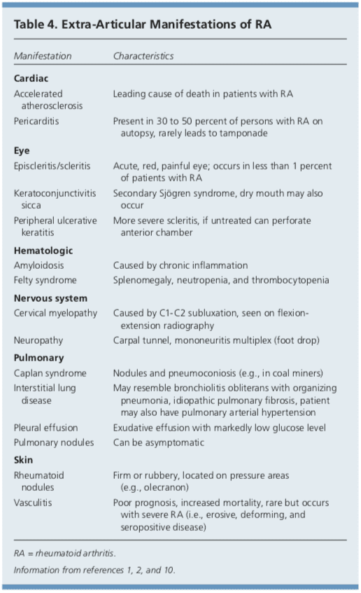
Although RA is considered a disease of the joints, it is also a systemic disease capable of involving multiple organ systems. Extra-articular manifestations of RA are included in Table 4.1,2,10
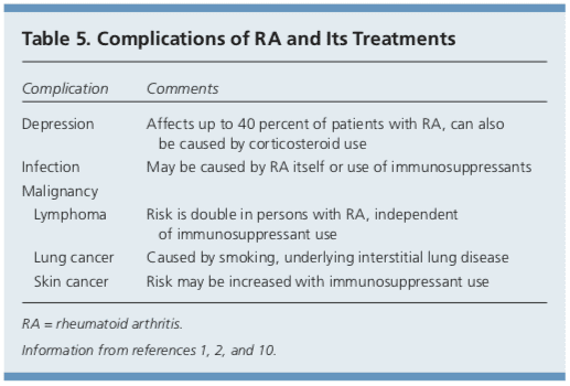
Patients with RA have a twofold increased risk of lymphoma, which is thought to be caused by the underlying inflammatory�process, and not a consequence of medical treatment.39 Patients with RA are also at an increased risk of coronary artery disease, and physicians should work with patients to modify risk factors, such as smoking, high blood pressure, and high cholesterol.40,41 Class III or IV congestive heart failure (CHF) is a contraindication for using TNF inhibitors, which can worsen CHF outcomes.21 In patients with RA and malignancy, caution is needed with continued use of DMARDs, especially TNF inhibitors. Biologic DMARDs, methotrexate, and leflunomide should not be initiated in patients with active herpes zoster, significant fungal infection, or bacterial infection requiring antibiotics.21 Complications of RA and its treatments are listed in Table 5.1,2,10
Prognosis
Patients with RA live three to 12 years less than the general population.40 Increased mortality in these patients is mainly due to accelerated cardiovascular disease, especially in those with high disease activity and chronic inflammation. The relatively new biologic therapies may reverse progression of atherosclerosis and extend life in those with RA.41
Data Sources: A PubMed search was completed in Clinical Queries using the key terms rheumatoid arthritis, extra-articular manifestations, and disease-modifying antirheumatic agents. The search included meta-analyses, randomized controlled trials, clinical trials, and reviews. Also searched were the Agency for Healthcare Research and Quality evidence reports, Clinical Evidence, the Cochrane database, Essential Evidence, and UpToDate. Search date: September 20, 2010.
Author disclosure: No relevant financial affiliations to disclose.
In conclusion, rheumatoid arthritis is a chronic, autoimmune disease which causes painful symptoms, such as pain and discomfort, inflammation and swelling of the joints, among others. The joint damage characterized as RA is symmetrical, meaning it generally affects both sides of the body. Early�diagnosis is essential for treatment of RA. The scope of our information is limited to chiropractic and spinal health issues. To discuss the subject matter, please feel free to ask Dr. Jimenez or contact us at�915-850-0900�.
Curated by Dr. Alex Jimenez
Additional Topic Discussion: Relieving Knee Pain without Surgery
Knee pain is a well-known symptom which can occur due to a variety of knee injuries and/or conditions, including�sports injuries. The knee is one of the most complex joints in the human body as it is made-up of the intersection of four bones, four ligaments, various tendons, two menisci, and cartilage. According to the American Academy of Family Physicians, the most common causes of knee pain include patellar subluxation, patellar tendinitis or jumper’s knee, and Osgood-Schlatter disease. Although knee pain is most likely to occur in people over 60 years old, knee pain can also occur in children and adolescents. Knee pain can be treated at home following the RICE methods, however, severe knee injuries may require immediate medical attention, including chiropractic care.
1. Etiology and pathogenesis of rheumatoid arthritis. In: Firestein GS, Kelley WN, eds. Kelley�s Textbook of Rheu- matology. 8th ed. Philadelphia, Pa.: Saunders/Elsevier; 2009:1035-1086. 2. Bathon J, Tehlirian C. Rheumatoid arthritis clinical and laboratory manifestations. In: Klippel JH, Stone JH, Crofford LJ, et al., eds. Primer on the Rheumatic Dis- eases. 13th ed. New York, NY: Springer; 2008:114-121. 3. Allaire S, Wolfe F, Niu J, et al. Current risk factors for work disability associated with rheumatoid arthritis. Arthritis Rheum. 2009;61(3):321-328. 4. MacGregor AJ, Snieder H, Rigby AS, et al. Characteriz- ing the quantitative genetic contribution to rheumatoid arthritis using data from twins. Arthritis Rheum. 2000; 43(1):30-37. 5. Orozco G, Barton A. Update on the genetic risk fac- tors for rheumatoid arthritis. Expert Rev Clin Immunol. 2010;6(1):61-75. 6. Balsa A, Cabezo?n A, Orozco G, et al. Influence of HLA DRB1 alleles in the susceptibility of rheumatoid arthritis and the regulation of antibodies against citrullinated proteins and rheumatoid factor. Arthritis Res Ther. 2010;12(2):R62. 7. McClure A, Lunt M, Eyre S, et al. Investigating the via- bility of genetic screening/testing for RA susceptibility using combinations of five confirmed risk loci. Rheuma- tology (Oxford). 2009;48(11):1369-1374. 8. Bang SY, Lee KH, Cho SK, et al. Smoking increases rheu- matoid arthritis susceptibility in individuals carrying the HLA-DRB1 shared epitope, regardless of rheumatoid factor or anti-cyclic citrullinated peptide antibody sta- tus. Arthritis Rheum. 2010;62(2):369-377. 9. Wilder RL, Crofford LJ. Do infectious agents cause rheu- matoid arthritis? Clin Orthop Relat Res. 1991;(265): 36-41. 10. Scott DL, Wolfe F, Huizinga TW. Rheumatoid arthritis. Lancet. 2010;376(9746):1094-1108. 11. Costenbader KH, Feskanich D, Mandl LA, et al. Smoking intensity, duration, and cessation, and the risk of rheu- matoid arthritis in women. Am J Med. 2006;119(6): 503.e1-e9. 12. Kaaja RJ, Greer IA. Manifestations of chronic disease during pregnancy. JAMA. 2005;294(21):2751-2757. 13. Guthrie KA, Dugowson CE, Voigt LF, et al. Does preg- nancy provide vaccine-like protection against rheuma- toid arthritis? Arthritis Rheum. 2010;62(7):1842-1848. 14. Karlson EW, Mandl LA, Hankinson SE, et al. Do breast- feeding and other reproductive factors influence future risk of rheumatoid arthritis? Results from the Nurses� Health Study. Arthritis Rheum. 2004;50(11):3458-3467. 15. Karlson EW, Shadick NA, Cook NR, et al. Vitamin E in the primary prevention of rheumatoid arthritis: the Women�s Health Study. Arthritis Rheum. 2008;59(11): 1589-1595. 16. Aletaha D, Neogi T, Silman AJ, et al. 2010 rheumatoid arthritis classification criteria: an American College of Rheumatology/European League Against Rheumatism collaborative initiative [published correction appears in Ann Rheum Dis. 2010;69(10):1892]. Ann Rheum Dis. 2010;69(9):1580-1588. 17. van der Helm-van Mil AH, le Cessie S, van Dongen H, et al. A prediction rule for disease outcome in patients with recent-onset undifferentiated arthritis. Arthritis Rheum. 2007;56(2):433-440. 18. Mochan E, Ebell MH. Predicting rheumatoid arthritis risk in adults with undifferentiated arthritis. Am Fam Physi- cian. 2008;77(10):1451-1453. 19. Ravelli A, Felici E, Magni-Manzoni S, et al. Patients with antinuclear antibody-positive juvenile idiopathic arthri- tis constitute a homogeneous subgroup irrespective of the course of joint disease. Arthritis Rheum. 2005; 52(3):826-832. 20. Wilson A, Yu HT, Goodnough LT, et al. Prevalence and outcomes of anemia in rheumatoid arthritis. Am J Med. 2004;116(suppl 7A):50S-57S. 21. Saag KG, Teng GG, Patkar NM, et al. American College of Rheumatology 2008 recommendations for the use of nonbiologic and biologic disease-modifying antirheu- matic drugs in rheumatoid arthritis. Arthritis Rheum. 2008;59(6):762-784. 22. Deighton C, O�Mahony R, Tosh J, et al.; Guideline Devel- opment Group. Management of rheumatoid arthritis: summary of NICE guidance. BMJ. 2009;338:b702. 23. AHRQ. Choosing medications for rheumatoid arthritis. April 9, 2008. http://www.effectivehealthcare.ahrq.gov/ ehc/products/14/85/RheumArthritisClinicianGuide.pdf. Accessed June 23, 2011. 24. Choy EH, Smith C, Dore? CJ, et al. A meta-analysis of the efficacy and toxicity of combining disease-modify- ing anti-rheumatic drugs in rheumatoid arthritis based on patient withdrawal. Rheumatology (Oxford). 2005; 4 4 (11) :1414 -1421. 25. Smedslund G, Byfuglien MG, Olsen SU, et al. Effective- ness and safety of dietary interventions for rheumatoid arthritis. J Am Diet Assoc. 2010;110(5):727-735. 26. Hagen KB, Byfuglien MG, Falzon L, et al. Dietary inter- ventions for rheumatoid arthritis. Cochrane Database Syst Rev. 2009;21(1):CD006400. 27. Wang C, de Pablo P, Chen X, et al. Acupuncture for pain relief in patients with rheumatoid arthritis: a systematic review. Arthritis Rheum. 2008;59(9):1249-1256. 28. Kelly RB. Acupuncture for pain. Am Fam Physician. 2009;80(5):481-484. 29. Robinson V, Brosseau L, Casimiro L, et al. Thermother- apy for treating rheumatoid arthritis. Cochrane Data- base Syst Rev. 2002;2(2):CD002826. 30. Casimiro L, Brosseau L, Robinson V, et al. Therapeutic ultrasound for the treatment of rheumatoid arthritis. Cochrane Database Syst Rev. 2002;3(3):CD003787. 31. Cameron M, Gagnier JJ, Chrubasik S. Herbal therapy for treating rheumatoid arthritis. Cochrane Database Syst Rev. 2011;(2):CD002948. 32. Brodin N, Eurenius E, Jensen I, et al. Coaching patients with early rheumatoid arthritis to healthy physical activ- ity. Arthritis Rheum. 2008;59(3):325-331. 33. Baillet A, Payraud E, Niderprim VA, et al. A dynamic exercise programme to improve patients� disability in rheumatoid arthritis: a prospective randomized con- trolled trial. Rheumatology (Oxford). 2009;48(4): 410-415. 34. Hurkmans E, van der Giesen FJ, Vliet Vlieland TP, et al. Dynamic Exercise programs (aerobic capacity and/or mus- cle strength training) in patients with rheumatoid arthri- tis. Cochrane Database Syst Rev. 2009;(4):CD006853. 35. Han A, Robinson V, Judd M, et al. Tai chi for treat- ing rheumatoid arthritis. Cochrane Database Syst Rev. 2004;(3):CD004849. 36. Evans S, Cousins L, Tsao JC, et al. A randomized con- trolled trial examining Iyengar yoga for young adults with rheumatoid arthritis. Trials. 2011;12:19. 37. Katchamart W, Johnson S, Lin HJ, et al. Predictors for remis- sion in rheumatoid arthritis patients: a systematic review. Arthritis Care Res (Hoboken). 2010;62(8):1128-1143. 38. Wolfe F, Zwillich SH. The long-term outcomes of rheu- matoid arthritis: a 23-year prospective, longitudinal study of total joint replacement and its predictors in 1,600 patients with rheumatoid arthritis. Arthritis Rheum. 1998;41(6):1072-1082. 39. Baecklund E, Iliadou A, Askling J, et al. Association of chronic inflammation, not its treatment, with increased lymphoma risk in rheumatoid arthritis. Arthritis Rheum. 2006;54(3):692-701. 40. Friedewald VE, Ganz P, Kremer JM, et al. AJC editor�s consensus: rheumatoid arthritis and atherosclerotic cardiovascular disease. Am J Cardiol. 2010;106(3): 442-447. 41. Atzeni F, Turiel M, Caporali R, et al. The effect of phar- macological therapy on the cardiovascular system of patients with systemic rheumatic diseases. Autoimmun Rev. 2010;9(12):835-839.
Arthritis is characterized as the inflammation of one or multiple joints. The most common symptoms of arthritis include pain and discomfort, swelling, inflammation, and stiffness, among others. Arthritis may affect�any joint in the human body, however, it commonly develops in the knee. � Knee arthritis can make everyday�physical activities difficult. The most prevalent types of arthritis are osteoarthritis and rheumatoid arthritis, although there are well over 100 distinct forms of arthritis, affecting children and adults alike. While there is no cure for arthritis, many treatment approaches can help treat the symptoms of knee arthritis.
Anatomy of the Knee
� The knee is the largest and strongest joint in the human body. It is made up of the lower end of the thigh bone,�or femur, the top end of the shin bone, or tibia, and the kneecap, or patella. The ends of the three bones are covered with articular cartilage, a smooth, slippery structure which protects and cushions the bones when bending and straightening the knee.
� Two wedge-shaped parts of cartilage, known as the meniscus, function as shock absorbers between the bones of the knee to help cushion the joint and provide stability. The knee joint is also surrounded by a thin lining known as the synovial membrane. This membrane releases a fluid which lubricates the cartilage and also helps reduce friction in the knee. The significant kinds of arthritis that affect the knee�include osteoarthritis, rheumatoid arthritis, and post-traumatic arthritis.
Osteoarthritis
� Osteoarthritis is the most common type of arthritis which affects the knee joint. This form of arthritis is a degenerative, wear-and-tear health issue which occurs most commonly in people 50 years of age and older, however, it may also develop in younger people.
� In osteoarthritis, the cartilage in the knee joint gradually wears away. As the cartilage wears away, the distance between the bones decreases. This can result in bone rubbing and it can�create painful bone spurs. Osteoarthritis generally develops slowly but the pain may worsen over time.
Rheumatoid Arthritis
� Rheumatoid arthritis is a chronic health issue which affects multiple joints throughout the body, especially the knee joint. RA is also symmetrical, meaning it often affects the same joint on each side of the human body.
� In rheumatoid arthritis, the synovial membrane that covers the knee joint becomes inflamed and swollen, causing knee pain, discomfort, and stiffness. RA is an autoimmune disease, which means that the immune system attacks its own soft tissues. The immune system attacks healthy tissue,�including tendons, ligaments and cartilage, as well as softens the bone.
Post-traumatic Arthritis
� Posttraumatic arthritis is a form of arthritis that develops after damage or injury to the knee. By way of instance, the knee joint may be harmed by a broken bone, or fracture, and result in post-traumatic arthritis years after the initial injury. Meniscal tears and ligament injuries can cause additional wear-and-tear on the knee joint, which over time can lead to arthritis and other problems.
Symptoms of Knee Arthritis
� The most common symptoms of knee arthritis include pain and discomfort, inflammation, swelling, and stiffness. Although sudden onset is probable, the painful symptoms generally�develop gradually over time. Additional symptoms of knee arthritis can be recognized as follows:
The joint may become stiff and swollen, making it difficult to bend and straighten the knee.
Swelling and inflammation may be worse in the morning, or when sitting or resting.
Vigorous activity might cause the pain to flare up.
Loose fragments of cartilage and other soft tissue may interfere with the smooth motion of the joints, causing the knee to lock or stick through motion. It could also creak, click, snap or make a grinding sound, known as crepitus.
Pain can cause a sense of fatigue or buckling from the knee.
Many individuals with arthritis may also describe increased joint pain with rainy weather and climate changes.
Diagnosis for Knee Arthritis
� During the patient’s appointment for diagnosis of knee arthritis, the healthcare professional will talk about the symptoms and medical history, as well as conduct a physical examination. The doctor may also order imaging diagnostic tests, such as X-rays, MRI or blood tests for further diagnosis. During the physical examination, the doctor will search for:
Joint inflammation, swelling, warmth, or redness
Tenderness around the knee joint
Assortment of passive and active movement
Instability of the knee joint
Crepitus, the grating sensation inside the joint, with motion
Pain when weight is placed on the knee
Issues with gait, or manner of walking
Any signs of damage or injury to the muscles, tendons, and ligaments surrounding the knee joint
Involvement of additional joints (an indicator of rheumatoid arthritis)
Imaging Diagnostic Tests
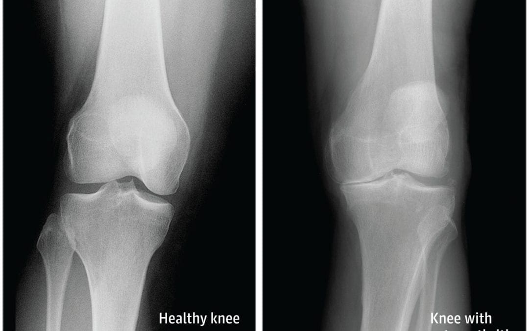
X-rays. These imaging diagnostic tests produce images of compact structures, such as bones. They can help distinguish among various forms of arthritis. X-rays for knee arthritis may demonstrate a portion of the joint distance, changes in the bone as well as the formation of bone spurs, known as osteophytes.
Additional tests. Sometimes, magnetic resonance imaging, or MRI, scans, computed tomography, or CT,�scans, or bone scans are required to ascertain the condition of the bone and soft tissues of the knee.
Blood Tests
� Your doctor may also recommend blood tests to determine which type of arthritis you have. With some kinds of arthritis, such as rheumatoid arthritis, blood tests can help with the proper identification of the disease.
Although the knee joint is one of the strongest and largest joints in the human body, it is often prone to suffering damage or injury, resulting in a variety of conditions. In addition, however, other health issues, such as arthritis, can affect the knee joint. In network for most insurances of El Paso, TX, chiropractic care can help ease painful symptoms associated with knee arthritis, among other health issues. Dr. Alex Jimenez D.C., C.C.S.T. Insight
�
Treatment for Knee Arthritis
Non-surgical Treatment
� Non-surgical treatment approaches are often recommended before considering surgical treatment for knee arthritis. Healthcare professionals may recommend a variety of treatment options, including chiropractic care, physical therapy, and lifestyle modifications, among others.
� Lifestyle modifications. Some lifestyle modifications can help protect the knee joint and impede the progress of arthritis. Minimizing physical activities which aggravate the condition, will put less strain on the knee. Losing weight may also help lessen stress and pressure on the knee joint, resulting in less painful symptoms and increased function.
� Chiropractic care and physical therapy.�Chiropractic care utilizes full body chiropractic adjustments to carefully restore any spinal misalignments, or subluxations, which may�be causing symptoms, including arthritis. The doctor may also recommend physical therapy to create an individualized exercise and physical activity program for each patient’s needs.�Specific exercises will help increase range of motion and endurance, as well as help strengthen the muscles in the lower extremities.
� Assistive devices. Using assistive devices, such as a cane, shock-absorbing shoes or inserts, or a brace or knee sleeve, can decrease painful symptoms. A brace helps with function and stability, and may be particularly useful if the arthritis is based on one side of the knee. There are two types of braces that are often used for knee arthritis: A “unloader” brace shifts weight from the affected section of the knee, while a “support” brace helps support the entire knee load.
� Drugs and/or medications. Several types of medications are useful in treating arthritis of the knee. Since individuals respond differently to medications, your doctor will work closely with you to determine the medications and dosages which are safe and effective for you.
Surgical Treatment
� The healthcare professional may recommend surgical treatment if the patient’s knee arthritis causes severe disability and only if the problem isn’t relieved with non-surgical treatment. Like all surgeries, there are a few risks and complications with surgical treatment for knee arthritis. The�doctor will discuss the possible problems with the patient.
� Arthroscopy. During arthroscopy, physicians use instruments and small incisions to diagnose and treat knee joint problems. Arthroscopic surgery isn’t frequently used in the treatment of arthritis of the knee. In cases where osteoarthritis is accompanied with a degenerative meniscal tear, arthroscopic surgery may be wise to treat the torn meniscus.
� Cartilage grafting. Normal cartilage tissue may be taken from a tissue bank or through a different part of the knee to fill out a hole in the articular cartilage. This process is typically considered only for younger patients.
� Synovectomy. The lining damaged by rheumatoid arthritis is eliminated to reduce swelling and pain.
� Osteotomy. In a knee osteotomy, either the tibia (shinbone) or femur (thighbone) is cut then reshaped to relieve stress and pressure on the knee joint. Knee�osteotomy is utilized when early-stage osteoarthritis has damaged one facet of the knee joint. By changing the weight distribution, this can relieve and enhance the function of the knee.
� Total or partial knee replacement (arthroplasty).�The�doctor will remove the damaged bone and cartilage, then place new plastic or metal surfaces to restore the function of the knee�and its surrounding structures.
� Following any type of surgery for knee�arthritis will involve a period of recovery. Recovery time and rehabilitation will depend on the type of surgery performed. It’s essential to talk with your healthcare professional to determine the best treatment option for your�knee arthritis. The scope of our information is limited to chiropractic and spinal health issues. To discuss the subject matter, please feel free to ask Dr. Jimenez or contact us at�915-850-0900�.
� Curated by Dr. Alex Jimenez �
�
Additional Topic Discussion: Relieving Knee Pain without Surgery
� Knee pain is a well-known symptom which can occur due to a variety of knee injuries and/or conditions, including�sports injuries. The knee is one of the most complex joints in the human body as it is made-up of the intersection of four bones, four ligaments, various tendons, two menisci, and cartilage. According to the American Academy of Family Physicians, the most common causes of knee pain include patellar subluxation, patellar tendinitis or jumper’s knee, and Osgood-Schlatter disease. Although knee pain is most likely to occur in people over 60 years old, knee pain can also occur in children and adolescents. Knee pain can be treated at home following the RICE methods, however, severe knee injuries may require immediate medical attention, including chiropractic care.
IFM's Find A Practitioner tool is the largest referral network in Functional Medicine, created to help patients locate Functional Medicine practitioners anywhere in the world. IFM Certified Practitioners are listed first in the search results, given their extensive education in Functional Medicine