Understanding Chiropractic Wedges: Their Role in Pain Relief and Spinal Health
Chiropractic care helps people feel better by fixing problems in the spine and body without surgery or strong medicines. One tool that chiropractors often use is called a wedge. These are simple, triangle-shaped blocks made from foam or other firm materials. They are placed on parts of the body, such as the neck, hips, or feet. The idea is to use gravity—the Earth’s natural pull—to gently stretch and align the body. This can help correct spinal curves, ease pain, and improve overall body function (Diamond State Chiropractic, n.d.).
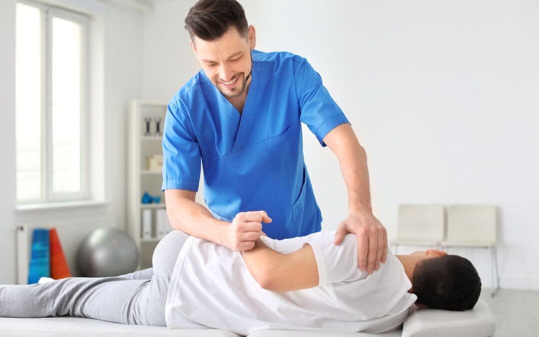
Wedges are not like hard adjustments where the chiropractor pushes on the spine. Instead, they let the body relax and correct itself slowly. Patients lie on them for a few minutes, and gravity does the work. This makes them good for people who want gentle care, such as older adults or pregnant individuals. They can help with back pain, neck strain, and even headaches by improving the body’s alignment (Tiger Lily Chiropractic, n.d.).
In this article, we’ll look at how these wedges work, the different types, and why they fit into a bigger picture of health care. We’ll also discuss how clinics that combine different treatments can improve patient outcomes.
What Are Chiropractic Wedges, and How Do They Work?
Chiropractic wedges are basic tools that look like small ramps. They come in different sizes and shapes, but most are firm enough to support the body’s weight. When a person lies on one side, the wedge lifts a specific area, such as the neck or pelvis. This creates a gentle pull that stretches tight muscles and helps bones return to their proper positions.
The main goal is to restore the spine’s natural curves. The spine isn’t straight; it has gentle bends that help us stand tall and move easily. If these curves become flat or twisted due to poor posture, injuries, or daily stress, it can lead to pain. Wedges use the body’s own weight to fix this over time (Core Chiropractic, n.d.).
Here’s how they typically work:
Placement: The chiropractor places the wedge at the right spot based on the body’s needs.
Time: Patients relax on it for 5 to 10 minutes, sometimes longer, as they get used to it.
Gravity’s Role: No pushing or twisting—just letting gravity pull things into alignment.
Safety: Always start slow to avoid strain, and stop if it hurts (Pure Health, n.d.).
This passive method means no sudden moves, making it comfortable for most people. It’s often part of a plan that includes other care, such as exercises or advice on sitting better.
Types of Chiropractic Wedges
There are a few main kinds of wedges, each for a different part of the body. They target specific issues but can help the whole body feel better.
Neck Wedges (Cervical Wedges)
These are for the upper spine, which includes the neck. Many people lose the natural curve in their neck from looking down at phones or computers all day. This is called forward head posture, and it puts extra pressure on the neck and shoulders.
To use a neck wedge:
Lie on your back on a flat surface.
Place the wedge so the flat side is against your shoulders, and your head rests on the sloped part.
Relax for 5-10 minutes, letting gravity stretch the neck.
Start with short times and build up (YouTube – Cordova & Siegmund, n.d.).
Benefits include less neck pain, fewer headaches, and better posture. It can even help with things like dizziness or tingling in the arms by taking stress off nerves (Pure Health, n.d.). One clinic notes that consistent use, along with adjustments, helps the curve come back and makes changes last longer (Chiropractic First, n.d.).
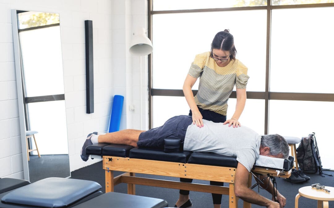
Pelvic Wedges or SOT Blocks
These are used in the Sacro Occipital Technique (SOT). They go under the hips or pelvis while the person lies face down. The wedges act like a see-saw, using gravity to balance the lower spine and hips.
How they’re placed:
Two wedges under the hips, angled to fix tilts or twists.
The patient lies still, and gravity corrects imbalances.
They are beneficial for conditions such as low back pain, sciatica, or uneven hips (Tiger Lily Chiropractic, n.d.).
They help with conditions like scoliosis or coccydynia (tailbone pain) by aligning the pelvis without hard thrusts. This is ideal for people who can’t tolerate stronger adjustments, such as those with acute pain or older individuals (Walkley Chiropractic Group, n.d.). Dr. Alexander Jimenez, a chiropractor with over 30 years of experience, notes that misaligned hips can cause pain that spreads to the back, legs, and even the knees. He uses non-invasive methods, such as decompression, to fix this, which pairs well with wedge techniques (Jimenez, n.d.a; Jimenez, n.d.b).
Foot Wedges
These smaller wedges go under the feet or in shoes. They fix problems with how the feet roll in or out, called pronation or supination. Bad foot mechanics can affect the knees, hips, and spine.
Uses include:
Placing them to encourage better foot movement.
Helping with pain in the feet, ankles, or higher up the body.
Unlike stiff inserts, they promote natural motion (PhysioFlexx Ayrshire, n.d.).
They can ease nagging aches or prevent injuries by improving the body’s overall movement. For example, if one foot turns in too much, it might tilt the pelvis and cause back issues (Boroondara Osteopathy, n.d.).
Benefits of Using Wedges in Chiropractic Care
Wedges offer many advantages because they’re simple and effective. They don’t require fancy equipment, and patients can often use them at home after learning how to use them.
Key benefits:
Pain Relief: They reduce pressure on nerves and joints, helping with back, neck, and hip pain (Diamond State Chiropractic, n.d.).
Better Alignment: Restore natural spine curves to improve posture and reduce strain (Core Chiropractic, n.d.).
Gentle for Everyone: Safe for pregnant people, older individuals, or those recovering from injuries (Walkley Chiropractic Group, n.d.).
No Side Effects: Unlike pills, they work naturally without risks (National Center for Complementary and Integrative Health [NCCIH], n.d.).
Long-Term Help: When used regularly, they help adjustments last and prevent problems from recurring (Pure Health, n.d.).
Studies show that about 11% of U.S. adults used chiropractic care in 2022, often for pain, and tools like wedges play a big role (NCCIH, n.d.).
Conditions Treated with Wedges
Wedges aren’t a cure-all, but they help with many common issues. Chiropractors check the body first to see if they’re right for you.
Common conditions:
Neck and Shoulder Pain: From poor posture or stress (YouTube – Cordova & Siegmund, n.d.).
Low Back Pain and Sciatica: By balancing the pelvis (Tiger Lily Chiropractic, n.d.).
Scoliosis: Gentle corrections to ease curves (Diamond State Chiropractic, n.d.).
Coccydynia (Tailbone Pain): Using cushions or wedges to reduce pressure while sitting or lying (El Paso Chiropractor Blog, 2019).
Headaches: Less tension in the neck means fewer migraines (Integrated Chiropractic of Boca, n.d.).
Hip Misalignment: Fixes uneven hips that cause limping or leg pain (Jimenez, n.d.a).
Dr. Jimenez notes that hip issues often stem from daily habits, such as carrying heavy bags on one side. He combines alignments with lifestyle changes for better results (Jimenez, n.d.b).
Integrative Clinics and Holistic Approaches
Many chiropractic clinics now take a holistic view, meaning they look at the whole person—not just the spine. This includes mixing wedges with other treatments for better healing.
In an integrative clinic, highly trained experts work together. They might use:
Manual adjustments to move bones.
Physical therapy for strength and flexibility.
Acupuncture to ease pain and inflammation.
Nutritional advice to support the body’s repair (Involve Health, n.d.).
This team approach helps mobility, reduces pain, and boosts quality of life. It’s like what the NCCIH describes: care that combines different methods for overall wellness (NCCIH, n.d.; All Cure Spine and Sports, n.d.).
For example, a patient with back pain might get wedge sessions, then exercises, and tips on eating anti-inflammatory foods. Clinics like Nexus Chiropractic even offer seat wedges for better sitting posture, helping people who work at desks (Nexus Chiropractic, n.d.).
Dr. Jimenez’s practice in El Paso, Texas, shows this well. As a DC, APRN, and FNP-BC, he blends chiropractic with functional medicine. He looks at factors such as diet, stress, and genes to address root causes. For sciatica, he uses adjustments and self-massage tools, including wedge-like supports. His patients report less pain and better movement after integrative plans (Jimenez, n.d.a; Jimenez, n.d.b).
Other benefits of multidisciplinary care:
Faster Healing: Combining therapies speeds up recovery (Dallas Accident and Injury Rehab, n.d.).
Less Medication: Natural methods cut down on pills, including opioids (All Cure Spine and Sports, n.d.).
Personalized Plans: Care fits your life, like adding positive psychology for stress (Involve Health, n.d.).
Prevention: Learn habits to stay healthy in the long term (Poets Corner Medical Centre, n.d.).
Medical doctors often see chiropractors as helpful partners. They value how chiropractic restores movement without surgery (AICA, n.d.).
How to Use Wedges Safely at Home
Some chiropractors teach patients to use wedges at home. Videos show simple steps, like for lumbar or neck stretches (Facebook – West Chiropractic, n.d.; YouTube – Pelvic Wedges, n.d.).
Tips:
Always get checked by a pro first.
Start with 1-2 minutes and add time slowly.
Use on a firm surface, not a soft bed.
Relax fully—don’t tense up.
Stop if you feel pain and talk to your doctor (Pure Health, n.d.).
Consistency matters. Using them daily, along with healthy habits, leads to big changes.
Clinical Observations from Dr. Alexander Jimenez
Dr. Alexander Jimenez has seen thousands of patients over 30 years. He notes that many pains start with small imbalances, such as in the hips or spine. In his clinic, he uses digital X-rays to spot issues, then non-invasive fixes like decompression. While he doesn’t always mention wedges, his focus on gentle alignment aligns with their use. For example, in treating sciatica, he combines adjustments with home tools like foam rollers, which are similar to wedges for pressure relief (Jimenez, n.d.b).
He stresses integrative care: “Addressing the whole person—body, nutrition, and mind—leads to lasting health.” His work with veterans and athletes shows how these methods improve life without drugs (Jimenez, n.d.a).
Conclusion
Chiropractic wedges are a smart, gentle way to support the body’s healing. They fix alignments, ease pain, and fit into bigger health plans. Whether for neck curves, pelvic balance, or foot mechanics, they offer real benefits. In integrative clinics, like Dr. Jimenez’s, they team up with other therapies for the best results. If you’re dealing with pain, talk to a chiropractor—they can show if wedges are right for you.
Why Neuropathy Treatment Costs So Much: Insights from El Paso Back Clinic® in El Paso, TX
Neuropathy is a nerve damage condition that leads to pain, numbness, tingling, or weakness, often in the feet and hands. It can stem from diabetes, injuries, or other health issues. At El Paso Back Clinic® in El Paso, TX, a top wellness chiropractic care center, experts like Dr. Alexander Jimenez help patients manage this through custom, non-invasive treatments. But why does neuropathy therapy cost a lot? It involves long-term care, special tests, complex treatments, and pricey meds. Factors such as regular specialist visits and experimental options add up. Plus, there are hidden costs from missing work. This article breaks down these reasons and offers tips on how El Paso Back Clinic® makes care more affordable and effective for locals in El Paso, TX.
The Need for Long-Term Care in Neuropathy Treatment at El Paso Back Clinic®
Neuropathy is not a quick fix. It is a lasting condition that needs ongoing care to ease symptoms and stop it from getting worse. This long-term nature is a major driver of high costs, as patients return for treatment over time.
Ongoing Check-Ups: Doctors monitor progress and adjust plans, leading to more visits.
Symptom Control: Pain relief might need weekly sessions for months.
Avoiding Worse Problems: Without care, issues like infections or falls can lead to significant hospital bills.
Research shows that neuropathy linked to multiple myeloma can add $16,600 monthly to healthcare costs compared to $15,090 without it (Binder et al., 2019). For diabetic cases, yearly costs can hit $27,931, over four times higher than diabetes alone (Petersen et al., 2023). At El Paso Back Clinic®, Dr. Jimenez uses functional medicine to address root causes, such as inflammation, which can reduce long-term expenses by focusing on natural healing (Jimenez, n.d.a). The clinic offers flexible plans without insurance headaches, making ongoing care easier for El Paso residents (El Paso Back Clinic, n.d.a).
Lifestyle changes are part of the plan as well. Patients receive support with diet and exercise to improve nerve health, but these add costs for experts. Still, at this El Paso, TX clinic, integrated care means better results with fewer future bills.
Specialist Tests and Diagnostic Costs for Neuropathy in El Paso, TX
Identifying the cause of neuropathy requires advanced testing, which is not cheap but is vital to the right treatment.
Nerve Tests: Studies like conduction checks cost $100 to $1,000.
Muscle Tests (EMG): These range from $200 to $500 and assess how muscles respond.
Imaging and Biopsies: MRIs and nerve samples help identify damage, driving costs higher.
Clinics report diagnostic fees ranging from $100 to $1,000, depending on the need (Northstar Joint and Spine, n.d.). Some places repeat tests unnecessarily, adding thousands (Foundation for Peripheral Neuropathy, n.d.). Neuropathy’s complexity, with each nerve different, makes diagnosis tough (London Pain Clinic, n.d.).
At El Paso Back Clinic®, tests such as digital X-rays and nerve checks are included in affordable packages. Dr. Jimenez, with his dual expertise as a chiropractor and nurse practitioner, ensures tests are targeted, saving patients in El Paso, TX, money (Jimenez, n.d.b; El Paso Neuropathy Center, n.d.). This wellness chiropractic approach uses non-invasive methods to diagnose without extra waste.
The High Price of Medications for Neuropathy Relief
Drugs are common for neuropathy, but brand names make them expensive.
Top Brands: Lyrica can cost $200 to $500 per month.
Cheaper Choices: Generics like gabapentin are $10 to $50, but not for all.
Mix of Meds: Multiple pills mean higher totals.
Pregabalin costs more than gabapentin but may reduce overall visits (Sicras-Mainar et al., 2017). For challenging cases, expenses climb with failed trials (Petersen et al., 2023). Side effects require additional monitoring, increasing costs (Cleveland Clinic, 2023).
El Paso Back Clinic® focuses on reducing med reliance through chiropractic and functional medicine. Dr. Jimenez prescribes when needed but prefers natural options like acupuncture to manage pain, cutting drug costs for El Paso, TX patients (Jimenez, n.d.a; Health Coach Clinic, n.d.). Their neuropathy plans include effective prescriptions tailored to minimize symptoms at an affordable cost (Dralexjimenez.com, 2026).
Complex Treatments and Clinic Packages at El Paso Back Clinic®
Treatments can involve technology and multiple sessions, often bundled.
Therapy Rounds: Physical or laser therapy may require 9-12 visits, priced at $600 to $4,200 each.
Devices: TENS units range from $30 to $100, but professional sessions add up.
Advanced Options: Injections or decompression can be $5,000+.
Packages range from $500 to $5,000 yearly (Advantage Health Center, n.d.). Some use laser and bioelectrical for $3,000 to $6,000 over 12 visits (Olympic Spine, n.d.). Nerve healing is slow, so multi-session plans are key (Creekside Chiropractic, n.d.).
In El Paso, TX, El Paso Back Clinic® offers chiropractic care packages for peripheral neuropathy, using adjustments and rehabilitation to ease nerve pain at lower costs (Push as Rx, n.d.). Their affordable plans avoid copays, making complex care accessible (Sciatica Clinic, n.d.). Dr. Jimenez integrates spinal decompression and nutrition for better, faster results.
Regular Meetings with Specialists for Neuropathy Care
Specialist visits are frequent and pricey.
Pain Experts: Higher fees for complex handling.
Foot Specialists: Podiatrists prevent issues with ongoing costs.
Team Approach: Weekly at first, then as needed.
Sessions start at $100, with total costs reaching thousands for complex cases (Northstar Joint and Spine, n.d.). Some setups have brief doctor visits, followed by nurse-led billing (Foundation for Peripheral Neuropathy, n.d.).
At El Paso Back Clinic®, specialists like Dr. Jimenez develop treatment plans with input from chiropractors and nurse practitioners. This El Paso, TX clinic uses team-based care for neuropathy, with routine visits focused on progress and cost control through efficiency (Yelp, n.d.; El Paso Neuropathy Center, n.d.). Military discounts and insurance help too.
Alternative or Experimental Treatments Offered in El Paso, TX
When basics fail, alternatives cost more.
Stem Cells: $5,000 to $50,000, often out-of-pocket.
Acupuncture: $50 to $150 per session; multiple sessions may be needed.
New Tech: TENS or stimulation adds fees.
Trends warn of costly devices without proof (Instagram Reel, 2024). Latest examples include spinal stimulation, which is new and expensive (DVC Stem, n.d.).
El Paso Back Clinic® includes alternatives such as electroacupuncture in its plans, avoiding unproven, high-cost options. Dr. Jimenez’s functional medicine uses evidence-based options for neuropathy, making them affordable for El Paso locals (Jimenez, n.d.a).
Beyond Medical Bills: Lost Productivity and Other Costs
Neuropathy hits more than wallets—work suffers.
Work Absences: Pain causes missed days.
Lower Output: 18% more lost time in diabetic cases.
Family Help: Indirect costs from caregivers.
Monthly extras can be $1,509, including work losses (Binder et al., 2019). Indirect costs, such as leave, account for 48% of the total (Sicras-Mainar et al., 2017). Aids and travel add up (Foundation for Peripheral Neuropathy, n.d.).
In El Paso, TX, El Paso Back Clinic® provides rehabilitation to help patients return to work faster, reducing these losses. Their wellness focus builds strength and reduces downtime (Millennium LC, n.d.).
Specialized Practitioners and Individualized Plans at El Paso Back Clinic®
Experts craft custom plans, which work well but cost.
Full Checks: Look at life, genes, and more.
Mixed Therapies: Functional medicine, manual care, changes.
Whole-Person View: Fix causes, not just pain.
Dr. Alexander Jimenez, DC, APRN, FNP-BC, IFMCP, CFMP, ATN, with 30+ years of experience, leads at El Paso Back Clinic®. He uses “Neuro-Gen” and nutrition for neuropathy, avoiding drugs/surgery. Plans include adjustments, acupuncture, and lifestyle to heal nerves (Jimenez, n.d.a; Jimenez, n.d.b). This clinic in El Paso, TX, is patient-centered, with telemedicine for convenient follow-up.
For diabetic neuropathy, chiropractic care can help improve comfort and overall health (Health Coach Clinic, n.d.). Plans are affordable and prioritize well-being (El Paso Back Clinic, n.d.b).
Wrapping Up: Managing Neuropathy Therapy Costs in El Paso, TX
Neuropathy treatment is pricey due to chronic care, tests, meds like Lyrica, packages, visits, alternatives, and work losses. But at El Paso Back Clinic® in El Paso, TX, Dr. Jimenez and team offer value with custom, natural plans. Call 915-850-0900 for affordable wellness chiropractic care that reduces long-term costs.
Binder, L. M., Chimenti, R. L., Sluka, K. A., & Vardaxis, V. G. (2019). Cost of peripheral neuropathy in patients receiving treatment for multiple myeloma: A US administrative claims analysis. PMC, PMC6444783. https://pmc.ncbi.nlm.nih.gov/articles/PMC6444783/
Common Fastpitch Softball Injuries and How El Paso Back Clinic’s Integrative Chiropractic Care Can Help
Fastpitch softball is a tough sport that asks a lot from players. Pitchers use the underhand windmill throw frequently, and everyone must move quickly and change direction quickly. This leads to pain in muscles and bones. The most common are overuse problems in the shoulder and elbow, like rotator cuff strains and UCL tears from all that pitching. Then there are sudden hurts, such as ACL tears in the knee, ankle sprains, and breaks from sliding, diving, or running into others. Players also deal with finger and hand issues, lower back pain, and concussions. At El Paso Back Clinic in El Paso, TX, they use integrative chiropractic care. This is a gentle, whole-body approach that includes spinal adjustments, muscle therapy, and rehab exercises. It addresses both acute injuries and the root causes of overuse. This care helps softball players heal faster, get stronger, and prevent re-injury. Led by Dr. Alexander Jimenez, DC, APRN, FNP-BC, the clinic focuses on athletes with personalized plans.
Common Injuries in Fastpitch Softball
Fastpitch softball can cause injuries due to its speed and repeated moves. Pitchers throw hard and often, putting stress on their arms. Other players dive, slide, and run, which can twist joints or cause impacts. Research shows shoulder and elbow overuse is the top issue for pitchers because of the windmill pitch (Rothman Orthopaedics, n.d.; Andrews Sports Medicine, n.d.). Lower-body problems result from quick stops and turns (Sports Medicine Clinics, 2025). Head injuries come from hits or crashes (Children’s Health, n.d.).
Here are some main overuse injuries:
Rotator cuff strains: Repeated throwing inflames the shoulder muscles, causing pain. This hits pitchers and throwers hard (Share UPMC, 2020; HDP Chiro, n.d.).
UCL tears: The elbow ligament gets stretched or torn due to the pitching force. Young players who overdo it are at risk (UC Health, n.d.; North Central Surgical, n.d.).
Sudden, acute injuries include:
ACL tears: Knee ligament rips during fast changes in direction. It can keep players out for months (Andrews Sports Medicine, n.d.; PubMed, n.d.).
Ankle sprains: Ankles twist while running or sliding into bases (Rock Valley PT, n.d.; Children’s Hospital, 2022).
Fractures: Breaks in fingers, hands, or wrists from dives or ball hits (Summit Orthopedics, 2022; Therapy Partners Group, n.d.).
Other common problems are:
Finger and hand injuries: From catching or batting (UC Health, n.d.).
Lower back pain: Caused by twisting or bad pitching form (North Central Surgical, n.d.; Share UPMC, 2020).
Concussions: Brain injuries from collisions or head hits (Children’s Health, n.d.; YouTube, n.d.).
These often stem from excessive play without breaks (PubMed, n.d.; PMC, n.d.). Strains and sprains are frequent in arms and legs (PMC, n.d.). To prevent them, use warm-ups, good technique, rest, and pitch limits (Rothman Orthopaedics, n.d.; UC Health, n.d.; NCYS, 2022).
Integrative Chiropractic Care at El Paso Back Clinic
At El Paso Back Clinic, integrative chiropractic care treats the whole body without surgery or meds. It’s holistic, meaning it looks at everything that affects health. The clinic combines chiropractic care with functional medicine and sports rehabilitation to address injuries and their causes (El Paso Back Clinic, n.d.; Integrative Chiro Center, n.d.). Dr. Alexander Jimenez and his team use evidence-based ways to help athletes.
Key parts of their care:
Spinal adjustments: These correct spinal misalignments to reduce pain, improve mobility, and support nerve function (Injury2Wellness, n.d.; SCUHS, n.d.).
Soft tissue therapy: Techniques such as massage reduce swelling and promote muscle healing (SCUHS, n.d.; Peoria Spine and Sport, n.d.).
Functional rehabilitation: Exercises build strength, balance, and flexibility to prevent re-injury (Push as RX, n.d.; Dallas Accident and Injury Rehab, n.d.).
The clinic also offers nutrition, stress management, and lifestyle tips to support full recovery (El Paso Back Clinic, n.d.). This differs from basic care by addressing root causes of softball injuries, such as poor posture or weak muscles (Chiropractic Sports Care, n.d.; El Paso Back Clinic, n.d.).
Benefits for Softball Players at El Paso Back Clinic
El Paso Back Clinic helps softball players recover quickly, play better, and avoid injuries. Their care corrects alignment and reduces inflammation to promote faster healing (SCUHS, n.d.). Players gain more power from balanced bodies, leading to stronger pitches and quicker moves (Dallas Accident and Injury Rehab, n.d.). Prevention is key—they spot problems early (Push as RX, n.d.; El Paso Back Clinic, n.d.).
Dr. Alexander Jimenez shares from his work: Overuse in softball causes inflammation and nerve issues. His methods, such as adjustments and nutrition, can help without surgery (Dr. Alexander Jimenez, n.d.; Dr. Alexander Jimenez LinkedIn, n.d.). He treats shoulders, knees, and backs with movement checks to stop repeats. This fits softball, where arm strain is common.
Benefits include:
Quicker recovery: Adjustments reduce pain and swelling so players return soon (Injury2Wellness, n.d.; SCUHS, n.d.).
Better performance: Stronger muscles and joints mean harder throws and faster runs (Dallas Accident and Injury Rehab, n.d.).
Injury prevention: Regular visits address imbalances, reducing overuse risk (El Paso Back Clinic, n.d.; Push as RX, n.d.).
Studies and videos support this. One shows that therapy for softball injuries is beneficial (YouTube, n.d.). At the clinic, athletes receive custom plans that include rehabilitation and education (El Paso Back Clinic, n.d.).
If you’re in El Paso or nearby, like Horizon City, contact El Paso Back Clinic today. Call +1-915-850-0900 or schedule an appointment. Locations include 11860 Vista Del Sol, Ste 128. Discover how Dr. Jimenez can help your game.
In the end, fastpitch softball risks injuries, but El Paso Back Clinic’s integrative care offers real help. It heals holistically and builds strength. Players stay on the field longer and stronger.
Sciatica Self-Massage at Home (The El Paso Back Clinic Approach to Safer Relief)
Sciatica is a nerve irritation pattern, not just a tight muscle. It often feels like burning, aching, tingling, or “electric” pain that can start in the low back or buttock and travel into the thigh, calf, and foot. Many people in El Paso experience sciatica after long hours of sitting, driving, or heavy lifting, or after an old injury that never fully healed. At El Paso Back Clinic, sciatica care is commonly described as integrative—meaning hands-on chiropractic care plus soft-tissue work, rehab, and (when appropriate) decompression strategies to reduce nerve pressure and help the body heal instead of just “chasing symptoms.”
Self-massage can be an effective home tool when done correctly. The goal is to relax the tissues around the irritated nerve pathway—especially the glutes, piriformis, low back muscles, hamstrings, and sometimes the calf—without smashing the nerve itself.
The safety rule that matters most: don’t “dig into” the sciatic nerve
If you press directly on the most “zappy” spot, you can flare symptoms. Instead, aim for gentle, targeted pressure that feels like a controlled release.
Use the “hurts good” rule:
Keep pressure 0–3 out of 10 (mild to moderate discomfort)
Avoid 4–10 out of 10 (too aggressive)
If symptoms worsen, stop right away and reduce pressure next time
Tools that work well at home
You do not need expensive equipment. These basic tools are enough for most people:
Tennis ball (beginner-friendly pressure)
Foam roller (great for slow myofascial release)
Two tennis balls taped together or in a sock (to work beside the spine more safely)
Heat pack (before or after)
Many sciatica massage guides recommend simple tools like tennis balls and foam rollers because they help you reach deep glute and hip muscles without overworking your hands.
Step-by-step: a simple self-massage routine for sciatica relief
Start with heat (optional, but helpful)
Apply heat to the lower back or glutes for 10–15 minutes. Heat can help muscles relax, so you do not need to apply as much pressure during a massage.
Tip: Heat should feel soothing, not scorching.
Trigger point release for the glutes and piriformis (tennis ball)
This is one of the most helpful self-massage steps because the piriformis and nearby glute muscles can tighten and irritate the sciatic nerve pathway.
How to do it:
Sit on the floor (or a firm bed) and place a tennis ball under one buttock.
Lean your weight into the ball until you find a tender “knot.”
Hold steady pressure for 20–45 seconds while breathing slowly.
Move the ball 1–2 inches and repeat on 2–4 spots.
Keep it safe:
If pain becomes sharp, numbness increases, or symptoms travel farther down the leg, stop immediately.
Low back muscle release (two tennis balls—NOT on the spine)
At El Paso Back Clinic, massage and soft-tissue work are considered a key part of sciatica treatment because relaxing tight tissues can reduce pressure on irritated structures. A safe home approach is to use two tennis balls so that pressure is applied beside the spine.
How to do it:
Tape two tennis balls together (or place them in a sock).
Lie on your back with knees bent.
Place the balls on either side of the spine, not on the bone.
Make tiny shifts and pauses—no fast rolling.
Work for 1–2 minutes, then rest.
Myofascial release for hamstrings (foam roller)
If your hamstrings are tight, they can “pull” on the pelvis and keep the low back and hip region tense. Slow foam rolling is often described as a form of self-myofascial release that warms and loosens tissue over time.
How to do it:
Sit with the roller under the back of your thigh.
Roll slowly and pause on tight spots for 20–30 seconds.
Don’t chase pain—stay in the 0–3/10 range.
Calf massage for referred pain (hands or roller)
Some sciatica patterns show up strongly in the calf or foot. Gentle calf work may help reduce guarding and improve comfort.
How to do it:
Use your hands to squeeze and glide from ankle toward knee.
Pause on a tender spot and breathe.
Keep pressure light to moderate.
What to avoid (so you don’t flare symptoms)
Heavy pressure on the “electric” pain spot
Fast rolling over the lower back or buttocks
Long sessions that leave you sore for 1–2 days
Pressing on the bone (spine, sacrum ridge, hip bone)
If you feel worse after self-massage, your body is telling you the dose was too high. Reduce pressure and shorten the next session.
Why chiropractic + massage often works better than either alone
Self-massage can help relieve muscle tension, but some cases of sciatica also involve spinal joint restriction, disc irritation, or nerve root pressure. That is why integrative chiropractic care is often paired with soft-tissue work.
On El Paso Back Clinic, sciatica care is described as focusing on addressing sources of pain (not only masking it), and the clinic also highlights combining chiropractic adjustments with therapeutic massage and non-surgical decompression options.
Common integrative components include:
Targeted chiropractic adjustments to improve motion and reduce irritation
Myofascial release/therapeutic massage to reduce spasms and improve circulation
Non-surgical spinal decompression (when appropriate) to reduce pressure on discs/nerve roots
Clinical observations from Dr. Alexander Jimenez
Across sciatica-focused education on the clinic’s site, the recurring theme is that lasting relief often improves when care addresses both sides of the problem:
tissue tension (glutes/piriformis/low back tightness), and
spinal mechanics (how joints/discs and nerve pathways are loading under stress).
When to stop home care and get evaluated quickly
Get urgent medical evaluation if you have:
New or worsening leg weakness
Loss of bowel or bladder control
Numbness in the saddle area
Severe pain with fever, unexplained weight loss, or major trauma
These may indicate a condition requiring immediate care beyond self-massage.
Fitness Optimization in El Paso, TX: How to Organize a Weekly Workout Plan With Warm-Ups, Cool-Downs, and Integrative Chiropractic Support
A woman doing her weekly workout
A weekly workout plan should do two things at the same time:
Help you get stronger, fitter, and more mobile
Help you stay consistent without getting hurt or burned out
That balance matters even more in El Paso, Texas, where heat, dry air, and busy schedules can make training feel harder than expected. A smart plan incorporates strength training, cardio, mobility, and recovery—and includes warm-ups and cool-downs in every session.
This guide is written for real life. It is geared to the El Paso Back Clinic approach: improving movement quality, addressing posture and joint mechanics, and supporting safer training through an integrative model that blends chiropractic and clinical assessment. ()
Why most people struggle with weekly workout planning
Many people start with motivation, then hit one of these problems:
They do too much too fast (and flare up pain)
They skip warm-ups and feel stiff or strained
They train hard but don’t recover well
They repeat the same muscle groups without enough rest
They don’t have a simple weekly structure that they can repeat
A better plan is not “perfect.” It is repeatable.
A common starting target for beginners and intermediate exercisers is 3–5 workout days per week, depending on schedule, recovery, and current fitness level. (Mayo Clinic, 2023; EōS Fitness, 2024) ()
What a balanced weekly workout plan includes
A strong weekly plan usually includes these building blocks:
Strength training (2–3 days/week)
Cardio (2–3 days/week)
Mobility (most days, even 5–10 minutes helps)
Recovery (at least 1 full rest day, plus lighter days)
Many gyms and fitness instructors recommend alternating training styles throughout the week—such as upper body, lower body, and cardio—to give muscles time to recover while you stay active. (Grinder Gym, 2025; ISSA, 2022)
El Paso-specific training: heat, hydration, and timing
El Paso’s climate can change how workouts feel, especially if you train outdoors. Dry air can increase fluid loss, and heat can accelerate fatigue.
Simple El Paso-friendly adjustments:
Train early morning or later evening outdoors when possible
Build hydration into your plan, not as an afterthought
Hydration tip: If you sweat heavily or train longer, you may need electrolytes—especially during hot weather—based on your personal needs and health status. (American College of Sports Medicine, 2007)
Warm-ups and cool-downs: the 5–10 minute habit that protects progress
If you only change one thing in your training week, make it this:
Warm up for 5–10 minutes (dynamic movement)
Cool down for 5–10 minutes (gradual slowdown + stretching/breathing)
Why warm-ups matter
Warm-ups help your body transition from rest to work. Mayo Clinic explains that warm-ups prepare the cardiovascular system, raise temperature, increase blood flow to muscles, and may lower injury risk. (Mayo Clinic, 2023) ()
Why cool-downs matter
Cooling down helps your body transition back toward rest. Mayo Clinic Press emphasizes that cooldown supports recovery and helps the body transition out of high-intensity exercise more smoothly. (Mayo Clinic Press, 2025) ()
A simple warm-up you can reuse for almost any workout (5–10 minutes)
Keep it easy. The goal is to feel warmer, looser, and more “ready,” not exhausted.
Warm-up (choose this as your default):
2 minutes of easy movement
brisk walk, light bike, easy row
2 minutes dynamic mobility (pick 3–4)
arm circles
hip circles
ankle rocks
thoracic (upper back) rotations
2–4 minutes workout-specific prep
strength day: 1–2 lighter sets of your first lift
cardio day: start slower and gradually build pace
Mayo Clinic Press notes that warm-up duration depends on intensity, but 5–10 minutes is a solid baseline for many people, with longer warm-ups for higher-intensity work. (Mayo Clinic Press, 2025) ()
A simple cool-down you can reuse (5–10 minutes)
Cool-downs work best when they are consistent.
Cool-down template:
3–5 minutes gradual slowdown
walk slowly, easy cycling, gentle movement
2–5 minutes stretching + breathing
hamstrings
hip flexors
calves
chest/shoulders
gentle low back rotation (if comfortable)
Mayo Clinic explains that warm-ups and cool-downs are often the same activity, performed at a lower intensity before and after the workout. (Mayo Clinic, 2023) ()
The best weekly workout schedules for beginners and intermediates (3–5 days/week)
Below are three schedules you can choose from. Pick the one you can follow most weeks.
Option A: 3-day plan (simple and sustainable)
This is perfect if you are starting again, staying consistent, or managing pain flare-ups.
Day 1 (Mon): Full-body strength + short walk
Day 2 (Wed): Cardio + mobility
Day 3 (Fri): Full-body strength + core
Weekend: 1 light activity day + 1 full rest day
Many weekly workout guides recommend 2–3 strength sessions and at least one rest day for recovery. (Health, n.d.) ()
Option B: 4-day plan (upper/lower split + cardio)
This is a popular plan for steady progress.
Mon: Lower body strength
Tue: Upper body strength
Thu: Lower body strength + core
Sat: Cardio + mobility (or a class)
Splitting upper/lower body helps prevent repeating the same muscle groups on back-to-back days and makes recovery easier to manage. (ISSA, 2022; Grinder Gym, 2025) ()
Option C: 5-day plan (shorter sessions, more frequency)
This works well if you like shorter workouts and a daily structure.
Mon: Strength (full body)
Tue: Cardio
Wed: Strength (upper)
Thu: Mobility + easy cardio
Fri: Strength (lower)
Sat: Optional class or easy walk
Sun: Rest
EōS Fitness emphasizes building a weekly plan based on your goals and starting level, often incorporating strength, cardio, and recovery. (EōS Fitness, 2024) ()
What to do inside each strength workout (so it’s organized)
A clean structure keeps you from wandering around the gym and doing random exercises.
Strength session structure (45–60 minutes):
Warm-up: 5–10 minutes
Main lift: 10–15 minutes
Assistance work: 15–25 minutes
Core: 5–10 minutes
Cool-down: 5–10 minutes
Main lift examples:
squat pattern (leg press or squat)
hinge pattern (deadlift variation or hip hinge)
press (dumbbell press)
pull (row or pulldown)
Assistance work examples:
glute bridges or hip thrusts
split squats or step-ups
face pulls or band work for shoulders
hamstring curls
carries (farmer carry)
This aligns with structuring training days around major patterns (push/pull/lower) to build balanced strength and avoid overuse. (Grinder Gym, 2025; ISSA, 2022) ()
Cardio planning: simple is better than perfect
Cardio should support your life, not crush you.
Great El Paso-friendly cardio options:
incline treadmill walking (easy on joints)
stationary bike
rowing machine
brisk outdoor walking (timing matters in heat)
Easy weekly cardio goals:
2 days of steady cardio (20–40 minutes)
1 optional interval day (shorter, only if you tolerate it)
Health.com outlines weekly schedules that combine strength and cardio while protecting recovery. (Health, n.d.) ()
Mobility and recovery: the glue that holds the week together
Recovery is not “doing nothing.” It is training your body to stay ready for the next workout.
Recovery habits that work:
sleep consistency
hydration plan
protein and balanced meals
walking on rest days
mobility work for hips, ankles, upper back, and shoulders
Simple mobility “micro-dose” (5 minutes):
1 minute hip flexor stretch (each side)
1 minute calf stretch (each side)
1 minute thoracic rotations
1 minute shoulder mobility
This kind of daily movement keeps joints from stiffening, especially if you sit a lot.
How integrative chiropractic supports routine optimization
Many people don’t need more willpower. They need:
better joint motion
better movement patterns
better recovery
fewer flare-ups
The El Paso Back Clinic approach: integrative care and movement-focused support
The El Paso Back Clinic describes an integrated model led by Dr. Alex Jimenez, DC, APRN, FNP-BC, combining chiropractic care and clinical assessment within a multidisciplinary setting. (El Paso Back Clinic, n.d.)
From a routine-optimization standpoint, that integrative approach can help people who struggle with:
recurring neck or low back tightness during training
posture-related strain (desk work, long driving, “tech neck”)
limited hip or shoulder mobility
compensation patterns (one side always “takes over”)
The clinic also discusses advanced collaboration and diagnostics, including imaging relationships when needed for complex cases—especially when symptoms do not match what someone expects from “normal soreness.” (El Paso Back Clinic, n.d.) ()
Clinical observations from Dr. Jimenez (fitness-focused takeaways)
Across the clinic’s educational content, Dr. Jimenez emphasizes:
improving posture and movement quality to reduce repeated strain patterns (El Paso Back Clinic, n.d.) ()
using mobility and functional training to build resilience and prevent re-injury (El Paso Back Clinic, n.d.) ()
integrating training structure with recovery so people can stay consistent long-term (El Paso Back Clinic, n.d.) ()
In simple terms: train with a plan, move better, recover better.
A weekly “checklist” you can follow
Use this to keep your week on track:
✅ 3–5 workouts completed (based on your plan)
✅ Warm-up done every workout (5–10 minutes) (Mayo Clinic, 2023)
pain that worsens with training, even after deloading
trouble figuring out what movements are safe for your body
If you want clinic support, El Paso Back Clinic provides contact and appointment options, including online scheduling information listed on their site. (El Paso Back Clinic, n.d.) ()
Navigating Car Accident Claims in El Paso, Texas: Pre-Existing Conditions, the Eggshell Skull Rule, and Care at El Paso Back Clinic
The doctor explains an X-ray to the patient and points at the computer screen. The patient wears a cervical collar
Car accidents are common in El Paso, Texas. They can cause new injuries or worsen existing ones. People often wonder if a past health issue, like back pain or arthritis, will block them from getting help after a crash. Texas law offers protection. You can still claim money for injuries even with prior conditions. This article covers the rules, what you can get paid for, and the steps to follow. It highlights the “eggshell skull rule” and why quick medical care is key. In El Paso, El Paso Back Clinic stands out for expert care for auto accidents and worsening conditions.
Understanding the Eggshell Skull Rule
The eggshell skull rule is an important legal concept. It means that if someone causes an accident, they must pay for all resulting damage. This applies even if the injured person had a weakness from a prior condition. It’s like breaking a fragile egg—you can’t blame the thin shell. The rule is also known as the “thin skull rule” or “take your victim as you find them” (Amtz Law, n.d.).
Simply put, the at-fault person takes full responsibility. They can’t use your old health problems to avoid paying. However, the accident must cause new damage or aggravate the existing issue. If your condition had worsened on its own, that might not be covered (Gutierrez Law Firm, n.d.a). For instance, if you had mild back arthritis and the crash resulted in severe pain that required therapy, the at-fault driver is responsible for covering that additional harm.
This rule originated in prior legal cases. It safeguards those who are more vulnerable. In Texas, it’s used in car accident lawsuits to ensure fair compensation (Reyes Law, n.d.).
Applying the Eggshell Skull Rule in Texas and El Paso
Texas fully supports the eggshell skull rule. In El Paso, if a car accident aggravates your pre-existing condition, you can pursue a claim. The law holds the at-fault party liable for all injuries resulting from the crash, including those amplified by prior issues (GDL Firm, n.d.).
El Paso has busy highways, such as I-10, which leads to frequent accidents. Local laws follow Texas standards. For example, if you had an old neck injury and a collision causes whiplash on top of it, the rule helps you recover costs. Insurance companies may argue that your pain stems solely from the prior condition to reduce payments (BHW Law Firm, n.d.). Strong evidence can counter this.
You have two years from the accident date to file in El Paso under the statute of limitations (No Bull Law, n.d.). Act fast to avoid missing out.
Typical Pre-Existing Conditions Impacted: Chronic back pain, sciatica, herniated discs, fibromyalgia, or degenerative disc disease.
Signs of Aggravation: Increased pain, new movement limitations, or the need for additional medical treatment.
El Paso-Specific Risks: Border traffic and dust storms increase crash chances, often affecting backs and necks.
Compensation Options for Aggravated Conditions
When an accident worsens your condition, Texas allows claims for various damages. The eggshell skull rule ensures coverage for the full extent of harm (Siegfried & Jensen, n.d.). This includes bills, lost income, and emotional distress.
Possible compensations include:
Medical Expenses: Costs for new therapies, adjustments, or surgeries due to the aggravation, plus future care.
Wage Loss: Earnings missed from work because of heightened symptoms.
Pain and Suffering: Payment for added physical discomfort and mental strain, such as stress from chronic pain.
Reduced Quality of Life: If daily activities or hobbies become harder.
Long-Term Disability: For permanent worsening, like ongoing sciatica.
Amounts depend on severity. Minor aggravations may yield smaller settlements, while persistent issues, such as the need for regular chiropractic care, may increase them (Reyes Law, n.d.). Age factors in—younger victims may experience greater impacts over time.
In El Paso, solid documentation boosts settlements (Abraham Watkins, n.d.).
Proving Your Case for Compensation
To win, show that the accident directly worsened your condition. Use medical records from before and after to illustrate changes (St. Louis Injury Law, n.d.). This “before-and-after” approach is crucial.
Key steps:
Seek Immediate Care: Visit a doctor soon after. Discuss your history and new symptoms.
Maintain Documentation: Collect bills, notes, and X-ray scans.
Log Daily Effects: Journal pain levels and activity changes.
Expert Testimony: Have a physician explain the connection.
Accident Evidence: Include reports, photos, and statements.
Honesty about your past is vital—concealing it can weaken your claim (Gage Mathers, n.d.). Courts assess if the aggravation ties to the crash or is a natural progression.
Handling Insurance Challenges
Insurers aim to minimize payouts. They may blame your pre-existing condition entirely. They could demand full records to deny claims (Romanow Law Group, n.d.). Avoid broad agreements without advice.
Strategies:
Use Legal Support: Have an attorney negotiate on your behalf.
Reject Low Offers: Initial proposals are often insufficient.
Challenge Rejections: Present evidence linking to the accident.
Recognize Strategies: Beware of their experts minimizing damage.
El Paso attorneys familiar with local rules can help (Ellis & Thomas, n.d.).
Benefits of Specialized Auto Accident Clinics in El Paso
Post-accident, choose a clinic expert in auto injuries. This ensures proper documentation and healing. El Paso Back Clinic excels in this, offering chiropractic care for whiplash, back pain, and aggravated conditions (El Paso Back Clinic, n.d.).
Their approach includes:
Chiropractic Adjustments: To align the spine and ease nerve pressure.
Physical Therapy: To rebuild strength and mobility.
Spinal Decompression: For herniated discs and sciatica.
Functional Medicine: Addressing root causes with nutrition and lifestyle.
Seeing them early helps record aggravations, aiding claims (Your Back in Line Now, n.d.). They coordinate with attorneys for seamless support.
Insights from Dr. Alexander Jimenez at El Paso Back Clinic
Dr. Alexander Jimenez, DC, APRN, FNP-BC, leads El Paso Back Clinic. With dual expertise in chiropractic and nursing, he treats complex cases like auto-aggravated back issues (El Paso Back Clinic, n.d.).
He observes that crashes often intensify conditions like degenerative discs or fibromyalgia. Treatments blend adjustments, acupuncture, and rehab. He emphasizes non-invasive methods, using diagnostic tools such as X-rays to establish links.
Patients praise quick relief. For example, Gale Grijalva recovered from accident-related back pain, resuming activities. Dr. Jimenez’s team offers personalized plans to prevent surgery.
The clinic’s 30,000+ sq ft facility includes gyms and meal prep, supporting full recovery.
Wrapping Up
Dealing with car accidents in El Paso is challenging, especially with pre-existing conditions. Texas’s eggshell skull rule allows compensation for aggravations with proper proof. Seek prompt medical attention, document everything, and consult legal counsel. El Paso Back Clinic, led by Dr. Jimenez, provides top chiropractic and rehab for healing and claims. Contact them at +1-915-850-0900 or visit https://elpasobackclinic.com/ for help.
References
Abraham Watkins. (n.d.). Do Pre-Existing Conditions Disqualify Me From Damages in a Personal Injury Case?Abraham Watkins.
Amtz Law. (n.d.). How Pre-Existing Conditions Affect Your Personal Injury Claim.Amtz Law.
BHW Law Firm. (n.d.). Pre-Existing Injury and Accident in Texas.BHW Law Firm.
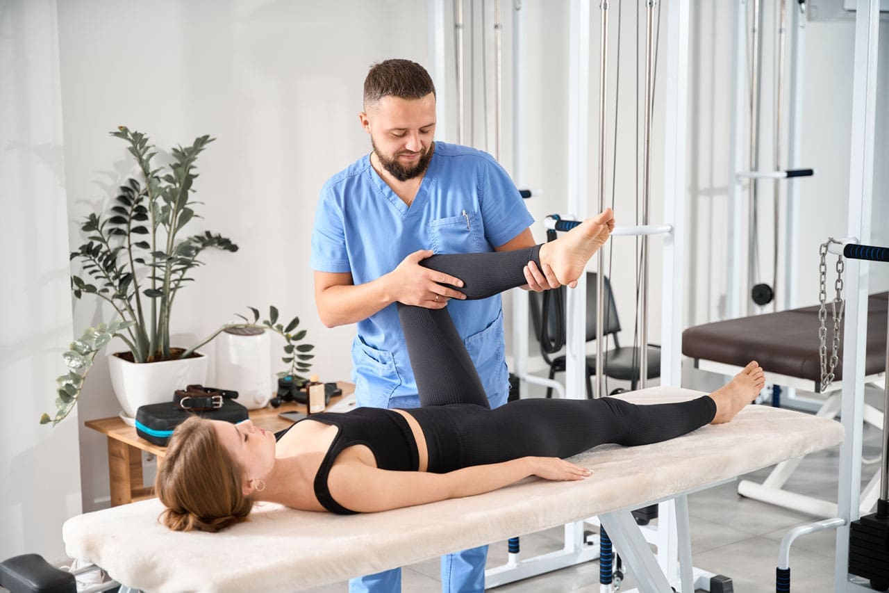
Optimal Joint Movement: Enhancing Mobility and Stability at El Paso Back Clinic
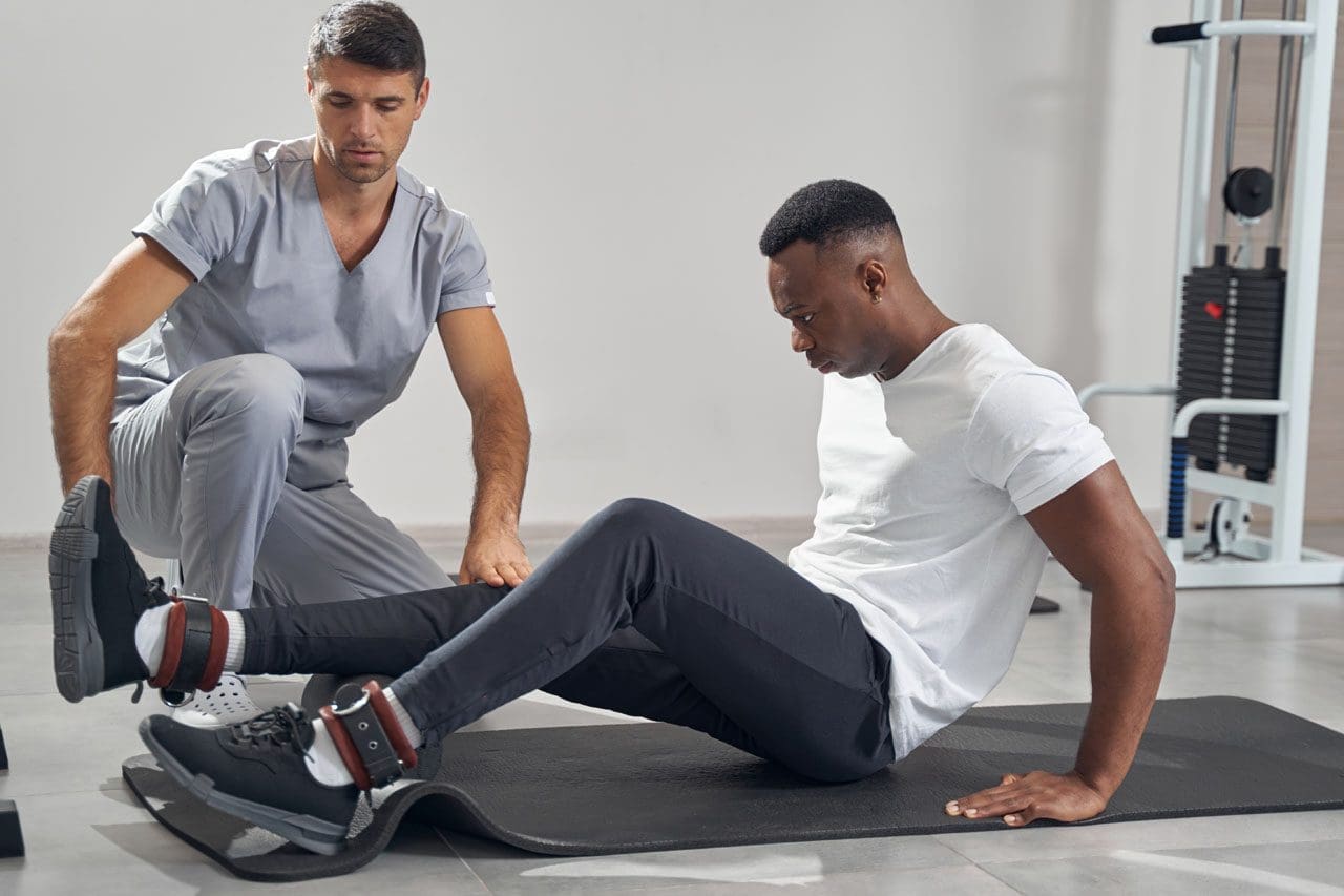
A chiropractor or Nurse Practitioner works with a patient in a rehabilitation center to improve joint mobility.
Optimal joint movement is essential for an active, pain-free life. At El Paso Back Clinic in El Paso, TX, we specialize in helping people achieve this through personalized chiropractic care. This article explains what optimal joint movement means, why it’s important, and how our clinic’s integrative approaches can restore it. Whether you’re dealing with back pain, sports injuries, or daily stiffness, our team, led by Dr. Alex Jimenez, DC, APRN, FNP-BC, uses spinal adjustments, rehabilitation, and functional medicine to get you moving better. Discover how we support joint health to improve function in everyday tasks and athletic pursuits.
Understanding Optimal Joint Movement
Optimal joint movement is the ability to move your joints through their full natural range of motion (ROM) smoothly, without pain, and with good control. It’s often referred to as high-quality mobility, blending flexibility with strength for daily activities and sports (University of Colorado Anschutz Medical Campus, n.d.).
At El Paso Back Clinic, we define it as moving joints efficiently while maintaining balance between mobility (active movement) and stability (joint control). This ensures muscles, ligaments, and tendons work together properly (National Academy of Sports Medicine, n.d.; Mainstay Medical, n.d.). For instance, a healthy shoulder should lift overhead to 180 degrees without strain, allowing you to reach shelves or throw a ball (Verywell Health, 2023a).
When injury or prolonged sitting disrupts this, mobility declines, leading to awkward movements elsewhere in the body (University of Colorado Anschutz Medical Campus, n.d.). Our clinic addresses this through holistic care, combining adjustments, soft-tissue therapy, and exercises to reduce inflammation and improve coordination.
Key Elements of Optimal Movement:
Full ROM: Joints reach their natural limits, like knee flexion to 140 degrees for squatting (The GO KNEE, n.d.).
Smooth Control: No jerking or pain, thanks to strong muscles and clear nerve signals.
Balance: Mobility for range, stability to prevent wobbles or injuries (ACE Fitness, n.d.a).
The Importance of Mobility and Stability Balance
At El Paso Back Clinic, we emphasize the balance between mobility and stability for peak performance. Mobility allows free movement, while stability keeps joints secure during activities (ACE Fitness, n.d.b). This synergy is key in our treatments.
Think of the body as a chain: Ankles and hips need mobility for steps, while knees and lower back provide stability (Motus Physiotherapy, n.d.; NASM, n.d.). If an ankle stiffens due to injury, the knee compensates, increasing the risk of pain (Physical Therapy at MJC, n.d.). Our chiropractic adjustments and rehab programs restore this chain, enhancing joint function.
Integrative care at our clinic—including spinal decompression and strength training—supports this balance, reducing the risk of injury and improving mobility (Peninsula Wellness Partners, n.d.).
Common Disruptions to Joint Mobility
Life factors can hinder optimal joint movement. Injuries cause swelling and tightness, limiting ROM (Frozen Shoulder Clinic, n.d.; Musculoskeletal Key, n.d.). A sedentary lifestyle, common in desk jobs, tightens muscles and stiffens joints (Dr. Ong Kee Leong, n.d.).
At El Paso Back Clinic, we see this in patients with back pain or sciatica, where poor posture leads to compensation and strain in other areas (OMassageT, n.d.). Aging, arthritis, or repetitive motions worsen it (Arthritis Foundation, n.d.; Chesapeake Regional, n.d.).
Typical Causes:
Trauma: Sprains create hard end-feels, stopping movement early (Physiopedia, n.d.c).
Inactivity: Shortens tissues, reducing flexibility (Dr Ong Kee Leong, n.d.).
Health Conditions: Arthritis limits ROM, causing bony sensations (Physiopedia, n.d.c).
Habits: Bad ergonomics unbalance the kinetic chain (OMassageT, n.d.).
Without correction, this increases fall risk and reduces quality of life. Our clinic’s diagnostic tools, such as digital X-rays, identify issues early.
Why Prioritize Optimal Joint Movement?
Good joint movement enhances everything from walking to sports. It prevents pain and boosts efficiency (OneStep, n.d.). At El Paso Back Clinic, we help athletes improve power and reduce injuries through better ROM (Activ Therapy, n.d.).
For daily life, it means easier tasks without fatigue (Baliston, n.d.). In walking, ankle flexion aids balance; poor ROM shortens strides (Baliston, n.d.). Our programs keep joints lubricated and muscles strong (Arthritis Foundation, n.d.).
At El Paso Back Clinic, maintenance starts with assessment. We measure ROM against norms using tools like goniometers (Physical Therapy at MJC, n.d.; Trainerize, n.d.). Then, we recommend exercises.
Regular activity, such as stretching, helps keep joints flexible (Arthritis Foundation, n.d.; Royal City Physiotherapy, n.d.). Our mobility drills focus on control for real-world use (Royal City Physiotherapy, n.d.).
Practical Tips:
Warm-Ups: Shoulder circles or ankle rolls (Chesapeake Regional, n.d.).
Stretching: Hold for 30 seconds on tight spots (Verywell Health, 2023a).
Strength Work: Squats for knee stability (ACE Fitness, n.d.b).
Activity: Low-impact, like swimming (Arthritis Foundation, n.d.).
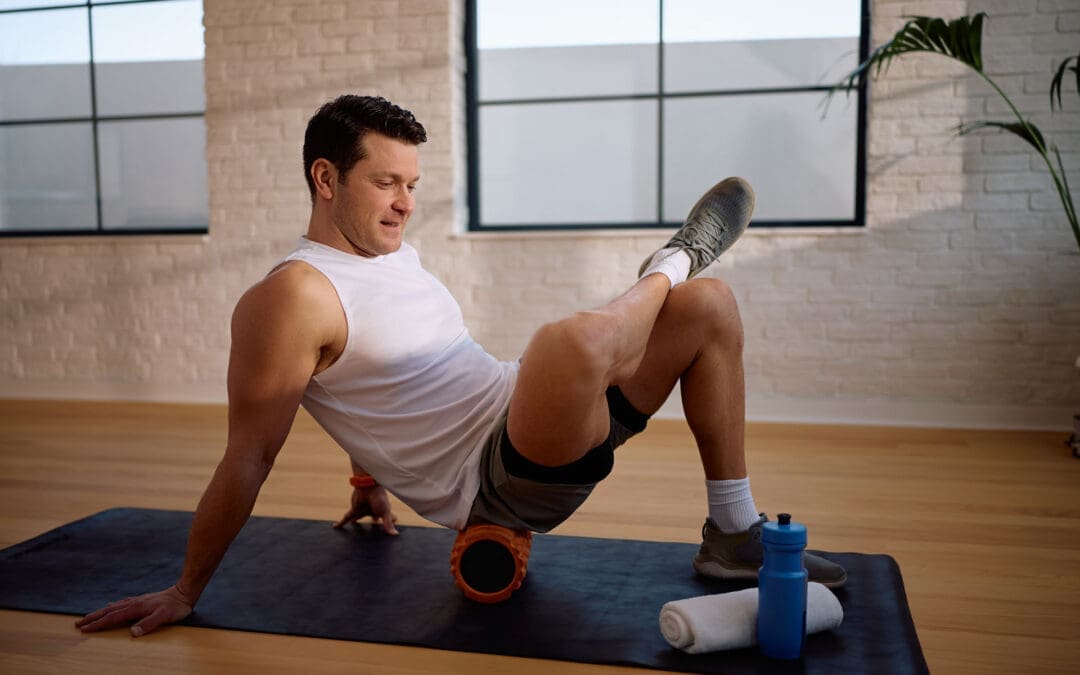
Tools: Foam rollers for self-care (Muscle and Motion, n.d.).
Visit our East Side location for personalized plans.
Integrative Chiropractic Care at El Paso Back Clinic
Our clinic offers holistic chiropractic care to restore joint movement. Led by Dr. Alex Jimenez, we combine adjustments, therapy, and guidance (Peninsula Wellness Partners, n.d.; Evolved Health Chiropractic, n.d.).
Adjustments realign joints, easing inflammation and nerves (Rodgers Stein Chiropractic, n.d.a; Rodgers Stein Chiropractic, n.d.b). Soft tissue work and rehab build muscle support (Evolved Health Chiropractic, n.d.).
This approach enhances mobility, strengthens areas, and reduces risks (Core Integrative Health, n.d.; Duca Chiropractic, n.d.). Joint mobilization gently increases ROM (Smart Sports Medicine, n.d.).
Our Services:
Spinal Adjustments: Restore alignment for better ROM (Chiropractic Omaha, n.d.).
Functional Medicine: Addresses root causes, such as nutrition (TXMAC, n.d.).
Rehab: Exercises for long-term health (Duca Chiropractic, n.d.).
Clinical Insights from Dr. Alex Jimenez at El Paso Back Clinic
Dr. Alex Jimenez, DC, APRN, FNP-BC, heads El Paso Back Clinic, with over 30 years of experience in integrative care. At our facilities, he blends chiropractic, functional medicine, and rehab for joint issues (Jimenez, n.d.a; Jimenez, n.d.b).
His observations: Adjustments alleviate nerve-related issues, restoring ROM in cases of back pain or sciatica (Jimenez, n.d.a). Patients from accidents or sports regain mobility through tailored plans (Jimenez, n.d.a).
Dr. Jimenez focuses on root causes with nutrition and exercises, preventing surgery (Jimenez, n.d.b). For hips or knees, agility programs balance mobility and stability (Jimenez, n.d.a). Our holistic model empowers patients and aligns with evidence supporting better function (Jimenez, n.d.b).
At El Paso Back Clinic, optimal joint movement is achievable with our expert care. Balance mobility and stability to overcome disruptions. Visit elpasobackclinic.com or our El Paso locations for help from Dr. Jimenez’s team.
IFM's Find A Practitioner tool is the largest referral network in Functional Medicine, created to help patients locate Functional Medicine practitioners anywhere in the world. IFM Certified Practitioners are listed first in the search results, given their extensive education in Functional Medicine