Gut Health for Faster Recovery—El Paso Back Clinic
Why your gut matters when you’re healing
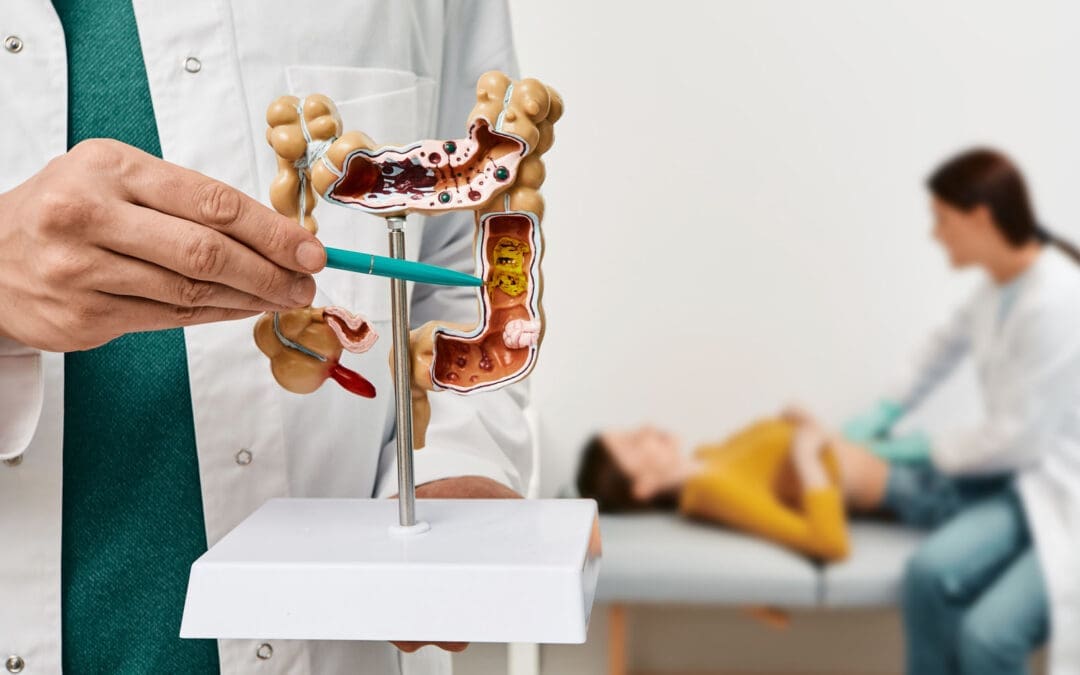
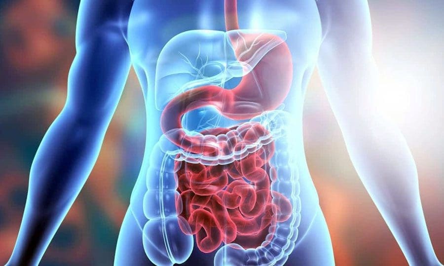
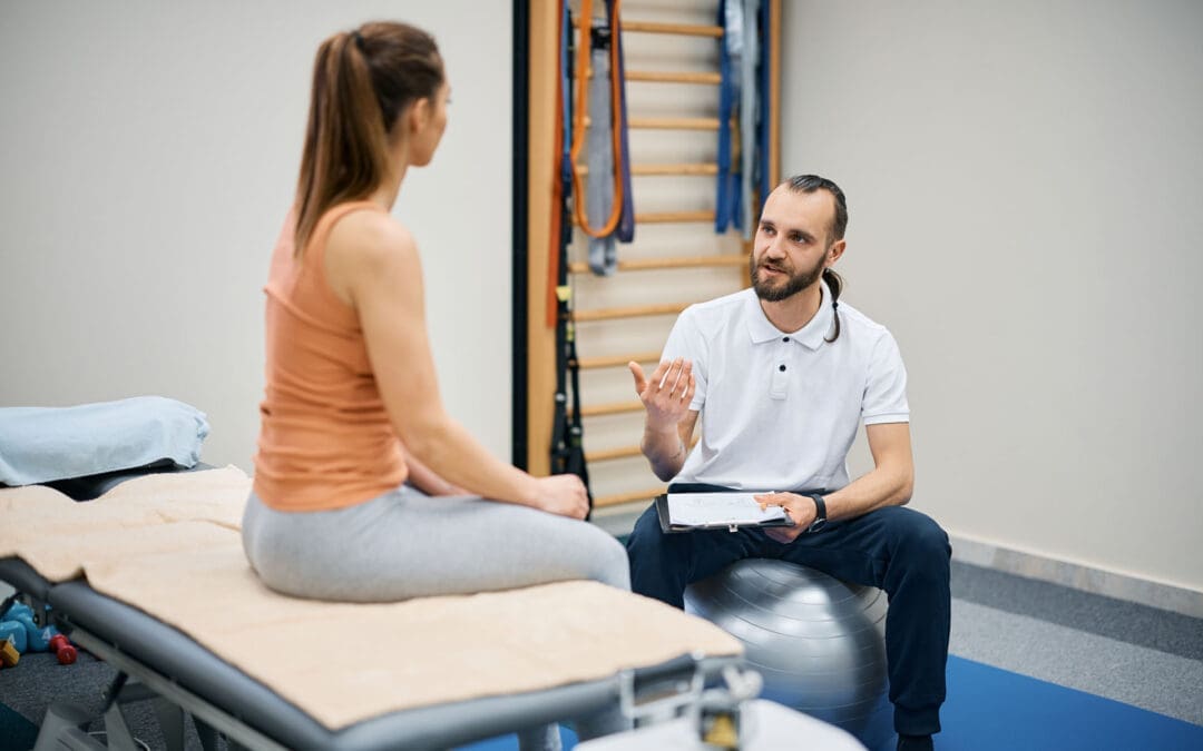
After a back or neck injury—from daily strain, sports, work, or a car crash—pain and limited mobility can dominate your life. But there’s a powerful helper inside you: the gut microbiome. These trillions of microbes influence digestion, inflammation, immunity, energy, and even sleep. When they fall out of balance (called dysbiosis), bloating, irregular stools, fatigue, and higher inflammation can slow your rehab progress. The positive news is that simple daily steps can reset the balance and support your recovery. (Cleveland Clinic, 2023/2022). (Cleveland Clinic)
At El Paso Back Clinic, we often combine spine-focused care—such as chiropractic adjustments when appropriate, therapeutic exercise, soft-tissue work, and, if indicated, imaging—with practical gut-support strategies, helping patients recover more comfortably and steadily. (Dr. Alex Jimenez, El Paso clinic pages). (El Paso, TX Doctor Of Chiropractic)
Dysbiosis in plain language
Dysbiosis means your gut community is out of balance—too many “unhelpful” species, not enough beneficial ones, or less diversity overall. Diets high in refined sugars and ultra-processed foods, repeated courses of antibiotics, stress, poor sleep, and alcohol/environmental toxins are common triggers. (Cleveland Clinic, 2024; Better Health Channel, 2023; USDA ARS, 2025). (Cleveland Clinic)
Ultra-processed foods tend to be low in fiber and high in additives; over time, they’re linked with inflammation and a less favorable gut environment—exactly what you don’t want while healing. (Cleveland Clinic Newsroom, 2023). (Cleveland Clinic)
How “unhealthy” bacteria gain ground
Unwanted bacteria flourish when conditions favor them. Three everyday drivers:
Low fiber, high ultra-processed intake. Beneficial microbes feed on plant fibers and resistant starches from beans, whole grains, vegetables, and fruit. Starve them, and opportunistic species take over. (Wilson et al., 2020; Singh et al., 2017). (PMC)
Antibiotics and antimicrobials. Essential when needed, but they can also reduce helpful species; rebuilding with fiber-rich foods (and sometimes probiotics) helps restore balance. (Cleveland Clinic, 2024). (Cleveland Clinic)
Stress and poor sleep. Both alter motility and immune signaling via the brain–gut axis, nudging the microbiome toward dysbiosis. (Better Health Channel, 2023). (Better Health Channel)
SIBO: a special case to know about
Small Intestinal Bacterial Overgrowth (SIBO) happens when excess bacteria build up in the small intestine, which normally has low counts. Symptoms can include bloating, abdominal discomfort, diarrhea, early fullness, weight loss, or malnutrition. (Mayo Clinic, 2024). (Mayo Clinic)
Treatment often pairs targeted antibiotics with nutrition and root-cause fixes (e.g., motility support or addressing structural issues). Without tackling the cause, SIBO can recur. (Mayo Clinic, 2024). (Mayo Clinic)
If you notice persistent bloating, pain, or weight loss, ask your clinician about evaluation and a phased plan that treats the cause, then carefully re-expands fibers and fermented foods.
How better gut habits speed musculoskeletal recovery
Lower, steadier inflammation: A fiber-rich, plant-forward pattern boosts short-chain fatty acids (SCFAs) like butyrate that help protect the gut lining and may dampen systemic inflammation tied to pain. (Singh et al., 2017). (PMC)
Energy and participation: Balanced digestion supports energy, sleep, and mood—key drivers of successful physical therapy and home exercise. (Cleveland Clinic, 2022). (Cleveland Clinic)
Medication tolerance: If you need antibiotics or other meds, a microbiome-friendly plan can reduce GI side effects. (Cleveland Clinic, 2024). (Cleveland Clinic)
The El Paso Back Clinic approach (dual-scope care)
Our team—led by Alexander Jimenez, DC, APRN, FNP-BC—blends chiropractic care with nurse-practitioner medical evaluation. When appropriate, we use X-ray/MRI to clarify the diagnosis, and we coordinate conservative therapies with nutrition and lifestyle coaching. For injury cases, we also provide the documentation insurers and attorneys require. (El Paso, TX Doctor Of Chiropractic)
Common elements of a plan:
Dual-scope assessment: History, neuro/orthopedic testing, and imaging when indicated to pinpoint pain drivers (joint, nerve, soft tissue). (El Paso, TX Doctor Of Chiropractic)
Conservative therapies: Chiropractic adjustments (as indicated), therapeutic exercise, massage/soft-tissue work; acupuncture may be added to modulate pain and stress. (El Paso, TX Doctor Of Chiropractic)
Gut-support basics: Plant variety, fiber targets, and live-culture foods; stress and sleep tools that calm the gut–brain axis. (Cleveland Clinic Magazine; Penn State Health). (Cleveland Clinic)
Medical-legal readiness: Structured notes, imaging reports, and measurable outcomes for personal-injury and MVA cases. (El Paso, TX Doctor Of Chiropractic)
Clinical observation: Patients with back/neck pain who improve sleep and add one fermented food daily—while increasing beans/whole grains and veggies—often report less bloating and steadier energy within weeks, which helps them stay consistent with rehab.
A 4–6 week “gut-reset” that fits rehab
1) Make plants the base (daily)
Aim for colorful vegetables and fruits, beans/lentils 4–5 days/week, and whole grains (oats, barley, brown rice, quinoa). These choices feed beneficial microbes and boost SCFAs. (Wilson et al., 2020). (PMC)
2) Add one fermented food most days
Yogurt or kefir with live active cultures, kimchi, sauerkraut, or kombucha. Not all fermented foods have live microbes after processing—check the label. (Healthline; Cleveland Clinic Magazine). (Healthline)
3) Tame ultra-processed foods
Swap sugary drinks for water/unsweetened tea; favor whole-grain staples; keep packaged snacks as occasional treats. (Cleveland Clinic, 2023). (Cleveland Clinic)
4) Support sleep and stress
Target 7–9 hours with a consistent wind-down; try 5 minutes of slow breathing before bed; walk 20–30 minutes most days, and add two short strength sessions weekly. (Better Health Channel, 2023). (Better Health Channel)
5) Medications—coordinate with your clinician
Don’t stop prescribed meds on your own. If antibiotics are necessary, ask whether a food-first strategy and a short-term probiotic make sense for you. (Cleveland Clinic, 2024). (Cleveland Clinic)
6) Hygiene matters
Wash hands, rinse produce, and avoid kitchen cross-contamination to reduce exposure to harmful bacteria. (Better Health Channel, 2023). (Better Health Channel)
Two-week starter plan (easy, budget-minded)
Breakfast: Oats + kefir or yogurt + berries + nuts.
Dinner: Slow-cooker chili or lentil curry; salad with olive oil; baked potato (cool leftovers for resistant starch).
Snacks: Fruit + nut butter; carrots + hummus; plain popcorn; small kefir smoothie.
Small, steady changes add up; focus on what you can repeat during busy treatment weeks. (Penn State Health, 2018). (Penn State)
When to seek medical care now
Unintended weight loss, blood in stool, fever, severe or night-time symptoms, or a history of GI surgery.
Talk with your clinician about evaluation, including possible SIBO testing when appropriate. (Mayo Clinic, 2024). (Mayo Clinic)
Local help in El Paso
If you’re recovering from a back or neck injury and want a plan that connects spine care, gut health, and documentation for injury cases, our team can help you build a sustainable routine while we treat the root musculoskeletal drivers. (El Paso Back Clinic/Dr. Jimenez). (El Paso, TX Doctor Of Chiropractic)
Your Spine, Your Life: An El Paso-Ready Guide to Strong, Flexible, Pain-Resistant Backs
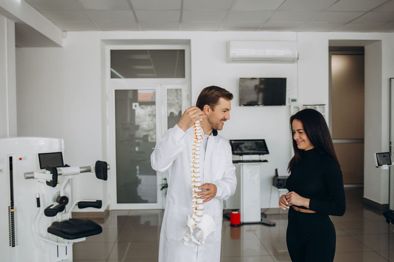
A young woman is performing a spine checkup at a vertebra clinic.
What “spinal health” means (and why it matters here in El Paso)
Spinal health refers to the proper structure, alignment, and function of the spine, enabling it to support the body, facilitate movement, and protect the spinal cord—the pathway for nerve signals between the brain and the body. Good spinal health comes from regular exercise, posture awareness, a nutrient-dense diet, steady hydration, and a healthy weight. Poor spinal health can lead to chronic pain, nerve irritation or damage, and a lower quality of life (Raleigh Orthopaedics, 2024; Orthopedic Specialists of Southwest Florida [OSSWF], 2024; National Spine Health Foundation, 2024).
How a healthy spine supports your whole body
Support & alignment: Your spine acts like a central pillar that shares load with the hips and legs and keeps you upright (Premier Spine & Sports Medicine, n.d.).
Movement & shock absorption: Curves, discs, and joints allow for safe bending and twisting, enabling you to lift, reach, and play (Raleigh Orthopaedics, 2024).
Nerve protection: The spinal column shields the spinal cord and nerve roots, so signals move clearly. Irritation can cause pain, tingling, or weakness (Cary Orthopaedics, 2023).
Quality of life: Ongoing spine issues can lead to fatigue, poor sleep, headaches, and reduced participation in work or sports (Raleigh Orthopaedics, 2024).
Common problems we see—and why early action helps
Strains/sprains and facet irritation from long sitting, poor lifting form, or sudden loads
Disc problems that can press on nearby nerves and create radiating symptoms
Spinal stenosis (narrowing) that pinches nerves
Degenerative changes related to age, low activity, smoking, or extra weight
Most cases respond to conservative care when initiated early, including movement, postural changes, targeted exercises, and load management (OSSWF, 2024).
Red flags—don’t wait: radiating pain, numbness, weakness, headaches, or loss of function. Seek a prompt exam (Cary Orthopaedics, 2023; Suarez Physical Therapy, n.d.).
An El Paso Back Clinic–style plan: simple steps that fit your day
1) Movement you can keep
20–30 minutes of low-impact cardio most days (e.g., walking, cycling, swimming).
Core & hip strength 2–3 days/week: planks, side planks, glute bridges, and bird-dogs.
Mobility after warm-ups: thoracic open-books, hip-flexor, and hamstring stretches (National Spine Health Foundation, 2024; Mobility Project PT, 2024).
2) Posture that holds up at work and home
Sit: feet flat, hips back in the chair, lumbar support, screen at eye level.
Stand: weight balanced, knees soft, ears over shoulders.
Micro-breaks: move every 30–45 minutes (National Spine Health Foundation, 2024).
3) Ergonomics you actually feel
The chair is high enough so the hips are level with or slightly above the knees.
Keyboard and mouse close; forearms supported; shoulders relaxed.
Lift with a hip hinge, keep the load close, and exhale as you stand.
4) Sleep & stress recovery
Neutral neck/back with a supportive mattress and the right pillow height.
Side sleepers: pillow between knees. Back sleepers: pillow under knees.
Use breathing drills, short walks, and stretch breaks to lower tension (Raleigh Orthopaedics, 2024).
5) Hydration & healthy weight
Steady water intake supports disc hydration and tissue recovery (Centeno-Schultz Clinic, n.d.).
A healthy body weight lowers compressive load on joints and discs (Raleigh Orthopaedics, 2024).
Nutrition for a stronger spine (simple and local-friendly)
Protein for muscle and connective-tissue repair
Omega-3s (salmon, trout, walnuts) to help regulate inflammation
Calcium & vitamin D for bone strength
Magnesium for nerve and muscle function
Colorful fruits/vegetables for antioxidants that support recovery
Water for disc hydration and nutrient transport These habits reduce inflammation and support healing (Watkins Family Chiropractic, 2023; OSSWF, 2024).
Four-week “Borderland Back Reset” (minimal gear, steady progress)
Week 1 — Start easy
Daily: 10-minute walk + 5 minutes mobility (open-books, hip-flexor, hamstrings).
Core set (3x/week): plank 20 s, side plank 15 s/side, glute bridge 10 reps.
Posture: Raise the screen and add a small lumbar roll.
Week 2 — Build consistency
Daily: 15–20 minutes walk/cycle + mobility.
Core set (3x/week): plank 25–30 s, side plank 20 s/side, bridge 12 reps; add bird-dog 6/side.
Nutrition: add one serving of leafy greens and one serving of lean protein to each meal (Watkins Family Chiropractic, 2023).
Week 3 — Strength + recovery
Cardio most days: 20–25 minutes.
Light hinge pattern (backpack or kettlebell) 1–2 days/week; focus on form.
Before bed, do slow breathing for 5 minutes.
Week 4 — Re-test & adjust
Compare flexibility, pain, and energy levels with those of Week 1.
Keep what helps; trim what doesn’t.
If numbness, weakness, or radiating pain persists, book an exam (Cary Orthopaedics, 2023; Suarez Physical Therapy, n.d.).
Real-world injuries: work, sports, and motor-vehicle accidents (MVAs)
Work: Desk roles need posture breaks and lumbar support; physical jobs need task rotation, hip-hinge training, and planned recovery.
Sports: Combine mobility, core/hip strength, and gradual return to play.
MVAs: Even “minor” collisions can cause whiplash or soft-tissue injury. A stepwise evaluation, along with imaging when necessary, guides safe return and documentation (OSSWF, 2024).
Inside our integrative approach in El Paso
(Clinical observations from Dr. Alexander Jimenez, DC, APRN, FNP-BC, Nurse Practitioner and Chiropractor)
Dual-scope diagnosis: We blend chiropractic and medical perspectives. Your exam includes a detailed history, movement, and neurological screens, as well as, when necessary, advanced imaging to clarify the problem and rule out potential red flags (Jimenez, n.d.; see Imaging/Diagnostics and Personal-Injury topics).
Evidence-based conservative care:
Chiropractic adjustments to restore motion and reduce joint irritation
Therapeutic exercise to build core/hip strength and mobility
Manual therapy/massage for tight or sensitive tissues
Acupuncture as part of an integrative plan when appropriate
Lifestyle coaching on posture, lifting, sleep, and stress (Prestige Health & Wellness, n.d.; Mobility Project PT, 2024; Raleigh Orthopaedics, 2024)
Documentation & advocacy: For work, sports, personal, and MVA cases, we document the mechanism of injury, exam findings, functional limits, and response to care. When claims or legal issues arise, clear records and appropriate imaging support decision-making (Jimenez, n.d.; Rangeline Chiropractic, n.d.).
Myths vs. facts (short and clear)
Myth: “If my back hurts, I should rest all day.” Fact: Gentle movement and short walks often speed recovery; long bed rest adds stiffness (National Spine Health Foundation, 2024).
Myth: “Only heavy lifting causes back pain.” Fact: Prolonged sitting, poor ergonomics, stress, and sleep problems also drive pain (National Spine Health Foundation, 2024; Raleigh Orthopaedics, 2024).
The El Paso Back Clinic checklist
☐ Break up sitting every 30–45 minutes
☐ Screen at eye level; use lumbar support
☐ 10–15 minutes daily core + mobility
☐ 20–30 minutes low-impact cardio most days
☐ Hydrate across the day
☐ Build meals around protein + produce + healthy fats
☐ Sleep with neutral neck/back alignment
☐ Seek care quickly for red flags or lasting symptoms
Sciatica Relief for Teachers: El Paso Back Clinic’s Chiropractic Solutions
A teacher helping an elementary school girl using a tablet computer
Introduction: Supporting Teachers’ Health in El Paso
Teaching is a rewarding yet demanding profession, especially in vibrant communities like El Paso, Texas. Teachers spend long hours standing, sitting, and moving in ways that strain their bodies. These daily tasks can lead to sciatica, a painful condition caused by irritation of the sciatic nerve, which runs from the lower back down the legs. Symptoms like sharp leg pain, numbness, or tingling can disrupt lesson plans and classroom energy.
At El Paso Back Clinic®, led by Dr. Alexander Jimenez, DC, APRN, FNP-BC, we understand the unique challenges educators face. Prolonged sitting during grading, standing for lessons, poor posture over desks, and the physical demands of managing classrooms increase sciatica risks. Our clinic specializes in chiropractic care, integrative medicine, and functional rehabilitation to help teachers manage pain and prevent flare-ups. Using manual adjustments, ergonomic advice, and targeted exercises, we aim to restore spinal health and enhance quality of life.
This article examines why teachers are prone to sciatica, how our clinic’s chiropractic and integrative approaches can provide relief, and offers practical steps for achieving lasting wellness. Drawing on Dr. Jimenez’s 30+ years of expertise, we’ll share clinical insights and real-world solutions tailored for El Paso’s educators.
What Is Sciatica and Why Does It Affect Teachers?
Sciatica occurs when the sciatic nerve, the body’s longest nerve, becomes compressed or irritated. This nerve starts in the lower spine, travels through the hips, and extends down each leg. Common symptoms include burning pain, tingling, or weakness in one leg, often worsening with sitting or standing. For teachers, this can mean discomfort during classes or while grading at home.
The teaching environment in El Paso schools, from bustling elementary classrooms to high school lecture halls, creates perfect conditions for sciatica. Standing for long periods during lessons or playground duty fatigues back muscles, pressing on spinal discs (Bomberg Chiropractic, 2023). Sitting at desks or in cramped staff rooms can shorten hip muscles, tilt the pelvis, and pinch the nerve (East Bay Chiropractic Office, 2023). Poor posture, like slouching over lesson plans, further irritates the nerve roots (Scoliosis Center of Utah, n.d.).
Dr. Jimenez sees this often at El Paso Back Clinic. His advanced neuromusculoskeletal imaging, such as X-rays and MRIs, pinpoints disc bulges or muscle imbalances that cause sciatica in teachers. By addressing these root causes, our clinic helps educators stay active without pain.
How Teachers’ Daily Routines Trigger Sciatica
Teachers’ days are a mix of physical and mental demands. Standing to deliver lessons or monitor halls strains the lower back, increasing nerve pressure (Boyne Ergonomics, n.d.). Sitting for hours on outdated chairs compresses the spinal discs, a key factor in triggering sciatica (Bomberg Chiropractic, 2023). Bending to assist students or lifting heavy teaching materials—such as projectors or book boxes—can strain the piriformis muscle, which is located near the sciatic nerve.
Poor posture is a major culprit. Leaning over desks or hunching at computers curves the spine unnaturally, squeezing nerve roots (Scoliosis Center of Utah, n.d.). Stress from managing classrooms or meeting tight deadlines can cause muscle tension, leading to inflammation (Paragon Chiropractic, n.d.). In El Paso, where teachers often juggle bilingual classes and extracurricular duties, these risks accumulate.
Dr. Jimenez’s clinic frequently treats educators with sciatica from these habits. His dual-scope approach—combining chiropractic exams with diagnostic imaging—reveals how daily tasks, such as carrying heavy bags, can lead to spinal misalignment. Our tailored treatments at El Paso Back Clinic, including adjustments and massage, address these issues directly, helping teachers move freely.
The Impact of Prolonged Sitting and Standing
Teachers switch between sitting and standing constantly—standing for morning assemblies, sitting for parent-teacher meetings, then standing again for labs. Prolonged sitting, especially on hard classroom chairs, increases disc pressure by up to 30%, irritating the sciatic nerve (Bomberg Chiropractic, 2023). Standing too long without breaks tightens hip flexors, pulling the spine out of alignment (Boyne Ergonomics, n.d.).
This back-and-forth strains stabilizing muscles, risking micro-tears in discs that pinch nerves. In El Paso’s active school settings, teachers may stand for over four hours daily, increasing the odds of back pain by 50% (Abundant Life Chiropractor, 2023). At El Paso Back Clinic, Dr. Jimenez utilizes advanced imaging to identify these strains, which are often seen in teachers following minor classroom injuries or slips. Our spinal decompression therapy gently stretches the spine, relieving nerve pressure and promoting healing.
Simple fixes can help: switch positions every 20 minutes, use cushioned mats for standing, or adjust your desk height to a comfortable level. These small changes, guided by our clinic’s ergonomic coaching, significantly reduce the risk of sciatica.
Poor Posture: A Hidden Cause of Nerve Pain
Posture shapes spinal health. Teachers often slouch over their desks or lean forward to engage students, curving their spines into a “C” shape. This compresses the lumbar vertebrae, irritating sciatic nerve roots (Scoliosis Center of Utah, n.d.). Low computer screens can cause neck craning, which can lead to lower back strain.
In El Paso classrooms, crouching to help young students or writing on low boards exacerbates this issue. Over time, uneven muscle pull misaligns the spine, trapping the nerve. At El Paso Back Clinic, Dr. Jimenez uses posture assessments to spot these habits early. His chiropractic adjustments realign the vertebrae, while acupuncture relaxes tight muscles, such as the piriformis, easing nerve pressure (Jimenez, n.d.a).
Posture tips: Keep your ears over your shoulders, use a lumbar-support chair, and raise screens to eye level. Our clinic offers workshops for El Paso teachers to build these habits, preventing chronic pain.
Physical Demands: The Active Side of Teaching
Teaching isn’t just standing or sitting—it’s dynamic. Lifting stacks of textbooks, bending to pick up dropped items, or dashing to manage recess chaos strains the back. These motions can herniate discs or inflame muscles near the sciatic nerve (East Bay Chiropractic Office, 2023). In El Paso, where teachers may handle heavy bilingual materials or sports equipment, risks grow.
Sudden twists, such as grabbing a falling projector, mimic sports injuries that Dr. Jimenez treats. His clinic documents these as work-related injuries for insurance purposes, utilizing massage and exercise to aid in tissue healing. Advanced imaging ensures an accurate diagnosis, detecting layered issues such as sprains and nerve compression (Jimenez, n.d.b).
Safe habits reduce risks: Lift with bent knees, use carts for supplies, and stretch before engaging in active duties. El Paso Back Clinic’s tailored plans help teachers stay strong and pain-free.
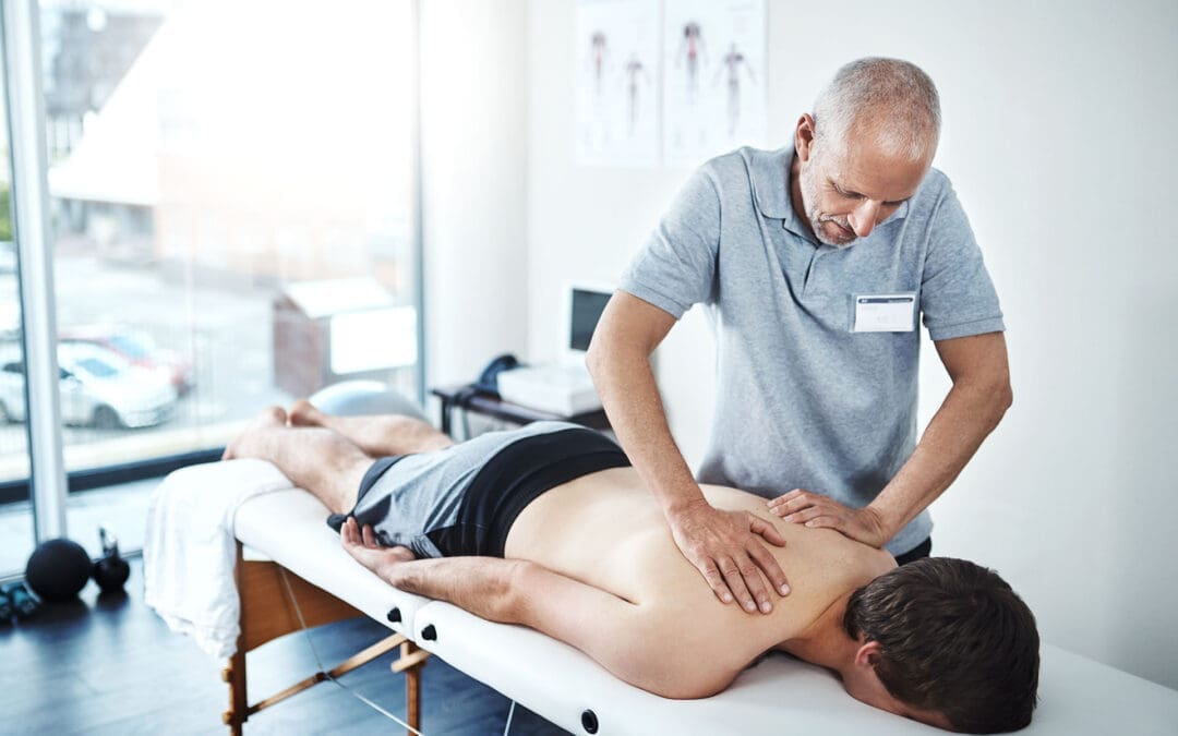
Chiropractic Care at El Paso Back Clinic: Targeted Relief
Chiropractic care is a cornerstone for sciatica relief. At El Paso Back Clinic, our manual adjustments realign the spine, reducing nerve irritation and inflammation (Active Health Center, n.d.). Teachers notice less leg pain and better mobility after sessions. Our spinal decompression therapy gently stretches the spine, retracting bulging discs that pinch nerves (Bomberg Chiropractic, 2023).
Dr. Jimenez’s expertise shines here. With over 30 years of experience treating patients in El Paso, including educators, Dr. [Last Name] combines chiropractic care with integrative methods, such as acupuncture, to provide natural pain relief. Advanced imaging ensures precise adjustments, targeting the exact cause of sciatica (Jimenez, n.d.a). Regular visits prevent flare-ups, letting teachers focus on students, not pain.
Restoring Spinal Alignment and Nerve Function
Adjustments are quick, targeted thrusts that realign vertebrae, freeing the sciatic nerve. This boosts blood flow and reduces inflammation, key for teachers battling daily strain (AFC Adherence, n.d.). At our clinic, Dr. Jimenez pairs adjustments with soft tissue work to release tight hips, a common issue among educators.
Our approach restores function holistically. Teachers regain flexibility for classroom tasks, and consistent care prevents future issues. Jimenez’s diagnostic tools, such as MRIs, ensure that treatments match each patient’s needs, offering El Paso educators reliable relief (Jimenez, n.d.b).
Reducing Inflammation Naturally
Inflammation fuels sciatica pain and swelling of the tissues around the nerve. Our adjustments improve spinal motion, reducing this swelling (Active Health Center, n.d.). We add ice or heat therapy to speed relief, tailored to each teacher’s symptoms.
Dr. Jimenez enhances this approach with nutrigenomics, recommending anti-inflammatory foods, such as salmon, to support the healing process. For El Paso teachers, this integrative approach means less pain and faster recovery from classroom strains (Jimenez, n.d.a).
Lifestyle Changes: Ergonomics and Exercises for Teachers
El Paso Back Clinic goes beyond adjustments, offering practical advice. Ergonomic tips include adjustable chairs, footrests, and raised monitors to reduce strain (Boyne Ergonomics, n.d.). For teachers, we recommend lumbar pillows and standing desks for grading.
Exercises are key: planks strengthen the core, and piriformis stretches loosen the hips (Alliance Orthopedics, n.d.). Dr. Jimenez designs home routines, such as knee-to-chest stretches, to accommodate busy schedules. Our massage therapy supports recovery, ensuring El Paso educators stay active.
Preventing Flare-Ups: Daily Habits for Long-Term Relief
Preventing sciatica means tracking triggers. Long sits or heavy lifts? Take breaks or use carts. Heat eases tight muscles; cold calms acute pain (Abundant Life Chiropractor, 2023). Weekly core workouts and posture apps help maintain proper alignment.
Dr. Jimenez’s clinic emphasizes prevention. Our exercise plans, paired with stress-reducing yoga, help teachers avoid chronic issues. Legal documentation supports work-injury claims, ensuring access to care (Jimenez, n.d.b).
Integrative Care: A Team Approach at El Paso Back Clinic
We combine chiropractic care with physical therapy, acupuncture, and massage to facilitate a comprehensive recovery. Physical therapy builds strength with moves like bridges (Active Health Center, n.d.). Acupuncture calms the nerves, making it ideal for reducing teachers’ stress (Jimenez, n.d.a). Short movement breaks, such as stretching during class, boost circulation.
Our clinic’s integrative model, led by Dr. Jimenez, treats sciatica holistically, addressing work or personal injuries with detailed records for insurance.
Dr. Alexander Jimenez’s Expertise: A Beacon for El Paso Teachers
Dr. Jimenez, with dual credentials as a chiropractor and nurse practitioner, brings unmatched care to El Paso. His clinic treats sciatica from classroom strains, sports injuries, or accidents, using imaging to diagnose precisely. Treatments such as adjustments, massage, and tailored exercises can help the body heal naturally, thereby preventing long-term issues.
For teachers, Jimenez’s legal documentation supports work claims, ensuring coverage. His functional medicine approach, including nutrition and acupuncture, empowers educators to thrive (Jimenez, n.d.a; Jimenez, n.d.b).
Practical Tips for El Paso Teachers
Morning Stretch: Try cat-cow (10 reps) to loosen the spine.
Classroom Ergonomics: Use lumbar-support chairs; raise boards to waist height.
Breaks: March in place every 30 minutes to ease nerve pressure.
Nutrition: Eat berries and fish to combat inflammation, according to Jimenez’s advice.
Conclusion: Empowering El Paso Educators
Sciatica doesn’t have to slow down El Paso’s teachers. At El Paso Back Clinic, Dr. Jimenez and our team offer chiropractic care, integrative therapies, and practical tips to relieve pain and prevent issues. From adjustments to ergonomic tweaks, we help educators stay healthy and focused on inspiring students.
Visit us at 11860 Vista Del Sol Dr, Suite 128, El Paso, TX, or call 915-850-0900 to start your pain-free journey.
Preventing Sports & Back Injuries: The El Paso Back Clinic Approach
Athletes, weekend warriors, and active individuals often push their bodies to the limit. Without smart preparation and care, minor misalignments or imbalances can lead to back pain, sprains, or more serious injuries. At El Paso Back Clinic, our mission is to prevent injuries before they occur, maintain spine health, and support long-term performance and wellness.
In this article, you’ll learn how a multifaceted strategy—involving movement, conditioning, chiropractic, integrative therapies, and recovery—can reduce injury risk. We’ll also show how El Paso Back Clinic applies these principles in real-world care.
Why Back & Sports Injuries Occur
Biomechanical Stress & Misalignment
Even small spinal misalignments or joint restrictions can change movement mechanics. Over time, stresses that should spread evenly across tissues become concentrated on certain discs, muscles, or ligaments, making them vulnerable (Mount Sinai, n.d.; Emery & Meeuwisse, 2008).
Overuse and Repetition
Playing the same sport repeatedly without variation often leads to overuse injuries—microtears that accumulate faster than the body can heal. Many youth and amateur athletes suffer from this because they skip rest phases (Nationwide Children’s, n.d.; CHOP, n.d.).
Fatigue, Poor Technique, and Weakness
When muscles fatigue, the muscle fibers break down. A runner might collapse inward at the knee, or a basketball player might land with improper form. These movement faults under fatigue cause injury (Walker Physical Therapy, n.d.; PWR Physio, n.d.).
Insufficient Recovery
Without proper rest, nutrition, and tissue repair, microdamage lingers. Eventually, the body’s threshold is crossed, and a dramatic injury occurs.
Core Prevention Pillars
At El Paso Back Clinic, we emphasize these foundational pillars:
1. Dynamic Warm-Up & Mobility Routines
Warm-ups aren’t just stretching—they’re activation drills, joint movements, and controlled progressions that prepare muscles and joints. Cooling down, stretching, and mobility work afterward help flush byproducts and reduce stiffness (First Physio Plus, n.d.; Garden State Pain, n.d.).
2. Technique Monitoring and Movement Quality
We routinely analyze movement—such as running gait, jumping, squatting, and twisting—to identify harmful patterns. By coaching technique and correcting faults, we reduce stress on the back and joints (GPOA, n.d.; Walker Physical Therapy, n.d.).
3. Balanced Strength, Stability & Flexibility
Having a strong core, glutes, and stabilizers protects the lumbar spine. We design programs that incorporate strength, balance, flexibility, and endurance to create a well-rounded system (PWR Physio, n.d.; Walker Physical Therapy, n.d.).
4. Strategic Rest and Load Management
We guide patients and athletes in periodization, which involves alternating high and low loads, scheduling rest days, and monitoring fatigue to prevent overtraining (Bayfront Health, n.d.; Fick PT & Performance, n.d.).
5. Nutrition, Hydration & Recovery Support
Good hydration and nutrients (protein, vitamins, minerals) are essential for tissue repair. A poor diet hinders recovery and increases the risk of injury (LI Spine Med, 2024).
The Role of Chiropractic & Back Clinic Services
El Paso Back Clinic (under Dr. Jimenez) stands out by combining back/spine care with integrative therapies. Here’s how chiropractic and back-clinic services help prevent injuries:
Spinal Alignment & Joint Function
Chiropractic adjustments and spinal mobilizations help maintain vertebral alignment, ease restrictions, and ensure joints move properly. This reduces compensatory stress on surrounding tissues (Dallas Accident & Injury Rehab, n.d.; Evolved Health Chiropractic, n.d.).
Posture, Movement Pattern Correction & Neuromuscular Feedback
We assess posture and movement patterns across the kinetic chain. Correcting compensations (e.g., pelvic tilt, scoliosis curves) helps protect the spine during sport demands (Dallas Accident & Injury Rehab, n.d.; Evolved Health Chiropractic, n.d.).
Proper nerve input from spinal segments supports muscle activation and timing. By improving the communication between the spine and joints and the surrounding muscles, we help the body respond more effectively under stress (Fremont Chiropractic, n.d.; Young Chiropractic, n.d.).
Versatile Soft-Tissue & Myofascial Work
Muscles, fascia, and connective tissues often tighten, pulling on the spine. Techniques, such as soft-tissue work, instrument-assisted release, and myofascial release, help reduce tension and restore balance (Garmon Chiropractic, n.d.).
Monitoring & Maintenance Care
We often schedule preventive “maintenance” visits. Even when patients feel fine, small dysfunctions can arise. Regular check-ins allow us to catch them early—before they develop into problems.
Integrative Therapies & Supportive Methods
To maximize prevention, El Paso Back Clinic layers on integrative and complementary care:
Physical Therapy & Exercise Therapy
Sometimes muscles need retraining. Our clinic can collaborate with or provide therapeutic exercise programs that focus on weakness, imbalance, mobility deficits, and sport-specific drills (Current Physical Therapy, 2025).
Massage, Trigger Point Work & Soft-Tissue Modulation
Massage and trigger point therapy enhance circulation, alleviate adhesions, and promote muscular recovery. These help tissues remain supple and resilient (Primary Health & Wellness, n.d.).
Acupuncture & Electro-Acupuncture
Using needles or micro-current stimulation, we stimulate healing, reduce inflammation, and modulate pain. These methods pair well with structural work (clinic’s integrative model).
Kinesio Taping & Supportive Bracing
Taping techniques provide gentle support, reduce stress on soft tissues, and enhance proprioception during dynamic phases of sports (Premier Injury Clinics of DFW, n.d.).
Nutritional & Functional Medicine Guidance
As part of Dr. Jimenez’s broader practice, we assess systemic contributors—such as nutrition, inflammation, and hormonal balance—to optimize the body’s healing environment.
Putting It Together: How El Paso Back Clinic Builds a Preventive Protocol
Here’s how our clinic might structure a prevention plan for an athlete or active individual:
Watch performance metrics, fatigue trends, and pain signals
Adjust load or interventions accordingly
Over time, this layered approach builds resilience—spines become more stable, tissues more durable, and neuromuscular control more refined.
Why Choose El Paso Back Clinic
Dual Expertise for Spine & Whole-Body Health
At El Paso Back Clinic, Dr. Jimenez offers both advanced back-centric care and integrative medicine. The clinic’s services extend beyond symptom relief to encompass systemic wellness, functional movement, and injury prevention (El Paso Back Clinic, n.d.).
Local Focus, Tailored to El Paso Athletes
We are familiar with the terrain, climate, demands, and sports culture in El Paso. Our protocols are adapted to local conditions—heat, elevation, sports trends—and we serve individuals, teams, schools, and sports clubs.
Evidence-Informed, Patient-Centered Approach
Our protocols integrate best practices from sports medicine, chiropractic research, and functional health models. We emphasize care plans unique to each patient—not cookie-cutter templates.
Support for Injury, Recovery & Prevention
Whether someone has already been injured or is simply seeking preventive care, our clinic handles a spectrum: back pain, sports injuries, work injuries, and even personal injury/auto trauma.
Summary & Next Steps
Preventing back and sports injuries is not about a single fix. It’s about a synergistic strategy: warm-ups, monitoring technique, balanced conditioning, spinal care, integrative therapies, and smart recovery. El Paso Back Clinic weaves these together in a real-world, locally tuned model.
If you are an athlete or an active person looking to protect your spine and enhance your performance, consider a preventive evaluation. Contact us to begin your tailored, resilience-building program.
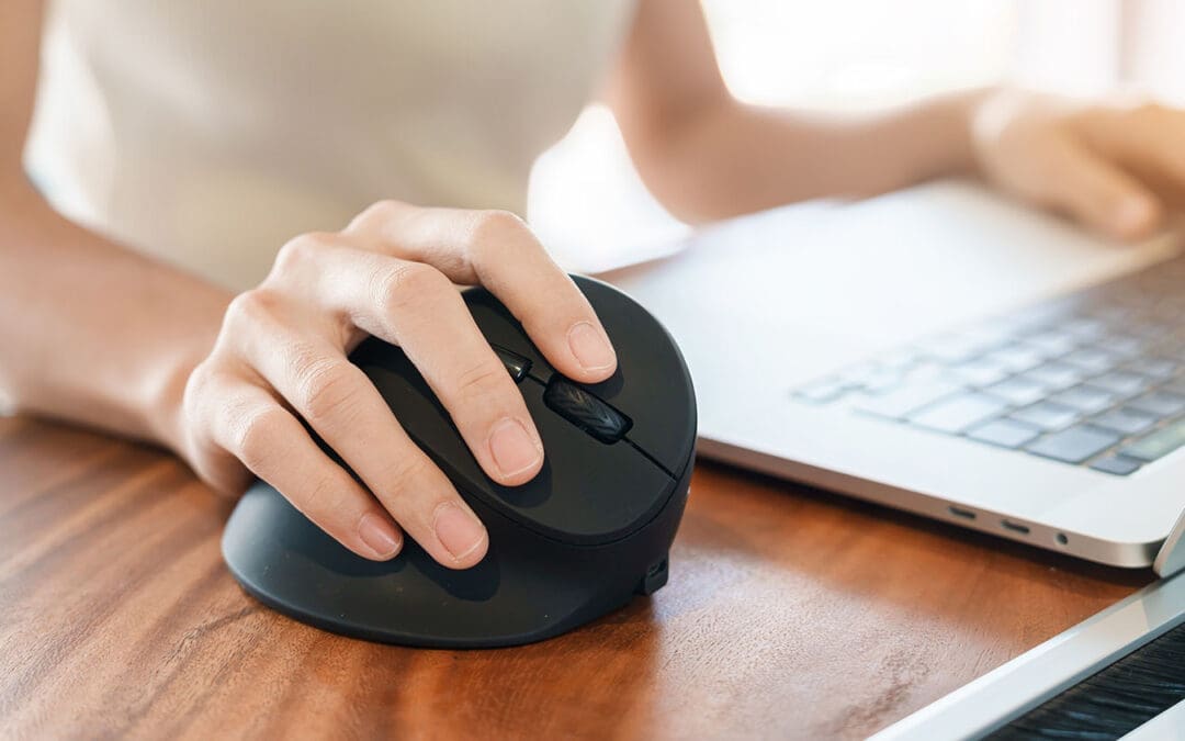
El Paso Back Clinic’s Guide to Ergonomic Mice for Pain-Free Hands
Spending hours at a computer can strain your hands, wrists, and arms, especially after injuries from accidents or repetitive tasks. At El Paso Back Clinic in El Paso, TX, led by Dr. Alex Jimenez, DC, APRN, FNP-BC, we specialize in providing holistic solutions to help patients overcome pain. An ergonomic mouse, designed to fit your hand’s natural shape, reduces strain and helps prevent conditions like carpal tunnel syndrome and tendonitis. Paired with our chiropractic care, advanced diagnostics, and integrative therapies, it supports recovery and long-term wellness. This article explains how El Paso Back Clinic uses ergonomic tools and expert care to restore health and prevent future issues.
Why Choose an Ergonomic Mouse?
Unlike standard flat mice, an ergonomic mouse curves to match your hand, often tilting upright in a manner similar to a handshake grip. This keeps your wrist straight, easing muscle and nerve strain (Goldtouch, 2023a). At El Paso Back Clinic, we recommend these for patients with desk jobs or those recovering from accidents.
Traditional mice twist your forearm, pinching nerves. Ergonomic designs hold your arm neutrally, reducing fatigue (Logitech, n.d.). For example, Logitech’s MX Vertical tilts at 57 degrees, cutting wrist tension (Logitech, n.d.). Our patients report less pain after switching, helping them work or recover comfortably.
Pick a mouse with thumb rests or adjustable angles to suit your hand. Our clinic guides you to the best choice for your needs (ProtoArc, 2023).
Supporting Natural Posture for Comfort
Your hand’s position affects your entire arm. Regular mice force your wrist to bend inward, stressing bones and nerves (ZDNet, 2023). An ergonomic mouse reduces this twist, called pronation, keeping your hand in a relaxed position (Goldtouch, 2023a).
Studies show these mice cut muscle effort by up to four times (Logitech, n.d.). They also help ease shoulder and neck tension, which is crucial for those recovering from injuries (Kosak Chiropractic, n.d.). At El Paso Back Clinic, we have seen patients benefit from this switch, especially those who have experienced motor vehicle accidents (MVAs) or repetitive strain injuries.
Reducing Repetitive Strain Injuries
Repetitive strain injuries (RSI) from constant clicking cause tingling, numbness, or pain (EffyDesk, 2023). Ergonomic mice minimize hand movements, featuring curves that allow fingers to rest naturally (Goldtouch, 2023b).
Thumb rests stop over-gripping, and lightweight designs make moving easier (ProtoArc, 2023). Our patients, from office workers to MVA survivors, use these to avoid worsening injuries. This supports healing during rehabilitation.
Preventing Carpal Tunnel and Tendonitis
Carpal tunnel syndrome squeezes the wrist’s median nerve, causing tingling or a weak grip. Tendonitis inflames tendons from overuse (FlexiSpot, n.d.). Both are common in desk workers and individuals who have been in accidents. Ergonomic mice open the wrist’s tunnel, reducing pressure by up to 30% (Goldtouch, 2023a).
They also limit bends that inflame tissues (ZDNet, 2023). For tendonitis, less forearm twist eases elbow strain, preventing long-term damage (Lowery Chiropractic, n.d.). El Paso Back Clinic patients who use these mice often stop the progression of injury early.
Setting Up Your Workstation for Health
An ergonomic mouse works best with a properly set-up desk. At El Paso Back Clinic, we recommend adjusting your chair to a 90-degree elbow angle with your feet flat. Keep your mouse at elbow height to avoid reaching (Kosak Chiropractic, n.d.).
Use a keyboard tray to maintain a straight wrist position and set your monitor at eye level to prevent neck strain (Kosak Chiropractic, n.d.). Take hourly breaks—stretch wrists, roll shoulders—to boost blood flow (EffyDesk, 2023). Our team offers personalized tips to make your workspace support recovery.
El Paso Back Clinic’s Holistic Healing Approach
Our clinic blends chiropractic adjustments, acupuncture, and rehabilitation to treat pain holistically. Adjustments realign joints, easing nerve pressure and swelling (Rozenhart Chiropractic, n.d.). For wrist pain, we target hand-to-elbow alignment to relieve carpal tunnel (Lowery Chiropractic, n.d.).
We utilize integrative therapies, such as ultrasound to warm tissues and electrical stimulation to calm nerves (Lowery Chiropractic, n.d.). Nutrition counseling helps reduce inflammation, thereby aiding recovery (Evolve Chiropractic, n.d.). Dr. Jimenez creates custom plans to address the causes of injuries, not just their symptoms.
Dr. Alex Jimenez’s Expertise in Injury Care
Dr. Alex Jimenez, a chiropractor and nurse practitioner, leads El Paso Back Clinic with dual expertise. He treats work, sports, personal, and MVA injuries using advanced neuromusculoskeletal imaging and dual-scope diagnosis to pinpoint issues like nerve compression (Jimenez, n.d.a).
For MVAs, he links whiplash to arm pain, using scans to guide treatment (Jimenez, n.d.b). Care includes adjustments, exercises, and massage to restore function. Acupuncture boosts natural healing (Evolve Chiropractic, n.d.). We also manage legal documentation for injury claims, easing patient stress (Jimenez, n.d.a).
A recent patient, following a motor vehicle accident (MVA), utilized an ergonomic mouse and our care plan. Pain dropped 70% in weeks, avoiding surgery (Jimenez, n.d.b). Dr. Jimenez focuses on natural healing over medication.
Targeted Therapies for Lasting Relief
We pair ergonomic tools with rehab. Grip exercises strengthen the hands, while wrist stretches build flexibility (EffyDesk, 2023). Acupuncture targets specific pain points, and massage helps loosen muscles (Rozenhart Chiropractic, n.d.).
Dr. Jimenez utilizes electro-acupuncture for nerve recovery, which has been shown to be effective for chronic pain (Jimenez, n.d.a). Patients track their progress with pain logs to achieve steady improvement. Our El Paso clinic provides these therapies for seamless care.
Success Stories at El Paso Back Clinic
Anna, a receptionist, switched to an ergonomic mouse and received our adjustments. Her wrist pain faded in weeks, improving her work (Goldtouch, 2023a). Carlos, an MVA survivor, worked with Dr. Jimenez. Adjustments and exercises restored his arm strength (Jimenez, n.d.b).
These stories show our approach delivers. Small changes, combined with expert care, transform lives.
Building a Pain-Free Future
Start with an ergonomic mouse and a tuned workspace. Experience the benefits of our chiropractic care, acupuncture, and nutrition for lasting health. Short walks and breathing exercises boost recovery (Evolve Chiropractic, n.d.).
Visit El Paso Back Clinic for a custom plan. Dr. Jimenez’s team treats all injuries naturally, from desk strain to MVAs (Jimenez, n.d.a). Act early to stay pain-free.
Conclusion: Heal with El Paso Back Clinic
An ergonomic mouse supports natural hand posture, cutting strain. Paired with our chiropractic and integrative care, it helps prevent and manage issues such as carpal tunnel syndrome. Dr. Jimenez’s expertise ensures effective recovery. Call +1 (915) 850-0900 to start your pain-free journey today.
Understanding Nerve Conditions of the Spine: Causes, Symptoms, and Treatments
The spine is a critical part of the body, serving as a highway for nerves that transmit signals between the brain and the rest of the body. When something goes wrong with these nerves—whether they’re compressed, irritated, or damaged—it can lead to a range of uncomfortable symptoms like pain, numbness, tingling, or weakness. These issues, known as nerve-related spine conditions, can affect the back, arms, or legs and stem from various causes, including injuries, degenerative conditions, or infections. In this article, we’ll explore these conditions, their symptoms, causes, and how they’re diagnosed and treated, with a special focus on integrative approaches like those used by Dr. Alexander Jimenez, a chiropractor and nurse practitioner in El Paso, Texas. We’ll also look at how chiropractic care, targeted exercises, massage therapy, acupuncture, and integrative medicine can promote healing and prevent long-term problems.
What Are Nerve-Related Spine Conditions?
Nerve-related spine conditions happen when the spinal nerves or spinal cord are compressed, irritated, or damaged. The spine is made up of bones called vertebrae, which protect the spinal cord—a bundle of nerves that carries messages to and from the brain. Between the vertebrae are intervertebral discs, which act as cushions, and small openings called foramina, where nerve roots exit the spinal cord to connect to other parts of the body. When these nerves or the spinal cord itself are affected, it can disrupt the signals, leading to symptoms like pain, numbness, tingling, or weakness (Mayo Clinic Health System, n.d.).
Some of the most common nerve-related spine conditions include:
Radiculopathy: Often referred to as a “pinched nerve,” this condition occurs when a nerve root is compressed or irritated as it exits the spine. It can cause pain, numbness, or weakness that radiates along the nerve’s path. For example, lumbar radiculopathy can lead to sciatica, a condition characterized by pain that shoots from the lower back down the leg (Cleveland Clinic, n.d.).
Spinal stenosis refers to the narrowing of the spinal canal, which puts pressure on the spinal cord or nerve roots. It’s often caused by aging or degenerative changes and can lead to symptoms like back pain, numbness, or difficulty walking (HSS Education, n.d.).
Herniated or Bulging Discs: Discs can bulge or herniate (when the inner gel-like material pushes out), pressing on nearby nerves. This can cause pain, tingling, or weakness in the arms or legs, depending on where the disc is located (Penn Medicine, n.d.).
Degenerative Conditions: Conditions like arthritis or bone spurs can narrow the spaces where nerves travel, causing compression and symptoms like pain or stiffness (Health Central, n.d.).
Trauma or Injury: Accidents, such as car crashes or falls, can damage the spine and compress nerves, leading to immediate or delayed symptoms (Verywell Health, n.d.).
Infections or Structural Abnormalities: Infections, tumors, or abnormal spine alignment (like scoliosis) can also press on nerves, causing similar symptoms (MSD Manuals, n.d.).
These conditions can range from mild annoyances to serious issues requiring immediate medical attention, especially if they cause severe symptoms like loss of bladder or bowel control, which may indicate cauda equina syndrome, a medical emergency (Verywell Health, n.d.).
Symptoms of Nerve-Related Spine Conditions
The symptoms of nerve-related spine conditions depend on where the nerve compression or damage occurs and the severity of the condition. Common symptoms include:
Pain: This can be sharp, burning, or aching and may stay in one spot (like the neck or lower back) or radiate to other areas, such as the arms, buttocks, or legs. For example, sciatica often causes burning pain that travels from the lower back to the legs (Penn Medicine, n.d.).
Numbness or Tingling: These sensations, often described as “pins and needles,” can occur in the hands, arms, feet, or legs, depending on the affected nerve (Cleveland Clinic, n.d.).
Weakness: Muscle weakness in the arms, hands, or legs can make it hard to lift objects, walk, or maintain balance. In severe cases, it can cause issues like foot drop, where a person struggles to lift their foot while walking (Johns Hopkins Medicine, n.d.).
Loss of Coordination: Compression of the spinal cord (myelopathy) can affect fine motor skills, making tasks like buttoning a shirt or writing difficult (Verywell Health, n.d.).
Balance Issues: Spinal stenosis or myelopathy can cause trouble walking or maintaining balance, sometimes described as feeling like “walking through mud” (Spine-health, n.d.).
Loss of Bladder or Bowel Control: This is a rare but serious symptom that requires immediate medical attention, as it may signal cauda equina syndrome (HSS Education, n.d.).
Symptoms can develop suddenly, like after an injury, or gradually, as with degenerative conditions like arthritis. If you experience severe or worsening symptoms, especially loss of bladder or bowel control, seek medical care right away.
Causes of Nerve-Related Spine Conditions
Nerve-related spine conditions can have many causes, ranging from natural aging to sudden injuries. Here are some of the main culprits:
Degenerative Changes: As people age, the spine can undergo wear and tear. Osteoarthritis can cause bone spurs, and degenerative disc disease can lead to bulging or herniated discs, both of which can press on nerves (Mayo Clinic Health System, n.d.).
Herniated or Bulging Discs: When a disc’s inner material bulges or herniates, it can push against nearby nerves, causing pain or numbness. This is a common cause of radiculopathy, including sciatica (Penn Medicine, n.d.).
Spinal Stenosis: The spinal canal can narrow due to thickened ligaments, bone spurs, or other changes, putting pressure on the spinal cord or nerve roots (Cleveland Clinic, n.d.).
Trauma: Car accidents, sports injuries, or falls can fracture vertebrae, dislocate joints, or cause swelling that compresses nerves, leading to severe consequences. For example, a car crash can lead to whiplash, which may cause nerve damage in the neck (Solomon Law, n.d.).
Infections: Spinal infections, like abscesses, can press on the spinal cord or nerves, causing pain and neurological symptoms (MSD Manuals, n.d.).
Structural Abnormalities: Conditions like scoliosis (abnormal spine curvature) or tumors can compress nerves, leading to symptoms like pain or weakness (Johns Hopkins Medicine, n.d.).
Inflammatory or Autoimmune Conditions: Diseases like rheumatoid arthritis can cause inflammation that compresses nerves, contributing to symptoms (OrthoTOC, n.d.).
Each cause can lead to different symptoms and requires specific diagnostic and treatment approaches to address the root issue.
Diagnosing Nerve-Related Spine Conditions
Diagnosing nerve-related spine conditions starts with a doctor asking about your symptoms and medical history, followed by a physical exam to check for numbness, weakness, reflexes, and posture. Depending on the findings, additional tests may be needed to pinpoint the cause (Penn Medicine, n.d.). Common diagnostic tools include:
Imaging tests, such as X-rays, CT scans, or MRIs, can reveal the spine’s structure, including bones, discs, and nerves, to identify compression or damage (Spine Info, n.d.).
Nerve Conduction Studies (NCS) and Electromyography (EMG): These tests assess the function of nerves and muscles, and can help confirm nerve damage (Spine Info, n.d.).
Myelogram: A special X-ray or CT scan with contrast dye can highlight pressure on the spinal cord or nerves (Spine Info, n.d.).
Dr. Alexander Jimenez, a chiropractor and nurse practitioner in El Paso, Texas, uses a dual-scope approach to diagnosis, combining his expertise in chiropractic care and advanced nursing. His clinic utilizes advanced neuromusculoskeletal imaging techniques, such as MRIs and CT scans, to obtain a clear picture of the spine’s condition. Dr. Jimenez correlates patient injuries—whether from work, sports, car accidents, or personal incidents—with clinical findings to create a precise diagnosis. This approach ensures that the treatment plan targets the specific cause of the nerve issue, whether it’s a herniated disc, spinal stenosis, or trauma-related damage (Jimenez, n.d.).
Treatment Options for Nerve-Related Spine Conditions
Treatment for nerve-related spine conditions depends on the cause, severity, and symptoms. Most doctors start with conservative (non-surgical) treatments, moving to surgery only if needed. Here’s an overview of common treatments:
Non-Surgical Treatments
Medications: Over-the-counter pain relievers, such as ibuprofen, or prescription medications, like gabapentin, can help manage pain and inflammation (Spine Info, n.d.).
Physical Therapy: Targeted exercises can strengthen muscles, improve posture, and reduce pressure on nerves. Physical therapy is often effective for radiculopathy and spinal stenosis (Cleveland Clinic, n.d.).
Epidural Steroid Injections: These deliver anti-inflammatory medication directly to the affected nerve root, reducing pain and swelling (Penn Medicine, n.d.).
Chiropractic Care: Adjustments and manipulations can realign the spine, relieving pressure on nerves. Dr. Jimenez’s clinic utilizes chiropractic techniques to treat conditions such as sciatica and herniated discs, with a focus on restoring spinal alignment (Jimenez, n.d.).
Massage Therapy: This can relax tight muscles, improve blood flow, and reduce nerve irritation, especially for conditions caused by muscle tension or spasms (Inova, n.d.).
Acupuncture: By stimulating specific points, acupuncture can reduce pain and promote natural healing, often used alongside other treatments (Total Spine Ortho, n.d.).
Activity Modification: Avoiding activities that worsen symptoms, like heavy lifting, can help the spine heal (Penn Medicine, n.d.).
Surgical Treatments
If conservative treatments are not effective, surgery may be necessary. Common procedures include:
Laminectomy: Removes part of a vertebra to create more space for nerves, often used for spinal stenosis (Spine Info, n.d.).
Microdiscectomy: Removes part of a herniated disc that’s pressing on a nerve, commonly used for radiculopathy (Spine Info, n.d.).
Spinal Fusion: Fuses vertebrae together to stabilize the spine, used for severe degenerative conditions or trauma (Inova, n.d.).
Dr. Jimenez’s clinic takes an integrative approach, combining chiropractic care with targeted exercises, massage therapy, and acupuncture to treat nerve-related spine conditions. For example, a patient with sciatica resulting from a herniated disc may receive spinal adjustments to realign the spine, exercises to strengthen core muscles, and acupuncture to alleviate pain. This holistic approach addresses the root cause while promoting long-term healing and preventing future problems (Jimenez, n.d.).
Dr. Alexander Jimenez’s Integrative Approach in El Paso
Dr. Alexander Jimenez, a chiropractor and nurse practitioner in El Paso, Texas, has extensive experience treating nerve-related spine conditions caused by work, sports, personal, or motor vehicle accident injuries. His clinic uses a dual-scope approach, blending chiropractic expertise with advanced medical knowledge to provide comprehensive care. Here’s how his clinic handles these cases:
Treating Different Types of Injuries
Work Injuries: Repetitive motions or heavy lifting at work can lead to conditions like herniated discs or radiculopathy. Dr. Jimenez uses spinal adjustments, targeted exercises, and ergonomic advice to relieve nerve compression and prevent recurrence (Jimenez, n.d.).
Sports Injuries: Athletes may suffer nerve compression from trauma or overuse. The clinic employs chiropractic care, physical therapy, and massage to restore function and reduce pain, helping athletes return to their activities (Jimenez, n.d.).
Personal Injuries: Falls or other accidents can cause nerve damage. Dr. Jimenez’s team uses advanced imaging to assess the injury and creates personalized treatment plans, often including acupuncture and exercise (Jimenez, n.d.).
Motor Vehicle Accident (MVA) Injuries: Car crashes can cause whiplash or other trauma that compresses nerves. The clinic provides detailed diagnostic assessments, including MRIs, to identify nerve damage and offers treatments like spinal adjustments and massage to promote healing (Solomon Law, n.d.; Jimenez, n.d.).
Medical Care and Legal Documentation
Dr. Jimenez’s clinic is skilled in handling the medical and legal aspects of injury cases, especially for MVAs. They provide thorough documentation of injuries, diagnoses, and treatments, which is critical for insurance claims or legal cases. For example, if a patient has radiculopathy from a car accident, the clinic documents the injury’s impact on their daily life, the diagnostic findings (like MRI results), and the treatment plan. This detailed paperwork supports patients in legal proceedings while ensuring they receive proper medical care (Jimenez, n.d.).
Integrative Medicine for Healing and Prevention
Dr. Jimenez’s approach emphasizes integrative medicine, combining chiropractic care with other therapies to address the cause of nerve issues and enhance overall health. For instance:
Chiropractic Adjustments: Realign the spine to relieve nerve pressure, effective for conditions like sciatica or herniated discs.
Targeted Exercises: Strengthen muscles around the spine to improve stability and prevent future injuries.
Massage Therapy: Reduces muscle tension and improves circulation, aiding in nerve healing.
Acupuncture: Stimulates natural pain relief and promotes recovery, especially for chronic pain.
Lifestyle Changes: Advice on posture, ergonomics, and nutrition helps prevent long-term problems (Jimenez, n.d.).
This integrative approach not only treats the immediate symptoms but also focuses on long-term health, reducing the risk of chronic pain or recurring issues.
How Integrative Medicine Promotes Healing
Integrative medicine, as practiced by Dr. Jimenez, combines conventional medical treatments with complementary therapies to address the whole person, not just the symptoms. For nerve-related spine conditions, this approach offers several benefits:
Natural Healing: Chiropractic care and acupuncture stimulate the body’s natural healing processes, reducing reliance on medications (Total Spine Ortho, n.d.).
Pain Reduction: Therapies such as massage and acupuncture can help reduce pain levels, thereby improving quality of life (Inova, n.d.).
Improved Function: Exercises and adjustments restore mobility and strength, helping patients return to normal activities (Cleveland Clinic, n.d.).
Prevention: By addressing underlying causes, like poor posture or weak muscles, integrative medicine reduces the risk of future nerve problems (Jimenez, n.d.).
For example, a patient with spinal stenosis might receive adjustments to improve spinal alignment, exercises to strengthen their core, and massage to relax tight muscles. Over time, these treatments can reduce nerve compression, improve mobility, and prevent the condition from worsening.
Preventing Long-Term Problems
Preventing long-term nerve-related spine issues involves addressing the root causes and maintaining spinal health. Here are some strategies:
Maintain Good Posture: Proper posture reduces strain on the spine and nerves (Mayo Clinic Health System, n.d.).
Stay Active: Regular exercise, especially core-strengthening workouts, supports the spine and prevents injuries (Cleveland Clinic, n.d.).
Utilize ergonomics: Adjust workstations or lifting techniques to prevent repetitive strain (Jimenez, n.d.).
Manage Weight: Excess weight can put pressure on the spine, worsening nerve conditions (Health Central, n.d.).
Seek Early Treatment: Addressing symptoms early with chiropractic care or physical therapy can prevent conditions like radiculopathy from becoming chronic (Spine Info, n.d.).
Dr. Jimenez’s clinic emphasizes these preventive measures, educating patients on lifestyle changes to keep their spines healthy and reduce the risk of future nerve issues.
Conclusion
Nerve-related spine conditions, like radiculopathy, spinal stenosis, and herniated discs, can cause significant discomfort and disrupt daily life. These conditions stem from various causes, including degenerative changes, trauma, infections, or structural issues, and lead to symptoms like pain, numbness, tingling, and weakness. Through proper diagnosis using imaging and clinical assessments, doctors can pinpoint the cause and recommend treatments, ranging from medications and physical therapy to surgery in severe cases. Integrative approaches, like those used by Dr. Alexander Jimenez in El Paso, combine chiropractic care, targeted exercises, massage therapy, and acupuncture to treat injuries from work, sports, or accidents while promoting natural healing. By addressing the root cause and focusing on prevention, these methods can help patients recover and avoid long-term problems. If you’re experiencing symptoms of a nerve-related spine condition, consult a healthcare provider to explore your treatment options and start your journey to recovery.
Genetics, Stiffness, and Flexibility: Understanding the Back’s Natural Limits
Introduction: Why Flexibility Matters for Spinal Health
Flexibility is often thought of as a skill we can train, like strength or endurance. But in reality, flexibility begins with genetics. Some people are born naturally limber, while others experience tightness in their muscles and connective tissues no matter how much they stretch. This is not always a problem—it is a normal variation in human biology.
At the El Paso Back Clinic, under the care of Dr. Alexander Jimenez, DC, APRN, FNP-BC, patients learn that stiffness has many causes: genetics, aging, lifestyle habits, and sometimes injuries. Through chiropractic adjustments, advanced imaging, and integrative care, the clinic helps individuals restore mobility, manage stiffness, and prevent long-term complications.
How Genetics Shapes Flexibility
Collagen and Connective Tissue
Ligaments, tendons, and fascia are made from collagen. Some people are genetically predisposed to tighter collagen, while others inherit looser connective tissues that allow more joint motion (Xcode Life, n.d.).
Muscle Fiber Balance
Fast-twitch fibers create power but are less flexible, while slow-twitch fibers support endurance and mobility. Genetics dictates the proportion of these fibers in each person’s body (PMC, 2020).
Genetic Syndromes and Flexibility Extremes
Ehlers-Danlos Syndrome (EDS): Causes extreme flexibility from connective tissue fragility.
Inherited Stiffness Disorders: Families can pass down congenital stiffness across generations (JAMA Pediatrics, 2000).
These differences show why two people can do the same stretching routine but achieve very different results.
Stiffness as a Normal Range of Human Variation
Not every stiff person has a medical problem. Many people naturally sit at the less flexible end of the spectrum, which is completely normal (Quora, n.d.).
Alexander Orthopaedics (2023) reports that gender, bone shape, and joint design also influence how flexible someone can be. For example, women tend to have greater flexibility in certain joints due to hormonal differences, while men often have more rigid tissue structures.
At El Paso Back Clinic, Dr. Jimenez helps patients understand that stiffness does not always mean something is wrong—but it can increase the risk of injury if not properly managed.
When Stiffness Becomes a Medical Concern
Stiff Person Syndrome (SPS)
SPS is a rare autoimmune condition that leads to severe rigidity, spasms, and difficulty walking. It is distinct from natural stiffness and requires medical treatment (Hopkins Medicine, n.d.; MSU Healthcare, 2024).
Genetic Disorders of Rigidity
Some families inherit congenital disorders that lock joints into restricted motion. These are uncommon but important to recognize in clinical settings (JAMA Pediatrics, 2000).
Injury-Related Stiffness
Motor vehicle accidents (MVAs), workplace injuries, and sports trauma can cause scar tissue, joint misalignment, and muscle guarding that worsen stiffness. Dr. Jimenez frequently sees these cases at El Paso Back Clinic.
Aging, Lifestyle, and the Stiff Back
Age-Related Tissue Changes
Over time, collagen stiffens, cartilage thins, and joint capsules lose elasticity. Even flexible individuals in youth often report stiffness as they age (PMC, 2020).
Lifestyle Habits
Sedentary behavior shortens connective tissue.
Repetitive work tasks create uneven strain.
Lack of stretching allows muscles to tighten.
At El Paso Back Clinic, patients often present with stiffness that is a combination of genetic and lifestyle factors, requiring a tailored treatment plan.
Case Studies from El Paso Back Clinic
Case 1: Lifelong Stiffness Meets Injury
A 45-year-old man reported lifelong tightness, which worsened after an MVA. Imaging revealed whiplash compounded by rigid connective tissue. With chiropractic adjustments, massage, and guided rehab, he restored safe mobility while respecting his natural limits.
Case 2: Athletic Stiffness and Performance
A 20-year-old track athlete experienced poor hamstring flexibility, which led to recurring strains. Rather than forcing an extreme range of motion, Dr. Jimenez built a plan focusing on functional mobility, hip stability, and performance-specific conditioning.
Case 3: Sedentary Aging and Stiff Joints
A 68-year-old office worker complained of chronic back stiffness. With chiropractic care, acupuncture, and mobility training, stiffness eased enough to improve daily activities and quality of life.
Chiropractic and Integrative Solutions for Stiffness
At El Paso Back Clinic, treatment is not one-size-fits-all. Dr. Jimenez employs an integrative approach that combines chiropractic with medical and functional strategies:
Chiropractic Adjustments: Correct spinal alignment and improve motion.
Massage Therapy: Loosens tight fascia and muscles.
Acupuncture: Reduces spasms and supports nervous system balance.
Targeted Exercise: Builds mobility without overstretching joints.
Functional Medicine: Focuses on diet, inflammation, and tissue repair.
By blending these treatments, patients can improve mobility and manage stiffness effectively.
Sports, Flexibility, and Injury Prevention
Flexibility influences athletic performance—but both extremes have risks.
Too flexible: Joints may lack stability.
Too stiff: Risk of muscle strain or joint injury.
Dr. Jimenez helps athletes at El Paso Back Clinic find their optimal flexibility zone. This may mean increasing mobility in some cases or focusing on stability and strength in others.
Legal and Diagnostic Support in Personal Injury Cases
One unique aspect of Dr. Jimenez’s work is his dual-scope role in both chiropractic and medical diagnosis. For personal injury cases, this includes:
Advanced imaging (MRI, CT, X-ray)
Dual medical and chiropractic reports
Coordination with attorneys and insurers
Documentation of stiffness-related limitations
This ensures patients receive not only effective treatment but also the proper legal support for compensation and care continuity.
Lifestyle Practices to Support Mobility
While genetics can’t be changed, lifestyle makes a difference:
Daily Stretching for sustained tissue pliability.
Hydration to keep connective tissues healthy.
Balanced Nutrition to reduce inflammation and support collagen.
Regular Movement to prevent stiffness from inactivity.
Mind-Body Exercise, such as yoga or tai chi.
El Paso Back Clinic encourages patients to adopt these habits alongside clinical care.
Conclusion: Living Well with Natural Stiffness
Some people are naturally stiff. Others are naturally flexible. Both variations are normal, shaped by genetics, age, and lifestyle. What matters is managing stiffness in ways that prevent injury, restore comfort, and support long-term health.
At El Paso Back Clinic, Dr. Alexander Jimenez uses his dual expertise to evaluate stiffness, provide integrative treatment, and guide patients toward healthier mobility—whether recovering from injury, aging with stiffness, or simply working within genetic limits.
IFM's Find A Practitioner tool is the largest referral network in Functional Medicine, created to help patients locate Functional Medicine practitioners anywhere in the world. IFM Certified Practitioners are listed first in the search results, given their extensive education in Functional Medicine