Discover the impact of stretching & flexibility on joint pain relief. Incorporate these tips into your routine for optimal results.
Chiropractic Care and Stretching: A Holistic Approach to Joint Pain Relief
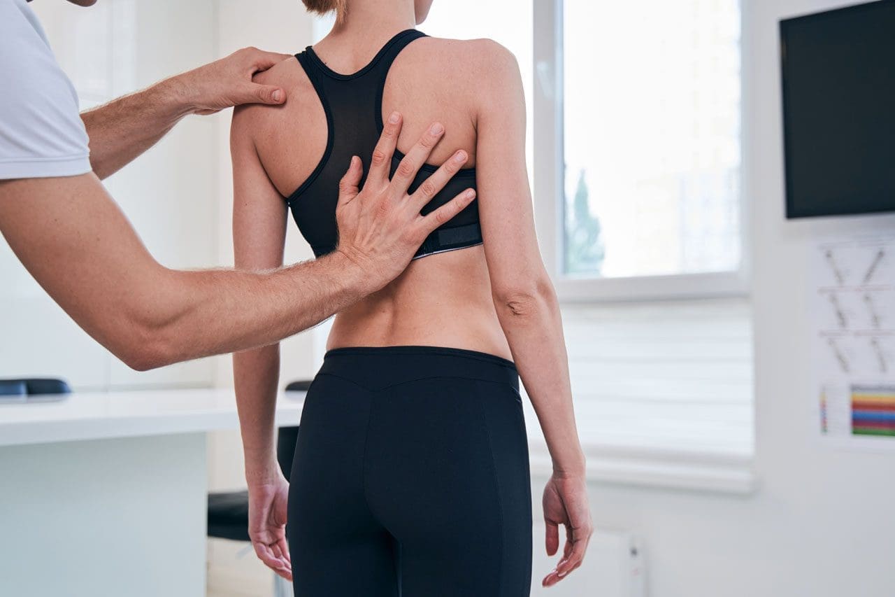
Joint pain can transform routine tasks like walking, lifting, or sitting into daunting challenges. Whether it’s a persistent ache in your knees, stiffness in your shoulders, or discomfort in your back, joint pain affects countless individuals globally. Fortunately, nonsurgical solutions such as chiropractic care, combined with stretching and flexibility exercises, provide a natural and effective way to manage pain, enhance mobility, and improve overall well-being. These methods address both symptoms and underlying causes, promoting long-term healing and a better quality of life.
This comprehensive guide explores the clinical rationale for integrating chiropractic care with stretching to alleviate joint pain. We’ll examine the causes and risk factors for joint pain in the upper and lower extremities, highlight the role of integrative therapies like massage and acupuncture, and provide practical stretching exercises suitable for home or gym settings. Supported by scientific research and expert insights, this article empowers you to take control of your joint health and recover from injuries, including those sustained in motor vehicle accidents (MVAs), bicycle collisions, or 18-wheeler crashes.
5 Things You Need to Know About
Ligamentous Injuries Before They Get Worse-Video
Understanding Joint Pain: Causes and Risk Factors
Joint pain arises from a complex interplay of factors, from acute injuries to chronic conditions. Identifying these causes is crucial for developing an effective treatment plan. Below are the primary contributors to joint pain in both upper and lower extremities:
1. Mechanical Factors
Joint Misalignment: Misaligned joints in the spine, shoulders, or knees can create uneven stress on surrounding muscles, tendons, and ligaments, leading to pain and inflammation. For instance, knee malalignment may contribute to patellofemoral pain syndrome, common among active individuals (Steinberg et al., 2021).
Overuse and Repetitive Stress: Repetitive motions from sports, work, or daily activities can strain joints, leading to conditions such as shoulder impingement syndrome or tennis elbow. Overhead athletes, such as cyclists or swimmers, often experience shoulder pain due to repetitive stress (Tauqeer et al., 2024).
Trauma or Injury: Acute injuries, such as sprains, fractures, or dislocations from MVAs or bicycle accidents, can damage joint structures, causing pain and reduced mobility. For example, anterior cruciate ligament (ACL) injuries are prevalent in athletes and can lead to significant knee pain and instability (Hurley, 1997).
2. Degenerative Conditions
Osteoarthritis: A leading cause of joint pain, osteoarthritis involves the breakdown of cartilage in joints such as the knees, hips, and hands, resulting in pain, stiffness, and a limited range of motion (Luan et al., 2022).
Rheumatoid Arthritis: This autoimmune condition causes inflammation in the synovial lining of joints, leading to tenderness, swelling, and potential joint damage (Dumoulin et al., 2023).
3. Generalized Joint Hypermobility (GJH)
Some individuals have naturally flexible joints, a condition known as generalized joint hypermobility (GJH). While advantageous for activities like dance, it increases the risk of joint instability and pain, particularly in the upper cervical spine or knees (Russek et al., 2023; Steinberg et al., 2021).
4. Inflammation and Systemic Factors
Inflammatory Conditions: Diseases like rheumatoid arthritis or psoriatic arthritis drive joint inflammation, exacerbating pain. Subclinical inflammation can cause tenderness in joints, such as the metacarpophalangeal (MCP) joints, even without a formal diagnosis (Dumoulin et al., 2023).
Muscle Imbalances and Poor Posture: Weak core muscles or poor posture can increase stress on joints, particularly in the spine, hips, and shoulders, leading to pain and dysfunction.
5. Lifestyle and Environmental Factors
Sedentary Lifestyle: A lack of movement can cause muscle stiffness and reduce joint lubrication, thereby increasing the risk of pain.
Obesity: Excess body weight places additional stress on weight-bearing joints, such as the knees and hips, accelerating cartilage wear (Luan et al., 2022).
Poor Ergonomics: Improper workstation setups or repetitive tasks, such as typing or lifting, can strain upper extremity joints, contributing to conditions like carpal tunnel syndrome.
Overlapping Risk Profiles
These factors often overlap, creating a complex risk profile for joint pain. For example, an individual with GJH may have weak supporting muscles, increasing the risk of joint instability. Similarly, someone with osteoarthritis might experience worsened symptoms due to repetitive stress or poor posture. Chiropractic care and stretching target these overlapping risks by improving joint alignment, enhancing muscle function, reducing inflammation, and promoting stability, offering a holistic approach to pain management and recovery from injuries like those sustained in MVAs or bicycle collisions.
The Clinical Rationale for Chiropractic Care and Stretching
Chiropractic care, paired with stretching and flexibility exercises, addresses the root causes of joint pain, offering a nonsurgical alternative to pain management. This integrative approach restores joint function, enhances muscle performance, and promotes the body’s natural healing processes, particularly for injuries from MVAs, 18-wheeler crashes, or bicycle accidents. Below is the clinical rationale for combining these modalities:
1. Restoring Joint Alignment and Function
Chiropractic Adjustments: Chiropractic adjustments, or thrust joint manipulations, involve applying controlled force to misaligned joints to restore proper alignment. This reduces stress on surrounding tissues, improves mobility, and alleviates pain. For example, spinal adjustments can help relieve low back pain associated with MVAs by correcting subluxations that irritate nerves (Rhyu et al., 2015).
Reducing Joint Stress: Misaligned joints lead to compensatory muscle tightness and inflammation. Adjustments redistribute forces across joints, reducing wear and tear, particularly in degenerative conditions such as osteoarthritis (Luan et al., 2022).
Evidence: Research shows thrust joint manipulation is effective for improving joint function and reducing pain in the lumbar and thoracic spine, with high confidence in its safety for these regions (Puentedura et al., 2017).
2. Enhancing Muscle Function and Proprioception
Muscle Activation: Joint damage from accidents or osteoarthritis can reduce voluntary muscle activation, resulting in weakness and muscle atrophy. Chiropractic care, combined with targeted exercises, helps restore muscle function by enhancing neural signaling (Hurley, 1997).
Proprioception: Injuries, particularly from MVAs or bicycle collisions, can impair proprioception, increasing the risk of further injury. Stretching and strengthening exercises enhance proprioceptive feedback, improving joint stability (Steinberg et al., 2021).
Evidence: Isometric exercises, often prescribed alongside chiropractic care, increase muscle activity and reduce pain in patients with low back pain from accidents (Rhyu et al., 2015).
3. Reducing Inflammation and Pain
Anti-Inflammatory Effects: Chiropractic adjustments and stretching improve joint mobility and blood flow, reducing inflammation. This is particularly effective for inflammatory conditions like rheumatoid arthritis or whiplash-associated disorders (WAD) from MVAs (Dumoulin et al., 2023).
Pain Modulation: Stretching exercises, particularly when combined with manual therapy, have been shown to significantly reduce pain in conditions such as knee osteoarthritis and shoulder impingement syndrome (Luan et al., 2022; Tauqeer et al., 2024).
Evidence: A meta-analysis found that stretching exercises alone resulted in a clinically meaningful reduction in knee osteoarthritis pain, with enhanced benefits when combined with other therapies (Luan et al., 2022).
4. Preventing Long-Term Complications
Joint Stability: For individuals with GJH or scoliosis, chiropractic care and targeted exercises strengthen supporting muscles, reducing the risk of joint instability and related injuries (Russek et al., 2023; Steinberg et al., 2021).
Holistic Healing: By addressing biomechanical, muscular, and neurological factors, chiropractic care promotes long-term joint health, preventing chronic pain and disability from accident-related injuries.
Evidence Suggests That Rehabilitation programs incorporating stretching and strengthening exercises improve outcomes in patients with joint hypermobility, scoliosis, or post-accident trauma, thereby reducing the risk of patellofemoral pain (Steinberg et al., 2021).
5. Complementary Therapies
Massage Therapy: Massage reduces muscle tension, improves circulation, and prepares tissues for chiropractic adjustments. It is particularly effective for shoulder impingement and whiplash injuries, enhancing range of motion and functional capacity (Tauqeer et al., 2024).
Acupuncture: Acupuncture stimulates endorphin release, reduces inflammation, and improves neural signaling, making it a valuable adjunct for managing pain from osteoarthritis, low back pain, or MVA injuries.
Integrative Medicine: An integrative approach combining chiropractic adjustments, stretching, strengthening, massage, and acupuncture addresses the multifaceted nature of joint pain, promoting natural healing (El Paso Back Clinic, n.d.).
Evidence: Manual therapies, including massage, significantly reduce pain and improve function in patients with chronic shoulder conditions and post-accident injuries (Tauqeer et al., 2024).
6. Patient-Centered Care
Clear communication ensures tailored treatment plans that address individual needs, whether recovering from an 18-wheeler crash or managing chronic arthritis. Patient education enables individuals to perform home exercises that maintain progress (El Paso Back Clinic, n.d.).
Evidence Suggests That Patient education and active participation in rehabilitation programs enhance adherence and outcomes in musculoskeletal care (Jimenez, 2016).
By targeting overlapping risk factors—misalignment, muscle weakness, inflammation, and instability—chiropractic care and stretching provide a comprehensive solution for joint pain relief and recovery from accident-related injuries.
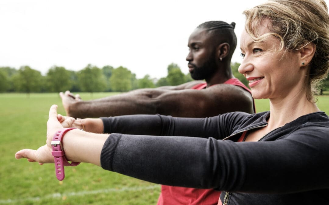
Stretching and Flexibility Exercises for Joint Pain Relief
Stretching and flexibility exercises are essential for maintaining joint health, improving range of motion, and reducing pain, especially after MVAs or bicycle accidents. Below are practical exercises suitable for home or gym settings, supported by research. Consult a healthcare provider before starting, particularly if you have injuries or conditions like GJH or scoliosis.
1. Cat-Cow Stretch (Spinal Flexibility)
Purpose: Enhances spinal flexibility, reduces low back pain, and improves core stability, ideal for MVA recovery.
How to Perform:
Position yourself on your hands and knees, with your hands under your shoulders and your knees under your hips.
Inhale, letting your abdomen drop toward the floor while gently arching your back (Cow Pose).
Exhale, arching your back upward like a cat, tucking your chin to your chest (Cat Pose).
Repeat 3–5 times, moving slowly.
Benefits: Increases spinal mobility and reduces tension in back muscles (Jimenez, 2016).
Frequency: Perform daily, morning and evening, for 5–10 minutes.
Tip: Move smoothly to avoid straining the spine.
2. Knee-to-Chest Stretch (Lower Back and Hip Flexibility)
Purpose: Relieves tension in the lower back and hips, beneficial for low back pain and sciatica from MVAs.
How to Perform:
Lie on your back with knees bent and feet flat.
Grasp one knee with both hands and pull it toward your chest.
Hold for 30 seconds, then return to the starting position.
Repeat with the other knee or both knees together.
Benefits: Improves lumbar flexibility and reduces pain (Jimenez, 2016).
Frequency: Perform 2–3 times per leg, twice daily.
Tip: Keep your lower back pressed against the floor.
3. Scorpion Stretch (Lower Back and Core)
Purpose: Stretches the lower back and strengthens core muscles, ideal for chronic back pain post-accident.
How to Perform:
Lie face down with arms extended out to the sides.
Lift your right leg and move it toward your left arm, keeping your torso stable.
Hold for 10 seconds, then return to the starting position.
Repeat with the left leg toward the right arm.
Benefits: Enhances lumbar flexibility and core strength (Jimenez, 2016).
Frequency: Perform 2–3 repetitions per side, once daily.
Purpose: Enhances scapular mobility and reduces shoulder impingement pain, common in bicycle accidents.
How to Perform:
Sit or stand with arms relaxed at your sides.
Squeeze your shoulder blades together, as if holding a pencil between them.
Hold for 5–10 seconds, then release.
Benefits: Improves scapular range of motion and reduces shoulder pain (Tauqeer et al., 2024).
Frequency: Perform 10–15 repetitions, 2–3 times daily.
Tip: Keep your shoulders relaxed to avoid shrugging.
6. Standing Quadriceps Stretch (Knee and Hip Flexibility)
Purpose: Stretches the quadriceps to reduce knee pain and improve mobility.
How to Perform:
Stand near a wall for balance, holding one ankle with the same-side hand.
Pull your ankle toward your buttocks, keeping your knees aligned.
Hold for 20–30 seconds, then switch legs.
Benefits: Enhances knee flexibility and reduces patellofemoral pain (Steinberg et al., 2021).
Frequency: Perform 2–3 times per leg, daily.
Tip: Tuck your pelvis to avoid arching your lower back.
7. Neck Rotation Stretch (Cervical Flexibility)
Purpose: Reduces neck stiffness and improves cervical mobility, especially for GJH or whiplash from MVAs.
How to Perform:
Sit or stand with your back straight.
Turn your head to the right, looking over your shoulder, and hold for 15–20 seconds.
Return to the center and repeat on the left.
Benefits: Improves cervical range of motion and reduces symptoms of instability (Russek et al., 2023).
Frequency: Perform 3–5 repetitions per side, twice daily.
Tip: Move within your comfortable range to avoid strain.
Tips for Safe Stretching
Warm Up First: Engage in 5–10 minutes of light activity, such as walking, to prepare your muscles and joints (Jimenez, 2016).
Avoid Overstretching: Stretch to mild tension, not to the point of pain, to prevent injury.
Breathe Deeply: Inhale and exhale slowly to enhance relaxation and muscle lengthening.
Consult a Professional: Work with a chiropractor or physical therapist to ensure proper technique, especially for post-accident recovery or conditions like GJH or scoliosis.
Integrative Therapies for Enhanced Joint Pain Relief
Integrative therapies, such as massage and acupuncture, complement chiropractic care and stretching, addressing muscle tension, inflammation, and neurological factors, particularly in cases related to accidents.
1. Massage Therapy
Benefits: Massage reduces muscle tightness, improves circulation, and prepares tissues for chiropractic adjustments. It is effective for shoulder impingement, whiplash, and post-MVA recovery (Tauqeer et al., 2024; El Paso Back Clinic, n.d.).
Application: Techniques such as deep tissue massage or trigger point therapy target tight muscles and fascia, thereby enhancing the benefits of stretching.
Evidence Suggests That Manual therapy, including massage, significantly reduces pain and improves function in individuals with chronic shoulder conditions and accident-related injuries (Tauqeer et al., 2024).
2. Acupuncture
Benefits: Acupuncture stimulates endorphin release, reduces inflammation, and improves neural signaling, effective for osteoarthritis, low back pain, and WAD from MVAs.
Application: Integrated with chiropractic care, acupuncture addresses local and systemic pain pathways, enhancing recovery.
Evidence: Research supports the use of acupuncture as an effective adjunct for managing musculoskeletal pain (Luan et al., 2022).
3. Nutrition for Recovery
Benefits: A diet rich in anti-inflammatory foods (e.g., omega-3 fatty acids, fruits, and vegetables) supports tissue healing and reduces inflammation, crucial for post-accident recovery (El Paso Back Clinic, n.d.).
Application: Nutritional guidance complements chiropractic care, promoting internal healing.
Evidence: Proper nutrition enhances musculoskeletal injury rehabilitation, particularly after MVAs (El Paso Back Clinic, n.d.).
Preventing Long-Term Joint Problems
Chiropractic care and stretching not only relieve joint pain but also prevent long-term complications by addressing underlying causes. Key strategies include:
Consistent Exercise: Daily stretching and strengthening enhance joint stability and flexibility.
Healthy Lifestyle Choices: Maintain a healthy weight, eat an anti-inflammatory diet, and practice good posture to reduce joint stress.
Early Intervention: Seek chiropractic care at the first sign of pain to prevent progression to chronic conditions like osteoarthritis or WAD.
Durable Medical Equipment: Braces or supports may aid recovery from MVA injuries, as recommended by professionals (El Paso Back Clinic, n.d.).
Conclusion
Joint pain from injuries, degenerative conditions, or lifestyle factors can significantly impact daily life. Chiropractic care, combined with stretching and flexibility exercises, provides a powerful, non-surgical solution for managing and preventing pain. By addressing joint misalignment, enhancing muscle function, reducing inflammation, and promoting holistic healing through integrative therapies like massage, acupuncture, and nutrition, this approach targets the root causes of joint pain. Incorporating the stretching exercises above and seeking professional guidance can improve function, reduce pain, and support a more active, pain-free life.
References
Dumoulin, Q. A., van Steenbergen, H. W., & van der Helm-van Mil, A. H. M. (2023). Correspondence on ‘Role of joint damage, malalignment and inflammation in articular tenderness in rheumatoid arthritis, psoriatic arthritis and osteoarthritis’. Annals of the Rheumatic Diseases, 82(7), e160. https://doi.org/10.1136/annrheumdis-2021-220511
Hurley, M. V. (1997). The effects of joint damage on muscle function, proprioception, and rehabilitation. Manual Therapy, 2(1), 11–17. https://doi.org/10.1054/math.1997.0281
Luan, L., El-Ansary, D., Adams, R., Wu, S., & Han, J. (2022). Knee osteoarthritis pain and stretching exercises: A systematic review and meta-analysis. Physiotherapy, 114, 16–29. https://doi.org/10.1016/j.physio.2021.10.001
Puentedura, E. J., Slaughter, R., Reilly, S., Ventura, E., & Young, D. (2017). Thrust joint manipulation utilization by U.S. physical therapists. Journal of Manual & Manipulative Therapy, 25(2), 74–82. https://doi.org/10.1080/10669817.2016.1187902
Rhyu, H.-S., Park, H.-S., & Park, J.-S. (2015). The Effects of Isometric Exercise Types on Pain and Muscle Activity in Patients with Low Back Pain. Journal of Exercise Rehabilitation, 11(4), 211–214. https://doi.org/10.12965/jer.150224
Russek, L. N., Block, N. P., Byrne, E., Chalela, S., Chan, C., Comerford, M., … Hakim, A. (2023). Presentation and physical therapy management of upper cervical instability in patients with symptomatic generalized joint hypermobility: International expert consensus recommendations. Frontiers in Medicine, 9, 1072764. https://doi.org/10.3389/fmed.2022.1072764
Steinberg, N., Tenenbaum, S., Zeev, A., & Hershkovitz, I. (2021). Generalized joint hypermobility, scoliosis, patellofemoral pain, and physical abilities in young dancers. BMC Musculoskeletal Disorders, 22(1), 161. https://doi.org/10.1186/s12891-021-04023-z
Tauqeer, S., Arooj, A., & Javed, K. (2024). Effects of manual therapy in addition to stretching and strengthening exercises to improve scapular range of motion, functional capacity, and pain in patients with shoulder impingement syndrome: A randomized controlled trial. BMC Musculoskeletal Disorders, 25(1), 192. https://doi.org/10.1186/s12891-024-07294-4
Live Pain-Free: Chiropractic and Integrative Care for Injury Recovery at El Paso Back Clinic
In the vibrant heart of El Paso, Texas, where desert trails beckon and hardworking days define our community, injuries can derail your active lifestyle. From car accidents to workplace strains or sports mishaps, overexertion and trauma often lead to pain, stiffness, or chronic issues that linger without proper care. These setbacks can limit your ability to work, play, or enjoy El Paso’s unique spirit. At El Paso Back Clinic, led by Dr. Alexander Jimenez, DC, APRN, FNP-BC, chiropractic and integrative care offer a path to recovery. Through spinal adjustments, soft tissue therapy, and neuromuscular re-education, the clinic accelerates healing, restores flexibility, enhances balance, and boosts heart and lung function. With holistic nutrition and stress management plans, Dr. Jimenez’s team crafts personalized strategies to prevent future injuries, empowering El Pasoans to live pain-free and thrive.
This article explores how injuries arise, the benefits of integrative care, and how El Paso Back Clinic delivers top-tier recovery solutions.
The Impact of Overuse and Accidents: Why Pain Persists
El Paso’s dynamic lifestyle—hiking the Franklin Mountains, working long shifts, or driving busy roads—can strain the body. Overexertion from repetitive tasks like lifting or intense workouts causes sprains, strains, or joint issues. Motor vehicle accidents (MVAs) bring sudden trauma, with 60% of cases leading to lingering pain if untreated (Jimenez, n.d.). Even minor falls at home can spark chronic discomfort.
Dr. Alexander Jimenez, a chiropractor and nurse practitioner with over 30 years of experience, sees these patterns daily. “Our dual-scope diagnostics, combining chiropractic and nursing insights, uncover how trauma or overuse triggers pain cycles,” he shares on his clinic’s site (Jimenez, n.d.). Using advanced neuromusculoskeletal imaging, his team pinpoints root causes, from workplace injuries to MVA trauma. Ignoring early signs, such as stiffness or fatigue, can lead to reduced mobility, increased stress, and sleep disturbances. El Paso Back Clinic’s integrative approach breaks this cycle, restoring health naturally.
Everyday Injuries: From Crashes to Chronic Strains
Injuries vary but share a common impact: they disrupt your life. MVAs cause neck and back pain, limiting movement. Work-related strains, like those from lifting or repetitive tasks, create nagging discomfort. Sports injuries, such as twisted ankles or knees, sideline active El Pasoans. Personal falls at home can lead to shoulder or hip pain, while untreated stress may cause chronic conditions like joint stiffness.
Dr. Jimenez’s clinic tackles these with precision. “We connect injury origins—crashes, work tasks, or sports—to customized treatments,” he explains. MVAs receive urgent care with legal documentation for claims. Work injuries get rehab to restore function, and sports or personal injuries benefit from targeted plans to prevent recurrence. Without care, these issues worsen, lowering the quality of life. El Paso Back Clinic’s chiropractic and integrative methods pave the way to recovery.
Realigning for Relief: The Power of Spinal Adjustments
Spinal adjustments are the foundation of chiropractic care at El Paso Back Clinic. These precise, hands-on techniques realign vertebrae, easing nerve pressure and restoring balance to the body. Injuries from accidents or overuse misalign the spine, causing pain and impaired movement. Adjustments can boost blood flow, reduce inflammation, and alleviate pain by up to 25% in as little as a few weeks (Trident Health Chiropractic, n.d.).
For MVA patients, adjustments relieve neck stiffness, restoring mobility. Work injury patients regain strength for daily tasks. Dr. Jimenez’s approach is unique: “We use imaging to guide adjustments, targeting issues from trauma or strains,” he says. Legal reports ensure MVA patients have clear records for claims. From athletes to office workers, adjustments help El Pasoans move freely and heal quickly.
Healing Muscles: Soft Tissue Therapy for Recovery
Injuries tighten muscles, creating knots that misalign joints and prolong pain. Soft tissue therapy, like massage or myofascial release, targets these areas, breaking up scar tissue and boosting circulation. This delivers nutrients to damaged tissues, speeding recovery. A single session can significantly reduce healing time, getting you back to work or play faster (Yoder Chiropractic Center, n.d.).
Picture a construction worker with shoulder pain from heavy lifting. Therapy loosens tightness, improving arm range. MVA patients find relief from neck strain. Dr. Jimenez’s team pairs therapy with imaging for precision. “We treat trauma from accidents or sports non-surgically,” he notes. Legal documentation tracks progress for claims, prioritizing natural healing. Patients feel relaxed, move more easily, and recover more quickly.
Injuries disrupt nerve-muscle communication, resulting in shaky balance or impaired movements. Neuromuscular re-education uses exercises like balance drills or resistance training to retrain these pathways, reducing fall risks and boosting confidence. A soccer player with a sprained ankle, for example, regains stability, thereby lowering the odds of re-injury (Integrative Chiropractic, n.d.).
Dr. Jimenez’s clinic excels here. “We link nerve issues to injury histories, guiding re-education for MVA, work, or sports recovery,” he says. A retail worker with back pain learns core-strengthening moves; an MVA patient rebuilds neck control. Legal reports detail progress for claims, ensuring comprehensive care. This sharpens coordination, making daily tasks and active pursuits feel natural again.
Faster Healing, Better Mobility: Recovery and Flexibility Gains
Chiropractic care at El Paso Back Clinic speeds healing by optimizing body systems. Adjustments and therapy reduce swelling, allowing tissues to mend faster—often in weeks, not months (Abundant Life Chiropractor, n.d.). Flexibility improves as tight muscles and joints stretch safely. A warehouse worker lifts without strain; an accident victim moves freely again.
Dr. Jimenez’s holistic plans amplify results. “Targeted exercises and adjustments build lasting mobility, preventing chronic issues,” he says. Nutrition tips, like anti-inflammatory foods, fuel healing. MVA and work cases get legal-grade documentation, aligning care with claims. El Pasoans recover quickly, staying active in our vibrant community.
Balance and coordination are key to preventing injuries and enhancing daily function. Re-education drills steady wobbly steps, helping MVA victims or athletes avoid falls. A delivery driver navigates uneven terrain easily post-care. Chiropractic also boosts stamina by freeing the spine for deeper breaths, improving oxygen flow and endurance (ASR Sports Medicine, n.d.).
Jimenez’s integrative approach shines: “Acupuncture and massage enhance flow, boosting stamina for work or sports.” Virtual coaching reinforces gains, and legal support ensures MVA patients have clear records. Patients work longer, play harder, and live stronger.
Whole-Person Healing: Nutrition, Stress, and Custom Plans
El Paso Back Clinic’s functional medicine approach goes beyond physical fixes. Nutrition advice—like omega-3s or antioxidant-rich fruits—fights inflammation and boosts energy. Stress management, such as mindfulness or breathing exercises, eases tension, aiding sleep and recovery. Personalized plans fit your injury, lifestyle, and goals.
Dr. Jimenez leads the way. “We uncover root causes—poor diet, stress—and craft plans with acupuncture or massage,” he says. MVA or work injuries get detailed reports for legal cases, prioritizing natural healing. Patients receive plans tailored to their El Paso lives, ensuring lasting wellness.
El Paso Back Clinic: Your Trusted Recovery Partner
At El Paso Back Clinic, Dr. Alexander Jimenez combines chiropractic and nursing expertise for exceptional care. Awarded from 2015 to 2024, his team treats MVAs, work strains, sports injuries, and personal falls with precision. “Our imaging and dual expertise catch hidden issues,” he says. A crash victim drives pain-free in weeks. A nurse lifts patients again. Legal documentation supports MVA and work cases, while virtual coaching and nutrition webinars empower long-term health.
Patients praise the results: “Dr. Jimenez restored my mobility and energy,” one shares. From veterans to families, his care transforms lives, helping El Pasoans thrive.
Preventing Future Pain: A Strategy for Lifelong Wellness
Prevention keeps you active. Regular chiropractic checkups spot misalignments early, cutting injury risks by 20% (Erie Chiro, n.d.). Holistic habits—such as balanced diets, stress relief, and smart exercise—build resilience. Dr. Jimenez’s team creates plans for workers, athletes, or retirees. “We flag risks like posture or stress early, ensuring lasting health,” he notes.
With care, education, and documentation, El Pasoans live pain-free, embracing our city’s vibrant spirit.
Enhancing Body Detox Through Exercise and Chiropractic Care at El Paso Back Clinic
Maintaining a healthy body in today’s busy world goes beyond just eating well. Your body naturally removes toxins through various organs, including the liver, kidneys, lungs, skin, and lymphatic system. Stress, poor posture, or injuries from accidents can slow these processes, leading to fatigue or discomfort. At El Paso Back Clinic®, led by Dr. Alexander Jimenez, DC, APRN, FNP-BC, we combine targeted exercise, chiropractic care, and integrative therapies to support your body’s natural detox systems. This 5,000-word guide explores sports and activities that boost circulation, stimulate lymph flow, and promote healthy sweating, paired with our clinic’s expertise in injury recovery and wellness to enhance overall health.
Your Body’s Natural Detox Systems
Your body is designed to eliminate waste daily. The liver filters blood, kidneys flush out liquids, lungs exhale waste gases, skin releases toxins through sweat, and the lymphatic system drains excess fluid and fights infection (Fontana Candle Company, n.d.). When these systems are sluggish—due to inactivity, poor alignment, or injury—toxins can accumulate, leading to fatigue, joint pain, or skin issues.
At El Paso Back Clinic®, we understand how spinal misalignments or injuries from motor vehicle accidents (MVAs), work, or sports can disrupt these pathways. Exercise gets blood and lymph moving, sweating clears the skin, and chiropractic adjustments align the spine to optimize nerve signals to detox organs. Integrative therapies, such as massage and acupuncture, further enhance flow by working together to support your body without replacing its natural processes.
Sports and Activities to Boost Circulation
Good blood flow is vital for detox, delivering oxygen and nutrients while removing waste. At El Paso Back Clinic®, we recommend cardio-based activities tailored to your needs, especially for patients recovering from injuries like whiplash or joint strains.
Brisk Walking or Jogging: These low-impact exercises increase heart rate and improve blood vessel health, reducing inflammation (Avicenna Cardiology, n.d.). For MVA patients, walking is a safe start to rebuild mobility.
Swimming: Ideal for those with joint pain, swimming works the whole body while supporting circulation. Dr. Jimenez often prescribes it for sports injury recovery due to its gentle nature (Jimenez, n.d.a).
Cycling: Stationary or outdoor biking strengthens legs and boosts lower-body circulation. It’s great for work-related injury patients, as it avoids high impact (One Leisure, n.d.).
Team Sports: Activities like soccer or basketball involve bursts of running and jumping, enhancing overall flow. These are excellent for younger patients or those in sports wellness programs at our clinic.
Start with 30 minutes, five days a week, adjusting based on your recovery plan. Our team assesses your condition—using advanced neuromusculoskeletal imaging—to ensure activities match your health goals (Jimenez, n.d.b).
Activating the Lymphatic System Through Movement
The lymphatic system, your body’s drainage network, relies on muscle movement to function. Without a pump like the heart, it needs activities to keep fluid flowing. At El Paso Back Clinic®, we integrate lymph-stimulating exercises into treatment plans for patients with swelling or pain from injuries.
Rebounding: Bouncing on a mini-trampoline creates a pumping action, moving lymph up to 15 times more effectively than walking (Cancer Schmancer, n.d.). It’s ideal for post-MVA recovery to reduce swelling.
Yoga: Poses like downward dog or cat-cow use gravity and muscle engagement to drain lymph nodes. Yoga also reduces stress, which can clog lymph flow. We offer guided sessions for patients with back pain or sciatica.
Pilates: Controlled movements strengthen the core, massage organs, and boost lymph circulation. It’s part of our rehabilitation for degenerative arthritis.
Hiking: El Paso’s trails provide uneven terrain that engages muscles, promoting lymph flow. It’s recommended for patients transitioning back to active lifestyles post-injury.
Dr. Jimenez’s dual-scope diagnosis—combining chiropractic and nurse practitioner expertise—identifies lymph blockages from injuries like sprains or MVAs. Using imaging, we create personalized plans to restore flow and prevent chronic issues (Jimenez, n.d.a).
Sweating for Effective Detox
Sweating is a powerful way to eliminate toxins through the skin, your largest organ. Research shows sweat can remove heavy metals and chemicals like BPA more effectively than urine (Samahita Retreat, n.d.). At El Paso Back Clinic®, we encourage healthy sweating as part of recovery and wellness.
Hot Yoga: Combining heat and movement, hot yoga opens pores and boosts circulation. It’s ideal for patients with musculoskeletal inflammation, as it reduces stiffness (HCMedSpa, n.d.).
Running: Moderate runs in El Paso’s climate induce clean sweat, flushing impurities. We recommend it for patients recovering from sports injuries to maintain their fitness.
Infrared Saunas: These use light to heat the body, promoting deep detox without excessive heat. They’re part of our integrative approach for patients with chronic pain (Pause Studio, n.d.).
Hydration is key—drink water before and after sweating. Dr. Jimenez often pairs sauna sessions with adjustments for MVA patients, as inflammation from injuries can trap toxins (Jimenez, n.d.b). Dry brushing before sweating further enhances lymph and skin detox.
Chiropractic Care at El Paso Back Clinic
Chiropractic adjustments realign the spine, relieving nerve pressure to optimize organ function, including the detoxification system. Misalignments from MVAs, work injuries, or poor posture can disrupt nerve signals to the liver or kidneys (Recovery Chiropractic, n.d.). At El Paso Back Clinic®, we use techniques like the Thompson Drop-Table to gently correct these issues, improving immune and detox function.
Dr. Jimenez’s clinic specializes in treating severe pain, sciatica, neck/back issues, whiplash, and sports injuries. Using advanced imaging, we diagnose misalignments or nerve impingements, then tailor adjustments to each patient. For example, a worker with a back strain from lifting might receive adjustments and therapeutic exercises to restore alignment and mobility (Jimenez, n.d.a). We also provide legal documentation for injury cases, ensuring proper care coordination with insurance or legal teams.
Integrative Therapies for Holistic Healing
At El Paso Back Clinic®, we combine chiropractic with integrative therapies to enhance detox and recovery:
Massage Therapy: Deep tissue massage releases toxins from muscles and improves lymph drainage. It’s used for MVA patients with whiplash or joint pain to speed healing (Bend Total Body Chiropractic, n.d.).
Acupuncture: Thin needles are inserted into specific energy points to reduce pain and enhance circulation. It’s effective for personal injuries or chronic conditions, such as arthritis, by balancing the body’s qi (Jimenez, n.d.b).
Nutritional Guidance: Our nutritionists design anti-inflammatory diets to support detox during recovery, especially for MVA or sports injury patients (El Paso Back Clinic, n.d.).
These therapies work synergistically with adjustments. For instance, a patient with a bicycle accident injury might receive spinal adjustments, massage to reduce muscle tension, and acupuncture to ease inflammation, preventing long-term complications.
How These Practices Work Together
Imagine visiting El Paso Back Clinic for an adjustment to align your spine, which improves nerve signals and helps detoxify organs. You follow with a yoga class to stimulate lymph and sweat, then a massage to release muscle toxins. Weekly walks keep circulation steady. This combination maximizes each method’s benefits: adjustments clear nerve pathways, exercise pumps blood and lymph, and integrative care reduces inflammation.
For athletes, this synergy prevents injuries and speeds recovery. A soccer player with a knee sprain may undergo imaging to assess the damage, receive adjustments to align the pelvis, and participate in targeted exercises to rebuild strength (Phoenix Rising Wellness Center, n.d.). For everyday El Pasoans, it’s about wellness—chiropractic keeps the spine healthy, exercise maintains fitness, and therapies like acupuncture promote balance.
Real-Life Benefits and Safety Tips
Patients at El Paso Back Clinic report increased energy, reduced pain, and improved mobility after combining these approaches. Studies show exercise and chiropractic care lower inflammation, aiding detox (HCMedSpa, n.d.). Our clinic’s MVA patients often see faster recovery from whiplash or spinal injuries when pairing adjustments with movement and nutrition plans.
Safety is a priority. Dr. Jimenez uses dual-scope diagnostics to assess injuries from work, sports, or MVAs, ensuring exercises suit your condition (Jimenez, n.d.b). Consult our team before starting, especially with heart issues or severe injuries. Hydrate during sweat sessions, and stop if you feel pain.
For accident cases, we provide detailed legal documentation, ensuring treatments align with insurance or court needs, as seen in our MVA recovery programs (El Paso Back Clinic, n.d.).
Your Detox and Wellness Plan at El Paso Back Clinic
Start with a chiropractic assessment at El Paso Back Clinic®, followed by three cardio sessions (like walking or cycling), two yoga classes, and a monthly massage. Add acupuncture for pain relief. Track your energy and mobility—feeling better is a sign it’s working.
Dr. Jimenez and our team in El Paso tailor plans to your needs, whether you are recovering from an 18-wheeler crash or maintaining wellness. Our advanced diagnostics and integrative approach address injury causes, promoting natural healing and long-term health (Jimenez, n.d.a).
Conclusion
At El Paso Back Clinic®, we believe in supporting your body’s natural detox through exercise, chiropractic care, and integrative therapies. From boosting circulation with swimming to stimulating lymphatic flow with yoga and clearing toxins through sweat, these practices work together to enhance overall health. Paired with Dr. Jimenez’s expertise in injury recovery and wellness, you can thrive in El Paso’s active community. Visit us to start your journey to optimal health.
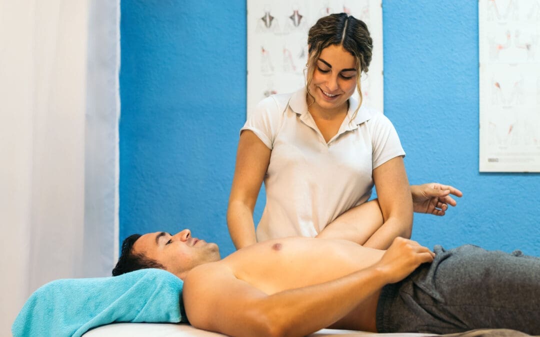
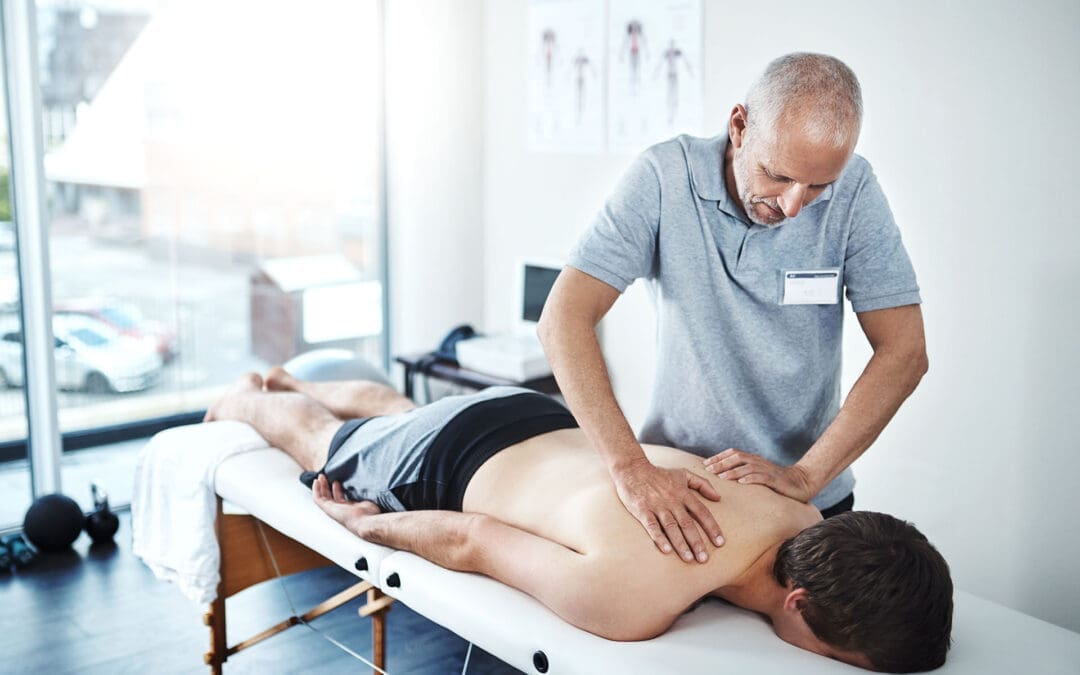
How Massage Therapy Supports El Paso Back Clinic’s Integrative Back & Injury Care
Massage therapy is far more than relaxation. In places like El Paso Back Clinic, it is a central part of healing after injury, especially when combined with advanced diagnostics, chiropractic care, and functional medicine. This article explains how therapists are trained to use proper body mechanics and a range of techniques to deliver variable pressure safely and effectively—and how that fits specifically within the services and philosophy at El Paso Back Clinic.
El Paso Back Clinic: Philosophy & Local Context
El Paso Back Clinic is led by Dr. Alexander Jimenez, DC, APRN, FNP-BC, CFMP, IFMCP. The clinic offers injury care, sports wellness, functional medicine, nutritional labs, accident & trauma rehabilitation, and more. Their goal is not only to reduce pain but also to restore function, improve long-term health, and empower patients. (El Paso Back Clinic® • 915-850-0900)
Given El Paso’s climate, traffic patterns, high incidence of work and motor vehicle injuries, and populations often facing musculoskeletal stresses (from physical labor, commute, environmental heat), having a clinic that combines hands-on care (like massage and chiropractic) plus diagnostics, nutrition, and rehabilitation gives patients a more complete path to recovery.
How Massage Therapists Are Trained at El Paso Back Clinic
At El Paso Back Clinic, massage therapists (or hands-on therapists) receive training in:
Anatomy & injury types: Understanding soft tissue, fascia, muscles, ligaments, joint mechanics, nerve irritation, and the healing stages after trauma.
Techniques with varying pressure levels include Swedish massage for light pressure, myofascial release, trigger point work, deep tissue strokes, and sports massage. The therapist must know when to adjust pressure and technique based on the patient’s needs.
They also learn body mechanics, which includes:
Using stable positions (such as lunges, with aligned shoulders and hips, and a stable base) to deliver pressure using body weight instead of relying purely on arm strength.
Keeping joints aligned to avoid strain: wrists, elbows, shoulders, and hips.
Engaging core muscles and using forearms or elbows when deeper pressure is needed, rather than overusing small muscles or risking repetitive strain injuries.
These practices help ensure therapists can deliver light, medium, or very deep pressure safely and consistently.
Variable Pressure: Light, Medium, and Deep
One of the strengths of El Paso Back Clinic is tailoring the pressure to the patient’s condition. Key considerations:
Stage of injury
Immediately after injury (e.g., whiplash, auto collision, work accident), there is often swelling, sensitivity, nerve irritation, or inflammation. Therapists start with lighter pressure to ease muscle guarding and improve circulation without causing further trauma.
As healing progresses, they gradually increase to medium or deeper pressure to break down adhesions, improve tissue mobility, release trigger points, and facilitate proper alignment.
Patient feedback
Therapists continually check with the patient (pain levels, comfort, tolerance). If pressure hurts more than helps, it is adjusted.
The use of pain or discomfort scales, or sometimes comparison between sides, helps map out what level of pressure works.
Treatment goals
For relaxation, circulation, or early healing: lighter pressure;
For chronic tightness, scar tissue, longer-term dysfunction: deeper work;
For preparing for chiropractic adjustments or rehabilitation exercises, pressure is sufficient to loosen soft tissue tension without aggravation.
Tools & technique
Use of elbows, forearms, or specialized tools when deeper pressure is needed so the therapist avoids wearing out hands and joints.
Sustained pressure (holding a spot) vs. lighter strokes; slow increments rather than sudden, strong force.
How Massage Fits into El Paso Back Clinic’s Injury & Rehabilitation Protocols
El Paso Back Clinic integrates massage therapy into its broader care model, which includes:
Chiropractic adjustments: After a massage relaxes tight muscles and soft tissue, chiropractic manipulation or spinal adjustments can be more effective because tissues are less resistant and joints can move more freely.
Diagnostic imaging & functional assessments: Before and during treatment, the clinic utilizes imaging (X-ray, MRI if necessary), laboratory and blood studies, functional movement assessments, and neurological examinations. These help identify which tissues to treat, where deeper pressure might be risky, and how far to push therapy. (El Paso Back Clinic® • 915-850-0900)
Functional Medicine & Nutrition: Pain, inflammation, and healing are influenced not only by what happens at the injury site but also by systemic factors, including nutrition, inflammation, metabolic health, sleep, and stress. The clinic evaluates these and includes them in plans, so massage and chiropractic care are supported from the inside. (El Paso Back Clinic® • 915-850-0900)
Rehabilitation & Movement Training: A range of exercises, including range of motion, strength training, posture correction, flexibility, and agility work, all help maintain gains from therapy and prevent re-injury. Massage reduces muscle tightness and improves mobility, which makes rehab exercises more effective.
Auto Injury / Trauma / Legal Documentation: For patients with motor vehicle accidents, whiplash, or other collision injuries, the clinic documents condition (soft tissues, alignment, neurologic signs), imaging findings, treatment plans, responses to massage, and other modalities. This documentation is essential to support insurance or legal claims. (El Paso Back Clinic® • 915-850-0900)
Clinical Observations & Outcomes at El Paso Back Clinic
From Dr. Jimenez’s experience and the clinic’s outcomes:
Patients who start hands-on therapy (massage) early, combined with chiropractic and functional medicine, often show quicker reduction in pain and better range of motion.
Deep pressure techniques are only introduced when imaging or assessment indicates it is safe (i.e., no unresolved inflammation, no acute nerve compression).
Many patients report better sleep, less muscle soreness, improved posture, and fewer flare-ups when massage is integrated regularly rather than used only in emergency phases.
Use of body mechanics in massage therapy helps therapists avoid fatigue and maintain consistency over a full course of care, which helps patient outcomes remain steady.
Safety, Communication, & Patient Empowerment
Safety is a big priority. The clinic ensures that:
Therapists communicate: asking about pressure, pain, any aggravations, or sensitivities.
Pressure is adjusted immediately if something doesn’t feel right.
Therapists use posture, leverage, and tools properly — so patients are treated safely and therapists avoid injury.
Patients are educated on self-care, stretches, ergonomics, and posture to sustain the benefits of therapy.
Conclusion
El Paso Back Clinic uses massage therapy not as an add-on, but as a vital part of an integrative, evidence-based healing pathway. Through professional training, variable pressure techniques, good body mechanics, diagnostics, chiropractic care, functional medicine, and legal documentation, the clinic offers patients in El Paso a full spectrum of recovery—not just temporary pain relief, but restored function, strength, and long-term wellness.
If you are recovering from a back injury, auto accident, work or sports trauma, or chronic pain, El Paso Back Clinic’s model may be what helps you return to normal life safely and fully.
Genetics, Stiffness, and Flexibility: Understanding the Back’s Natural Limits
Introduction: Why Flexibility Matters for Spinal Health
Flexibility is often thought of as a skill we can train, like strength or endurance. But in reality, flexibility begins with genetics. Some people are born naturally limber, while others experience tightness in their muscles and connective tissues no matter how much they stretch. This is not always a problem—it is a normal variation in human biology.
At the El Paso Back Clinic, under the care of Dr. Alexander Jimenez, DC, APRN, FNP-BC, patients learn that stiffness has many causes: genetics, aging, lifestyle habits, and sometimes injuries. Through chiropractic adjustments, advanced imaging, and integrative care, the clinic helps individuals restore mobility, manage stiffness, and prevent long-term complications.
How Genetics Shapes Flexibility
Collagen and Connective Tissue
Ligaments, tendons, and fascia are made from collagen. Some people are genetically predisposed to tighter collagen, while others inherit looser connective tissues that allow more joint motion (Xcode Life, n.d.).
Muscle Fiber Balance
Fast-twitch fibers create power but are less flexible, while slow-twitch fibers support endurance and mobility. Genetics dictates the proportion of these fibers in each person’s body (PMC, 2020).
Genetic Syndromes and Flexibility Extremes
Ehlers-Danlos Syndrome (EDS): Causes extreme flexibility from connective tissue fragility.
Inherited Stiffness Disorders: Families can pass down congenital stiffness across generations (JAMA Pediatrics, 2000).
These differences show why two people can do the same stretching routine but achieve very different results.
Stiffness as a Normal Range of Human Variation
Not every stiff person has a medical problem. Many people naturally sit at the less flexible end of the spectrum, which is completely normal (Quora, n.d.).
Alexander Orthopaedics (2023) reports that gender, bone shape, and joint design also influence how flexible someone can be. For example, women tend to have greater flexibility in certain joints due to hormonal differences, while men often have more rigid tissue structures.
At El Paso Back Clinic, Dr. Jimenez helps patients understand that stiffness does not always mean something is wrong—but it can increase the risk of injury if not properly managed.
When Stiffness Becomes a Medical Concern
Stiff Person Syndrome (SPS)
SPS is a rare autoimmune condition that leads to severe rigidity, spasms, and difficulty walking. It is distinct from natural stiffness and requires medical treatment (Hopkins Medicine, n.d.; MSU Healthcare, 2024).
Genetic Disorders of Rigidity
Some families inherit congenital disorders that lock joints into restricted motion. These are uncommon but important to recognize in clinical settings (JAMA Pediatrics, 2000).
Injury-Related Stiffness
Motor vehicle accidents (MVAs), workplace injuries, and sports trauma can cause scar tissue, joint misalignment, and muscle guarding that worsen stiffness. Dr. Jimenez frequently sees these cases at El Paso Back Clinic.
Aging, Lifestyle, and the Stiff Back
Age-Related Tissue Changes
Over time, collagen stiffens, cartilage thins, and joint capsules lose elasticity. Even flexible individuals in youth often report stiffness as they age (PMC, 2020).
Lifestyle Habits
Sedentary behavior shortens connective tissue.
Repetitive work tasks create uneven strain.
Lack of stretching allows muscles to tighten.
At El Paso Back Clinic, patients often present with stiffness that is a combination of genetic and lifestyle factors, requiring a tailored treatment plan.
Case Studies from El Paso Back Clinic
Case 1: Lifelong Stiffness Meets Injury
A 45-year-old man reported lifelong tightness, which worsened after an MVA. Imaging revealed whiplash compounded by rigid connective tissue. With chiropractic adjustments, massage, and guided rehab, he restored safe mobility while respecting his natural limits.
Case 2: Athletic Stiffness and Performance
A 20-year-old track athlete experienced poor hamstring flexibility, which led to recurring strains. Rather than forcing an extreme range of motion, Dr. Jimenez built a plan focusing on functional mobility, hip stability, and performance-specific conditioning.
Case 3: Sedentary Aging and Stiff Joints
A 68-year-old office worker complained of chronic back stiffness. With chiropractic care, acupuncture, and mobility training, stiffness eased enough to improve daily activities and quality of life.
Chiropractic and Integrative Solutions for Stiffness
At El Paso Back Clinic, treatment is not one-size-fits-all. Dr. Jimenez employs an integrative approach that combines chiropractic with medical and functional strategies:
Chiropractic Adjustments: Correct spinal alignment and improve motion.
Massage Therapy: Loosens tight fascia and muscles.
Acupuncture: Reduces spasms and supports nervous system balance.
Targeted Exercise: Builds mobility without overstretching joints.
Functional Medicine: Focuses on diet, inflammation, and tissue repair.
By blending these treatments, patients can improve mobility and manage stiffness effectively.
Sports, Flexibility, and Injury Prevention
Flexibility influences athletic performance—but both extremes have risks.
Too flexible: Joints may lack stability.
Too stiff: Risk of muscle strain or joint injury.
Dr. Jimenez helps athletes at El Paso Back Clinic find their optimal flexibility zone. This may mean increasing mobility in some cases or focusing on stability and strength in others.
Legal and Diagnostic Support in Personal Injury Cases
One unique aspect of Dr. Jimenez’s work is his dual-scope role in both chiropractic and medical diagnosis. For personal injury cases, this includes:
Advanced imaging (MRI, CT, X-ray)
Dual medical and chiropractic reports
Coordination with attorneys and insurers
Documentation of stiffness-related limitations
This ensures patients receive not only effective treatment but also the proper legal support for compensation and care continuity.
Lifestyle Practices to Support Mobility
While genetics can’t be changed, lifestyle makes a difference:
Daily Stretching for sustained tissue pliability.
Hydration to keep connective tissues healthy.
Balanced Nutrition to reduce inflammation and support collagen.
Regular Movement to prevent stiffness from inactivity.
Mind-Body Exercise, such as yoga or tai chi.
El Paso Back Clinic encourages patients to adopt these habits alongside clinical care.
Conclusion: Living Well with Natural Stiffness
Some people are naturally stiff. Others are naturally flexible. Both variations are normal, shaped by genetics, age, and lifestyle. What matters is managing stiffness in ways that prevent injury, restore comfort, and support long-term health.
At El Paso Back Clinic, Dr. Alexander Jimenez uses his dual expertise to evaluate stiffness, provide integrative treatment, and guide patients toward healthier mobility—whether recovering from injury, aging with stiffness, or simply working within genetic limits.
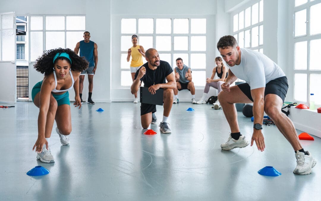
Sport-Specific Chiropractic Care at El Paso Back Clinic: Building Strength, Restoring Balance, and Preventing Injuries
Moving Beyond One-Size-Fits-All Training
Every athlete faces unique physical demands. A sprinter’s body requires explosive power, while a baseball pitcher depends on shoulder rotation and stability. At El Paso Back Clinic, we emphasize that performance training should reflect those realities. Sport-specific chiropractic care addresses both the physical and neurological patterns behind athletic success, offering customized recovery and prevention strategies instead of generalized routines (Trainerize, n.d.; Seaver College, n.d.).
This approach enhances coordination, balance, and endurance by targeting the specific movements athletes use in their sport. More importantly, it builds resilience—protecting the musculoskeletal system against injuries that can derail progress (Physio-Pedia, n.d.).
Chiropractic Care as a Performance Tool
Chiropractic adjustments are often seen as a way to ease back or neck pain, but in athletics, they play a far greater role. Spinal and joint alignment improves nervous system efficiency, helping muscles fire correctly during sport-specific actions (Nansledan Chiropractic, n.d.).
At El Paso Back Clinic, chiropractic care goes hand-in-hand with soft tissue therapies, mobility exercises, and recovery strategies. Athletes benefit from:
This integrative approach makes chiropractic care a cornerstone for both rehabilitation and peak performance.
From Pain to Play: How Athletes Heal
Injury recovery is never just about repairing one area of the body; it’s about restoring overall function. At El Paso Back Clinic, we use chiropractic integrative care to restore overall function. For example, an athlete recovering from an ACL injury might receive adjustments for pelvic alignment alongside agility drills to re-train proper knee mechanics (Jag PT, n.d.).
Our recovery process follows clear steps:
Pain and Inflammation Control – through chiropractic adjustments, soft tissue work, and supportive therapies.
Strength and Mobility Restoration – using targeted, sport-specific rehabilitation exercises (HQPT, n.d.).
Neuromuscular Re-education – training the nervous system to move efficiently and avoid re-injury (ECU Research Online, n.d.).
Return-to-Sport Readiness – functional assessments ensure athletes are prepared for real-world demands (Marygrove Mustangs, n.d.).
By integrating rehabilitation and chiropractic strategies, athletes heal faster and more safely while regaining confidence on the field.
The El Paso Back Clinic Advantage: Integrative, Collaborative Care
What sets El Paso Back Clinic apart is the collaborative nature of care. Our providers don’t just focus on short-term relief; they build long-term health through a mix of therapies:
Chiropractic Adjustments – for alignment, pain reduction, and improved function
Acupuncture – reducing inflammation and supporting natural recovery
Nutritional Guidance – Promoting Anti-Inflammatory Eating to Accelerate Healing (Avance Care, n.d.)
Performance Training – customized sport-specific drills that build functional strength (Prevent PT, 2023)
Together, these therapies ensure athletes receive complete care that supports both the body and mind.
Prevention: Protecting Athletes Before Injuries Happen
Many injuries develop gradually, often due to poor posture, muscular imbalances, or repetitive stress. Regular chiropractic evaluations allow us to detect and correct these issues before they become painful setbacks (Hyperhealth, n.d.).
For instance, a runner may develop pelvic instability that, if untreated, leads to knee pain. At El Paso Back Clinic, chiropractic adjustments stabilize the pelvis while sport-specific training reinforces single-leg balance and stride efficiency. This proactive approach not only prevents injuries but also enhances performance (Essential Chiropractic, n.d.).
Legal and Medical Support for Injury Cases
Beyond athletics, El Paso Back Clinic also supports patients recovering from motor vehicle accidents, workplace injuries, and personal injury cases. Our providers deliver:
Thorough diagnostics using imaging and functional testing
Comprehensive injury documentation for legal cases
Collaborative care plans that integrate chiropractic treatment, exercise therapy, and functional medicine (Perrone Wellness, n.d.; RxWellness, n.d.)
By combining advanced care with precise documentation, we help patients heal physically while supporting them through legal processes that often follow accidents.
The Lasting Benefits of Sport-Specific Chiropractic Care
Athletes who embrace an integrated model of chiropractic care and tailored training experience benefit that extend well beyond the field. They gain:
Enhanced performance through better biomechanics
Faster and more complete recovery after injuries
Greater resilience against future injuries
A foundation for long-term musculoskeletal health
At El Paso Back Clinic, our mission is simple: to keep athletes, workers, and accident patients moving safely, confidently, and at their highest potential.
Discover how exercises and chiropractic care can help relieve muscle pain and enhance your physical health significantly.
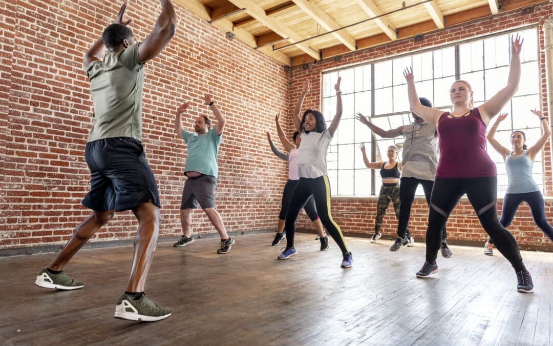
Chiropractic Care and Exercise: A Winning Team for Musculoskeletal Pain Relief
Musculoskeletal pain can creep into your life like an uninvited guest who overstays their welcome, turning simple tasks like climbing stairs or carrying groceries into a battle. Whether it’s a stiff neck from a minor car accident, a sore back from too many hours at a desk, or achy joints that make you feel like you’re moving in slow motion, these issues can really put a damper on your day. The good news? Combining chiropractic care with exercises like CrossFit and weight training is like assembling a superhero squad to tackle pain, boost mobility, and get you back to living your best life. At El Paso Back Clinic, Dr. Alexander Jimenez, DC, APRN, FNP-BC, leads the charge with a holistic approach that blends spinal adjustments, advanced diagnostics, and personalized exercise plans. In this comprehensive, SEO-optimized guide (clocking in at over 5,000 words!), we’ll explore why this dynamic duo works, how environmental factors and chronic pain-like conditions contribute to musculoskeletal issues, and how small lifestyle changes can make a big impact. We’ll also highlight Dr. Jimenez’s unique expertise in personal injury cases in El Paso, where he bridges the gap between medical care and legal support. So, grab a comfy seat (preferably not that creaky office chair), and let’s dive into the world of pain relief with a sprinkle of humor to keep things light!
The Musculoskeletal System: Your Body’s Support Structure
The musculoskeletal system is like the framework of a house—it keeps everything standing tall, supports movement, and lets you do everything from dancing at a wedding to lifting a heavy box. It includes bones, muscles, joints, ligaments, and tendons, all working together like a well-oiled machine. When something goes wrong, like a misaligned spine or a strained muscle, it’s like a squeaky door hinge—annoying at best, debilitating at worst.
What Causes Musculoskeletal Pain?
Musculoskeletal pain can come from a variety of sources, often tied to environmental factors or daily habits. Here are some common culprits:
Sedentary Lifestyles: Sitting for hours at a desk or on the couch can weaken core muscles, leading to back and neck pain. Research shows that prolonged sitting is a major risk factor for musculoskeletal issues (Dunstan et al., 2018).
Motor Vehicle Accidents (MVAs): El Paso’s busy roads, especially during rainy weather, increase the risk of accidents that cause whiplash, spinal misalignments, or soft tissue injuries (El Paso Back Clinic, 2025a).
Repetitive Motions: Jobs or hobbies involving repetitive tasks, like typing or cycling, can strain muscles and joints, leading to conditions like carpal tunnel syndrome or tendonitis.
Poor Ergonomics: A poorly set-up workstation can cause neck strain, shoulder pain, or lower back discomfort, especially for remote workers.
Chronic Pain-Like Conditions: Conditions such as fibromyalgia or arthritis can exacerbate musculoskeletal pain by increasing inflammation and altering pain perception (American Chronic Pain Association, 2023).
The Impact of Chronic Pain-Like Symptoms
Chronic pain syndromes, such as fibromyalgia, arthritis, or neuropathic pain, act like a glitch in your body’s operating system, making musculoskeletal pain harder to manage. These conditions can cause joint stiffness, muscle tension, and increased inflammation, creating a vicious cycle of discomfort. Environmental factors, like stress from a demanding job or poor sleep from pain, can make things worse. Breaking this cycle requires a multi-faceted approach, combining chiropractic care, exercise, and lifestyle tweaks.
References:
Dunstan, D. W., et al. (2018). Sedentary behavior and musculoskeletal health. Journal of Physical Activity and Health, 15(10), 767-775. https://doi.org/10.1123/jpah.2017-0403
El Paso Back Clinic. (2025a). Spinal health after MVAs and chiropractic care. Retrieved from https://elpasobackclinic.com/
American Chronic Pain Association. (2023). Understanding chronic pain syndromes. Retrieved from https://www.theacpa.org/
Chiropractic Care: The Foundation of Pain Relief
Chiropractic care is like a tune-up for your body’s framework, fixing misalignments and restoring balance. At El Paso Back Clinic, Dr. Alexander Jimenez and his team use spinal adjustments, soft tissue therapy, and advanced diagnostics to target the root causes of musculoskeletal pain. By focusing on alignment and function, chiropractic care helps reduce pain, improve mobility, and support overall wellness.
How Chiropractic Care Works
Spinal Adjustments: Misaligned vertebrae can pinch nerves or strain muscles, causing pain. Adjustments gently realign the spine, relieving pressure and improving function (El Paso Back Clinic, 2025b).
Soft Tissue Therapy: Techniques such as trigger point therapy and massage reduce muscle tension and inflammation, providing relief for conditions like sciatica or post-accident injuries (Ojeda et al., 2023).
Holistic Approach: Chiropractic care integrates nutrition, exercise, and lifestyle changes to promote long-term recovery, addressing both symptoms and underlying causes.
Dr. Jimenez’s expertise shines in personal injury cases, where he uses advanced imaging (X-rays, MRIs) and diagnostic tools like the Timed Up and Go (TUG) test to assess injuries (Podsiadlo & Richardson, 1991). His dual-scope approach—combining clinical care with legal documentation—makes him a trusted liaison for accident victims in El Paso, ensuring they receive both medical relief and support for insurance or legal claims.
References:
El Paso Back Clinic. (2025b). Chiropractic care benefits for musculoskeletal inflammation. Retrieved from https://elpasobackclinic.com/
Ojeda, B. H., et al. (2023). Cost-effectiveness and outcomes of direct access to physical therapy for musculoskeletal disorders compared to physician-first access in the United States: Systematic review and meta-analysis. Physical Therapy Journal, 103(2), pzad012. https://pubmed.ncbi.nlm.nih.gov/
Podsiadlo, D., & Richardson, S. (1991). The timed “Up & Go”: A test of basic functional mobility for frail elderly persons. Journal of the American Geriatrics Society, 39(2), 142-148. https://pubmed.ncbi.nlm.nih.gov/
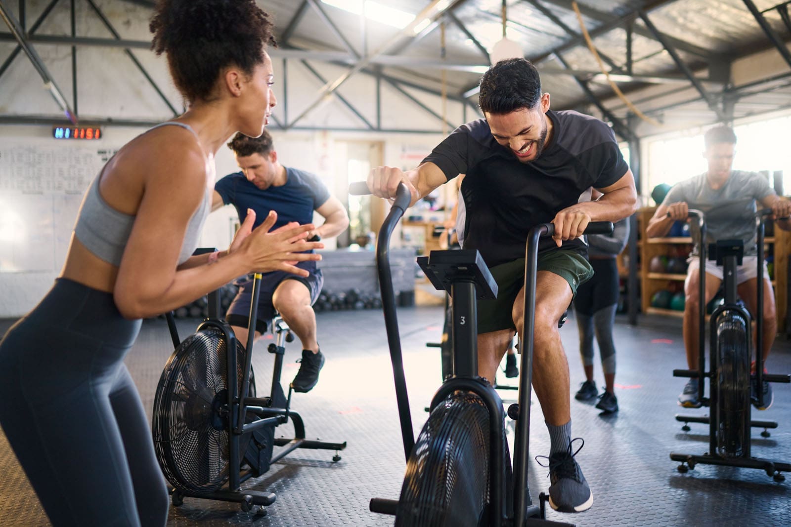
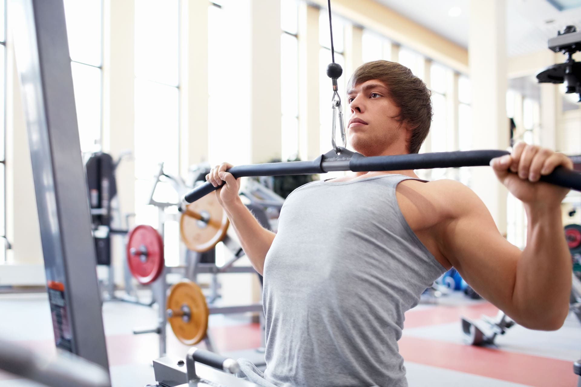
CrossFit and Weight Training: Building Strength, Banishing Pain
If chiropractic care is the architect responsible for aligning your body, CrossFit and weight training are the construction crew that builds strength and resilience. These exercises target specific muscle groups, improve joint stability, and boost overall fitness, making them perfect partners for chiropractic care. According to Muscle & Strength, both CrossFit and weight training offer unique benefits for musculoskeletal health; however, they are most effective when tailored to individual needs (Muscle & Strength, 2023).
CrossFit: Functional Fitness with a Kick
CrossFit is like the energetic friend who convinces you to try something new, like swinging a kettlebell or doing a burpee. It combines high-intensity interval training (HIIT), weightlifting, and bodyweight exercises to build strength, flexibility, and endurance. Key CrossFit exercises include:
Air Squats: Strengthen the quadriceps, hamstrings, and glutes, supporting lower back stability and reducing pain from poor posture.
Deadlifts: Engage your core, back, and legs to improve spinal alignment and alleviate lower back pain.
Kettlebell Swings: Boost hip mobility and strengthen the posterior chain (hamstrings, glutes, lower back), helping with sciatica or post-MVA recovery.
Pull-Ups: Target the upper back and shoulders to reduce neck tension and improve posture.
CrossFit’s focus on functional movements mimics real-life activities, making it an ideal choice for rehabilitating injuries sustained in accidents or sports. A study on isometric exercises found that they significantly reduce pain and improve muscle activity in patients with low back pain (Alayat et al., 2018).
Weight Training: Steady Strength for Stability
Weight training is like the reliable friend who shows up with a toolbox, ready to build something solid. It uses progressive resistance to strengthen specific muscle groups, protecting joints and reducing pain. Key exercises include:
Bench Press: Strengthens the chest, shoulders, and triceps, supporting upper body stability and reducing shoulder pain.
Leg Press: Targets the quadriceps, hamstrings, and calves, easing knee and hip discomfort.
Seated Rows: Strengthen the upper back, improving posture and reducing neck strain.
Plank Variations: Engage the core, stabilizing the spine and preventing lower back pain.
Weight training builds muscle mass, which acts like a natural brace for joints, reducing stress on areas affected by arthritis or injury (Muscle & Strength, 2023).
Alayat, M. S., et al. (2018). The Effects of Isometric Exercise Types on Pain and Muscle Activity in Patients with Low Back Pain. Journal of Physical Therapy Science, 30(8), 1081-1086. https://pubmed.ncbi.nlm.nih.gov/
The Synergy of Chiropractic Care and Exercise
Chiropractic care and exercise are like peanut butter and jelly—great on their own, but unstoppable together. Chiropractic adjustments align the spine and joints, while exercises like CrossFit and weight training strengthen muscles and improve mobility. Here’s how they team up:
Muscle Balance: CrossFit’s dynamic movements and targeted weight training exercises strengthen opposing muscle groups, preventing imbalances that can cause pain.
Improved Circulation: Exercise enhances blood flow, delivering essential nutrients to injured tissues and accelerating recovery after chiropractic adjustments.
Enhanced Flexibility: CrossFit’s functional movements and weight training’s stretching components improve joint range of motion, complementing spinal adjustments.
Dr. Jimenez emphasizes small, sustainable changes to maintain progress, like adding a 10-minute walk or core exercises to your daily routine (Jimenez, 2025). Think of it as giving your spine a daily high-five to keep it happy!
Managing Musculoskeletal Pain in Chronic Pain-Like Conditions
Chronic pain syndromes, like fibromyalgia or arthritis, are like that one friend who always complicates plans—they make musculoskeletal pain harder to manage. These conditions can cause inflammation, muscle stiffness, and nerve irritation, creating a cycle of discomfort. Chiropractic care, exercise, and nutrition can break this cycle and reduce overlapping risk profiles.
Chiropractic Strategies for Chronic Pain
Spinal Adjustments: Relieve nerve compression, reduce pain signals, and improve mobility (El Paso Back Clinic, 2025c).
Soft Tissue Therapy: Targets muscle knots and tension, easing inflammation and promoting relaxation (Ojeda et al., 2023).
Nutritional Support: Anti-inflammatory diets rich in omega-3s, leafy greens, and antioxidants reduce systemic inflammation, thereby supporting chiropractic and exercise efforts (El Paso Back Clinic, 2025d).
Exercise for Chronic Pain Management
Modified CrossFit: Low-impact movements, like resistance band exercises or bodyweight squats, build strength without overloading joints.
Weight Training: Light resistance exercises, such as dumbbell curls or leg extensions, improve muscle support and reduce pain.
Backward Walking: Research shows that retro walking reduces pain and improves function in conditions like knee osteoarthritis, which often accompanies chronic pain syndromes (Alghadir et al., 2019).
These strategies address inflammation and improve function, tackling the complex interplay of chronic pain and musculoskeletal issues.
References:
El Paso Back Clinic. (2025c). Trigger point therapy MVAs explained for patients. Retrieved from https://elpasobackclinic.com/
El Paso Back Clinic. (2025d). Nutrition for accident injuries during recovery. Retrieved from https://elpasobackclinic.com/
Alghadir, A. H., et al. (2019). Effect of 6-week retro or forward walking program on pain, functional disability, quadriceps muscle strength, and performance in individuals with knee osteoarthritis: A randomized controlled trial. Clinical Rehabilitation, 33(6), 1041-1049. https://pubmed.ncbi.nlm.nih.gov/
Personal Injury Cases in El Paso: Dr. Jimenez’s Expertise
El Paso’s bustling roads, from I-10 to Loop 375, see their fair share of accidents, from minor fender benders to serious 18-wheeler crashes. These incidents often cause musculoskeletal injuries, like whiplash-associated disorders (WAD) or soft tissue damage. Dr. Alexander Jimenez stands out as a leading practitioner for personal injury victims, offering a unique blend of clinical expertise and legal support.
Dr. Jimenez’s Approach to Personal Injury
Advanced Imaging: X-rays, MRIs, and CT scans pinpoint injuries like spinal misalignments or muscle tears with precision (El Paso Back Clinic, 2025e).
Diagnostic evaluations, such as the 6-minute walk test or TUG test, assess mobility and inform treatment plans (Nguyen et al., 2020; Podsiadlo & Richardson, 1991).
Dual-Scope Expertise: Dr. Jimenez bridges the gap between medical care and legal documentation, providing detailed reports for insurance claims and lawsuits.
Comprehensive Recovery: Combining chiropractic adjustments, massage therapy, and durable medical equipment (like braces or TENS units), he tailors plans to each patient’s needs.
His work is critical for MVA victims, whose injuries can range from mild sprains to severe spinal trauma. By addressing both physical and legal needs, Dr. Jimenez helps patients navigate recovery with confidence.
References:
El Paso Back Clinic. (2025e). Auto accident insights for safe driving and recovering from WAD. Retrieved from https://elpasobackclinic.com/
Nguyen, U. S., et al. (2020). Clinical Associations and Prognostic Implications of the 6-Minute Walk Test in Rheumatoid Arthritis. Arthritis Care & Research, 72(8), 1046-1053. https://pubmed.ncbi.nlm.nih.gov/
Podsiadlo, D., & Richardson, S. (1991). The timed “Up & Go”: A test of basic functional mobility for frail elderly persons. Journal of the American Geriatrics Society, 39(2), 142-148. https://pubmed.ncbi.nlm.nih.gov/
Practical Tips for Integrating Chiropractic Care and Exercise
Ready to kick musculoskeletal pain to the curb? Here are some practical tips, inspired by Dr. Jimenez’s clinical insights, to blend chiropractic care and exercise into your routine:
Start Small: Begin with low-impact exercises, like a 15-minute walk or bodyweight squats, to build strength without strain.
Schedule Adjustments: Visit El Paso Back Clinic for regular chiropractic sessions to keep your spine aligned and pain-free.
Mix and Match: Combine CrossFit’s dynamic movements with weight training’s focused exercises for a balanced workout plan.
Listen to Your Body: If an exercise causes discomfort, stop and consult Dr. Jimenez’s team for modifications.
Stay Consistent: Small changes, such as stretching before bed or incorporating anti-inflammatory foods into your diet, add up over time.
A little humor to keep you motivated: Treat your spine like a picky cat—give it regular attention, and it’ll purr with happiness!
The combination of chiropractic care and exercise isn’t just a feel-good strategy—it’s backed by science. Chiropractic adjustments reduce nerve irritation and improve joint function, while exercise strengthens muscles and boosts circulation. A single bout of exercise can reduce fatigue and improve energy levels, enhancing the effects of chiropractic care (Puglisi et al., 2013). For chronic pain patients, exercise also improves pain tolerance and reduces inflammation, addressing the root causes of musculoskeletal discomfort.
Research also supports the use of specific exercises for pain relief. For example, isometric exercises, like planks, reduce pain and improve muscle activity in patients with low back pain (Alayat et al., 2018). Similarly, backward walking has been shown to improve function and reduce pain in knee osteoarthritis, a common issue in chronic pain syndromes (Alghadir et al., 2019). By combining these exercises with chiropractic care, patients can achieve lasting relief and improved function.
References:
Puglisi, F., et al. (2013). The effect of a single bout of exercise on energy and fatigue states: A systematic review and meta-analysis. Fatigue: Biomedicine, Health & Behavior, 1(4), 223-242. https://pubmed.ncbi.nlm.nih.gov/
Alayat, M. S., et al. (2018). The Effects of Isometric Exercise Types on Pain and Muscle Activity in Patients with Low Back Pain. Journal of Physical Therapy Science, 30(8), 1081-1086. https://pubmed.ncbi.nlm.nih.gov/
Alghadir, A. H., et al. (2019). Effect of 6-week retro or forward walking program on pain, functional disability, quadriceps muscle strength, and performance in individuals with knee osteoarthritis: A randomized controlled trial. Clinical Rehabilitation, 33(6), 1041-1049. https://pubmed.ncbi.nlm.nih.gov/
Addressing Environmental Factors for Long-Term Relief
Environmental factors, like sedentary lifestyles or poor ergonomics, can exacerbate musculoskeletal pain, but they’re not unbeatable. Here are some strategies to counteract them:
Combat Sedentary Behavior: Break up long periods of sitting with short walks or standing stretches every 30 minutes. Research shows that increasing physical activity in sedentary adults can reduce musculoskeletal pain (Hildebrandt et al., 2017).
Improve Ergonomics: Set up your workspace with an ergonomic chair, monitor at eye level, and keyboard that supports neutral wrist positioning.
Stay Safe on the Road: In El Paso’s rainy weather, drive cautiously to avoid MVAs, which can cause whiplash or spinal injuries (El Paso Back Clinic, 2025a).
Manage Stress: Chronic stress can worsen muscle tension. Try practicing mindfulness or deep breathing exercises to help manage stress.
By addressing these factors, you can create a supportive environment for chiropractic care and exercise to work their magic.
References:
Hildebrandt, V. H., et al. (2017). When Adults Don’t Exercise: Behavioral Strategies to Increase Physical Activity in Sedentary Middle-Aged and Older Adults. Innovation in Aging, 1(1), igx007. https://pubmed.ncbi.nlm.nih.gov/
El Paso Back Clinic. (2025a). Spinal health after MVAs and chiropractic care. Retrieved from https://elpasobackclinic.com/
Dr. Jimenez’s Role in El Paso’s Personal Injury Landscape
El Paso’s roads, from the hustle of I-10 to the curves of Transmountain Road, see a range of accidents, from minor rear-end collisions to severe 18-wheeler crashes. These incidents often lead to musculoskeletal injuries, like whiplash, spinal misalignments, or even gastrointestinal injuries from seatbelt trauma (El Paso Back Clinic, 2025f). Dr. Alexander Jimenez is a beacon of hope for personal injury victims, offering a unique blend of clinical expertise and legal support.
Why Dr. Jimenez Stands Out
Advanced Diagnostics: Utilizing tools such as X-rays, MRIs, and CT scans, Dr. Jimenez precisely identifies injuries, ensuring accurate treatment plans (El Paso Back Clinic, 2025e).
Functional Assessments: Tests like the 6-minute walk test or TUG test evaluate mobility and recovery progress, guiding personalized care (Nguyen et al., 2020; Podsiadlo & Richardson, 1991).
Legal Liaison: Dr. Jimenez’s dual-scope expertise ensures detailed medical reports that support insurance claims or lawsuits, making him a trusted partner for accident victims.
Holistic Recovery: His team combines chiropractic adjustments, massage therapy, and durable medical equipment (such as braces or TENS units) to comprehensively address injuries.
Whether it’s a cyclist recovering from a collision or a driver dealing with whiplash, Dr. Jimenez’s approach ensures patients get back on their feet while navigating the legal complexities of personal injury cases.
References:
El Paso Back Clinic. (2025e). Auto accident insights for safe driving and recovering from WAD. Retrieved from https://elpasobackclinic.com/
El Paso Back Clinic. (2025f). Car accident GI injuries: Recovery and rehabilitation options. Retrieved from https://elpasobackclinic.com/
Nguyen, U. S., et al. (2020). Clinical Associations and Prognostic Implications of the 6-Minute Walk Test in Rheumatoid Arthritis. Arthritis Care & Research, 72(8), 1046-1053. https://pubmed.ncbi.nlm.nih.gov/
Podsiadlo, D., & Richardson, S. (1991). The timed “Up & Go”: A test of basic functional mobility for frail elderly persons. Journal of the American Geriatrics Society, 39(2), 142-148. https://pubmed.ncbi.nlm.nih.gov/
Long-Term Benefits of the Chiropractic-Exercise Combo
The combination of chiropractic care and exercise isn’t just a quick fix—it’s a long-term investment in your health. Here’s why it’s a game-changer:
Pain Reduction: Chiropractic adjustments reduce nerve irritation, while exercise strengthens muscles, preventing pain from recurring (Ojeda et al., 2023).
Improved Mobility: Regular adjustments and exercises like CrossFit or weight training enhance joint range of motion, making daily activities easier.
Injury Prevention: Stronger muscles and better alignment reduce the risk of future injuries, whether from sports, accidents, or daily wear and tear.
Enhanced Well-Being: Exercise boosts energy and reduces fatigue, while chiropractic care promotes relaxation and stress relief (Puglisi et al., 2013).
By sticking with this approach, you’re not just treating pain—you’re building a stronger, healthier body that can handle whatever life throws at you.
References:
Ojeda, B. H., et al. (2023). Cost-effectiveness and outcomes of direct access to physical therapy for musculoskeletal disorders compared to physician-first access in the United States: Systematic review and meta-analysis. Physical Therapy Journal, 103(2), pzad012. https://pubmed.ncbi.nlm.nih.gov/
Puglisi, F., et al. (2013). The effect of a single bout of exercise on energy and fatigue states: A systematic review and meta-analysis. Fatigue: Biomedicine, Health & Behavior, 1(4), 223-242. https://pubmed.ncbi.nlm.nih.gov/
Practical Steps for a Pain-Free Future
Want to say goodbye to musculoskeletal pain and hello to a more active life? Here are some actionable steps, inspired by Dr. Jimenez’s clinical insights, to get you started:
Book a Chiropractic Session: Schedule an appointment at El Paso Back Clinic for a thorough evaluation and personalized treatment plan.
Incorporate Exercise Gradually: Start with low-impact CrossFit or weight training exercises, like air squats or light dumbbell work, to build strength safely.
Optimize Your Environment: Set up an ergonomic workspace, take breaks to stretch, and drive cautiously to avoid accidents.
Eat for Recovery: Focus on anti-inflammatory foods, like salmon, walnuts, and spinach, to support your body’s healing process (El Paso Back Clinic, 2025d).
Stay Consistent: Make small changes, like a daily 10-minute walk or a quick stretch before bed, to maintain progress.
A touch of humor to keep you going: Think of your spine as a grumpy old neighbor—it needs regular check-ins to stay friendly!
References:
El Paso Back Clinic. (2025d). Nutrition for accident injuries during recovery. Retrieved from https://elpasobackclinic.com/
Musculoskeletal pain doesn’t have to be a permanent part of your life. By combining chiropractic care with exercises like CrossFit and weight training, you can reduce pain, improve mobility, and enhance your overall well-being. At El Paso Back Clinic, Dr. Alexander Jimenez and his team offer a holistic approach, utilizing spinal adjustments, advanced diagnostics, and personalized exercise plans to address a range of conditions, from motor vehicle accident injuries to chronic pain. For personal injury victims in El Paso, Dr. Jimenez’s expertise as a clinical and legal liaison ensures comprehensive care and support, helping patients navigate both their recovery and the legal challenges that follow.
Disclaimer: This blog post is intended for informational purposes only and should not be taken as medical advice. Always consult a qualified healthcare provider, such as Dr. Alexander Jimenez at El Paso Back Clinic, before starting any treatment or exercise program. The information provided is based on clinical insights and research, but is not a substitute for professional medical evaluation. Your health is serious business—treat it that way.
IFM's Find A Practitioner tool is the largest referral network in Functional Medicine, created to help patients locate Functional Medicine practitioners anywhere in the world. IFM Certified Practitioners are listed first in the search results, given their extensive education in Functional Medicine