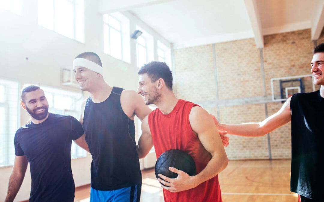
When You Don’t Stretch: Why Muscles Get Stiff, Movement Gets Harder, and Injuries Become More Likely
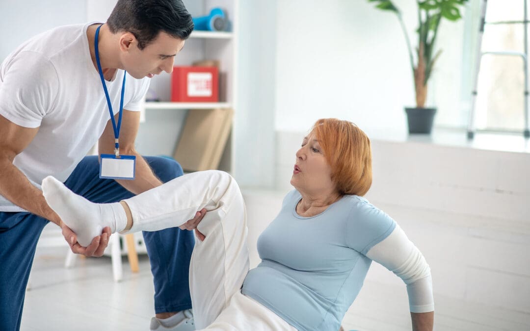
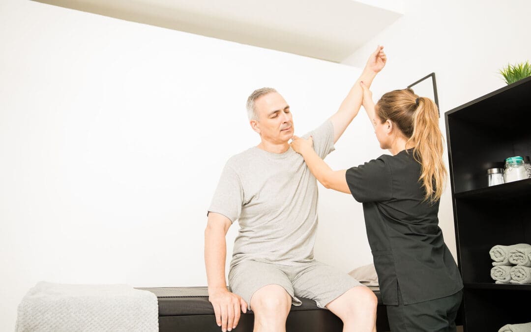
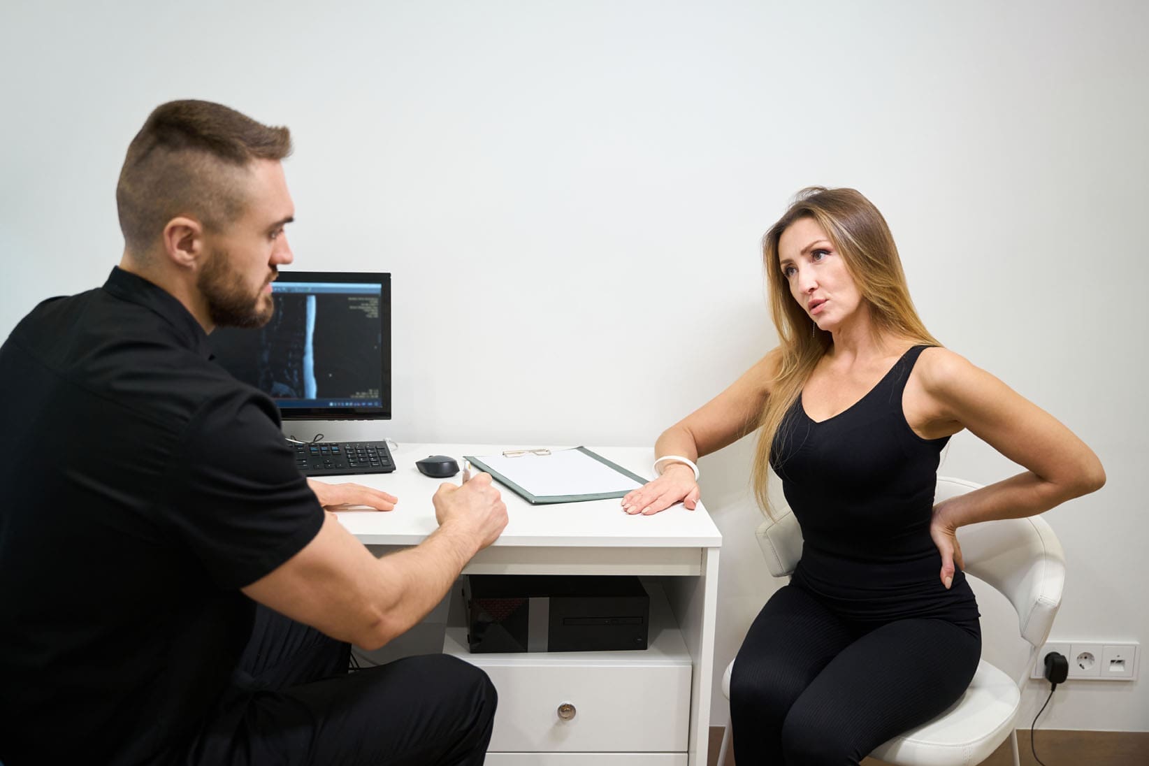
A patient with chronic back pain does targeted stretches.
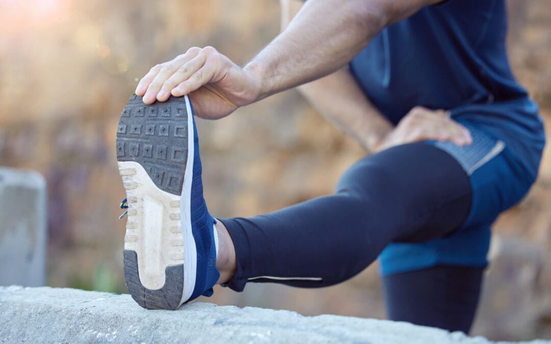
If you rarely stretch, your body can start to feel “tight,” which can change how you move. Many people notice they can’t bend, twist, squat, reach overhead, or turn their head as easily as they used to. Over time, this can affect your flexibility, your range of motion (how far a joint can move), and how smooth and efficient your daily movements feel.
At El Paso Back Clinic, Dr. Alexander Jimenez, DC, APRN, FNP-BC, often explains this: when mobility decreases, the body starts to “compensate.” That means you move around a stiff area instead of through it, and those workarounds can build up stress in nearby joints and muscles (Jimenez, n.d.-a). This is one reason people can develop recurring back pain, neck stiffness, hip tightness, or shoulder irritation even without a single big injury.
What “Muscle Stiffness” Really Means
Muscle stiffness usually feels like tightness, soreness, or difficulty moving. It can happen after overuse, after you’ve been still for a long time, or when your muscles stay “stuck” in a more contracted state (Tarantino, 2025). Osmosis
Osmosis notes that stiffness can appear after a long period of minimal motion (such as bed rest or inactivity) or after new exercise that causes temporary muscle cell damage (Tarantino, 2025). Osmosis
Key idea: When your body doesn’t move a joint through its normal range often enough, the muscles and tissues around it can start to feel restricted. That restriction can make normal tasks think harder than they should.
Do Muscles Actually “Shorten” If You Don’t Stretch?
You’ll hear people say, “If you don’t stretch, your muscles will shorten.” That statement is partly true, but it needs context.
Adidas explains that the word “shorten” can be misleading: for most people, it feels like shortening because mobility and flexibility decrease when stretching is skipped, even if the muscle is not literally shrinking in everyday life (Adidas, 2025). adidas
Harvard Health adds an important clarification: without regular stretching, muscles can become tight, and when you need them for activity, they may not extend fully, increasing the risk of joint pain, strains, and muscle damage (Harvard Health Publishing, 2024). Harvard Health
So the practical takeaway is simple:
Skipping stretching often leads to less mobility and flexibility
Tight muscles can reduce how far joints can move
Tight muscles can make injuries more likely when you suddenly “ask more” of your body
How Tight Muscles Reduce Range of Motion
Range of motion (ROM) is the movement around a joint or body part. When ROM is limited, you can’t move that body part through its usual, healthy motion (Jimenez, n.d.-b). El Paso Back Clinic® • 915-850-0900
El Paso Back Clinic explains how tightness—especially in areas like the hips and ankles—can reduce ROM and limit potential for form and strength. When posture and form are compromised, pain and injury risk can rise (Jimenez, n.d.-b). El Paso Back Clinic® • 915-850-0900
What limited ROM can look like in real life
You might notice:
You can’t turn your head fully when driving
You bend from your lower back instead of your hips
You can’t squat without your heels lifting
Your shoulders feel “pinched” when reaching into a cabinet
Your hamstrings feel tight when you try to walk fast
And here’s the tricky part: your body still gets the job done—just with more strain.
Why Stiffness Can Raise Injury Risk
Harvard Health explains that tight muscles may be more easily damaged when they are suddenly stretched during strenuous activity (Harvard Health Publishing, 2024). Harvard Health
That’s why injuries often show up in moments like:
A weekend game after sitting all week
A sudden sprint to catch something
Lifting a heavy box with “cold” hips and hamstrings
A long drive followed by quick unloading or bending
Mayo Clinic also notes that better flexibility can help joints move through full ROM and may decrease injury risk, while emphasizing that stretching must be done correctly (Mayo Clinic Staff, n.d.). Mayo Clinic
Common Reasons People Stop Stretching (And How to Fix Them)
Most people don’t skip stretching because they don’t care. They skip it because it feels confusing, time-consuming, or uncomfortable.
Common barriers
“I don’t have time.”
“Stretching hurts.”
“I’m not flexible, so it doesn’t work for me.”
“I only need stretching if I work out.”
Better, more realistic reframes
You only need 5–10 minutes a few times a week to start seeing benefits (Mayo Clinic Staff, n.d.). Mayo Clinic
Stretching should create tension, not pain (Mayo Clinic Staff, n.d.). Mayo Clinic
Flexibility improves over weeks to months, not days (Harvard Health Publishing, 2024). Harvard Health
Stretching supports everyday movement, not just workouts (Harvard Health Publishing, 2024). Harvard Health
Safe Stretching Basics (So You Don’t Make Things Worse)
This matters: stretching done poorly can backfire.
Mayo Clinic recommends:
Don’t stretch cold muscles—warm up 5–10 minutes first
Don’t bounce
Hold stretches about 30 seconds (longer for problem areas)
Don’t stretch into pain (Mayo Clinic Staff, n.d.). Mayo Clinic
The American Heart Association adds:
Stretch when muscles are warm
Hold 10–30 seconds and repeat 3–5 times
Stretch slowly and smoothly (American Heart Association, 2024). www.heart.org
Quick safety checklist
Warm up first (easy walk, gentle movement)
Move slowly
Breathe
No bouncing
Stop if you feel sharp pain, numbness, or joint pain
A Simple 10-Minute Daily Stretch Routine for Real Life
This is designed for normal adults: busy schedules, stiff hips, tight neck, and lots of sitting.
Step 1: Warm up (1–2 minutes)
Walk around the house
March in place
Gentle arm circles
Step 2: Do these 6 stretches (about 8 minutes total)
1) Hip flexor stretch (1 minute each side) Helps if you sit a lot and feel tight in the front of your hips.
2) Hamstring stretch (1 minute each side) Harvard points out that tight hamstrings from sitting can limit how well you extend your leg and support walking mechanics (Harvard Health Publishing, 2024). Harvard Health
3) Calf stretch (45 seconds each side) Helpful for ankle mobility, walking, and squatting mechanics.
4) Chest opener (45 seconds) Stand in a doorway and gently open the chest to reduce rounded-shoulder posture.
5) Upper back reach (45 seconds) Hug yourself and gently pull your shoulder blades apart.
6) Neck side stretch (30 seconds each side) Gentle only—never crank your neck.
Step 3: Add “micro-mobility” during your day (optional but powerful)
Stand up every hour for 30–60 seconds
Do 5 bodyweight squats to a chair
Do 10 shoulder rolls
Take a 3-minute walk after meals
These small habits often matter as much as one long stretch session.
Stretching After Workouts: What You Should Know
Adidas explains the difference clearly:
Dynamic movement is best before workouts (prepares your body)
Static stretching is typically better after workouts, when you’re warm (Adidas, 2025). adidas
Mayo Clinic also cautions that stretching cold muscles can increase injury risk and notes that some intense activities may not benefit from heavy stretching right before performance (Mayo Clinic Staff, n.d.). Mayo Clinic
A balanced approach
Before exercise: warm up + dynamic mobility
After exercise: gentle static stretching + breathing
On rest days: short, consistent flexibility routine
When Stiffness Is a Sign You Need More Than Stretching
Sometimes the problem is not just “tight muscles.” You may have:
Joint restrictions that block movement
Spine or pelvis alignment issues affecting mechanics
Inflammation around a joint
Pain patterns that keep muscles “guarded”
A nerve-related problem (numbness, tingling, weakness)
El Paso Back Clinic notes that limited ROM in areas like the back, neck, or shoulders can be linked to the body being out of natural alignment, repetitive motions, or wear and tear (Jimenez, n.d.-b). El Paso Back Clinic® • 915-850-0900
If stretching doesn’t help—or makes symptoms worse—it’s smart to get assessed.
The El Paso Back Clinic Approach: Integrative Chiropractic + Nurse Practitioner Support
This is where integrative care can be a game-changer: you’re not only “stretching more,” you’re also finding out why you’re tight and building a plan that fits your body.
What chiropractic care can add
El Paso Back Clinic describes a “restoration” approach that may include:
Soft tissue work (to reduce tightness and improve circulation)
Adjustments (to address misalignments and support mobility)
Nurse practitioners are advanced practice clinicians who assess, diagnose, and treat illnesses and injuries and support chronic condition management (American Nurses Association, n.d.). ANA Healthgrades also describes NPs performing screenings and physical exams, ordering lab work, documenting care, and diagnosing certain conditions (Prosser, 2025). Healthgrades Resources
Why the combo helps stiffness and pain
Together, a chiropractor + NP team can:
Screen for red flags (nerve symptoms, systemic issues)
Decide when imaging or labs are appropriate
Build a movement plan that matches your pain level
Address sleep, stress, inflammation, and recovery habits
Track progress using measurable goals (like ROM improvements)
Dr. Jimenez’s Mobility & Flexibility materials emphasize that “great mobility” supports functional movement without ROM restrictions and that people who don’t stretch often may experience stiffened muscles that reduce effective movement (Jimenez, n.d.-a). El Paso Back Clinic® • 915-850-0900
Red Flags: When to Stop Stretching and Get Checked
Call a clinician promptly if you have:
Numbness, tingling, or weakness in an arm/leg
Loss of balance, clumsiness, or trouble walking
Severe pain that doesn’t improve
Pain after trauma (car accident, fall, sports collision)
Fever, unexplained swelling, or sudden intense stiffness
Muscle stiffness can sometimes be related to underlying medical issues, and diagnosis may require an exam and follow-up testing, depending on the cause (Tarantino, 2025). Osmosis
The Bottom Line
If you don’t stretch regularly, it’s common to feel tighter and less mobile over time. That stiffness can reduce range of motion, make daily tasks harder, and increase your risk of injury when you suddenly push your body. The good news is that you don’t need extreme flexibility. You need consistent, safe mobility work—and when required, professional support to restore movement and reduce pain.
A practical plan usually includes:
Small daily stretching habits
Better warm-ups and recovery routines
Strength + mobility (not stretching alone)
Integrative evaluation when pain, ROM loss, or repeated flare-ups keep returning
Fast Sports Injury Help Online: How Telemedicine Guides Diagnosis, Rehab, and Return to Play
A massage therapist treats the injury of a professional athlete at El Paso Back Clinic
Telemedicine is changing how athletes get help after an injury. When a chiropractor and a nurse practitioner (NP) work together online, they can guide recovery from many sports injuries without the need for an in-office visit. This is especially helpful for athletes who travel, live far from clinics, or are balancing school, work, family, and training.
In this article, we’ll break down how an integrated chiropractor–NP telemedicine team can:
Do virtual exams from a distance
Share treatment plans and coordinate care
Support at-home rehab, nutrition, and mental health
Help with urgent issues like a possible concussion during games
Reduce unnecessary ER visits while still protecting your safety
1. Why telemedicine matters for sports injuries
Telemedicine is more than a video call. It is a structured way to deliver health care at a distance using secure video, phone, apps, and online tools. Johns Hopkins Medicine notes that telemedicine improves comfort, convenience, and access, especially for people who would otherwise struggle to travel or fit visits into a busy schedule. Hopkins Medicine
For athletes, that matters because:
Practices and games already take up time.
Travel teams may compete hours away from home.
Injuries often happen suddenly—during a weekend tournament, camp, or late-night match.
Telehealth physical therapy and sports services now let athletes receive full evaluations and guided rehab sessions from home, with real-time video coaching. SportsMD+1 Research shows telehealth physical therapy is effective for many orthopedic and sports-related conditions, including non-surgical and post-surgical rehab. PMC
At the same time, sports medicine researchers have shown that telehealth can support concussion care, including baseline testing, diagnosis, and follow-up—especially in rural or resource-limited settings. PMC+1
2. What is an integrated chiropractor + NP telemedicine team?
An integrated team means the chiropractor and nurse practitioner work together instead of in separate silos.
The nurse practitioner (NP) focuses on your overall health, medical history, medications, imaging, and underlying conditions (like asthma, diabetes, or heart issues).
The chiropractor focuses on your spine, joints, muscles, and movement patterns, using guided tests, posture checks, and therapeutic exercises delivered remotely.
In Dr. Alexander Jimenez’s clinical model in El Paso, Texas, the same provider is both a board-certified family nurse practitioner and a chiropractor, which allows one clinician to blend medical and musculoskeletal care through telemedicine for neck pain, low back pain, headaches, and sports injuries. El Paso, TX Doctor Of Chiropractic+2El Paso, TX Doctor Of Chiropractic+2
When the chiropractor and NP are separate providers, they can still share:
Notes and findings in the same electronic health record
Imaging reports and lab results
Exercise programs and rehab goals
Messages with athletic trainers, physical therapists, and coaches
This two-pronged approach helps create one unified plan that covers:
Functional goals (return to sport, position-specific demands)
3. How a virtual sports injury exam works
A telemedicine visit is structured and systematic, not just a quick chat.
3.1 Before the visit
You’ll usually:
Complete an online intake form about symptoms, past injuries, and sport.
Upload any previous X-rays, MRIs, or reports, if available.
Test your camera, microphone, and Wi-Fi connection. SportsMD+1
3.2 During the visit: what the NP does
The nurse practitioner can:
Take a detailed medical history:
How the injury happened
Any prior concussions, surgeries, or chronic conditions
Current medications and allergies
Screen for red flags like chest pain, severe shortness of breath, uncontrolled bleeding, or signs of serious head injury. telehealth.hhs.gov+1
Order diagnostic imaging (X-ray, MRI, CT) if needed.
Write or adjust prescriptions, such as:
Pain medications (when appropriate)
Muscle relaxants
Anti-inflammatory medications
Coordinate referrals to orthopedics, neurology, or emergency care if telemedicine alone is unsafe. OrthoLive+1
3.3 During the visit: what the chiropractor does
Over secure video, the chiropractor can:
Observe posture and alignment (standing, sitting, walking).
Guide you through movement tests, for example:
Bending, rotating, or side-bending the spine
Squats, lunges, or single-leg balance
Shoulder or hip range of motion
Identify pain patterns that suggest sprain, strain, tendinopathy, or joint irritation. sportsandexercise.physio+1
Teach safe at-home movements, such as:
Gentle mobility drills
Core stability exercises
Isometrics to protect healing tissue
In his telemedicine work, Dr. Jimenez describes using these virtual exams to track changes in pain, strength, and mobility from week to week, adjusting exercise progressions and ensuring athletes are not overloading injured tissue. El Paso, TX Doctor Of Chiropractic+1
3.4 Typical flow of a telemedicine sports injury visit
NP and chiropractor (or dual-licensed provider) review your history and goals.
Guided movement and functional tests help narrow down the likely diagnosis.
The NP decides whether imaging or labs are needed.
The chiropractor designs initial movement and pain-reduction strategies.
You leave with a clear home plan and follow-up schedule.
4. Building a shared treatment plan online
After the virtual exam, the team builds a plan that blends medical and musculoskeletal care. Telehealth orthopedic and sports practices report four consistent benefits from this style of care: improved access, reduced costs, better quality and safety, and higher patient satisfaction. OrthoLive
Clear guidelines for when to go to urgent care or ER
Chiropractic and movement actions
Joint and spinal stabilization work
Mobility and flexibility progression
Posture and movement training specific to your sport position
Rehab schedule
How often you meet on video
How many daily or weekly exercises
When to retest speed, strength, or sport-specific skills
Telehealth sports physiotherapy services emphasize that virtual care works best when the athlete receives personalized exercise programs, regular online check-ins, and careful progression from injury to return to play. sportsandexercise.physio+1
5. Conditions that respond well to integrated telemedicine care
Research and real-world practice show that many sports injuries can be evaluated and managed, at least partly, through telemedicine. SportsMD+1
5.1 Common injuries suited for telemedicine
Mild to moderate ankle sprains
Knee pain related to overuse (patellofemoral pain, mild tendinopathy)
Back and neck pain from training load, lifting, or collisions
Mild muscle contusions without signs of fracture
Telehealth physical therapy has shown promise in non-operative and post-operative sports rehab, especially when therapists guide exercise, monitor progress, and adjust programs in real time. PMC+1
5.2 How the NP and chiropractor divide roles
The NP can:
Confirm whether the injury is stable enough for home care.
Check for other health issues (asthma, heart conditions, bleeding disorders).
Manage medications and monitor side effects.
The chiropractor can:
Analyze movement patterns that caused or worsened the injury.
Dr. Jimenez’s clinical work often combines telemedicine visits with in-clinic follow-ups, advanced imaging review, and collaboration with physical therapy and sports training teams to keep athletes progressing without re-injury. El Paso, TX Doctor Of Chiropractic+1
6. Telemedicine and concussion: quick decisions from a distance
Concussions and suspected head injuries are a special case. A missed or delayed diagnosis can put an athlete at serious risk.
A systematic review found that telehealth has been used successfully for concussion baseline testing, diagnosis, and management, especially in military and rural settings. PMC+1 Another review focused on sideline telehealth, where sports medicine physicians assist trainers in real time through video connections during games. PMC+1
SportsMD describes “teleconcussion,” where athletes can quickly access concussion specialists via telehealth instead of waiting days or weeks for in-person care. SportsMD
6.1 How telemedicine helps when you suspect a concussion
During or shortly after a game, a telemedicine visit can help:
Review how the head impact occurred (direct hit, whiplash, fall).
Check acute symptoms, such as:
Headache
Dizziness
Nausea or vomiting
Vision changes
Confusion or memory loss
Guide a brief neurological exam and balance checks via video. PMC+1
Decide whether the athlete must leave the game immediately and seek emergency care.
Telemedicine programs in school sports have also been used to minimize risk by providing teams with rapid access to sports medicine expertise, rather than relying solely on coaches to decide whether a player is safe to continue. NFHS+1
6.2 Role of the integrated team
The NP can determine whether emergency imaging or ER evaluation is needed, arrange teleconcussion follow-ups, and manage symptom-relief medications when appropriate.
The chiropractor can later help with neck pain, posture, and vestibular-related issues—such as balance and coordination problems—once the acute phase is stable and medical clearance is given.
7. At-home rehab and return-to-play through telemedicine
Telehealth lets rehab follow you to your home, hotel room, or training camp.
Telehealth physical therapy programs show several key benefits: increased accessibility, reduced travel burden, and the ability to continue personalized plans even when athletes are on the road. SportsMD+2SportsMD+2
7.1 Common tele-rehab tools
An integrated chiropractor–NP team may use:
Video exercise sessions where the provider:
Demonstrates exercises
Watches your form from different angles
Makes real-time corrections
Secure messaging for quick questions about pain flare-ups or modifications. ATI+1
Remote monitoring apps, where you log:
Pain levels
Step counts or training minutes
Completion of home exercises
Progress checks every 1–2 weeks to advance the plan or adjust if pain increases.
7.2 Examples of tele-rehab goals
Acute phase (first days)
Protect the injured area
Control swelling and pain
Maintain gentle mobility where safe
Subacute phase (1–4 weeks)
Restore the normal range of motion
Begin light strengthening and balance work
Fix faulty movement patterns
Return-to-play phase
Add power, agility, and sport-specific drills
Monitor for any return of pain or instability
Clear the athlete for full competition once the criteria are met
Telehealth sports physio services emphasize a “injury to return-to-play” continuum, where the same remote team oversees each phase to avoid gaps in care. sportsandexercise.physio+1
8. Lifestyle, nutrition, and mental health support from afar
Sports injuries are never just physical. Pain, sudden time off from sport, and stress about losing a starting spot can weigh heavily on athletes.
Telemedicine makes it easier to address the whole person, not just the injured body part:
Nutrition – Remote visits can cover:
Protein and calorie needs during healing
Anti-inflammatory food choices
Hydration strategies for training and games SportsMD+1
Sleep and recovery habits – Online coaching about sleep routines, stretching, and scheduling lighter days can support healing. SportsMD
Mental health – some telemedicine platforms connect athletes with sports psychologists or counselors for stress, anxiety, or mood changes after injury. Programs that highlight telemedicine for athlete health care note that virtual visits help athletes stay engaged in care without derailing their training or school schedules. Nully Medical LLC+2Nully Medical LLC+2
In Dr. Jimenez’s integrative model, telemedicine visits often combine pain management, mobility training, nutritional guidance, and coaching on long-term wellness so that athletes return to sport stronger and healthier, not just “cleared.” El Paso, TX Doctor Of Chiropractic+2LinkedIn+2
9. Benefits for remote and traveling athletes
Telemedicine is especially valuable if you:
Live in a rural area with limited access to sports medicine. Hopkins Medicine+1
Travel often for tournaments, camps, or professional seasons. Nully Medical LLC+1
Have trouble arranging rides, time off work, or childcare. Hopkins Medicine+1
Telehealth platforms built for sports and orthopedic care highlight these advantages:
Faster access to specialists who may be in another city or state. OrthoLive+1
Fewer missed practices or school days.
Less time sitting in traffic or waiting rooms.
Continuous oversight of rehab, even during road trips. SportsMD+1
In school and youth sports, telemedicine programs have also been used to minimize risk by providing real-time medical input during events and improving response to injuries. NFHS+1
10. When telemedicine is not enough: red flags
Telemedicine is powerful, but it is not a replacement for emergency or in-person care when certain warning signs are present. National telehealth guidance stresses that some situations require hands-on exams or urgent evaluation. telehealth.hhs.gov+1
If you experience any of the following, seek in-person or emergency care immediately:
Loss of consciousness, seizure, or severe confusion after a hit to the head
Repeated vomiting, severe headache, or worsening neurologic symptoms
Clear deformity of a bone or joint, or inability to bear weight at all
Suspected fracture with severe swelling or visible misalignment
Chest pain, shortness of breath, or signs of allergic reaction
Suspected spinal injury with numbness, weakness, or loss of bowel/bladder control
In these cases, telemedicine can still play a role after emergency care—for follow-up visits, rehab planning, and coordination between specialists, the NP, and the chiropractor. PMC+1
11. Clinical observations from Dr. Alexander Jimenez, DC, APRN, FNP-BC
1. Telemedicine speeds up early decisions. Athletes can be evaluated within hours of an injury—sometimes the same day—without waiting for an in-person slot. This helps determine quickly whether an athlete can manage at home, needs imaging, or must seek urgent or emergency care.
2. Dual-scope evaluation reduces gaps. Because Dr. Jimenez is both a chiropractor and an NP, he can:
Interpret imaging and lab results
Address inflammation, pain, and sleep issues medically
Analyze biomechanics, joint function, and movement patterns
Coordinate with attorneys and athletic organizations when injuries occur in organized sports or school settings El Paso, TX Doctor Of Chiropractic+1
3. Telemedicine helps keep athletes compliant. Through secure messaging and remote check-ins, many athletes are more likely to complete their exercises and follow nutrition or recovery plans. This lines up with broader telehealth research showing high patient satisfaction and good adherence when care is accessible and flexible. OrthoLive+1
4. Hybrid care works best. Dr. Jimenez often uses a hybrid model: telemedicine for triage, education, home-based rehab progressions, and imaging review, plus targeted in-clinic visits for hands-on care when necessary. This mirrors national trends where telemedicine is integrated into, not replacing, in-person sports and orthopedic care. El Paso, TX Doctor Of Chiropractic+1
12. Practical tips for athletes using telemedicine for sports injuries
To get the most out of a telemedicine visit with an NP and chiropractor, prepare like you would for a big game.
Before your visit
Write down:
When and how the injury happened
What makes it better or worse
Medications and supplements you take
Set up your space:
Good lighting
Enough room to walk, squat, or lie down
A stable surface for your phone or laptop
Have gear ready:
Resistance bands or light weights (if you have them)
A chair, wall, or countertop for balance work
During your visit
Be honest about your pain level and limitations.
If you are worried about a concussion, clearly describe all symptoms, even if they seem minor. SportsMD+1
Ask about clear return-to-play criteria:
Pain goals
Strength targets
Functional tests (jumping, sprinting, cutting)
After your visit
Follow the home exercise program and track your progress.
Use the patient portal or app to ask questions if pain changes or if you have trouble with a movement. ATI+1
Schedule regular follow-up telehealth visits so your plan can be adjusted as you improve.
13. Putting it all together
An integrated chiropractor and nurse practitioner telemedicine team gives athletes a powerful, flexible way to:
Get fast evaluations after a sports injury
Receive coordinated medical and musculoskeletal care
Follow individualized rehab plans at home
Access nutrition and mental health support
Lower the chance of unnecessary ER visits, while still protecting safety
From major health systems like Johns Hopkins to specialized sports platforms, and from youth leagues to professional levels, the evidence continues to grow that telemedicine—when used wisely—can make sports medicine more accessible, more coordinated, and more athlete-friendly. InjureFree+3Hopkins Medicine+3OrthoLive+3
In real-world practice, clinicians like Dr. Alexander Jimenez show how blending chiropractic care, nurse practitioner expertise, and telemedicine can keep athletes moving forward—even when they are injured, on the road, or far from a clinic. El Paso, TX Doctor Of Chiropractic+2El Paso, TX Doctor Of Chiropractic+2
Kim, B. I., et al. (2022). Telehealth physical therapy for sports medicine and orthopedic care. Journal of Telemedicine and Telecare. (Summary from PMC article). PMC
Sports and Activities for TBI Recovery: The Role of Nurse Practitioners and Integrative Chiropractic Care
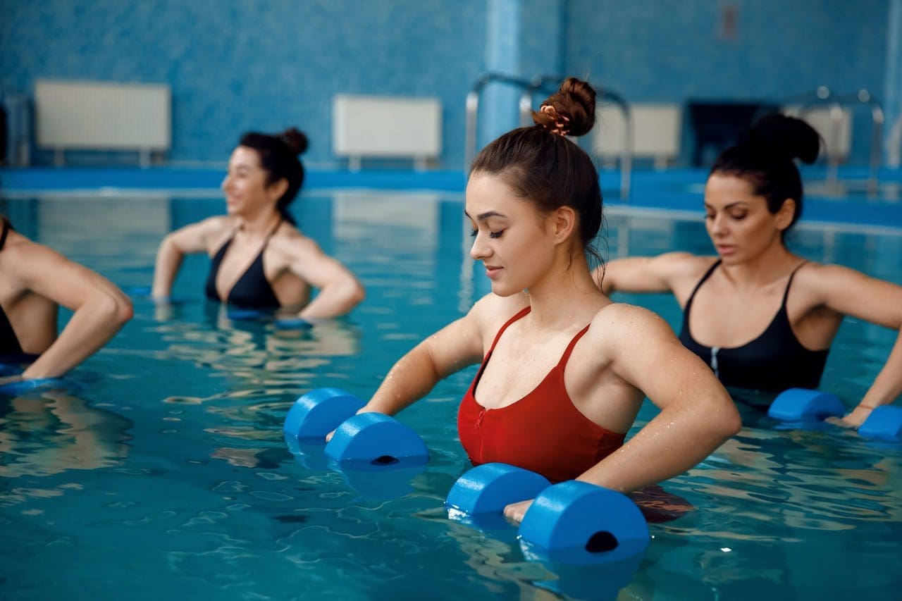
aquatic rehabilitation class for various injuries, including traumatic brain injuries
Traumatic brain injuries, or TBIs, happen when a sudden bump or blow to the head damages the brain. These injuries can come from car crashes, falls, or even sports accidents. Recovering from a TBI takes time and involves many steps to get back strength, balance, and clear thinking. One great way to help is through sports and activities tailored to a person’s needs. These are called adaptive sports. They can boost physical health and also lift moods by making people feel connected and strong. Along with that, healthcare experts like nurse practitioners and chiropractors play big parts in guiding recovery. Nurse practitioners help manage overall health and meds, while chiropractors focus on fixing spine issues and easing pain. This team approach, often called integrative care, mixes different treatments for better results.
In this article, we’ll look at sports that support TBI recovery, such as adaptive basketball and swimming. We’ll also cover calming activities such as tai chi and hiking. Then, we’ll explain how nurse practitioners and chiropractors fit into the picture, drawing on expert perspectives such as Dr. Alexander Jimenez, who combines chiropractic and nursing skills. By the end, you’ll see how these elements work together to create a comprehensive recovery plan.
Understanding TBIs and the Need for Active Recovery
A TBI can mess with how you move, think, and feel. Mild ones, like concussions, might cause headaches or dizziness. Severe ones can lead to long-term problems with balance or memory. The brain has a cool ability called neuroplasticity, which means it can rewire itself to heal. Activities that get you moving help spark this process by building new connections in the brain.
Doctors say rest is key right after a TBI, but then it’s time to add gentle exercise. Starting slow prevents more harm and builds up skills step by step. For example, light walking can improve blood flow to the brain, helping it heal faster. As you get better, more fun activities like games or outdoor adventures can keep things exciting and motivating.
Why activities matter: They improve strength, coordination, and mood.
Start small: Begin with easy tasks at home, like puzzles or stretching.
Build up: Move to group activities for social support.
Research shows that staying active after a TBI lowers the risk of depression and helps people get back to daily life sooner.
Adaptive Sports for Physical and Mental Healing
Adaptive sports are regular sports modified with special tools or rules so everyone can join, regardless of their limitations. For TBI survivors, these sports target balance, hand-eye skills, and thinking on your feet. They also build confidence by letting you achieve goals in a safe way.
Many groups offer adaptive sports programs, making it easy to find local options. Here’s a look at some top ones for TBI recovery:
Adaptive Basketball: Played in wheelchairs or with lower hoops, this sport boosts coordination and teamwork. It helps with quick decisions and arm strength, which TBIs often weaken. Groups like the National Wheelchair Basketball Association run events where players connect and stay motivated.
Cycling: Use adaptive bikes with extra wheels for stability. Cycling improves leg strength and heart health while being low-impact on joints. It’s great for building endurance without straining the brain too much.
Swimming: Water supports your body, making movements easier. Adaptive swimming uses floats or lanes for safety. It enhances balance and breathing control, plus the calm water reduces stress.
Canoeing: In adaptive versions, boats have seats or handles for support. Paddling builds upper body strength and focus. Being on water also calms the mind, helping with anxiety from TBIs.
These sports aren’t just exercise—they create social bonds. Playing with others fights loneliness, a common issue after brain injuries. Studies note that adaptive sports like these keep people active and linked to their communities. One review found that they improve gait and balance in patients with brain injury.
Other Rehabilitative Activities to Enhance Balance and Well-Being
Not all recovery needs to be high-energy. Slower activities like tai chi or hiking can rebuild skills without overwhelming the brain. These focus on mindful movement, which also supports mental health.
Tai Chi: This gentle martial art uses slow, flowing movements to improve balance and focus. For TBI patients, it reduces falls by strengthening core muscles. Classes often adapt poses for sitting if standing is difficult.
Hiking: Adaptive hiking uses trails with smooth paths or walking sticks. It increases heart rate and provides a refreshing change of scenery. Nature-based activities like this restore energy both physically and emotionally.
Adaptive Water Sports: Beyond swimming, try kayaking or water aerobics. These use buoyancy to reduce pressure on the body while improving coordination. Special gear, like life vests, ensures safety.
Home activities can start the process. Activities like balloon tosses or chair yoga build hand-eye coordination and flexibility. Online videos make it easy to try. As skills grow, add group classes for more challenge. Experts say even simple mobilizing, like walking circuits, aids recovery.
Special tools might be needed based on your strengths. For example, use bigger balls in games or stabilizers in cycling. Always check with a doctor to match activities to your healing stage.
The Role of Nurse Practitioners in Coordinating TBI Care
Nurse practitioners (NPs) are advanced nurses who can diagnose, treat, and manage health issues. In TBI recovery, they act as coordinators, making sure all parts of care fit together smoothly.
NPs monitor your overall health during activities. They check for signs like fatigue or headaches that might mean you’re pushing too hard. They also manage meds for pain or mood, adjusting doses as you improve. For instance, if swimming causes dizziness, an NP might suggest changes or add rest days.
In integrative teams, NPs work with other experts to create safe plans. They ensure activities like canoeing don’t clash with your meds or other treatments. Their focus on whole-person care includes emotional support to help with stress during recovery.
Dr. Alexander Jimenez, a chiropractor and family nurse practitioner, notes that NPs play a key role in linking brain health to daily wellness. His observations show they help with sleep and nutrition, which in turn boost activity benefits. This approach ensures activities are effective and safe.
Integrative Chiropractic Care: Supporting Spine and Pain Management
Chiropractors specialize in spine health, which is crucial after a TBI since head injuries often affect the neck. Integrative chiropractic combines adjustments with other therapies, such as exercises, for full recovery.
Chiropractors realign the spine to ease pressure on nerves, reducing headaches and improving balance. For TBI patients, this can help with dizziness from vestibular issues. They also manage pain without heavy meds, using hands-on techniques.
In recovery plans, chiropractors include exercises such as postural training and balance drills. These complement sports by building a strong base. For example, after an adaptive basketball session, a session might address any spine shifts from play.
Dr. Jimenez’s clinical work highlights how chiropractic aids brain healing. He uses gentle adjustments to improve blood flow and nerve function, key for TBIs. His teams integrate this with nutrition and rehab activities, such as light walking, to prevent reinjury. One method he supports is vestibular rehab, which pairs well with sports for better coordination.
Benefits of integrative chiropractic:
Reduces inflammation and pain.
Improves mobility for activities.
Prevents future issues through education.
Combining chiropractic with NP care creates a strong support system. NPs handle meds and monitoring, while chiropractors focus on physical fixes.
Combining Sports, Activities, and Professional Care for Best Results
The best TBI recovery programs combine adaptive sports, calming activities, and expert guidance. Start with a plan from your healthcare team. For example, begin with tai chi for balance, then add cycling as strength grows.
Community outings, like group hikes, apply skills in real life. These build confidence and social ties. Equine therapy, like therapeutic riding, is another option—horses’ movements aid gait and emotional health.
Dr. Jimenez observes that nutrition supports this, like anti-inflammatory foods for brain repair. His work shows that stress management is key, as it affects outcomes.
Track progress with tools like journals or apps. Adjust as needed with your NP or chiropractor. Over time, this leads to independence and joy in activities.
Challenges and Tips for Success
Recovery isn’t always smooth. Fatigue or setbacks can happen. Tips include:
Listen to your body—rest when needed.
Use adaptive gear for safety.
Join support groups for motivation.
With patience, most people see big gains. Studies show stepwise returns to activity, like in sports protocols, work well.
Conclusion
Recovering from a TBI through sports like adaptive basketball or activities like hiking builds both the body and the mind. Nurse practitioners coordinate safe care, while integrative chiropractic handles pain and alignment. Experts like Dr. Jimenez show how this blend speeds healing. Stay active, seek help, and celebrate small wins to pave the way for a brighter path ahead.
Gentle Recovery Strategies After Traumatic Brain Injury: Exercises, Chiropractic Care, and Holistic Support for Lasting Healing
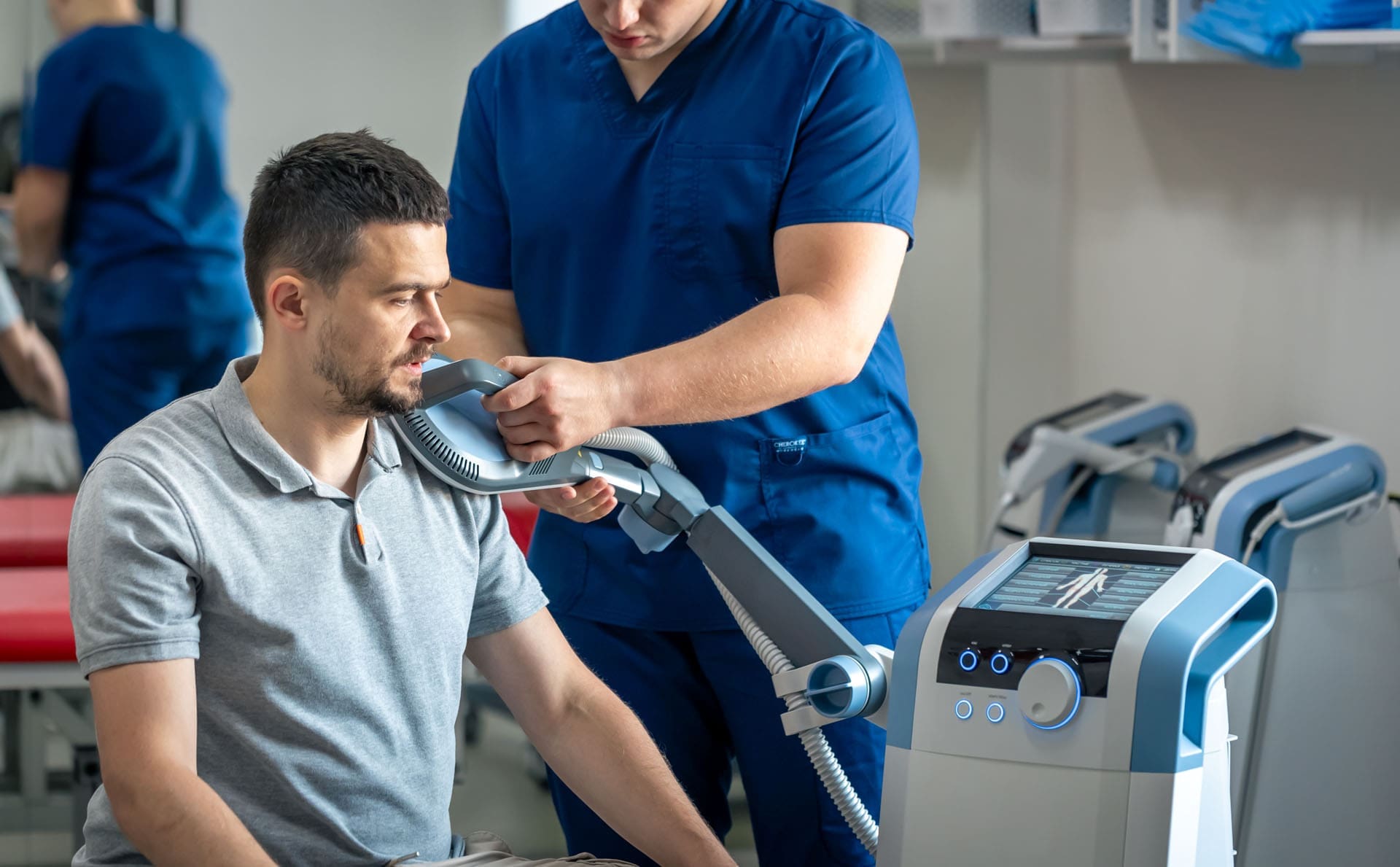
Electromagnetic therapy of the back: a physiotherapist doctor works with a patient with a traumatic brain injury from an occupational accident.
Traumatic brain injury (TBI) affects millions of people every year. A sudden blow or jolt to the head can cause headaches, dizziness, memory problems, neck pain, and poor balance. While the brain needs time and rest to heal, the body also needs gentle movement to recover fully. Early, safe exercises for the neck, core, and balance can speed healing, reduce pain, and lower the risk of falls. Integrative chiropractic care helps restore nerve signals and alignment. Nurse practitioners guide the whole recovery process. When these approaches work together, many people feel stronger and clearer months faster than with rest alone.
This 3,000-word guide uses simple language and proven steps. Every exercise and idea comes from military health guides, rehab centers, and clinical experts. Always get a doctor’s okay before starting. Stop any move that causes sharp pain or new dizziness.
Why Neck Pain Is So Common After TBI
When the head snaps forward and back—like in a car crash or sports hit—the neck takes a huge force. Doctors call this whiplash-associated disorder. Muscles tighten, joints get stiff, and nerves can become irritated. Many people also develop forward head posture, where the head sits inches in front of the shoulders. Each inch forward adds about 10 pounds of stress to the neck muscles (Healthline, 2023a).
Left alone, tight neck muscles pull on the skull base, worsening headaches. They also make balance harder because the brain receives mixed signals from the upper neck. Gentle stretches and posture exercises can effectively address this issue early on.
Common Neck Problems After TBI
Muscle spasms and knots
Stiffness that limits turning the head
Headaches that start at the base of the skull
Forward head posture from pain guarding
Dizziness when moving the head quickly
Safe Neck Stretches to Start in the First Weeks
These four stretches appear on official military and rehab fact sheets. Have them sit in a firm chair with feet flat on the floor. Breathe slowly. Hold each stretch 15–30 seconds and repeat 3–5 times, 2–3 times a day.
Chin Tuck – Slide your chin straight back (like making a double chin) until you feel a stretch behind the neck. Do not tilt down. This is the single best move to fight forward head posture (U.S. Department of Defense, 2020; Healthline, 2023a).
Side Bend – Sit tall. Slowly drop one ear toward the same shoulder until you feel a stretch on the opposite side. Keep your nose pointing forward. Use the hand on top of the head for a gentle extra pull if comfortable (Achieve Brain & Spine, n.d.).
Neck Rotation – Turn your head to look over one shoulder as far as comfortable. Hold, then switch sides. Move only the neck, not the shoulders (U.S. Department of Defense, 2020).
Upper Trapezius Stretch – Sit and place one hand under your thigh to anchor the shoulder. With the other hand, gently pull the head away and slightly forward. You will feel the stretch along the side and back of the neck (Healix Therapy, n.d.).
Tip: Warm the neck first with a warm shower or heating pad for 10 minutes.
Core and Trunk Exercises That Protect the Neck and Brain
A weak core forces the neck muscles to work overtime to keep the head steady. Simple seated core moves wake up the deep stomach and back muscles without jarring the brain.
Do these 3–4 times a week. Start with 8–10 repetitions and build to 15–20.
Sitting Marching – Sit tall with hands on thighs. Lift one knee toward the chest while keeping the back straight, then lower slowly. Alternate legs. This exercise turns on the lower abs and hip flexors (Flint Rehab, 2023a).
Lateral Trunk Flexion (Side Bends) – Sit tall. Slowly slide one hand down the side of the thigh as you bend to that side. Use the opposite core muscles to pull yourself back upright. Works the obliques and reduces side-to-side sway (Illinois Department of Central Management Services, n.d.).
Seated Trunk Extension – Cross arms over chest. Lean forward 10–15 degrees, then slowly sit back tall using the back muscles. Keep the chin tucked to protect the neck (Flint Rehab, 2023a).
Seated Weight Shifts – Scoot forward on the chair so feet are flat and knees are at 90 degrees. Shift weight side to side or front to back while keeping the trunk tall. This exercise is particularly beneficial for promoting early balance (Flint Rehab, 2023a).
Balance Exercises That Are Safe After TBI
Poor balance is one of the biggest fall risks after brain injury. Start every balance exercise seated or holding onto a sturdy surface.
Beginner Level (Weeks 1–4)
Reach in different directions while seated
Heel raises and toe raises while holding a counter
March in place, holding onto a chair
Intermediate Level (Weeks 4–8)
Stand with feet together, eyes open, then eyes closed for 10–20 seconds
Single-leg stance holding a chair (5–10 seconds each leg)
Stand on a firm cushion or folded towel (Neofect, n.d.)
Advanced Level
Tandem stance (heel-to-toe) with arms out
Walk heel-to-toe in a straight line
Step over small objects while watching your feet
Do balance work for 5–10 minutes daily. Progress only when the easier level feels straightforward.
Gentle Yoga and Breathing for Brain and Body Recovery
Modified yoga poses calm the nervous system and safely stretch the entire spine.
Seated Cat-Cow – Hands on knees. Inhale and arch the back while lifting the chest and chin slightly. Exhale and round the back while tucking the chin. Move slowly with the breath (Flint Rehab, 2023b).
Seated Side Stretch – Inhale arms overhead. Exhale and lean to one side, keeping both sit bones on the chair. Hold 3–5 breaths on each side.
Chair Warrior II – Sit sideways on the chair. Extend one leg back and bend the front knee. Reach arms out for a gentle chest and hip opener.
Yoga improves balance by 36% and reduces anxiety in brain-injury patients (Flint Rehab, 2023b).
How Integrative Chiropractic Care Helps TBI Recovery
Chiropractic care is not just about “cracking” the back. Doctors of chiropractic trained in brain-injury care use gentle techniques to:
Remove pressure on nerves, leaving the spine
Restore normal motion to stiff neck joints
Reduce muscle spasms with soft-tissue therapy
Improve blood flow and oxygen to the brain
Correct forward head posture that slows healing
Studies and clinical reports show that spinal adjustments can reduce headache frequency, improve sleep, and speed return to work after concussion (Calibration Mansfield, n.d.; Northwest Florida Physicians Group, n.d.; Pinnacle Health Chiropractic, n.d.).
Dr. Alexander Jimenez, DC, APRN, FNP-BC, a dual-credentialed chiropractor and family nurse practitioner in El Paso, Texas, has treated thousands of patients with TBI, including veterans. He combines precise cervical adjustments, soft-tissue work, and functional neurology exercises. “The upper neck houses sensors that tell the brain where the head is in space. When those joints are stuck, the brain gets fuzzy signals, and balance suffers,” Dr. Jimenez explains in his clinical teaching (Jimenez, 2025). His patients often report clearer thinking and less dizziness within weeks of starting care.
The Important Role of Nurse Practitioners in TBI Care
Nurse practitioners (NPs) are trained to manage complex patients from head to toe. In TBI recovery, they:
Watch for worsening symptoms (increased swelling, seizures, mood changes)
Coordinate physical therapy, chiropractic, counseling, and medications
Teach patients and families what is normal and what needs quick attention
Adjust care plans as healing progresses
Provide follow-up visits to catch problems early (Ackerman, 2012; Mayo Clinic, 2024; Nursing Center, 2023)
Because NPs spend more time with patients than many doctors, they often spot small improvements or setbacks first. Dr. Jimenez, who also holds APRN and FNP-BC credentials, uses this whole-person view in his clinic every day.
Sample 6-Week Gentle Recovery Plan
Week 1–2 (Very Gentle Phase)
5–10 minutes of chin tucks and side bends twice daily
Sitting, marching 2 sets of 10 each leg
Deep breathing for 3 minutes
Short walks with a partner
Week 3–4 (Add Core and Balance)
Add lateral trunk flexion and seated trunk extension
Begin seated weight shifts and reaching
One chiropractic visit for evaluation and gentle adjustment
Week 5–6 (Build Strength and Confidence)
Add standing balance drills with support
Try modified cat-cow and seated yoga stretches
Increase reps to 15–20
Weekly chiropractic care and NP follow-up
Rest for at least one full day between harder sessions. Keep a simple journal: note pain level (0–10), dizziness, and energy. Share it with your team.
Drink water all day (half your body weight in ounces)
Eat protein and colorful vegetables at every meal
Limit screen time in the first weeks—use blue-light glasses if needed
Join an online TBI support group for encouragement
Walk outside in nature when symptoms allow
When to Call the Doctor Right Away
Stop exercising and seek help if you have:
Sudden severe headache
Vomiting or vision changes
Worsening confusion or slurred speech
Seizure or loss of consciousness
Final Thoughts: Healing Is Possible and Often Faster Than You Think
A traumatic brain injury feels overwhelming at first, but the brain and body are built to heal. Gentle neck stretches, core work, balance drills, chiropractic adjustments, and strong nurse practitioner guidance give your recovery the best chance. Start small, stay consistent, and celebrate every tiny win.
Thousands of people—including veterans treated by Dr. Alexander Jimenez—return to work, sports, and family life after TBI by using exactly these safe, evidence-based steps. You can too.
How Head Injuries Affect Movement—and How Chiropractic Care Gives It Back
A physiotherapist is conducting a consultation on a possible traumatic brain injury; the patient complains of back pain and mobility problems.
Head injuries and traumatic brain injuries (TBIs) can turn simple steps into big challenges. A fall, a car crash, or a sports hit can damage the brain and the nerves that tell your body how to walk, reach, or stand tall. This guide explains exactly how these injuries cause muscle fatigue, shaky balance, stiff joints, and even paralysis. You will also learn how gentle chiropractic adjustments, soft-tissue work, and targeted exercises help people move better, feel less pain, and live fuller lives.
What Happens Inside the Body After a Head Injury
When the skull jolts, the brain bounces inside. That sudden movement can tear tiny nerve wires and swell delicate tissues. The messages that once zipped from brain to legs now arrive late, weak, or not at all (Model Systems Knowledge Translation Center, 2023).
Muscle Fatigue Hits Fast
Even mild TBIs make muscles tire in minutes instead of hours. A short walk to the mailbox can feel like a marathon. Dr. Alexander Jimenez, a chiropractor and nurse practitioner in El Paso, Texas, sees this every week. “Patients tell me their legs feel like wet sandbags after five minutes of standing,” he says in his clinic videos (Jimenez, 2025).
Balance Becomes a Wobbly Game
The brain’s balance center sits deep inside the cerebellum. When it gets bruised, the ground seems to tilt. People sway, stumble, or freeze in place. One study found that even “mild” head injuries change walking patterns enough to raise fall risk by 50% (Brain Injury Association of America, 2024).
Coordination Turns Clumsy
Reaching for a coffee cup can knock over the whole table. Fine finger skills vanish. Buttons stay undone, handwriting turns shaky, and stairs feel like mountains. Physiopedia refers to this as “loss of motor dexterity” (Physiopedia, 2024).
Pain and Tiredness Make Everything Worse
Chronic headaches, neck pain, and shoulder aches are common after TBIs. When pain flares, muscles guard and stiffen. Add normal daily fatigue, and movement shuts down completely (Irvine, 2023).
Symptom Questionnaire:
From Stiffness to Locked Joints: The Contracture Trap
If a person rests too much to avoid pain, muscles shorten like dried rubber bands. Joints freeze. Doctors call these locked positions contractures. Elbows, knees, and ankles can bend only a few degrees. Contractures typically develop within weeks and become permanent within months if left untreated (Physiopedia, 2024).
Headway, a UK brain-injury charity, warns: “Lack of movement is the biggest enemy of recovery” (Headway, 2023).
How Chiropractic and Integrative Care Unlock the Body
Chiropractors do more than crack backs. They use gentle moves, hands-on muscle work, and brain-retraining exercises to restart motion and calm pain.
1. Spinal Adjustments Re-Open Nerve Highways
Misaligned neck bones pinch nerves that control arms and legs. A precise chiropractic adjustment lifts that pressure. Blood and cerebrospinal fluid flow better. Patients often feel looser the same day (Northwest Florida Physicians Group, 2023).
Dr. Jimenez films before-and-after videos: one patient who dragged her foot for two years took ten smooth steps after three visits (Jimenez, 2025).
2. Soft-Tissue Therapy Melts Tight Muscles
Fascia—the thin sleeve around every muscle—can knot after injury. Chiropractors use tools and fingers to smooth these knots. Shoulders drop, necks turn, and hips swing again (Function First, 2024).
3. Balance Boards and Eye-Tracking Drills Rewire the Brain
Simple wobble boards teach the brain to steady the body. Following a finger with the eyes rebuilds coordination pathways. These “neuro-drills” are fun and fast. Most patients notice steadier steps in four weeks (HML Functional Care, 2024).
4. Stretching Plans Stop Contractures Before They Start
Daily 10-minute routines keep joints supple. A chiropractor demonstrates the exact angle and hold time to ensure muscles lengthen safely (NR Times, 2024).
5. Posture Fixes End Headache Cycles
Slumped shoulders strain the neck and starve the brain of oxygen. One posture taping session plus two adjustments can cut headache days in half (Cognitive FX, 2024).
Real Stories That Prove It Works
Mark, age 34, car crash survivor “I couldn’t lift my toddler. After six weeks of chiropractic care, I carried her across the park.” (Patient testimonial, Apex Chiropractic, 2024)
Sarah, age 19, soccer concussion “Balance boards felt silly—until I walked the graduation stage without my cane.” (Crumley House, 2024)
Midday 10-minute walk with trekking poles, Soft-tissue massage on tight calves
Evening Wobble-board “surfing” while brushing teeth, Gentle foam-roll under guidance
Follow this for 90 days, and most people regain 70–80% of normal motion (Impact Medical Group, 2024).
When to See a Chiropractic Neurologist
Look for these red-flag signs:
Your legs drag or cross when you walk
Arms stay glued to your sides
You fall more than once a month
Painkillers no longer help
A chiropractic neurologist assesses your gait on video, tests eye reflexes, and develops a customized plan (NeuroChiro, 2024).
Science Backs the Gentle Touch
A 2022 review of 14 studies found that spinal adjustments, combined with exercise, reduced TBI pain by 41% more than exercise alone (Jimenez, 2025). Another trial showed that balance scores increased by 28 points in eight weeks with integrative care (PMC, 2022).
Safe, Drug-Free, and Covered by Many Insurances
Chiropractic care for head injuries is a non-invasive approach. No needles, no scalpels, no opioids. Most auto-insurance PIP plans and major health plans pay for 12–20 visits (Sam’s Chiropractic, 2024).
Your Next Step Today
Call a local chiropractor who lists “TBI” or “concussion” on their website.
Bring a 1-page list: “I trip, my left knee locks, headaches every afternoon.”
Traumatic Brain Injury Recovery: Effective Exercises and Chiropractic Care for Head Injuries
Rehabilitation exercises after an auto accident with head injuries.
Traumatic brain injury, or TBI, happens when a strong hit to the head harms the brain. This can come from falls, car crashes, sports, or other accidents. Head injuries are much like TBIs because they often involve the same kinds of damage to the brain and body. Recovery from these injuries requires time and effort. It focuses on getting back physical strength, mental sharpness, and balance. Rehabilitation utilizes a combination of exercises to aid recovery. These include activities that get the heart pumping, build muscle, improve steadiness, and sharpen the mind. Chiropractic care can also play a significant role, particularly in addressing issues such as headaches and dizziness. This article examines ways to recover, with a strong focus on training and improving step by step.
People with TBI or head injuries often face problems like pain, trouble moving, forgetfulness, or feeling off-balance. Starting recovery early is crucial, but it must be done slowly and safely. Doctors and therapists guide the process. Exercises help the brain rewire itself through something called neuroplasticity. This means the brain can create new pathways to repair damaged ones. Training helps build these paths. Recovery is not limited to a single type of exercise. It combines various types to cater to all needs. Let’s dive into the details.
Physical Exercises for Strength and Aerobic Health
Physical exercises are a big part of getting better from TBI or head injuries. They help rebuild muscle, boost energy, and enhance overall bodily function. Start slow because rushing can cause more harm. Always check with a doctor first.
Aerobic activities get the heart rate up without too much strain. Walking is a simple start. It can be done inside or outside, and it helps blood flow to the brain. This brings oxygen and nutrients for the healing process. Jogging on a treadmill or using a stationary bike are other options. Swimming is great too because the water supports the body, making movement easier. Aim for 150 minutes a week of moderate aerobic work, spread out over days. This could be 20 to 40 minutes per session, three to four times a week. These activities lower the risk of other health issues like heart problems or diabetes, which can slow recovery. They also lift mood and reduce tiredness.
Strength training builds muscle power. This is important because injuries can weaken muscles. Squats are a good exercise. Stand with your feet apart, as if your shoulders are wide, bend your knees as if sitting back in a chair, then stand up. Do this 10 times. Rows work the back and arms. Sit or stand, pull your elbows back like squeezing something between your shoulder blades. Use light weights or resistance bands if possible. Bicep curls are simple: Hold a water bottle, bend your elbow to bring it to your shoulder, then lower it. Repeat 10 times per arm. For legs, try seated marching. Sit in a chair and lift one knee up, then the other, like walking in place. These exercises help with daily tasks, such as getting up from a chair or carrying objects.
Other strength moves include push-ups against a wall or chair for the chest and arms. Shoulder presses: Lift arms overhead with light weights. Do these in sets, with rests in between. Strength training should be done two to three times a week, focusing on the larger muscle groups. It helps with posture and stops falls. As you become stronger, add more reps or increase the weight. But listen to your body. If it hurts, stop and rest.
Seated exercises are beneficial for individuals who are unable to stand or walk. Seated hip rotations: Sit and turn your hips side to side. This builds core strength. Alternating heel-toe raises: Lift your heels, then your toes, while sitting. These improve lower-body control and blood flow. Arm push: Push a bottle across a table with your wrist. This strengthens arms without much effort. Mixing aerobic and strength training keeps the workout fun and covers more ground for recovery.
Balance Exercises to Regain Stability
Balance problems are common after TBI or head injuries. They can cause falls and make walking hard. Balance training helps the brain and body work together better. It uses neuroplasticity to fix these issues.
Tandem stance is a basic exercise. Stand with one foot right in front of the other, like on a tightrope. Hold for 30 seconds, then switch feet. If it’s too hard, spread feet wider. Close your eyes to make it tougher once you’re ready. Weight shifts: Stand with your feet apart, shift your weight to one side, and lift the other foot slightly. Hold 30 seconds per side. This builds steadiness.
Romberg stance: Stand with feet together, eyes closed. Hold as long as you can, up to two minutes. It trains the body to use senses apart from sight for balance. Alternating heel-to-toe raises: Stand and rise on your toes, then rock back onto your heels. Do it 10 times. This strengthens legs and improves coordination.
For more challenge, use tools. A gym ball: Sit on it and reach for objects. This makes the surface unstable, forcing better control. Balance boards: Stand on a wobbly board and try not to lose your balance. Start with help. Walking on various surfaces, such as grass or sand, trains the body to adapt.
Vestibular exercises help with dizziness. These include head turns while focusing on a point, as well as eye movements such as following a finger. They retrain the inner ear and brain. Do balance work daily, but in short sessions to avoid fatigue. Progress slowly from a seated to a standing position. Good balance means safer movement and less fear of falling.
Mix balance with other training. For example, do squats while on one leg. Or walk while turning your head. This makes exercises more realistic. Recovery improves when training mimics daily activities.
Cognitive Exercises for Mental Sharpness
Mental skills can be affected after TBI or head injuries. Aspects such as memory, focus, and problem-solving require improvement. Cognitive exercises challenge the brain to build new connections.
Try new things: Walk a different path or try a new food. This sparks neuron growth. Use your non-dominant hand for tasks such as brushing your teeth. It activates the other side of the brain and strengthens thinking. Brain-training games: Play chess, Sudoku, or apps like Lumosity. These improve logic and memory.
Memorization: Recall a grocery list or song lyrics. Start small and build up. Draw maps from memory, like your route to the store. This boosts spatial thinking. Read out loud: It works reading, speaking, and listening parts of the brain.
Puzzles and games: Jigsaw puzzles or board games like Connect Four help develop planning and hand-eye coordination skills. Mental math: Add numbers in your head or count backwards by sevens. Keep a journal of senses: Note what you see, hear, and smell each day. This mixes memory and senses.
Start slow with easy tasks. Increase difficulty as you improve. Do 15-20 minutes a day. Combine with physical exercises for a complete recovery. Cognitive training helps with daily life, like remembering names or following recipes.
Integrative Chiropractic Therapy for Support
Chiropractic care helps with TBI and head injury recovery. It focuses on the spine and nervous system. This can help alleviate headaches and dizziness caused by injuries.
Adjustments align the spine, reducing nerve pressure. This improves blood flow to the brain and cuts inflammation. Craniosacral therapy: Light touch on the head and spine boosts fluid flow around the brain. It helps with headaches and brain function.
Chiropractors offer lifestyle tips, such as healthy eating and adequate sleep. They also suggest exercises, such as those for strength and balance. Combining chiropractic care with physical therapy can accelerate recovery. It addresses both body and mind.
For long-term care, regular visits prevent chronic pain. Chiropractic supports neuroplasticity by stimulating the nervous system. It’s non-invasive and can be used in conjunction with other treatments.
Insights from Dr. Alexander Jimenez
Dr. Alexander Jimenez, a chiropractor with over 30 years of experience, shares observations on TBI and head injuries. He uses integrative care for recovery. His work includes functional medicine to fix root causes. For injuries, he emphasizes the importance of prompt action with rehabilitation programs. These include exercises for mobility and nerve health. He helps with symptoms like pain and weakness through adjustments and nutrition. His clinic focuses on achieving full healing without the use of drugs or surgery.
Jimenez notes that personalized plans are most effective. He combines chiropractic with exercises to boost recovery. His insights demonstrate how training can rebuild strength and function after head injuries.
Putting It All Together for Recovery
Recovery from TBI or head injuries needs a mix of exercises and care. Focus on training: Do aerobic exercises for heart health, strength training for muscles, balance training for stability, and cognitive exercises for the mind. Add chiropractic for extra support. Start slow, be consistent, and track progress. With time, these steps lead to a better quality of life.
Always work with pros. Recovery is a journey, but training makes it possible.
Achieve pain relief with heel pain chiropractic care targeting Achilles tendon concerns for a better quality of life.
Understanding Achilles Tendon Heel Pain: A Comprehensive Guide to Chiropractic Care and Natural Recovery
Heel pain affecting the Achilles tendon is one of the most common complaints among active individuals, weekend warriors, and even those with sedentary lifestyles. This debilitating condition can significantly impact your quality of life, limiting your ability to walk, run, or even stand comfortably. While many people immediately think of medications or surgery as solutions, chiropractic care offers a comprehensive, non-invasive approach to addressing the root causes of Achilles tendon pain and promoting natural healing. This guide explores the anatomy, biomechanics, causes, and evidence-based treatments for Achilles tendon heel pain, with a special focus on how chiropractic care can restore function and reduce discomfort.
Understanding the Achilles Tendon: The Body’s Strongest and Most Vulnerable Tendon
The Achilles tendon holds the distinction of being both the largest and strongest tendon in the human body, yet it remains paradoxically one of the most commonly injured structures in the lower extremity. This remarkable structure connects the powerful calf muscles to the heel bone, creating a critical link in the kinetic chain that allows us to walk, run, jump, and stand on our toes.
Despite its impressive strength, the Achilles tendon is uniquely vulnerable to injury. Research shows that this tendon can bear loads up to 12 times body weight during running and up to 3,500 Newtons of force before rupture. However, a hypovascular area exists approximately 2 to 6 centimeters proximal to the calcaneal insertion, where blood supply is significantly reduced. This zone of poor vascularity makes the tendon particularly susceptible to degenerative changes and injury.
Understanding the complexity of the Achilles tendon helps us appreciate why a comprehensive, whole-body approach like chiropractic care can be so effective. Rather than simply treating the symptoms at the site of pain, chiropractors evaluate the entire musculoskeletal system to identify biomechanical imbalances that may contribute to excessive stress on the tendon.
Anatomy and Biomechanics of the Achilles Tendon: A Marvel of Engineering
Structural Composition
The Achilles tendon, also known as the calcaneal tendon or triceps surae tendon, is formed by the confluence of three muscles: the gastrocnemius (with its medial and lateral heads) and the soleus muscle. The gastrocnemius originates from the posterior aspect of the femoral condyles, while the soleus arises from the posterior surface of the fibula and medial border of the tibia. These muscles coalesce distally to form the common Achilles tendon, which inserts onto the middle portion of the posterior calcaneal surface.
Compositionally, the Achilles tendon consists of approximately 95% type I collagen fibers, which provide exceptional tensile strength and flexibility. The remaining 5% includes type III collagen, elastin (accounting for up to 2% of dry mass), proteoglycans, and glycosaminoglycans. This hierarchical structure organizes into fibrils, fibers, and fascicles bound together by small matrix molecules.
A distinctive feature of the Achilles tendon is its spiral configuration. As the tendon descends toward its insertion, the fibers rotate approximately 90 degrees, causing the medial gastrocnemius fibers to become superficial while the lateral gastrocnemius and soleus fibers become deeper. This spiraling creates an area of concentrated stress but also confers a significant mechanical advantage during propulsion activities.
The Paratenon: A Unique Protective Sheath
Unlike many tendons, the Achilles does not possess a true synovial sheath. Instead, it is surrounded by a paratenon—a thin layer of loose connective tissue that provides a significant portion of the tendon’s blood supply and allows for gliding movement of up to 2-3 centimeters. The paratenon contains elastin and extends into the tendon, binding collagen bundles together while permitting movement among them.
Blood Supply and Vulnerability
The vascular supply to the Achilles tendon comes from three sources: the musculotendinous junction, vessels in the surrounding connective tissue (primarily the paratenon), and the osteotendinous junction. The vascular territories can be classified into three regions, with the midsection supplied by the peroneal artery and the proximal and distal sections supplied by the posterior tibial artery. This arrangement leaves a relatively hypovascular area in the mid-portion of the tendon—precisely where most pathology occurs.
Biomechanical Properties
The Achilles tendon demonstrates nonlinear mechanical properties at low strains, exhibiting what is known as a “toe region” in its force-displacement curve. This nonlinearity arises from the uncrimping of collagen fibers and an associated increase in collagen alignment as load is applied. Under polarized light, tendons exhibit periodic banding due to their waveform configuration known as “crimp,” which extends hierarchically from macro- to nano-structural scales.
At higher strains, the tendon deforms linearly prior to yield and rupture. While traditionally described as viscoelastic (containing both elastic and viscous components), recent evidence in humans suggests that its elastic properties dominate. These spring-like properties allow the Achilles tendon to store and release energy efficiently during ambulation, delivering explosive propulsion while protecting soft tissues from damage.
Functions of the Achilles Tendon in the Lower Body and Extremities
Primary Function: Plantarflexion
The primary function of the Achilles tendon is to enable plantarflexion of the foot—the movement that points the toes downward and lifts the heel off the ground. This action is fundamental to virtually all lower extremity movements, including walking, running, jumping, climbing stairs, and standing on tiptoes. The gastrocnemius muscle also contributes to knee flexion, adding another dimension to lower extremity function.
Force Transmission and Lever Action
The calcaneus (heel bone) acts as a lever arm for the triceps surae muscles, and the Achilles tendon serves as the critical link that transmits force from the calf muscles to the heel bone. This arrangement allows for efficient transfer of muscular force to the foot during the propulsive phase of gait. The heel bone projects posterior to the tibia and fibula, creating a mechanical advantage that amplifies the force generated by the calf muscles.
Shock Absorption and Energy Storage
During walking, the heel can absorb approximately 110% of body weight, and during running, this increases to 200% of body weight. The Achilles tendon, in conjunction with the plantar fascia and the specialized fat pad beneath the heel, functions as part of an integrated shock absorption system. The elastic properties of the tendon allow it to store mechanical energy during the loading phase of gait and release it during toe-off, improving efficiency and reducing metabolic cost.
Role in Postural Control and Balance
Vibration studies have demonstrated that the Achilles tendon plays a crucial role in postural orientation and balance. When the tendon is vibrated without visual input, subjects experience movement backwards and the illusion of forward body tilt. This occurs because vibrations stimulate muscle spindles in the calf muscles, alerting the brain to body position and initiating compensatory movements through the central nervous system.
Integration with the Kinetic Chain
The Achilles tendon does not function in isolation but rather as an integral component of the lower extremity kinetic chain. Problems with foot alignment, ankle mobility, knee position, hip alignment, or even spinal posture can alter the biomechanical forces acting on the Achilles tendon. This interconnected system explains why chiropractors examine the entire body when evaluating Achilles tendon pain, rather than focusing solely on the local area of discomfort.
Factors Leading to the Development of Heel Pain Associated with the Achilles Tendon
Achilles tendinopathy develops through a complex interplay of intrinsic and extrinsic factors that create an imbalance between the loading demands placed on the tendon and its capacity to adapt and recover.
Intrinsic Risk Factors
Muscle Strength and Weakness: Expert consensus identifies muscle strength, particularly plantarflexor weakness, as the primary modifiable risk factor for Achilles tendinopathy. Studies of military recruits have shown that plantarflexor strength is predictive of tendinopathy development. When the calf muscles are weak or fatigued, the Achilles tendon must bear disproportionate loads, increasing the risk of microtrauma and degeneration.
Previous Injuries and Incomplete Rehabilitation: A history of prior Achilles tendinopathy or incomplete recovery from previous injuries significantly increases the risk of recurrent problems. Residual strength deficits, altered neuromuscular control, and persistent structural changes may explain why previous injury is such a strong risk factor.
Age and Degenerative Changes: While age itself is not directly causal, age-related reductions in tendon vascularity, collagen quality, and muscle strength contribute to increased vulnerability. Achilles tendinopathy is most commonly seen in individuals aged 30-50 years, with middle-aged recreational athletes being particularly susceptible.
Anatomical Factors: Foot structure and alignment play crucial roles in tendinopathy development. Excessive pronation (rolling inward of the foot), high arches (pes cavus), flat feet (pes planus), limited ankle dorsiflexion, varus alignment with functional hyperpronation, leg length discrepancies, and excessive tibial torsion can all alter the distribution of forces through the Achilles tendon.
Systemic Conditions: Metabolic and systemic diseases can affect tendon health and increase vulnerability to injury. These include diabetes mellitus, thyroid and parathyroid disorders, gout, collagen deficiencies, hypercholesterolemia, and autoimmune conditions. Blood group O has also been associated with increased incidence of Achilles tendinopathy and rupture.
Genetic Factors: Family history appears to be a risk factor, with individuals who have a positive family history of Achilles tendinopathy having a five-fold greater risk for such injuries. Genetic factors may influence collagen structure, muscle fiber composition, and tendon morphology.
Extrinsic Risk Factors
Training Errors and Load Management: Changes in loading patterns represent the most consistently ranked extrinsic risk factor for Achilles tendinopathy. Sudden increases in training volume or intensity, particularly after layoffs or recovery periods, create a mismatch between tendon capacity and demands. Other problematic training errors include changes in training type (such as adding hill work), alterations in training due to events or competitions, excessive training intensity, inadequate recovery between sessions, and abrupt increases in weekly distance.
Overuse and Insufficient Recovery: The concept of “training errors” encompasses insufficient recovery periods between bouts of activity. Studies have shown that muscle weakness and fatigue may persist for up to 47 days after a single exercise session, even when individuals report feeling “recovered”. Continuing to train despite ongoing neuromuscular deficits inadvertently increases tendinopathy risk.
Footwear Issues: Improper footwear can contribute to Achilles problems through inadequate support, insufficient cushioning, worn-out shoes, heel counter pressure against the posterior heel, and inappropriate shoes for specific activities. For cyclists, low saddle height resulting in excessive ankle dorsiflexion during pedaling may be a causative factor.
Training Surface: Hard or uneven training surfaces can increase impact forces and alter biomechanics, contributing to overload of the Achilles tendon. Sudden changes in training surface (such as moving from a treadmill to outdoor pavement) can precipitate symptoms.
Medications: Certain medications, particularly fluoroquinolone antibiotics, corticosteroids, and anabolic steroids, have been associated with increased risk of Achilles tendinopathy and rupture. These medications may affect collagen synthesis, reduce tendon strength, or impair healing processes.
Pathophysiology: From Overload to Degeneration
The pathological process in Achilles tendinopathy represents failed healing or degenerative changes resulting from continuous overloading. Contrary to traditional understanding, chronic Achilles tendinopathy is not primarily an inflammatory condition (despite the term “tendinitis” suggesting inflammation). Histological examination reveals degenerative changes including collagen disorganization, increased type III collagen production, proteoglycan accumulation, neovascularization (new blood vessel formation), and nerve ingrowth.
The repetitive microtrauma hypothesis suggests that excessive stretching and loading create microscopic tears in the collagen matrix. When recovery time is insufficient, these microtears accumulate, overwhelming the tendon’s repair mechanisms. The resulting degenerative process involves tenocyte death, collagen fiber disruption, and areas filled with vessels and nerves that may contribute to pain.
How Heel Pain Can Mimic Sciatica Pain: Understanding Referred Pain Patterns
One of the most clinically challenging aspects of heel pain is its potential to mimic or coexist with sciatica, creating diagnostic complexity and potentially delaying appropriate treatment.
The Sciatic Nerve and Referred Pain
The sciatic nerve is the largest nerve in the human body, originating from the L4-S3 nerve roots in the lower spine and traveling through the buttocks and down the leg to the foot. When this nerve becomes compressed or irritated—a condition known as sciatica—it can produce pain, tingling, numbness, and weakness that radiates along its entire pathway.
Critically, sciatica can cause referred pain in the heel through several mechanisms. The L5-S1 nerve root provides segmental innervation to the posterior thigh, gluteal muscles, and the anterior, posterior, and lateral leg muscles, as well as sensory innervation to the heel. Compression or irritation of this nerve root can produce heel pain that patients may attribute to a local foot problem rather than a spinal origin.
Distinguishing Features
Understanding the differences between true Achilles tendinopathy and sciatica-related heel pain is essential for appropriate treatment:
Achilles Tendinopathy Characteristics:
Pain localized to the posterior heel and along the tendon
Tenderness to palpation of the Achilles tendon
Morning stiffness that improves with initial movement but may worsen with continued activity
Pain worsens with activities that load the tendon (walking, running, jumping)
No radiating pain up the leg
No neurological symptoms such as numbness or tingling
Sciatica-Related Heel Pain Characteristics:
Pain radiating from the lower back through the buttocks and down the leg to the heel
Numbness, tingling, or burning sensations in the leg or foot
Weakness in the leg or foot muscles
Pain may worsen with prolonged sitting, standing, or certain movements
Possible positive findings on nerve tension tests (straight leg raise)
Possible altered reflexes, particularly the ankle reflex
Chiropractic Care for Leg Instability- Video
The Double-Crush Phenomenon
Clinicians must also be aware of the “double-crush syndrome,” in which concomitant proximal and distal nerve entrapments occur simultaneously. A patient could have both lumbar radiculopathy causing sciatic symptoms and local nerve compression in the foot (such as tarsal tunnel syndrome), creating complex and overlapping pain patterns.
Clinical Implications for Chiropractic Care
The potential for sciatica to cause or contribute to heel pain underscores the importance of comprehensive evaluation by healthcare providers trained in whole-body assessment. Chiropractors are uniquely positioned to evaluate both spinal and peripheral sources of heel pain, ensuring that treatment addresses all contributing factors rather than focusing exclusively on local symptoms.
Clinical Rationale: Why Chiropractic Care Can Help Reduce Heel Pain in the Achilles Tendon
Chiropractic care offers a comprehensive, evidence-informed approach to managing Achilles tendon heel pain that addresses both local tissue dysfunction and systemic biomechanical imbalances.
Addressing Biomechanical Dysfunction in the Kinetic Chain
A fundamental principle of chiropractic philosophy is that the body functions as an integrated kinetic chain, where dysfunction in one area creates compensatory stress in others. The feet serve as the foundation for the entire skeletal system, and problems with foot alignment can cause cascading effects upward through the ankles, knees, hips, and spine.
Research confirms this interconnected relationship. Excessive pronation causes the arches to flatten and ankles to roll inward, forcing internal leg rotation, pelvic tilt, and lower back stress. Conversely, spinal misalignments, pelvic imbalances, or hip dysfunction can alter gait patterns and weight distribution, placing abnormal stress on the Achilles tendon.
Chiropractic adjustments to the spine, pelvis, hips, knees, ankles, and feet help restore proper alignment throughout the kinetic chain. By correcting misalignments (subluxations) in these areas, chiropractors reduce compensatory strain on the Achilles tendon and surrounding structures.
Improving Joint Mobility and Function
Limited ankle dorsiflexion and restricted foot and ankle joint mobility are established risk factors for Achilles tendinopathy. Chiropractic adjustments specifically directed at the foot and ankle can improve joint mobility, decrease pain, and enhance function.
A case series published in the Journal of Physical Therapy Science demonstrated that the addition of joint mobilization and manipulation to eccentric exercise programs produced immediate within-session improvements in pain, heel raise repetitions, and pressure pain thresholds in runners with chronic Achilles tendinopathy. At discharge and nine-month follow-up, patients showed improvements in self-reported function, pain levels, joint mobility, ankle motion, and single-leg heel raises.
The mechanisms underlying these improvements include decreased nociceptive reflex excitability, enhanced conditioned pain modulation, reduction of bilateral hyperalgesia following unilateral joint mobilization, and improved biomechanical function through restored joint alignment.
Enhancing Blood Flow and Tissue Healing
The Achilles tendon receives relatively poor blood supply, particularly in the vulnerable mid-portion region. Chiropractic care can assist in recovery from tendon injuries by improving circulation to the area. Manual techniques, including soft tissue mobilization and specific adjustments, may stimulate local blood flow, enhancing nutrient delivery and waste removal from the healing tendon.
Research on manual therapy for tendinopathy suggests that soft tissue and joint-directed techniques can decrease pain, improve mobility, and enhance muscle performance—all relevant for persons with Achilles tendinopathy. While manual therapy effects are typically short-lived, when performed prior to exercise and reinforced with subsequent activities, it can reduce pain sensitivity and increase range of motion, allowing individuals to participate more actively in tendon loading progressions.
Reducing Compensatory Muscle Tension
Muscle imbalances and excessive tension in the calf muscles (gastrocnemius and soleus) contribute to increased stress on the Achilles tendon. Chiropractors employ various soft tissue techniques, including myofascial release, trigger point therapy, instrument-assisted soft tissue mobilization (IASTM), and massage therapy to release tension in the calf muscles, plantar fascia, and surrounding structures.
These techniques not only provide pain relief but also improve tissue extensibility and flexibility, allowing for more normal tendon loading during movement. By addressing muscle dysfunction both locally and remotely (including muscles of the hip and trunk), chiropractors help reduce the overall stress on the Achilles tendon.
Neurological Effects and Pain Modulation
Chiropractic adjustments and manual therapy techniques produce neurophysiological effects that extend beyond simple mechanical realignment. Research demonstrates that spinal and extremity manipulation can increase stimulability of alpha motor neurons, enhance activity in corticospinal pathways, alter sensorimotor cortex activity, and modulate pain processing through effects on the central nervous system.
Joint mobilization has been associated with reduced sensitivity to pain, improved pain tolerance, and enhanced endogenous pain inhibition mechanisms. These neurological effects complement the biomechanical benefits of chiropractic care, providing multi-level pain relief for patients with Achilles tendinopathy.
Evidence Supporting Chiropractic Management
Multiple studies and case reports support the effectiveness of chiropractic care for Achilles tendinopathy:
A 2012 case study published in the Journal of Canadian Chiropractic Association reported successful management of midportion Achilles tendinopathy using chiropractic interventions, with rapid cessation of pain.
Research comparing chiropractic adjustments with daily stretching to custom orthotics alone found significant improvements in both groups, with the chiropractic care group showing advantages in pain ratings at the 15-day mark.
Case series combining manual therapy with eccentric exercise have demonstrated superior outcomes compared to exercise alone, particularly in the first four weeks of treatment.
Dr. Alexander Jimenez’s Clinical Approach: Integrating Advanced Diagnostics with Dual-Scope Care
Dr. Alexander Jimenez, DC, APRN, FNP-BC, CFMP, IFMCP, brings a unique dual-licensed perspective to the treatment of Achilles tendon heel pain and lower extremity injuries. As both a licensed chiropractor and a board-certified Family Practice Nurse Practitioner, Dr. Jimenez combines the biomechanical expertise of chiropractic care with advanced diagnostic capabilities and medical management.
Comprehensive Patient Evaluation
Dr. Jimenez’s approach begins with thorough patient assessment that examines not only the local site of pain but the entire musculoskeletal and neuromuscular system. His evaluation protocols include detailed medical history taking, comprehensive physical examination of the spine, pelvis, lower extremities, and feet, gait analysis to identify biomechanical abnormalities, orthopedic and neurological testing, and assessment of functional movement patterns.
This comprehensive evaluation allows Dr. Jimenez to identify all contributing factors to a patient’s heel pain, including spinal misalignments affecting the kinetic chain, muscle imbalances and weakness, joint restrictions in the foot and ankle, faulty foot mechanics (pronation/supination), and potential systemic factors influencing tissue health.
Advanced Imaging and Diagnostic Evaluation
One of Dr. Jimenez’s distinguishing characteristics is his ability to integrate advanced imaging and diagnostic evaluations into treatment planning. His dual licensure enables him to order and interpret diagnostic studies including musculoskeletal ultrasound for visualizing tendon structure and pathology, magnetic resonance imaging (MRI) to assess tendon integrity, inflammation, and surrounding structures, X-rays to evaluate bone alignment and rule out fractures or bone spurs, and specialized laboratory testing when systemic conditions are suspected.
This diagnostic capability ensures accurate diagnosis and allows for monitoring of treatment progress and tissue healing. Dr. Jimenez can differentiate between insertional and non-insertional Achilles tendinopathy, identify concurrent pathologies such as retrocalcaneal bursitis or Haglund’s deformity, assess the degree of tendon degeneration or partial tearing, and rule out other causes of heel pain that may require different treatment approaches.
Integrative Treatment Protocols
Dr. Jimenez’s treatment philosophy emphasizes integrative, patient-centered care that combines the best of chiropractic medicine, functional medicine, and evidence-based rehabilitation. His protocols for Achilles tendon heel pain typically include chiropractic adjustments to the spine, pelvis, and lower extremity joints to restore proper biomechanics, extremity-specific adjustments targeting the foot and ankle, soft tissue therapies including myofascial release and instrument-assisted techniques, customized rehabilitation exercises with progressive loading, functional medicine interventions addressing nutrition, inflammation, and tissue healing, and patient education on activity modification and injury prevention.
Dr. Jimenez recognizes that successful outcomes require addressing not just the symptoms but the underlying causes of tendon pathology. His functional medicine training allows him to consider factors such as nutritional deficiencies affecting collagen synthesis, metabolic conditions influencing tissue healing, inflammatory status and oxidative stress, hormonal imbalances that may affect musculoskeletal health, and lifestyle factors contributing to injury risk.
Collaborative Care Model
When necessary, Dr. Jimenez collaborates with orthopedic surgeons, physical therapists, podiatrists, and other specialists to ensure patients receive comprehensive care. If he determines that a patient would benefit from interventions outside his scope of practice, such as advanced imaging procedures, specialized injections, or surgical consultation, he refers to the appropriate providers while continuing to support the patient’s overall recovery.
This collaborative approach ensures that patients receive the most appropriate care for their specific condition while benefiting from Dr. Jimenez’s unique ability to bridge conventional medical evaluation with chiropractic and functional medicine interventions.
Different Nonsurgical Treatments for Heel Pain in the Lower Extremities
Effective management of Achilles tendon heel pain typically requires a multimodal approach combining various evidence-based interventions.
Eccentric Exercise Programs
Eccentric exercise has the highest level of evidence supporting its ability to reduce Achilles tendinopathy pain (Grade A recommendation). The most well-known protocol is the Alfredson protocol, which consists of a 12-week program where patients perform eccentric heel-drop exercises—three sets of 15 repetitions, twice daily, seven days a week.
The exercises are performed in two variations: with the knee straight (emphasizing the gastrocnemius muscle) and with the knee bent (emphasizing the soleus muscle). Patients start in a raised heel position, slowly lower the heel below the level of the step over three seconds (eccentric phase), then use the opposite leg to return to the starting position.
For insertional Achilles tendinopathy, the protocol is modified to reduce ankle dorsiflexion range of motion, avoiding compression of soft tissues at the tendon insertion. This modification has been shown to decrease pain from baseline to follow-up, with 67% of patients able to resume pre-injury activity levels.
Acupuncture for Achilles Tendinopathy
Acupuncture offers a holistic approach to treating Achilles tendinopathy by addressing both pain and underlying inflammation. A 2013 randomized controlled trial found that acupuncture intervention significantly improved pain and activity in patients with chronic Achilles tendinopathy compared to eccentric exercises alone.
In this study, the mean VISA-A score improved to 67.1 points in the acupuncture group versus 48.5 points in the control group after eight weeks—an 18.6-point advantage for acupuncture. Pain scores diminished by 3.7 cm after activity and 3.2 cm at rest in the acupuncture group, significantly more than the control group.
Mechanisms of Acupuncture for Tendinopathy:
Stimulates the body’s natural anti-inflammatory response, reducing pain and swelling
Enhances blood circulation to the Achilles tendon, promoting faster healing and recovery
Releases endorphins and neurochemicals that provide pain relief
Deactivates myofascial trigger points in the calf muscles contributing to tendon stress
Addresses underlying energy imbalances according to traditional Chinese medicine principles
Electroacupuncture, which adds electrical stimulation to traditional needle placement, has shown particular promise. A case series reported successful treatment using direct tendon needle insertion with electrostimulation at specific acupuncture points, resulting in symptomatic reduction and functional improvement.
Rest, Ice, Compression, and Elevation (RICE)
The RICE protocol remains a foundational component of acute Achilles tendon injury management. Rest or immobilization allows the tendon time to heal without continued mechanical stress. Doctors may recommend walking boots to keep the foot immobile for two to three weeks after an Achilles tendinitis diagnosis.
Ice application 2-3 times daily for 5-10 minutes helps alleviate pain and swelling. Compression with flexible bandaging during the first few days after injury reduces swelling. Elevation of the foot three times daily for 15 minutes also helps manage inflammation.
Extracorporeal Shock Wave Therapy (ESWT)
When exercise programs are unsuccessful, extracorporeal shock wave therapy appears to be the next best non-operative treatment option to reduce Achilles tendinopathy pain (Grade B recommendation). ESWT uses acoustic waves to stimulate healing processes in tendon tissue.
A randomized controlled trial by Rompe and colleagues found that ESWT was more effective at reducing pain than full-range eccentric exercise in patients who had not responded to other conservative treatments. The ESWT group experienced pain reduction from 7.0 to 3.0, while the eccentric exercise group improved from 6.8 to 5.0. Multiple clinical trials have demonstrated 2-point or greater decreases in pain with ESWT.
Physical Therapy and Manual Therapy
Physical therapy combines therapeutic exercise, manual techniques, and modalities to address Achilles tendinopathy comprehensively. Manual therapy techniques include joint mobilization to improve ankle and foot mobility, soft tissue mobilization targeting the calf muscles and surrounding tissues, trigger point release for myofascial dysfunction, nerve mobilization when neural tension is present, and massage therapy to improve circulation and reduce muscle tension.
Research demonstrates that adding manual therapy to eccentric exercise protocols can enhance rehabilitation outcomes, particularly in the first four weeks of treatment when manual therapy accelerates recovery compared to exercise alone.
Orthotic Interventions
Custom foot orthoses help correct biomechanical abnormalities contributing to Achilles stress. These devices support proper arch height, correct pronation or supination issues, balance leg length discrepancies, redistribute pressure across the foot, and improve overall foot alignment.
Heel lifts can temporarily reduce strain on the Achilles tendon by decreasing ankle dorsiflexion during walking and standing. While heel lifts do not address underlying pathology, they can provide symptomatic relief during the healing phase.
Nutritional Interventions
Emerging research supports nutritional supplementation to enhance tendon healing. Vitamin C plays a crucial role in collagen synthesis, and supplementation (particularly when combined with gelatin or hydrolysed collagen) may accelerate tendon recovery.
Studies suggest that 15 grams of gelatin containing 225mg of vitamin C taken one hour before resistance training increases collagen synthesis twofold. Vitamin C supplementation aids tendon healing through antioxidant activity, promotion of type I collagen fiber production, and reduction of oxidative stress parameters.
Various Stretches and Exercises to Stretch and Strengthen the Achilles Tendon
A comprehensive exercise program for Achilles tendinopathy should include eccentric strengthening, progressive loading, flexibility work, and neuromuscular training.
The Alfredson Eccentric Protocol
Exercise 1: Straight-Knee Heel Drops
Stand on the edge of a step on the affected leg, with the heel hanging over the edge. Rise up on the toes using both legs for assistance. Slowly lower the affected heel below the level of the step over 3 seconds (eccentric phase only). Use the unaffected leg to return to the starting position. Perform 3 sets of 15 repetitions, twice daily.
Exercise 2: Bent-Knee Heel Drops
Perform the same movement as above, but with the knee slightly bent (approximately 15-20 degrees). This variation emphasizes the soleus muscle. Slowly lower the heel below step level over 3 seconds. Return to starting position using the opposite leg. Perform 3 sets of 15 repetitions, twice daily.
Progression: As pain decreases and strength improves, add weight using a backpack or weighted vest to increase the load on the tendon. The exercises should be performed “into pain”—meaning some discomfort is acceptable and even desirable, as it indicates appropriate tendon loading.
Calf Stretching Exercises
Gastrocnemius Stretch: Stand facing a wall with the affected leg straight behind you and the unaffected leg bent in front. Keep the heel of the back leg on the ground and lean forward until a stretch is felt in the upper calf. Hold for 20-30 seconds, repeat 3 times, perform 3 times daily.
Soleus Stretch: Similar to the gastrocnemius stretch, but bend the knee of the back leg while keeping the heel on the ground. This targets the deeper soleus muscle. Hold for 20-30 seconds, repeat 3 times, perform 3 times daily.
Plantar Fascia Stretch: Sit with the affected leg crossed over the opposite knee. Pull the toes back toward the shin until a stretch is felt along the bottom of the foot and heel. Hold for 20-30 seconds, repeat 3 times, perform 3 times daily.
Progressive Strengthening Exercises
Double-Leg Heel Raises: Stand on a flat surface with feet hip-width apart. Rise up on both toes as high as possible. Lower back down with control. Perform 3 sets of 15-20 repetitions once daily.
Single-Leg Heel Raises (on flat surface): Progress to performing heel raises on the affected leg only when double-leg raises can be performed pain-free. Rise up on one toe as high as possible. Lower with control. Perform 3 sets of 10-15 repetitions once daily.
Calf Raises with Weight: Add progressive resistance using a backpack, weighted vest, or holding dumbbells to increase load through the Achilles tendon. Perform 3 sets of 8-12 repetitions 2-3 times per week.
Balance and Proprioception Training
Single-Leg Balance: Stand on the affected leg with eyes open for 30-60 seconds. Progress to eyes closed when able. Perform 3 sets once daily.
Wobble Board Training: Stand on an unstable surface (wobble board or balance pad) on the affected leg. Maintain balance for 30-60 seconds. Perform 3 sets once daily.
Return-to-Activity Progressions
Gradual return to running and jumping activities should follow a structured progression based on symptom response and functional testing:
Phase 1: Walking program—begin with 20 minutes daily, gradually increasing duration and pace.
Phase 3: Continuous jogging—transition to sustained jogging at comfortable pace.
Phase 4: Running with progressive intensity increases—gradually increase speed and distance.
Phase 5: Sport-specific training—incorporate movements specific to the athlete’s sport.
Throughout all phases, monitoring pain response is critical. The pain-monitoring model suggests that some discomfort during activity (rated 3-5 out of 10) is acceptable if pain returns to baseline within 24 hours and does not progressively worsen session to session.
Conclusion and Important Disclaimer
Achilles tendon heel pain represents a complex condition influenced by anatomical vulnerability, biomechanical dysfunction, training errors, and systemic factors. Understanding the intricate anatomy and biomechanics of this remarkable tendon helps us appreciate why comprehensive, whole-body approaches like chiropractic care can be so effective in promoting healing and preventing recurrence.
Chiropractic care addresses Achilles tendinopathy through multiple mechanisms: restoring proper alignment throughout the kinetic chain, improving joint mobility in the foot and ankle complex, enhancing blood flow to the relatively hypovascular tendon, reducing compensatory muscle tension and imbalances, and modulating pain through neurophysiological effects. When combined with evidence-based interventions such as eccentric exercise programs, acupuncture, appropriate rest and activity modification, progressive strengthening and stretching, nutritional support for tissue healing, and patient education, chiropractic care offers a comprehensive, non-invasive pathway to recovery.
The clinical insights from practitioners like Dr. Alexander Jimenez, DC, APRN, FNP-BC, CFMP, IFMCP, demonstrate the value of integrating advanced diagnostics with chiropractic and functional medicine approaches. His dual-scope practice allows for comprehensive evaluation of patient injuries, correlation with advanced imaging findings, and development of individualized treatment protocols that address both local tissue pathology and systemic contributing factors.
Serious Note and Medical Disclaimer
IMPORTANT: This article is intended for informational and educational purposes only and should be taken seriously as part of your health education. The information provided is not a substitute for professional medical advice, diagnosis, or treatment. You should NEVER disregard professional medical advice or delay seeking medical care because of information you have read in this article. Always consult with a qualified healthcare provider, such as a physician, chiropractor, or physical therapist, before beginning any new treatment program, exercise regimen, or making changes to your existing healthcare routine. If you are experiencing severe or worsening heel pain, sudden inability to bear weight, signs of tendon rupture (sudden “pop” with immediate severe pain and weakness), or symptoms that have not improved with conservative care, seek immediate medical evaluation. The exercises and treatments discussed in this article should only be performed under the guidance of a qualified healthcare professional who can assess your individual condition and provide personalized recommendations. Improper performance of exercises or inappropriate treatment can potentially worsen your condition or cause new injuries. Individual results may vary significantly based on the specific nature and severity of your condition, your overall health status, adherence to treatment recommendations, and other factors. No guarantees of specific outcomes can be made. The references to Dr. Alexander Jimenez and his clinical approach are provided for informational purposes to illustrate integrative treatment philosophies. They do not constitute an endorsement or recommendation of any specific provider. You should make your own informed healthcare decisions in consultation with providers you have selected. Please take this disclaimer seriously and prioritize your health by working with qualified healthcare professionals for the diagnosis and treatment of any musculoskeletal condition.
References
American Academy of Family Physicians. (2018). Heel pain: Diagnosis and management. American Family Physician, 97(2), 86-93. https://pubmed.ncbi.nlm.nih.gov/29365222/
Chimenti, R. L., & Dilger, C. P. (2019). Nonsurgical treatment options for insertional Achilles tendinopathy. Foot and Ankle Clinics, 24(3), 505-513. https://pmc.ncbi.nlm.nih.gov/articles/PMC7216975/
Dilger, C. P., & Chimenti, R. L. (2022). Successful treatment of Achilles tendinopathy with electroacupuncture: Two cases. Medical Acupuncture, 29(3). https://pmc.ncbi.nlm.nih.gov/articles/PMC5512315/
Dimou, E. S., Brantingham, J. W., & Wood, T. (2004). Conservative therapy for plantar fasciitis: A narrative review of randomized controlled trials. Journal of Canadian Chiropractic Association, 49(4). https://pmc.ncbi.nlm.nih.gov/articles/PMC1839987/
Doral, M. N., et al. (2010). Functional anatomy of the Achilles tendon. Knee Surgery, Sports Traumatology, Arthroscopy, 18(5), 638-643. https://pubmed.ncbi.nlm.nih.gov/20182867/
Jimenez, A. (2024). El Paso, TX, doctor of chiropractic. Injury Medical & Chiropractic Clinic. https://dralexjimenez.com
Martin, R. L., et al. (2021). Management of plantar heel pain: A best practice guide. British Journal of Sports Medicine, 55(19), 1106-1118. https://pubmed.ncbi.nlm.nih.gov/33785535/
Pearson, S. J., et al. (2013). Acupuncture for chronic Achilles tendinopathy: A randomized controlled study. Acupuncture in Medicine, 31(1), 142-146. https://pubmed.ncbi.nlm.nih.gov/23263998/
Reinking, M. F., et al. (2015). A Delphi study of risk factors for Achilles tendinopathy. International Journal of Sports Physical Therapy, 10(6). https://pmc.ncbi.nlm.nih.gov/articles/PMC5046962/
Silbernagel, K. G., et al. (2015). A proposed return-to-sport program for patients with midportion Achilles tendinopathy. Journal of Orthopaedic & Sports Physical Therapy, 45(11). https://www.jospt.org/doi/10.2519/jospt.2015.5885
IFM's Find A Practitioner tool is the largest referral network in Functional Medicine, created to help patients locate Functional Medicine practitioners anywhere in the world. IFM Certified Practitioners are listed first in the search results, given their extensive education in Functional Medicine